formation of a functional maize centromere after loss of
TRANSCRIPT

Formation of a Functional Maize Centromere after Loss ofCentromeric Sequences and Gain of Ectopic SequencesC W
Bing Zhang,a,b,1 Zhenling Lv,a,1 Junling Pang,b,c,1 Yalin Liu,a,b Xiang Guo,a,b Shulan Fu,a Jun Li,a Qianhua Dong,a,b
Hua-Jun Wu,c Zhi Gao,d Xiu-Jie Wang,c,1 and Fangpu Hana,1,2
a State Key Laboratory of Plant Cell and Chromosome Engineering, Institute of Genetics and Developmental Biology, ChineseAcademy of Sciences, Beijing 100101, ChinabUniversity of Chinese Academy of Sciences, Beijing 100049, Chinac State Key Laboratory of Plant Genomics, Institute of Genetics and Developmental Biology, Chinese Academy of Sciences, Beijing100101, ChinadDivision of Biological Sciences, University of Missouri, Columbia, Missouri 65211-7400
ORCID ID: 0000-0001-8393-3575 (F.H.).
The maize (Zea mays) B centromere is composed of B centromere–specific repeats (ZmBs), centromere-specific satelliterepeats (CentC), and centromeric retrotransposons of maize (CRM). Here we describe a newly formed B centromere in maize,which has lost CentC sequences and has dramatically reduced CRM and ZmBs sequences, but still retains the molecularfeatures of functional centromeres, such as CENH3, H2A phosphorylation at Thr-133, H3 phosphorylation at Ser-10, and Thr-3immunostaining signals. This new centromere is stable and can be transmitted to offspring through meiosis. Anti-CENH3chromatin immunoprecipitation sequencing revealed that a 723-kb region from the short arm of chromosome 9 (9S) wasinvolved in the formation of the new centromere. The 723-kb region, which is gene poor and enriched for transposons,contains two abundant DNA motifs. Genes in the new centromere region are still transcribed. The original 723-kb regionshowed a higher DNA methylation level compared with native centromeres but was not significantly changed when it wasinvolved in new centromere formation. Our results indicate that functional centromeres may be formed without the knowncentromere-specific sequences, yet the maintenance of a high DNA methylation level seems to be crucial for the properfunction of a new centromere.
INTRODUCTION
The centromere is a unique chromosomal region that ensuresaccurate segregation of chromosomes during mitosis and mei-osis in eukaryotes. A normal eukaryotic chromosome only hasone centromere, typically located within a heterochromatic re-gion mainly composed of repetitive sequences (Jiang et al.,2003; Ma et al., 2007; Henikoff and Furuyama, 2010). Althoughthe DNA sequences of centromeres are highly variable amongspecies, common specific epigenetic modifications have beenfound within centromeric regions of all higher eukaryotes. Typ-ical epigenetic marks of functional centromeres include thebinding of a conserved variant of conventional histone H3,termed CENP-A in mammals or CENH3 in plants (Henikoff et al.,2001; Cleveland et al., 2003; Birchler et al., 2011), as well as thepresence of H2A phosphorylation at Thr-133 (Dong and Han,2012) and H3 phosphorylation at Ser-10 (Houben et al., 1999).
Newly established centromeres, termed neocentromeres,have been found in several organisms, including humans (Choo,2001; Marshall et al., 2008; Burrack and Berman, 2012). Inhumans, over 90 neocentromeres have been reported on 20different chromosomes (Marshall et al., 2008). In plants, neo-centromeres formed in barley (Hordeum vulgare) and oat (Avenasativa)-maize (Zea mays) addition lines can also be stablytransmitted to offspring (Nasuda et al., 2005; Topp et al., 2009).Neocentromeres are usually formed at noncentromeric locationsand may lack certain centromere-specific DNA sequences. Di-centric chromosomes contain another form of epigeneticallydetermined centromere. Chromosomal rearrangement, or denovo centromere formation, can produce a chromosome con-taining two centromeric regions. To be stably inherited, onecentromere of the dicentric chromosome must be inactivated,as a chromosome with two functional centromeres will sufferchromosomal breakage (Han et al., 2006, 2007; Zhang et al.,2010; Gao et al., 2011). If both centromeres remain active andthe chromosome pulls apart, the broken ends of the chromosomewill subsequently fuse and undergo the breakage-fusion-bridge(BFB) cycle to form a new dicentric chromosome (McClintock,1939, 1941). The mechanism of how neocentromeres are formedis unknown, but identification of neocentromeres can shed lighton the understanding of centromere structural variation and themechanism of centromere formation.Maize has been widely used as a model organism for cen-
tromere studies. In addition to the standard A chromosomes,some maize lines possess a supernumerary B chromosome. The
1 These authors contributed equally to this work.2 Address correspondence to [email protected] author responsible for distribution of materials integral to the findingspresented in this article in accordance with the policy described in theInstructions for Authors (www.plantcell.org) is: Fangpu Han ([email protected]).C Some figures in this article are displayed in color online but in black andwhite in the print edition.W Online version contains Web-only data.www.plantcell.org/cgi/doi/10.1105/tpc.113.110015
This article is a Plant Cell Advance Online Publication. The date of its first appearance online is the official date of publication. The article has been
edited and the authors have corrected proofs, but minor changes could be made before the final version is published. Posting this version online
reduces the time to publication by several weeks.
The Plant Cell Preview, www.aspb.org ã 2013 American Society of Plant Biologists. All rights reserved. 1 of 11

centromeres of both A and B chromosomes contain twotypes of centromere-specific sequences: the 156-bp satelliterepeat CentC and centromeric retrotransposon of maize(CRM) (Jiang et al., 1996; Ananiev et al., 1998; Nagaki et al.,2003; Sharma et al., 2008; Birchler and Han, 2009). In addi-tion, the centromeres of B chromosomes also contain twomegabase blocks of B centromere–specific repeats (ZmBs)that are interspersed with the CentC and CRM elements andform a 700-kb core region (Alfenito and Birchler, 1993; Jinet al., 2005; Lamb et al., 2005). Translocations between the Band A chromosomes (or B-A translocated chromosomes)provide good materials for centromere studies (Birchler et al.,2011).
Previously, we reported a dicentric chromosome named Dic-15 containing a large and a small B centromere generated fromthe cross of two B-A translocated chromosomes TB-9Sb-Dp9and T3-5(+) (Han et al., 2009). In the stable dicentric chromo-some Dic15, the small centromere is inactivated. However, theinactive centromere can be reactivated when separated from thelarge active centromere by intrachromosomal recombination.Dic-15 can generate many distinct progenies through this formof recombination. We describe here a progeny of Dic-15 namedsDic-15 (based on its centromere coming from the small cen-tromere of Dic15). This newly formed B centromere has no de-tectable CentC sequences and strongly reduced CRM and Brepeat sequences. Using chromatin immunoprecipitation se-quencing (ChIP-Seq), we found that the new centromere con-tains a 723-kb-long CENH3 binding domain with sequencesimilarity to a region within the short arm of maize chromosome9 (9S). The 723-kb region has a higher content of repetitivesequences compared with average euchromatic regions andcontains only six protein-coding genes, all of which are activelytranscribed. This region is also enriched for two kinds of DNAmotifs, which are shared with centromeres in maize. Both the723-kb neocentromere region and its parental sequences onchromosome 9S have a higher DNA methylation level than
euchromatin regions, suggesting that the maintenance of a highDNA methylation level, similar to the native centromeres, is im-portant for the formation of a new centromere.
Figure 1. Cytological Analysis of Chromosome sDic-15.
(A) Somatic metaphase chromosomes of a maize line containing sDic-15, probed with the centromere-specific tandem repeat CentC (green) and Bchromosome–specific ZmBs (magenta). Bar = 10 mm.(B) Somatic metaphase chromosomes of maize probed with CRM (green) and ZmBs (magenta).Arrows indicate the B chromosome, and insets show a higher magnification view of the B chromosome.
Figure 2. Immunolocalization Analysis of Chromosome sDic-15.
(A) Immunostaining with anti-CENH3 (magenta) antibodies on somaticmetaphase chromosomes on sDic-15 (arrow).(B) Immunostaining with ph-H3-Ser10 (magenta) antibodies and FISHwith ZmBs (green) sequences on sDic-15 (arrow).(C) Immunostaining with ph-H2A-Thr133 (magenta) antibodies and FISHwith ZmBs (green) sequences on sDic-15 (arrow).(D) Immunostaining with ph-H3-Thr3 (magenta) antibodies and FISH withZmBs (green) sequences on sDic-15 (arrow). Bar = 10 mm.
2 of 11 The Plant Cell

RESULTS
Identification of A Dicentric Chromosome sDic-15 witha Newly Formed Centromere
Our previous work identified a maize line with a dicentric chro-mosome, Dic-15, which contains both a large and a small ver-sion of the B centromere (Han et al., 2009). Further analysisrevealed that Dic-15 contains two copies of the 9S (short arm ofchromosome 9), which allowed it to undergo centromere activitystate changes either through intrachromosomal recombinationor by a centromere reactivation process (Han et al., 2009). Fromour analyses of Dic-15 progenies, we discovered a new chro-mosome called sDic-15. The sDic-15 chromosome containedtwo altered B repeat signals, both of which no longer containeddetectable levels of CentC, as determined by fluorescence insitu hybridization (FISH), and had dramatically reduced CRMsequences compared with native centromeres (Figures 1A and1B). sDic-15 also contained a small amount of B repeat se-quence in the centromeres, as detected by FISH (Figures 1A and1B). Strong telomere signals were detected at the distal end ofthe sDic-15 chromosome (see Supplemental Figure 1 online),indicating that the newly formed chromosome was a stablelinear chromosome.
Functional Analysis of the Newly Formed DicentricChromosome sDic-15
FISH analysis of sDic-15 using centromeric-specific sequencesas probes indicated that both centromeres of sDic-15 have al-tered centromeric sequences, including deletion of the CentCsequences. Both of the centromeres appeared to be stable in
mitosis, as no breakage of sDic15 was observed in root tipsamples from 230 seedlings. We used antibodies againstCENH3, H2A phosphorylation at Thr-133 (Dong and Han, 2012),and H3 phosphorylation at Ser-10 and Thr-3 to characterize thecentromere activity of sDic-15. Only one centromere exhibiteddetectable immunostaining signals with the four antibodies(Figure 2). We also detected the CENH3 protein signals in mei-osis and confirmed that only one centromere of sDic-15 wasactive (see Supplemental Figure 2 online).
The sDic-15 Chromosome Can Be Stably Inheritedthrough Meiosis
To test whether sDic-15 can be stably transmitted during meiosis,we collected tassel samples from several greenhouse-grownplants containing sDic-15 and analyzed the meiotic behavior ofpollen mother cells. Although the linear sDic-15 chromosomecontained a large fragment of 9S, it did not pair with chromo-some 9, and there were no crossovers detected (Figure 3A).sDic-15 exhibited behavior typical of a univalent chromosome(Figure 3B). A total of 156 from four plants were examined, andduring meiosis I, sDic-15 randomly moved to one pole of the cell(Figures 3C and 3D). The sister chromatids separated at ana-phase II (Figure 3E). We then screened the progeny of sDic-15and found that all examined seedlings that contained a singlecopy of sDic-15 showed no breakage of the sDic-15 chromo-some during mitosis in dividing cells collected from root tips.However, we did observe chromosome fragments at a ratherlower frequency (see Supplemental Figure 3 online); nine out of239 plants contained a monocentric chromosome fragment thatwas likely to be the result of breakage of anaphase II bridges(Han et al., 2009).
Figure 3. Meiotic Analysis of One Copy of Chromosome sDic-15.
Examination of meiotic chromosome behavior of maize sDic-15 in pollen mother cells. ZmBs sequences are labeled in magenta, and CRM is labeled ingreen. Arrows point to the sDic-15 chromosome. Bars = 10 mm.(A) Diakinesis.(B) Metaphase I. sDic-15 has moved to the plate with the other bivalents.(C) Anaphase I. sDic-15 has moved to one pole randomly.(D) Early prophase II. sDic-15 still stays at one pole.(E) Metaphase II. sDic-15 has moved to the plate with the other chromosome.(F) Tetrad. sDic-15 sister chromatids are separated to the two poles.
New Centromere Formation in Maize 3 of 11

Figure 4. Mapping Reads Resulting from ChIP with CENH3 Antibodies along Maize Chromosome 9.
The mapping results of chromosome 9 in 10-kb windows in three samples are shown in the top half of the figure. The x axis marks the position on thechromosome, and the centromere region is indicated by the green box. The y axis indicates the number of reads mapping uniquely to each position,normalized as per million mapped reads. There is a significant peak in the sDic-15 sample, which is 723-kb long, spanning from 54,447,000 to55,170,000 bp, and is marked with a magenta box comparing with the two control lines [“control” and “T3-5(+)”], which do not contain sDic-15. Thebottom half of the figure is the detailed layout of the 723-kb region using IGV, a visualization tool. Four tracks represent the ChIP-Seq mapping results of“control” and “sDic-15,” and the distribution of genes and transposable elements (TEs), which are represented using gray bars with white arrowheadsthat indicate the direction of each TE in this region.
4 of 11 The Plant Cell

A 723-kb Sequence Was Used for NewCentromere Formation
Based on the FISH results, we predicted that a nontypicalcentromeric sequence might be responsible for the maintenanceof centromeric functions of sDic-15. In order to identify the DNAsequences associated with CENH3 in sDic-15, we performeda chromatin immunoprecipitation (ChIP) experiment using nucleiisolated from young leaf tissue of maize plants with or withoutsDic-15. The ChIP experiment was performed using maize-specific CENH3 antibodies. FISH experiments using the ChIPedDNA from sDic-15 plants as probes revealed that majority of theChIPed sequences were CRM and CentC sequences, and theywere highly enriched in the centromeric regions of maize chro-mosomes. However, compared with the small centromere of Dic-15, the neocentromere of sDic-15 had dramatically reduced CRMand lacked detectable CentC sequences (see SupplementalFigure 4 online).
The high-quality ChIPed DNAs were then sequenced usingthe Illumina platform, a total of 51.4 and 39.6 million 100-nucleotide-long reads were obtained for the control and sDic-15samples, respectively. Most of the sequenced reads had perfectmatches on the assembled B73 genome (Schnable et al., 2009).
Nonredundant reads with unique genomic mapping loci wereused in the comparison of control plants and samples with sDic-15. A 723-kb-long region with significant read enrichment wasfound in the sDic-15 sample but not in the syntenic genomicregion on 9S of the control sample, spanning from 54,447,000 to55,170,000 bp (Figure 4). By labeling the 4-kb sequence froma single-copy gene GRMZM2G057743 within the 723-kb regionas the FISH probe, we detected a signal that colocalized with
ZmBs on the sDic-15 chromosome and chromosome 9 (Figure5). This FISH result confirmed that the 723-kb region originatedfrom chromosome 9 and also demonstrated that the 723-kbregion was involved in the formation of the sDic-15 centromere.As the progenitor of sDic-15, Dic-15 originated from the tug of
war between the large centromere of TB-9Sb-Dp9 and the smallcentromere of T3-5(+). We wondered whether the 723-kb regionwas involved in centromere formation when T3-5(+) was cre-ated. We conducted ChIP-Seq for T3-5(+) using CENH3 anti-bodies. Mapping of ChIPed DNA sequences associated withCENH3 revealed no significant peak in the T3-5(+) sample(Figure 4). This finding demonstrated that the 723-kb region wasnot involved in centromere formation of the misdivision derivativeT3-5(+). These data indicated that sDic-15 gained sequencesfrom 9S when Dic-15 released a new dicentric chromosomethrough BFB cycles to rebuild a new CENH3 binding domain andfinally formed a new centromere.
Two DNA Motifs Are Enriched in the 723-kb Regionand in Known Centromeric Regions
The 723-kb CENH3 binding region of sDic-15 was significantlyenriched for transposable elements (88.67% retrotransposonsand 2.29% DNA transposons) compared with the average genometransposon content of maize (P < 1e-4, t test; see SupplementalFigure 5 online). In addition to transposons, the region also con-tained six protein-coding genes (see Supplemental Table 1 online).In order to examine the sequence characteristics of the 723-
kb region, we performed motif analysis (see Methods). We foundtwo significantly enriched motifs (E-value < 1e-100) in this region(Figure 6). Aside from this 723-kb region, these motifs weremore likely to be enriched in the centromeric region of normalchromosomes, especially motif-1, which was almost exclusivelypresent in the 723-kb region as well as the centromere region ofchromosome 9 (Figure 6B). To further confirm the distributionpattern of the two motifs, we screened the motifs on chromo-somes 2 and 5, which contain the best-assembled centromeresequences of the maize chromosomes. We found that similar tothe observation of chromosome 9, the centromeres of chro-mosomes 2 and 5 were also enriched for these two motifs(Figures 6C and 6D). These two motifs had no overlap with theCentC and CRM sequences. The enrichment of these motifs inthe centromeric regions indicated that they might serve as a newtype of characteristic sequences for centromere formation.
The 723-kb Region of sDic-15 Was Highly Methylatedyet Maintained Active Gene Transcription
During the process of centromere formation, many factors havebeen identified to influence neocentromere formation (Choo,2000). Chemical modifications such as methylation of DNA maybe one of the ways to mark a region to form a new centromere.To determine the role that DNA methylation may have played information of the new centromere, we examined DNA methyla-tion by bisulfite sequencing of the CENH3-ChIPed DNA se-quences (Lister et al., 2009). By comparing the methylation levelof the 723-kb region on sDic-15 with its syntenic parental region
Figure 5. FISH Pattern of sDic-15 Using DNA Sequences from the723-kb Region.
The ZmBs sequences are labeled in magenta, a 4-kb sequence fromGRMZM2G057743, which is from the 723-kb region, is labeled in green.sDic-15 is marked by the arrowhead, and the arrows mark chromosome 9.
New Centromere Formation in Maize 5 of 11

on 9S using the maize whole-genome bisulfite sequencing re-sults as a reference (SRP011933), we found that the two regionshad similar DNA methylation levels (Figure 7). The total amountof CpG methylation sites within the 9S region was 219,193, witha rate of ;0.317 [219193/(219193+473315)], which was nearlyidentical to the rate of CpG methylation for sDic-15 [154481/(154481+330405)�0.319]. Then, we compared their methylationlevels with known centromeric regions (Cen2 and Cen5) as wellas with randomly selected genomic regions (Rand-1000) andfound that the methylation level of the 723-kb region, either onsDic-15 or on the normal 9S, was similar to the native centro-meric regions and higher than most of the randomly selectedgenomic regions (Figure 7A). Similar trends were found when we
analyzed the CG and CHG methylation contexts, but the CHH-type methylation was low in all sequence categories (Figures 7Bto 7D). There was no significant DNA methylation level changewithin the 723-kb region after neocentromere formation.Furthermore, we wondered whether the high DNA methylation
level could affect the transcription of the genes in this region orwhether the status of genes was altered with the process ofnew centromere formation. By comparing the transcriptionlevel of these protein-coding genes by quantitative RT-PCRusing seedlings from maize with or without the sDic-15 chro-mosome, we found that three genes within the 723-kb regionwere actively expressed (Figure 8; see Supplemental Table 2online).
Figure 6. Two Significantly Enriched Motifs Found in the 723-kb Region and in the Centromeric Regions.
(A) The pattern of the two significant motifs found in the 723-kb region.(B) The distribution of the two motifs on chromosome 9. The red box indicates the 723-kb region, and the blue box shows the suspected centromericregion of the chromosome 9 based on the ChIP-Seq data in the first lane. The red bars in lane 2 represent motif-1, and the black bars in lane 3 representmotif-2.(C) The distribution of the two motifs on chromosome 2. The blue box indicates the centromeric region.(D) The distribution of the two motifs on chromosome 5. The blue box indicates the centromeric region.
6 of 11 The Plant Cell

DISCUSSION
The centromere is an essential region of a chromosome requiredfor faithful chromosomal separation as it serves as the platformfor kinetochore assembly. Alterations in centromere structure,sequence, and organization have been found in differentorganisms (Stimpson and Sullivan, 2010). In maize and wheat(Triticum aestivum), stable dicentric chromosomes have beenfound with one inactive centromere, even though the inactivatedcentromeres contain all the DNA elements considered to benecessary for functional centromeres (Han et al., 2006; Gaoet al., 2011; Zhang et al., 2011; Fu et al., 2012). In barley, a chro-mosome that has lost detectable centromeric repeat sequencescan be recovered with full centromere function (Nasuda et al.,
2005), demonstrating that DNA sequences alone are insufficientfor centromere formation in plants.Thus, the question is raised whether a functional centromere
or neocentromeres loses its original centromeric DNA elementsor do they gain other sequences to wrap the nucleosome toform centromeric chromatin? In our analysis, the newly formedcentromere seemed to be created by centromere breakage andfusion. In the process of its formation, most of the native cen-tromeric repetitive sequences were lost and a unique genomicregion (a 723-kb region of chromosome 9S) was recruited forCENH3 loading and seeding of a new centromere. We haveshown that this newly formed centromere was from an inactiveB centromere in Dic-15, and in the sDic15 it was functional andstably transmitted.
Figure 7. Comparison of DNA Methylation Levels between the 723-kb Region and Other Genomic Regions.
DNA methylation level, with 1 representing complete methylation, of the 723-kb region on sDic-15 and normal chromosome 9 compared with 1000randomly selected genomic regions (Rand-1000) and two well-assembled centromere sequences, centromeres 2 and 5 (Cen2 and Cen5). The Rand-1000 regions, each 350 kb in length, were selected randomly from chromosomes 1 to 10. The Cen2 region was defined from 92,200,000 to 95,000,000bp on chromosome 2, while Cen5 from 102,000,000 to 106,550,000 bp on chromosome 5. The methylation level includes all CpG contexts of selectedregions. The dashed line highlights the 75th percentile of the Rand-1000 regions. The x axis represents the different region selected from the chro-mosome, and the y axis represents the DNA methylation level.(A) Methylation level including all CpG contexts of selected regions. The dashed line highlights the 75th percentile of the Rand-1000 regions.(B) CG methylation level. The dashed line highlights the 75th percentile of the Rand-1000 regions.(C) CHG methylation level. The dashed line highlights the 75th percentile of the Rand-1000 regions.(D) CHH methylation level. The dashed line highlights the 25th percentile of the Rand-1000 regions.[See online article for color version of this figure.]
New Centromere Formation in Maize 7 of 11

When we screened the self-progeny of maize containing onecopy of Dic-15, several newly formed centromeres with alteredstructures were found. Based on previous knowledge (Han et al.,2009), the intrachromosomal recombination of one copy of Dic-15could form new chromosomes only with large or small centromeresor new dicentric chromosomes containing small centromeres (Hanet al., 2009). We predicted that sDic-15 would be produced ina manner similar to a dicentric chromosome with two small cen-tromeres, undergoing similar intrachromosomal recombination,bridge formation, and BFB cycle (see Supplemental Figure 6online).
Although sDic15 came from the inactive centromere of Dic15,it has recovered centromeric function during formation. As ourresults show, the active centromere possesses all of the bio-chemical markers of a functional centromere, and it also can betransmitted to offspring through meiosis. So the newly formedcentromere behaves just like a typical centromere.
Our results have shown that the typical centromere sequenceis different from the conventional sequences. The 723-kb regionhas an abundance of transposable elements compared with
other genomic sequences. Genes from the centromeric regionof both human neocentromeres and plant centromeres can beactively transcribed (Saffery et al., 2003; Nagaki et al., 2004; Mayet al., 2005; Wong et al., 2006; Alonso et al., 2010; Wu et al.,2011; Gong et al., 2012). Most of the transcripts are associatedwith epigenetic modifications to maintain centromere identity.Recently, mammalian centromere researchers have suggestedthat active transcription is important for centromere function(Chan et al., 2012). We also observed active gene transcriptionwithin the new centromere region.Satellite DNA and centromeric retrotransposons are major
components of centromeres in plants and animals. However,the failure to identify conserved sequences among speciesand the emergence of neocentromeres without centromericsequences raise the question of whether centromere functionis sequence dependent (Dawe, 2005). In our analysis, wefound that the newly formed centromere contains two signif-icant DNA motifs, which are also enriched in the normal cen-tromere regions in maize. This gives us a hint that these motifsmay have a functional role in some aspect of the centromereformation.In recent years, accumulating evidence has revealed the im-
portance of epigenetic factors, including DNA methylation andhistone modifications, in the establishment of centromericchromatin (Choo, 2001; Schueler and Sullivan, 2006). In thenewly formed sDic-15 centromere, the DNA methylation patternof the parental region in 9S of the 723-kb CENH3 binding se-quences had a higher DNA methylation level compared withother euchromatin regions (Figure 7). Such high methylationmight confer this region with the ability to form a neocentromere,as a similar DNA methylation level was observed in nativecentromeres. It is possible that during the formation of sDic-15,the loss of CentC and reduction of CRM sequences in thecentromere was caused by intrachromosomal recombinationand BFB cycles, and the 723-kb region was subsequently re-cruited due to its high DNA methylation levels and proximity tothe original centromere region.In summary, we discovered a newly formed centromere in
maize, sDic-15, which has almost completely lost CentCsequences and contains significantly reduced CRM and ZmBssequences compared with canonical maize centromeres. Weidentified the centromere of sDic-15 as a neocentromere se-quence and characterized its sequence characteristics andepigenetic features. Our results indicate that specific sequencecharacters plus a high DNA methylation level might be neces-sary for the formation of neocentromeres in maize.
Figure 8. Transcription Analysis of Genes from the 723-kb Region.
Expression levels of GRMZM5G820607, GRMZM2G427058, GRMZM2G533389,and GRMZM2G057743 were examined by quantitative RT-PCR inseedlings without (control, n = 4) and with sDic-15 (sDic-15, n = 4). Thecolumns and error bars represent the mean relative mRNA expressionlevel and SD from three independent experiments.
Table 1. Statistics of the Mapping Results of ChIP-Seq Data
Categories
Control (Pair-End 100Nucleotides)
sDic15 (Pair-End 100Nucleotides)
T3-5(+) (Pair-End 100Nucleotides)
Reads Percentage Reads Percentage Reads Percentage
Total reads 51,453,782 100% 39,616,030 100% 37,775,428 100%Total mapped 39,326,480 76.43% 23,467,483 59.24% 28,738,416 76.08%Uniquely and nonredundantly mapped 33,992,185 66.06% 16,569,775 41.83% 24,602,730 65.13%
8 of 11 The Plant Cell
METHODS
Plant Materials
Dicentric chromosome Dic-15 (Han et al., 2009), telocentric chromosomeT3-5(+) (Kaszás and Birchler, 1998), and sDic-15 developed from theprogeny of Dic-15 were kindly provide by James A. Birchler (University ofMissouri-Columbia). These materials were planted in our greenhouse andwere used for ChIP and cytogenetic analyses. sDic-15 seedlings werescreened by FISH using probes combining the ZmBs (B chromosome-specific repeat) (Alfenito and Birchler, 1993), CentC (centromeric satelliterepeats) (Ananiev et al., 1998), and CRM (Zhong et al., 2002) sequences asprobes. Several seedlings (150) containing one copy of sDic-15 werescreened. The candidate seedlings were transferred to the greenhouse forfurther analysis. Male inflorescences at the meiotic stage were fixed inethanol:acetic acid (3:1, v/v) for 4 h at room temperature and transferred to70% ethanol and stored at 220°C.
DNA Probe Preparation
For mitotic and meiotic analysis, ZmBs was labeled with Alexa Fluor-488-5-dUTP or Alexa Fluor-594-5-dUTP, and the CentC and CRM sequenceswere labeled with Alexa Fluor-488-5-dUTP, both by the nick translationmethod as previously described (Kato et al., 2004). The 4-kb sequencefrom the 723-kb region was labeled with Alexa Fluor-488-5-dUTP (forprimer sequences, see Supplemental Table 3 online).
Immunolocalization in Mitotic and Meiotic Cells
Immunolocalization formitosis andmeiosis were performed as described (Hanet al., 2009). The maize (Zea mays) CENH3 antibody is a rabbit polyclonalantiserum and was raised against the peptides C-RPGTVALREIRKYQKSSTSATPERAAGTGGR-C (GL Biochem). A monoclonal rabbit antibody (04-817) raised against histone H3 phosphorylated at Ser-10 was obtained fromMillipore. The phosphorylated H2A antibody was previously described (Dongand Han, 2012). The images were taken as a confocal z-stack (Zeiss LSM 710NLO), processed with Photoshop CS 3.0.
Meiotic Analysis
Anthers of variousmeiotic stageswere collected and slides were prepared asdescribed (Gao et al., 1999). After hybridization, the slideswere washed in 23SSC (13 SSC is 0.15 M NaCl and 0.015 M sodium citrate) and mounted inmounting medium (containing 1.5 mg/mL 49,6-diamidino-2-phenylindole;Vector Laboratories). The FISH images were recorded using an epi-fluorescence Olympus BX61 microscope equipped with a cooled charge-coupled device camera operated with MetaMorph software and processedwith Photoshop CS 3.0.
ChIP and ChIP-Seq
ChIP was performed as previously described (Nagaki et al., 2003). Ap-proximately 20 g of fresh leaf tissue was collected from young sDic-15
seedlings containing zero or one copy of sDic15 as well as T3-5(+). Thechromatin was digested with micrococcal nuclease (Sigma-N3755) andwas used for ChIP experiments using the maize CENH3 antibody. TheChIPed DNA was prepared for Illumina sequencing as described pre-viously (Lister et al., 2009). The enriched DNA sample was sequencedusing Illumina’s sequencer to generate pair-ended 100-bp sequencereads. Sequences are available at the National Center for BiotechnologyInformation Gene Expression Omnibus database under accession numberGSE43140 (Table 1).
Mapping of ChIP-Seq Reads to the Maize Genome
The reads from ChIP-Seq were mapped to the reference genome withBWA software (Li and Durbin, 2009), allowing at most three mismatches.Reads with unique genomic mapping locus were chosen for furtheranalysis. Identical reads were merged to avoid redundancy caused byPCR reaction. The abundance of reads was calculated by reads per millionvalues with 10-kb windows sliding along the examined genomic regions.
Quantitative RT-PCR
Total RNA was extracted using TRIzol reagent (Invitrogen) and possiblecontaminating DNA was removed using DNase I (New England Biolabs).cDNA synthesis was performed using Moloney Murine Leukemia VirusReverse Transcriptase (Promega). Quantitative RT-PCR was performedusing SYBR Green PCR Master Mix (Roche) on a Roche 480 LightCyclereal-time PCR system (Roche 480) according to the manufacturer’s in-structions. cDNA equivalent to 20 ng RNA was amplified with 200 nMforward and reverse primers in a 20-mL reaction (for primer sequences,see Supplemental Table 4 online). Dissociation curves were performed toconfirm specific amplifications without primer dimer formation. Se-quencing experiments were also performed to confirm the identity of eachRT-PCR product for the transcription assay of the genes in the 723-kbregion. Calculations and statistics in the analysis were as follows.
The comparative CT (for cycle threshold) method was used for dataanalysis in transcription detection assay and quantitative RT-PCR anal-ysis (Schmittgen and Livak, 2008). The△CT value was calculated as (CT =CT test gene 2 CT internal control). Value of △△CT was calculated as(△△CT = △CT test gene 2 △CT experimental control) for fold changeanalysis of transcripts. Differences between experiment groups werecompared by Student’s t test. Significant differences were considered atP < 0.05. Data analysis was performed with SPSS 19.0 software (IBM).
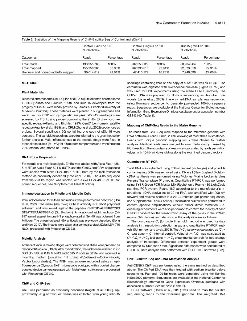
ChIP-Bisulfite-Seq and DNA Methylation Analysis
Anti-CENH3 ChIP was performed using the same method as describedabove. The ChIPed DNA was then treated with sodium bisulfite beforesequencing. Pair-end 100-bp reads were generated using the IlluminaHiSeq2000 platform. Sequences are available at the National Center forBiotechnology Information Gene Expression Omnibus database withaccession number GSM1057283 (Table 2).
BRAT software (Harris et al., 2010) was used to map the bisulfitesequencing reads to the reference genome. The weighted DNA
Table 2. Statistics of the Mapping Results of ChIP-Bisulfite-Seq of Control and sDic-15
Categories
Control (Pair-End 100Nucleotides)
Control (Single-End 100Nucleotides)
sDic15 (Pair-End 100Nucleotides)
Reads Percentage Reads Percentage Reads Percentage
Total reads 193,955,788 100% 282,553,129 100% 30,204,964 100%Total mapped 155,258,280 80.05% 262,236,518 92.81% 22,023,510 72.91%Uniquely and nonredundantly mapped 96,614,812 49.81% 47,415,179 16.78% 7,248,039 24.00%
New Centromere Formation in Maize 9 of 11

methylation level of selected regions was calculated using methodsdiscussed (Schultz et al., 2012), using cytosines that were covered by atleast two reads. Basically, the methods can be expressed in the following
formula: ∑n
i¼1Ci
�∑n
i¼1ðCi þ TiÞ, where i represents the positions of cytosine
meeting the coverage criteria, C represents reads supporting methylatedcytosine, and T represents reads supporting unmethylated cytosine. Perlscripts and IGV tools (Robinson et al., 2011) were used in the process ofdata analysis.
Analysis of Motifs
Self-BLAST of the 723-kb region was conducted using BLASTn with “-FT” parameter. Low-complexity sequences were masked and extractedusing perl script. Motifs were searched using MEME software (Bailey andElkan, 1994) using the sequences of the 723-kb region as data set, withthe following parameters: “-dna -minw 6 -maxw 50.” The distributionpattern of the motifs on the whole genome was searched using FIMOsoftware (Grant et al., 2011). Only sites with scores greater than zero werekept for further analysis. The final results were shown using IGV tools.
Accession Numbers
Sequence data from this article can be found in the Arabidopsis GenomeInitiative or GenBank/EMBL databases under the following accessionnumbers: GSE43140, GSM1057283, and SRP011933.
Supplemental Data
The following materials are available in the online version of this article.
Supplemental Figure 1. FISH Detection Using Telomeres in sDic-15.
Supplemental Figure 2. CENH3 Detection for sDic-15 Meiotic Cells.
Supplemental Figure 3. FISH Detection the Fragment from the sDic-15.
Supplemental Figure 4. FISH Pattern Using ChIPed DNA as Probes.
Supplemental Figure 5. The TE and LTR Distribution on the 723 kb.
Supplemental Figure 6. The Model of Chromosome sDic-15Formation.
Supplemental Table 1. Detailed Information on the 15 Genes Locatedin the 723-kb Region of sDic-15.
Supplemental Table 2. Expression of Six Protein-Coding Genes in the723-kb Region of sDic-15.
Supplemental Table 3. FISH Probe Primers Used in This Study.
Supplemental Table 4. qRT-PCR Primers Used in This Study.
ACKNOWLEDGMENTS
We thank Jinsheng Lai (Chinese Agriculture of University) for the B73seedling DNA methylation data. James A Birchler supplied the seeds andIngo Schubert and Andreas Houben provided critical reading of the article.Ryan Douglas helped edit the revised article. This work is supported by theNational Natural Science Foundation of China (31130033 and 31071083), theNational Basic Research Program of China (973 Program 2011CB944601),and the Chinese Academy of Sciences (KSCX2-EW-R-0103).
AUTHOR CONTRIBUTIONS
F.H., Z.L., and X.-J.W designed the research. B.Z., Z.L., J.P., Y.L., S.F., X.G., Z.G., H.-J.W., Q.D., and J.L. performed the research. B.Z., Z.L., J.P.,X.-J.W., and F.H. analyzed the data. F.H. and X.-J.W. wrote the article.
Received January 25, 2013; revised May 19, 2013; accepted May 23,2013; published June 14, 2013.
REFERENCES
Alfenito, M.R., and Birchler, J.A. (1993). Molecular characterizationof a maize B chromosome centric sequence. Genetics 135: 589–597.
Alonso, A., Hasson, D., Cheung, F., and Warburton, P.E. (2010). Apaucity of heterochromatin at functional human neocentromeres.Epigenetics Chromatin 3: 6.
Ananiev, E.V., Phillips, R.L., and Rines, H.W. (1998). Chromosome-specific molecular organization of maize (Zea mays L.) centromericregions. Proc. Natl. Acad. Sci. USA 95: 13073–13078.
Bailey, T.L., and Elkan, C. (1994). Fitting a mixture model by ex-pectation maximization to discover motifs in biopolymers. Proc. Int.Conf. on Intell. Syst. Mol. Biol. 2: 28–36.
Birchler, J.A., Gao, Z., Sharma, A., Presting, G.G., and Han, F.(2011). Epigenetic aspects of centromere function in plants. Curr.Opin. Plant Biol. 14: 217–222.
Birchler, J.A., and Han, F. (2009). Maize centromeres: Structure,function, epigenetics. Annu. Rev. Genet. 43: 287–303.
Burrack, L.S., and Berman, J. (2012). Neocentromeres and epigeneticallyinherited features of centromeres. Chromosome Res. 20: 607–619.
Chan, F.L., Marshall, O.J., Saffery, R., Kim, B.W., Earle, E., Choo,K.H.A., and Wong, L.H. (2012). Active transcription and essentialrole of RNA polymerase II at the centromere during mitosis. Proc.Natl. Acad. Sci. USA 109: 1979–1984.
Choo, K.H.A. (2000). Centromerization. Trends Cell Biol. 10: 182–188.Choo, K.H.A. (2001). Domain organization at the centromere and
neocentromere. Dev. Cell 1: 165–177.Cleveland, D.W., Mao, Y.H., and Sullivan, K.F. (2003). Centromeres
and kinetochores: From epigenetics to mitotic checkpoint signaling.Cell 112: 407–421.
Dawe, R.K. (2005). Centromere renewal and replacement in the plantkingdom. Proc. Natl. Acad. Sci. USA 102: 11573–11574.
Dong, Q., and Han, F. (2012). Phosphorylation of histone H2A isassociated with centromere function and maintenance in meiosis.Plant J. 71: 800–809.
Fu, S., Gao, Z., Birchler, J., and Han, F. (2012). Dicentricchromosome formation and epigenetics of centromere formation inplants. J. Genet. Genomics 39: 125–130.
Gao, Z., Fu, S., Dong, Q., Han, F., and Birchler, J.A. (2011).Inactivation of a centromere during the formation of a translocationin maize. Chromosome Res. 19: 755–761.
Gao, Z., Han, F.P., He, M.Y., Ma, Y.Z., and Xin, Z.Y. (1999).Characterization of genome and chromosomes in octoploid wheat-wheatgrass amphiploid Zhong 2 using fluorescence in situ hybridizationand chromosome pairing analysis. Acta Bot. Sin. 41: 25–28.
Gong, Z., Wu, Y., Koblízková, A., Torres, G.A., Wang, K., Iovene,M., Neumann, P., Zhang, W., Novák, P., Buell, C.R., Macas, J.,and Jiang, J. (2012). Repeatless and repeat-based centromeresin potato: Implications for centromere evolution. Plant Cell 24:3559–3574.
Grant, C.E., Bailey, T.L., and Noble, W.S. (2011). FIMO: Scanning foroccurrences of a given motif. Bioinformatics 27: 1017–1018.
Han, F., Gao, Z., and Birchler, J.A. (2009). Reactivation of an inactivecentromere reveals epigenetic and structural components forcentromere specification in maize. Plant Cell 21: 1929–1939.
Han, F., Lamb, J.C., Yu, W., Gao, Z., and Birchler, J.A. (2007).Centromere function and nondisjunction are independent componentsof the maize B chromosome accumulation mechanism. Plant Cell 19:524–533.
10 of 11 The Plant Cell
Han, F.P., Lamb, J.C., and Birchler, J.A. (2006). High frequency ofcentromere inactivation resulting in stable dicentric chromosomesof maize. Proc. Natl. Acad. Sci. USA 103: 3238–3243.
Harris, E.Y., Ponts, N., Levchuk, A., Roch, K.L., and Lonardi, S.(2010). BRAT: Bisulfite-treated reads analysis tool. Bioinformatics26: 572–573.
Henikoff, S., Ahmad, K., and Malik, H.S. (2001). The centromereparadox: Stable inheritance with rapidly evolving DNA. Science 293:1098–1102.
Henikoff, S., and Furuyama, T. (2010). Epigenetic inheritance ofcentromeres. Cold Spring Harb. Symp. Quant. Biol. 75: 51–60.
Houben, A., Wako, T., Furushima-Shimogawara, R., Presting, G.,Kunzel, G., Schubert, I., and Fukui, K. (1999). Short communication:The cell cycle dependent phosphorylation of histone H3 is correlatedwith the condensation of plant mitotic chromosomes. Plant J. 18: 675–679.
Jiang, J.M., Birchler, J.A., Parrott, W.A., and Dawe, R.K. (2003). Amolecular view of plant centromeres. Trends Plant Sci. 8: 570–575.
Jiang, J.M., Nasuda, S., Dong, F.G., Scherrer, C.W., Woo, S.S.,Wing, R.A., Gill, B.S., and Ward, D.C. (1996). A conservedrepetitive DNA element located in the centromeres of cerealchromosomes. Proc. Natl. Acad. Sci. USA 93: 14210–14213.
Jin, W.W., Lamb, J.C., Vega, J.M., Dawe, R.K., Birchler, J.A., andJiang, J. (2005). Molecular and functional dissection of the maize Bchromosome centromere. Plant Cell 17: 1412–1423.
Kaszás, E., and Birchler, J.A. (1998). Meiotic transmission ratescorrelate with physical features of rearranged centromeres in maize.Genetics 150: 1683–1692.
Kato, A., Lamb, J.C., and Birchler, J.A. (2004). Chromosome paintingusing repetitive DNA sequences as probes for somatic chromosomeidentification in maize. Proc. Natl. Acad. Sci. USA 101: 13554–13559.
Lamb, J.C., Kato, A., and Birchler, J.A. (2005). Sequencesassociated with A chromosome centromeres are present throughoutthe maize B chromosome. Chromosoma 113: 337–349.
Li, H., and Durbin, R. (2009). Fast and accurate short read alignmentwith Burrows-Wheeler transform. Bioinformatics 25: 1754–1760.
Lister, R., et al. (2009). Human DNA methylomes at base resolutionshow widespread epigenomic differences. Nature 462: 315–322.
Ma, J., Wing, R.A., Bennetzen, J.L., and Jackson, S.A. (2007). Plantcentromere organization: A dynamic structure with conservedfunctions. Trends Genet. 23: 134–139.
Marshall, O.J., Chueh, A.C., Wong, L.H., and Choo, K.H.A. (2008).Neocentromeres: New insights into centromere structure, diseasedevelopment, and karyotype evolution. Am. J. Hum. Genet. 82:261–282.
May, B.P., Lippman, Z.B., Fang, Y.D., Spector, D.L., andMartienssen, R.A. (2005). Differential regulation of strand-specifictranscripts from Arabidopsis centromeric satellite repeats. PLoSGenet. 1: e79.
McClintock, B. (1939). The behavior in successive nuclear divisionsof a chromosome broken at meiosis. Proc. Natl. Acad. Sci. USA 25:405–416.
McClintock, B. (1941). The stability of broken ends of chromosomesin zea mays. Genetics 26: 234–282.
Nagaki, K., Cheng, Z.K., Ouyang, S., Talbert, P.B., Kim, M., Jones,K.M., Henikoff, S., Buell, C.R., and Jiang, J.M. (2004). Sequencingof a rice centromere uncovers active genes. Nat. Genet. 36: 138–145.
Nagaki, K., Talbert, P.B., Zhong, C.X., Dawe, R.K., Henikoff, S.,and Jiang, J.M. (2003). Chromatin immunoprecipitation reveals thatthe 180-bp satellite repeat is the key functional DNA element ofArabidopsis thaliana centromeres. Genetics 163: 1221–1225.
Nasuda, S., Hudakova, S., Schubert, I., Houben, A., and Endo, T.R.(2005). Stable barley chromosomes without centromeric repeats.Proc. Natl. Acad. Sci. USA 102: 9842–9847.
Robinson, J.T., Thorvaldsdóttir, H., Winckler, W., Guttman, M.,Lander, E.S., Getz, G., and Mesirov, J.P. (2011). Integrativegenomics viewer. Nat. Biotechnol. 29: 24–26.
Saffery, R., Sumer, H., Hassan, S., Wong, L.H., Craig, J.M.,Todokoro, K., Anderson, M., Stafford, A., and Choo, K.H.A.(2003). Transcription within a functional human centromere. Mol.Cell 12: 509–516.
Schmittgen, T.D., and Livak, K.J. (2008). Analyzing real-time PCRdata by the comparative C(T) method. Nat. Protoc. 3: 1101–1108.
Schnable, P.S., et al. (2009). The B73 maize genome: Complexity,diversity, and dynamics. Science 326: 1112–1115.
Schueler, M.G., and Sullivan, B.A. (2006). Structural and functionaldynamics of human centromeric chromatin. Annu. Rev. GenomicsHum. Genet. 7: 301–313.
Schultz, M.D., Schmitz, R.J., and Ecker, J.R. (2012). ‘Leveling’ theplaying field for analyses of single-base resolution DNA methylomes.Trends Genet. 28: 583–585.
Sharma, A., Schneider, K.L., and Presting, G.G. (2008). Sustainedretrotransposition is mediated by nucleotide deletions and interelementrecombinations. Proc. Natl. Acad. Sci. USA 105: 15470–15474.
Stimpson, K.M., and Sullivan, B.A. (2010). Epigenomics of centromereassembly and function. Curr. Opin. Cell Biol. 22: 772–780.
Topp, C.N., Okagaki, R.J., Melo, J.R., Kynast, R.G., Phillips, R.L.,and Dawe, R.K. (2009). Identification of a maize neocentromere inan oat-maize addition line. Cytogenet. Genome Res. 124: 228–238.
Wong, N.C., Wong, L.H., Quach, J.M., Canham, P., Craig, J.M.,Song, J.Z., Clark, S.J., and Choo, K.H.A. (2006). Permissivetranscriptional activity at the centromere through pockets of DNAhypomethylation. PLoS Genet. 2: e17.
Wu, Y., Kikuchi, S., Yan, H., Zhang, W., Rosenbaum, H., Iniguez, A.L., and Jiang, J. (2011). Euchromatic subdomains in rice centromeresare associated with genes and transcription. Plant Cell 23: 4054–4064.
Zhang, M., Zhao, H., Xie, S., Chen, J., Xu, Y., Wang, K., Zhao, H.,Guan, H., Hu, X., Jiao, Y., Song, W., and Lai, J. (2011). Extensive,clustered parental imprinting of protein-coding and noncodingRNAs in developing maize endosperm. Proc. Natl. Acad. Sci. USA108: 20042–20047.
Zhang, W., Friebe, B., Gill, B.S., and Jiang, J. (2010). Centromereinactivation and epigenetic modifications of a plant chromosomewith three functional centromeres. Chromosoma 119: 553–563.
Zhong, C.X., Marshall, J.B., Topp, C., Mroczek, R., Kato, A.,Nagaki, K., Birchler, J.A., Jiang, J.M., and Dawe, R.K. (2002).Centromeric retroelements and satellites interact with maizekinetochore protein CENH3. Plant Cell 14: 2825–2836.
New Centromere Formation in Maize 11 of 11
DOI 10.1105/tpc.113.110015; originally published online June 14, 2013;Plant Cell
Hua-Jun Wu, Zhi Gao, Xiu-Jie Wang and Fangpu HanBing Zhang, Zhenling Lv, Junling Pang, Yalin Liu, Xiang Guo, Shulan Fu, Jun Li, Qianhua Dong,
Ectopic SequencesFormation of a Functional Maize Centromere after Loss of Centromeric Sequences and Gain of
This information is current as of February 19, 2018
Supplemental Data /content/suppl/2013/05/24/tpc.113.110015.DC1.html
Permissions https://www.copyright.com/ccc/openurl.do?sid=pd_hw1532298X&issn=1532298X&WT.mc_id=pd_hw1532298X
eTOCs http://www.plantcell.org/cgi/alerts/ctmain
Sign up for eTOCs at:
CiteTrack Alerts http://www.plantcell.org/cgi/alerts/ctmain
Sign up for CiteTrack Alerts at:
Subscription Information http://www.aspb.org/publications/subscriptions.cfm
is available at:Plant Physiology and The Plant CellSubscription Information for
ADVANCING THE SCIENCE OF PLANT BIOLOGY © American Society of Plant Biologists