formulation and in vitro evaluation of zidovudine microspheres
TRANSCRIPT

FORMULATION AND IN VITRO EVALUATION OF ZIDOVUDINE
MICROSPHERES
Dissertation work submitted toTHE TAMIL NADU Dr. M.G.R. MEDICAL UNIVERSITY, CHENNAI
In partial fulfillment of the award of degree of
MASTER OF PHARMACY(Pharmaceutics)
March 2009
PSG COLLEGE OF PHARMACYPSG Institute of Medical Sciences & Research
Peelamedu, Coimbatore – 641004

CertificateThis is to certify that the dissertation entitled FORMULATION AND
IN VITRO EVALUATION OF ZIDOVUDINE MICROSPHERES
was carried out by PRASATH.R, in the Department of Pharmaceutics, PSG
College of Pharmacy, PSG Institute of Medical Sciences & Research,
Peelamedu, Coimbatore, which is affiliated to The Tamilnadu Dr. M.G.R.
Medical University, Chennai, under the supervision and guidance of
Prof. A. K. Chandrasekharan, M.Pharm., Principal, PSG College of Pharmacy,
PSG IMS & R, Coimbatore.
Prof. A.K.CHANDRASEKHARAN, M.Pharm., Principal,
PSG College of Pharmacy,Place: Coimbatore PSG IMS & R,Date : Coimbatore – 641 004.

Certificate
This is to certify that the dissertation entitled
FORMULATION AND IN VITRO EVALUATION OF ZIDOVUDINE
MICROSPHERES was carried out by PRASATH.R, in the Department of
Pharmaceutics, PSG College of Pharmacy, PSG Institute of Medical Sciences
& Research, Peelamedu, Coimbatore, which is affiliated to The Tamilnadu Dr.
M.G.R. Medical University, Chennai, under my co-guidance and supervision to
my fullest satisfaction.
S.M. HABIBUR RAHMAN, M.Pharm.,(Ph.D.)Lecturer,
Department of Pharmaceutics,PSG College of Pharmacy,
Place: Coimbatore PSG IMS & R,Date: Coimbatore - 641 004.

Certificate
This is to certify that the dissertation entitled
FORMULATION AND IN VITRO EVALUATION OF ZIDOVUDINE
MICROSPHERES was carried out by PRASATH.R, in the Department of
Pharmaceutics, PSG College of Pharmacy, PSG Institute of Medical Sciences
& Research, Peelamedu, Coimbatore, which is affiliated to The Tamilnadu Dr.
M.G.R. Medical University, Chennai, under the co-guidance and direct
supervision of
Mr. S.M. Habibur Rahman, M.Pharm., (Ph.D.), Lecturer, Department of
Pharmaceutics, PSG College of Pharmacy, PSG IMS & R, Coimbatore-4.
.
Dr. C. VIJAYA RAGHAVAN, M.Pharm.,Ph.D.Vice Principal,
Head, Department of Pharmaceutics,PSG College of Pharmacy,
Place: Coimbatore PSG IMS & R,Date: Coimbatore - 641 004.

ACKNOWLEDGEMENT
I take this opportunity with pride and immense pleasure in expressing my deep sense of gratitude to Prof..A.K.Chandrasekharan M.Pharm., Principal, PSG College of Pharmacy, PSG IMS&R, Coimbatore, whose guidance was unforgettable, invaluable. His impressive, innovative ideas and constructive suggestion has made the presentation of my work a grand success.
My sincere gratitude to our beloved Principal Dr. C. Vijaya Raghavan, M.Pharm., Ph.D., Vice Principal and Head, Department of Pharmaceutics, PSG College of Pharmacy, PSG IMS&R, Coimbatore, for providing every need from time to time to complete this work successfully.
I submit my sincere thanks to our beloved Managing Trustee of PSG Sons and Charities, Shri. G.Rangaswamy for providing all the facilities to carryout this work. I express my heartfelt sincere thanks to my Co Guide Mr. S. M. Habibur Rahman M.Pharm (Ph.D), Lecturer Department of Pharmaceutics, PSG College of Pharmacy, PSG IMS&R, Coimbatore, for his valuable suggestions and support during this study.
I am elated to place on record my profound sense of gratitude to Mr. V.Sankar, M.Pharm., (Ph.D)., Mrs.S Durgaramani, M.Pharm.(Ph.D.,),Assistant Professors and Mr.S.Subhramanyam,M.Pharm., (Ph.D.,), Lecturer, Department of Pharmaceutics, PSG College of Pharmacy, PSG IMS&R, Coimbatore, for their constructive ideas at each and every stage of the project.
I extend my profound gratitude to Miss. K.Y. Kavitha, M.Pharm., Assistant Professor, Department of Pharmaceutical Analysis, for her co-operation and valuable suggestions during the tenure of my work.
I would like to thank Dr.. R. Saraswathi & Mrs Soja who have guided me in my Ist year with their innovative ideas and moral support.

I would like to thank Dr. M. Ramanathan, M.Pharm., Ph.D., Professor & Head, Department of Pharmacology giving me all the mental strength throughout my career.
I would like to thank Mrs.R.Chithra, B.Sc. Bio Chemistry, Ms. Sheela Gracy, D.Pharm., Lab Technicians, Librarians and Non Teaching Staff for their kind co-operation during this work.
It is privilege to extend my special thanks to my dearest Lovable Parents, without whose unconditional love and support; this process of my learning would have been incomplete. And they are also the backbone for all successful endeavors in my life.
Words can’t express my sincere gratitude and obligation to my dear batch mates Jayesh.V.N, John.R, Harikrishnan.V, Arunprasath.B, Siddharth.S, V.Harikrishnan, Muthukumar ,Velayutham K, Praveen .C and to all other batch mates who directly helped during my work.
I would like to thank my Juniors, and to all other batch mates who directly or indirectly helped during my work.
Above all, I humbly submit my dissertation work, into the hands of Almighty, who is the source of all wisdom and knowledge for the successful completion of my thesis.
I wish to thank of Mrs. Mini Nair and Mr. Niranjan,M/s. Saraswathi Computer Centre for framing project work in a beautiful manner.
My sincere thanks to all those who have directly or indirectly helped me to complete this project work.

CONTENTSChapter No.
Topics Page No.
I INTRODUCTION AND OBJECTIVE OF INVESTIGATION
1
II INTRODUCTION 3
Factors governing design of controlled release dosage form
6
Classification of orally controlled release systems 8
Future directions in controlled delivery 9
Microspheres and Microcapsules 14
Ethyl cellulose microspheres 33
III Review of Literature 35
IV Drug and Polymer Profile 48
V Experimental work 57
Preformulation studies 61
Drug content analysis 71
Particle size and shape analysis 80
In vitro evaluation studies 88
VI Summary and conclusion 95
Bibliography
LIST OF TABLES
Table No.
TitlePage No.
1 Classification of microencapsulation methods. 11
2 Proposed mechanism for uptake of microspheres 32
3 Formulation of zidovudine microspheres using
ethylcellulose60
4 Calibration data for the estimation of zidovudine 63
5 Amount of drug loaded in ethylcellulose microspheres using 50 and 100 ml of dispersion medium
72
6 Percentage encapsulation efficiency of ethylcellulose microspheres using 50 and 100 ml of dispersion medium
75
7 Percentage yield of ethylcellulose microspheres using 50 and 100 ml of dispersion medium
78
8 Calibration of eyepiece micrometer 82
9 Particle size determination by optical microscopic method
using 50ml of dispersion medium83
10 Particle size determination by optical microscopic method
using 100ml of dispersion medium85
11 Percentage of drug released for formulations prepared
using 50 ml of dispersion medium89
12 Percentage of drug released for formulations prepared using 100 ml of dispersion medium
92

LIST OF FIGURES
Fig. No
TitlePage
No
1 Hypothetical plasma drug concentration-time profile 4
2 Various configurations of a)microcapsules and b)microspheres
16
3 Stadard graph of Zidovudine 64
4 IR Specrum of zidovudine 65
5 IR Specrum of ethylcellulose 67
6 IR Specrum of Formulation 68
7 Amount of drug loaded in Ethylcellulose microspheres using 50 and 100 ml of dispersion medium
72
8 Percentage encapsulation efficiency of ethylcellulose microspheres using 50 and 100 ml of dispersion medium
75
9 Percentage yield of ethylcellulose microspheres using 50 and 100 ml of dispersion medium
78
10 Scanning electron microscope pictures of ethyl cellulose microspheres
81
11 Scanning electron microscope pictures of ethyl cellulose microspheres
81
12 Particle size determination by optical microscope method using 50 of dispersion medium
84
13 Particle size determination by optical microscope method using 100 ml of dispersion medium
86
14 Percentage release of zidovudine microspheres prepared using 50 ml of dispersion medium
90
15 Percentage release of zidovudine microspheres prepared using 100 ml of dispersion medium
93

INTRODUCTION AND OBJECTIVE OF THE INVESTIGATION
Novel drug delivery systems are being employed both
experimentally and therapeutically to alter the distribution of drugs in the
body with an idea of delivering them more efficiently and reducing the
toxicity of the existing drugs. One such area in which research is gaining
much attention is “controlled drug delivery” because of its enhanced
efficacy along with reduced toxicity.
It have been reported that 40% of the compounds developed in
pharmaceutical industry are poorly soluble in water. A limiting factor to the
in vivo performance of poorly water soluble drugs after oral administration
is their inadequate ability to become wetter and getting dissolved in the
gastrointestinal (GI) fluid.
Microspheres have more advantages over other soluble carrier’s
liposomes. Liposomes have low drug entrapment efficiency, rapid leakage
of water soluble drugs especially in the presence of blood components,
poor stability and method of preparation which are not compatible with
large scale production requirements. However microspheres can entrap
various molecules in a stable and reproducible way.

Zidovudine is a nucleoside reverse transcriptase inhibitor (NRTI)
with activity against Human Immunodeficiency Virus Type 1 (HIV-1).
Zidovudine is phosphorylated to active metabolites that compete for
incorporation into viral DNA. They inhibit the HIV reverse transcriptase
enzyme competitively and act as a chain terminator of DNA synthesis. The
bioavailability of zidovudine can be increased by making the drug to get
released in a controlled manner by incorporating the drug in certain
biodegradable polymers.
The aim of this study was to formulate and evaluate
microencapsulated controlled release preparations of Zidovudine using
ethyl cellulose as the retardant material with high entrapment efficiency
and extended release. Ethylcellulose is a natural, biodegradable polymer
and the method adopted for preparing microsphere was water-in-oil-in-oil
(w/o/o) double emulsion solvent diffusion technique.
The plan of work was designed as follows:-
Preparation of zidovudine microspheres using water-in-oil-in-oil
(w/o/o) double emulsion solvent diffusion technique.
Determination of size and shape of microspheres.
Estimation of percentage yield of the microspheres and is drug
entrapment efficiency.
Study of in vitro release from prepared microspheres.

INTRODUCTION
Over the years we have learnt the optimization of drug therapy is of very
great importance and in the process patience convenience, compliance,
safety has to be taken into account.
For many decades treatment of an acute disease or chronic illness has
been mostly accomplished by deliver of drugs to patients using various
pharmaceutical dosage forms like capsules, pills etc. These types of dosage
forms require frequent dosing and thus causing inconvenience to patients.
There are three types of drug delivery i.e. controlled delivery, targeted
delivery and modulated release. Targeted delivery refers to the systemic
administration of drug carrier with the goal of delivering drug to specific cell
types, tissues and organs. Controlled release refers to the use of delivery
device with the objective of releasing the drug into the patient body at a
predominant rate at a specific time or with specific release profiles. Modulated
release implies release of the drug at variable rate which is controlled by
various factors such as bio feedback, environmental conditions, sensor input
or an external control device.
3

Controlled drug delivery is the one which delivers the drug at a
specific rate locally or systemically for a specific period of time.
Controlled release dosage forms are being referred by a number of
terminologies such as delayed action, extended action, prolonged release,
slow release, gradual release, sustained release and timed release dosage
forms.
Fig 1. Hypothetical plasma drug concentration-time profile
4

OBJECTIVE OF CONTROLLED RELEASE
Increase in patient compliance by obtaining extended duration of
action
Targeted drug action by using carriers or chemical derivatization to
deliver drugs to a particular target cell type.
Localized drug action by spatial or controlled release system adjacent
to or in the diseased tissue or organ.
Advantages
1) Decrease in toxicity and adverse effects.
2) More consistent and prolonged therapeutic effect.
3) Improved patience compliance.
4) Reduced dosing frequency.
5) More uniform blood concentration.
6) A greater selectivity in pharmacological activity.
7) Avoidance of night time dosing.
8) Better drug utilization.
Disadvantages
1) Stability problems.
2) Toxicity due to dose dumping.
5

3) Increased cost.
4) More rapid development of drug tolerance.
5) Increased variability among dosage units.
6) Poor in vitro and in vivo correlation.
7) Need for additional patient education and counseling.
8) Decreased systemic availability.
9) Retrieval of drug is difficult in case of toxicity or hypersensitivity
reactions.
FACTORS GOVERNING THE DESIGN OF CONTROL RELEASE DOSAGE
FORM:-
Drug related
• Aqueous solubility
• Partition coefficient
• Molecular size
• Drug stability
• Protein binding
Biological factors
• Absorption.
• Cardiac rhythm.
6
• Distribution.
• Elimination.
• Duration of action.
• Margin of safety.
• Side effects.
• Diseased sates.
Physiological factors
• Prolonged drug absorption.
• Variability in GI emptying time.
• GI blood flow.
Pharmacological factors
• Sensitivity/tolerance.
• Changes in drug effect upon multiple dosing.
Pharmacokinetic factors
• Dose dumping.
• First pass metabolism.
• Enzyme induction/inhibition upon multiple dosing.
• Variability in urinary pH and its effect on drug elimination. (Jain., 1997)
7
CLASSIFICATION OF ORAL CONTROLLED RELEASE SYSTEMS:
A) Continuous release systems
1) Dissolution controlled systems
a) Matrix type
b) Reservoir type
2) Diffusion controlled release systems
a) Matrix type
b) Reservoir type
3) Dissolution and diffusion controlled release systems.
4) Ion exchange resin drug complexes.
5) Osmotic pressure controlled systems.
6) Slow dissolving salts and complexes.
7) pH dependant formulations
8) Hydrodynamic pressure controlled systems.
B) Delayed transit and continuous release systems.
1) Altered density systems
a) High density.
b) Low density.
c) Floating.
2) Mucoadhesive systems.
8

3) Size based systems.
C) Delayed release systems.
1) Intestinal release systems.
2) Colonic release systems.
Future developments in controlled drug delivery
The most exciting opportunities in controlled drug delivery lies in the
area of responsive delivery systems, with which it will be possible to deliver
drugs through implantable devices in response to a measured drug level or to
deliver a drug precisely to a targeted site. Much of the developments of novel
materials in controlled delivery are focusing on the preparation and use of the
responsive polymers with specifically designed microscopic and macroscopic
structural and chemical features.
Such systems include,
Copolymers with desirable hydrophilic/hydrophobic interactions
Complexation networks responding via hydrogen or ionic bonding
Block or graft copolymers.
Dendrimers or star polymers as nanoparticles for immobilization of
enzymes, drugs, peptides, or other biological agents.
9

MICROENCAPSULATION
Microencapsulation is one of the most intriguing fields in the area of
drug delivery systems. It is an interdisciplinary field that requires knowledge of
the field of pure polymer science, familiarity with emulsion technology and an
in-depth understanding of drug and protein stabilization. This method is also
used to make controlled release dosage forms. Solids, liquids, gels can be
entrapped inside one or more polymeric coatings. The structure of
microcapsules depends on the type of manufacturing process and mostly
spherical. Pellets, liposomes, multiple emulsions, microcapsules and
microspheres are common examples of microcapsulate drug delivery
systems. (Edith mathiowitz., 2002)
10

Table: 1 Classification of microencapsulation methods
PROCESS COATING MATERIAL SUSPENDED MEDIUM
Interfacial polymerization
Water soluble & insoluble monomers
Aqueous/organic solvent
Complex coacervation Water soluble polyelectrolyte
Water
Coacervation Hydrophobic polymers Organic solvent
Thermal denaturation Proteins Organic
Salting-out Water soluble polymer water
Solvent evaporation Hydrophilic or hydrophobic polymers
Organic solvent or water
Hot melt Hydrophilic or hydrophobic polymers
Aqueous/organic solvent
Solvent removal Hydrophilic or hydrophobic polymers
Organic solvents
Spray drying Hydrophilic or hydrophobic polymers
Air, Nitrogen
Phase separation Hydrophilic or hydrophobic polymers
Aqueous/organic solvent.
(i) Reasons for microencapsulation
Sustained and controlled release
Taste and odor masking
Protection of drug against environment
Delay of volatilization
Enteric coating
11

Detoxification
Conversion of oils and other liquids into solids for ease of handling
Separation of flow of powders
Isolation from tissues
Safe handling of toxic substances
(ii) Properties of drugs used in microencapsulation
The core of microcapsules formulated may contain one or more drugs
either a single drug or combination of drugs with suitable additives to form a
liquid or solid phase. Liquid core may be composed of polar or non polar
substantial that comprises the active ingredient or that acts as vehicle for
dissolved or suspended drugs. The solvent properties of liquids critically
influence the rate of drug release and selection of coating materials. For
drugs of low solubility with known bioavailability problems associated with low
rates of dissolution, decrease in particle size of suspended drugs may be
important in enhancing in vivo absorption. Smaller microparticles also have
faster release rates because of their increased surface area per unit volume
or weight of core material. Solid cores are used more frequently than liquid
cores. Very small core particles tend to give tend to give aggregation problem
during production, because of the attraction force present at the surface of the
12

particles. Larger particles can cause problems because of their rapid
sedimentation rate. The shapes of these cores are also important. Density of
cores is very important for controlling the transit time in GI tract. Increasing
the density was the most important factor in prompting the retention of pellets
in stomach. Decreasing density is important in floating type dosage forms.
Swelling of the core with disruption of coating leads to uncontrolled drug
release.
(iii) Properties of polymers used for microencapsulation
The selection of appropriate coating material dictates to a major
degree the resultant physical and chemical properties of the microcapsules.
The coating material should be capable of forming a film that is cohesive with
the core material and the ideal coating material should be compatible and non
reactive with the core material. The polymer coating should provide desired
coating properties such as strength, impermeability, optical properties,
flexibility and stability. (Jain., 1997).
13

MICROSPHERES AND MICROCAPSULES
Microspheres can be defined as spherical empty particles ranging in
size from 1 to 1000 micrometer. Microspheres are characteristically free
flowing powder consisting of proteins and synthetic polymers, which are
biodegradable in nature.
Microcapsules consist of a well defined core and a well defined
envelope; the core can be solid, liquid or gas, the envelope is made of
continuous process or non porous polymeric phase.
It is also defined as a spherical particle with size varying from 50 nm
to 2 mm containing a core substance.
Microspheres have been investigated for intravenous and intra arterial
targeting and delivery systems. Microspheres and Microcapsules have been
injected in the vessels to ensure passive targeting of the drugs. The drug
release is controlled by diffusion through the polymer matrix and/or by erosion
of the polymer. The role of microspheres and microparticles depend on their
size and site of injection.
Microparticles of diameter smaller than 2µm can be injected in an
intravenous, intra arterial and intra peritoneal manner in order to target the
reticuloendothelial system (RES). Intravenous injection of Microspheres of
14

size from 3-12 micrometer is intended to block the capillaries of the lungs,
liver and spleen.(Vyas., 2002)
Vessels can be hyperselectively embolized with drug loaded particulate
materials of more than 10 micrometer. Microspheres of 100-300 micrometer
size are the most appropriate embolic agents. They reach the intra lesional
precapillary arteries and cause reduction of blood flow.
Drug incorporation methods
1. The drug can be dissolved or homogeneously dispersed in the polymer
(Matrix device-microsphere type).
2. Drugs can be dissolved or homogenously dispersed in the polymer
(Matrix device- Microsphere type).
15

Fig: 2 various configurations of a)microcapsules and b)microspheres
16

17

(i) Advantages of microspheres
1. Taste and odor masking.
2. Protection of the drugs against the environment (moisture, light, heat,
and/or oxidation) and vice versa (prevention of pain on injection).
3. Delay of volatilization.
4. Conversion of oils and other liquids to solids for ease of handling.
5. Separation of incompatible materials (other drugs or excipients
such as buffers)
6. Safe handling of toxic substances.
7. Improvement of flow of powders.
8. Aid in dispersion of water-insoluble substances in aqueous media.
9. Production of sustained release, controlled release and targeted
medications.
(ii) Route of administration
Route of administration is selected depending on the drug properties,
disease states being treated and the age condition of the patient. Desirable
properties of microspheres to be used for delivery will also change depending
on route of administration.
18

a) Oral delivery
Oral delivery is the simplest route of drug administration. Constraints of
the oral route are numerous on the whole it offers potential danger than
parental route. The relatively brief transit time of about 12 hours through the
GI tract limits the duration of action that can be expected via the oral route.
Recently, it has been reported that microspheres of less than 10µm in size
are taken up by peyer’s patches and may increase the retention time in the
stomach. Also microspheres made of polymers with mucoadhesive properties
get attached to stomach or intestine and prolong the residence time in the
stomach. Bioavailability of drugs with limited solubility in the stomach or
intestine and small absorption rate constant can be increased by increasing
the retention time in the stomach. Improved drug delivery was observed in
mucoadhesive propertied microspheres when compared to non
mucoadhesive propertied microspheres administered alone.(Jain., 1997)
b) Parenteral delivery
Most of the microsphere based controlled delivery systems are
developed with the aim of using them for parenteral administration. Drug
released is completely absorbed in this case. Microspheres used for
parenteral delivery should be sterile and should be dispersible and compatible
in a suitable vehicle for injection. Surfactants in small concentration are often
19

necessary for reconstituting hydrophobic particles for injection in aqueous
vehicles, which are reported for certain adverse tissue reactions and affect
the release of incorporated drug.
PROCESS
Polymers used
Synthetic polymers
Non-biodegradable
PMMA
Acrolein
Glycidyl methaacrylate
Epoxy polymers
Biodegradable
Lactides and glycolides
Polyalkyl cyano acrylates
Polyanhydrides
20

NATURAL MATERIALS
Proteins
Albumin
Gelatin
Collagen
Carbohydrates
Starch
Agarose
Chitosan
Chemically modified carbohydrates
DEAE cellulose
Poly (acryl dextran)
Poly(acryl starch)
Prerequisites for ideal microparticulate carriers
Longer duration of action
Control of content release
Increase of therapeutic efficacy
Protection of drug
21

Reduction of toxicity
Biocompatibility
Sterilizability
Relative stability
Dispersability or water solubility
Bioresorbability
Targetability
Polyvalent
(iv) Preparation of microspheres
The preparation of microspheres should satisfy certain criteria,
The ability to incorporate reasonably high concentrations of drug.
Stability of the penetration after synthesis with clinically acceptable
shelf life.
Controllable particle size and dispersability in aqueous vehicles for
injection.
Release of active ingredient with good control over a wide time scale.
Biocompatibility with a controllable biodegradability.
Susceptibility to chemical modification.
22

(v) Methods of preparation
The method of preparation and its choice are equivocally determined
by some formulation and technology related factors as mentioned below,
The particle size requirement.
The drug or protein should not be adversely affected by the process.
Reproducibility of the release profile and the method.
No stability problem.
There should be no toxic products associated with the final product.
1. Solvent evaporation
This technique is based on the evaporation of the initial phase of an
emulsion by agitation. Initially the polymeric supporting material was
dissolved in a volatile organic solvent. The active ingredient that has to be
encapsulated is then dispersed of and dissolved in organic solution to form
suspension, emulsion or solution. In the following step the organic phase is
emulsified under agitation in a dispersing phase consisting of the non solvent
of the polymer, which is immiscible with the organic solvent, which contains
an appropriate tension active additive. Once emulsion is stabilized, agitation
is maintained and solvent is the creation of solid microspheres. On the
completion of solvent evaporation process, the microspheres held in
23

suspension in the continuous phase recovered by filtration of centrifugation
and are washed and dried.
2. Thermal and cross linking
Microspheres made from natural polymers are prepared by a cross
linking process; the polymers include gelatin, albumin, starch and dextran.
Water in oil emulsion is prepared, where the water phase is a solution of the
polymer, which contains the drug to be incorporated. The oil phase is a
suitable vegetable oil or oil-organic solvent mixture containing an oil- soluble
emulsifier. Once the desired water-oil emulsion is formed, the water soluble
polymer is solidified by some kind of cross linking process. This requires a
thermal treatment or the addition of chemical cross linking agent like
glutaraldehyde. In the chemical and thermal cross linking process both the
amount of chemical and intensity of heat applied are critical in determining the
swelling properties and release profiles of microspheres. If glutaraldehyde is
used as cross linking agent, residual amounts can have toxic effects.
24

3. Precipitation
A variation on the evaporation method is the precipitation method. The
emulsion consists of polar droplets dispersed in a non polar medium. Solvent
may be removed from the droplets by the use of a co solvent. The resulting
increase in the polymer-drug concentration causes a precipitation forming a
suspension.
4. Freeze drying
The technique involves the freezing of the emulsion; the relative
freezing points of the continuous-phase solvent is usually organic and is
removed by sublimation at low temperature and pressure. Finally, the
dispersed phase solvent of the droplets is removed by sublimation, leaving
polymer-drug particles.
3. Spray drying and spray congealing
Spray drying and spray congealing methods are based on the drying of
the mist of polymer and drug in air. Depending upon the removal of the
solvent or the cooling of the solution, the two processes are named spray
drying and spray congealing respectively. The polymer is first dissolved in an
organic volatile solvent such as dichloromethane, acetone etc. the drug in the
solid form is then dispersed in the polymer solution under high-speed
homogenization. This dispersion is then atomized in a stream of hot air. The
25

atomization leads to formation of small droplets or the fine mist form which
the solvent evaporates instantaneously leading to the formation of
microspheres in a size range of 1-100µm. Microspheres are separated from
the hot air by means of the cyclone separator while the traces of solvent are
removed by vacuum drying.(Jain., 1997).
Very rapid solvent evaporation, however leads to the formation of
microparticles.
Advantages
→ Feasibility of the operation under aseptic conditions.
→ The process is rapid.
→ Suitable for both batch and bulk manufacturing.
→ This technique can be use to encapsulate large number of drugs.
4. Phase separation and coacervation techniques
Phase separation method is specially designed for preparing the
reservoir type of the system, i.e. to encapsulate water soluble drugs e.g.
peptides, proteins, however some of the preparations are of matrix type
particularly, when the drug is hydrophilic in nature e.g. steroids. In matrix type
device, the drug or the protein is soluble in polymer phase. The process is
based on the principle of decreasing the solubility of the polymer in the
26

organic phase to affect the formation of the polymer rich phase called
coacervates. The coacervation can be brought about by addition of the third
component to the system which results in the formation of two phases, one is
rich in polymer and the other, i.e. supernatant, depleted of the polymer. This
is achieved by the addition of the salt, non-solvent addition, addition of the
incompatible polymer or change in pH.
In this technique, the polymer is first dissolved in a suitable solvent and
then the drug is dissolved by making its aqueous solution. Phase separation
is then accomplished by changing the solution conditions by using any of the
method above. The process is carried out under continuous stirring to control
the size of microparticles.
(V) Loading of drug
The active components are loaded over the microspheres principally
using two methods, i.e. during preparation of the microsphere or after the
formation of microspheres by incubating them with drug or protein. The active
component can be loaded by means of the physical entrapment, chemical
linkage and surface absorption.
Maximum absorption can be achieved by incorporating the drug
during the time of preparation but it may get affected by many other
processes such as method of preparation, presence of additives (e.g. cross
27

linking agent, surfactants, stabilizers etc), the loading is carried out in
preformed microspheres by incubating them with a very high concentration of
drug in a suitable solvent. The drug gets loaded in these microspheres
through diffusion or penetration through the pores in the microspheres as well
as adsorption in their surface. The solvent is then removed leaving the drug
loaded microsphere.
The Freundlich model is applied to determine the adsorption of the
drugs. The Freundlich equation is,
X/M = KCpeq
Where,
K= Constant related to the capacity of adsorbent for the adsorbate.
P= Constant related to the affinity of the adsorbent for the adsorbate.
(vi) Release of drug from microspheres
The rate of drug release from the microspheres dictates their
therapeutic action. Drug release is governed by the molecular structure of the
drug and the resistance of polymer to degradation and the surface area and
porosity of microspheres.
Reservoir delivery systems extend the residence time of drug within
the systemic circulation originally focused on zero-order dissolution kinetics.
28

In porous polymeric systems the rate of drug release is dictated by the
device surface area, which is linked directly to its shape. The rate of release
from microspheres may be result of polymer erosion or diffusion.
The internal structure may vary as a function of microencapsulation
process employed. Reservoir microcapsules have a core of drug coated with
polymer. The drug is distributed homogenously throughout the polymeric
matrix in monolithic microspheres.
The mechanism of drug release at a controlled rate from microspheres
include diffusion of drug through a polymeric excipients, diffusion of trapped
drug as polymer erodes, and release of drug through pores in the polymeric
excipients. Increasing the surface area, by reducing the particle size, results
in an increased release rate. The path length travelled by the drug in the
matrix can be controlled by manipulating the microsphere loading.
Microspheres with high drug content release the outline ingredient more
rapidly than those with a low load. Thicknesses of the polymer also influence
release rate.
29
Mechanism of drug release
Factors affecting the release of drug from the particulate system in
relation with drug, microspheres and bio environment.
Drug
Position in microspheres.
Molecular weight.
Concentration.
Interaction with matrix.
Microspheres
Type and amount of matrix polymer.
Size and density of microspheres.
Extent of cross linking, denaturation or polymerization.
Adjuvants.
Environment
pH.
Polarity.
Presence of enzyme.
30

The geometry of carrier, i.e., whether it is reservoir type where the drug
is present as a core, or matrix type in which drug is dispersed throughout the
carrier, governs overall release profile of drug or active ingredients. (Jain.,
1997).
SYSTEMS
Reservoir type system
Release from the reservoir type system with rate controlling membrane
proceeds by first penetration of water through the membrane followed by
dissolution of the drug in the penetrating dissolution fluid. The dissolved drug
after partitioning through the membrane diffuses across the stagnant diffusion
layer. The release is essentially governed by the Fick’s first law of diffusion
as, J= −Ddcdx
Where,
J = flux per unit area.
D = diffusion coefficient.
Diffusion across the membrane determines the effectiveness of the
carrier system. The release rate from the carriers can be modified by
changing both the composition and thickness of the polymeric membrane.
31

Matrix type system
Release profile of the drug from this type of the device critically
depends on the state of drug whether it is dissolved or dispersed in the
polymer matrix.
In case of the drug dissolved in the polymeric matrix, amount of drug,
and the nature of polymer affect the release profile. If this is the case the
amount of drug appearing in the receptor phase at “t” is approximated by two
separate equations. The first equation determines the 60% of drug release
profile at the later stage.
dMtdt
=2Mx D /π∫2t 1 /2
dMtdt
= 8 DM x / ∫ 2 expπ2 Dt+/∫ 2
Where,
l = thickness of polymer slab.
D = diffusion coefficient.
Mx = total amount of drug present in the matrix.
Mt = amount of drug released in time “t”.
When the drug is dispersed throughout the polymer matrix then the
release profile follows higuchi’s equation.
dM t
dt =
A2
(2DCsCo)
32

Where,
A = area of matrix.
Cs = solubility of the drug in matrix.
Co = total concentration in the matrix.
(iv) Fate of microsphere in the body
The knowledge about the fate of microspheres after parenteral
administration is very important for designing a drug delivery system. The
biological fate of administered microspheres has been studied by
radiolabelling techniques. 14C, 131I, 125I, and 99Tc are being used for
radiolabelling. Fluoroscein labeling has also been found useful for studying
the fate of microspheres in vivo.
Table: 2 proposed mechanism for uptake of microspheres
SITE SIZE RANGE FATEEnterocyte/Endocyte <220 nm RES uptakeParacellular uptake 100-200 nm Unknown
Intestinal macrophage 1 µm MLNPersorption 5-150 µm Blood and Excretory fluids
Peyer’s Patches 20 nm - 10 µm PP and MLNFollicle associated
epithelium< 750 nm MLN
33

(v) Applications of microspheres.
Microspheres in vaccine delivery
Antigen release
Immune system
Targeting using particulate carriers (ocular, intranasal, oral)
Magnetic microspheres, Imaging
Monoclonal antibodies.
Microsponges: topical porous microspheres.(Jain., 1997).
ETHYL CELLULOSE MICROSPHERES
Ethyl cellulose microspheres were prepared based on the principle
of double emulsion solvent diffusion technique with certain modifications. The
rational for selecting EC for the shell material was that, this substance is
commonly used as an additive in foods and drug because of its high
inertness, and forms a stable, semi-permeable capsular membrane.
Ethyl cellulose is synthetic polymer; synthetic polymers have the
advantage that they can be easily and reproducibly prepared. This can be
copolymerized with one another to alter the physical, chemical and
mechanical properties and can be prepared as low or high molecular weight
material by suitable reaction conditions. Chemical bonds which are
susceptible to degradation include amides, esters, orthoesters, acetyls,
34

glycosides and related groups. biodegradability of the polymer depends on
many factors such as polymer structure, molecular weight ,physical form of
the polymer and environment in which the polymer is placed since many
proteolytic enzymes specifically catalyzes the hydrolyses of peptide linkages
adjacent to substituted proteins, substituted polymers containing benzyl,
hydroxyl ,carboxyl-methyl and phenyl groups have been prepared to improve
biodegradability.
DOUBLE EMULSION SOLVENT DIFFUSION TECHNIQUE:
The technique was based on formation of an emulsion by agitation.
Initially the drug and the polymeric material are dissolved in the solvent. Then
the primary emulsion was formed by adding water little by little into the
solvent, drug and polymer mixture which stirring was done simultaneously
using a mechanical stirrer. After the emulsion was formed the stirring was
stopped. Then the oily phase was taken along with surfactant and the primary
emulsion formed was added to the oil phase slowly while stirring was
continued at a constant speed. The process was continued for two hours and
then a stabilizing agent was added in order to harden the preformed
microspheres and stirring was further continued for one more hour.
Then the product formed was washed with an suitable compound and
then filtered in order to remove the oily phase and air dried.
35

REVIEW OF LITERATURE
Chowdary et al., (1989) prepared on microcapsules using calcium
alginate and reported that method based on emulsification of a solution of
sodium alginate containing the drug in an immiscible liquid medium followed
by curing with calcium chloride to result in spherical calcium alginate
microcapsules was reported. Aspirin, diazepam and nitrofurantoin were
encapsulated by this method. The microcapsules were found to be slow and
spread over extended period of time. The release mechanism was found to
be of diffusion type.
Khawla abu-izza et al., (1996) prepared zidovudine-loaded (AZT-
loaded) sustained release microspheres and optimized using response
surface methodology. Entrapment efficiency, yield, and percentage of loose
surface crystals were investigated. All the investigated response variables
were found to be highly dependent on the formulation variables, with strong
interactions observed between the formulation variables. It was found that
optimum overall desirability of AZT microspheres can be obtained at low
levels of SDS and ethyl acetate concentrations and at intermediate levels of
drug to polymer ratio. An optimized formulation was prepared under these
36

experimental conditions and evaluated for individual responses and overall
desirability. It is clear that the loading efficiency was
highly dependent on the D: P ratio. As the theoretical loading increased, the
loading efficiency increased significantly. A fixed amount of AZT is probably
lost to the aqueous phase during the formation of microspheres and this loss
obviously has a more detrimental effect on the loading efficiency of
microspheres with relatively lower drug content.
Perumal., (2001) proposed a method to prepare modified release of
ibuprofen by emulsion solvent diffusion technique. The technique was
optimized for following process variables: the absence or presence of baffles
in the reaction vessel, agitation rate and drying time. Thereafter, the influence
of various formulation factors on the microencapsulation efficiency, in vitro
drug release and micrometric properties were examined. The variables
included the methaacrylic polymer, Eudragit RS 100, ibuprofen and the
volume of ethanol used during microencapsulation. The results obtained were
then interpreted on a triangular phase diagram to map the region of
microencapsulation, as well as those formulations that yielded suitable
modified release ibuprofen microspheres.
37

Gonzalez-Rodriguez et al., (2002) prepared Alginate /chitosan
particulate systems for diclofenac sodium release by ionic gelation (Ca2+ and
Al3+) and characterized by scanning electron microscopy and differential
scanning calorimetry. The release of diclofenac sodium was prevented at
acidic pH, while it was complete in a few minutes when pH is raised up to 6.4
and 7.2.The alginate / chitosan ratio and nature of gelifying cation allow a
control of the release rate of the drug.
Bhupender Singh et al., (2002) Designed, developed and optimized
controlled release microcapsules of diltiazem hydrochloride reported that,
microcapsules were formulated as per factorial design taking rate controlling
coat polymer and emulgent that is ethylcellulose and span 80 respectively.
The release of drug follows fickian drug release. The release was found to be
quite regulated for controlled release purpose (t80% ≈ 9.5hr) with little dose
dumping (release up to 16hr≈ 99%).
Sajeev et al., (2002) formulated and evaluated microencapsulated
preparations of diclofenac sodium (DFS) using different proportions of ethyl
cellulose (EC) as the retardant material to extend the release. The formulated
microcapsules were then compressed into tablets to obtain controlled release
oral formulation. Phase separation coacervation technique was used to
38
prepare microcapsules of DFS using different proportions of EC in
cyclohexane Physical characteristics of microcapsules and their tablets, in
vitro release pattern of the designed microcapsules and their tablets prepared
from them were studied using USP dissolution apparatus (USP 2000) type II
(paddle method) in triple distilled water. The prepared microcapsules were
white free and owing and spherical in shape. All tablets were good of quality
with respect to appearance, drug content uniformity, hardness, weight
variation, friability, and thickness uniformity a good correlation was obtained
between drug release (t60) and proportion of in the microcapsules. In the case
of tabletted microcapsules, very good correlation could be established
between release rate constant (k) and proportion of EC. All the formulations
were highly stable and possessed reproducible release kinetics across the
batches.
Mishara et al., (2003) prepared chitosan – alginate microcapsules for
colon specific delivery of metronidazole and reported that microcapsules
prepared by calcium chloride cross – linking method with different
concentration of sodium alginate and chitosan. Then they were treated for
three different coating with reduced molecular weight chitosan, guar gum and
enteric coatings with cellulose acetate pthalate. Chitosan concentration
significantly affected the strength and flexibility of membrane. Drug loading
39

was decreased with increase in the weight of either encapsulating polymer or
chitosan and different coatings. In vitro drug release was found to be
decreased with increasing chitosan and sodium alginate concentrations.
Among the three coatings reduced molecular weight chitosan coating gave
much lower drug release and exhibit colon specificity.
Sharbaraya et al., (2003) prepared chitosan microspheres of
metaprolol tartarate by phase separation emulsification technique and
microspheres are investigated for sustained release. Percentage yield was
found to be 80-86 percent. The size range of microspheres varied from 3.5-
31.5 µm. the in vitro release studies were carried out at different pH for a
period of 10 hours and was compared with that of pure drug.
Dandagi et al., (2004) prepared microcapsules of verapamil
hydrochloride by Ionotropic gelation technique and reported that, increase in
speed of rotation of calcium chloride (counter-ion) solution, leads to decrease
in pellet size. Also it was found that with the increase in harvesting time, the
pellet formed in turn decreased the drug entrapment efficiency. The release of
the drug from micro pellets was found to be following non-Fickian diffusion
mechanism, which accounts for the prolonged release of Verapamil
Hydrochloride.
40
Chowdary et al., (2004) prepared ethyl cellulose microspheres of
glipizide by industrially feasible emulsion-solvent evaporation technique and
the microspheres were investigated. The microspheres were discrete and free
flowing. Encapsulation efficiency was in the range of 81-91 percent. Glipizide
release from microspheres was slow, diffusion controlled and extended over a
period of 10 d depending on the core: coat ratio, wall thickness and size of
microspheres. In the in vivo, the microspheres produced a sustained
hypoglycemic effect 6 d in normal rabbits. These microspheres were found to
be suitable for parenteral controlled release.
Cui-Yun Yu et al., (2005) prepared alginate based microparticles for
the sustained release of antineoplastic drugs. The drug loaded microparticles
were fabricated using a very convenient method under very mild conditions,
i.e., directly shredding the drug loaded beads into microparticles in a
commercial food processor. The mean sizes of the obtained microparticles
were between 100 and 200µm. To effectively sustain the drug release,
alginate microparticles were reinforced by chitosan during gelation. The drug
release from the chitosan-reinforced alginate microparticles was obviously
slower than that from the unreinforced microparticles. The effect of the
reinforcement conditions on the drug release property of the microparticles
was studied, and the optimized concentration of chitosan solution for
41
reinforcement was identified. The effects of drug feeding concentration and
pH value of the release medium on the drug release were investigated. The
in vitro release shows that chitosan reinforcement could effectively sustain the
release of the drugs with low molecular weights. And the efficiency of
reinforcement is affected by the concentration of chitosan solution.
Martinac et al., (2005) prepared loratidine-loaded microspheres by
spray drying of dispersions, emulsions and suspensions differing in polymeric
composition and solvents used. Composed microspheres were
obtained by spray drying of two phase systems of Chitosan and ethyl
cellulose (EC). Microspheres differed in EC/CM weight ratio (0:1, 1:2 and 1:3)
and in loratidine/polymers weight ratio (1:6 and 1:8). Tensile studies showed
that both, EC/CM ratio, and the type of spray dried system influenced the
bioadhesive properties of microspheres in a way that microspheres with
higher Chitosan content were more bioadhesive and microspheres prepared
from suspensions were more bioadhesive than those prepared from
emulsions, regardless of same polymeric combination.
Gohel et al., (2005) Prepared and optimized sugar crosslinked gelatin
microspheres of Diclofenac sodium reported that sugar (e.g. glucose,
fructose) can induce cross linking of gelation for the preparation of modified
release microspheres. The microspheres which were prepared by emulsion
42

crosslinking method revealed that, the parameters such as drug to gelation
ratio, volume of light liquid paraffin and stirring rate were found to affect the
morphology and drug release of microspheres.
Sunit Kumar Sahoo et al., (2005) prepared Eudragit microspheres of
stavudine by solvent evaporation technique. The prepared microspheres were
characterized for their micromeritic properties and drug loading, as well by
Fourier transform infrared spectroscopy (FTIR), differential scanning
calorimetry, x-ray powder diffractometry and scanning electron microscopy.
The drug-loaded microspheres showed 67-91% of entrapment and release
was extended up to 6 to 8 h. The infrared spectra and differential scanning
calorimetry thermographs showed stable character of stavudine in the drug-
loaded microspheres and revealed the absence of drug-polymer interactions.
The best-fit release kinetics was achieved with Higuchi plot followed by zero
order and First order. The release of stavudine was influenced by the drug to
polymer ratio and particle size & was found to be diffusion controlled.
Malay Kumar Das et al., (2006) prepared zidovudine-loaded
ethylcellulose microspheres by w/o/o double emulsion solvent diffusion
method with high entrapment capacity and sustained release is described.
The prepared microspheres were characterized by entrapment efficiency, in
vitro release behavior, differential scanning calorimetry (DSC) and scanning
electron microscopy (SEM). The drug-loaded microspheres showed 32 and
43

55% entrapment capacity. The DSC thermograms confirmed the absence of
any drug-polymer interaction. SEM studies showed that the microspheres
were spherical and porous in nature. The in vitro release profiles from
microspheres of different polymer-drug ratios were best fitted to Higuchi
model with high correlation coefficient and the n value obtained from
Korsmeyer-Peppas model was ranged between 0.23 and 0.54. The drug
release was found to be diffusion controlled mechanism.
Sameer Sharma et al., (2006) prepared low density multiparticulate
system for pulsatile release of meloxicam and reported that floating pulsatile
drug delivery system was developed using porous calcium silicate (Florite
RE) and sodium alginate, for time and site specific drug release of
meloxicam. Drug adsorbed Florite RE powder was used to prepare calcium
alginate beads by ionotropic gelation method, using 3 factorial designs and
evaluated. The floating time was controlled by density of beads and
hydrophobic character of drug. A pulsatile release of meloxicam was
demonstrated by a simple drug delivery system which could be useful in
chronopharmacotherapy of rheumatoid arthritis.
44

Das et al., (2006) prepared Zidovudine-ethylcellulose microspheres
were prepared by water-in-oil-in-oil double emulsion solvent diffusion method.
Spherical free flowing microspheres having an entrapment efficiency of 32-
54% were obtained. The effect of polymer-drug ratio, surfactant concentration
for secondary emulsification process, volume of processing medium and
stirring speed of secondary emulsification process was evaluated with respect
to entrapment efficiency and in vitro drug release behaviors. The in vitro
release profiles from microspheres of different polymer-drug ratios were
applied on various kinetic models. The higher drug loading typically results in
lower encapsulation efficiency due to higher concentration gradients resulting
the drug to diffuse out of the polymer/solvent droplets to the external
processing medium. The viscosity of the polymer solution at higher drug
loading was very high and was responsible for the formation of larger
polymer/solvent droplets. It caused a decrease rate of entrapment of drug due
to slower hardening of the larger particles, allowing time for drug diffusion out
of the particles, which tends to decrease encapsulation efficiency. The
attempt to prepare controlled release microspheres of zidovudine with
increased entrapment efficiency was successful, even though the entrapment
efficiency was still lower compared to the same process reported for other
hydrophilic drugs.
45

Amitava Ghosh et al., (2007) prepared lamivudine incorporated
microspheres composed of ethyl cellulose as release controlling polymeric
material. Microspheres were prepared from various methods, namely,
modified w/o/o emulsion solvent evaporation method, o/w/o type emulsion
solvent evaporation method, thermal change technique, Nobel Quasi
emulsion solvent diffusion method, meltable dispersion method, phase
separation coacervation method (non-solvent addition technique). The
prepared microspheres were evaluated for parameters such as, percentage
yield, drug entrapment efficiency, particle size determination, drug polymer
interaction, stability studies, and in vitro drug release kinetic study. The drug
polymer ratio and drug load kept constant throughout the current
investigation. Among the methods adopted in this study, thermal change
method was most successful in sustaining the release of lamivudine from
ethyl cellulose microspheres.
Li Jun et al., (2007) prepared procaine haemoglobin microcapsules of
chitosan –sodium alginate. Microcapsules were prepared by using an
emulsification –gelation method. Microcapsules possess a relatively narrow
and normal Gaussian distribution. The procaine hemoglobin released from
microcapsules were extended for more than one month. Chitosan-sodium
46

alginate –hemoglobin microcapsules were expected to become an artificial
oxygen carrying therapeutic agent with SR for intravenous injection.
Tamizharasi et al., (2007) Formulated, characterized and carried out
In-vitro release kinetics of Aceclofenac loaded poly (ε -caprolactone)
microspheres and reported that, drug to carrier ratio-(1:4) showed highest
drug entrapment and the drug released up to 15 hour and found to be
sustained. There was no interaction between drug and polymer.
Prakash et al., (2007) prepared and evaluated microcapsules for the
controlled release of lamivudine using various cellulose polymers. The
microcapsules were prepared by the solvent evaporation method. The
prepared microcapsules were characterized for the percent drug content,
entrapment efficiency, FTIR, DSC, scanning electron microscopy (SEM) and
in vitro dissolution studies. The entrapment efficiency was 76-86%. The
release of drug from the microcapsules extended up to 8 to 12 hours. FTIR
and DSC thermograms showed the stable character of lamivudine in the
microcapsules. SEM revealed that the microcapsules were porous in nature.
The release kinetics data and characterization studies proved that drug
release from microcapsules was diffusion – controlled and that the
microcapsules were stable.
47

Dong Xun Li et al., (2007) prepared ibuprofen microcapsule by spray
drying technique. To develop a novel ibuprofen-loaded gelatin microcapsule
with bioavailability enhancement, the effect of spray-drying conditions, gelatin,
ibuprofen and sodium lauryl sulfate on the ibuprofen solubility and the amount
of ethanol encapsulated in gelatin microcapsule were investigated. The
ibuprofen solubility and amount of encapsulated ethanol increased as inlet
temperature and amount of sodium lauryl sulfate increased, reached
maximum at 105oC and 0.6%, respectively and after that followed a rapid
decrease. This ibuprofen loaded gelatin microcapsule gave significantly
higher initial plasma concentrations, Cmax and AUC of ibuprofen than did
ibuprofen powder, indicating that the drug from gelatin microcapsule could be
more orally absorbed in rats. Thus, the ibuprofen-loaded gelatin microcapsule
developed using spray drying technique with gelatin was a more effective oral
dosage form for poorly water-soluble ibuprofen.
Parul Trivedi et al., (2008) prepared and characterized of aceclofenac
microspheres. The microspheres were prepared by o/w emulsion – solvent
evaporation technique using Eudragit S 100, RL 100 and RS 100] to provide
controlled release and minimizes local side effects by avoiding the drug
release in the upper gastro intestinal track. Prepared microspheres were
subjected to micromeritic evaluation, drug loading studies, and in-vitro drug
48

release studies. The drug polymer concentration in the dispersed phase
influence the particle size and drug release properties. All the formulations at
higher pH follow the Matrix-Higuchi model.
Das et al., (2008) prepared Furosemide loaded alginate microspheres
by ionic cross linking technique. Morphology and release characteristics
reported that, entrapment efficiency and particle size increased with the
increased sodium alginate concentration. The kinetic modeling of the release
data indicate that furosemide release from the alginate microspheres follow
anomalous transport mechanism after an initial lag period when the drug
release mechanism was found to be Fickian diffusion controlled.
49

DRUG AND POLYMER PROFILE
DRUG PROFILE
ZIDOVUDINE
Empirical formula : C10H13N5O4
Relative molecular mass : 267.2
Melting point : 106-112oC
Chemical name : 1-[(2R, 4S, 5S)-4-azido-5-(hydroxymethyl)
tetrahydrofuran-2-yl]-5-methylpyrimidine-
2, 4(1H, 3H)-Dione;
48

Description : A white or brownish powder.
A dideoxynucleoside compound in which
the 3'-hydroxy group on the sugar
moiety has been replaced by an azido
group. This modification prevents the
formation of phosphodiester linkages
which are needed for the completion of
nucleic acid chains. The compound is a
potent inhibitor of HIV replication, acting
as a chain-terminator of viral DNA during
reverse transcription.
Solubility : Soluble in ethanol (750 g/l) TS (ethanol
95%) , sparingly soluble in water.
Category : Anti-HIV Agent, Antimetabolite,
Nucleoside and Nucleotide Reverse
Transcriptase Inhibitors.(Tripathi., 2008).
Storage : Zidovudine should be kept in a tightly
closed container, protected from light.
49

PHARMACODYNAMICS
Mechanism of action
Zidovudine is a nucleoside reverse transcriptase inhibitor (NRTI)
with activity against Human Immunodeficiency Virus Type 1 (HIV-1).
Zidovudine is phosphorylated to active metabolites that compete for
incorporation into viral DNA. They inhibit the HIV reverse transcriptase
enzyme competitively and act as a chain terminator of DNA synthesis. The
lack of a 3'-OH group in the incorporated nucleoside analogue prevents
the formation of the 5' to 3' phosphodiester linkage essential for DNA chain
elongation, and therefore, the viral DNA growth is terminated.
Absorption
Rapid and nearly complete absorption from the gastrointestinal tract
following oral administration; however, because of first-pass metabolism,
systemic bioavailability of zidovudine capsules and solution is
approximately 65% (range, 52 to 75%). Bioavailability in neonates up to
14 days of age is approximately 89%, and it decreases to approximately
61% and 65% in neonates over 14 days of age and children 3 months to
12 years, respectively. Administration with a high-fat meal may decrease
the rate and extent of absorption.(Tripathi., 2008).
50

Half life
0.5-3 hours.
Distribution
The apparent volume of distribution of zidovudine, following
oral administration, is 1.6 ± 0.6 L/kg; and binding to plasma protein is low,
< 38%.
Biotransformation
Hepatic metabolized by glucuronide conjugation to major, inactive
metabolite, 3′-azido-3′-deoxy-5′- O-beta-D-glucopyranuronosylthymidine
(GZDV).
Elimination
Zidovudine is eliminated from the body primarily by renal excretion
following metabolism in the liver (glucuronidation). In patients with severely
impaired renal function (CrCl<15 mL/min), dosage reduction is
recommended. Although the data are limited, zidovudine concentrations
appear to be increased in patients with severely impaired hepatic function
which may increase the risk of hematologic toxicity.
Drug interaction
Concomitant use of zidovudine with stavudine should be avoided
since an antagonistic relationship has been demonstrated.
Some nucleoside analogues affecting DNA replication, such as
ribavirin, antagonize the antiviral activity of zidovudine against HIV-1; 51

concomitant use of such drugs should be avoided.
Concomitant use of zidovudine with doxorubicin should be avoided
since an antagonistic relationship has been demonstrated.
Precautions
Zidovudine is eliminated from the body primarily by renal excretion
following metabolism in the liver (glucuronidation).
Patients should be informed that the major toxicities of zidovudine
are neutropenia and/or anemia.
Patients should be informed that other adverse effects of
zidovudine include nausea and vomiting.
Adverse effects
Asthenia Headache
Malaise Anorexia
Constipation Nausea
Vomiting
POLYMER PROFILE
ETHYL CELLULOSE
52

Synonyms
Aquacoat ECD, Aqualon E 462, ethocel, surelease.
Chemical name
Cellulose ethyl ether
Description
Ethocel is tasteless, free flowing, white to light tan-coloured powder.
Density
0.4 g/cm3.
Glass transition temperature
129 -133º C.
Solubility
Ethyl cellulose is practically insoluble in glycerin, propylene glycol,
water.
Ethyl cellulose that contains less than 46.5% of ethoxy group is
freely soluble in chloroform, Methyl acetate, tetrahydrofuran, aromatic
hydrocarbons and ethanol.53

It is also freely soluble in ethanol, ethyl acetate, methanol and
toluene.(Handbook of pharmaceutical exicipients, 2003).
Specific gravity
1.12-1.15 g/cm3.
Viscosity
5-100 m Pascal (7-100 cp).
Functional category:
Coating agent, flavoring fixative, tablet binder, tablet filler, viscosity
increasing agent.
Method of manufacture
Ethyl cellulose is prepared by treating purified cellulose (sourced
from chemical-grade cotton, linters and wood pulp) with an alkaline
solution, followed by ethylation of the alkali.
The manner ethyl group is added to cellulose can be described by
the degree of substitution (DS). The DS designates the average number of
hydroxyl positions on the anhydroglucose unit that have been reacted with
ethyl chloride. Since each anhydroglucose unit of the cellulose molecule
has three hydroxyl groups, the maximum DS is three.
54

Stability and storage conditions
Ethylcellulose is stable and slightly hygroscopic material. chemically
resistant to alkali and more sensitive to acid materials. Ethylcellulose is
subjected to oxidative degradation in the sunlight or UV radiation at
elevated temperature. This can be prevented by using an antioxidant and
chemical additives that absorb light in 230-340 nm range.
It should be stored at a temperature not exceeding 32oC and in the
areas away from all sources of heat. It should not be stored next to
peroxides or other oxidizing agents.
Safety
Ethylcellulose is widely used in oral and in topical pharmaceutical
formulations. It is also used in food products. Ethylcellulose is not
metabolized following oral consumption and therefore it is a non calorific
substance. Because ethylcellulose it is not recommended for parenteral
formulations; parenteral usage may be harmful to the kidneys.
Ethyl cellulose is generally regarded as a nontoxic non
allergenic and nonirritating material. Ethylcellulose intake is not considered
as a health hazard and WHO has not specified and acceptable daily
intake.
Incompatibilities
It is incompatible with microcrystalline wax and paraffin wax.
55

Applications in pharmaceutical formulations:
To mask the unpleasant taste of drug.
Hydrophobic coating agent for tablets and granules.
Modified release of the drug.
To improve the stability of the formulation.
Used in cosmetics and food products.
Thickness agent in creams, lotions and gels.
Binders in tablets.
56

EXPERIMENTAL WORK
MATERIALS USED
Zidovudine
• Gift sample from Ranbaxy laboratories.
Ethyl cellulose
• Lobachemie
Acetonitrile
• Qualigens
Dichloromethane
• Lobachemie
Light liquid paraffin
• Lobachemie
Span 20
• Lobachemie
n-Hexane
• Lobachemie
57

INSTRUMENTS AND EQUIPMENTS USED
UV spectrophotometer
• SHIMADZU UV 1650 PC
Electronic balance
• AND HR-200
Dissolution test apparatus
• LAB INDIA, DISSO 2000
FT-IR
• SHIMADZU FTIR-8400 S
Mechanical stirrer
• REMI MOTORS
IR - Hydraulic pellet press
• M - 15 KBr Press
pH Meter
• LI-120, Elico
Scanning electron microscope
• Jeol JSM - 6400
58

METHODS
Preparation of ethyl cellulose microspheres by double emulsion
solvent diffusion technique.
Method Employed
All microspheres were prepared by the w/o/o double emulsion
solvent diffusion method .The effect of various formulation and processing
factors on microspheres characteristics were investigated by changing
polymer-drug ratio and the volume of external oil phase. Weighed amounts
of ethylcellulose and zidovudine (AZT) (Different Ethylcellulose: AZT ratios
were 1:0.25, 1:0.5, 1:0.75 and 1:1) were dissolved in 5 mL of a mixture of
acetonitrile and dichloromethane (1:1). The initial w/o emulsion was
formed by adding 2 mL of deionized water to the drug-polymer solution
with constant stirring at 500 rpm for 5 min. The w/o primary emulsion was
then slowly added to light liquid paraffin (variable volume of 50 mL,
100 mL) containing Span 20 (concentration of 2% w/v) as a surfactant
with constant stirring (speed 1000) for 2 h. The n-hexane (10 mL) was
added to harden the formed microspheres and the stirring was further
continued for 1 h. The resulting microspheres were separated by
decantation, freed from liquid paraffin by repeated washing with n-hexane
(3 × 50 mL) and finally air dried over a period of 12 h.
59

Table: 3 Formulation of zidovudine microspheres using
Ethylcellulose
S.NO
Formulation zidovudine (mg)
Ethyl Cellulose
(mg)
Drug:polymer
Ratio
Light liquid
paraffin (ml)
1 F1 250 mg 1000 mg 0.25:1 50
2 F2 500 mg 1000 mg 0.5:1 50
3 F3 750 mg 1000 mg 0.75:1 50
4 F4 1000 mg 1000 mg 1:1 50
5 F5 250 mg 1000 mg 0.25:1 100
6 F6 500 mg 1000 mg 0.5:1 100
7 F7 750 mg 1000 mg 0.75:1 100
8 F8 1000 mg 1000 mg 1:1 100
• Medium A- 50 ml light liquid paraffin.
• Medium B- 100 ml light liquid paraffin.
60

PREFORMULATION STUDIES
Standard graph of zidovudine
• Preparation of stock solution:
• 100 mg of zidovudine dissolved in 100 ml of PH 7.4 phosphate
buffer to get a concentration of 100 µg/ml.
• From this take 2 ml and made up to 100 ml with PH 7.4 phosphate
buffer to get a concentration of solution 20 µg/ml.
Preparation of various concentrations of zidovudine solution
• 1ml of the stock solution was taken was made up to 10 ml with pH
7.4 phosphate buffer to give 2µg/ml.
• 2ml of the stock solution was taken was made up to 10 ml with pH
7.4 phosphate buffer to give 4µg/ml.
• 3ml of the stock solution was taken was made up to 10 ml with pH
7.4 phosphate buffer to give 6µg/ml.
• 4ml of the stock solution was taken was made up to 10 ml with pH
7.4 phosphate buffer to give 8µg/ml.
• 5ml of the stock solution was taken was made up to 10 ml with pH
7.4 phosphate buffer to give 10µg/ml.
• 6ml of the stock solution was taken was made up to 10 ml with pH
7.4 phosphate buffer to give 12µg/ml.
• 7ml of the stock solution was taken was made up to 10 ml with pH
61
7.4 phosphate buffer to give 14µg/ml.
• 8ml of the stock solution was taken was made up to 10 ml with pH
7.4 phosphate buffer to give 16µg/ml.
• 9ml of the stock solution was taken was made up to 10 ml with pH
7.4 phosphate buffer to give 18µg/ml.
• 10ml of the stock solution was taken which contains 2µg/ml.
Procedure
Various concentrations of zidovudine (2, 4, 6, 8, 10, 12, 14, 16, 18,
20 µg/ml) were prepared as mentioned above. Absorbance of the solution
was measured against reagent blank at 266 nm using UV
spectrophotometer. A standard graph between concentration Vs
absorbance was plotted. A straight line passing through the origin was
obtained.
62

Table 4 : Calibration data for the estimation of zidovudine(2-20 µg/ml)
Concentration (µg/ml) Absorbance at 266 nm.
0 0.0000
2 0.1151
4 0.2180
6 0.3181
8 0.4132
10 0.5092
12 0.6391
14 0.7140
16 0.8030
18 0.9107
20 0.9920
63

Fig. 3 Stadard graph of Zidovudine
0
0.2
0.4
0.6
0.8
1
1.2
0 2 4 6 8 10 12 14 16 18 20
Concentration mcg/ml
Ab
sorb
an
ce a
t 2
66
nm
64

INTERACTION STUDY
Equipment
1. IR- hydraulic pellet press
2. Perkin Elmer FTIR.
Infrared (IR) absorption spectroscopy
To investigate any possible interactions between the drug and the
polymers, the IR spectra of pure drug zidovudine and its physical mixtures
(1:1) with Ethylcellulose were carried out using shimadzu IR-470
spectrophotometer (Tokyo, Japan).The samples were prepared
as KBr disks compressed under a pressure of 6 ton/nm2. The wavelength
selected ranged between 400-4000 cm-1 in a Perkin Elmer FTIR
spectrophotometer. The IR spectrum of the physical mixture was
compared with those of pure drug and polymers and matching was done
to detect any appearance or disappearance of peaks.
Procedure
First a pinch of drug was added to the dried KBr, and triturated in
mortar. The pellet was made by using pellet plate technique, and kept in
IR chamber and scanned to get the spectra. The same procedure was
followed for the combination of drug-polymer too.
65

By comparing the IR spectrum of drug and drug-polymer
combinations, it was inferred that in both cases the characteristic peaks
were obtained at same wave numbers. So from this it was concluded that
no structural changes had occurred for the drug when combined with the
polymers.
66

Fig
. 4 I
R S
pecr
um
of
Zid
ovu
dine
67

Fig
. 5 I
R S
pec
rum
of
Eth
ylce
llul
ose
68
Fig
. 6
IIR
SP
EC
TR
UM
OF
F
OR
MU
LA
TIO
N
69
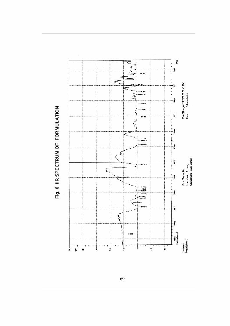
Results and Discussion
The compatibility between zidovudine and the selected polymer
ethyl cellulose was evaluated using FTIR peak matching method. The IR
spectra of pure drug, polymer and physical mixtures are shown in spectra
above (fig.4, fig.5 and fig.6) respectively. There was no disappearance of
peak in polymer drug mixture, which confirmed the absence of any
chemical interaction between drug and polymer.
Drug content analysis
UV spectrophotometric method was employed to verify the
presence of drug in microspheres.30 mg of formulation was taken and the
drug was extracted with phosphate buffer pH 7.4 and absorbance was
measured using UV spectrophotometer at 266nm. The amount of
zidovudine in the microspheres was estimated with the help of standard
graph. A study was performed on the percentage yield and percentage
encapsulation efficiency.
Drug encapsulation efficiency =
Amount of drug bound to the microspheres mg Total amount of applied drug mg
70

Table 5 : Amount of drug loaded in Ethylcellulose microspheres using 50 and 100 ml of dispersion medium.
Drug:Polymer
Amount of drug loaded
50 ml of dispersion medium
100 ml of dispersion medium
0.25:1 6.31±0.5 5.74±0.5
0.5:1 11.85±0.3 11.61±0.7
0.75:1 13.25±0.5 13.06±0.6
1:1 16.66±0.5 15.69±0.2
Fig. 7 Amount of drug loaded in Ethylcellulose microspheres using 50 and 100 ml of dispersion medium
0
2
4
6
8
10
12
14
16
18
Qua
ntity
of d
rug
load
ed
0.25:1 0.5:1 0.75:1 1:01
Drug : Polymer
Medium A Medium B
71

Results and Discussion
The amount of drug loaded in the microspheres prepared using 50
and 100 ml of the light liquid paraffin was represented in fig.7. It was found
that the amount of drug getting loaded into the microspheres increased
when the amount of drug used for the preparation of the microspheres.
The amount of drug getting loaded was found to be 6.31±0.5, 11.85±0.3,
13.25±0.5 and 16.66±0.5 mg when the volume of dispersion medium was
low i.e. 50ml and 5.74±0.5, 11.61±0.7, 13.06±0.6 and 15.69±0.2 mg when
the volume of dispersion medium was high i.e. 100ml.
From the above results it was found that the amount of drug getting
loaded into the microspheres was high when the volume of medium was
low and less when the volume of medium was high.
The polymer ratio was found to be inversely proportional to drug
loading capacity.
72

Percentage Encapsulation efficiency of ethylcellulose microspheres
The amount of zidovudine present in the microspheres was
determined by extracting into phosphate buffer (pH 7.4). 40 mg of
microspheres were crushed and powdered by using a mortar and pestle
and 30mg of accurately weighed powder was extracted into 100ml of
phosphate buffer (pH 7.4) by stirring at 1000 rpm for 2 h. The solution was
filtered, diluted 100 times and estimated for zidovudine content using
UV/Visible spectrophotometer at 266 nm. (Das et al., 2005).
Percentage entrapped
(%) =Amount of drug bound to the microspheres mg
Total amount of applied drug mg ×100
Table 6: Percentage encapsulation efficiency of ethyl cellulosemicrospheres using 50 and 100 ml of dispersion medium
Drug:Polymer
Percentage of drug loaded
50 ml of dispersion medium
100 ml of dispersion medium
0.25:1 51.22±0.5 48.12±0.6
0.5:1 54.29±0.6 52.18±0.2
0.75:1 58.26±0.3 57.27±0.5
1:1 65.12±0.5 62.57±0.4
73

Fig. 8 Percentage encapsulation efficiency of ethyl cellulose microspheres using 50 and 100 ml of dispersion medium
0
10
20
30
40
50
60
70
Per
cen
tag
e o
f dr
ug
entr
app
ed
0.25:1 0.5:1 0.75:1 1:01
Drug : Polymer
Medium A Medium B
74

Results and Discussion
The percentage of drug loaded in the microspheres prepared using
50 and 100 ml of the light liquid paraffin was represented in fig.8. It was
found that the percentage of drug getting loaded into the microspheres
was directly proportional to the amount of drug. The amount of drug
getting loaded was found to be 51.22±0.5, 54.29±0.6, 58.26±0.3 and
65.12±0.8 % when the volume of dispersion medium was low i.e. 50ml and
48.12±0.6, 52.18±0.2, 57.27±0.5 and 62.57±0.4 % when the volume of
dispersion medium was high i.e. 100ml.
From the above results it was found that the percentage of drug
getting loaded into the microspheres was high when the volume of
medium was low and less when the volume of medium was high.
Percentage yield of ethylcellulose microspheres
The percentage yield of the microspheres was calculated by
accurately weighing out the amount of product obtained at the end of each
batch of each batch of formulation. The formula given below was used to
calculate the % yield of the microspheres. (Sunit Kumar Sahoo et al.,
2005).
Percentage yield (%) =Amount of recored microparticle mg
The theoratical content mg ×100
75

Table 7: Percentage yield of ethylcellulose microspheres using
50 and 100 ml of dispersion medium
Drug:Polymer
Percentage yield
50 ml of dispersion
medium
100 ml of dispersion
medium
0.25:1 49.04 50.24
0.5:1 45.08 44.9
75:1 57.0 56.9
1:1 61.8 59.8
Fig. 9 Percentage yield of Ethylcellulose Microspheres using 50 and 100 ml of dispersion medium.
0
10
20
30
40
50
60
70
Pe
rce
nta
ge y
ield
0.25:1 0.5:1 0.75:1 1:01
Drug : Polymer
Medium A Medium B
76

Results and Discussion
The percentage yield of microspheres by using various drug
polymer ratio and different volume of dispersion medium was shown in fig
9. The percentage yield in 50 ml of dispersion medium was found to be
49.04%, 45.08%, 57.0% and 61.8%. The percentage yield in 100 ml of
distribution medium was found to be 50.24%, 44.9%, 56.9% and 59.8%.
From the above results it was found that the percentage yield was
high when the volume of dispersion medium used was 50 ml.
DETERMINATION OF SIZE AND SHAPE OF MICROSPHERES
Determination of size and shape of microspheres using scanning
electron microscope:-
The surface morphology and internal texture of zidovudine
microspheres were observed by a scanning electron microscope (SEM).
SEM photographs were taken on 3kv magnification at 18oC. Before
scanning the particles were sputtered with platinum to make the surface
conductive. The photograph shows that the particles are spherical enough
and have a smooth surface.
77

Fig. 10, 11 Scanning electron microscope pictures of ethyl cellulose microspheres
78
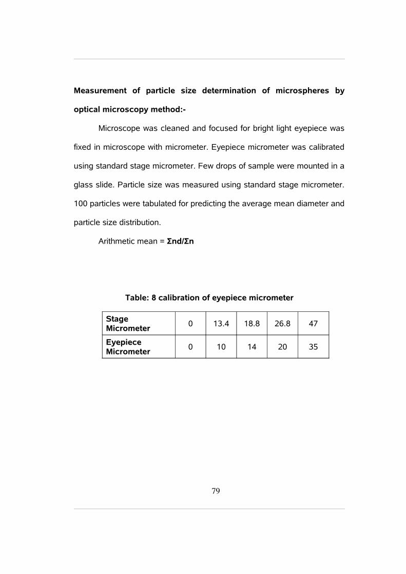
Measurement of particle size determination of microspheres by
optical microscopy method:-
Microscope was cleaned and focused for bright light eyepiece was
fixed in microscope with micrometer. Eyepiece micrometer was calibrated
using standard stage micrometer. Few drops of sample were mounted in a
glass slide. Particle size was measured using standard stage micrometer.
100 particles were tabulated for predicting the average mean diameter and
particle size distribution.
Arithmetic mean = Σnd/Σn
Table: 8 calibration of eyepiece micrometer
Stage Micrometer
0 13.4 18.8 26.8 47
Eyepiece Micrometer
0 10 14 20 35
79

Table: 9 Particle size determination by optical microscopic method for formulations prepared using 50 ml dispersion medium
Formulation code
Particle size range (µm)
Mean diameter(µm) (d)
Number of particles (n)
nxd
F1
0-100 50 8 400100-200 150 5 750
200-300 250 4 1000
300-400 350 54 18900400-500 450 21 9450500-600 550 8 4400
F2
0-100 50 4 200100-200 150 7 1050
200-300 250 21 5250
300-400 350 48 16800
400-500 450 13 5850
500-600 550 7 3850
F3
0-100 50 4 200
100-200 150 4 600
200-300 250 18 4000300-400 350 52 18200400-500 450 17 7650500-600 550 5 2750
F4
0-100 50 5 250
100-200 150 7 1050
200-300 250 15 3750
300-400 350 47 16450
400-500 450 20 9000
500-600 550 6 3300
80

Fig.12 Particle size determination by optical microscope method
using
for formulations prepared using 50 ml dispersion medium
0
10
20
30
40
50
60
0-100 100-200 200-300 300-400 400-500 500-600
Particle size range
No.
of
Par
ticle
s
F1 F2 F3 F4
Results and Discussion
The number of particles present in each size range was shown in
fig 12 for the formulations made using 50 ml of light liquid paraffin and
drug polymer ratio of (0.25:1, 0.5:1, 0.75:1, 1:1). The highest number of
particles were found in the range of 300-400 µm.
81

Table: 10 Particle size determination by optical microscopic method
for formulations prepared using 100 ml dispersion medium.
Fig. 13 Particle size determination by optical microscope method for
formulations prepared using 100 ml dispersion medium
0
10
20
30
40
50
60
0-100 100-200 200-300 300-400 400-500 500-600
Particle size range
No.
of P
artic
les
F5 F6 F7 F8
82

Results and discussion
The number of particles present in each size range was shown in
fig 13 for the formulations made using 100 ml of light liquid paraffin and
drug polymer ratio of 0.25:1, 0.5:1, 0.75:1 and 1:1. The highest number of
particles was found in the range of 300-400 µm.
Arithmetic mean:
F1 = 349.00 µm F 5= 307.00 µm
F 2= 330.00 µm F 6= 352.00 µm
F 3= 334.00 µm F 7= 351.00 µm
F 4= 338.00 µm F 8= 337.00 µm
83
4.5 In Vitro Drug Release Studies
The USP basket-type dissolution (Type II) test apparatus was used
for in vitro release studies. A weighed quantity of the microspheres was
suspended in 900 ml of phosphate buffer (pH 7.4). The dissolution
medium was stirred at 100 rpm and maintained at constant temperature
(37±1 0 C). At preset time intervals 5 ml aliquots were withdrawn at 0, 1, 2,
4, 6, 12, and 24 hours and replaced by an equal volume of fresh
dissolution medium to maintain sink condition. After suitable dilution, the
samples were analyzed at 266 nm using UV-Visible spectrophotometer.
Calculation
To calculate the actual percentage release of drug it is necessary to
calculate the theoretical release of the drug. It can be calculated by using
the general formula,
Theoretical release = (total amount o drug loaded in 1mg of
microspheres) x (amount of microspheres taken for in vitro studies)
The exact percentage release of drug can be obtained by the
general formula:
Exact percentage release or
Cumulative release =Actual Re lease
Theoretical Re leaseX 100
84

IN VITRO DRUG RELEASE STUDIES
Table: 11 Percentage of drug released for formulations prepared using 50 ml of dispersion medium.
S.No Time(hrs)
Cumulative percentage release
F1 F2 F3 F4
1 0 0.98±0.02 0.82±0.08 0.53±0.05 0.73±0.04
2 1 12.74±0.04 15.7±0.05 21.06±0.07 20.78±0.05
3 2 20.09±0.06 20.66±0.07 24.0±0.05 24.2±0.08
4 4 23.52±0.05 24.51±0.04 29.33±0.04 30.56±0.05
5 6 25.98±0.07 26.17±0.05 34.66±0.08 33.74±0.04
6 12 36.76±0.07 44.07±0.07 48.0±0.04 45.72±0.07
7 24 57.85±0.08 60.06±0.04 61.3±0.05 64.05±0.08
85

Chapter V Experimental WorkFig. 14 Percentage release of zidovudine microspheres prepared using 50 ml of dispersion medium
0
10
20
30
40
50
60
70
0 1 2 4 6 12 24
Time in hours
Cu
mu
lativ
e p
erce
nta
ge
rele
ase
F1 F2 F3 F4
Formulation and In Vitro Evaluation of Zidovudine Microspheres
86

Results and Discussion:
The in vitro release profile was shown in fig.14. All the formulation
was found to show sustained release. The release profile of F1, F2, F3 &
F4 was found to be 57.85%, 60.06%, 61.3% & 64.05% respectively at the
end of 24 hours. The results suggest that the polymer concentration
having a significant influence on the release profile. It was found to be
inversely proportional. As the drug polymer ratio increase there was a low
release profile. 1:1 drug polymer ratio can be further evaluated in vivo to
optimize the formulation.
87
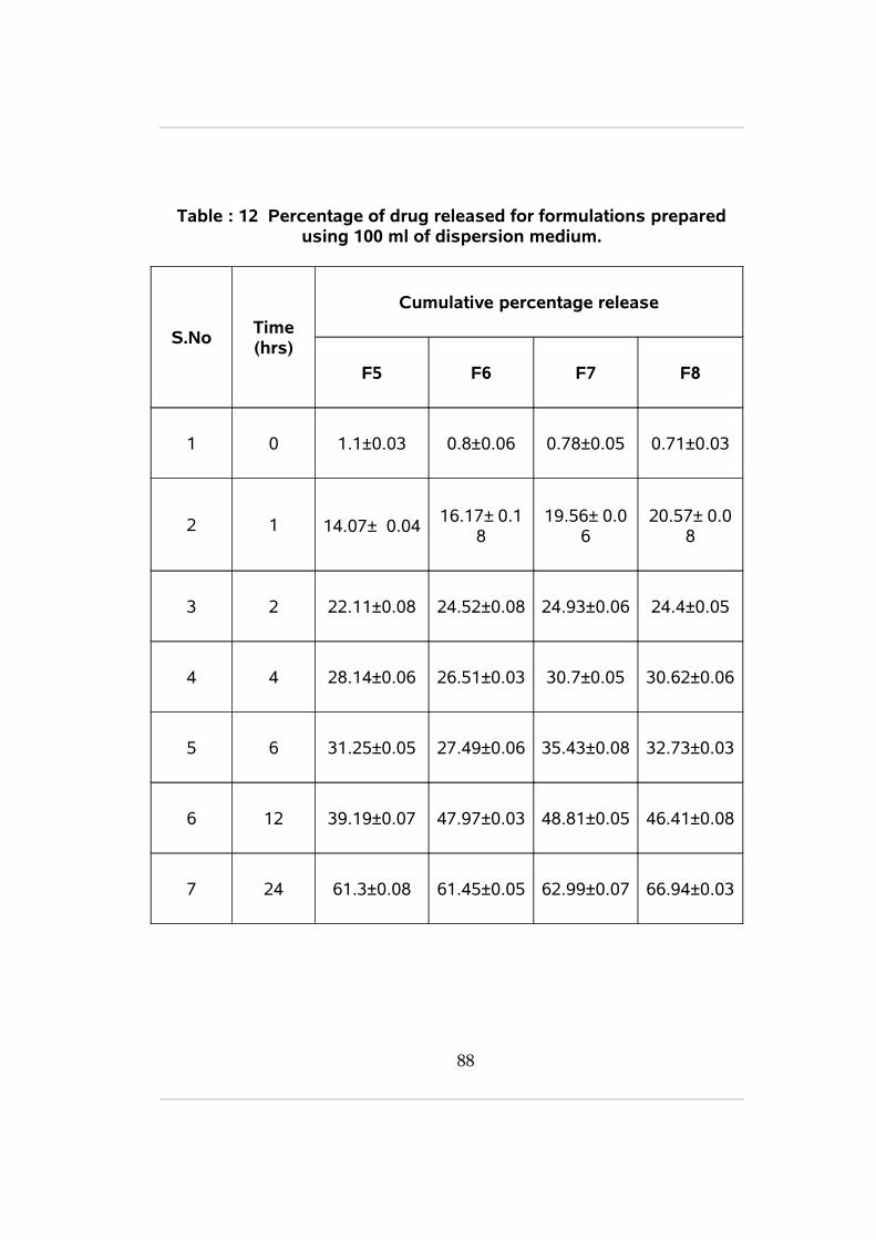
Table : 12 Percentage of drug released for formulations prepared using 100 ml of dispersion medium.
S.NoTime (hrs)
Cumulative percentage release
F5 F6 F7 F8
1 0 1.1±0.03 0.8±0.06 0.78±0.05 0.71±0.03
2 1 14.07± 0.0416.17± 0.1
819.56± 0.0
620.57± 0.0
8
3 2 22.11±0.08 24.52±0.08 24.93±0.06 24.4±0.05
4 4 28.14±0.06 26.51±0.03 30.7±0.05 30.62±0.06
5 6 31.25±0.05 27.49±0.06 35.43±0.08 32.73±0.03
6 12 39.19±0.07 47.97±0.03 48.81±0.05 46.41±0.08
7 24 61.3±0.08 61.45±0.05 62.99±0.07 66.94±0.03
88

Fig. 15 Percentage release of zidovudine microspheres prepared using 100 ml of dispersion medium
0
10
20
30
40
50
60
70
80
0 1 2 4 6 12 24
Time in hours
Cum
ula
tive
pe
rce
nta
ge r
ele
ase
F5 F6 F7 F8
Formulation and In Vitro Evaluation of Zidovudine Microspheres
89

Results and Discussion
The in vitro release profile was shown in Fig.15. All the formulation
was found to show sustained release. The release profile of F5, F6, F7 &
F8 was found to be 61.3%, 61.45%, 62.99% & 66.94% respectively at the
end of 24 hours. The results suggest that the polymer concentration
having a significant influence on the release profile. It was found to be
inversely proportional. As the drug polymer ratio increase there was a low
release profile. 1:1 drug polymer ratio can be further evaluated in vivo to
optimize the formulation.
90

SUMMARY AND CONCLUSION
SUMMARY
Novel drug delivery system is finding more attention in
pharmaceutical industries due to its various advantages. In the line
sustained/controlled release formulations are more focused for improving
the drug bioavailability with less dosing frequency. Zidovudine is one such
compound having a poor bioavailability with high dosing frequency. The
present investigation is focused to develop a suitable sustained release
formulation for achieving better bioavailability and reduce dosing
frequency.
Compatibility Studies
Zidovudine was formulated as microspheres using ethylcellulose.
Compatibility studies were carried out using FTIR. The results suggest no
evidence of any interaction. Different batches containing various
concentrations of drug, polymer with varying volume of dispersion medium
was prepared by double emulsion solvent diffusion technique.
In Vitro release studies
The in vitro release suggest that low volume of dispersion medium
and low polymer ratio (F4) showed 64.05% of release at the end of 24 hrs
with 65.12 % entrapment.
95

CONCLUSION
It is evident that zidovudine loaded ethylcellulose microspheres
showed better sustained release profile which is up to 24 hrs. The
prepared formulation can be further evaluated to achieve a better
bioavailability. IN VIVO studies can be carried out to optimize the
Zidovudine microspheres using ethyl cellulose. The pharmacokinetic
behavior of the formulated microspheres can be studies for optimization of
the formulation.
96

BIBLIOGRAPHY
Amitava Ghosh., Udaya Kumar Nayak., Partha Roy., 2007. Development,
evaluation and method selection for the preparation of lamivudine
microspheres.
Bhupender Singh., and Agrwal, R., 2002. Design development and
optimization of controlled release microcapsules of diltiazem
hydrochloride, Ind J.Pharm.Sci; 64(4): p 378-385.
Brahmankar, D.M., Sunil B., Jaiswal., 1995. Biopharmaceutics and
Pharmacokinetics, A treatise. First edition, p 335-337.
Chowdary, K.P.R., Rao, N.K., and Malathi, K., 2004. Ethylcellulose
microspheres of Glipizide; characterization, in vitro and in vivo
evaluation. Ind. J. Pharm. p.1-33.
Cui-Yun Yu., Xi-Chen Zhang., Fang-Zhou Zhoua., Xian-Zheng Zhang., Si-
Xue Chenga., Ren-Xi Zhuoa., Cui-Yun Yu A.B., Xi-Chen Zhang A.,
Fang-Zhou Zhoua., Xian- Zheng Zhang., Si-Xue Chenga., Ren-Xi
Zhuoa., 2008. Sustained release of antineoplastic drugs from
chitosan-reinforced alginate microparticle drug delivery systems. Int
J Pharm sci p 1-30.
Dandagi, PM., Manvi, F.V., Gadad, V.S., Masitholimaath, A.P., 2004.
Microencapsulation of Verapamil Hydrochloride by Ionotropic
gelation technique Ind J Pharm Sci;66(5): p 631 635.
97

Das, M.K., and senapati, P.C., 2008. Furosemide loaded alginate
microspheres prepared by ionic cross linking technique:
morphology and release characteristics.Ind J Pharm Sci 70 (1):
p 77- 84.
Das, M.K., Rama Rao, K., 2007. Microencapsulation of zidovudine by
double emulsion solvent diffusion technique using ethylcellulose. J.
Microencapsulation. 69 (2) p -244-250.
Edith mathiowitz, Encyclopedia of controlled drug delivery 2002. Volume 2,
John Willy and sons inc. p. 493-543, 669-670, 1031.
Gohel, M.C., Parikh, R.K., Amin, A.F., Surati, A.K., 2005. Preparation and
formulation optimization of sugar cross linked gelatin microspheres
of diclofenac sodium Ind. J.pharm.sci;67(5): p 575-581.
Indian Pharmacopoeia 1996. Vol -2, A-144-145.
Jain, N.K., 1997. Controlled and novel drug delivery,.
CBS publishers. New Delhi.p. 236-256.
Khawla Abu-Izza., Lucila Garcia-Contreras., Robert Lu, D., 1996.
Preparation and evaluation of zidovudine-loaded sustained-release
Microspheres. 2. Optimization of multiple response variables.
Journal of Pharmaceutical Sciences 85( 6), p.572-576.
Lee, J.H., Park T.G., Choi H.K., 2000. Effect of formulation and processing
variables on the characteristics of microspheres for water soluble
drugs prepared by w/o/o double emulsion solvent diffusion method.
Int J of Pharm 196: p.75–78.
98

Leon Lachman., Herbert A., Lieberman., Jospeh L., kanig., 1990. The
theory and practice of industrial pharmacy Third edition, Varghese
publishing house. p.430 - 456
Li Jun., Zhang Jijuan., ZHAO YU., YAN Xinjuan., 2007. Preparation of
procaine haemoglobin microcapsules of chitosan –sodium alginate.
Natural Science.24(3): p .239- 242.
Gonzalez, M.L., Rodriguez Holgado, M.A., Sanchez lafuente, C., Rabasco.
A.M., 2002. Alginate / Chitosan particulate systems for diclofenac
sodium release Int J Pharm .Sci. 27(3): p .539- 542.
Malay Kumar Das., Kalakuntala Rama Rao., 2006. Evaluation Of
Zidovudine Encapsulated Ethylcellulose Microspheres Prepared By
Water-In-Oil-In-Oil (W/O/O) Double Emulsion Solvent Diffusion
Technique. Acta Poloniae Pharmaceutica and Drug Research,63
(2) p.141-148.
Mandal, T.K., Shekleton, M., Onyebueke, E., Washington, L., Penson, T.,
1996. Effect of formulation and processing factors on the
characteristics of biodegradable microcapsules of Zidovudine. J of
Microencapsulation 13:p.545–557.
Mandal, T.K., Tenjarla, S., 1996. Preparation of biodegradable
microcapsules of Zidovudine using solvent evaporation: Effect of
the modification of aqueous phase. Int J of Pharmaceutics
137(3):p.187–197.
99

Manjunatha, K.M., .Ramana, M.V., and Satyanarayana, D., 2007. Design
and evaluation of diclofenac sodium controlled drug delivery
systems. Int J.Pharm Sci. p384-388.
Martinac, A., Filipovic-Gracic, J., Vomovich, D., perissatii, B., and
Franceschinis, E., 2005. Preparation of loratidine loaded
microspheres by spray drying technique. Int. J. Pharm. 291(1-2),
p.69-77.
Mishara., 2003.Development of chitosan – alginate microcapsules for
colon specific delivery of Metronidazole. int J of pharm 234(3), p
78-86.
Parul Trivedi, A.M., Verma, L., garud, N., 2008.Preparation and
characterization of aceclofenac microspheres. Asian J of
Pharm,p.110-115.
Perumal, D., Dangor, C.M., Alcock, R.S., Hurbans, N., and Moopanar,
K.R., 1999. Effect of formulation variables and micrometric
properties of modified release ibuprofen microspheres.
J.Microencapsulation. 16(4), p.475-487.
Perumal, D., 2003. Microencapsulation of ibuprofen and eudragit RS 100
by emulsion solvent diffusion technique. Int. J. pharm. 218(7),
p.(1-11).
Prakash, K., Raju, P.N., Shanta, K.K., Lakshmi, M N., 2007. Preparation
characterization of lamivudine microcapsules using various
100

cellulose polymers Tropical Journal of Pharmaceutical Research, 6
(4): p.841-847.
Rama rao., Prakash, K., Senapati., Das, M.K., 2005. Formulation and
in vitro evaluation of ethyl cellulose microspheres containing
zidovudine. J. of Microencapsulation, 22(8): p.863–876.
Remington, The science and practice of pharmacy.2002. 20 th edition,
Lippincot Williams and Wilkins..(I and II), p.313, 740-741, 890,
1043-1044,1553.
Robinson, J.R., Lee, H., 2005.Controlled drug delivery, Fundamentals and
applications. Maecek Dekker, Inc. Second edition(29); 373-374.
Sajeev, C., Vinay, G., Archana, R., and Saha, R.N., 2002. Oral controlled
release formulation of diclofenac sodium by microencapsulation
with ethylcellulose. J. Microencapsulation. 19(6), 753-760.
Sameer Sharma., Atmaram Pawar., 2006. Low density multiparticulate
system for pulsatile release of meloxicam; J. microencapsulation
21(7): p 150–158.
Sunit Kumar Sahoo., Abdul Arif Mallick., Barik, B.B., Prakash., Senapati,
C.B., 2005. Formulation and in vitro Evaluation of Eudragit®
Microspheres of Stavudine Tropical Journal of Pharmaceutical
Research,4,(1), p. 369-375.
101

Tamizharasi,S., Rathi, J.C., Rathi, V., 2007. Formulation, characterization
and in-vitro release kinetics of aceclofenac loaded
poly (β-caprolactone) microspheres. Indian Drugs 44(12),
p 973-975.
Tripathi, K.D., 2008. Essentials of medical pharmacology. Vallabh
prakashan.8th edition. p. 767,770-771,774-777.
Vyas., S.P., and Khar, R., 2002. Targeted and controlled drug delivery
novel carrier systems. Vallabh prakashan. Second edition,
p.417-457.
102