formulation of pegylated and hesylated biopharmaceuticals · formulation of pegylated and hesylated...
TRANSCRIPT

Dissertation
zur Erlangung des Doktorgrades
der Fakultät für Chemie und Pharmazie der
Ludwigs-Maximilians-Universität München
Formulation of PEGylated and HESylated
Biopharmaceuticals
von
Robert Liebner
aus Halle/ Saale, Deutschland
2015

II

III
Erklärung
Diese Dissertation wurde im Sinne von § 7 der Promotionsordnung vom 28. November
2011 von Herrn Prof. Dr. Gerhard Winter betreut.
Eidesstattliche Versicherung
Diese Dissertation wurde eigenständig und ohne unerlaubte Hilfe erarbeitet.
Frankfurt am Main, 22.08.2015
……………………………
(Robert Liebner)
Dissertation eingereicht am: 08.07.2015
1. Gutachter: Prof. Dr. Gerhard Winter
2. Gutachter: Prof. Dr. Wolfgang Frieß
Mündliche Prüfung am: 30.07.2015

IV
Für meine Familie in Liebe und Dankbarkeit.

V
Acknowledgments
The presented thesis was prepared under the supervision of Prof. Dr. Gerhard Winter at
the facilities at the Department of Pharmacy, Pharmaceutical Technology and Bio-
pharmaceutics at the Ludwig-Maximilians-University in Munich, Germany.
First of all I would like to express my deepest gratitude to my doctoral father Prof. Dr.
Gerhard Winter for being a member of his research group. During that time he guided
me through all phases of the PhD program with his excellent and continuous scientific
input as well as personal advice. Furthermore he facilitated the contribution to a number
of research conferences in Europe and the United States. Additionally, he was the initia-
tor of many different social and scientific events, which were participated by professors
and scientists from different universities and pharmaceutical companies to facilitate the
exchange of our experiences and to extend our knowledge in different fields.
My deepest gratitude has to go to Dr. Ahmed Besheer for his outstanding and excellent
guidance. He initiated a collaboration with Fresenius Kabi and was significantly respon-
sible for the success of the project. During regular meetings and discussions he taught
me how to deal with success and breakdowns and fostered my scientific approaches. He
invested a lot of effort and personal resources into the project with an interminable pas-
sion. Thank you for being a great supervisor!
In addition, I have to thank Prof. Dr. Wolfgang Frieß for being the second referee of the
thesis and the fruitful discussions during e.g. progress reports and personal meetings.
My lab mates Roman, Christian and Marie- Paule provided a very harmonious and
funny atmosphere. I am sorry for some mood swings from time to time and I really ap-
preciate the relationship with each of you within and outside of the lab. Thank you!
All other group members of the Winter and Frieß labs has to be named in the same way.
I have to thank for the constructive help and the development of a number analytical
methods. Beyond the labs we shared a lot of funny moments and events.

VI
Additionally, I have to thank Dr. Frank Hacket, Dr. Thomas Hey, Dr. Martin Meyer, Dr.
Helmut Knoller, Dr. Peter Vorstheim, Dr. Bernd Sundermann and Prof. Dr. Crispulo
Gallegos from Fresenius Kabi for the funding, the material support and the continuous
as well as excellent scientific input.
Finally I would like to thank my girlfriend Christiane for her continuous encouragement
and her love.

VII
Table of Contents
Chapter I - General Introduction .............................................................. 1
1. General Introduction ............................................................................................. 3
1.1. Half-life modulation of biologics ..................................................................... 3
1.2. The bioconjugation polymer PEG and the effect of PEGylation on biologics . 6
1.3. PEGylation chemistry and PEG reagents ......................................................... 7
1.3.1. Random PEGylation ..................................................................................... 7
1.3.2. Site-specific PEGylation of the N-terminus ................................................. 9
1.3.3. Site-specific PEGylation of thiol groups .................................................... 10
1.3.4. Further strategies for PEGylation ............................................................... 13
1.4. Limitations of PEG and PEGylation technology ............................................ 13
1.4.1. Quality of the polymer and PEGylation chemistry ..................................... 13
1.4.2. Effect on activity upon conjugation ............................................................ 14
1.4.3. Toxicity ....................................................................................................... 15
1.4.4. Immunogenicity .......................................................................................... 16
1.4.5. Effect on protein stability ........................................................................... 17
1.4.6. Effect on viscosity ...................................................................................... 17
1.4.7. Behavior during and after lyophilization .................................................... 18
1.5. Alternative strategies for half-life extension based on biodegradable polymers
19
1.6. HES and HESylation ...................................................................................... 19
1.6.1. HESylation chemistry and HES reagents ................................................... 21
1.6.2. Random HESylation ................................................................................... 22
1.6.3. Site-specific HESylation ............................................................................. 23
1.6.4. Quality of the polymer HES ....................................................................... 25
1.6.5. Activity ....................................................................................................... 25
1.6.6. Toxicity ....................................................................................................... 26
1.6.7. Effect on protein stability ........................................................................... 26
1.6.8. Effect on viscosity ...................................................................................... 27
1.6.9. Lyophilization ............................................................................................. 27
1.7. Polysialylation ................................................................................................ 28

VIII
1.8. Recombinant PEG mimetics ........................................................................... 28
1.8.1. PASylation .................................................................................................. 28
1.8.2. XTENylation ............................................................................................... 29
1.9. Aims of the thesis ............................................................................................ 31
1.10. References ....................................................................................................... 32
Chapter II – Half-life extension through HESylation............................ 45
2. Protein HESylation for half-life extension: Synthesis, characterization and
pharmacokinetics of HESylated anakinra ................................................................... 47
2.1. Introduction ..................................................................................................... 49
2.2. Experimental procedures ................................................................................. 50
2.2.1. Materials ...................................................................................................... 50
2.2.2. Synthesis and purification of HESylated anakinra ...................................... 50
2.2.3. Determination of conjugate concentration .................................................. 51
2.2.4. SEC-MALLS measurement for the determination of the molar mass and
size of the conjugates .................................................................................................. 51
2.2.5. Dynamic light scattering (DLS) .................................................................. 52
2.2.6. Fourier transform infrared spectroscopy ..................................................... 52
2.2.7. Surface plasmon resonance measurements for in vitro binding affinity ..... 52
2.2.8. Microscale Thermophoresis (MST) for in vitro binding affinity ................ 53
2.2.9. Microcalorimetry ......................................................................................... 53
2.2.10. Thermal stability ......................................................................................... 54
2.2.11. Pharmacokinetic study ................................................................................ 54
2.3. Results ............................................................................................................. 55
2.3.1. Molar mass and size .................................................................................... 55
2.3.2. Protein conformation ................................................................................... 57
2.3.3. Microcalorimetry and thermal stability ....................................................... 58
2.3.4. In vitro binding affinity ............................................................................... 60
2.3.5. Pharmacokinetic profiles of native and HESylated anakinra ...................... 62
2.4. Discussion ....................................................................................................... 64
2.5. Conclusions ..................................................................................................... 68
2.6. References ....................................................................................................... 68
IX
Chapter III – Highly concentrated formulations ................................... 75
3. Head to Head Comparison of the Formulation and Stability of Concentrated
Solutions of HESylated versus PEGylated Anakinra ................................................. 77
3.1. Introduction ..................................................................................................... 79
3.2. Materials and Methods .................................................................................... 80
3.2.1. Materials ..................................................................................................... 80
3.2.2. Synthesis and Purification of the Protein Conjugates ................................. 80
3.2.2.1. PEGylation .................................................................................................. 80
3.2.2.2. HESylation .................................................................................................. 81
3.2.3. SEC–MALLS Measurement for the Determination of the Molecular
Weight and Size of the Conjugates ............................................................................. 82
3.2.4. Hydrodynamic Radius ................................................................................ 82
3.2.5. FTIR ............................................................................................................ 83
3.2.6. Biacore Analysis for in vitro Binding Affinity ........................................... 83
3.2.7. Preparation of Highly Concentrated Solutions ........................................... 83
3.2.8. Differential Scanning Calorimetry .............................................................. 84
3.2.9. Viscosity ..................................................................................................... 84
3.2.10. Storage Stability: SEC ................................................................................ 84
3.3. Results ............................................................................................................. 85
3.3.1. Synthesis and Purification .......................................................................... 85
3.3.2. Determination of the Molecular Weight and Hydro-dynamic Size ............ 85
3.3.3. FTIR Analyses for Changes in Secondary Structure .................................. 87
3.3.4. In Vitro Binding Affinity ............................................................................ 88
3.3.5. Properties of Highly Concentrated Solutions ............................................. 90
3.3.5.1. Viscosity Measurement ............................................................................... 90
3.3.5.2. Thermal Analysis ........................................................................................ 92
3.3.5.3. Thermal Stability upon Storage at 40°C ..................................................... 94
3.4. Discussion ....................................................................................................... 95
3.5. Conclusion ...................................................................................................... 99
3.6. References ..................................................................................................... 100

X
Chapter IV – Freeze- dried formulations ............................................. 107
4. Freeze-drying of HESylated IFNα-2b: Effect of HESylation on storage stability
and benchmarking to PEGylation ............................................................................. 109
4.1. Introduction ................................................................................................... 111
4.2. Materials and Methods .................................................................................. 113
4.2.1. Materials .................................................................................................... 113
4.2.2. Synthesis and purification of HESylated interferon α-2b ......................... 113
4.2.3. Synthesis and purification of PEGylated interferon α-2b ......................... 114
4.2.4. Synthesis and purification of HESylated rh-met-G-CSF .......................... 115
4.2.5. Pretreatment of PEGylated rh-met-G-CSF ............................................... 115
4.2.6. Quality control of protein conjugates ........................................................ 116
4.2.6.1. UV-measurement ...................................................................................... 116
4.2.6.2. SDS-PAGE ................................................................................................ 116
4.2.6.3. Size-exclusion and reversed phase chromatography ................................. 116
4.2.6.4. AF4-MALLS measurement for the determination of the molar mass, size of
the conjugates and conjugate stoichiometry .............................................................. 117
4.2.6.5. Dynamic light scattering (DLS) ................................................................ 117
4.2.6.6. Peptide mapping ........................................................................................ 117
4.2.7. Freeze-Drying Microscopy ....................................................................... 118
4.2.8. Sample preparation for lyophilization ....................................................... 118
4.2.9. Tg´ measurement ....................................................................................... 118
4.2.10. Freeze-drying protocol .............................................................................. 119
4.2.11. Tg and degree of crystallization ................................................................ 119
4.2.12. Residual moisture ...................................................................................... 119
4.2.13. Turbidity measurement ............................................................................. 119
4.2.14. Particle count - Light obscuration ............................................................. 120
4.2.15. Monomer recovery – Size exclusion chromatography .............................. 120
4.2.16. Conformational changes - FTIR ................................................................ 120
4.3. Results ........................................................................................................... 120
4.3.1. Preparation and quality control of the polymer modified interferon samples
120
4.3.2. Lyomicroscopy .......................................................................................... 126
XI
4.3.3. Thermal analysis of the frozen formulations ............................................ 127
4.3.4. Optical evaluation of the freeze-dried samples ......................................... 128
4.3.5. Cake properties after lyophilization and storage ...................................... 128
4.3.5.1. Glass transition temperature and degree of crystallization ....................... 128
4.3.5.2. Residual moisture ..................................................................................... 129
4.3.5.3. Conformational stability after lyophilization and storage ........................ 130
4.3.6. Colloidal stability after lyophilization and storage ................................... 131
4.3.6.1. Turbidity and particle counts .................................................................... 131
4.3.6.2. Monomer recovery and soluble aggregates .............................................. 132
4.4. Discussion ..................................................................................................... 133
4.5. Conclusion .................................................................................................... 136
4.6. References ..................................................................................................... 137
Chapter V – Highly concentrated lyophilized formulations ............... 143
5. Protein HESylation for the use of highly concentrated and freeze-dried
formulations of HES-anakinra conjugates: storage stability and the benchmark to
PEGylation ................................................................................................................ 145
5.1. Introduction ................................................................................................... 147
5.2. Materials and Methods .................................................................................. 148
5.2.1. Synthesis and purification ......................................................................... 148
5.2.2. Sample preparation ................................................................................... 148
5.2.3. Tg´ measurement ...................................................................................... 149
5.2.4. Freeze-drying protocol .............................................................................. 149
5.2.5. Tg and degree of crystallization ................................................................ 150
5.2.6. Residual moisture ..................................................................................... 150
5.2.7. Turbidity measurement ............................................................................. 150
5.2.8. Particle count - Light obscuration ............................................................. 151
5.2.9. Monomer recovery – Size exclusion chromatography ............................. 151
5.3. Results ........................................................................................................... 151
5.3.1. Cake properties after lyophilization and storage ...................................... 152
5.3.1.1. Glass transition temperature and residual moisture .................................. 152
5.3.1.2. Degree of crystallization ........................................................................... 155
5.3.2. Protein quality after reconstitution ........................................................... 156

XII
5.3.2.1. Colloidal stability after lyophilization and storage ................................... 156
5.3.2.2. Monomer recovery and soluble aggregates ............................................... 156
5.4. Discussion ..................................................................................................... 159
5.5. Conclusion ..................................................................................................... 161
5.6. References ..................................................................................................... 162
Chapter VI – Summary and attachments ............................................. 165
6. Summary of the thesis ....................................................................................... 167
7. Publications associated with this thesis ............................................................. 171
7.1. Research articles ............................................................................................ 171
7.2. Book chapter ................................................................................................. 171
8. Presentations associated with this thesis ........................................................... 171
8.1. Oral presentations .......................................................................................... 171
8.2. Poster presentations ....................................................................................... 172

XIII
List of Abbreviations
ABC Accelerated blood clearance
API Active pharmaceutical ingredient
AUC Area under the curve
BfArM Federal Institute for Drugs and Medical Devices
BSA Bovine serum albumin
CA Columinic acid
CD Circular dichroism
CD20 B-lymphocyte antigen 20
CHO Chinese hamster ovary
CL Clearance
cmax Maximum concentration of a drug observed after its administra-
tion
CSE Citrate-saline-ethylenediaminetetraacetic acid
DLS Dynamic light scattering
DMARDs Disease modifying antirheumatic drugs
DNA Deoxyribonucleic acid
DSC Differential scanning calorimetry
E. coli Escherichia coli
EPO Erythropoietin
FcRn Neonatal Fc receptor
FDA Food and Drug Administration
FTIR Fourier transform infrared spectroscopy
G-CSF Granulocyte colony-stimulating factor
HEPES 4-(2-hydroxyethyl)-1-piperazineethanesulfonic acid
HER2 Human epidermal growth factor receptor 2
HES Hydroxyethyl starch
hGH Human growth hormone
HLE Half-life extension
HSA Human serum albumin
IFN Interferon
ITC Isothermal calorimetriy
kDa Kilodalton

XIV
LO Light obscuration
mAb Monoclonal antibodies
MHC Major histocompatibility complex
Mn Number average molar mass
MS Molar substitution
MST Microscale Thermophoresis
Mw Mass average molar mass
MW Molar mass
MWCO molecular weight cutoff
NaCl Sodium chloride
Neu5Ac 5-N-acetylneuraminic acid
NH4OH Ammonium hydroxide
P-20 Polysorbate 20
PAS Peptide based on proline, alanine and serine
PD Pharmacodynamics
PDI Polydispersity Index
PEG Polyethylene glycol
Ph. Eur. Pharmacopoea Europaea
PK Pharmacokinetic
PPI Protein- protein- interaction
PRAC Pharmacovigilance Risk Assessment Committee
PSA Polysialic acid
rhIL1-ra Recombinant human interleukin-1 receptor antagonist
RI Refractive index
RNA Ribonucleic acid
RP-HPLC Reversed phase High-performance liquid chromatography
rpm Rounds per minute
s.c. Subcutaneous
scFv Single chain Fv regions
SCID Severe combined immunodeficiency disease
SE- HPLC Size exclusion High-performance liquid chromatography
SEC-MALLS Size exclusion chromatography - multi angle laser light scattering
SPR Surface plasmon resonance
t1/2 Half-life

XV
Tc Critical collapse temperature
TFF Tangential flow filtration
Tg Glass transition temperature
Tg´ Glass transition temperatures of the maximally freeze-concen-
trated matrix
Tm Melting temperature
TNF-α Tumor necrosis factor alpha
TRIS Tris(hydroxymethyl)aminomethane
USP United States Pharmacopeia
UV-VIS Ultraviolet–visible
VEGF Vascular endothelial growth factor


1
Chapter I - General Introduction

2

3
1. General Introduction
1.1. Half-life modulation of biologics
The discovery of recombinant DNA technology in the early 1980s has led to a rapidly ex-
panding market for diagnostic and therapeutic biologics covering a broad range of human
illnesses. Today, protein- and peptide-based drugs comprise over 200 approved products and
hundreds of potential candidates in clinical trials. These compounds can be classified as bio-
pharmaceuticals, which 1) mimic native proteins and operate as replacement therapies, 2)
serve as antagonist therapy or 3) stimulate and mobilize malfunctioning body proteins [1].
Problems with biopharmaceuticals often involve a suboptimal physicochemical profile, typi-
cally caused by either a tendency to aggregate, limited solubility or proteolytic instability.
Additionally, a molecular weight below the renal cutoff (MW < 60 kDa) can restrain their
pharmacokinetic effectiveness, resulting in a plasma half-life of just minutes to hours. Mono-
clonal antibodies (mAbs) tend to avoid this issue, with a molecular weight of around 150 kDa
and a naturally-mediated FcRn recycling mechanism, which together yield a plasma half-life
of days to weeks [2]. Rapid elimination is thus associated with hydrodynamically smaller va-
rieties of biopharmaceuticals such as cytokines, growth factors, peptides and protein scaf-
folds. The efficiency of these drugs is limited by their short circulation time, which must be
overcome by frequent injections [3]. However, simultaneous circulation time enhancement
and improved physicochemical properties can be obtained by covalent linkage of the active
pharmaceutical ingredient (API) to biocompatible polymers. Among the first successful at-
tempts in this regard were the experiments performed by Davies and Abuchowsky in the
1970s, who improved blood circulation of bovine liver catalase and bovine serum albumin by
the chemical attachment of polyethylene glycol (PEG) [4, 5]. The first PEGylated protein
was approved by the Food and Drug Administration (FDA) in the early 1990s: the PEGylated
version of the adenosine deamidase (Adagen®) for the treatment of severe combined immu-
nodeficiency disease (SCID), an autosomal recessive genetic disorder induced by adenosine
deficiency. At least ten PEGylated biopharmaceuticals are approved today and PEGylation is
considered to be the gold standard for half-life extension (HLE) [6]. Table 1 summarizes the
commercial benefit of the PEGylated products currently on the market (adapted from [7]).
Therapeutic compounds usually profit from conjugation with PEG. PEGylation reduces glo-
merular filtration by substantially increasing hydrodynamic size to above the renal cut-off,
thereby slowing down kidney clearance. Additional benefits include protection of the drug

4
from interactions with catabolic and proteolytic factors and the immune system [8, 9]. Physi-
cochemical properties are likewise improved, due to an increase in thermal stability, attenuat-
ed aggregation and enhanced solubility [10].
Most of the approved biopharmaceutical drugs are recombinant replicas of naturally occurring
human proteins. Next generation biologics include an emerging class of alternative protein
scaffolds like affibodies, Adnectins, anticallins or DARPins [11], which are engineered to
recognize particular target structures. These small, specific binders are designed to have affin-
ity to common targets such as TNF-α, CD20 or VEGF and are based on a robust, single-chain
polypeptide framework with remarkable conformational tolerance [12-14]. As with the first
generation of biologics, rapid elimination by the kidneys could prove to be the Achilles’ heel
of these highly specific, unique molecules. Therefore, half-life extension technologies can be
expected to play an important role in market entry.

5
Drug PEGylated
protein
Market
entry
Sales 2013
(M US$)1
Indication Company
PEG–adenosine
deaminase (Adagen®)
Adenosine
deaminase 1990 65
Severe combined
immunodeficiency
disease (SCID)
Enzon
PEG–asparaginase
(Oncaspar®)
Asparaginase 1994 55 Acute lymphoblastic
leukemia Enzon
PEG–interferon α-2b
(PegIntron®)
Interferon α-2b 2000 496 Hepatitis C Schering-Plough
PEG-interferon α-2a
(Pegasys®)
Interferon α-2a 2002 1,416 Hepatitis C Roche
Pegvisomant,
(Somavert®)
Growth
hormone
receptor
antagonist
2002 217 Acromegaly Pfizer
PEG-filgrastim
(Neulasta®)
Granulocyte
colony
stimulating
factor
2002 4,392 Neutropenia Amgen
Pegaptanib
(MacugenTM
)
PEG-anti-
VEGF aptamer 2004 8
Wet age-related
macular degeneration
Eyetech
Pharmaceuticals/
Pfizer
PEG-epoetin-β
(Mircera®)
Erythropoetin 2007 459 Renal anemia Roche
Certolizumab Pegol
(Cimzia®)
Fab fragment
against TNF-α 2008 789
Rheumatoid arthritis
and Crohn’s disease UCB
Pegloticase
(Krystexxa®)
Urate oxidase 2010 26 Chronic gout Savient
Pharmaceuticals
Peginesatide
(Omontys®)*
Erythropoiesis
stimulating
agent
2012 n.a.
Anemia due to
chronic kidney
disease
Affymax and
Takeda
1 From www.evaluategroup.com *recalled in 2013 and now withdrawn from the market
Table 1: Marketed PEGylated proteins and peptides, their year of approval, global sales in
2013 and the companies which first commercialized them (adapted from [7])

6
1.2. The bioconjugation polymer PEG and the effect of
PEGylation on biologics
As the name suggests, polyethylene glycol is a nonionic polyether with a chemical structure
of HO(CH2CH2O)nH which can be synthesized by an anionic ring opening polymerization of
ethylene oxide initiated by nucleophilic attack of a hydroxide ion on the epoxide ring [15].
However, when PEG is used for polypeptide modification, it must typically be of the hetero-
bifunctional variety. Specifically, one of the hydroxyl end groups must be capped with a me-
thyl group to create a monomethoxylated PEG (structure mPEG: CH3O - (CH2CH2O)n -
CH2CH2OH), while the second end group is modified with a functional group amenable to the
conjugation step. This heterobifunctionality enables straightforward conjugation while pre-
venting crosslinking of multiple polypeptides. In this approach, synthesis is initiated by nu-
cleophilic attack of a methoxide ion, as opposed to hydroxide, on the epoxide ring. The final
product is amphiphilic in nature. The oxygen molecules are responsible for PEG’s hydrophilic
character; while the hydrophobic tendency is caused by the ethylene subunits. As such, PEG
is a surface active molecule, soluble both in water and in a number of organic solvents [16].
Its solubility in water over a wide range of molar masses is especially remarkable due to the
fact that its two neighbors namely poly(methylene glycol) and poly(propylene glycol), are
insoluble in water [17]. The apparently more polar character of PEG is derived from a strong
tendency of the oxygen atoms to form hydrogen bonds between 2-3 water molecules; this
results in extraordinary hydration of the polymer with high conformational flexibility and
chain mobility [17]. The exact water-binding capacity ranges from 2-3 water molecules per
subunit up to 16 molecules [18, 19] depending on the method used for quantification [20, 21].
In general, PEG is considered to be non-toxic, non-immunogenic and biocompatible and is
therefore approved by the FDA for parenteral usage [22]. Coupling the API of interest to PEG
will in most cases drastically improve the physicochemical properties of the conjugate. For
instance, hydrophobic drugs become soluble in an aqueous environment after PEGylation.
One of the most oft-cited examples of this phenomenon is interferon β-1b [8]. Native IFNβ-1b
is indicated for the treatment of multiple sclerosis and approved as Betaferon® (Bayer
HealthCare) or Extavia® (Novartis). However, this formulation requires the addition of human
serum albumin to stabilize the protein and preserve solubility after reconstitution. Basu et al.
reported that unmodified IFNβ-1b began to precipitate as insoluble aggregates within 7 days
at neutral pH in the absence of a detergent. In contrast, covalent coupling of a 40 kDa PEG
was able to maintain solubility during that time [8]. The modified protein also profited from
7
an improved pharmacokinetic profile, a lower tendency toward aggregation and reduced im-
munogenicity [8].
1.3. PEGylation chemistry and PEG reagents
1.3.1. Random PEGylation
For conjugation to therapeutic proteins and peptides, pharmaceutical grade PEG reagents are
commercially available in linear or branched architectures and with a variety of different end
group linker moieties for subsequent coupling. These linkers can either react directly or after
an activation step with particular functional groups on the surface of the protein, in both cases
forming a covalent bond. When linking to proteins, available conjugation targets for PEG
include amino acids like lysine, cysteine, histidine, arginine, aspartic acid, glutamic acid, ser-
ine, threonine and tyrosine, as well as the N-terminal amino group and the C-terminal carbox-
ylic acid [19]. For random PEGylation, lysine, with its primary amine side group, is an attrac-
tive target due to the fact that it can represent up to 10% of the primary sequence in many
proteins. This ɛ-amino group represents a nucleophilic target for a number of electrophilic
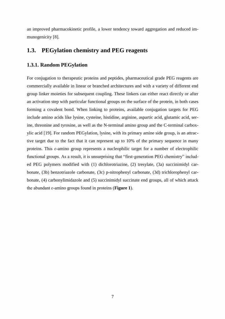
functional groups. As a result, it is unsurprising that “first-generation PEG chemistry” includ-
ed PEG polymers modified with (1) dichlorotriazine, (2) tresylate, (3a) succinimidyl car-
bonate, (3b) benzotriazole carbonate, (3c) p-nitrophenyl carbonate, (3d) trichlorophenyl car-
bonate, (4) carbonylimidazole and (5) succinimidyl succinate end groups, all of which attack
the abundant ɛ-amino groups found in proteins (Figure 1).

8
Figure 1: Activated PEG-derivatives for the chemical coupling to ɛ-amino groups
(adapted from [19])
Such reactions are rapid and straightforward to optimize and scale, but are dominated by a
lack of selectivity, resulting in a number of positional isoforms and differences in the total
number of coupled PEG chains per protein [19]. The reactivity of the functional group and the
protein to PEG ratio control the prevalence of side reactions with other nucleophiles on the
protein surface, namely the N-terminal amino group, the imidazole nitrogens of histidine resi-
dues and the side chains of serine, threonine, tyrosine and cysteine residues [23]. The exist-

9
ence of a number of positional isoforms led to concerns about the reproducibility of drug
batches and may have contributed to higher antigenicity of the modified drug and poor clini-
cal outcomes [3]. Furthermore, unstable linkages between PEG and the protein were some-
times used, which triggered degradation of the PEGylated drug during manufacturing and
storage [24]. An additional problem was caused by the presence of diols, representing up to
15% by mass in batches of mPEG, which resulted in API crosslinking and the formation of
aggregates [3, 19]. However, several PEG conjugates which emerged from “first-generation
PEG chemistry,” such as Adagen® and Oncospar
®, did in fact receive regulatory approval.
The historical evolution of improvements to PEGylation chemistry is presented in Table 2.
Decade PEG reagents General observations Applications
1970 – 1980
PEG-chloro triazine [5]
PEG-succinimidyl succinate
PEG-tresyl
Immunogenic or toxic starting
material, highly polydisperse
PEG, lack of selectivity
Research studies,
enzyme modification
for biocatalysis
1980 – 1990
PEG-aldehyde [25]
PEG-succinimidyl carbonate
PEG-pNO2
phenyl carbonate
PEG-AA-NHS [26]
PEG-carbonylimidazole, etc.
Site-specific conjugation, less
polydisperse PEG, absence of
diols
Enzyme replacement
therapy
1990 – 2000 [27]
Branched PEG
PEG-NHS
PEG-maleimide
PEG-OPSS
Improved selectivity, marketing of
PEGylated drugs
Cytokines, hormones,
anticancer drug tar-
geting
2000 - 2014
Enzymatic coupling [28]
Disulfide coupling [29]
Releasable PEGs [30]
Forked PEGs
Star PEGs
Monodisperse PEGs
Detailed chemical and biological
characterization of conjugates,
combination of genetic engineer-
ing and PEGylation in the design
and discovery of new drugs, more
stringent regulatory requirements
Non-protein drug
PEGylation, oligonu-
cleotide
PEGylation, cell
PEGylation
AA = amino acids; NHS = N-hydroxysuccinimide; OPSS = ortho-pyridyldisulfide
Table 2: History of PEGylation (adapted from [31])
1.3.2. Site-specific PEGylation of the N-terminus
PEG chemistry from the so-called “second generation” was developed to eliminate first-
generation pitfalls by reducing polydispersity (also for high molecular weight PEGs) and diol
content. Improvements to the stability of the linkers were also introduced by creating new
functional moieties that enabled a more tunable conjugation process. The use of propionalde-
10
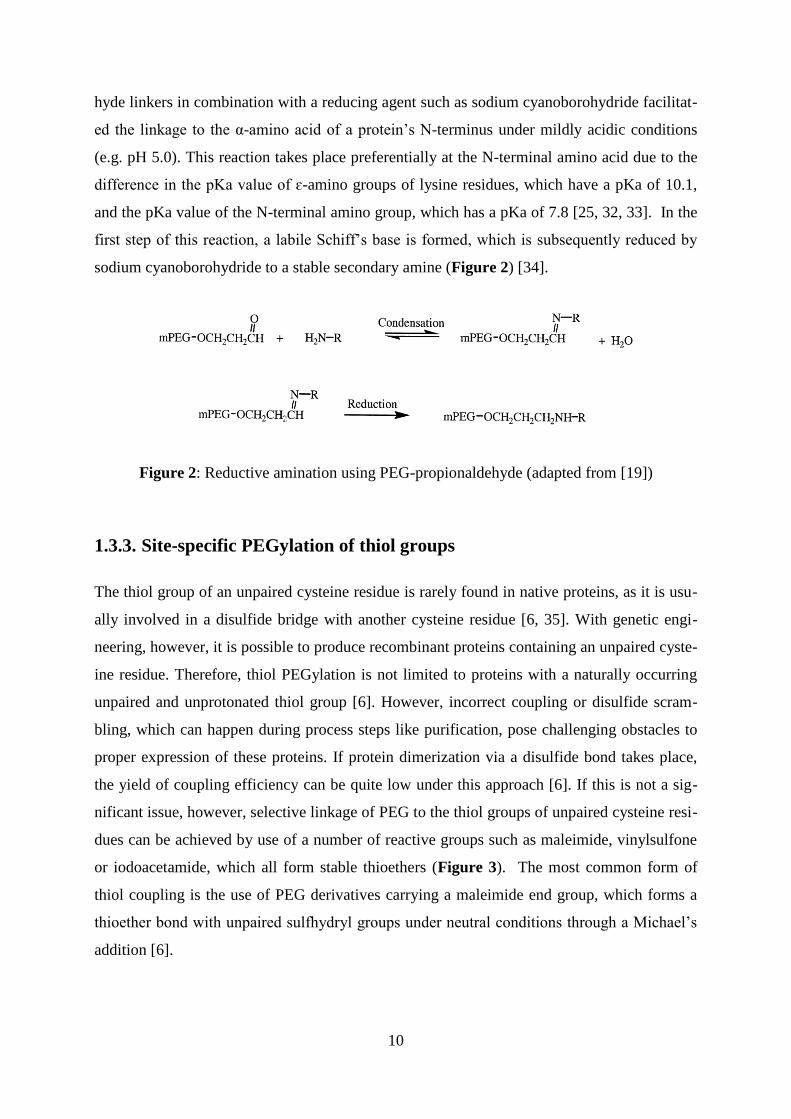
hyde linkers in combination with a reducing agent such as sodium cyanoborohydride facilitat-
ed the linkage to the α-amino acid of a protein’s N-terminus under mildly acidic conditions
(e.g. pH 5.0). This reaction takes place preferentially at the N-terminal amino acid due to the
difference in the pKa value of ɛ-amino groups of lysine residues, which have a pKa of 10.1,
and the pKa value of the N-terminal amino group, which has a pKa of 7.8 [25, 32, 33]. In the
first step of this reaction, a labile Schiff’s base is formed, which is subsequently reduced by
sodium cyanoborohydride to a stable secondary amine (Figure 2) [34].
Figure 2: Reductive amination using PEG-propionaldehyde (adapted from [19])
1.3.3. Site-specific PEGylation of thiol groups
The thiol group of an unpaired cysteine residue is rarely found in native proteins, as it is usu-
ally involved in a disulfide bridge with another cysteine residue [6, 35]. With genetic engi-
neering, however, it is possible to produce recombinant proteins containing an unpaired cyste-
ine residue. Therefore, thiol PEGylation is not limited to proteins with a naturally occurring
unpaired and unprotonated thiol group [6]. However, incorrect coupling or disulfide scram-
bling, which can happen during process steps like purification, pose challenging obstacles to
proper expression of these proteins. If protein dimerization via a disulfide bond takes place,
the yield of coupling efficiency can be quite low under this approach [6]. If this is not a sig-
nificant issue, however, selective linkage of PEG to the thiol groups of unpaired cysteine resi-
dues can be achieved by use of a number of reactive groups such as maleimide, vinylsulfone
or iodoacetamide, which all form stable thioethers (Figure 3). The most common form of
thiol coupling is the use of PEG derivatives carrying a maleimide end group, which forms a
thioether bond with unpaired sulfhydryl groups under neutral conditions through a Michael’s
addition [6].

11
Figure 3: Thiol reactive PEGs. (1) PEG maleimide, (2) PEG vinylsulfone, (3) PEG
iodoacetamid and (4) PEG orthopyridyl disulfide
Table 3 illustrates the different PEG agents used for approved PEG-drug conjugates, includ-
ing MW, linker structure and type of modification (adapted from [36]).

12
Trade
name Drug
PEG agent
(MW and linker structure) Type of modification
Adagen®
PEG-adenosine de-
aminase
Linear 5 kDa /
NHS-ester
HyperPEGylation –
random on predomi-
nantly ε-amino groups
Oncaspar®
PEG-asparaginase Linear 5 kDa /
NHS-carbonate
HyperPEGylation –
random on predomi-
nantly ε-amino groups
Neulasta®
PEG-G-CSF
Linear 20 kDa /
PEG-aldehyde and reducing
agent
MonoPEGylation –
specific N-terminal α-
amino group
PegIntron®
PEG-interferon α-2b Linear 12 kDa /
PEG-p-nitrophenyl carbonate
MonoPEGylation –
random on predomi-
nantly ε-amino groups
of lysine residues
Pegasys®
PEG-interferon α-2a Branched 40 kDa / PEG-
NHS-ester
MonoPEGylation –
random on predomi-
nantly ε-amino groups
of lysine residues
Somavert®
PEG-human growth
hormone receptor an-
tagonist
Linear 5 kDa /
NHS-ester
HyperPEGylation –
random on predomi-
nantly ε-amino groups
of lysine residues
Mircera® PEG-epoetin-β
Linear 30 kDa /
PEG-aldehyde and reducing
agent
MonoPEGylation –
random on predomi-
nantly ε-amino groups
of lysine residues
Cimzia®
PEG-anti-TNF-α Fab′ Branched 40 kDa / PEG-
maleimide
MonoPEGylation –
specific on a thiol
group of unpaired cys-
teine residue
Krystexxa®
PEG-uricase Linear 10 kDa /
PEG-p-nitrophenyl carbonate
HyperPEGylation –
random on predomi-
nantly ε-amino groups
of lysine residues
Omontys®
PEG-hematide Branched 40 kDa / PEG-
aldehyde and reducing agent
MonoPEGylation –
specific on a secondary
amine group between
the dimeric peptide
Table 3: PEGylation chemistry used in approved PEG-drugs

13
1.3.4. Further strategies for PEGylation
A number of excellent articles and reviews have described additional novel – and sometimes
highly sophisticated – pathways to PEGylation [27, 37-40]. For instance, a reducible linkage
can be facilitated by the formation of a disulfide bridge using PEG-orthopyridyl disulfide
[19]. A variety of other linker structures and approaches have been reported to create releasa-
ble and noncovalent PEG conjugates [6]. A more exotic approach to PEGylation utilizes an
enzymatic pathway, which involves the addition of transglutaminase (TGase), discovered
primarily by Sato et al. [41]. The reaction takes place between a PEG derivative carrying a
primary amino end group and the γ-carboxamide group of glutamine residues, yielding a
highly selective conjugation [37, 42].
1.4. Limitations of PEG and PEGylation technology
1.4.1. Quality of the polymer and PEGylation chemistry
Polyethylene glycol is a synthetic polymer and may therefore be characterized by a certain
polydispersity. Low molecular weight oligomers (3-5 kDa) have a polydispersity value
(Mw/Mn) of less than 1.01; this value can increase up to 1.2 for higher molecular weight
will be directly transformed into a broader polydispersity of the PEGylated protein, which can
lead to batch-to-batch variations. Consequences include changes in pharmacokinetics between
batches and hampered analysis and characterization [44].
For the conjugation process, only activated PEG derivatives can be used, which may contain
certain amounts of impurities that influence conjugate synthesis and stability after coupling
[45]. For protein conjugation, commercially available monomethoxy PEG (mPEG) reagents
can contain considerable amounts of diol PEG, up to 15% by mass, due to the presence of
trace amounts of water during polymerization [19]. Another common type of contaminant in
PEG polymers is peroxides. The initial amount of peroxides present at the time of manufac-
ture can increase upon storage due to the presence of oxygen, light exposure or metal-induced
auto-oxidation [46]. This can cause a loss of protein stability or activity upon PEG coupling
[47]. Auto-oxidation leads to formation of hydroperoxides as well as peroxide free radicals,
which promote PEG chain scission and increased polydispersity [48, 49]. This undesirable
reaction is not unique to PEG and can also occur for polysorbate-based surfactants, polox-
amers and other substances which contain a number of ethylene oxide units [48]. Optimized

14
storage conditions, such as storage away from light, under an inert atmosphere (argon or ni-
trogen), in the presence of antioxidants such as 2-tert-butyl-4-methoxyphenol and at tempera-
tures below -15°C can attenuate the accumulation of peroxides [48]. Another chemical impu-
rity was reported by Zhang et al., who described the presence of a monomethoxy polyeth-
ylene glycol (mPEG)-acetaldehyde impurity in batches of mPEG–aldehyde. Storage of a
PEGylated protein derived from this raw material for 12 months at 2-8°C initiated a slow hy-
drolysis of the acetal bond, which resulted in dePEGylation of the protein [50]. Finally, con-
taminants can impact the reactivity of the functionalized PEG polymer, influencing the degree
of PEGylation during the conjugation process and leading to batch-to-batch variations; as
such, reactivity of the PEG derivative must be evaluated before each conjugation step [49].
Today, a number of different coupling strategies are available to attach functionalized PEG
polymers to nucleophilic targets such as amino or thiol groups on the protein surface. Thiol
coupling performed by maleimide chemistry is not stable under alkaline conditions and can
undergo ring opening leading to release of the protein. PEG reagents carrying an iodo-
containing active group can generate iodine during the conjugation, which can interact with
tyrosine residues. Succinimidyl esters do not couple selectively to amino groups and can also
react with tyrosine and cysteine residues, creating unstable linkages that can slowly hydrolyze
during storage [51]. The formation of multi-PEGylated species can drastically reduce the
yield of the desired mono-PEGylated drug [52]. Additionally, positional isomers of a
PEGylated protein cause heterogeneity in the final product [49]. In conclusion, comprehen-
sive quality control of the raw material, validation of the conjugation process and a well-
designed battery of physicochemical characterization methods [50] are required to provide the
consistent quality and reproducibility of the PEGylated drug needed to obtain approval by
regulatory authorities and ensure patient safety [53].
1.4.2. Effect on activity upon conjugation
PEGylation tends to decrease the in vitro and in vivo activity of the protein, sometimes drasti-
cally. This diminished activity is not surprising; PEG has a high shielding effect, protecting
proteins from unwanted interactions but also impeding target or receptor recognition. A de-
tailed discussion is given by Kubetzko et al., who pointed out that the decline in conjugate
activity is due to decreased association of the conjugate with the binding partner. The dissoca-
tion constant, in contrast, is in general unaffected [54]. In most cases, activity of a protein is
related to a short sequence in the primary structure. Therefore, the conjugation site plays a
major role. Basu and coworkers utilized a library of different versions of PEGylated IFNβ-1b
15
to describe how the conjugation site (random conjugation on lysine residues, N-terminal cou-
pling or the linkage to free thiol groups) influences the conjugate’s antiviral activity [8]. Peg-
asys®, an approved PEGylated version of IFNα-2a made by Roche, has a very low residual
activity – as low as 7% compared to the unmodified protein. However, the conjugate exhibits
a 70-fold increase in serum half-life and a 50-fold increase in the mean plasma residence time
in mice, outweighing the reduction in activity [55].
Size, shape and length of the polymer chains can also influence circulation time, absorption
rate and biological activity. In general, an increase in chain length prolongs circulation time,
but reduces residual in vitro activity. Branched PEGs, or a molecule comprising two chains of
identical length, prolong circulation more than linear derivatives of the same nominal molecu-
lar weight, but will also reduce the residual in vitro activity to a greater extent than a linear
chain of the same molecular weight [56].
1.4.3. Toxicity
One of the main limitations of PEG is its non-biodegradability. In general, successful prolon-
gation of the circulation time and eventual renal filtration requires PEG of a certain molar
mass, usually less than 30-40 kDa [7]. The elimination route for PEG can differ from case to
case, mainly driven by the fate of the protein or peptide portion of the conjugate. For exam-
ple, when a conjugate is taken up by a cell via receptor-mediated endocytosis, PEG will also
be absorbed. In this case, PEG can induce vacuolization of the cell, which is not observed for
administration of PEG or unmodified protein alone. This phenomenon has been reported for a
number of cell types from different tissues and organ types, including the kidneys, liver,
spleen and bone marrow [57, 58]. Essentially, for so long as treatment with the PEGylated
drug continues, the protein will be present in cellular vacuoles, which can be verified by im-
munostaining [58]. If intracellular degradation of the protein portion of the conjugate takes
place, the vacuoles generated will contain PEG alone, which lysosomal proteases are typically
unable to degrade [59]. In theory, degradation of PEG requires an etherase, which cleaves
ether linkages, but these enzymes are not commonly found in mammalian cells [58]. The de-
gree of vacuolization and size of vacuoles formed are highly dependent on both the dosing
interval and the total amount of the modified drug. Bendele and colleagues showed clear dos-
age dependencies for vacuolization when rats were treated with anywhere from 4 to 40 mg
PEGylated API per kg body weight. The highest dosage led to the largest vacuoles in kidney
tissue. After cessation of drug administration, vacuoles were not completely eliminated from
the cells, even after a period of 3 months [58]. Smaller vacuoles could regress, but additional

16
studies by Webster et al. showed that larger vacuoles can persist for at least 2 months [59]. In
the worst-case scenario, consequences can include a failed restoration of cells to status quo,
which can then be followed by cell death [60]. During drug development, toxicity studies are
routinely employed, but must be evaluated more critically when selecting dosing regimens,
especially when the treatment requires chronic administration of high doses of a PEGylated
drug.
1.4.4. Immunogenicity
Both PEG and PEGylated systems are widely used in pharmaceutical research, clinical appli-
cations, food additives and cosmetic products since PEG is in general non-immunogenic, bio-
compatible and non-toxic. However, the common use of PEG entails continuous exposure to
the polymer. Even as early as 1984, Richter and Åkerblom published the existence of anti-
PEG-antibodies, which were found in 0.2% of healthy test donors. By the early 2000s, this
number had increased to 25%, probably due to both continuous exposure to PEG and the de-
velopment of higher-sensitivity methods to detect anti-PEG antibodies [61]. In the field of
PEG-coated drug-delivery systems as well as PEGylated proteins and peptides, anti-PEG-
antibodies have already been described [37, 62, 63]. In these applications, the linker between
polymer and conjugation site plays a remarkable role. For PEG derivatives carrying an aro-
matic linker or where the link is quite close to heterocyclic groups in the protein, immunogen-
icity of the conjugate can significantly increase [63]. Especially for PEGylated liposomes and
particles, accelerated blood clearance (ABC) can occur upon second administration. This so-
called ABC phenomenon includes the production of anti-PEG antibodies with an IgM sub-
type, which fosters rapid elimination of the PEG conjugate from the body. For approved
PEGylated proteins such as asparaginase, uricase and certolizumab pegol, induction of anti-
PEG antibodies and concomitant accelerated elimination has already been reported; the net
effect is to increase the number of non-responders in the patient population [64-66]. Arm-
strong et al. found a clear relationship between pre-existing antibodies to PEG and diminished
clinical response and suggested routine screening to monitor clearance rate and response, var-
iables which can inform the decision to adjust dosing or administer alternative, non-
PEGylated therapies [67].

17
1.4.5. Effect on protein stability
The physical stability of a protein is associated with two main thermodynamic aspects: colloi-
dal and conformational stability [68]. Conformational, or thermodynamic, stability is correlat-
ed to the melting temperature (Tm) in solution, or the point at which the protein starts to un-
fold into a nonnative state [69, 70]. PEGylation has, in most cases, been widely reported to
increase the thermodynamic stability, resulting in higher Tm values. For example, recombinant
human endostatin showed an increase in melting temperature of 15°C, whereas PEGylated α-
chymotrypsin was reported to have a 6°C higher Tm compared to the unmodified protein [71,
72]. In contrast, Gonnelli et al. found a decrease in melting temperature for PEGylated azurin;
Plesner and colleagues observed a similar effect with calorimetric studies on PEGylated bo-
vine serum albumin (BSA) over a series of different PEG chain lengths [73, 74].
PEG is of high osmotic activity [75], exhibits amphiphilic behavior and can bind to hydro-
phobic patches or aromatic clusters [76, 77]. In principle, specific adsorption of PEG on the
surface of the protein can induce partial dehydration [73]. Furthermore, PEG can act as a pre-
cipitant due to unfavorable preferential exclusion of PEG at higher temperatures [78].
PEGylation has been widely reported to improve colloidal stability by physically separating
monomers from one another, leading to reduced protein-protein-interaction and therefore,
reduced aggregation [8, 79, 80]. Contrary reports given by Veronese et al. reported a higher
aggregation tendency for PEGylated G-CSF (linked by conjugation to a buried thiol group),
caused predominantly by a subtle conformational change in the protein that exposes residues
with a more hydrophobic character [81]. In the end, PEGylation cannot be assumed to im-
prove conformational and colloidal stability in general, but must be investigated on a case-by-
case basis.
1.4.6. Effect on viscosity
PEG and PEGylated biopharmaceuticals exhibit nonlinear increases in viscosity with increas-
ing concentration in solution [82, 83]. Such altered physicochemical properties are primarily
driven by the architecture of the chosen PEG polymer. At higher concentrations, linear PEG
polymers are especially prone to chain entanglement, which increases the viscosity of the so-
lution. In the case of PEGylated canine hemoglobin, the viscosity increase was dependent on
the number of coupled PEG molecules, whereas the viscosity of the unmodified counterpart
was nearly constant over the range of measured concentrations [83]. This viscosity effect is
driven by both the length of the PEG chain and its branching factor. In general, highly

18
branched polymers have a lower intrinsic viscosity compared to linear versions with compa-
rable molecular weight [84]. The main cause is rooted in the different topology of linear vs.
branched polymers. A highly branched polymer architecture allows the molecule to act more
like a hard sphere, which is less prone to chain entanglement when compared to a linear, more
flexible polymer chain.
1.4.7. Behavior during and after lyophilization
One potential drawback to use of PEG is the fact that PEG, when used as a bulking agent or
when chemically grafted to a protein, tends to phase-separate during freeze-drying – an initial
step toward crystallization [85, 86]. As a consequence, PEG conjugates experience a stronger
tendency toward protein degradation if nascent crystallization is not suppressed by amorphous
lyoprotectants and bulking agents [86, 87]. During and immediately after the lyophilization
process, PEG crystallization is not immediately ruinous but will increase during storage, es-
pecially at elevated temperatures [88]. The route most commonly used to overcome crystalli-
zation is the addition of disaccharides like sucrose, which are frequently used to stabilize pro-
teins during freeze-drying and subsequent storage in the dried state by forming hydrogen
bonds that inhibit unfolding [89]. These sugars tend to remain amorphous during dehydration
and can also decrease crystallization [90]. For a freeze-dried formulation of a PEGylated pro-
tein, high sucrose-to-PEG weight ratios are required (≥ 5 [88]) to suppress PEG-induced crys-
tallization [85, 86, 91].
19
1.5. Alternative strategies for half-life extension based on
biodegradable polymers
Half-life extension is becoming an essential component of the industrial development of small
therapeutic peptides and proteins such as hormones, growth factors, cytokines, coagulation
factors and enzymes [92]. PEGylation technology is by far the gold-standard for half-life ex-
tension but suffers from a number of shortcomings. The last decade has seen rapid growth in
novel, alternative half-life extension technologies, including the use of other hydrophilic pol-
ymers, the development of recombinant PEG-mimicking polypeptide chains and the evolution
of albumin-binding molecules. Additionally, genetic engineering of the Fc region has been
used to alter the half-life of IgG molecules, opening new possibilities for the expansion of
next-generation antibody-based drugs. An entire book has been written on the topic, with ex-
cellent reviews and case studies by contributors from industry and academia [92]. All availa-
ble methods can be divided into two main strategies. Strategy 1: Reducing renal filtration by
increasing the hydrodynamic size of the protein, which can be achieved by chemical linkage
to a polymer or fusion with large recombinant polypeptides. Strategy 2: The use of methods
which increase the size of the molecule and keep the drug in circulation by using the natural
recycling mechanism mediated by the Fc neonatal receptor. Therefore, the molecule of inter-
est has to be chemically linked or fused to either albumin or the Fc part of an IgG antibody
[93, 94]. The following discussion is focused on techniques wherein biodegradable polymers
or large polypeptides are fused to the molecule of interest.
1.6. HES and HESylation
HES is the semi-synthetic and water-soluble version of poorly soluble waxy maize starch
fragments, which can be synthesized by ethylene oxide-mediated hydroxyethylation of starch
under alkaline conditions (Figure 4). The polysaccharide is constructed of amylose (linear
glucose polymer based on α-1,4-glycosidic bonds) and amylopectin (linear glucose polymer
based on α-1,4-glycosidic bonds with branching points based on α-1,6-glycosidic bonds).

20
Figure 4: Hydroxyethylation of starch
The naturally occurring starch molecule exhibits a short serum half-life due to fast enzymatic
digestion by serum amylase. Hydroxyethylation improves the solubility of starch in water,
decreases the viscosity of starch solutions and notably lowers its biodegradability. The modi-
fication predominantly occurs at position C2, followed by C6 and C3 on the starch molecule.
Hydroxyethylation at position C2 significantly hinders the degrading enzyme (α-amylase)
from reaching its cleavage site and increases the circulation time. Factors like molar mass and
the average number of hydroxyethyl groups per glucose subunit (C2/C6 ratio) can be a tuna-
ble tool to extend the half-life of the HES conjugates from minutes up to hours [95, 96].
The HES production process is divided into three steps (Fig. 5). First, amylopectin-rich starch
is cleaved using acid or enzymatic hydrolysis to adjust the molecular weight. Next, the result-
ing starch fragments are hydroxyethylated using ethylene oxide under alkaline conditions.
The degree of hydroxyethylation is controlled primarily by the reaction time. Purification
and/or fractionation as a final polishing step are then applied to adjust the polydispersity of
the resulting HES. A detailed description is given in the Patent EP0402724 A1 [97].

21
Figure 5: Preparation of hydroxyethyl starch (adapted from [98])
Hydroxyethylstarch is widely used as a plasma volume expander (PVE) due to its high bio-
compatibility and biodegradability, and can be administered in doses of up to 200 g/day [96].
The structural similarity to glycogen (the human glucose storage moiety) is thought to be the
reason for its low immunogenicity and the correspondingly low incidence of HES hypersensi-
tivity [99]. Additionally, and in contrast to other PVEs such as dextran or albumin, HES dis-
plays a lack of bacterial/viral contamination hazards. Undesirable drug interactions, such as
the interaction of ACE inhibitors with albumin, are also absent [99].
In conclusion, HES is characterized by a combination of desirable properties like excellent
biocompatibility, tunable biodegradability and high tolerable doses. As a result, HESylation
for half-life extension represents a promising alternative to PEGylation technology. And in
fact, global pharmaceutical companies such as Octapharma, Boehringer Ingelheim, Bayer
HealthCare and Sandoz have already begun using HESylation® technology for the develop-
ment of novel drug candidates that require extended half-life [100-103].
1.6.1. HESylation chemistry and HES reagents
HESylation, as the name suggests, involves the covalent coupling of HES to the molecule of
interest. HESylation technology was first demonstrated with a HES-albumin conjugate in the
1970s by Richter and de Belder [98]. They used those conjugates to immunize rabbits and
obtain antisera for HES diagnostic purposes; HES had at that point already been in clinical
use for several years as a plasma volume expander [104].

22
1.6.2. Random HESylation
Early results for HESylated proteins such as hemoglobin were obtained by a random conjuga-
tion step involving cyanogen bromide activation of HES or a periodate oxidation to obtain an
amination of the aldehyde groups for novel blood substitutes [104-106]. These studies were
then supported by clinical results [107-109]. However, these HES conjugates faced some
manufacturing limitations and suffered from toxicity problems. As with PEGylation, random
HES linkage resulted in conjugation to a number of lysine residues with poorly controllable
stoichiometry. Additionally, the multivalent HES molecules (with numerous BrCN-activated
or aldehyde sites) in combination with poorly defined multivalent reaction sites often resulted
in polymerization of the proteinaceous reaction partner. Tolerance of such conjugates was
quite low in animal experiments due to the high fraction of covalently linked high molecular
weight aggregates. Factors like stoichiometry and conjugate size were also not well-controlled
under the chosen conditions [98]. At that point, the main focus shifted to use of the monova-
lent aldehyde functionality of the terminal glucose unit, which is not involved in glycosidic
bond formation and is thus available for further derivatization steps. The reaction of hypoio-
dide under mild alkaline conditions resulted in selective and quantitative conversion of the
aldehyde into an aldonic acid [98]. With this single carboxyl end group it was possible to
HESylate by an EDC-mediated addition to protein amino groups or by forming a reactive
ester using disuccinimidyl carbonate. Although reactive NHS esters are highly sensitive to
hydrolysis, the HESylation achieved was sufficient to obtain high yields of HES-albumin con-
jugates and HES-nucleic acid molecules in aqueous solution [110, 111]. However, the high
reactivity of the esters triggered the formation of unwanted linkages with thiol and protein
hydroxyl groups. These drawbacks led to research activities on the implementation of specific
linker structures, including use of aliphatic diamines or hexamethylendiamine to obtain an
amino-HES structure. This structure was then used either as is or as an intermediate for reac-
tion with another bifunctional linker, resulting in reactive components for thiol modifications
or aldehyde- or amine-reactive functionalized groups, for example [112]. Figure 4 illustrates
a number of different coupling possibilities.

23
Figure 4: Chemistry of regioselective modification of the reducing end group
(adapted from [98])
1.6.3. Site-specific HESylation
HESylation® technology, similar to PEGylation technology, at one point faced the problem of
ensuring site-directed polymer coupling with well-defined stoichiometry: characteristics
which are considered advantageous from a regulatory point of view. As mentioned above,
stable and ready-to-use forms with a variety of linker structures became available to fulfill
such requirements [113]. Site-directed HESylation was applied to a number of low molecular
weight substances like amphotericin B, peptides and proteins such as an erythropoietin mi-
metic peptide, erythropoietin, interferon α-2b and anakinra [98, 114, 115].
For instance, erythropoietin mimetic peptide was coupled via its single thiol group to an acti-
vated HES polymer carrying a number of maleimide groups which could link up to five mole-
cules of the peptide while retaining their functionality. Additionally, the HESylated derivative
showed excellent efficiency, better than the peptide alone and comparable to that of erythro-
poietin (EPO) and Aranesp® (Darbepoietin alpha) [116]. In the case of EPO, HESylation was
OH
H
O
H
R2O
H
OR3
H
OR1
HESHN
O
O
H
O
H
R2O
H
OR3
H O
OR1
HESO
H
O
H
R2O
H
OR3
H OH
OR1
HESH2N ProteinI2, OH-
Protein
OH
H
O
H
R2O
H
OR3
H
OR1
HES
O
OH T, -H2O
linkerless approaches:
lactonizationmild oxidation
of reducing end
aminolysis of
the lactone
O
H
O
H
R2O
H
OR3
H OH
OR1
HES I2, OH- OH
H
O
H
R2O
H
OR3
H
OR1
HES
O
OH
N
O
O
O O
O
N
O
O OH
H
O
H
R2O
H
OR3
H
OR1
HES
O
O N
O
O
H2N Protein OH
H
O
H
R2O
H
OR3
H
OR1
HESHN
O
Protein
mild oxidation
of reducing end
activation using
active esters
in situ
O
H
O
H
R2O
H
OR3
H OH
OR1
HES OH
H
O
H
R2O
H
OR3
H
OR1
HESH2N Protein
NaCNBH3
HN Protein
direct reductive
amination (inefficient)
bifunctional linker approaches:
O
H
O
H
R2O
H
OR3
H OH
OR1
HES O
H
O
H
R2O
H
OR3
H O
OR1
HES H2N-Linker-X OH
H
O
H
R2O
H
OR3
H
OR1
HESHN
O
Linker
X
oxidation
lactonization
O
H
O
H
R2O
H
OR3
H OH
OR1
HES H2N-Linker-X
NaCNBH3
OH
H
O
H
R2O
H
OR3
H
OR1
HESHN
Linker
X
reductive
amination
examples for X:
HES N
O
O
R-SH
HES N
O
O
S
R
HES
O
O
H+
HES
ONaCNBH3
R-NH2
HES
HN
R
HES
R-CHO
HES
O
N
O
NH2 R
R1, R2, R3:
-OH or hydroxyethyl

24
performed either at the N-terminal amino group or the glycosylation site of the protein [98].
The latter coupling was achieved by oxidation of the glycan structure using galactose oxidase
to obtain an aldehyde functionality, which was used for reactive coupling to aminated HES
molecules. In vivo studies in dogs performed for such conjugates showed that half-life can be
tuned by varying the molar mass (MW) and molar substitution (MS). The HES conjugate with
high MW and high MS led to threefold longer half-life compared to that of the commercially
available glycosylated form of EPO, Aranesp®. Additionally, a PD study performed in mice
showed a fourfold increase in hematocrit using HES-EPO over and above that of the unmodi-
fied counterpart and a 1.5-fold increase compared to Aranesp® [98]. Commercially available
PEG-EPO (Mircera®) served as a benchmark and showed comparable in vitro and in vivo bio-
activity profiles.
HESylation of the N-terminal amino group of interferon α-2b performed by regioselective
conjugation with an aldehyde-containing HES derivative under acidic conditions yielded a 1:1
coupling stoichiometry > 80%. In vivo experiments performed in rabbits compared HES-IFNα
to its PEGylated counterpart, resulting in comparable pharmacokinetics [98]. Additionally,
conjugation at the N-terminus was performed in order to obtain improvements in product ho-
mogeneity relative to approved PEGylated versions of interferons such as PegIntron® and
Pegasys®, which both yield a number of positional isoforms upon conjugation. Both PEG-
drugs are derived from a more random mono-PEGylation using NHS-activated PEG deriva-
tives, which are known for their limited selectivity for amino groups [98].
In conclusion, several chemical options have been successfully developed for covalent cou-
pling of HES to molecules of interest. However, chemical modifications can have some det-
rimental effects on very sensitive proteins. Under those circumstances, biocatalysis can be
applied as a gentler alternative to strictly chemical modification strategies. Additionally, en-
zymes are known for their high specificity and selectivity, which can significantly increase
the yield of site-specific conjugation of HES, especially for candidates which involve very
complex structures. Following previous reports of enzymatically-mediated PEGylation [41,
42], a feasibility study was performed for enzymatic catalysis of HES conjugation using
transglutaminase [117]. In particular, recombinant, microbially sourced transglutaminase
(rMTG) catalyzes the addition of a primary amine to an acyl residue. Glutamine residues car-
ry a gamma-carboxamide group and can act as an acyl donor. HES is then modified by esteri-
fication using N-carbobenzyloxy glutaminyl glycin and hexamethylene diamine to obtain an
amino-HES, which acts as an amino donor substrate for the conjugation step. In this case, the

25
amino-HES derivative was used for HESylating dimethylcasein and monodansyl cadaverine
[117].
1.6.4. Quality of the polymer HES
Use of HES for HESylation requires that two main aspects of raw material quality be consid-
ered, namely the polydispersity of the polymer (which depends on the MW) and the chemical
stability of the chosen linker structure. The latter aspect is likely similar to what is required of
commercialized PEG derivatives. Based on the natural origin of the polymer, high polydisper-
sity (up to 4.5) is to be expected for commercially available HES-based plasma volume ex-
panders [99]. However, by polymer fractionation, it is possible to obtain HES fractions with
much narrower size distributions for further derivatization steps. Such fractions, with a much
lower polydispersity of 1.3, were used to form HESylated anakinra from a chosen HES deriv-
ative [115]. For HESylated erythropoietin mimetic peptide, HES200/0.5 was fractionated to a
size distribution of 130 ± 20 kDa in molecular weight, leading to greater homogeneity in the
resulting conjugate [116]. In conclusion, a library of different activated HES derivatives with
a variety of linker structures is available in pharmaceutical grade, wherein aspects like poly-
dispersity are drastically improved.
1.6.5. Activity
Polymer conjugation to proteins is known to reduce the specific activity of the protein due to
steric hindrance, which inhibits the interaction between the protein and its intended receptor.
For example, the PEGylated form of interferon α-2a has a residual activity of just 7% that of
the unmodified protein [118]. Therefore, it can be supposed that HESylation will also de-
crease the conjugate activity in comparison to native protein. Site-specific conjugation, how-
ever, can greatly improve activity and binding affinity, as it can reduce the interference in
protein–receptor interactions. HESylation of anakinra lowered the initial in vitro binding af-
finity from kD = 0.05 nM (unmodified protein) to 0.32 nM (conjugate) [115]. However, the
impaired residual activity was more than compensated for by a 6.5-fold longer half-life and a
45-fold increase in AUC [115]. For the case of HESylated versions of EPO, the selective
attachment of the glycosylation site or to the N-terminus resulted in conjugates with a residual
in vitro activity of approximately 20-40% of the activity of the unmodified EPO standard
[98]. A common EPO efficiency marker (hematocrit) showed that the conjugate’s PK profiles

26
were greatly improved upon HES coupling and comparable in both in vitro and in vivo per-
formance to its marketed PEGylated counterpart (Mircera®) [98].
1.6.6. Toxicity
From a toxicological point of view, HES has been reported as safe since its first launch in the
US in the 1970s. In September 2013, all intravenous HES products were associated with an
increased risk of kidney injury and mortality especially for patients with sepsis, burn victims
or the critically ill [119]. Over 40 years the potential immunological risk has been deemed
clinically insignificant and is considerably lower than that of dextran or albumin. This is be-
lieved to be due to the structural similarity of HES and glycogen, which also contains a
branched glucose polymer backbone [120, 121]. In June 2013, the European Medicines
Agency´s Pharmacovigilance Risk Assessment Committee (PRAC) recommended suspending
marketing authorizations for infusion solutions containing hydroxyethyl starch. The German
Federal Institute for Drugs and Medical Devices (BfArM) triggered this review based on three
recent studies [122-124]. The purpose of these studies was to compare HES as a plasma vol-
ume expander with crystalloids in a population of critically ill patients. Patients with sepsis
showed an increased risk of kidney injury that required dialysis. Additionally, Perner and
Brunkhorst found a higher risk of mortality for patients treated with HES [122, 123]. Present-
ly, HES solutions can be used in patients for the treatment of hypovolaemia caused by acute
loss of blood, where infusion solutions based on crystalloids are considered to be insufficient.
Treatment regimens with HES solutions should not exceed 24 hours and patient kidney func-
tion must be monitored [119].
Despite these hazards, the use of HES for HESylation of proteins and peptides will result in
significantly lower doses of HES in the final conjugate when compared to PVEs. Even so,
routinely performed toxicity studies must be evaluated to ensure the HESylated conjugate
exhibits a satisfactory safety profile.
1.6.7. Effect on protein stability
Prior to our studies, little was known about the effect of HESylation on the stability of the
modified protein. We recently showed for HES-anakinra that site-specific HESylation can
drastically improve protein stability by increasing the thermodynamic stability and reducing
the tendency toward aggregation [115]. The HESylated protein exhibited an increase in melt-

27
ing temperature of 4.5 K, an enhanced melting enthalpy and possible refolding upon cooling,
an effect which could not be observed for the unmodified protein.
1.6.8. Effect on viscosity
Generally, the development of highly concentrated protein solutions is associated with many
analytical and formulation challenges [125]. Besides a higher tendency towards aggregation
due to molecular crowding at high concentrations, proteins are liable to reversibly self-
associate under crowded conditions, leading to an increase in viscosity. The effect on viscosi-
ty is dramatically enhanced for polymer-protein conjugates due to the presence of the poly-
meric component, which tends to entangle at high concentrations. However, highly concen-
trated formulations are still common, especially for repeatedly dosed protein drugs which are
self-administrated by subcutaneous injection in the chronically ill. In the literature, very little
is written about the formulation of concentrated solutions of polymer-protein conjugates. It
can be inferred that the stiff and branched architecture of HES will be less prone to polymer
chain entanglement, acting as more of a hard sphere, than flexible, chain-like polymers such
as PEG [99], which worked well in case of highly concentrated PEG- and HESylated anakinra
[126].
1.6.9. Lyophilization
HES has as long history of use as a cryoprotectant for red blood cells and human tissue [127-
129]. In freeze-drying, frozen solutions of HES have a relatively high glass transition temper-
ature compared to that of disaccharides like trehalose or sucrose. In addition, HES shows ex-
cellent glass-forming properties, characterized by solid cakes with high Tgs; it also serves as a
Tg-modifying agent [130]. The use of HES as bulking agent and/or lyoprotectant unfortunate-
ly failed in most of the reported trials of freeze-dried biologics due to its high molecular
weight and its inability to form sufficient hydrogen bonds with the protein [131]. Garzon-
Rodriguez et al. reported that when used in combination with disaccharides, HES (MW = 200
kDa) enabled greater stability of freeze-dried IL-11 compared to formulations which included
only disaccharides or only HES. The formulations which included HES showed higher col-
lapse temperatures and higher glass transition points, leading to improved storage stability
and a potentially more economic drying process [132]. However, the effect of HESylation,
lyophilization and subsequent storage on protein stability has not been described until now,

28
nor has the question been answered of whether or not the remarkable properties of HES will
be transferred to the HES conjugate during freeze-drying.
1.7. Polysialylation
Polysialic acid (PSA), also known as columinic acid (CA), is a naturally occurring biode-
gradable polysaccharide and can be used for HLE of biopharmaceuticals. This homopolymer
is composed of α-2,8-linked 5-N-acetylneuraminic acid (Neu5Ac) and was discovered on the
capsule of neuroinvasive bacteria. The natural function of PSA is to provide a hydrophilic
stealth coat for various bacteria. A bacterial surface thus coated can avoid host complement
activation and defensive phagocytosis when entering the human body [133]. PSA coating en-
ables organisms to evade the host immune response because PSA is already prevalent in the
human body [134]. For example, PSA is known to coat neurons and is involved in nervous
system development and repair [135]. When PSA is covalently linked to proteins, the ex-
treme hydrophilicity of the polymer increases the conjugate’s hydrodynamic size and pro-
vides a protective coating against catabolic enzymes, opsonins, neutralizing antibodies and
receptors on phagocytic cells, thus prolonging the presence of the conjugate in the blood
stream [136]. Xenetic Bioscience is already using a modified CA for coupling to therapeutic
molecules. In order to be used for coupling, the non-reducing end must be either cleaved by
periodate oxidation to form an aldehyde group for direct coupling by reductive amination or
attachment of another activated linker [136]. Recent publications describing polysialylated
insulin using a 22 and 39 kDa PSA showed a threefold increase in the hypoglycemic effect
compared to unmodified insulin [136]. Similar approaches were achieved for Fab fragments
or a modified antitumor single chain Fv region (scFv), which both experienced increased half-
life, reduced immunogenicity and improved tumor uptake [137, 138]. As another example,
polysialylation of asparaginase provided an effective coating against proteolytic enzymes,
improved its pharmacokinetic profile and decreased protein immunogenicity depending on the
degree of coupling of PSA chains [139, 140].
1.8. Recombinant PEG mimetics
1.8.1. PASylation
T The term ‘recombinant PEG mimetics’ describes genetic engineering of large polypeptide
sequences containing a number of selected amino acids. Fusion of this polypeptide with pro-

29
teins and peptides increases their hydrodynamic volume and provides a completely biode-
gradable conjugate. XL-Protein GmbH, in one of the first approaches, fused a PEG mimetic
based on the amino acids proline, alanine and serine (PAS) to a number of therapeutic pro-
teins [141]. The concept is to maintain PEG-like properties in an engineered, recombinantly
produced polypeptide, which should include a large hydrodynamic size, the absence of posi-
tive or negative charges in the polymer backbone, high aqueous solubility and a random
coiled structure. Therefore, hydrophobic and charged amino acid side chains were not consid-
ered. Carboxamide-containing side chains from Asn and Gln were also eliminated due to their
known tendency toward aggregation as well as their role in protein folding pathologies such
as Huntington’s disease [142]. Threonine, a β-branched amino acid, was also excluded due to
its pronounced propensity for β-sheet formation. Histidine was not considered because of the
high binding affinity of the imidazole side chain for metal ions, as well as its basic character.
Due to poor solubility with increasing peptide length and its compacting effect on a random
coiled and unfolded polypeptide chain, glycine was also excluded [143]. The final PAS mole-
cule is a flexible and uncharged polypeptide chain and can range in length from 200 to 600
amino acid residues; the neutral charge does not provoke changes in the isoelectric point of
the molecule of interest upon fusion. The well-defined amino acid sequence tends to form a
random coiled structure, which is highly dependent on the length of the polypeptide and can
be assessed by CD-spectroscopy [144]. A PAS 600 amino acids in length is characterized by a
MW of 50 kDa and appears as a 0.5 MDa molecule in analytical size exclusion chromatog-
raphy [145]. The PASylated molecule can experience a 10- to 100-fold increase in circulation
time depending on the length of the chosen polypeptide. PASylation also enables conjugate
production in an E. coli-based cell line, avoiding the use of mammalian or CHO cells. The
PAS molecule itself is believed to be eliminated by intracellular enzymatic digestion but is
highly resistant to serum proteases [145]. PASylation of three relevant protein therapeutics
(hGH, IFNα-2b and a recombinant Fab fragment of the humanized anti-HER2 antibody 4D5
(trastuzumab) proved that PAS extends half-life by retarding conjugate renal clearance [144].
In addition, the modified proteins showed higher thermal resistance, observed as higher melt-
ing points, while retaining their native charge and high in vitro binding affinity [144].
1.8.2. XTENylation
The XTEN technology from Amunix Inc. is a second approach to producing genetic PEG
mimetics. The polypeptide sequence is based on alanine, glutamic acid, glycine, proline, ser-
ine and threonine. The exclusion of hydrophobic amino acids like phenylalanine, isoleucine,

30
leucine, methionine, valine, tryptophan and tyrosine promotes a maximal hydrodynamic radi-
us and avoids generation of a compact polypeptide structure, which can induce protein aggre-
gation. From the immunological point of view, the hydrophobicity of such amino acids is
considered to play a critical role in polypeptide recognition by the immune system when the
reaction is driven by binding to MHC receptors [146]. The negatively charged amino acids in
the sequence provide for an unstructured chain with strong intramolecular repulsion. This is
necessary both to prevent chain collapse and to produce a highly flexible polymer backbone.
The molecule shows high resistance to plasma proteases and remains intact in the blood-
stream for a significant period of time [147]. Degradation is driven mainly by internalization
into cells and subsequent rapid destruction. Therefore, it can be supposed that long-term in-
tracellular accumulation will be decreased [147]. In contrast to PASylation, XTENylation
involves a conscious decision to make the overall net charge of the molecule negative after
coupling. Adding polypeptide sequences of 864 amino acids based on ala (72 aa), glu (144
aa), gly (144 aa), pro (144 aa), ser (216 aa) and thr (144 aa) to exenatide resulted in a 125-fold
increase in half-life and a depot effect upon s.c. injection [146]. The lack of immunogenic
potential has been proven in mice and rabbits, which were treated by weekly injection of the
conjugate over a period of 6 weeks. However, XTENylation can lower the bioactivity of the
conjugate relative to the native protein. In the case of XTENylated glucagon, activity de-
creased to 15% that of the native form [148]. Additionally, the modification of human growth
hormone (VRS317 with a MW = 119 kDa) showed a 12-fold reduction in in vitro potency due
to significantly attenuated receptor recognition, caused primarily by the highly negatively
charged surface of the conjugate [149]. However, XTENylation yielded a net positive benefit
due to reduced receptor-mediated clearance and a reduction in side effects, such as lipoatro-
phy at the injection site, observed for other long-acting derivatives like PEGylated hGH [149,
150]. This novel construct caused a sustained pharmacodynamic response over the course of
one month, an effect which promises to increase patient compliance and reduce possible side
effects of repeated dosing [149]. XTENylation can also improve the physicochemical proper-
ties of hydrophobic peptides like glucagon, resulting in improved hydrophilicity as well as
improved stability in liquid formulation. Improved colloidal stability could enable formulators
to avoid an additional lyophilization step recommended for the native form of glucagon [148].

31
1.9. Aims of the thesis
Many clinically relevant improvements have been achieved by grafting polyethylene glycol to
proteins and peptides; this strategy has resulted in more than ten approved PEGylated drugs.
However, PEGylation suffers from a number of shortcomings. These shortcomings inspired
an investigation, in collaboration with Fresenius Kabi, into whether or not some of these
drawbacks might be overcome by protein HESylation while preserving the favorable physico-
chemical properties enabled by PEGylation.
Chapter II describes the effect of HESylation® on the model protein anakinra. A battery of
analytical methods is used to characterize the conjugate’s physicochemical properties, such as
conformational and colloidal stability. These analytical results are supported by pharmacoki-
netic results from in vivo experiments.
Little is known about the effect of polymer conjugation on APIs intended for use in highly
concentrated formulations. Therefore, the aim of chaper III is to use anakinra as a model pro-
tein for comparing HESylation and PEGylation regarding their effect on the physicochemical
characteristics of the protein, as well as its formulation and stability in the challenging situa-
tion of highly concentrated protein solutions. To wit, the influence on viscosity, thermody-
namic stability and storage stability are explored.
Finally, chapters IV and V are dedicated to an evaluation of the stability of two PEGylated
and HESylated model proteins upon freeze-drying and subsequent storage at elevated temper-
atures. Lyophilization of PEGylated proteins still represents a challenge, mainly due to the
high tendency of PEG to crystallize during freeze-drying. Thus, its impact on storage stability
at elevated temperatures in the presence/absence of lyoprotectants is the main focus of these
studies. Finally, the influence of HESylation on protein stability upon lyophilization is evalu-
ated, for both dilute and highly concentrated protein samples.

32
1.10. References
[1] D.E. Szymkowski, Creating the next generation of protein therapeutics through rational
drug design, Current opinion in drug discovery & development, 8 (2005) 590-600.
[2] R. Keizer, A.R. Huitema, J.M. Schellens, J. Beijnen, Clinical Pharmacokinetics of
Therapeutic Monoclonal Antibodies, Clin Pharmacokinet, 49 (2010) 493-507.
[3] J.M. Harris, R.B. Chess, Effect of pegylation on pharmaceuticals, Nat Rev Drug Discov, 2
(2003) 214-221.
[4] A. Abuchowski, J.R. McCoy, N.C. Palczuk, T. van Es, F.F. Davis, Effect of covalent
attachment of polyethylene glycol on immunogenicity and circulating life of bovine liver
catalase, Journal of Biological Chemistry, 252 (1977) 3582-3586.
[5] A. Abuchowski, T. van Es, N.C. Palczuk, F.F. Davis, Alteration of immunological
properties of bovine serum albumin by covalent attachment of polyethylene glycol, Journal of
Biological Chemistry, 252 (1977) 3578-3581.
[6] G. Pasut, F.M. Veronese, State of the art in PEGylation: The great versatility achieved
after forty years of research, Journal of Controlled Release, 161 (2012) 461-472.
[7] A. Besheer, R. Liebner, M. Meyer, G. Winter, Challenges for PEGylated Proteins and
Alternative Half-Life Extension Technologies Based on Biodegradable Polymers, in:
Tailored Polymer Architectures for Pharmaceutical and Biomedical Applications, American
Chemical Society, 2013, pp. 215-233.
[8] A. Basu, K. Yang, M. Wang, S. Liu, R. Chintala, T. Palm, H. Zhao, P. Peng, D. Wu, Z.
Zhang, J. Hua, M.-C. Hsieh, J. Zhou, G. Petti, X. Li, A. Janjua, M. Mendez, J. Liu, C.
Longley, Z. Zhang, M. Mehlig, V. Borowski, M. Viswanathan, D. Filpula,
Structure−Function Engineering of Interferon β-1b for Improving Stability, Solubility,
Potency, Immunogenicity, and Pharmacokinetic Properties by Site-Selective Mono-
PEGylation, Bioconjugate Chemistry, 17 (2006) 618-630.
[9] J.S. Kang, P.P. DeLuca, K.C. Lee, Emerging PEGylated drugs, Expert Opinion on
Emerging Drugs, 14 (2009) 363-380.
[10] R.S. Rajan, T. Li, M. Aras, C. Sloey, W. Sutherland, H. Arai, R. Briddell, O. Kinstler,
A.M.K. Lueras, Y. Zhang, H. Yeghnazar, M. Treuheit, D.N. Brems, Modulation of protein
aggregation by polyethylene glycol conjugation: GCSF as a case study, Protein Science, 15
(2006) 1063-1075.

33
[11] T. Hey, E. Fiedler, R. Rudolph, M. Fiedler, Artificial, non-antibody binding proteins for
pharmaceutical and industrial applications, Trends in Biotechnology, 23 (2005) 514-522.
[12] T. Wurch, A. Pierré, S. Depil, Novel protein scaffolds as emerging therapeutic proteins:
from discovery to clinical proof-of-concept, Trends in Biotechnology, 30 (2012) 575-582.
[13] M. Gebauer, A. Skerra, Engineered protein scaffolds as next-generation antibody
therapeutics, Current Opinion in Chemical Biology, 13 (2009) 245-255.
[14] A. Skerra, Alternative non-antibody scaffolds for molecular recognition, Current Opinion
in Biotechnology, 18 (2007) 295-304.
[15] D.S. Pisal, M.P. Kosloski, S.V. Balu-Iyer, Delivery of therapeutic proteins, Journal of
Pharmaceutical Sciences, 99 (2010) 2557-2575.
[16] J. Israelachvili, The different faces of poly(ethylene glycol), Proceedings of the National
Academy of Sciences, 94 (1997) 8378-8379.
[17] U. Wattendorf, H.P. Merkle, PEGylation as a tool for the biomedical engineering of
surface modified microparticles, Journal of Pharmaceutical Sciences, 97 (2008) 4655-4669.
[18] J. González-Valdez, M. Rito-Palomares, J. Benavides, Advances and trends in the
design, analysis, and characterization of polymer–protein conjugates for “PEGylaided”
bioprocesses, Analytical and Bioanalytical Chemistry, 403 (2012) 2225-2235.
[19] M.J. Roberts, M.D. Bentley, J.M. Harris, Chemistry for peptide and protein PEGylation,
Advanced Drug Delivery Reviews, 54 (2002) 459-476.
[20] L. Ninni, H. Burd, W.H. Fung, A.J.A. Meirelles, Kinematic Viscosities of Poly(ethylene
glycol) Aqueous Solutions, Journal of Chemical & Engineering Data, 48 (2003) 324-329.
[21] S. Kirinčič, C. Klofutar, Viscosity of aqueous solutions of poly(ethylene glycol)s at
298.15 K, Fluid Phase Equilibria, 155 (1999) 311-325.
[22] G. Pasut, F.M. Veronese, Polymer-drug conjugation, recent achievements and general
strategies, Progress in Polymer Science, 32 (2007) 933-961.
[23] S. Jevševar, M. Kunstelj, V.G. Porekar, PEGylation of therapeutic proteins,
Biotechnology Journal, 5 (2010) 113-128.
[24] J.M. Harris, Laboratory synthesis of polyethylene glycol derivatives, Journal of
Macromolecular Science, Part C, 25 (1985) 325-373.
[25] O.B. Kinstler, D.N. Brems, S.L. Lauren, A.G. Paige, J.B. Hamburger, M.J. Treuheit,
Characterization and Stability of N-terminally PEGylated rhG-CSF, Pharmaceutical Research,
13 (1996) 996-1002.

34
[26] F.M. Veronese, B. Saccà, P. Polverino de Laureto, M. Sergi, P. Caliceti, O. Schiavon, P.
Orsolini, New PEGs for Peptide and Protein Modification, Suitable for Identification of the
PEGylation Site, Bioconjugate Chemistry, 12 (2001) 62-70.
[27] F.M. Veronese, Peptide and protein PEGylation: a review of problems and solutions,
Biomaterials, 22 (2001) 405-417.
[28] H. Sato, M. Ikeda, K. Suzuki, K. Hirayama, Site-Specific Modification of Interleukin-2
by the Combined Use of Genetic Engineering Techniques and Transglutaminase,
Biochemistry, 35 (1996) 13072-13080.
[29] S. Balan, J.-w. Choi, A. Godwin, I. Teo, C.M. Laborde, S. Heidelberger, M. Zloh, S.
Shaunak, S. Brocchini, Site-Specific PEGylation of Protein Disulfide Bonds Using a Three-
Carbon Bridge, Bioconjugate Chemistry, 18 (2006) 61-76.
[30] H. Zhao, K. Yang, A. Martinez, A. Basu, R. Chintala, H.-C. Liu, A. Janjua, M. Wang, D.
Filpula, Linear and Branched Bicin Linkers for Releasable PEGylation of Macromolecules:
Controlled Release in vivo and in vitro from Mono- and Multi-PEGylated Proteins,
Bioconjugate Chemistry, 17 (2006) 341-351.
[31] F. Veronese, A. Mero, The Impact of PEGylation on Biological Therapies, BioDrugs, 22
(2008) 315-329.
[32] O. Kinstler, G. Molineux, M. Treuheit, D. Ladd, C. Gegg, Mono-N-terminal
poly(ethylene glycol)-protein conjugates, Advanced Drug Delivery Reviews, 54 (2002) 477-
485.
[33] H. Lee, I.H. Jang, S.H. Ryu, T.G. Park, N-Terminal Site-Specific Mono-PEGylation of
Epidermal Growth Factor, Pharmaceutical Research, 20 (2003) 818-825.
[34] M.D. Bentley, M.J. Roberts, J.M. Harris, Reductive amination using poly(ethylene
glycol) acetaldehyde hydrate generated in situ: Applications to chitosan and lysozyme,
Journal of Pharmaceutical Sciences, 87 (1998) 1446-1449.
[35] S. Brocchini, A. Godwin, S. Balan, J.-w. Choi, M. Zloh, S. Shaunak, Disulfide bridge
based PEGylation of proteins, Advanced Drug Delivery Reviews, 60 (2008) 3-12.
[36] M. Farys, C.L. Ginn, G.O. Badescu, K. Peciak, E.M. Pawlisz, H. Khalili, S.J. Brocchini,
Chemical and Genetic Modification, in: Pharmaceutical Sciences Encyclopedia, John Wiley
& Sons, Inc., 2010.
[37] F.M. Veronese, G. Pasut, PEGylation, successful approach to drug delivery, Drug
Discovery Today, 10 (2005) 1451-1458.

35
[38] A. Fontana, B. Spolaore, A. Mero, F.M. Veronese, Site-specific modification and
PEGylation of pharmaceutical proteins mediated by transglutaminase, Advanced Drug
Delivery Reviews, 60 (2008) 13-28.
[39] M.J. Roberts, M.D. Bentley, J.M. Harris, Chemistry for peptide and protein PEGylation,
Advanced Drug Delivery Reviews, 64, Supplement (2012) 116-127.
[40] D. Filpula, H. Zhao, Releasable PEGylation of proteins with customized linkers,
Advanced Drug Delivery Reviews, 60 (2008) 29-49.
[41] H. Sato, Enzymatic procedure for site-specific pegylation of proteins, Advanced Drug
Delivery Reviews, 54 (2002) 487-504.
[42] A. Mero, B. Spolaore, F.M. Veronese, A. Fontana, Transglutaminase-Mediated
PEGylation of Proteins: Direct Identification of the Sites of Protein Modification by Mass
Spectrometry using a Novel Monodisperse PEG, Bioconjugate Chemistry, 20 (2009) 384-389.
[43] J. Zhang, Y.-J. Zhao, Z.-G. Su, G.-H. Ma, Synthesis of monomethoxy poly(ethylene
glycol) without diol poly(ethylene glycol), Journal of Applied Polymer Science, 105 (2007)
3782-3786.
[44] F. Veronese, A. Mero, G. Pasut, Protein PEGylation, basic science and biological
applications, in: F. Veronese (Ed.) PEGylated Protein Drugs: Basic Science and Clinical
Applications, Birkhäuser Basel, 2009, pp. 11-31.
[45] R.W. Payne, B.M. Murphy, M.C. Manning, Product development issues for PEGylated
proteins, Pharmaceutical Development and Technology, 16 (2011) 423-440.
[46] R. Thirumangalathu, S. Krishnan, P. Bondarenko, M. Speed-Ricci, T.W. Randolph, J.F.
Carpenter, D.N. Brems, Oxidation of Methionine Residues in Recombinant Human
Interleukin-1 Receptor Antagonist: Implications of Conformational Stability on Protein
Oxidation Kinetics, Biochemistry, 46 (2007) 6213-6224.
[47] V. Kumar, D. Kalonia, Removal of peroxides in polyethylene glycols by vacuum drying:
Implications in the stability of biotech and pharmaceutical formulations, AAPS
PharmSciTech, 7 (2006) E47-E53.
[48] V. Gaberc-Porekar, I. Zore, B. Podobnik, V. Menart, Obstacles and pitfalls in the
PEGylation of therapeutic proteins, Current Opinion in Drug Discovery and Development, 11
(2008) 242.
[49] J.E. Seely, S.D. Buckel, P.D. Green, C.W. Richey, Making site-specific PEGylation
work., BioPharm Int, (2005) 1-7.

36
[50] B. Zhang, E.W. Towers, L. Poppe, S.L. Cockrill, Analytical characterization of a novel
degradation product in a PEGylated recombinant protein, Journal of Pharmaceutical Sciences,
100 (2011) 4607-4616.
[51] H.-W. Chih, B. Gikanga, Y. Yang, B. Zhang, Identification of amino acid residues
responsible for the release of free drug from an antibody–drug conjugate utilizing lysine–
succinimidyl ester chemistry, Journal of Pharmaceutical Sciences, 100 (2011) 2518-2525.
[52] B.K. Lee, J.S. Kwon, H.J. Kim, S. Yamamoto, E.K. Lee, Solid-Phase PEGylation of
Recombinant Interferon α-2a for Site-Specific Modification: Process Performance,
Characterization, and in vitro Bioactivity, Bioconjugate Chemistry, 18 (2007) 1728-1734.
[53] T. Viegas, F. Veronese, Regulatory strategy and approval processes considered for PEG-
drug conjugates and other nanomedicines, in: F. Veronese (Ed.) PEGylated Protein Drugs:
Basic Science and Clinical Applications, Birkhäuser Basel, 2009, pp. 273-281.
[54] S. Kubetzko, C.A. Sarkar, A. Plückthun, Protein PEGylation Decreases Observed Target
Association Rates via a Dual Blocking Mechanism, Molecular Pharmacology, 68 (2005)
1439-1454.
[55] P. Bailon, A. Palleroni, C.A. Schaffer, C.L. Spence, W.-J. Fung, J.E. Porter, G.K.
Ehrlich, W. Pan, Z.-X. Xu, M.W. Modi, A. Farid, W. Berthold, M. Graves, Rational Design
of a Potent, Long-Lasting Form of Interferon: A 40 kDa Branched Polyethylene Glycol-
Conjugated Interferon α-2a for the Treatment of Hepatitis C, Bioconjugate Chemistry, 12
(2001) 195-202.
[56] F.M. Veronese, P. Caliceti, O. Schiavon, Branched and Linear Poly(Ethylene Glycol):
Influence of the Polymer Structure on Enzymological, Pharmacokinetic, and Immunological
Properties of Protein Conjugates, Journal of Bioactive and Compatible Polymers, 12 (1997)
196-207.
[57] M.A. Young, A. Malavalli, N. Winslow, K.D. Vandegriff, R.M. Winslow, Toxicity and
hemodynamic effects after single dose administration of MalPEG-hemoglobin (MP4) in
rhesus monkeys, Translational research : the journal of laboratory and clinical medicine, 149
(2007) 333-342.
[58] A. Bendele, J. Seely, C. Richey, G. Sennello, G. Shopp, Short Communication: Renal
Tubular Vacuolation in Animals Treated with Polyethylene-Glycol-Conjugated Proteins,
Toxicological Sciences, 42 (1998) 152-157.
[59] R. Webster, V. Elliott, B.K. Park, D. Walker, M. Hankin, P. Taupin, PEG and PEG
conjugates toxicity: towards an understanding of the toxicity of PEG and its relevance to

37
PEGylated biologicals, in: F. Veronese (Ed.) PEGylated Protein Drugs: Basic Science and
Clinical Applications, Birkhäuser Basel, 2009, pp. 127-146.
[60] T. Henics, D.N. Wheatley, Cytoplasmic vacuolation, adaptation and cell death: A view
on new perspectives and features, Biology of the Cell, 91 (1999) 485-498.
[61] J. Armstrong, The occurrence, induction, specificity and potential effect of antibodies
against poly(ethylene glycol), in: F. Veronese (Ed.) PEGylated Protein Drugs: Basic Science
and Clinical Applications, Birkhäuser Basel, 2009, pp. 147-168.
[62] K. Knop, R. Hoogenboom, D. Fischer, U.S. Schubert, Poly(ethylene glycol) in Drug
Delivery: Pros and Cons as Well as Potential Alternatives, Angewandte Chemie International
Edition, 49 (2010) 6288-6308.
[63] P. Bailon, C.-Y. Won, PEG-modified biopharmaceuticals, Expert Opinion on Drug
Delivery, 6 (2009) 1-16.
[64] S.K. Singh, Impact of product-related factors on immunogenicity of biotherapeutics,
Journal of Pharmaceutical Sciences, 100 (2011) 354-387.
[65] N. Schlesinger, U. Yasothan, P. Kirkpatrick, Pegloticase, Nat Rev Drug Discov, 10
(2011) 17-18.
[66] Cimzia® Prescribing Information.
[67] J.K. Armstrong, G. Hempel, S. Koling, L.S. Chan, T. Fisher, H.J. Meiselman, G.
Garratty, Antibody against poly(ethylene glycol) adversely affects PEG-asparaginase therapy
in acute lymphoblastic leukemia patients, Cancer, 110 (2007) 103-111.
[68] E.Y. Chi, S. Krishnan, B.S. Kendrick, B.S. Chang, J.F. Carpenter, T.W. Randolph, Roles
of conformational stability and colloidal stability in the aggregation of recombinant human
granulocyte colony-stimulating factor, Protein Science, 12 (2003) 903-913.
[69] E. Chi, S. Krishnan, T. Randolph, J. Carpenter, Physical Stability of Proteins in Aqueous
Solution: Mechanism and Driving Forces in Nonnative Protein Aggregation, Pharmaceutical
Research, 20 (2003) 1325-1336.
[70] T. Menzen, W. Friess, High-throughput melting-temperature analysis of a monoclonal
antibody by differential scanning fluorimetry in the presence of surfactants, Journal of
Pharmaceutical Sciences, 102 (2013) 415-428.
[71] J.A. Rodríguez-Martínez, R.J. Solá, B. Castillo, H.R. Cintrón-Colón, I. Rivera-Rivera, G.
Barletta, K. Griebenow, Stabilization of α-chymotrypsin upon PEGylation correlates with
reduced structural dynamics, Biotechnology and Bioengineering, 101 (2008) 1142-1149.
[72] Y. Nie, X. Zhang, X. Wang, J. Chen, Preparation and Stability of N-Terminal Mono-
PEGylated Recombinant Human Endostatin, Bioconjugate Chemistry, 17 (2006) 995-999.
38
[73] M. Gonnelli, G.B. Strambini, No effect of covalently linked poly(ethylene glycol) chains
on protein internal dynamics, Biochimica et Biophysica Acta (BBA) - Proteins and
Proteomics, 1794 (2009) 569-576.
[74] B. Plesner, C.J. Fee, P. Westh, A.D. Nielsen, Effects of PEG size on structure, function
and stability of PEGylated BSA, European Journal of Pharmaceutics and Biopharmaceutics,
79 (2011) 399-405.
[75] L.J. Juszczak, B. Manjula, C. Bonaventura, S.A. Acharya, J.M. Friedman, UV Resonance
Raman Study of β93-Modified Hemoglobin A: Chemical Modifier-Specific Effects and
Added Influences of Attached Poly(ethylene glycol) Chains, Biochemistry, 41 (2001) 376-
385.
[76] E.L. Furness, A. Ross, T.P. Davis, G.C. King, A hydrophobic interaction site for
lysozyme binding to polyethylene glycol and model contact lens polymers, Biomaterials, 19
(1998) 1361-1369.
[77] K. Sasahara, H. Uedaira, Solubility of amino acids in aqueous poly (ethylene glycol)
solutions, Colloid and Polymer Science, 271 (1993) 1035-1041.
[78] R. Bhat, S.N. Timasheff, Steric exclusion is the principal source of the preferential
hydration of proteins in the presence of polyethylene glycols, Protein Science, 1 (1992) 1133-
1143.
[79] P. Yu, D. Qin, G. Qin, B. Fan, G. Ma, Z. Su, PEGylation of rhIL-1RA increased its
solution stability at room temperature, Process Biochemistry, 44 (2009) 1340-1345.
[80] K. Hinds, J.J. Koh, L. Joss, F. Liu, M. Baudyš, S.W. Kim, Synthesis and
Characterization of Poly(ethylene glycol)−Insulin Conjugates, Bioconjugate Chemistry, 11
(2000) 195-201.
[81] F.M. Veronese, A. Mero, F. Caboi, M. Sergi, C. Marongiu, G. Pasut, Site-Specific
Pegylation of G-CSF by Reversible Denaturation, Bioconjugate Chemistry, 18 (2007) 1824-
1830.
[82] S. Kirincic, C. Klofutar, Viscosity of aqueous solutions of poly(ethylene glycol)s at
298.15 K, Fluid Phase Equilibria, 155 (1999) 311-325.
[83] S.A. Acharya, V.N. Acharya, N.D. Kanika, A.G. Tsai, M. Intaglietta, B.N. Manjula,
Non-hypertensive tetraPEGylated canine haemoglobin: correlation between PEGylation, O2
affinity and tissue oxygenation, Biochem J, 405 (2007) 503-511.
[84] R.K. Kainthan, J. Janzen, E. Levin, D.V. Devine, D.E. Brooks, Biocompatibility Testing
of Branched and Linear Polyglycidol, Biomacromolecules, 7 (2006) 703-709.

39
[85] K.-i. Izutsu, S. Yoshioka, S. Kojima, T. Randolph, J. Carpenter, Effects of Sugars and
Polymers on Crystallization of Poly(ethylene glycol) in Frozen Solutions: Phase Separation
Between Incompatible Polymers, Pharmaceutical Research, 13 (1996) 1393-1400.
[86] B.S. Bhatnagar, S.M. Martin, D.L. Teagarden, E.Y. Shalaev, R. Suryanarayanan,
Investigation of PEG crystallization in frozen PEG–sucrose–water solutions. I.
Characterization of the nonequilibrium behavior during freeze-thawing, Journal of
Pharmaceutical Sciences, 99 (2010) 2609-2619.
[87] L. Kreilgaard, S. Frokjaer, J.M. Flink, T.W. Randolph, J.F. Carpenter, Effects of
Additives on the Stability of Recombinant Human Factor XIII during Freeze-Drying and
Storage in the Dried Solid, Archives of Biochemistry and Biophysics, 360 (1998) 121-134.
[88] B.S. Bhatnagar, S.W.H. Martin, T.S. Hodge, T.K. Das, L. Joseph, D.L. Teagarden, E.Y.
Shalaev, R. Suryanarayanan, Investigation of PEG crystallization in frozen and freeze-dried
PEGylated recombinant human growth hormone–sucrose systems: Implications on storage
stability, Journal of Pharmaceutical Sciences, 100 (2011) 3062–3075.
[89] S.D. Allison, B. Chang, T.W. Randolph, J.F. Carpenter, Hydrogen Bonding between
Sugar and Protein Is Responsible for Inhibition of Dehydration-Induced Protein Unfolding,
Archives of Biochemistry and Biophysics, 365 (1999) 289-298.
[90] W. Wang, Lyophilization and development of solid protein pharmaceuticals,
International Journal of Pharmaceutics, 203 (2000) 1-60.
[91] B.S. Bhatnagar, S.M. Martin, D.L. Teagarden, E.Y. Shalaev, R. Suryanarayanan,
Investigation of PEG crystallization in frozen PEG–sucrose–water solutions: II.
Characterization of the equilibrium behavior during freeze-thawing, Journal of
Pharmaceutical Sciences, 99 (2010) 4510-4524.
[92] R.E. Kontermann, in: Therapeutic Proteins: Strategies to Modulate Their Plasma Half-
lives, Wiley-VCH Verlag GmbH & Co. KGaA, 2012.
[93] R.E. Kontermann, Strategies for extended serum half-life of protein therapeutics, Current
Opinion in Biotechnology, 22 (2011) 868-876.
[94] M.S. Dennis, M. Zhang, Y.G. Meng, M. Kadkhodayan, D. Kirchhofer, D. Combs, L.A.
Damico, Albumin Binding as a General Strategy for Improving the Pharmacokinetics of
Proteins, Journal of Biological Chemistry, 277 (2002) 35035-35043.
[95] J. Treib, J.F. Baron, M.T. Grauer, R.G. Strauss, An international view of hydroxyethyl
starches, Intensive Care Medicine, 25 (1999) 258-268.
[96] C. Jungheinrich, T. Neff, Pharmacokinetics of Hydroxyethyl Starch, Clin Pharmacokinet,
44 (2005) 681-699.

40
[97] F. Cech, K. Henning, K. Sommermeyer, B. Weidler, Hydroxylethyl starch as plasma
expander and process for preparing, 1993 EP 0402724 A1.
[98] T. Hey, H. Knoller, P. Vorstheim, Half-Life Extension through HESylation®
, in:
Therapeutic Proteins, Wiley-VCH Verlag GmbH & Co. KGaA, 2012, pp. 117-140.
[99] A.I.H. Besheer, Nanomedicines based on modified hydroxyethyl starch: From synthesis
to in vivo evaluation, in, Halle (Saale), Martin-Luther-Universitat Halle-Wittenberg,
Dissertation, 2009.
[100] F. Kabi, Fresenius Kabi and Bayer Schering Pharma Enter into a Research,
Development and License Agreement Regarding HESylation® Technology, from:
http://ivglutamine.com/files/Hesylation_Press_release.pdf, 18th
of May 2009.
[101] F. Kabi, Fresenius Kabi and Octapharma Group enter into an Exclusive License,
Development and Supply Agreement for a HESylated recombinant protein, from:
http://ivglutamine.com/files/News_Hesylation_FreseniusKabi_Octapharma__1289POLU.pdf,
www.HESylation.com, 28th
of October 2009.
[102] F. Kabi, Fresenius Kabi and Boehringer Ingelheim RCV successfully coupled HES to a
therapeutic protein in an industrial scale applying Fresenius Kabi’s HESylation® Technology,
from:http://ivglutamine.com/files/2010-11-11_Hesylation_Fresenius_Kabi_press_release.pdf,
www.HESylation.com, 11th
of November 2010.
[103] F. Kabi, Fresenius Kabi licenses HESylation platform to Sandoz, from:
http://www.thepharmaletter.com/article/fresenius-kabi-licenses-hesylation-platform-to-sandoz
28th of August 2006.
[104] A.W. Richter, A.N. de Belder, Antibodies against hydroxyethylstarch produced in
rabbits by immunization with a protein-hydroxyetylstarch conjugate., International Archives
of Allergy and Immunology, 52 (1976) 307-314.
[105] M.D. Beez, W.D. Rothe, Kolloidales Volumenersatzmittel aus Hydroxyethylstärke und
Hämoglobin, 1997 DE 2616086 A1.
[106] J.T.F. Wong, Blood substitute based on hemoglobin, 1977 US 4064118 A.
[107] J.E. Baldwin, B. Gill, J.P. Whitten, H. Taegtmeyer, Synthesis of polymer-bound
hemoglobin samples, Tetrahedron, 37 (1981) 1723-1726.
[108] L.C. Cerny, D.M. Stasiw, E.L. Cerny, J.E. Baldwin, B. Gill, A hydroxyethyl starch-
hemoglobin polymer as a blood substitute Clin. Hemorheol., 2 (1982) 355-365.
[109] E. Maout, M. Grandgeorge, E. Dellacherie, Hydroxyethylstarch conjugated to human
hemoglobin for use in blood transfusion: comparison with dextran conjugates in: M. Yalpani
(Ed.) Carbohydrates and Carbohydrates polymers, ATL Press, Shrewsbury 1993, pp. 132-140.

41
[110] K. Sommermeyer, W. Eichner, S. Frie, C. Jungheinrich, R. Scharpf, K. Lutterbeck, J.
Hemberger, M. Orlando, Conjugate of hydroxyalkyl starch and an active agent, 2011 WO
2002080979 A2.
[111] K. Sommermeyer, Method for producing conjugates of polysaccharides and
polynucleotides, 2005 WO 2005074993 A2.
[112] H.S. Conradt, E. Grabenhorst, M. Nimtz, N. Zander, R. Frank, W. Eichner, HASylated
polypeptides, especially HASylated erythropoietin, 2006 EP 2316850 A3.
[113] F. Hacket, T. Hey, F. Hauschild, H. Knoller, M. Schimmel, K. Sommermeyer,
Hydroxyalkyl starch derivatives and process for their preparation, 2009 EP 2070950 A1.
[114] M. Orlando, Modification of Proteins and Low Molecular Weight Substances with
Hydroxyethyl Starch (HES): HESylation - a New Technology for Polymer Conjugation to
Biologically Active Molecules, 2003.
[115] R. Liebner, R. Mathaes, M. Meyer, T. Hey, G. Winter, A. Besheer, Protein HESylation
for half-life extension: Synthesis, characterization and pharmacokinetics of HESylated
anakinra, European Journal of Pharmaceutics and Biopharmaceutics, 87 (2014) 378-385.
[116] A. Greindl, C. Kessler, B. Breuer, U. Haberl, A. Rybka, M. Emgenbroich, A.J. Potgens,
H.G. Frank, AGEM400(HES), a novel erythropoietin mimetic peptide conjugated to
hydroxyethyl starch with excellent in vitro efficacy, The Open Hematology Journal, 4 (2010)
1-14.
[117] A. Besheer, T.C. Hertel, J. Kressler, K. Mäder, M. Pietzsch, Enzymatically catalyzed
HES conjugation using microbial transglutaminase: Proof of feasibility, Journal of
Pharmaceutical Sciences, 98 (2009) 4420-4428.
[118] C.S. Fishburn, The pharmacology of PEGylation: Balancing PD with PK to generate
novel therapeutics, Journal of Pharmaceutical Sciences, 97 (2008) 4167-4183.
[119] Hydroxyethyl starch solutions for infusion, from: http://www.ema.europe.eu/ema, 2013.
[120] M.E. Brecher, H.G. Owen, N. Bandarenko, Alternatives to albumin: Starch replacement
for plasma exchange, Journal of Clinical Apheresis, 12 (1997) 146-153.
[121] A. Besheer, G. Hause, J. Kressler, K. Mader, Hydrophobically modified hydroxyethyl
starch: synthesis, characterization, and aqueous self-assembly into nano-sized polymeric
micelles and vesicles, Biomacromolecules, 8 (2007) 359-367.
[122] A. Perner, N. Haase, A.B. Guttormsen, J. Tenhunen, G. Klemenzson, A. Åneman, K.R.
Madsen, M.H. Møller, J.M. Elkjær, L.M. Poulsen, A. Bendtsen, R. Winding, M. Steensen, P.
Berezowicz, P. Søe-Jensen, M. Bestle, K. Strand, J. Wiis, J.O. White, K.J. Thornberg, L.
Quist, J. Nielsen, L.H. Andersen, L.B. Holst, K. Thormar, A.-L. Kjældgaard, M.L. Fabritius,

42
F. Mondrup, F.C. Pott, T.P. Møller, P. Winkel, J. Wetterslev, Hydroxyethyl Starch 130/0.42
versus Ringer's Acetate in Severe Sepsis, New England Journal of Medicine, 367 (2012) 124-
134.
[123] F.M. Brunkhorst, C. Engel, F. Bloos, A. Meier-Hellmann, M. Ragaller, N. Weiler, O.
Moerer, M. Gruendling, M. Oppert, S. Grond, D. Olthoff, U. Jaschinski, S. John, R. Rossaint,
T. Welte, M. Schaefer, P. Kern, E. Kuhnt, M. Kiehntopf, C. Hartog, C. Natanson, M. Loeffler,
K. Reinhart, Intensive Insulin Therapy and Pentastarch Resuscitation in Severe Sepsis, New
England Journal of Medicine, 358 (2008) 125-139.
[124] J.A. Myburgh, S. Finfer, R. Bellomo, L. Billot, A. Cass, D. Gattas, P. Glass, J. Lipman,
B. Liu, C. McArthur, S. McGuinness, D. Rajbhandari, C.B. Taylor, S.A.R. Webb,
Hydroxyethyl Starch or Saline for Fluid Resuscitation in Intensive Care, New England
Journal of Medicine, 367 (2012) 1901-1911.
[125] S.J. Shire, Z. Shahrokh, J. Liu, Challenges in the development of high protein
concentration formulations, Journal of Pharmaceutical Sciences, 93 (2004) 1390-1402.
[126] R. Liebner, M. Meyer, T. Hey, G. Winter, A. Besheer, Head to Head Comparison of the
Formulation and Stability of Concentrated Solutions of HESylated versus PEGylated
Anakinra, J Pharm Sci, 104 (2015) 515-526.
[127] M.J.G. Thomas, E.S. Parry, S.G. Nash, S.H. Bell, A method for the cryopreservation of
red blood cells using hydroxyethyl starch as a cryoprotectant, Transfusion Science, 17 (1996)
385-396.
[128] A. Stolzing, Y. Naaldijk, V. Fedorova, S. Sethe, Hydroxyethylstarch in
cryopreservation - Mechanisms, benefits and problems, Transfus Apher Sci, 46 (2012) 137 -
147.
[129] C. Stoll, J.L. Holovati, J.P. Acker, W.F. Wolkers, Synergistic effects of liposomes,
trehalose, and hydroxyethyl starch for cryopreservation of human erythrocytes, Biotechnology
Progress, 28 (2012) 364-371.
[130] J. Carpenter, M. Pikal, B. Chang, T. Randolph, Rational Design of Stable Lyophilized
Protein Formulations: Some Practical Advice, Pharmaceutical Research, 14 (1997) 969-975.
[131] J.F. Carpenter, S.J. Prestrelski, T. Arakawa, Separation of Freezing- and Drying-
Induced Denaturation of Lyophilized Proteins Using Stress-Specific Stabilization: I. Enzyme
Activity and Calorimetric Studies, Archives of Biochemistry and Biophysics, 303 (1993) 456-
464.
[132] W. Garzon-Rodriguez, R.L. Koval, S. Chongprasert, S. Krishnan, T.W. Randolph, N.W.
Warne, J.F. Carpenter, Optimizing storage stability of lyophilized recombinant human

43
interleukin-11 with disaccharide/hydroxyethyl starch mixtures, Journal of Pharmaceutical
Sciences, 93 (2004) 684-696.
[133] G. Pasut, Polymers for Protein Conjugation, Polymers, 6 (2014) 160-178.
[134] K.J. Colley, K. Kitajima, C. Sato, Polysialic acid: Biosynthesis, novel functions and
applications, Critical Reviews in Biochemistry and Molecular Biology, 0 1-35.
[135] A. Mehanna, B. Mishra, N. Kurschat, C. Schulze, S. Bian, G. Loers, A. Irintchev, M.
Schachner, Polysialic acid glycomimetics promote myelination and functional recovery after
peripheral nerve injury in mice, Brain, 132 (2009) 1449-1462.
[136] G. Gregoriadis, S. Jain, I. Papaioannou, P. Laing, Improving the therapeutic efficacy of
peptides and proteins: A role for polysialic acids, International Journal of Pharmaceutics, 300
(2005) 125-130.
[137] A. Constantinou, A.A. Epenetos, D. Hreczuk-Hirst, S. Jain, M.P. Deonarain,
Modulation of Antibody Pharmacokinetics by Chemical Polysialylation, Bioconjugate
Chemistry, 19 (2008) 643-650.
[138] A. Constantinou, A.A. Epenetos, D. Hreczuk-Hirst, S. Jain, M. Wright, K.A. Chester,
M.P. Deonarain, Site-Specific Polysialylation of an Antitumor Single-Chain Fv Fragment,
Bioconjugate Chemistry, 20 (2009) 924-931.
[139] A.I. Fernandes, G. Gregoriadis, Polysialylated asparaginase: preparation, activity and
pharmacokinetics, Biochimica et Biophysica Acta (BBA) - Protein Structure and Molecular
Enzymology, 1341 (1997) 26-34.
[140] A.I. Fernandes, G. Gregoriadis, The effect of polysialylation on the immunogenicity
and antigenicity of asparaginase: implication in its pharmacokinetics, International Journal of
Pharmaceutics, 217 (2001) 215-224.
[141] A. Skerra, I. Theobald, M. Schlapschy, Biological active proteins having increased in
vivo and/or vitro stability, in, WO 2008155134 A1, 2008.
[142] Y. Furukawa, K. Kaneko, G. Matsumoto, M. Kurosawa, N. Nukina, Cross-Seeding
Fibrillation of Q/N-Rich Proteins Offers New Pathomechanism of Polyglutamine Diseases,
The Journal of Neuroscience, 29 (2009) 5153-5162.
[143] C.R. Cantor, P.R. Schimmel, Biophysical Chemistry, Part II: Techniques for the Study
of Biological Structure and Function, 1980, WH Freeman, NY, 539 191.
[144] M. Schlapschy, U. Binder, C. Börger, I. Theobald, K. Wachinger, S. Kisling, D. Haller,
A. Skerra, PASylation: a biological alternative to PEGylation for extending the plasma half-
life of pharmaceutically active proteins, Protein Engineering Design and Selection, 26 (2013)
489-501.

44
[145] U. Binder, A. Skerra, Half-Life Extension of Therapeutic Proteins via Genetic Fusion to
Recombinant PEG Mimetics, in: Therapeutic Proteins, Wiley-VCH Verlag GmbH & Co.
KGaA, 2012, pp. 63-80.
[146] V. Schellenberger, C.-w. Wang, N.C. Geething, B.J. Spink, A. Campbell, W. To, M.D.
Scholle, Y. Yin, Y. Yao, O. Bogin, J.L. Cleland, J. Silverman, W.P.C. Stemmer, A
recombinant polypeptide extends the in vivo half-life of peptides and proteins in a tunable
manner, Nat Biotech, 27 (2009) 1186-1190.
[147] S. Ding, M. Song, B.-C. Sim, C. Gu, V.N. Podust, C.-W. Wang, B. McLaughlin, T.P.
Shah, R. Lax, R. Gast, R. Sharan, A. Vasek, M.A. Hartman, C. Deniston, P. Srinivas, V.
Schellenberger, Multivalent Antiviral XTEN–Peptide Conjugates with Long in vivo Half-Life
and Enhanced Solubility, Bioconjugate Chemistry, 25 (2014) 1351-1359.
[148] N.C. Geething, W. To, B.J. Spink, M.D. Scholle, C.W. Wang, Y. Yin, Y. Yao, V.
Schellenberger, J.L. Cleland, W.P. Stemmer, J. Silverman, Gcg-XTEN: an improved
glucagon capable of preventing hypoglycemia without increasing baseline blood glucose,
PloS one, 5 (2010) e10175.
[149] J.L. Cleland, N.C. Geething, J.A. Moore, B.C. Rogers, B.J. Spink, C.-W. Wang, S.E.
Alters, W.P.C. Stemmer, V. Schellenberger, A novel long-acting human growth hormone
fusion protein (vrs-317): enhanced in vivo potency and half-life, Journal of Pharmaceutical
Sciences, 101 (2012) 2744-2754.
[150] P. Touraine, G.A. D'Souza, I. Kourides, R. Abs, P. Barclay, R. Xie, A. Pico, E. Torres-
Vela, B. Ekman, Lipoatrophy in GH deficient patients treated with a long-acting pegylated
GH, European Journal of Endocrinology, 161 (2009) 533-540.

45
Chapter II – Half-life extension through HESylation

46

47
2. Protein HESylation for half-life extension: Synthesis,
characterization and pharmacokinetics of HESylated
anakinra
This chapter has been published in the
European Journal of Pharmaceutics and Biopharmaceutics.
Robert Liebner†, Roman Mathaes†, Martin Meyer‡, Thomas Hey‡, Gerhard Winter†, Ahmed
Besheer†
† Department of Pharmacy, Pharmaceutical Technology & Biopharmaceutics, Ludwig-
Maximillians-University Munich, Butenandstr. 5, 81377 Munich, Germany
‡ I&D Center Carbohydrate Chemistry, Fresenius Kabi Deutschland GmbH, Pfingstweide 53,
61169 Friedberg, Germany
Research point and funding were managed by A. Besheer. The experiments were designed by
R. Liebner and A. Besheer. Except the synthesis and purification of the HES- anakinra conju-
gate, the pharmacokinetic studies and SEC-MALLS analysis, all experiments were executed
by R. Liebner. The text was written by R. Liebner, edited and corrected for publication by A.
Besheer. All conjugation and purification steps of preparing the HESylated protein were per-
formed by Sarah Bergmann, Fresenius Kabi.The in vivo experiments and evaluation of the
results were performed by an external contract research institute (Aurigon Life Science
GmbH). The SEC-MALLS experiments and evaluation of the results were performed by
Roman Mathaes. Additionally, PD Dr. Ralf Herrmann is kindly acknowledged for his excel-
lent guidedance in introducing me in the field of surface plasmon resonance. All MST meas-
urements were performed by R. Liebner in the facilities of NanoTemper Technologies. How-
ever, Dr. Stefan Duhr, Dr. Philipp Baaske and Dr. Moran Jerabek- Willemsen from the
NanoTemper team were highly involved in the experiments and contributed an excellent sci-
entific advice in the evaluation of the results.

48
Abstract
Half-life extension (HLE) is becoming an essential component of the industrial development
of small-sized therapeutic peptides and proteins. HESylation®
is a HLE technology based on
coupling drug molecules to the biodegradable hydroxyethyl starch (HES). In this study, we
report on the synthesis, characterization and pharmacokinetics of HESylated anakinra, where
anakinra was conjugated to propionaldehyde-HES using reductive amination, leading to a
monoHESylated protein. Characterization using size exclusion chromatography and dynamic
light scattering confirmed conjugation and the increase in molecular size, while fourier trans-
form infrared spectroscopy showed that the secondary structure of the conjugate was not af-
fected by coupling. Meanwhile, microcalorimetry and aggregation studies showed a signifi-
cant increase in protein stability. Surface plasmon resonance and microscale thermophoresis
showed that the conjugate retained its nanomolar affinity, and finally, the pharmacokinetics of
the HESylated protein exhibited a 6.5-fold increase in the half-life, and a 45-fold increase in
the AUC. These results indicate that HESylation®
is a promising HLE technology.
Keywords
HESylation®, anakinra, microcalorimetry, binding affinity, pharmacokinetics

49
2.1. Introduction
Since the recombinant DNA revolution in the 80s, there has been a constant increase in the
development of new protein therapies [1]. Except for monoclonal antibodies (MAbs), many of
the marketed and investigational biopharmaceuticals have a molecular size below the renal
clearance threshold (i.e. < 60 kDa), and are thus rapidly eliminated through the kidneys (such
as cytokines, growth factors, antibody fragments or protein scaffolds) [2]. This requires fre-
quent injection and is associated with reduced compliance. The extension of a protein’s half-
life can be achieved by the covalent coupling of water soluble polymers, such as polyethylene
glycol (PEG), as pioneered by Davies and Abuchowsky in the 70’s [3, 4]. Such covalent at-
tachment of hydrophilic polymers can have the added advantages of increasing protein solu-
bility, enhancing stability, reducing proteolysis and immunogenicity [5, 6].
PEG, however, is not biodegradable, raising concerns about possible vacuolization of the kid-
ney or the liver upon chronic administration of high doses of PEGylated proteins [7-9]. This
and other limitations have spurred activity to find other half-life extension (HLE) technolo-
gies, with many being pursued industrially and academically [10, 11]. Among these,
HESylation®
represents a promising HLE strategy [12]. The latter involves the covalent cou-
pling of hydroxyethyl starch (HES) to a biopharmaceutical to increase its size. HES is highly
biocompatible and biodegradable, and is clinically approved as one of the first line plasma
volume expanders (PVEs), with applied doses up to 200 g/day, making it an attractive hydro-
philic polymer for HLE [13-15].
In this work, we report on the synthesis, characterization and pharmacokinetics of a model
HESylated protein, namely recombinant human interleukin 1 receptor antagonist (rhIL-1ra,
also known as anakinra). Anakinra is a 17.26 kDa protein that binds to IL-1 receptor, blocking
the inflammatory action of IL-1, and is thus approved for adult patients with rheumatic arthri-
tis who show an inadequate response to other disease modifying anti-rheumatic drugs
(DMARDs) [16]. Due to its short half-life [16], anakinra has to be administrated by a daily
injection of 100 mg, making it an ideal candidate for HLE. Accordingly, the aim of this study
is to synthesize and purify HESylated anakinra, compare its physicochemical properties to the
native protein, and finally investigate the binding affinity and pharmacokinetic properties of
the modified protein.

50
2.2. Experimental procedures
2.2.1. Materials
Kineret® was obtained from SOBI (Stockholm, Sweden). Activated hydroxyethyl starch
(HES, with a weight average molar mass (Mw) of approximately 85 kDa, number average
molar mass (Mn) ~ 65 kDa, and a polydispersity of 1.3). was from Fresenius Kabi Deutsch-
land GmbH (Bad Homburg, Germany). The activated HES carries an aldehyde linker at its
reducing end group as shown in scheme 1. All other chemicals were analytical grade and used
as obtained.
Scheme 1. Activated HES carrying an aldehyde linker at its reducing end.
2.2.2. Synthesis and purification of HESylated anakinra
In a 1 L glass reaction vessel equipped with a thermojacket and a blade stirrer, 2.5 g protein
(19 mL; 134 mg/mL) were diluted into 220 mL 0.1 sodium acetate buffer (pH = 5.0) pre-
chilled to 5°C. Hydroxyethyl starch carrying a reactive propionaldehyde group was dissolved
in the same buffer to yield a 40% (w/v) solution and 75 ml of the HES solution (representing
a 3:1 molar ratio of polymer to protein) were combined with the protein under moderate stir-
ring. The reductive amination reaction was started by addition of 12.5 mL of a freshly pre-
pared 0.5 M NaCNBH3 solution in water and incubated over night (18 h) under moderate stir-
ring at 5°C.
The conjugate was purified by anion-exchange chromatography using Q Sepharose HP mate-
rial and an Äkta Purifier 100 chromatography system (both GE Healthcare, Munich, Germa-
ny). The system was operated at room temperature with a typical flow rate of 10 mL/min. The
column (XK 16/20) was first equilibrated with 5 CV of eluent A (10 mM Tris acetate, pH =
8.0). The reductive amination reaction mixture was 10 fold diluted with eluent A, the pH ad-
justed to pH = 8.0 using NH4OH solution and beta-mercaptoethanol added to a final concen-
tration of 1 mM to destroy potential disulfide-mediated protein dimers. Up to 800 mg of con-
jugate were then loaded onto the column at a flow rate of 10 mL/min, followed by a washing
51
step with 2 CV 5% eluent B (eluent A + 250 mM NaCl) to remove the unbound, excessive
HES polymer. The conjugate was eluted in a step gradient at 25% eluent B for 4 CV. Peak
fractions were collected and subsequently a buffer exchange to CSE (10 mM citrate, 140 mM
sodium chloride, 0.5 mM EDTA, pH = 6.5) buffer w/o Tween 80 and up-concentration per-
formed by TFF using a 50 cm2 PES (polyethersulfone) membrane capsule with a MWCO of
10 kDa operated on a MinimateTM
benchtop system (both from Pall, Dreieich, Germany). The
retentate of the TFF step was subjected to a 0.22 µm filtration step, aliquots of the filtered
solution shock-frozen in liquid nitrogen and stored at -80°C until further use.
2.2.3. Determination of conjugate concentration
Concentration determination was performed by UV spectroscopy using the extinction coeffi-
cient at 280 nm with an extinction coefficient of 13392 M-1
cm-1
corrected for the formulation
buffer and potential stray light contribution at 320 nm [17].
2.2.4. SEC-MALLS measurement for the determination of the
molar mass and size of the conjugates
The molar mass and size of native and HESylated anakinra were investigated by a size exclu-
sion chromatography-multi angle laser light scattering (SEC-MALLS) on the AF2000 Focus
(Postnova Analytics, Landsberg/Lech, Germany) equipped with a multi-angle laser light scat-
tering (MALLS) miniDAWN Tristar detector (Wyatt Technology, Dernbach, Germany), re-
fractive index detector (PN 3150, Postnova Analytics, Landsberg/Lech, Germany) and a
Shimadzu SPD-10A UV-VIS Detector (Shimadzu, Duisburg, Germany). For SEC separation,
a Superose 6 10/300 GL column (GE Healthcare, Uppsala, Sweden) was used with CSE buff-
er as the running phase including 0.02% sodium azide at a flow rate of 0.5 mL/min over 45
min. Each run was performed by the injection of 50 µg, based on the protein weight. The data
were analyzed by the Astra software version 5.0 (Wyatt Technology, Dernbach, Germany),
where the rms (root mean square) radii were calculated from the MALLS data. The protein
conjugate analyses were performed by the Wyatt protein conjugate application embedded in
the Astra 5.0 software. UV extinction coefficient for anakinra is 13392 M-1
cm-1
[17]; dn/dc is
0.1850 mL/ g for anakinra and 0.1460 mL/g for HES [18].

52
2.2.5. Dynamic light scattering (DLS)
The hydrodynamic radius was measured by DLS using the Malvern Zetasizer (Malvern In-
struments, Herrenberg, Germany). Native and HESylated anakinra were diluted to a final con-
centration of 1 mg/mL (based on the protein). Before scanning, all samples were filtered
through a 0.2 µm filter. The hydrodynamic radius is expressed as the Z- average, together
with the polydispersity index (PI) as an indication for the breadth of size distribution.
2.2.6. Fourier transform infrared spectroscopy
FTIR spectroscopy experiments were performed using the Bruker Tensor 27 FTIR with the
Bruker AquaSpec Cell (Bruker Optics, Ettlingen, Germany). The sensor was cooled with liq-
uid nitrogen and a constant gaseous nitrogen flow. The samples were analyzed at a concentra-
tion of 3 mg/mL (based on the protein part) in 240 scans against CSE buffer which was used
for background subtraction. Using the OPUS Software, the second derivative of the spectrum
for each sample was obtained in the range of 1600 to 1700 cm-1
. The curves were normalized
by vector normalization.
2.2.7. Surface plasmon resonance measurements for in vitro bind-
ing affinity
The binding affinity was analyzed by surface plasmon resonance (SPR) using a Biacore T100
instrument (Biacore AB, Uppsala, Sweden). The recombinant IL-1 receptor type I (R&D Sys-
tems, Wiesbaden, Germany) was immobilized by standard EDC/ NHS coupling on a CM3
chip in acetate buffer (cacetate = 10 mM; pH = 4.5) to final response of 569 RU. A kinetic study
was performed for native and HESylated anakinra at room temperature with a constant flow
rate of 30 µL/ min and 180 s for the on-rate and 900 s for the off-rate, after which, the recep-
tor was regenerated with 50 mM NaOH for 5 s. Different sample concentrations were pro-
duced by dilution in HBS-EP running buffer (contains HEPES 10 mM, sodium chloride 150
mM, EDTA 3 mM and P-20 0.05%). The binding data were subtracted from those obtained
by running the ligands on a control cell lacking the bound receptor, and analyzed for kinetic
and affinity characteristics using the Biacore T200 Evaluation Software. The curves were fit-
ted by non-linear regression according to the Langmuir binding isotherm using the 1:1 model.
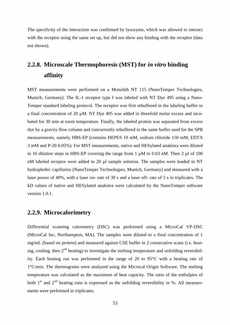
53
The specificity of the interaction was confirmed by lysozyme, which was allowed to interact
with the receptor using the same set up, but did not show any binding with the receptor (data
not shown).
2.2.8. Microscale Thermophoresis (MST) for in vitro binding
affinity
MST measurements were performed on a Monolith NT 115 (NanoTemper Technologies,
Munich, Germany). The IL-1 receptor type I was labeled with NT Dye 495 using a Nano-
Temper standard labeling protocol. The receptor was first rebuffered in the labeling buffer to
a final concentration of 20 µM. NT Dye 495 was added in threefold molar excess and incu-
bated for 30 min at room temperature. Finally, the labeled protein was separated from excess
dye by a gravity flow column and concurrently rebuffered in the same buffer used for the SPR
measurements, namely HBS-EP (contains HEPES 10 mM, sodium chloride 150 mM, EDTA
3 mM and P-20 0.05%). For MST measurements, native and HESylated anakinra were diluted
in 16 dilution steps in HBS-EP covering the range from 1 µM to 0.03 nM. Then 2 µl of 100
nM labeled receptor were added to 20 µl sample solution. The samples were loaded in NT
hydrophobic capillaries (NanoTemper Technologies, Munich, Germany) and measured with a
laser power of 40%, with a laser on- rate of 30 s and a laser off- rate of 5 s in triplicates. The
kD values of native and HESylated anakinra were calculated by the NanoTemper software
version 1.0.1.
2.2.9. Microcalorimetry
Differential scanning calorimetry (DSC) was performed using a MicroCal VP-DSC
(MicroCal Inc, Northampton, MA). The samples were diluted to a final concentration of 1
mg/mL (based on protein) and measured against CSE buffer in 2 consecutive scans (i.e. heat-
ing, cooling, then 2nd
heating) to investigate the melting temperature and unfolding reversibil-
ity. Each heating run was performed in the range of 20 to 95°C with a heating rate of
1°C/min. The thermograms were analyzed using the Microcal Origin Software. The melting
temperature was calculated as the maximum of heat capacity. The ratio of the enthalpies of
both 1st and 2
nd heating runs is expressed as the unfolding reversibility in %. All measure-
ments were performed in triplicates.
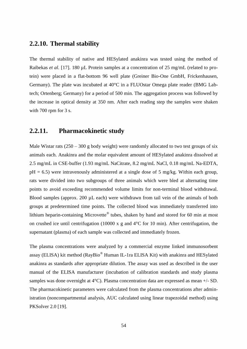
54
2.2.10. Thermal stability
The thermal stability of native and HESylated anakinra was tested using the method of
Raibekas et al. [17]. 180 µL Protein samples at a concentration of 25 mg/mL (related to pro-
tein) were placed in a flat-bottom 96 well plate (Greiner Bio-One GmbH, Frickenhausen,
Germany). The plate was incubated at 40°C in a FLUOstar Omega plate reader (BMG Lab-
tech; Ortenberg; Germany) for a period of 500 min. The aggregation process was followed by
the increase in optical density at 350 nm. After each reading step the samples were shaken
with 700 rpm for 3 s.
2.2.11. Pharmacokinetic study
Male Wistar rats (250 – 300 g body weight) were randomly allocated to two test groups of six
animals each. Anakinra and the molar equivalent amount of HESylated anakinra dissolved at
2.5 mg/mL in CSE-buffer (1.93 mg/mL NaCitrate, 8.2 mg/mL NaCl, 0.18 mg/mL Na-EDTA,
pH = 6.5) were intravenously administered at a single dose of 5 mg/kg. Within each group,
rats were divided into two subgroups of three animals which were bled at alternating time
points to avoid exceeding recommended volume limits for non-terminal blood withdrawal.
Blood samples (approx. 200 µL each) were withdrawn from tail vein of the animals of both
groups at predetermined time points. The collected blood was immediately transferred into
lithium heparin-containing Microvette® tubes, shaken by hand and stored for 60 min at most
on crushed ice until centrifugation (10000 x g and 4°C for 10 min). After centrifugation, the
supernatant (plasma) of each sample was collected and immediately frozen.
The plasma concentrations were analyzed by a commercial enzyme linked immunosorbent
assay (ELISA) kit method (RayBio® Human IL-1ra ELISA Kit) with anakinra and HESylated
anakinra as standards after appropriate dilution. The assay was used as described in the user
manual of the ELISA manufacturer (incubation of calibration standards and study plasma
samples was done overnight at 4°C). Plasma concentration data are expressed as mean +/- SD.
The pharmacokinetic parameters were calculated from the plasma concentrations after admin-
istration (noncompartmental analysis, AUC calculated using linear trapezoidal method) using
PKSolver 2.0 [19].

55
2.3. Results
Anakinra was coupled to HES at the N-terminus by a reductive amination reaction through a
single terminal aldehyde group on HES. After anion exchange chromatography, and re-
buffering into citrate buffer pH = 6.5, a highly purified mono-HESylated protein was ob-
tained, with a yield of 65%.
2.3.1. Molar mass and size
The molar mass and size of the obtained mono-conjugate were characterized using SEC-
MALLS as well as DLS, respectively. The combination of SEC for molecular separation with
MALLS, UV and RI for detection is a powerful method for an exact calculation of the molar
mass as well as the quantification of the extent of coupling and/or aggregation of the conju-
gated proteins. Results show that the weight average molar mass (Mw) of the native anakinra,
calculated by MALLS and UV detection, was 16.6 kDa, which is in good agreement with the
theoretical protein mass of 17.26 kDa (Figure 1 and Table 1). The HESylated protein (Fig-
ure 1 bottom) showed a Mw of 105.5 kDa, which is also quite close to the hypothetical mass
of 102.7 kDa.

56
Figure 1: SEC-MALLS chromatograms for determining the molar mass of native (top), and
HESylated anakinra (bottom). The molar mass distributions of the conjugate, the protein part
and the polymer part are expressed in red, green and blue lines, respectively.

57
Table 1: Weight average molar mass (Mw) and polydispersity index (PDI) for activated HES,
native and HESylated anakinra.
Sample MW [kDa] Polydispersity Hydrodynamic
diameter [nm]b
PDIb
Activated HES 85 1.3 - -
Native anakinra 16.6 a 1.00
a 4.36 ± 0.18 0.16 ± 0.03
HESylated anakinra 105.5 a 1.24
a 14.73 ± 0.25 0.20 ± 0.01
a determined by SEC-MALLS
b determined by DLS
The hydrodynamic size of the conjugate was determined using DLS as seen in Table 1. The
size of anakinra is 4.36 nm, which is quite comparable to the literature-reported value [6].
Meanwhile, the size of HES-conjugate is nearly three times that of native anakinra as seen in
Table 1.
2.3.2. Protein conformation
The effect of HESylation® on anakinra’s conformation was investigated using FTIR. The 2
nd
derivative spectrum of anakinra in Figure 2 shows the main characteristics of its secondary
structure, namely a predominant beta sheet arrangement (peak at 1640 cm-1
) and beta-turn
structures (peak at 1685 cm-1
) [20]. The spectrum of HESylated anakinra nearly superimposes
with that of the native protein, showing that HESylation® did not lead to significant changes
in anakinra’s secondary structure.

58
Figure 2: Second- derivative amide I FTIR spectra of native anakinra (black solid line), and
HESylated anakinra (red dotted line).
2.3.3. Microcalorimetry and thermal stability
Microcalorimetric measurements show that anakinra has a melting temperature of 58.0 ±
0.5°C, which is in agreement with the previously reported value of 56°C [21], whereas
HESylated anakinra shows a 4.8 K increase in the melting point in comparison to the native
protein (Figure 3 and Table 2). In addition to the observed increase in Tm, the enthalpy of
melting increases significantly upon polymer conjugation (Table 2). Furthermore, the protein
conjugate showed a high degree of refolding upon cooling (90%), contrary to the native pro-
tein, which precipitated upon denaturation (Figure 3 and Table 2). Meanwhile, the stress
stability study performed in a microwell-plate at 40°C (Figure 4) showed that native anakinra
aggregated rapidly as manifested by a rapid increase in turbidity after less than 30 min, simi-
lar to previously reported results [17], while the HESylated anakinra did not show an increase
in turbidity over several hours.

59
Figure 3: Microcalorimetry thermograms of native anakinra (black solid line) and HESylated
anakinra (red dotted line). Native anakinra aggregated directly after unfolding, leading to the
observed decrease in heat capacity, while HESylated anakinra did not.
Table 2: Thermodynamic properties of native and HESylated anakinra
Sample Tm
[°C]
Δ H
[kcal/ mole]
Δ cp
[kcal/mole/°C]
Unfolding re-
versibility
[%]
Native anakinra 58.0 ± 0.5 58.4 ± 0.1 11.7 ± 0.4 0
HESylated anakinra 62.8 ± 0.3 100.8 ± 1.0 19.4 ± 0.3 90.3 ± 0.9

60
Figure 4: Optical density at λ = 350 nm as a function of time for anakinra (black dots) and
HESylated anakinra (red dots) solutions (protein concentration = 25 mg/mL) stored at 40°C
for 500 min in a 96 well plate with eventual shaking after each reading step
2.3.4. In vitro binding affinity
The binding affinity was measured using SPR and MST. In case of SPR measurements, the
recombinant IL-1 receptor type I was immobilized with a low density to minimize mass
transport and rebinding effects. Table 3 illustrates the kD values of native and HESylated
anakinra including the on- and off-rates. The wild type protein showed the fastest on-rate fol-
lowed by a very slow off-rate (Figure 5 and Table 3), which led to a kD value of 0.05 nM.
The on-rate of the HESylated anakinra is nearly one order of magnitude lower than the native
protein, while the off-rates for both proteins are similar (Table 3), leading to a slightly higher
dissociation constant (kDHESylated anakinra = 0.32 nM). Meanwhile, MST results show that the
native protein had a kD of 3.85 nM, while the conjugate’s kD was 10.7 nM (Figure 6 and
Table 3).

61
Figure 5: SPR analyses for the binding of native anakinra (top) and HESylated anakinra (bot-
tom) to IL-1 receptor type I. All binding curves (black lines) were fitted by Langmuir (1:1)
binding isotherm (red lines).

62
Figure 6: MST measurement for of the binding of native and HESylated anakinra to IL-1
receptor type I as a function of ligand concentration.
Table 3: On- rates, off-rates and dissociation constants for native- and HESylated anakinra
from SPR measurements as well as dissociation constant from MST measurements
Sample
SPR MST
kon
[1/Ms]
koff
[1/s]
kD
[nM]
kD
[nM]
Native anakinra 8.54E+5 3.91E-5 0.05 3.85
HESylated anakinra 9.61E+4 3.11E-5 0.32 10.7
2.3.5. Pharmacokinetic profiles of native and HESylated anakinra
The pharmacokinetic properties of anakinra and HESylated anakinra were evaluated follow-
ing intravenous administration in male Wistar rats. No toxicity or adverse effects were noted
in the treated rats. Figure 7 illustrates the plasma concentration profiles during a 72 h experi-
mental period, and the pharmacokinetic parameters calculated from the data by noncompart-
mental analysis are summarized in Table 4. Figure 7 shows that plasma concentrations of
native and HESylated anakinra demonstrated an exponential declining pattern after i.v. ad-
ministration, but the elimination of HESylated anakinra was much slower than that of native
63
protein. The half-life of HESylated anakinra was increased about 6.5 times, as compared to
that of native anakinra. Additionally, HESylated anakinra showed a reduced clearance (CL)
and a marked increase (approximately 45 times) in the area under the plasma concentration
curve (AUC), as well as a marked decrease in the apparent volume of distribution, indicating
higher confinement to a smaller fluid volume, and lesser diffusion out of the blood compart-
ment.
Figure 7: Plasma level of native and HESylated anakinra after single intravenous administra-
tion to rats. Closed symbol: anakinra; open symbol: HESylated anakinra. Rats were injected
with 5 mg/kg anakinra or molar equivalent amount of HESylated anakinra.
Table 4: Pharmacokinetic parameters after anakinra and HESylated anakinra administration
to rats.
Parameter Native anakinra HESylated anakinra
AUC (h*ng/mL) 12,287 548,790
t1/2 (h) 1.7 10.8
CL (mL/h/kg) 403.3 9.1
Vz (mL/kg) 958 141

64
Results are expressed as mean (n = 3). Abbreviations: AUC, area under the curve; t1/2 (h):
apparent terminal elimination half-life; CL: observed clearance; Vz: apparent volume of dis-
tribution
2.4. Discussion
With the current developments in engineering protein molecules for a multitude of targets and
diseases, HLE became an important enabling strategy for many protein development pro-
grams. Due to the limitations of PEGylation [22, 23], newer technologies are being pursued,
and these can be grouped into 2 broad strategies: 1) physical increase in the size of the mole-
cule of interest above the renal glomerular filtration limit by coupling to biodegradable, water
soluble polymers (such as polysialic acid [24, 25]) or fusion to random-coil-forming polypep-
tides (such as Xten technology [26, 27], or PASylation [28]). The other strategy involves 2)
the use of a binding mechanism, such as binding to the Fc neonatal receptor (FcRn) to make
use of the FcRn natural recycling mechanism in the endothelial cells [10]. The latter can be
achieved by fusion to the Fc part of antibodies, or fusion to serum albumin [10].
In this study, we report on HESylation® as a promising HLE technology. It involves coupling
of hydroxyethyl starch (HES) to molecules of interest to increase their size and thus their cir-
culation time. HES is a semi-synthetic water-soluble polymer produced by the reaction of
starch with ethylene oxide [29]. It is degraded by the action of serum alpha-amylase, and the
rate of degradation can be tailored by controlling the extent of hydroxyethylation [30]. Such
favorable properties, in addition to its proven safety record and extensive clinical use as plas-
ma volume expander, render HES as an attractive alternative to PEG.
In this study, HESylation® was applied to anakinra. The latter is used for the treatment of
rheumatic arthritis, but has a terminal half-life of only 108 min in humans [16], thus many
researchers have attempted to increase its circulation time and reduce the dosing frequency
[31, 32]. Those attempts involved coupling it to PEG, either at the N-terminus [31], or at the
cysteine or lysine residues [32]. In the current study, we report a site-specific conjugation of
HES at the N-terminus of anakinra using reductive amination. The reaction makes use of a
single terminal aldehyde group on the activated HES, which forms a Schiff’s base with the
protein’s N-terminal-amino group, and is subsequently reduced to a secondary amino group.
The coupling chemistry is based on the difference in pKa between the ε-amino groups of ly-
sine and that of the N-terminal amino group, leading to a preferred reaction with the N-
terminus at slightly acidic conditions [33-35]. Previous reports on HESylation® described

65
coupling several peptides or small molecules to one HES chain [14, 36, 37], as well as site-
specific conjugation [12].
Despite the putative regioselectivity of the reductive amination reaction described above, it
can not be excluded that also certain exposed Lys residues become reactive with the polymer,
leading to an inhomogenous multi-HESylated product, especially when a high excess of the
modification reagent (i.e. activated HES) is used for a prolonged reaction time. To minimize
this risk, the small-scale conjugation process was directed towards maximizing homogeneity
of the product rather than yield. After preliminary optimization runs, it was possible to
identify conditions that produced predominantly mono-conjugated protein (as followed by
SEC analysis, see below), with a coupling efficiency restricted to ~80 %, and an overall pro-
cess yield of 65% after all purification steps. The predominately regioselective coupling at the
N-terminus was confirmed by peptide mapping after digestion by trypsin (Promega, Madison,
USA) for 20 h at 37°C, followed by analysis on a Q-Star® XL Hybrid LC/MS/MS system.
Results showed that, the heptapeptide at the N-terminus of anakinra could not be recovered
after HESylation, in contrast to the native protein, which confirms the N-terminal coupling
(data not shown).
SEC-MALS and RP-HPLC analysis showed that a predominantly mono-HESylated protein
was obtained after synthesis and purification, with a purity of > 98 %, containing less than
0.5% of non conjugated protein, free HES polymer as well as soluble aggregates. Additional-
ly, the calculated masses of the protein and the conjugate agree quite well with the theoretical
values. One obvious difference between native anakinra and the HESylated one is the broad
molar mass distribution of the latter (PDIHESylated anakinra = 1.244). This is quite close to
the PD of the activated HES molecule (= 1.3), and generally due to the natural origin of the
polymer, with polydispersities between 1.9 and 4.5 obtained for commercially available plas-
ma volume expanders based on HES [29]. However, by sample fractionation, it is possible to
obtain HES fractions with even narrower size distribution compared to the starting material
used for the protein conjugation in this study. Meanwhile, DLS results show that the hydro-
dynamic diameter of the anakinra increased from 4.36 to 14.73 nm for the HESylated protein.
This is essential for increasing the half-life, since it is known that molecules bigger than the
pores of the glomerular basement membrane (approximately 5.0 nm) have a reduced glomeru-
lar filtration rate [38].
The influence of HESylation® on the secondary structure of the protein was investigated using
FTIR. The vast majority of literature reports support the notion that polymer conjugation to
proteins has no effect on a protein’s secondary and tertiary structure [39, 40]. For example,
66
Kinstler et al. studied the effect of PEGylation on the secondary structure of GCSF by circular
dichroism, and found out that conjugation did not influence the protein conformation [33], a
result later confirmed by Rajan et al. in an FTIR study [39]. There are however few studies
which describe changes in protein conformation upon PEGylation [41, 42]. For instance, Yu
and coworkers reported changes in the circular-dichroism-measured secondary structure of
rhIL-1ra upon PEGylation [43]. Our results show that, HESylation® did not alter the second-
ary structure of anakinra, since the spectra before and after conjugation nearly superimpose.
In another set of experiments, the effect of HESylation® on the protein’s thermodynamic
properties and thermal stability was investigated. A number of studies have shown that
glycosylation or PEGylation increases a protein’s thermodynamic stability [44-46], such as
the work of Rodrigues-Martinez et al., who reported an increase of up to 6 K in the melting
temperature (Tm) of α-chymotrypsin upon PEGylation [47]. Similarly, Tm of recombinant
human endostatin increased by 15 K upon modification with a single 5 kDa PEG molecule at
the N-terminus [48]. Meanwhile, others, such as Gonnelli et al. [49] and Plesner et al. [50],
reported a reduction of Tm after polymer conjugation. The microcalorimetric measurements of
HESylated anakinra showed a 4.8 K increase in Tm and a 65% increase in the unfolding en-
thalpy as well as refolding upon cooling (which was missing in the native protein due to ag-
gregation). These improved thermodynamic properties showed a positive effect on the protein
stability under thermal stress, where the conjugated protein did not aggregate for more than
8 h, while the native protein started aggregation already after 30 min. Similar correlation be-
tween increased Tm and higher stability was reported in previous studies [45].
The in vitro binding affinity was also investigated as an important surrogate for the influence
of HES-conjugation on the protein activity. Polymer conjugation to proteins is known to re-
duce the protein activity due to steric hindrance of the interaction of the protein and the bind-
ing receptor, with activity dropping to as low as 7% as in the case of PEG-Interferon α-2a
[51]. Site-specific conjugation can greatly improve activity and binding affinity, as it can re-
duce the interference in protein-receptor interaction. In the current study, the binding kinetics
of the native and the conjugated anakinra were evaluated by surface plasmon resonance (SPR)
measurements as well as microscale thermophoresis (MST). The latter is a novel entirely op-
tical method for the determination of the binding affinity [52], where the migration of bio-
molecules along a laser-induced microscopic temperature gradient is mainly governed by pro-
tein hydration and surface charge [53]. A binding event leads to alterations of the protein hy-
dration shell, and accordingly changes in the thermophoretic motion [54]. It offers a number
of advantages, including minimum sample contamination or perturbation of the molecular

67
structure due to lack of protein-immobilization, as well as the ability to measure over a wide
concentration range (nM to mM), requiring very small sample volumes (< 3 µl) [52, 53].
SPR measurements of anakinra gave a dissociation constant (kD) of 0.05 nM, which is close
to the reported values of 0.12 [55] and 0.4 nM [56]. Meanwhile, it showed that the on-rate of
the HESylated protein decreased by an order of magnitude, most probably due to the shield-
ing effect of the polymer, and possibly the lower diffusion coefficient of the larger conjugate
[57], while the off-rates for both proteins are similar, indicating that once the ligand-receptor
complex is formed, its dissociation is not influenced by the coupled polymer. The dissociation
constants of native and modified anakinra were double-checked by MST, which showed
higher kD values for both proteins, but with the same trend, namely, that the HESylated pro-
tein had a multiple-fold higher dissociation constant than the native protein. The lower kD
values obtained by SPR have also been observed by Jecklin et al. when they compared SPR to
isothermal calorimetriy (ITC) and nanoelectrospray ionization mass spectrometry (nESI-MS)
[58]. They attributed this observation to issues of protein immobilization, where in case of
fast association rates, which are highly affected by mass transport, the binding of the ligand to
the protein on the sensor chip is faster than the diffusion of the ligand from the bulk solution
to the immobilized protein at the surface [58]. This problem is not present in MST leading to
higher apparent kD values. In general, one infers from both techniques that the conjugated
protein is still highly active, retaining a dissociation constant in the low nM range. It is also
worth noting that the minor reduction in binding affinity may not influence the in vivo per-
formance [5], as observed by Pearce et al. who produced hGH mutants with systematically
lower binding affinities down to 500-fold [59], and found out that the cellular proliferation of
the different mutants was unaffected until the affinity was reduced by more than 30-fold com-
pared to the wild-type protein [59].
Finally, the pharmacokinetic results of both native and HESylated anakinra in male Wistar
rats show that HESylation® significantly increased the half-life of the protein by 6.5 times, as
well as the AUC by 45 times. To compare these results with published information about
PEGylated anakinra, quite scarce data can be found in the literature. For instance, a patent by
Hakimi et al. describes the serum concentrations of native and PEGylated anakinra (conjugate
molar mass is 33 kDa, 46% protein content) for up to 24 h after subcutaneous injection into
C57BF/6 mice [60]. Analyzing their data using the noncompartmental analysis of PKSolver
2.0, native anakinra shows a t1/2 of 1.04 h (which is close to our estimate) while PEGylated
anakinra shows a t1/2 of 3.53 h. While it is difficult to directly compare the results of
HESylated anakinra to those of the published PEGylated one due to differences in the animal

68
species, molar mass of the polymer and injection route, the results generally show the poten-
tial of HESylation® for extending the circulation time of proteins.
2.5. Conclusions
The present study reports on the use of HES-conjugation for half-life extension of a model
protein, namely rhIL1ra (anakinra). The HESylated protein was synthesized using reductive
amination under mild acidic conditions, and purified by anion exchange chromatography,
giving a monoHESylated protein with a very good yield. Characterization of the HESylated
anakinra by SEC-MALLS and DLS identified the molar mass and size of the protein, respec-
tively, while FTIR showed that HESylation®
did not alter the protein’s secondary structure.
Additionally, microcalorimetry showed that HESylation® increased the proteins melting point
Tm by 4.5 K, as well as the melting enthalpy and refolding upon cooling, which translated into
higher stability towards aggregation at 40°C. Meanwhile, HESylated anakinra maintained its
high receptor binding affinity in the low nanomolar range as confirmed by SPR and MST.
Finally, the pharmacokinetic study of native and HESylated anakinra in Wistar rats showed a
marked increase in the protein’s half-life and AUC. This study shows that, while HESylation®
significantly improves the pharmacokinetic parameters of the model protein, anakinra, it does
not negatively affect its structure or binding affinity, and increases its stability, making
HESylation® an attractive HLE technology.
Acknowledgements
The authors would like to thank Sarah Bergmann for her technical assistance in preparing the
anakinra conjugate.
2.6. References
[1] B. Leader, Q.J. Baca, D.E. Golan, Protein therapeutics: a summary and pharmacological
classification, Nat Rev Drug Discov, 7 (2008) 21-39.
[2] J.M. Harris, R.B. Chess, Effect of pegylation on pharmaceuticals, Nat Rev Drug Discov, 2
(2003) 214-221.
[3] A. Abuchowski, J.R. McCoy, N.C. Palczuk, T. van Es, F.F. Davis, Effect of covalent at-
tachment of polyethylene glycol on immunogenicity and circulating life of bovine liver cata-
lase, Journal of Biological Chemistry, 252 (1977) 3582-3586.

69
[4] A. Abuchowski, T. van Es, N.C. Palczuk, F.F. Davis, Alteration of immunological proper-
ties of bovine serum albumin by covalent attachment of polyethylene glycol, Journal of Bio-
logical Chemistry, 252 (1977) 3578-3581.
[5] P. Bailon, C.-Y. Won, PEG-modified biopharmaceuticals, Expert Opinion on Drug Deliv-
ery, 6 (2009) 1-16.
[6] F.M. Veronese, G. Pasut, PEGylation, successful approach to drug delivery, Drug
Discovery Today, 10 (2005) 1451-1458.
[7] R. Webster, V. Elliott, B.K. Park, D. Walker, M. Hankin, P. Taupin, PEG and PEG conju-
gates toxicity: towards an understanding of the toxicity of PEG and its relevance to PEGylat-
ed biologicals, in: F. Veronese (Ed.) PEGylated Protein Drugs: Basic Science and Clinical
Applications, Birkhäuser Basel, 2009, pp. 127-146.
[8] M.A. Young, A. Malavalli, N. Winslow, K.D. Vandegriff, R.M. Winslow, Toxicity and
hemodynamic effects after single dose administration of MalPEG-hemoglobin (MP4) in rhe-
sus monkeys, Translational research : the journal of laboratory and clinical medicine, 149
(2007) 333-342.
[9] A. Bendele, J. Seely, C. Richey, G. Sennello, G. Shopp, Short Communication: Renal
Tubular Vacuolation in Animals Treated with Polyethylene-Glycol-Conjugated Proteins, Tox-
icological Sciences, 42 (1998) 152-157.
[10] R.E. Kontermann, Strategies to Extend Plasma Half-Lives of Recombinant Antibodies,
BioDrugs, 23 (2009) 93-109.
[11] R.E. Kontermann, Strategies for extended serum half-life of protein therapeutics, Current
Opinion in Biotechnology, 22 (2011) 868-876.
[12] T. Hey, H. Knoller, P. Vorstheim, Half-Life Extension through HESylation®, in: Thera-
peutic Proteins, Wiley-VCH Verlag GmbH & Co. KGaA, 2012, pp. 117-140.
[13] A. Besheer, G. Hause, J. Kressler, K. Mäder, Hydrophobically Modified Hydroxyethyl
Starch: Synthesis, Characterization, and Aqueous Self-Assembly into Nano-Sized Polymeric
Micelles and Vesicles, Biomacromolecules, 8 (2007) 359-367.
[14] A. Besheer, T.C. Hertel, J. Kressler, K. Mäder, M. Pietzsch, Enzymatically catalyzed
HES conjugation using microbial transglutaminase: Proof of feasibility, Journal of Pharma-
ceutical Sciences, 98 (2009) 4420-4428.
[15] M. Noga, D. Edinger, R. Kläger, S.V. Wegner, J.P. Spatz, E. Wagner, G. Winter, A. Be-
sheer, The effect of molar mass and degree of hydroxyethylation on the controlled shielding
and deshielding of hydroxyethyl starch-coated polyplexes, Biomaterials, 34 (2013) 2530-
2538.

70
[16] D. Burger, J.M. Dayer, IL-1Ra, in, J.J. Oppenheim, M. Feldmann (Eds.) Cytokine Refer-
ence, Academic Press, 2012, pp. 319–336.
[17] A.A. Raibekas, E.J. Bures, C.C. Siska, T. Kohno, R.F. Latypov, B.A. Kerwin, Anion
Binding and Controlled Aggregation of Human Interleukin-1 Receptor Antagonist, Biochem-
istry, 44 (2005) 9871-9879.
[18] W.-M. Kulicke, U. Kaiser, D. Schwengers, R. Lemmes, Measurements of the Refractive
Index Increment on Hydroxyethyl Starch as a Basis for Absolute Molecular Weight Determi-
nations, Starch - Stärke, 43 (1991) 392-396.
[19] Y. Zhang, M. Huo, J. Zhou, S. Xie, PKSolver: An add-in program for pharmacokinetic
and pharmacodynamic data analysis in Microsoft Excel, Computer Methods and Programs in
Biomedicine, 99 (2010) 306-314.
[20] B.S. Chang, R.M. Beauvais, T. Arakawa, L.O. Narhi, A. Dong, D.I. Aparisio, J.F. Car-
penter, Formation of an active dimer during storage of interleukin-1 receptor antagonist in
aqueous solution, Biophysical Journal, 71 (1996) 3399-3406.
[21] Y. Zhang, S. Roy, L.S. Jones, S. Krishnan, B.A. Kerwin, B.S. Chang, M.C. Manning,
T.W. Randolph, J.F. Carpenter, Mechanism for benzyl alcohol-induced aggregation of re-
combinant human interleukin-1 receptor antagonist in aqueous solution, Journal of Pharma-
ceutical Sciences, 93 (2004) 3076-3089.
[22] A. Besheer, R. Liebner, M. Meyer, G. Winter, Challenges for PEGylated Proteins and
Alternative Half-Life Extension Technologies Based on Biodegradable Polymers, in: Tai-
lored Polymer Architectures for Pharmaceutical and Biomedical Applications, American
Chemical Society, 2013, pp. 215-233.
[23] R.P. Garay, J.P. Labaune, Immunogenicity of polyethylene glycol (PEG), The Open
Conference Proceedings Journal, 2 (2011) 104-107.
[24] G. Gregoriadis, A. Fernandes, M. Mital, B. McCormack, Polysialic acids: potential in
improving the stability and pharmacokinetics of proteins and other therapeutics, Cellular and
Molecular Life Sciences CMLS, 57 (2000) 1964-1969.
[25] G. Gregoriadis, S. Jain, I. Papaioannou, P. Laing, Improving the therapeutic efficacy of
peptides and proteins: A role for polysialic acids, International Journal of Pharmaceutics, 300
(2005) 125-130.
[26] V. Schellenberger, C.-w. Wang, N.C. Geething, B.J. Spink, A. Campbell, W. To, M.D.
Scholle, Y. Yin, Y. Yao, O. Bogin, J.L. Cleland, J. Silverman, W.P.C. Stemmer, A recombi-
nant polypeptide extends the in vivo half-life of peptides and proteins in a tunable manner,
Nat Biotech, 27 (2009) 1186-1190.

71
[27] N.C. Geething, W. To, B.J. Spink, M.D. Scholle, C.W. Wang, Y. Yin, Y. Yao, V. Schel-
lenberger, J.L. Cleland, W.P. Stemmer, J. Silverman, Gcg-XTEN: an improved glucagon ca-
pable of preventing hypoglycemia without increasing baseline blood glucose, PloS one, 5
(2010) e10175.
[28] M. Schlapschy, U. Binder, C. Börger, I. Theobald, K. Wachinger, S. Kisling, D. Haller,
A. Skerra, PASylation: a biological alternative to PEGylation for extending the plasma half-
life of pharmaceutically active proteins, Protein Engineering Design and Selection, 26 (2013)
489-501.
[29] A.I.H. Besheer, Nanomedicines based on modified hydroxyethyl starch: From synthesis
to in vivo evaluation, in, Halle (Saale), Martin-Luther-Universitat Halle-Wittenberg,
Dissertation, 2009.
[30] C. Jungheinrich, T. Neff, Pharmacokinetics of Hydroxyethyl Starch, Clin Pharmacokinet,
44 (2005) 681-699.
[31] P. Yu, D. Qin, G. Qin, B. Fan, G. Ma, Z. Su, PEGylation of rhIL-1RA increased its solu-
tion stability at room temperature, Process Biochemistry, 44 (2009) 1340-1345.
[32] P. Yu, C. Zheng, J. Chen, G. Zhang, Y. Liu, X. Suo, G. Zhang, Z. Su, Investigation on
PEGylation strategy of recombinant human interleukin-1 receptor antagonist, Bioorganic &
Medicinal Chemistry, 15 (2007) 5396-5405.
[33] O.B. Kinstler, D.N. Brems, S.L. Lauren, A.G. Paige, J.B. Hamburger, M.J. Treuheit,
Characterization and Stability of N-terminally PEGylated rhG-CSF, Pharmaceutical Research,
13 (1996) 996-1002.
[34] H. Lee, I.H. Jang, S.H. Ryu, T.G. Park, N-Terminal Site-Specific Mono-PEGylation of
Epidermal Growth Factor, Pharmaceutical Research, 20 (2003) 818-825.
[35] O. Kinstler, G. Molineux, M. Treuheit, D. Ladd, C. Gegg, Mono-N-terminal
poly(ethylene glycol)-protein conjugates, Advanced Drug Delivery Reviews, 54 (2002) 477-
485.
[36] C. Kessler, A. Greindl, B. Breuer, U. Haberl, A. Rybka, M. Emgenbroich, H.-G. Frank,
A.J.G. Pötgens, Erythropoietin mimetic compound AGEM400(HES) binds to the same recep-
tor as erythropoietin but displays a different spectrum of activities, Cytokine, 57 (2012) 226-
237.
[37] A. Greindl, C. Kessler, B. Breuer, U. Haberl, A. Rybka, M. Emgenbroich, A.J. Potgens,
H.G. Frank, AGEM400(HES), a novel erythropoietin mimetic peptide conjugated to hydroxy-
ethyl starch with excellent in vitro efficacy, The Open Hematology Journal, 4 (2010) 1-14.

72
[38] C.J. Fee, Size comparison between proteins PEGylated with branched and linear
poly(ethylene glycol) molecules, Biotechnology and Bioengineering, 98 (2007) 725-731.
[39] R.S. Rajan, T. Li, M. Aras, C. Sloey, W. Sutherland, H. Arai, R. Briddell, O. Kinstler,
A.M.K. Lueras, Y. Zhang, H. Yeghnazar, M. Treuheit, D.N. Brems, Modulation of protein
aggregation by polyethylene glycol conjugation: GCSF as a case study, Protein Science, 15
(2006) 1063-1075.
[40] B.A. Luxon, M. Grace, D. Brassard, R. Bordens, Pegylated interferons for the treatment
of chronic hepatitis C infection, Clinical Therapeutics, 24 (2002) 1363-1383.
[41] P. Bailon, W. Berthold, Polyethylene glycol-conjugated pharmaceutical proteins, Phar-
maceutical Science & Technology Today, 1 (1998) 352-356.
[42] P. Stigsnaes, S. Frokjaer, S. Bjerregaard, M. van de Weert, P. Kingshott, E.H. Moeller,
Characterisation and physical stability of PEGylated glucagon, International Journal of Phar-
maceutics, 330 (2007) 89-98.
[43] P. Yu, G. Zhang, J. Bi, X. Lu, Y. Wang, Z. Su, Facile purification of mono-PEGylated
interleukin-1 receptor antagonist and its characterization with multi-angle laser light scatter-
ing, Process Biochemistry, 42 (2007) 971-977.
[44] D. Shental-Bechor, Y. Levy, Folding of glycoproteins: toward understanding the bio-
physics of the glycosylation code, Current Opinion in Structural Biology, 19 (2009) 524-533.
[45] T. Palm, R. Esfandiary, R. Gandhi, The effect of PEGylation on the stability of small
therapeutic proteins, Pharmaceutical Development and Technology, 0 (2011) 1-8.
[46] C. Dhalluin, A. Ross, L.-A. Leuthold, S. Foser, B. Gsell, F. Müller, H. Senn, Structural
and Biophysical Characterization of the 40 kDa PEG−Interferon α-2a and Its Individual Posi-
tional Isomers, Bioconjugate Chemistry, 16 (2005) 504-517.
[47] J.A. Rodríguez-Martínez, R.J. Solá, B. Castillo, H.R. Cintrón-Colón, I. Rivera-Rivera, G.
Barletta, K. Griebenow, Stabilization of α-chymotrypsin upon PEGylation correlates with
reduced structural dynamics, Biotechnology and Bioengineering, 101 (2008) 1142-1149.
[48] Y. Nie, X. Zhang, X. Wang, J. Chen, Preparation and Stability of N-Terminal Mono-
PEGylated Recombinant Human Endostatin, Bioconjugate Chemistry, 17 (2006) 995-999.
[49] M. Gonnelli, G.B. Strambini, No effect of covalently linked poly(ethylene glycol) chains
on protein internal dynamics, Biochimica et Biophysica Acta (BBA) - Proteins and Prote-
omics, 1794 (2009) 569-576.
[50] B. Plesner, C.J. Fee, P. Westh, A.D. Nielsen, Effects of PEG size on structure, function
and stability of PEGylated BSA, European Journal of Pharmaceutics and Biopharmaceutics,
79 (2011) 399-405.

73
[51] C.S. Fishburn, The pharmacology of PEGylation: Balancing PD with PK to generate
novel therapeutics, Journal of Pharmaceutical Sciences, 97 (2008) 4167-4183.
[52] C.J. Wienken, P. Baaske, U. Rothbauer, D. Braun, S. Duhr, Protein-binding assays in
biological liquids using microscale thermophoresis, Nat Commun, 1 (2010) 100.
[53] S. Duhr, D. Braun, Why molecules move along a temperature gradient, Proceedings of
the National Academy of Sciences, 103 (2006) 19678-19682.
[54] M. Jerabek-Willemsen, C.J. Wienken, D. Braun, P. Baaske, S. Duhr, Molecular interac-
tion studies using microscale thermophoresis, Assay and drug development technologies, 9
(2011) 342-353.
[55] M. Malyak, J.M. Guthridge, K.R. Hance, S.K. Dower, J.H. Freed, W.P. Arend, Charac-
terization of a Low Molecular Weight Isoform of IL-1 Receptor Antagonist, The Journal of
Immunology, 161 (1998) 1997-2003.
[56] M.F. Shamji, H. Betre, V.B. Kraus, J. Chen, A. Chilkoti, R. Pichika, K. Masuda, L.A.
Setton, Development and characterization of a fusion protein between thermally responsive
elastin-like polypeptide and interleukin-1 receptor antagonist: Sustained release of a local
antiinflammatory therapeutic, Arthritis & Rheumatism, 56 (2007) 3650-3661.
[57] S. Kubetzko, C.A. Sarkar, A. Plückthun, Protein PEGylation Decreases Observed Target
Association Rates via a Dual Blocking Mechanism, Molecular Pharmacology, 68 (2005)
1439-1454.
[58] M.C. Jecklin, S. Schauer, C.E. Dumelin, R. Zenobi, Label-free determination of protein–
ligand binding constants using mass spectrometry and validation using surface plasmon reso-
nance and isothermal titration calorimetry, Journal of Molecular Recognition, 22 (2009) 319-
329.
[59] K.H. Pearce, B.C. Cunningham, G. Fuh, T. Teeri, J.A. Wells, Growth Hormone Binding
Affinity for Its Receptor Surpasses the Requirements for Cellular Activity, Biochemistry, 38
(1998) 81-89.
[60] J. Hakimi, P. Kilian, P. Rosen, Polyethylene glycol protein conjugates, in, EP 0510356
A1, 1992.

74

75
Chapter III – Highly concentrated formulations

76

77
3. Head to Head Comparison of the Formulation and
Stability of Concentrated Solutions of HESylated versus
PEGylated Anakinra
This chapter has been published in the
Journal of Pharmaceutical Science.
Robert Liebner1, Martin Meyer
2, Thomas Hey
2, Gerhard Winter
1, Ahmed Besheer
1
1
Department of Pharmacy, Pharmaceutical Technology & Biopharmaceutics, Ludwig-
Maximillians-University Munich, Munich 81377, Germany
2 Innovation Centre Complex Formulations, Fresenius Kabi Deutschland GmbH, Friedberg
61169, Germany
Research point and funding were managed by A. Besheer. The experiments were designed by
R. Liebner and A. Besheer. Except the synthesis and purification of the HES- anakinra conju-
gate and SEC-MALLS analysis, all experiments were executed by R. Liebner. All conjugation
and purification steps of preparing the HESylated protein were performed by Sarah Berg-
mann, Fresenius Kabi. The SEC-MALLS experiments and evaluation of the results were per-
formed by Roman Mathaes. The text was written by R. Liebner, edited and corrected for pub-
lication by A. Besheer.

78
Abstract
Although PEGylation of biologics is currently the gold standard for half-life extension, the
technology has a number of limitations, most importantly the non-biodegradability of PEG
and the extremely high viscosity at high concentrations. HESylation is a promising alternative
based on coupling to the biodegradable polymer hydroxyethyl starch (HES). In this study, we
are comparing HESylation with PEGylation regarding the effect on the proteins physicochem-
ical properties, as well as on formulation at high concentrations, where protein stability and
viscosity can be compromised. For this purpose, the model protein anakinra is coupled to
HES or PEG by reductive amination. Results show that coupling of HES or PEG had practi-
cally no effect on the protein`s secondary structure, and that it reduced protein affinity by one
order of magnitude, with HESylated anakinra more affine than the PEGylated protein. The
viscosity of HESylated anakinra at protein concentrations up to 75 mg/mL was approximately
40% lower than that of PEG–anakinra. Both conjugates increased the apparent melting tem-
perature of anakinra in concentrated solutions. Finally, HESylated anakinra was superior to
PEG–anakinra regarding monomer recovery after 8 weeks of storage at 40°C. These results
show that HESylating anakinra offers formulation advantages compared with PEGylation,
especially for concentrated protein solutions.
Keywords
PEGylation; HESylation; anakinra; viscosity; protein aggregation; surface plasmon reso-
nance; FTIR; calorimetry; biopharmaceutical characterization

79
3.1. Introduction
There is currently a large growth in the development of therapeutic peptides and proteins,
with many academic institutions and industrial settings introducing engineered proteins hav-
ing new designs and functionalities [1,2]. However, for many biopharmaceuticals with a mo-
lecular weight lower than 60 kDa, such as some cytokines, antibody fragments, or protein
scaffolds, the rapid renal filtration and thus short half-life represents a hindrance for success-
ful commercialization due to the need for frequent administration and thereby reduced com-
pliance. Accordingly, these molecules require half-life extension (HLE) technologies to en-
sure their competitiveness [3]. Additionally, many of them need to be formulated as highly
concentrated solutions to enable self- administration of the required high doses by the subcu-
taneous route [4]. With more than 10 approved products on the market, PEGylation currently
represents the gold standard HLE technology [5]. The coupling of polyethylene glycol to pep-
tides and proteins was first introduced in the 1970s by Abuchowski et al. [6,7] and later found
its way to the market, not only because of extending the circulation time, but also because of
imparting many positive characteristics to molecules, such as increased solubility and stability
as well as reduced immunogenicity and susceptibility to proteolytic degradation [8]. How-
ever, PEGylation suffers from a number of shortcomings, most importantly the fact that PEG
is not biodegradable, leading to toxicity concerns, such as vacuolation of the kidney or liver
[9–11]. Another safety concern involves the presence of pre-existing or newly developed anti-
PEG antibodies, which can limit the therapeutic efficacy of PEGylated proteins [12]. Addi-
tionally, highly concentrated solutions of PEGylated proteins can have extremely high viscos-
ities, leading to challenges in production and administration [13,14]. These issues led to the
pursuit of better alternatives for PEGylation, with many approaches already commercially
under development [15,16]. These alternative technologies can be classified under two main
strategies. The first involves the same basic principle employed by PEGylation, that is, in-
creasing the hydrodynamic size of the biopharmaceutical above the renal glomerular filtration
threshold. This can be achieved by coupling to biodegradable polymers (such as polysialic
acid [17,18]) or fusion to non-folding polypeptides (such as Xten technology, [19,20] or
PASylation [21]). The second approach utilizes a binding mechanism, such as binding to the
Fc neonatal receptor (FcRn) to make use of the FcRn natural recycling machinery [15]. This
can be achieved by fusion to the Fc part of antibodies, or to albumin [15]. HESylation® is a
promising HLE technology that utilizes the first strategy [22]. It involves protein coupling to
the biodegradable and biocompatible semi synthetic polymer hydroxyethyl starch (HES).

80
HES is widely used as a plasma volume expander for decades, with patients tolerating doses
of more than 100 g of HES per day [23]. Such favorable properties have prompted its investi-
gation for biomedical applications [24, 25] and more importantly for polymer–protein conju-
gation as a promising substitute for PEG [26].
The protein molecule used in this study is anakinra (recombinant human interleukin 1 recep-
tor antagonist, rhIL1-ra). Anakinra is a 17 kDa protein that binds to the IL1 receptor, blocking
the inflammatory action of IL1 α and β, and is thus approved for adult patients with rheumatic
arthritis who show an inadequate response to other disease-modifying antirheumatic drugs
[27]. Because of its short half-life, anakinra has to be administrated by a daily injection of
100 mg, making it an ideal candidate for HLE. We have recently shown that HESylation of
anakinra maintains the binding affinity of the protein, has no effect on its secondary structure
or stability, and more importantly, increases the elimination half-life more than fivefold [28].
The aim of this study is to use anakinra as a model protein for comparing HESylation versus
PEGylation regarding their effect on the physicochemical characteristics of the protein, as
well as its formulation and stability, particularly at the challenging situation of high protein
concentrations.
3.2. Materials and Methods
3.2.1. Materials
Kineret® was purchased from SOBI (Stockholm, Sweden). Branched PEG propionaldehyd
(molecular weight (MW) = 40 kDa) was purchased from Jenkem Technologies (Allen, Tex-
as). Activated HES (with a weight average Mw of approximately 85 kDa, number average
MW (Mn) of approximately 65 kDa, and a polydispersity of 1.3) was from Fresenius Kabi
Deutschland GmbH (Bad Homburg, Germany). All chemicals were used in analytical grade
and purchased from VWR (Darmstadt, Germany).
3.2.2. Synthesis and Purification of the Protein Conjugates
3.2.2.1. PEGylation
Anakinra was conjugated to branched PEG propionaldehyde (MW = 40 kDa) on the N- ter-
minus by reductive amination in acetate buffer (cProtein = 8 mg/mL, cBuffer = 100 mM, pH =

81
5.0) in a 2.5 molar excess of the polymer using 20 mM sodium cyanoborohydride as the re-
ducing agent under gentle stirring for 16 h at 4°C. The reaction batch was rebuffered for 4 h
by a dialysis membrane (Cellu Sep, MW cutoff (MWCO) 3.5 kDa; Scienova GmbH, Jena,
Germany) against 5 L of a 20 mM TRIS buffer pH = 8.0 at 4°C before loading on a self-
packed XK 50/20 filled with Q Sepharose HP (GE Healthcare, Uppsala, Sweden) for anion-
exchange chromatography at a flow rate of 0.5 mL/min. The monoPEGylated protein was
separated from the multi-conjugates and the unreacted protein using 10 column volumes in a
linear salt gradient using Tris buffer with 250 mM NaCl on an ÄKTA Purifier (GE
Healthcare) at a flow rate of 3 mL/min. For further experiments, the pooled fractions of
mono-conjugate were rebuffered against citrate-saline- EDTA (CSE) buffer (cCitrate = 10 mM,
cNaCl = 140 mM, cEDTA = 0.5 mM, pH = 6.5) by tangential flow filtration (TFF) using a
MinimateTM
capsule with OmegaTM
10 kDa membrane (PALL Scientific, East Hills, New
York). The concentration was measured by UV-spectroscopy at 280 nm with an extinction
coefficient of 13,392 M−1
cm−1
[29].
3.2.2.2. HESylation
Anakinra was conjugated to HES propionaldehyde targeting protein’s N- terminus by reduc-
tive amination in acetate buffer. In a 1 L glass reaction vessel equipped with a thermojacket
and a blade stirrer, 2.5 g protein (19 mL; 134 mg/mL) were diluted into 220 mL 0.1 M sodi-
um acetate buffer (pH = 5.0) prechilled to 5°C. Hydroxyethyl starch (Mn ~ 68 kDa) carrying a
reactive propionaldehyde group (4-amino- N-(3-oxopropyl)butanamide [30]) was dissolved in
the same buffer to yield a 40% (w/v) solution and 75 mL of the HES solution (representing a
3:1 molar ratio of polymer to protein) were combined with the protein under moderate stir-
ring. The reductive amination reaction was started by addition of 12.5 mL of a freshly pre-
pared 0.5 M NaCNBH3 solution in water and incubated overnight (18 h) under moderate stir-
ring at 5°C. The conjugate was purified by anion-exchange chromatography using Q Se-
pharose HP material and an Äkta Purifier 100 chromatography system (both GE Healthcare,
Munich, Germany). The system was operated at room temperature with a typical flow rate of
10 mL/min. The column (XK 26/20) was first equilibrated with 5 CV of eluent A (10 mM
Tris acetate, pH = 8.0). The reductive amination reaction mixture was 10-fold diluted with
eluent A, the pH adjusted to pH 8.0 using NH4OH solution and beta-mercaptoethanol added
to a final concentration of 1 mM to destroy potential disulfide-mediated protein dimers. Up to
800 mg of conjugate was then loaded onto the column at a flow rate of 10 mL/min, followed
by a washing step with 2 CV 5% eluent B (eluent A + 250 mM NaCl) to remove the unbound,

82
excessive HES polymer. The conjugate was eluted in a step gradient at 25% eluent B for 4
CV. Peak fractions were collected and subsequently a buffer exchange to CSE (10 mM cit-
rate, 140 mM sodium chloride, 0.5 mM EDTA, pH = 6.5) buffer w/o Tween 80 and up-
concentration performed by TFF using a 50 cm2 PES (polyethersulfone) membrane capsule
with a MWCO of 10 kDa operated on a MinimateTM
benchtop system (both from Pall,
Dreieich, Germany). The retentate of the TFF step was subjected to a 0.22 µm filtration step,
aliquots of the filtered solution shock-frozen in liquid nitrogen and stored at −80°C until fur-
ther use.
3.2.3. SEC–MALLS Measurement for the Determination of the
Molecular Weight and Size of the Conjugates
The MW of native, PEGylated, and HESylated anakinra were investigated by size-exclusion
chromatography–multi- angle laser light scattering (SEC–MALLS) on the AF2000 Focus
(Postnova Analytics, Landsberg/Lech, Germany) equipped with a MALLS miniDAWN Tris-
tar detector (Wyatt Technology, Dernbach, Germany), refractive index detector (PN 3150;
Postnova Analytics) and a Shimadzu SPD-10A UV-VIS Detector (Shimadzu, Duisburg, Ger-
many). For SEC separation, a Superose 6 10/300 GL column (GE Healthcare) was used with
CSE buffer as the running phase including 0.02% sodium azide at a flow rate of 0.5 mL/min
over 45 min. Each run was performed by the injection of 50 µg, based on the protein weight.
The data were analyzed by the Astra software version 5.0 (Wyatt Technology). The protein
conjugate analyses were performed by the Wyatt protein conjugate application embedded in
the Astra 5.0 software. UV extinction coefficient for Anakinra was 13,392 M−1
cm−1
[29];
dn/dc for anakinra was 0.1850 mL/g, for PEG 0.1340 mL/g, [31] and HES 0.1460 mL/g [32].
3.2.4. Hydrodynamic Radius
The hydrodynamic radius was measured by dynamic light scattering (DLS) using the Malvern
Zetasizer (Malvern Instruments, Herrenberg, Germany). Native, PEGylated, and HESylated
anakinra were diluted to a final concentration of 1 mg/mL (based on the protein). Before
measurement, all samples were filtered through a 0.2 µm filter. The hydrodynamic radius is

83
expressed as the Z- average, together with the polydispersity index as an indication for the
size distribution.
3.2.5. FTIR
FTIR spectroscopy experiments were performed using the Bruker Tensor 27 FTIR with the
Bruker AquaSpec Cell (Bruker Optics, Ettlingen, Germany). The sensor was cooled with liq-
uid nitrogen and a constant gaseous nitrogen flow. The samples were analyzed at a concentra-
tion of 3 mg/mL (based on the protein part) in 240 scans against CSE buffer. Using the OPUS
Software, the second derivative of the spectrum for each sample was obtained in the range of
1600 to 1700 cm−1
. The curves were normalized by vector normalization.
3.2.6. Biacore Analysis for in vitro Binding Affinity
The binding affinity was analyzed by surface plasmon resonance (SPR) using a Biacore T100
instrument (Biacore AB, Uppsala, Sweden). The recombinant IL-1 receptor type I (R&D Sys-
tems, Wiesbaden, Germany) was immobilized by standard EDC/NHS coupling on a CM3
chip in acetate buffer (c = 20 mM, pH = 4.5) to a final response of ≈ 570 RU. A kinetic study
was performed for native, PEG- and HESylated anakinra by choosing 180 s for the on-rate
and 300 s for the off-rate with a constant flow rate of 30 µL/min measured at room tempera-
ture. After the 300 s dissociation of the analytes, the receptor was regenerated with Glycine 5
mM pH = 2.0 for 6 s. Different sample concentrations were diluted in HBS-EP running buffer
(contains HEPES 10 mM, sodium chloride 150 mM, EDTA 3 mM and P-20 0.005%). The
data were subtracted from an uncoated cell and normalized to their baseline using the
BIAcore T200 Evaluation Software. The overlaid curves showed the loss in activity by a de-
creased response at different concentrations.
3.2.7. Preparation of Highly Concentrated Solutions
Up-concentration of the protein solutions was performed by ultrafiltration in Vivaspin 20 cen-
trifugators using a MWCO of 10 kDa for the conjugates and 3.5 kDa for the unmodified pro-
tein. The tubes were placed into a Sigma 4K15 centrifuge (Sigma Laborzentrifugen GmbH,
Osterode am Harz, Germany) and rotated at 10,000 rpm (= 14,243 g) for 3– 4 h at 4°C. The

84
protein concentration was verified by UV measurement (NanoDrop; Thermo Scientific,
Schwerte, Germany) using the aforementioned extinction coefficient of the protein. For fur-
ther experiments, the samples were diluted in the formulation buffer to the desired protein
concentration.
3.2.8. Differential Scanning Calorimetry
Thermal properties of the highly concentrated protein solutions were investigated by differen-
tial scanning calorimetry on the Netzsch DSC (Netzsch- Gerätebau, Selb, Germany). There-
fore, 100 µL of native, PEGylated, and HESylated anakinra with a sample concentration of
25, 50, and 75 mg/mL (relabased on the protein) was filled in 100 µL aluminum crucibles,
hermetically sealed, and measured against CSE buffer. The folding process was followed by a
heating gradient from 20°C to 95°C with a heating rate of 1°C/ min. Data analysis was deter-
mined by the Proteus software. The curves were smoothed by the program to determine the
apparent Tm as the peak maximum of the melting process. Additionally, the temperature de-
scribing the onset of unfolding, Tonset, was determined by integrating the curve in Origin soft-
ware and fitting the curve using a Boltzmann equation as described by Menzen and Friess
[33].
3.2.9. Viscosity
The rheological properties as a function of protein concentration of the wild type and both
conjugates were investigated on the mVROC device (Rheosense Inc., San Ramon, CA, USA)
at 20°C. All samples were diluted in CSE buffer to 25, 50, and 75 mg/mL and analyzed at a
shear rate of 314 s−1
. The viscosity of Cimzia® (UCB, Brussels, Belgium) measured under
equivalent conditions served as a benchmark. The mentioned setup was also used to investi-
gate the influence of pH as well as sodium chloride on solution viscosity.
3.2.10. Storage Stability: SEC
The formation of soluble aggregates and the levels of monomeric protein were determined by
SE-HPLC measurements on a Spectra-Physics System P2000 (ThermoFisher Scientific, Hen-
ningsdorf, Germany) equipped with a TSKgel G3000 SWxl (TOSOH Bioscience GmbH,
Stuttgart, Germany) with CSE buffer as the running solvent at a flow rate of 0.5 mL/min over

85
35 min. The peak areas of the chromatograms were used to quantify the amount of soluble
aggregates and the monomeric fraction of each sample. Protein standards with a known con-
centration were stored at 2– 8°C and served as controls for the thermal incubation experiment.
All samples were diluted to 1 mg/mL and centrifuged to analyze the supernatant. Each SE-
HPLC run was performed by the injection of 50 µg of protein, detection wavelength was 280
nm.
3.3. Results
3.3.1. Synthesis and Purification
In case of both PEGylation and HESylation, the coupling efficiency was similar (approxi-
mately 75%). After purification using anion exchange chromatography, mono conjugated
protein could be obtained, with a purity > 98% for both HESylated and PEGylated anakinra as
determined by SEC and RP-HPLC, with a cumulated process yield of 65% – 70%.
3.3.2. Determination of the Molecular Weight and Hydro-
dynamic Size
Size-exclusion chromatography combined with MALLS, UV, and RI detectors was used for
the calculation of the MW as well as quantification of the extent of coupling and/or aggrega-
tion of the conjugated proteins. The MW of native anakinra was found to be 16.6 kDa, in
good agreement with the theoretical value of 17.26 kDa (Fig. 1a and Table 1). PEGylated
and HESylated anakinra showed a MW of 53.5 and 105.5 kDa, respectively (Figs. 1b and 1c
and Table 1), which is also quite close to the hypothetical masses of 57 and 102 kDa for
mono conjugated proteins. In case of PEGylated anakinra, a small peak (peak area < 0.3%) of
higher MW species can be observed before the monomer peak. It belongs to a dimer of mon-
oPEGylated protein (and not to a diPEGylated one) as confirmed by the doubling of the MW
of the monoconjugate to 105.6 kDa. Regarding MW distribution, HESylated anakinra showed
a broader distribution compared with the PEGylated protein (PDI HES-anakinra = 1.244 vs.
PDI PEG-anakinra = 1.004), which is due to the natural origin of HES.

86
Figure 1: SEC–MALLS experiment for determining the molar mass of (a) native, (b)
PEGylated, and (c) HESylated anakinra. The molar mass distributions of the conjugate, the
protein part, and the polymer part are expressed in black, dark gray, and light gray lines, re-
spectively.
Dynamic light scattering was used to determine the hydrodynamic size of the three proteins.
Results in Table 1 show that the diameter of anakinra is very close to that reported earlier

87
[34]. Meanwhile, polymer conjugation to PEG or HES increases the size by threefold to ap-
proximately 14.5 nm, with no significant difference between the two polymers used.
Sample MW [kDa] Polydispersity Hydrodynamic
diameter [nm]
PDI
Native anakinra 16.6 1.000 4.36 ± 0.18 0.16 ± 0.03
PEGylated anakinra 53.5 1.004 14.40 ± 0.27 0.08 ± 0.02
HESylated anakinra 105.5 1.244 14.73 ± 0.25 0.20 ± 0.01
Table 1: Weight Average Molecular Weight (MW) and Polydispersity as determined by SEC
MALLS measurements, as well hydrodynamic diameter and Polydispersity Index as deter-
mined by DLS Analysis for the native, PEGylated, and HESylated anakinra
3.3.3. FTIR Analyses for Changes in Secondary Structure
The second derivative spectrum of anakinra in Figure 2 shows the main predominant beta
sheet conformation (peak at 1640 cm−1
) and beta-turn structures (peak at 1685 cm−1
) [35]. The
spectrum of HESylated anakinra correlates well with that of the native protein, whereas
PEGylated anakinra shows a minor shift in the beta sheet peak from 1640 to 1641 cm−1
.
Therefore, it can be assumed that neither PEGylation nor HESylation under the chosen reac-
tion and purification conditions led to significant changes in the secondary structure of the
protein.

88
Figure 2: Second- derivative amide I FTIR spectra of native anakinra (solid line), PEGylated
anakinra (dashed line), and HESylated anakinra (dotted line).
3.3.4. In Vitro Binding Affinity
To investigate the effect of PEGylation and HESylation on the apparent binding affinity, the
binding kinetics of the native and conjugated anakinra was evaluated by SPR. Recombinant
IL-1 receptor type I was immobilized on a CM3 chip with a low density to minimize mass
transport and rebinding. Table 2 demonstrates the on- and off-rates, as well as the kD value of
native, PEGylated, and HESylated anakinra. The wild type protein showed the fastest on-rate
followed by a very slow off- rate (Fig. 3 and Table 2), which led to a kD value of 0.05 nM.
The on-rates of the PEGylated and HESylated anakinra are nearly one order of magnitude
lower than the native protein, whereas the off-rates for all three proteins are similar, indicating
that dissociation of the ligand-receptor complex is not influenced by the coupled polymer.
This leads to higher kD values for the protein conjugates relative to the native protein
(kDPEG-anakinra = 0.81 nM, kDHES-anakinra = 0.32 nM).

89
Figure 3: SPR analyses for the binding of (a) native anakinra, (b) PEGylated anakinra and (c)
HESylated anakinra to IL-1 receptor type I. All binding curves (dark gray lines) were fitted by
Langmuir (1:1) binding isotherm (black lines).

90
Sample
SPR
kon
[1/Ms]
koff
[1/s]
kD
[nM]
Native anakinra 8.54E+5 3.91E-5 0.05
PEGylated anakinra 4.75E+4 3.87E-5 0.81
HESylated anakinra 9.61E+4 3.11E-5 0.32
Table 2: On-rates, off-rates and dissociation constants for native, PEGylated and HESylated
anakinra from SPR measurements.
3.3.5. Properties of Highly Concentrated Solutions
3.3.5.1. Viscosity Measurement
In the current study, we measured the viscosity of all three proteins using the microchip tech-
nology of the mVROC system [36]. Results show that increasing the concentration of the na-
tive protein from 25 to 75 mg/mL (1.45– 4.35 mM) has little effect on viscosity. Meanwhile,
the viscosity of the PEGylated protein solution shows a dramatic increase of one order of
magnitude over the same molar concentration range (see Fig. 4). In comparison, the viscosity
of the HESylated protein is only approximately 60% of the PEGylated protein at the same
molar concentrations. Additionally, we studied the effect of pH and ionic strength on the vis-
cosity of the conjugates. Results show that the viscosity of anakinra did not change in slightly
alkaline conditions, but nearly doubled at pH 4 (Fig. 5, left). The same trend was seen with
anakinra conjugates, which showed much higher viscosity in acidic pH compared with pH =
6.5 or 8. The viscosity of all three proteins did not show any significant changes over the
studied salt concentration range (Fig. 5, right).

91
Figure 4: Viscosity of anakinra, PEGylated-, and HESylated anakinra as a function of conju-
gate concentration. Left: The x-axis represents the conjugate concentration in mM or the con-
centration of the protein part in mg/mL. Right: the x-axis represents the conjugate concentra-
tion in mg/mL. The lines are fitted by exponential function. The horizontal dashed line repre-
sents the viscosity of Cimzia® (conjugate concentration of 200 mg/mL), which is included as
a benchmark.
Figure 5: Viscosity of anakinra, PEGylated, and HESylated anakinra as a function of pH
(left) and NaCl concentration (right). Protein concentration was 50 mg/mL.
92
3.3.5.2. Thermal Analysis
The thermal properties of different concentrations of native and conjugated anakinra were
evaluated by DSC measurements of the different proteins at 25, 50, and 75 mg/mL protein
concentration. Anakinra is known to precipitate upon unfolding in thermal analysis experi-
ments, [37] and we had the same observation during earlier experiments [28]. Accordingly, a
robust determination of the protein melting temperature (Tm) was not possible, and the peak
maximum was regarded as apparent Tm. Additionally, we identified the temperature corre-
sponding to the onset of protein unfolding Tonset. The results in Figure 6 show that the appar-
ent Tm of anakinra decreases slightly with increasing concentration over the studied range.
Meanwhile, both protein conjugates show a much higher apparent Tm compared with native
anakinra over the studied concentration range, and show the same pattern of decreasing ap-
parent Tm with increasing concentration, except for HESylated anakinra, which shows a slight
increase in apparent Tm at the highest concentration tested (75 mg/mL protein concentration).
It is worth noting that PEGylated anakinra shows a slightly higher apparent Tm compared with
HESylated anakinra. As for Tonset, the values for native anakinra show a slight decrease with
increasing concentration, although the differences are not significant because of the large scat-
ter in values. Meanwhile, HESylated and PEGylated proteins have a much higher Tonset com-
pared with native anakinra, with a clear trend towards lower temperature with increasing con-
centration. There is no difference HESylated and PEGylated anakinra at 25 mg/mL, but at
higher concentrations, Tonset of PEGylated anakinra is higher, although with large scatter in
values.

93
Figure 6: DSC thermogram for native, HESylated, and PEGylated anakinra at a protein con-
centration of 75 mg/mL (a), the apparent melting temperature (Tm) of all three proteins as a
function of protein concentration (b), as well as the temperature corresponding to the onset of
unfolding (Tonset) as a function of protein concentration (c).

94
3.3.5.3. Thermal Stability upon Storage at 40°C
The thermal stability of anakinra and its conjugates was evaluated by storing them at three
different protein concentrations (25, 50, and 75 mg/mL) at 40°C for 8 weeks, and then evalu-
ated by estimating the monomer recovery by SEC. Results in Figure 7 show that native ana-
kinra at 75 mg/mL degrades rapidly, so that only 63.9% of the monomer is recovered after 8
weeks. In contrast, PEGylation leads to an improved protein stability and reduction in protein
loss, with a recovery of 71.9% at the end of the study for the 75 mg/mL concentration.
HESylation on the other hand results in a surprisingly high protein recovery of 88.2% mono-
mer at the same concentration and a lower degree of higher aggregates formation compared
with PEGylation (see Fig. 7).
Figure 7: SEC chromatograms for native, PEGylated, and HESylated anakinra samples at
three different protein concentrations (25, 50, and 75 mg/mL) stored at 40°C for 8 weeks
(Upper panel). The percentage of monomer recovery of the three proteins as a function stor-
age time (middle panel). The percentage of soluble aggregates for the three proteins as a func-
tion of storage time (lower panel).

95
3.4. Discussion
Although PEGylation is currently the benchmark technology in HLE, it suffers from a number
of drawbacks, including PEG’s non-biodegradability, [38] and the high viscosity of concen-
trated PEG solutions, complicating processing and injectability [3]. Accordingly, alternative
technologies are being pursued, with HESylation being a promising HLE approach [28]. In
the current study, we are comparing HESylation with PEGylation regarding the effect on the
physicochemical properties of proteins, as well as protein formulation at high concentration.
The latter case can be associated with protein stability and viscosity issues, and the aim is to
find out whether HESylation could offer advantages over PEGylation in this respect. For this
purpose, anakinra was used as a model protein. Anakinra is a small 17 kDa protein that binds
to IL-1 receptor with a short terminal half-life of 108 min [27]. As a result, daily administra-
tion of 100 mg protein as subcutaneous injection in the form of a highly concentrated solution
(150 mg/mL) is required. Additionally, the protein is well studied with many literature reports
about its biophysical properties and formulation, [34, 39– 42] thus serving as a useful model
protein. There are a few literature reports on the PEGylation of anakinra to increase its circu-
lation time and reduce the dosing frequency [43, 44]. They involved coupling PEG at the N-
terminus, [43] as well as at cysteine and lysine residues, [44] although with much smaller
PEG molecules compared with the ones used in the current study (5 vs. 40 kDa). The site-
specific conjugation at the N-terminus employed in this study using reductive amination has
been used in the development of Peg- filgrastim, a commercialized PEGylated form of GCSF
[45]. The coupling chemistry is based on the difference in pKa between the ε- amino groups
of lysine (pKa = 10.1) and that of the N- terminal amino group (pKa = 7.8), leading to a pre-
ferred Schiff’s base formation (and subsequent reduction) to the N-terminus at slightly acidic
conditions [46,47]. This chemical reaction is employed in the current study to couple anakinra
to HES at a single terminal aldehyde group, as described earlier [28]. Previous reports on
HESylation involved coupling several peptides or small molecules to one HES chain,
[26,48,49] as well as site-specific coupling [22] with a defined 1:1 stoichiometry. The physi-
cochemical analysis of the conjugates involved measurements using SEC–MALLS, DLS, and
FTIR. SEC– MALLS confirmed the conjugation and the purity of the mono- conjugated pro-
teins, where the MWs of the conjugates were in good agreement with the theoretical values
(see Table 1). The HESylated protein showed a higher polydispersity compared with the
PEGylated one. This is because of the natural origin of the polymer, with polydispersities for
commercially available, HES-based plasma volume expanders as high as 4.5 [50]. However,

96
by polymer fractionation, it is possible to obtain HES fractions with much narrower size dis-
tribution as in the case of the starting material used in this study. Although the two conjugates
differ considerably in MW (105.5 vs. 53.5 kDa for the HESylated and PEGylated proteins,
respectively), their hydrodynamic sizes are very close (see Table 1).This similarity in hydro-
dynamic size despite the difference in MW is attributed to the fact that HES is a rigid
branched polymer, [50] whereas PEG is very flexible and less branched (two branches only,
each having a MW of 20 kDa). This architecture of HES forces it to attain a smaller size
compared with flexible polymers with similar MW. As the main mechanism of HLE of both
polymer conjugates is based on the molecule’s hydrodynamic size (irrespective of the MW),
these two conjugates with similar sizes were chosen for the current study. FTIR analysis of
anakinra was employed frequently to identify changes in protein structure, such as structural
changes due to dimerization or aggregate formation in liquid form, [35,39] or due to interac-
tion with excipients [37].
Accordingly, it was employed in this study to evaluate the effect of polymer conjugation on
the protein’s secondary structure. In this respect, the majority of literature reports support the
opinion that conjugation has no effect on a protein’s secondary or tertiary structure [51,52].
However, a few studies describe changes in protein conformation upon PEGylation [8,53].
For instance, Yu et al. reported changes in the secondary structure of anakinra upon PEGyla-
tion. Our results show that, contrary to the aforementioned study, neither PEGylation nor HE-
Sylation led to significant changes in anakinra’s secondary structure, which is in accordance
with the conventional notion in this respect [51,52]. Surface plasmon resonance analysis was
used to evaluate the binding affinity of the conjugated proteins. The kD value of native ana-
kinra is quite close to the reported values of 0.12 [55] or 0.4 nM [56] (see Table 2). The on-
rates of the PEGylated and HESylated anakinra are nearly one order of magnitude lower than
the native protein, with the on-rate of PEGylated anakinra nearly half that of the HESylated
one (see Table 2). This reduction is probably because of the shielding effect of the polymers
[57]. The off-rates are, however, similar for all three proteins, indicating that the dissociation
of the ligand– receptor complex is not influenced by the coupled polymer. This leads to high-
er kD values for the protein conjugates relative to the native protein. These results show that
both conjugates remain highly active, with the HESylated protein being more affine than the
PEGylated one.
In the current study, the formulation of highly concentrated solutions of protein conjugates
was evaluated. Generally, the development of highly concentrated protein solutions is associ-

97
ated with many analytical and formulation challenges [58]. This is mainly because of the
higher tendency for aggregation upon protein crowding, making it one of the most important
protein degradation routes at high concentration [58]. Additionally, proteins are liable to re-
versibly self-associate in crowded conditions, leading to the increase in viscosity. This is ag-
gravated in case of polymer–protein conjugates because of the presence of the polymeric
component, which tends to entangle at high concentrations, leading to an extreme increase in
viscosity. Though the formulation of highly concentrated protein solutions is gaining increas-
ing attention, surprisingly little is known in the literature about the formulation of concentrat-
ed solutions of polymer– protein conjugates. In this study, we investigated the viscosity of the
highly concentrated conjugates as well as their thermal properties and thermal stability. On an
industrial scale, TFF is used for the final polishing and up- concentration of protein solutions.
Because of the demanding material consumption of high concentration solutions, and the lack
of sufficient protein amounts, we had to use small- scale methods for up-concentration, in-
cluding either dialysis or ultrafiltration with stirred cells or centrifugal filters. The method
employed for up-concentration can have an influence on protein stability as shown by Eppler
et al., [59] who demonstrated that the method showing the best comparability with TFF in
terms of protein stability was centrifugal filtration [59]. Accordingly, we employed this meth-
od in sample preparation. The mVROC instrument was used for viscosity measurements,
where it applies for this purpose a microchip-based technology [36]. It was used to measure
the viscosity of high concentration antibody solutions, and has the advantage of low sample
volume, thus saving material costs [60]. Additionally, it is devoid of measurement artifacts
due to surface tension effects as it is the case with the widely used cone and plate rheometers
[61]. Results show that the viscosity of PEGylated anakinra drastically increases with increas-
ing protein concentration in comparison with that of native anakinra (see Fig. 4). Similar be-
havior was reported earlier, where the viscosity of solutions of PEG or PEGylated proteins is
known to increase exponentially with the concentration [14,62]. For instance, the viscosity of
canine haemoglobin modified with up to six molecules of 5 kDa PEG increased “in a slightly
exponential fashion” with concentration [14]. Meanwhile, the HESylated protein shows ap-
proximately 60% of the viscosity of the PEGylated protein at equivalent molar concentra-
tions, which is clearly a benefit and would translate into a dramatic improvement in the force
required for injection. The latter is proportional to viscosity, [63,64] and is important in de-
termining the injectability either by patients or by autoinjectors [63,64]. The observed lower
viscosity of HESylated anakinra can be explained by the highly branched and stiff architec-
ture of HES, [50] which makes it closer to a hard sphere and less prone to entanglements

98
compared with the highly flexible PEG structure. pH and ionic strength are known to influ-
ence the viscosity of proteins [58,65,66]. For instance, salts can shield the protein surface
charges, and thus prevent attractive electrostatic interactions at high protein concentrations
[67]. Similarly, pH changes the protein surface charges, and accordingly the intermolecular
interactions [65]. As seen in Figure 5, the conjugates followed the same trend as native ana-
kinra, where they showed higher viscosities in acidic pH compared to pH 6.5 or 8, however
HESylated anakinra maintained its nearly 40% reduction in viscosity compared with its
PEGylated counterpart. Meanwhile, changing the salt concentration had no effect on viscosity
of all three proteins, indicating that the attractive electrostatic interactions do not play an im-
portant role at pH 6.5, and that the high viscosity of the conjugates is due to the polymer part
of the molecule, which is not affected by the employed NaCl concentrations. The thermal
properties of the three proteins at high concentrations were evaluated using DSC. The protein
melting temperature Tm represents the temperature at which 50% of the protein molecules are
folded and 50% are unfolded, and it is a thermodynamic parameter for a reversible process.
The peak maximum of the endotherm usually corresponds to Tm, but is amenable to errors if
the peak is asymmetric, thus curve analysis using robust models, such as the two-state model
could be used to correctly calculate Tm. However, anakinra is known to precipitate during
thermal analysis, [28,37] making the process of unfolding irreversible and hindering thermo-
dynamic analysis. Accordingly, the peak maximum was regarded as apparent Tm, and was
used for comparative analysis. Additionally, Tonset was computed to designate the onset of
protein unfolding. The latter is gaining importance as an indicator for protein stability, [68] as
it takes into account the width of the endothermic peak. Indeed, two proteins could have the
same Tm, but the protein with larger peak width (smaller Tonset) is expected to have lower
thermal stability [68]. Results show that, for all three proteins, the apparent melting tempera-
ture decreases with increasing concentration, a behavior reported earlier for some proteins
[69]. Additionally, one notices that conjugation to both polymers leads to increase in both
apparent Tm and Tonset. This is in agreement with many earlier reports showing that protein
PEGylation or glycosylation increase the protein melting point [70–72]. Finally, comparing
the apparent Tm for both protein conjugates shows that the PEGylated protein has a higher Tm
compared with the HESylated one, indicating a higher thermal stability, however, Tonset tem-
peratures show that there is little to no difference between the two conjugates. Finally, protein
stability was evaluated by storage at three different concentrations under elevated temperature
for 8 weeks. Results in Figure 7 show a significant decrease in monomer recovery of ana-
kinra with increasing protein concentration. This effect has already been observed for ana-

99
kinra and other proteins, [39,73] and is explained by increased macromolecular crowding and
high total volume occupancy [58,74–77]. Although both protein conjugates show improved
monomer recovery, HESylation shows a surprisingly higher monomer recovery compared
with PEGylation at all the three studied concentrations. The surprising difference in protein
stability might be due to differences in the nature of the polymers, where PEG is known to
change the polarity and dielectric constant of aqueous solutions [78]. Another possible reason
could be the well-known tendency of PEG to generate peroxides at high temperatures, [79–
81] which could have led to the observed increase in protein degradation compared with the
HESylated protein.
3.5. Conclusion
The present study reports on the synthesis of PEGylated and HESylated anakinra and the
comparison of the two polymer conjugates, regarding protein physicochemical properties and
formulation at high protein concentrations. Both conjugates were synthesized using reductive
amination under mild acidic conditions to produce predominantly mono-conjugated proteins,
as proven by the physicochemical characterization of the conjugates using SEC–MALLS ex-
periments. DLS measurements showed that both conjugates have similar hydrodynamic size
despite the large differences in MWs. FTIR analysis showed that there are practically no
changes in the secondary structure of the protein after either PEGylation or HESylation. In
vitro affinity studies performed by SPR detected higher kD values for the conjugates, mainly
influenced by the shielding effect of the polymers. Analysis of the conjugate properties at
high protein concentrations showed that the viscosity of HESylated anakinra was approxi-
mately 40% lower than its PEGylated counterpart at the same molar concentration. Thermal
analysis experiments pointed out that the apparent melting point of anakinra increased by
2– 4 K after coupling to HES or PEG, and that there is no significant difference between Tonset
for both conjugates. Finally, the storage at 40°C over 8 weeks showed a significantly superior
stability of the HESylated protein in comparison with the unmodified and the PEGylated
counterparts. In conclusion, the results of this study reveal that HESylation can provide for-
mulation advantages compared with PEGylation, especially for highly concentrated protein
solutions.

100
Acknowledgments
The authors would like to thank Sarah Bergmann for her technical assistance in preparation of
the anakinra conjugate.
3.6. References
[1] Hey T, Fiedler E, Rudolph R, Fiedler M. 2005. Artificial, non- antibody binding proteins
for pharmaceutical and industrial applications. Trend Biotechnol 23(10):514–522.
[2] Skerra A. 2007. Alternative non-antibody scaffolds for molecular recognition. Curr Opin
Biotechnol 18:295–304.
[3] Besheer A, Liebner R, Meyer M, Winter G. 2013. Challenges for PEGylated proteins and
alternative half-life extension technologies based on biodegradable polymers. In Tailored
polymer architectures for pharmaceutical and biomedical applications; Scholz C, Kressler J,
Eds., pp 215–233.
[4] Narasimhan C, Mach H, Shameem M. 2012. High-dose monoclonal antibodies via the
subcutaneous route: Challenges and technical solutions, an industry perspective. Ther Deliv
3(7):889– 900.
[5] Veronese FM, Mero A, Pasut G. 2009. Protein PEGylation, basic science and biological
applications. In PEGylated protein drugs: Basic science and clinical applications; Veronese
FM, Ed. Basel, Switzerland: Birkhäuser Verlag, p 113.
[6] Abuchowski A, McCoy JR, Palczuk NC, van Es T, Davis FF. 1977. Effect of covalent
attachment of polyethylene glycol on immunogenicity and circulating life of bovine liver
catalase. J Biol Chem 252(11):3582– 3586.
[7] Abuchowski A, van Es T, Palczuk NC, Davis FF. 1977. Alteration of immunological
properties of bovine serum albumin by covalent attachment of polyethylene glycol. J Biol
Chem 252(11):3578–3581.
[8] Bailon P, Berthold W. 1998. Polyethylene glycol-conjugated pharmaceutical proteins.
Pharm Sci Technol Today 1(8):352–356.
[9] Young MA, Malavalli A, Winslow N, Vandegriff KD, Winslow RM. 2007. Toxicity and
hemodynamic effects after single dose administration of MalPEG-hemoglobin (MP4) in rhe-
sus monkeys. Transl Res 149(6):333–342.
[10] Webster R, Elliott V, Park BK, Walker D, Hankin M, Taupin P. 2009. PEG and PEG
conjugates toxicity: Towards an understanding of the toxicity of PEG and its relevance to

101
PEGylated biologicals. In PEGylated protein drugs: Basic science and clinical applica-
tions;Veronese FM, Ed. Basel, Switzerland: Birkh, user Verlag, p 127–146.
[11] Bendele A, Seely J, Richey C, Sennello G, Shopp G. 1998. Short communication: Renal
tubular vacuolation in animals treated with polyethylene-glycol-conjugated proteins. Toxicol
Sci 42(2):152–157.
[12] Garay RP, Labaune JP. 2011. Immunogenicity of polyethylene glycol (PEG). Open Conf
Proc J 2:104–107.
[13] Payne RW, Murphy BM, Manning MC. 2010. Product development
issues for PEGylated proteins. Pharm Dev Technol 16:423–440.
[14] AcharyaSA,AcharyaVN,KanikaND,TsaiAG,IntagliettaM,Manjula BN. 2007. Non hyper-
tensive tetraPEGylated canine haemoglobin: Correlation between PEGylation, O2
affinity and tissue oxygenation. Biochem J 405:503–511.
[15] Kontermann RE. 2009. Strategies to extend plasma half-lives of recombinant antibodies.
BioDrugs 23(2):93–109.
[16] Kontermann RE. 2011. Strategies for extended serum half-life of protein therapeutics.
Curr Opin Biotechnol. 22(6):868–76.
[17] Gregoriadis G, Fernandes A, Mital M, McCormack B. 2000. Polysialic acids: Potential in
improving the stability and pharmacokinetics of proteins and other therapeutics. Cell Mol Life
Sci 57(13– 14):1964–1969.
[18] Gregoriadis G, Jain S, Papaioannou I, Laing P. 2005. Improving the therapeutic efficacy
of peptides and proteins: A role for polysialic acids. Int J Pharm 300(1–2):125–130.
[19] Geething NC, To W, Spink BJ, Scholle MD, Wang CW, Yin Y, Yao Y, Schellenberger
V, Cleland JL, Stemmer WP, Silverman J. 2010. Gcg-XTEN: An improved glucagon capable
of preventing hypoglycemia without increasing baseline blood glucose. PLoS One
5(4):e10175.
[20] Schellenberger V, Wang CW, Geething NC, Spink BJ, Campbell A, To W, Scholle MD,
Yin Y, Yao Y, Bogin O, Cleland JL, Silverman J, Stemmer WP. 2009. A recombinant poly-
peptide extends the in vivo half-life of peptides and proteins in a tunable manner. Nat Bio-
technol 27(12):1186–1190.
[21] Schlapschy M., Theobald I., Mack H., Schottelius M., Wester H.J., Skerra A. 2007. Fu-
sion of a recombinant antibody fragment with a homo-amino-acid polymer: Effects on bio-
physical properties and prolonged plasma half-life. Protein Eng Des Sel 20(6):273–284.
[22] Hey T, Knoller H, Vorstheim P. 2012. Half-Life Extension through

102
HESylation®. In Therapeutic proteins: Strategies to modulate their plasma half-lives; Konter-
mann R, Ed. Weinheim, Germany: Wiley- VCH Verlag Gmbh & Co. KGaA, pp 117- 140.
[23] Jungheinrich C, Neff T. 2005. Pharmacokinetics of hydroxyethyl starch. Clin Pharmaco-
kinet 44(7):681.
[24] Besheer A, Hause G, Kressler J, Mader K. 2007. Hydrophobically modified hydroxyeth-
yl starch: Synthesis, characterization, and aqueous self-assembly into nano-sized polymeric
micelles and vesicles. Biomacromolecules 8(2):359–367.
[25] Besheer A, Caysa H, Metz H, Mueller T, Kressler J, Mader K. 2010. Benchtop-MRI for
in vivo imaging using a macromolecular contrast agent based on hydroxyethyl starch (HES).
Int J Pharm 417(1–2):196–203.
[26] Besheer A, Hertel TC, Kressler J, Mäder K, Pietzsch M. 2009. Enzymatically catalyzed
HES conjugation using microbial transglutaminase: Proof of feasibility. J Pharm Sci
98(11):4420–4428.
[27] Burger D, Dayer JM. 2012. IL-1Ra. In Cytokine reference; Oppenheim JJ, Feldmann M,
Ed. Academic Press, pp 319–336.
[28] Liebner R, Mathaes R, Hey T, Meyer M, Winter G, Besheer A. 2014. Protein HESylation
for half-life extension: Synthesis, characterization and pharmacokinetics of HESylated ana-
kinra. Eur J Pharm Biopharm 87(2):378–385.
[29] Raibekas AA, Bures EJ, Siska CC, Kohno T, Latypov RF, Kerwin BA. 2005. Anion
binding and controlled aggregation of human interleukin-1 receptor antagonist. Biochemistry
44(29):9871– 9879.
[30] Hacket F, Hey T, Hauschild F, Knoller H, Schimmel M, Sommermeyer K. 2009. Hy-
droxyalkyl starch derivatives and process for theirpreparation. EP2070950 A1.
[31] Hasse H, Kany HP, Tintinger R, Maurer G. 1995. Osmotic virial coefficients of aqueous
poly(ethylene glycol) from laser-light scattering and isopiestic measurements. Macro-
molecules 28(10):3540–3552.
[32] Kulicke W-M, Kaiser U, Schwengers D, Lemmes R. 1991. Measurements of the refrac-
tive index increment on hydroxyethyl starch as a basis for absolute molecular weight determi-
nations. Starch 43(10):392–396.
[33] Menzen T, Friess W. 2013. High-throughput melting-temperature analysis of a monoclo-
nal antibody by differential scanning fluorimetry in the presence of surfactants. J Pharm Sci
102(2):415–428.

103
[34] Alford JR, Kwok SC, Roberts JN, Wuttke DS, Kendrick BS, Carpenter JF, Randolph
TW. 2008. High concentration formulations ofrecombinant human interleukin-1 receptor an-
tagonist: I. physical characterization. J Pharm Sci 97(8):3035–3050.
[35] Chang BS, Beauvais RM, Arakawa T, Narhi LO, Dong A, Aparisio DI, Carpenter JF.
1996. Formation of an active dimer during storage of interleukin-1 receptor antagonist in
aqueous solution. Biophys J 71(6):3399–3406.
[36] Pipe CJ, McKinley GH. 2009. Microfluidic rheometry. Mech Res Commun 36(1):110-
120.
[37] Zhang Y, Roy S, Jones LS, Krishnan S, Kerwin BA, Chang BS, Manning MC, Randolph
TW, Carpenter JF. 2004. Mechanism for benzyl alcohol-induced aggregation of recombinant
human interleukin-1 receptor antagonist in aqueous solution. J Pharm Sci 93(12):3076–3089.
[38] Rudmann DG, Alston JT, Hanson JC, Heidel S. 2013. High molecular weight polyeth-
ylene glycol cellular distribution and PEG-associated cytoplasmic vacuolation is molecular
weight dependent and does not require conjugation to proteins. Toxicol Pathol 41(7):970–
983.
[39] Alford JR, Kendrick BS, Carpenter JF, Randolph TW. 2008. High concentration formu-
lations of recombinant human interleukin-1 receptor antagonist: II. Aggregation kinetics. J
Pharm Sci 97(8):3005–3021.
[40] Wen ZQ, Cao X, Vance A. 2008. Conformation and side chains environment of recom-
binant human interleukin-1 receptor antagonist (rh-IL-1ra) probed by raman, raman optical
activity, and UV-resonance raman spectroscopy. J Pharm Sci 97(6):2228–2241.
[41] Stockman BJ, Scahill TA, Euvard A, Strakalaitis NA, Brunner DP, Yem AW, Deibel
MR. 1992. Interleukin-1 receptor antagonist protein: Solution secondary structure from
NOE’s and 1Hα and 13Cα chemical shifts. Bull Magnet Resonan 14(1–4):202–205.
[42] Stockman BJ, Scahill TA, Strakalaitis NA, Brunner DP, Yem AW, Deibel MR Jr.
1994.Solution structure of human interleukin-1 receptor antagonist protein.FEBS Lett
349(1):79-83.
[43] Yu PZ, Qin D, Qin GH, Fan B, Ma GH, Su ZG. 2009. PEGylation of rhIL-1RA in-
creased its solution stability at room temperature. Process Biochem 44(12):1340–1345.
[44] Yu PZ, Zheng CY,Chen J, Zhang GF,Liu YD,Suo XY,Zhang GC,Su ZG. 2007. Investi-
gation on PEGylation strategy of recombinant human interleukin-1 receptor antagonist. Bio-
org Med Chem 15(16):5396–5405.
[45] Kinstler O, Molineux G, Treuheit M, Ladd D, Gegg C. 2002. Mono- N-terminal
poly(ethylene glycol)-protein conjugates. Adv Drug Deliv Rev 54(4):477–485.

104
[46] Kinstler OB, Brems DN, Lauren SL, Paige AG, Hamburger JB, Treuheit MJ. 1996.
Characterization and stability of N-terminally PEGylated rhG-CSF. Pharm Res 13(7):996–
1002.
[47] Lee H, Jang IH, Ryu SH, Park TG. 2003. N-terminal site-specific mono-PEGylation of
epidermal growth factor. Pharm Res 20(5):818–825.
[48] Kessler C, Greindl A, Breuer B, Haberl U, Rybka A, Emgenbroich M, Frank HG, Pot-
gens AJ. 2012. Erythropoietin mimetic compound AGEM400(HES) binds to the same recep-
tor as erythropoietin but displays a different spectrum of activities. Cytokine 57(2):226-237.
[49] Greindl A, Kessler C, Breuer B, Haberl U, Rybka A, Emgenbroich M, Potgens AJ, Frank
HG. 2010. AGEM400(HES), a novel erethropoietin mimetic peptide conjugated to hydroxy-
ethyl starch with excellent in vitro efficacy. Open Hematol J 4:1–14.
[50] A.I.H. Besheer, Nanomedicines based on modified hydroxyethyl starch: From synthesis
to in vivo evaluation, in, Halle (Saale), Martin-Luther-Universitat Halle-Wittenberg,
Dissertation, 2009.
[51] Rajan RS, Li T, Aras M, Sloey C, Sutherland W, Arai H, Briddell R, Kinstler O, Lueras
AM, Zhang Y, Yeghnazar H, Treuheit M, Brems DN. 2006. Modulation of protein aggrega-
tion by polyethylene glycol conjugation: GCSF as a case study. Protein Sci 15(5):1063–1075.
[52] Luxon BA, Grace M, Brassard D, Bordens R. 2002. Pegylated interferons for the treat-
ment of chronic hepatitis C infection. Clin Ther 24(9):1363–1383.
[53] Stigsnaes P, Frokjaer S, Bjerregaard S, van de Weert M, Kingshott P, Moeller EH. 2007.
Characterisation and physical stability of PEGylated glucagon. Int J Pharm 330(1–2):89–98.
[54] Yu P, Zhang G, Bi J, Lu X, Wang Y, Su Z. 2007. Facile purification of mono-PEGylated
interleukin-1 receptor antagonist and its characterization with multi-angle laser light scatter-
ing. Process Biochem 42(6):971–977.
[55] Malyak M, Guthridge JM, Hance KR, Dower SK, Freed JH, Arend WP. 1998. Character-
ization of a low molecular weight isoform of IL-1 receptor antagonist. J Immunol
161(4):1997–2003.
[56] Shamji MF, Betre H, Kraus VB, Chen J, Chilkoti A, Pichika R, Masuda K, Setton LA.
2011. Development and characterization of a fusion protein between thermally responsive
elastin like polypeptide and interleukin-1 receptor antagonist. Arthritis Rheum 56(11):3650–
3661.
[57] Kubetzko S, Sarkar CA, Plückthun A. 2005. Protein PEGylation decreases observed tar-
get association rates via a dual blocking mechanism. Mol Pharmacol 68(5):1439–1454.

105
[58] Shire SJ,Shahrokh Z,Liu J. 2004. Challenges in the development of high protein concen-
tration formulations. J Pharm Sci 93(6):1390–1402.
[59] Eppler A, Weigandt M, Schulze S, Hanefeld A, Bunjes H. 2011. Comparison of different
protein concentration techniques within preformulation development. Int J Pharm
421(1):120–129.
[60] Härtl E, Winter G, Besheer A. 2013. Influence of hydroxypropyl- Beta-cyclodextrin on
the stability of dilute and highly concentrated immunoglobulin G formulations. J Pharm Sci
102(11):4121–4131.
[61] Jezek J, Rides M, Derham B, Moore J, Cerasoli E, Simler R, Perez-Ramirez B. 2011.
Viscosity of concentrated therapeutic protein compositions. Adv Drug Deliv Rev
63(13):1107–1117.
[62] Kirincic S, Klofutar C. 1999. Viscosity of aqueous solutions of poly(ethylene glycol)s at
298.15 K. Fluid Phase Equilib 155:311–325.
[63] Rathore N, Pranay P, Eu B, Ji W, Walls E. 2011. Variability in syringe components and
its impact on functionality of delivery systems. PDA J Pharm Sci Technol 65(5):468–480.
[649 Rathore N, Pranay P, Bernacki J, Eu B, Ji W, Walls E. 2012. Characterization of protein
rheology and delivery forces for combination products. J Pharm Sci 101(12):4472–4480.
[659 Shire SJ. 2009. Formulation and manufacturability of biologics. Curr Opin Biotechnol
20(6):708–714.
[66] Du W, Klibanov AM. 2011. Hydrophobic salts markedly diminish viscosity of concen-
trated protein solutions. Biotechnol Bioeng 108(3):632–636.
[67] Yadav S, Liu J, Shire SJ, Kalonia DS. 2010. Specific interactions in high concentration
antibody solutions resulting in high viscosity. J Pharm Sci 99(3):1152–1168.
[68] King AC, Woods M, Liu W, Lu Z, Gill D, Krebs MR. 2011. High- throughput measure-
ment, correlation analysis, and machine-learning predictions for pH and thermal stabilities of
Pfizer-generated antibodies. Protein Sci 20(9):1546–1557.
[69] Matheus S, Friess W, Mahler H-C. 2006. FTIR and nDSC as analytical tools for high-
concentration protein formulations. Pharm Res 23(6):1350–1363.
[70] Shental-Bechor D, Levy Y. 2009. Folding of glycoproteins: Toward understanding the
biophysics of the glycosylation code. Curr Opin Struct Biol 19(5):524–533.
[71] Palm T, Esfandiary R, Gandhi R. 2011. The effect of PEGylation on the stability of small
therapeutic proteins. Pharm Dev Technol 16(5):441–8.

106
[72] Dhalluin C, Ross A, Leuthold L-A, Foser S, Gsell B, Müller F,Senn H. 2005. Structural
and biophysical characterization of the 40 kDa PEG−interferon α-2a and its individual posi-
tional isomers. Bioconjug Chem 16(3):504–517.
[73] Treuheit M, Kosky A, Brems D. 2002. Inverse relationship of protein concentration and
aggregation. Pharm Res 19(4):511–516.
[74] Mahler H-C, Friess W, Grauschopf U, Kiese S. 2009. Protein aggregation: Pathways,
induction factors and analysis. J Pharm Sci 98(9):2909–2934.
[75] Fields GB, Alonso DOV, Stigter D, Dill KA. 1992. Theory for the aggregation of pro-
teins and copolymers. J Phys Chem 96(10):3974–3981.
[76] Minton AP. 2005. Influence of macromolecular crowding upon the stability and state of
association of proteins: Predictions and observations. J Pharm Sci 94(8):1668–1675.
[77] Wang W, Nema S, Teagarden D. 2010. Protein aggregation- Pathways and influencing
factors. Int J Pharm 390(2):89–99.
[78] Asaad N, den Otter MJ, Engberts JB. 2004. Aqueous solutions that model the cytosol:
Studies on polarity, chemical reactivity and enzyme kinetics. Org Biomol Chem 2(9):1404-
1412.
[79] Kumar V, Kalonia D. 2006. Removal of peroxides in polyethylene glycols by vacuum
drying: Implications in the stability of biotech and pharmaceutical formulations. AAPS
PharmSciTech 7(3):E47–E53.
[80] Kishore RSK, Pappenberger A, Dauphin IB, Ross A, Buergi B, Staempfli A, Mahler
H-C. 2011. Degradation of polysorbates 20 and 80: Studies on thermal autoxidation and hy-
drolysis. J Pharm Sci 100(2):721–731.
[81] Piedmonte DM, Treuheit MJ. 2008. Formulation of Neulasta® (peg- filgrastim). Adv
Drug Deliv Rev 60(1):50–58.

107
Chapter IV – Freeze- dried formulations

108

109
4. Freeze-drying of HESylated IFNα-2b: Effect of
HESylation on storage stability and benchmarking to
PEGylation
Parts of this chapter are prepared for submission to the
International journal of pharmaceutics.
All conjugation and purification steps involved in preparing the PEGylated and HESylated
proteins were performed by Sarah Bergmann, Fresenius Kabi. Georg Achleitner and his team
at Fresenius Kabi Austria/Graz conducted the freeze-drying microscopy measurements.
Thomas Hey and his team at Fresenius Kabi Germany/ Friedberg performed the quality con-
trol testing and the LC-MS analysis of the protein derivatives.

110
Abstract
The main objective of the current study was to investigate the behavior of a lyophilized, HE-
Sylated model protein in comparison with both its unmodified and PEGylated counter-parts.
Attachment of polyethylene glycol (PEG) as well as hydroxyethyl starch (HES) to the N-
terminus was achieved in a site-specific manner by reductive amination under mildly acidic
conditions that retain the protein’s secondary structure. Freeze-dried samples of the native,
PEG-modified and HESylated interferon α-2b were stored for up to three months at 4 and
40°C and subsequently analyzed by a battery of analytical methods to obtain insights into
conformational and colloidal changes during storage. In addition to other limitations, PEG
tends to crystallize during lyophilization – a phenomenon which negatively impacts protein
stability and must be suppressed by high amounts of amorphous lyoprotectants. HESylation®
is a promising alternative to that approach for the following reasons: HES remains totally
amorphous during lyophilization, facilitates a high glass transition temperature of the freeze-
dried cakes, provides effective shielding to prevent protein-protein-interaction (PPI) and
maintains storage stability even in the absence of additives, moving the field one step closer
to excipient-free lyophilized protein formulations. Additionally, we offer some general rec-
ommendations for lyophilization of PEGylated and HESylated proteins and describe the mod-
ification of G-CSF as a second model protein to provide improved understanding of the influ-
ence of both polymers on behavior during lyophilization.
Keywords
PEGylation, HESylation, freeze-drying, phase separation, interferon α-2b, crystallization,
FTIR

111
4.1. Introduction
Lyophilization is often implemented to increase the shelf life of labile protein and peptide
solutions. Currently over 25% of approved protein products are lyophilisates. Biologics like
cytokines, enzymes and peptides are known to experience limited physicochemical stability in
solution, as well as short in vivo half-lives due to molecular weights which fall below the re-
nal filtration threshold (MW < 60 kDa). Protein scaffolds, an emerging class of small, indi-
vidually engineered specific binders to common targets like TNF-α, CD20 or VEGF that are
based on a robust single chain polypeptide framework with remarkable conformational toler-
ance, represent another expanding area of resaerch [1-3]. The efficiency of these drugs is lim-
ited by their short circulation time, which must in general be overcome by frequent injections
[4]. Current state-of-the-art strategies increase plasma half-life of proteins by covalent at-
tachment of polyethylene glycol. PEGylation leads to an increased hydrodynamic size of the
conjugate, thereby slowing down kidney clearance and providing a protective coating that
diminishes both the immune response and the susceptibility to degradation by proteolytic en-
zymes [5]. At least 10 PEGylated protein therapeutics are approved today; this technology is
considered to be the gold standard for half-life extension (HLE) [6]. However, this well-
established technique has some limitations, especially within the field of lyophilization [7]. In
the case of Somavert® (PEG-hGH, Pfizer) and PegIntron
® (PEG-IFNα-2b, Schering-Plough)
an additional freeze-drying step is necessary to achieve reasonable long-term stability. Anoth-
er potential drawback is that when used as a bulking agent or chemically attached to a protein,
PEG tends to promote amorphous phase separation during freeze-drying, which is considered
a precursor to crystallization [8, 9]. A consequence of this behavior would be a stronger ten-
dency towards protein degradation if crystallization is not suppressed by amorphous lyopro-
tectants and bulking agents [9, 10]. During and immediately after the lyophilization process,
this crystallization has no serious impact, but its influence increases upon storage, especially
at higher temperatures [11]. Today, the most common route to overcoming crystallization is
the addition of disaccharides like sucrose, which are frequently used to stabilize proteins dur-
ing freeze-drying and subsequent storage in the dried state by forming hydrogen bonds that
inhibit unfolding [12]. These sugars tend to remain in an amorphous state when dehydrated
and can deter crystallization [13]. For freeze-dried formulations of PEGylated proteins, high
sucrose-to-PEG weight ratios are required to suppress PEG-induced crystallization [8, 9, 14].
In addition, other serious limitations of PEGylation technology include PEG’s non-
biodegradability, the increased viscosity of PEG-conjugate solutions at higher concentrations

112
and the rising number of reports describing the generation of anti-PEG-antibodies; these fac-
tors have spurred both academia and industry to investigate alternative HLE approaches [7,
15, 16]. Here, we present an alternative half-life extension technology based on the coupling
of hydroxyethyl starch (HES) to proteins and explore its behavior in freeze-dried formula-
tions. HES is a biocompatible and biodegradable semi-synthetic polymer derived from a natu-
ral source (e.g. maize starch) whose use during freeze-drying is already well-known in the
scientific community. However, its influence on protein stability following covalent attach-
ment to proteins has not yet been described. HES has been previously used successfully as a
cryoprotectant for red blood cells and human tissue [17-19]. When used in lyophilization,
HES exhibits a higher Tg’ value in solution compared to disaccharides like trehalose or su-
crose, imparts excellent glass-forming properties and enables high glass transition tempera-
tures of the solid cakes [20]. HES is generally more acceptable as a component of parenteral
formulations when compared to other Tg-modifying high molecular weight carbohydrates like
dextran [20] due to its lower antigenicity [21]. The use of HES as a bulking agent and/or lyo-
protectant unfortunately failed for most of the reported trials involving freeze-dried biologics
due to its high molecular weight and the inability to generate sufficient hydrogen bonding
with the protein [22]. Garzon-Rodriguez et al. reported that when used in combination with
disaccharides, HES (MW = 200 kDa) enabled greater stability of freeze-dried IL-11 compared
to formulations which included only disaccharides or only HES. The formulations which in-
cluded HES showed higher collapse temperatures and higher glass transition points, leading
to improved storage stability and a potentially more economic drying process [23]. Covalent
HESylation of several model proteins, on the other hand, has already proven its ability to pro-
long circulation [24, 25]. Additionally, site-specific HESylation of anakinra led to an im-
provement of both thermal and colloidal stability in comparison to the unmodified [24] and
PEGylated [26] molecules. In this report, we describe how HESylation influences the stability
of freeze-dried interferon α-2b as a model protein, already approved in its PEGylated version
in a lyophilized formulation (PegIntron®, Schering-Plough). We synthesized PEGylated and
HESylated versions of IFNα-2b by site-specific conjugation of the polymer to the protein’s N-
terminus and investigated their stability upon freeze-drying and long-term storage at elevated
temperatures. Additional experiments confirming the findings for IFNα-2b were conducted
with HES-modified G-CSF and its PEGylated counterpart, marketed under the brand name
Neulasta®.

113
4.2. Materials and Methods
4.2.1. Materials
Recombinant human interferon α-2b and met-G-CSF were obtained in API quality from
Sandoz (Kundl, Austria). Neulasta® was from Amgen (Thousand Oaks/CA, USA). Activated
hydroxyethyl starch carrying an aldehyde linker at its reducing end group [24] was from
Fresenius Kabi Deutschland GmbH (Bad Homburg, Germany). A 2x20 kDa branched PEG-
aldehyde was purchased from Jenkem Technology (Allen/TX, USA). All other chemicals
were at least of analytical grade and used as obtained.
4.2.2. Synthesis and purification of HESylated interferon α-2b
In a 6 L glass reaction vessel equipped with a thermal jacket and a blade stirrer, 30 mL of 2 M
acetic acid were added to 2.4 L protein solution (protein content 5 g; 2.1 mg/mL in 50 mM
sodium acetate buffer, 150 mM NaCl, pH = 4.5) to adjust the pH to 4. The solution was
chilled to 4°C. 200 g of hydroxyethyl starch (Mn ~ 60 kDa) carrying a reactive propionalde-
hyde group were dissolved in 400 mL of 0.1 M sodium acetate buffer (pH = 4.0) pre-chilled
to 5°C. 500 mL of the resulting HES reagent solution were added to the protein solution and
mixed for 15 min at a stirrer speed of 100 rpm. The reductive amination reaction was started
by addition of 4 g NaCNBH3 dissolved in 100 mL water and allowed to run overnight (21 h)
under moderate stirring at 5°C. In-process control samples were taken out and the reaction
mixture was then frozen in aliquots at -20°C and stored at this temperature until further use.
The HES-interferon conjugate was purified by cation-exchange chromatography using SP
Sepharose HP material and an Äkta Purifier 100 chromatography system (both from GE
Healthcare, Munich, Germany). The system was operated at room temperature with a typical
flow rate of 10 mL/min. The column (XK 26/20 packed with ~130 mL resin) was first equili-
brated with 5 column volumes (CV) of eluent A (20 mM sodium acetate, pH = 4.0). The re-
ductive amination reaction mixture was diluted 5-fold with water and the pH was adjusted to
pH = 4.0 using glacial acetic acid. 400 mg of conjugate were then loaded onto the column at a
flow rate of 10 mL/min, followed by a washing step with 2 CV eluent A to remove the un-
bound, excess HES polymer. The conjugate and unreacted protein were eluted using a linear
gradient from 0-100% eluent B (eluent B: 20 mM sodium acetate, 500 mM NaCl, pH = 4.0)
over 20 CV. Peak fractions containing the conjugate were collected; subsequently, a buffer
exchange was performed to 20 mM sodium acetate, pH = 4.5. The diafiltration step and the

114
final up-concentration to ~5 mg/mL (protein content) were performed by TFF using a 50 cm2
PES (polyethersulfone) membrane capsule with a MWCO of 10 kDa operated on a Mini-
mateTM
benchtop system (both from Pall, Dreieich, Germany). The retentate of the TFF step
was subjected to a 0.22 µm filtration step; aliquots of the filtered solution were flash-frozen in
liquid nitrogen and stored at -80°C until further use. Samples for lyo-microscopy experiments
were further concentrated to 20 mg/mL protein content using an Amicon Ultra 4 device
(MWCO 10 kDa).
4.2.3. Synthesis and purification of PEGylated interferon α-2b
320 mL of interferon solution (2.54 mg/mL in 50 mM sodium acetate buffer, 150 mM NaCl,
pH = 4.5) were subjected to diafiltration/ultrafiltration using a 50 cm2 PES (polyethersulfone)
membrane capsule with a MWCO of 3 kDa operated on a MinimateTM
benchtop system (both
from Pall, Dreieich, Germany). The protein solution was concentrated to ~10 g/L and the pH
adjusted to 4.0 using acetic acid. 75 mL of the concentrated interferon solution were trans-
ferred into a 100 mL glass reactor (Mettler Toledo) and cooled down to 5°C in a Mettler To-
ledo Easy Max 102 system. 4 g of a 40 kDa branched PEG aldehyde (Jenkem, PALD-40 k)
were dissolved in reaction buffer (0.1 M sodium acetate buffer, pH = 4.0) in a 50 mL Falcon
tube to yield a 20% (w/v) solution that was slowly added to the protein solution under gentle
mixing with a blade stirrer (100 rpm). The Falcon tube was rinsed with 14 mL reaction buffer
and the rinsing solution also transferred into the reactor. The reductive amination reaction was
started by addition of 4.5 mL of a 0.5 M NaCNBH3 solution freshly prepared in reaction
buffer. The reaction was allowed to run overnight (20 h) under moderate stirring (30 rpm) at
5°C. The reaction was quenched by addition of 20 mM glycine (final concentration) and sub-
jected to purification after removal of in-process control samples. The PEG-interferon conju-
gate was purified by cation-exchange chromatography using SP Sepharose HP material and
an Äkta Purifier 100 chromatography system (both from GE Healthcare, Munich, Germany).
The system was operated at room temperature with a typical flow rate of 10 mL/min. The
column (XK 26/20 packed with ~130 mL resin) was first equilibrated with 5 CV of eluent A
(20 mM sodium acetate, pH = 4.0). The reductive amination reaction mixture was diluted 5-
fold with water and 400 mg of conjugate were then loaded onto the column at a flow rate of
10 mL/min, followed by a washing step with 2 CV eluent A to remove the unbound, excess
polymer. The conjugate and unreacted protein were eluted using a linear gradient from 0-
40% eluent B (eluent B: 20 mM sodium acetate, 1 M NaCl, pH = 4.0) over 20 CV. Peak frac-
tions containing the conjugate were collected and subsequently a buffer exchange was per-

115
formed to 20 mM sodium acetate, pH = 4.5. The diafiltration step and the final up-
concentration to ~5 mg/mL (protein concentration) were performed by TFF using a 50 cm2
PES (polyethersulfone) membrane capsule with a MWCO of 10 kDa operated on a Mini-
mateTM
benchtop system (both from Pall, Dreieich, Germany). The retentate of the TFF step
was subjected to a 0.22 µm filtration step and the PEG-interferon α-2b conjugate solution
stored at 2-8°C until further use. Samples for lyo-microscopy experiments were further con-
centrated to 20 mg/mL protein content using an Amicon Ultra 4 device (MWCO 10 kDa).
4.2.4. Synthesis and purification of HESylated rh-met-G-CSF
25 mg G-CSF in 9 mL of an ice-cold 10 mM sodium acetate buffer, pH = 4.0, were combined
with 2 mL of a 40% (w/v) solution of hydroxyethyl starch (Mn ~ 60 kDa) carrying a reactive
propionaldehyde group and dissolved in G-CSF reaction buffer (0.1 M sodium acetate pH =
5.0) in a 50 mL Falcon tube. 0.43 mL of a 0.5 M NaCNBH3 solution, freshly prepared in
reac-tion buffer, was added and the reaction mixture incubated at 2-8°C on a roller-mixer (20
rpm) for 20 h. The reaction mixture was subjected to purification after removal of in-process
control samples. The HES-G-CSF conjugate was purified by cation-exchange chromatog-
raphy using SP Sepharose HP material prepacked in 5 mL HiTrap columns and an Äkta Puri-
fier 100 chromatography system (both GE Healthcare, Munich, Germany). The system was
operated at room temperature with a typical flow rate of 5 mL/min. The HiTrap 5 mL SP HP
column was first equilibrated with 5 CV of eluent A (20 mM sodium acetate, pH = 4.0). The
reductive amination reaction mixture was diluted 5-fold with water and the loaded onto the
column at a flow rate of 5 mL/min, followed by a washing step with 2 CV eluent A to remove
the un-bound, excess HES polymer. The conjugate and unreacted protein were eluted using a
linear gradient from 0-100% eluent B (eluent B: 20 mM sodium acetate, 100 mM NaCl, pH =
4.0) over 20 CV. Peak fractions containing the conjugate were collected and subsequently a
buffer exchange was performed to 10 mM sodium acetate, pH = 4.0 using an Amicon Ultra 15
de-vice (10 kDa MWCO). The retentate of the ultrafiltration step (~10 mg/mL protein con-
centra-tion) was subjected to a 0.22 µm filtration step, and aliquots of the filtered solution
were flash-frozen in liquid nitrogen and stored at -80°C until further use.
4.2.5. Pretreatment of PEGylated rh-met-G-CSF
Commercially available PEG20-G-CSF conjugate (Neulasta®
) is formulated in acetate buffer
containing sorbitol as a tonicity modifier. Sorbitol was removed by ultrafiltration against a 10
116
mM sodium acetate, pH = 4.0 formulation buffer using an Amicon Ultra 4 device (MWCO 10
kDa) for benchmarking against the lyo excipient-free formulation of the HESylated protein.
4.2.6. Quality control of protein conjugates
4.2.6.1. UV-measurement
Determination of protein and conjugate concentration was performed by UV spectroscopy
using the respective protein-specific extinction coefficient (IFNα-2b 279 nm: 17440 M-1 cm-
1; G-CSF 280 nm: 15720 M-1 cm-1) corrected for both the absorbance of formulation buffer
and potential stray light contribution at 320 nm. HES is almost transparent at this wavelength
and its contribution was thus neglected.
4.2.6.2. SDS-PAGE
The purity and mass distribution pattern of the conjugate samples was tested by SDS-PAGE
on 4-12% BisTris gels run in MOPS buffer (NuPAGE system, Life Technologies). Samples
were processed and gels stained with Coomassie according to the manufacturer’s instructions.
4.2.6.3. Size-exclusion and reversed phase chromatography
The content of soluble aggregates and unmodified protein was determined by SE-HPLC using
a 7.8 x 300 mm Biosep SEC-s3000 column (Phenomenex, Aschaffenburg, Germany)
equipped with the respective guard cartridge. 1x PBS with 10% (v/v) EtOH was used as elu-
ent at a flow rate of 1 mL/min. The absence of unreacted polymer and protein was also moni-
tored by RP-HPLC on a Jupiter C18 widepore column (5 µm, 300A; Phenomenex, Aschaf-
fenburg, Germany). Different methods were used for the separation of PEGylated and HESyl-
ated samples to address the differences in the hydrophilicity of the polymers. Separation of
the PEG conjugate and unmodified IFNα-2b was achieved in an acidic (0.1% (v/v) TFA) wa-
ter/acetonitrile system on a 4.6 x 50 mm column at a flow rate of 2 mL/min with a segmented
gradient program (2-40% B in 2.5 min, 40-46% B in 2.5 min, 46% B for 2.5 min). For HES
conjugations a gradient from 2-40% B in 2.5 min was used to elute the unreacted HES, fol-
lowed by 40-55% B in 2.5 min to obtain separation of HES-IFNα and free protein.

117
4.2.6.4. AF4-MALLS measurement for the determination of the
molar mass, size of the conjugates and conjugate
stoichiometry
The conjugate stoichiometry was investigated by aFFFF using a Wyatt (Wyatt Technology,
Dernbach, Germany) Eclipse system with triple detection (UV, RI, MALLS). A 300 mm sep-
aration chamber equipped with a 5 kDa MWCO regenerated cellulose membrane was used
with a 150 mM sodium acetate, pH 4.0 running buffer at a detector flow of 1 mL/min. The
components were controlled by AF4 Eclipse software (v. 2.5.1) and Astra V (v. 5.3.4.14) was
used for data evaluation (both from Wyatt). The refractive index increments used were 0.185
for the protein and 0.147 for the HES compound.
4.2.6.5. Dynamic light scattering (DLS)
Dynamic light scattering was used to determine the hydrodynamic radius of the conjugate
samples. Measurements were conducted in disposable cuvettes (UVette, Eppendorf,
Germany) with a sample volume of 100 µL diluted to 1 mg/mL in formulation buffer using a
Zetasizer Nano Series instrument (Malvern Instruments, Herrenberg, Germany), controlled by
software version 6.20.
4.2.6.6. Peptide mapping
The site-specific attachment of HES to the N-terminus of IFNα was proven by tryptic peptide
mapping analyzed by RP-HPLC-ESI-MS. The conjugate sample and unmodified protein were
cleaved with 7.5% (w/w) of trypsin (sequencing grade, Roche) for 18 h in phosphate buffer,
pH = 8 at 37°C. Samples were reduced and denatured with 50 mM DTT and 4.5 M guanidini-
um chloride (final concentrations) at 100°C for 1 min prior to loading on the RP-HPLC col-
umn. The chromatographic separation was conducted on a 2 x 150 mm Jupiter C18 column (5
µm, 300 A) in a segmented gradient (0-7 min, 3% B; 7-15 min, 3-10% B; 15-45 min, 10-50%
B; 45-65 min, 50-70% B; 65-70 min, 70% B; 70-90 min, 3% B) with eluent A (0.1% (v/v)
formic acid, 0.01% (v/v) TFA in water) and eluent B (0.1% (v/v) formic acid in acetonitrile).
Elution of the peptides was monitored at 214 nm. Peak identification and peptide assignment
was conducted based on the MS signal obtained from a MicrO-TOF system coupled to an ESI
device (Bruker Daltonik, Bremen, Germany).

118
4.2.7. Freeze-Drying Microscopy
For freeze-drying microscopy the lyostat 2 microscope (BTL, Winchester, UK) controlled by
Linksys 32 software (Linkam, Surrey, UK) was used. For the preparation of samples, a 15
mm round quartz slide was placed in the sample holder on the silver block of the freezing
stage. This block was coated with a drop of silicon oil to improve heat transfer from the silver
block to the glass cover slide. Approximately 2 µL of test solution were pipetted onto the
slide and subsequently covered by a second 9-mm cover glass slide. Precision-cut spacers
(height 0.025 mm) were located in-between both slides to maintain a constant sample height.
The cooling rate used throughout this study was 5°C/min. The heating rate was 1.0°C/min in
the temperature range of interest to allow a representative investigation of the first structural
changes in the product. The pressure in the freeze-drying chamber was below 1 Pa (0.01
mbar) when the sample was dried. Pictures of the respective sample were taken in 15-sec
intervals by using a digital camera system mounted on top of the microscope (200-fold mag-
nification). Pictures were then analyzed for onset of collapse, and full collapse temperature,
using the Linksys 32 software. The onset of collapse was defined as the temperature at which
first gaps and fissures were visible in the frozen sample adjacent to the sublimation interface.
Full collapse was defined as the temperature at which a full loss of structure (i.e. big holes,
melting) could be determined.
4.2.8. Sample preparation for lyophilization
Native, PEGylated and HESylated IFNα-2b were rebuffered into 25 mM citrate buffer at pH
5.0 using 70 mL Slide-A-Lyzer® dialysis cassettes (Thermo Scientific, Schwerte, Germany).
The protein concentration was determined by UV-VIS spectroscopy at 280 nm using an ex-
tinction coefficient of ɛ = 0.9051 (provided by the original protein supplier).
4.2.9. Tg´ measurement
The Tg’ was determined by differential scanning calorimetry on a Netzsch DSC 204 Phönix®
(Netzsch, Selb, Germany). 20 µL of each formulation was hermetically sealed in 40 µL alu-
minum pans, cooled from 20 to -70°C at a rate of 10°C/min and subsequently heated up to
20°C at a rate of 10°C/min to detect the glass transition temperature of the frozen solution.

119
4.2.10. Freeze-drying protocol
The freeze-drying process was performed in a Martin Christ Epsilon 2-6D pilot-scale freeze-
dryer (Martin Christ Gefriertrocknungsanlagen GmbH, Osterode am Harz, Germany) using
the following freeze-drying protocol. 2R vials (Vetter, Ravensburg, Germany) were filled
with 1 mL sample volume. After shelf loading the vials were frozen by cooling at a rate of
1°C/min from 20 to -45°C and holding at the final temperature for 90 min. Primary drying
was performed at 0.1 mbar by implementing a heating ramp from -45 to -25°C at a rate of
0.1°C/min and holding the final temperature for 30 h. Secondary drying was performed by
implementing an equivalent ramp up to 20°C and holding the system at 0.01 mbar for 15 h.
The vials were flooded with gaseous nitrogen, stoppered with Teflon-coated stoppers (West
Pharmaceutical Services, Eschweiler, Germany) at 800 mbar and afterwards manually sealed
with 13 mm flip off seals (West Pharmaceutical Services, Eschweiler, Germany).
4.2.11. Tg and degree of crystallization
The glass transition temperature of the freeze-dried cakes was determined by DSC measure-
ment. Under a pressurized, air-flooded glove box with a relative humidity between 5-10%, a
sample amount of 1-20 mg was weighed in aluminum pans and hermetically sealed. For DSC
measurement the pans were heated from 10 to 110°C, cooled to the starting point and heated
again to 180°C with a constant heating rate of 10°C/min. Due to structural relaxation of the
cakes (Tg overshoot), the samples had to be heated twice to determine the correct glass transi-
tion temperature. Additionally, the DSC thermograms imply further information about the
crystallization processes. Therefore, the enthalpy of crystallization as represented in J/g, was
determined by the AUC of the crystallization peak.
4.2.12. Residual moisture
The moisture content of the cakes was measured by a coulometric Karl-Fischer-Titrator (KF
373, Metrohm GmbH & Co, Filderstadt, Germany).
4.2.13. Turbidity measurement
The turbidity in 2 mL sample volumes was determined at 860 nm and given in formazine
nephelometric units (FNU) by using a NEPHLA turbidimeter (Dr. Lange, Düsseldorf, Ger-
many).

120
4.2.14. Particle count - Light obscuration
According to Ph. Eur. and USP specifications, particle analysis was performed by light obscu-
ration using a PAMAS SVSS-C particle counter (PAMAS, Rutersheim, Germany) [27]. Parti-
cles with a size of 1, 10 and 25 µm were quantified after a rinse volume of 0.5 mL in three
runs of 0.3 mL with an emptying rate and rinse rate of 10 mL/min.
4.2.15. Monomer recovery – Size exclusion chromatography
HP-SEC was performed on a Spectra System P2000 (Thermo Scientific, Germany) equipped
with a TSK guard column and a TSKgel G3000 SWxl (Tosoh Bioscience GmbH, Stuttgart,
Germany). After an injection of 50 µg protein, each run was performed by an isocratic elution
of the mobile phase, containing 25 mM citrate, 150 mM NaCl, 1% diethylene glycol, 10%
ethanol, pH 5.0 at 0.5 mL/min over 45 min followed by UV detection at 280 nm (buffer com-
position adapted from Kusterle et al. [28]).
4.2.16. Conformational changes - FTIR
FTIR spectroscopy was investigated on the Bruker Tensor 27 FTIR with the Aquaspec cell
(Bruker Optics, Ettlingen, Germany). The sensor was cooled with liquid nitrogen and flushed
with a constant gaseous nitrogen flow. The samples were analyzed at a protein concentration
of 3 mg/mL in 40 scans against pure formulation buffer, which was used for background sub-
traction. The curves were normalized by vector normalization using the OPUS Software to
detect changes in secondary structure in the second diversion of each spectrum.
4.3. Results
4.3.1. Preparation and quality control of the polymer modified
interferon samples
TThe SDS-PAGE analysis conducted on conjugate samples before and after chromatographic
purification reveals information on the conjugation turnover, the conjugate purity and poten-
tial changes in the mass distribution pattern due to purification (Fig. 1). The comparison of
the crude reaction mixtures on the left with the purified samples on the right proves that unre-
acted protein is efficiently depleted by the cation exchange chromatography step, since the

121
low molecular weight band at ~15-20 kDa is no longer detectable in the purified conjugates.
Furthermore, it can be seen that di-PEGylated protein was also removed during the purifica-
tion. The mass distribution pattern of the polydisperse HES conjugate, seen as a broad smear
on the gel, remains unchanged after purification.
Figure 1: SDS-PAGE analyses, 1: HES-IFNα reaction mix (non-red.) 2: PEG-IFNα reaction mix
(non-red.) 3: IFNα (non-red.) M1: SeeBlue® Plus2 Prestained Standard 4: IFNα (reducing) 5: HES-
IFNα purified (reducing) 6: PEG-IFNα purified (reducing) M2: HiMark® Standard
These observations are also confirmed by the chromatographic analyses shown in Fig. 2. For
the PEGylated sample an additional peak, eluting just before the main product, is visible in
the reaction mixtures; this peak is removed during purification and no longer appears in the
final product as assessed by both RP-HPLC and SE-HPLC. In the RP-HPLC analysis it can
also be seen that the unreacted polymer is removed in the ion exchange step, since a tiny peak
eluting in the first part of the gradient is no longer visible in the purified samples. Only negli-
gible amounts of free protein, which cannot be quantified using the methods applied here, are
present in the final product. The SEC analysis reveals the presence of only minor amounts of
soluble aggregates (< 0.5%). The data analysis is slightly impaired by peak fronting of the
HES conjugate, caused by its broader mass distribution and the presence of larger HES mole-
cules conjugated to the protein. Some of these molecules fall outside of the separation range

122
of the SEC column used here and led to a “pseudo” peak at the void volume that could also be
assigned as a soluble aggregate. But re-analysis of the samples on a column with a broader
separation range (Superose 6, GE Healthcare) unambiguously confirmed the absence of ag-
gregates in all conjugate preparations (data not shown).
Figure 2: A: SEC analyses of PEG-IFNα, B: SEC analyses of HES-IFNα, C: RP-HPLC
analyses of PEG-IFNα and D: RP-HPLC analyses of HES-IFNα before and after purifcation
A A 2x20 kDa branched PEG was chosen for the modification of IFNα-2b since it is used in a
number of approved drugs, including PEGasys®, a randomly mono-PEGylated IFNα-2a. In
order to obtain an HES conjugate having a comparable biologic effect it was necessary to use
an HES polymer of higher molecular weight to address the conformational and flexibility dif-
ferences between the two polymers: While the linear PEG chains adopt an extended coil con-
formation due to their high flexibility, the HES molecule has a more condensed, globular,

123
tree-like shape since it contains rigid glycosidic bonds and a large number of branches. The
hydrodynamic radius of the resulting conjugates was comparable as shown by DLS measure-
ments. As is to be expected, the polydispersity was slightly higher for the HESylated sample
(see Table 1).

124
Table 1: Weight average molar mass (Mw) and polydispersity index (PDI) for the activated polymers, native protein and conjugates.
Sample Hydrodynamic
radius [nm]b
PDI b
Conjugate Polymer Protein Polymer/ Protein Ratio
Mw by
RI/MALLS
[kDa]a
Mn by
RI/MALLS
[kDa]a
Mw by
RI/MALLS
[kDa]a
Mn by
RI/MALLS
[kDa]a
Mw by
RI/MALLS
[kDa]a
Mn by
RI/MALLS
[kDa]a
Modifier in conjugate/
modifier before
conjugationa
IFN-2b 2.1 0.26 n.a. n.a. n.a. n.a. 19.8 19.2 n.a.
PEG-IFN 6.1 0.21 68.4 65.8 47.9 46.1 20.5 19.7 1.14
HES-IFN 5.9 0.54 101.2 85.8 82.0 61.4 19.2 18.9 1.01
Activated PEG 4.7 0.23 n.a. n.a. 42.8 40.4 n.a. n.a. n.a.
Activated HES 4.7 0.55 n.a. n.a. 81.0 60.5 n.a. n.a. n.a.
a determined by AF4-MALLS
b determined by DLS

125
Table 1 also shows the results of aFFFF analysis followed by triple detection. The combina-
tion of light scattering data, refractive index measurements and UV absorption at 280 nm al-
lows a virtual split of the conjugate into its single components, thus yielding information on
its stoichiometry. It should be noted that the molecular weight determined for the protein is
close to the theoretical value. For HESylated IFNα-2b the molecular weight calculated for the
polymer fraction is almost identical to the value determined for the HES reagent as assessed
before conjugation, clearly suggesting a 1:1 stoichiometry. The value is slightly higher than 1
in the case of the PEG conjugate, although the other analysis methods employed clearly show
a mono-PEGylated protein and successful depletion of di-PEGylated species. It is also evident
that the Mw deduced for the protein fraction is slightly higher than for the other conjugates. A
potential explanation for this phenomenon may be the presence of some loose conjugate di-
mers leading to a virtual increase in the average molecular weight. The conjugation site of the
polymer on the protein was determined by peptide mapping for the HESylated sample (Fig.
3). The RP-HPLC/ESI-MS analysis of the tryptic digest showed the complete disappearance
of the N-terminal peptide peak in the conjugate sample. The polydisperse nature and high
hydrophilicity of the HES polymer resulted in a broad and front-shifted peak representing the
HESylated N-terminal peptide T1, while all other peaks remain unchanged, clearly pointing
towards the N-terminus as the predominant modification site.
Figure 3: Peptide Mapping of HES-IFNα
15 18 20 22 24 26 28 30 32 34 36 38 40 42 44 46 48 50-10
50
100
150
200
250
300
350
400
HES-IFN
IFN
T1
T1-HES?
Elution time [min]
A210nm
[m
AU
]
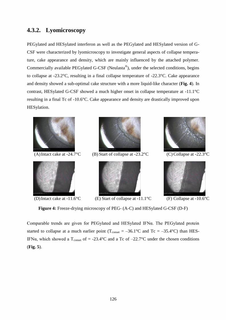
126
4.3.2. Lyomicroscopy
PEGylated and HESylated interferon as well as the PEGylated and HESylated version of G-
CSF were characterized by lyomicroscopy to investigate general aspects of collapse tempera-
ture, cake appearance and density, which are mainly influenced by the attached polymer.
Commercially available PEGylated G-CSF (Neulasta®), under the selected conditions, begins
to collapse at -23.2°C, resulting in a final collapse temperature of -22.3°C. Cake appearance
and density showed a sub-optimal cake structure with a more liquid-like character (Fig. 4). In
contrast, HESylated G-CSF showed a much higher onset in collapse temperature at -11.1°C
resulting in a final Tc of -10.6°C. Cake appearance and density are drastically improved upon
HESylation.
(A) Intact cake at -24.7°C
(B) Start of collapse at -23.2°C
(C) Collapse at -22.3°C
(D) Intact cake at -11.6°C
(E) Start of collapse at -11.1°C
(F) Collapse at -10.6°C
Figure 4: Freeze-drying microscopy of PEG- (A-C) and HESylated G-CSF (D-F)
Comparable trends are given for PEGylated and HESylated IFNα. The PEGylated protein
started to collapse at a much earlier point (Tconset = –36.1°C and Tc = –35.4°C) than HES-
IFNα, which showed a Tconset of = -23.4°C and a Tc of –22.7°C under the chosen conditions
(Fig. 5).

127
(A) Intact cake at -37.4°C
(B) Start of collapse at -36.1°C
(C) Collapse at -35.4°C
(D) Intact cake at -24.7°C
(E) Start of collapse at -23.4°C
(F) Collapse at -22.7°C
Figure 5: Freeze-drying microscopy of PEG- (A-C) and HESylated IFNα-2b (D-F)
4.3.3. Thermal analysis of the frozen formulations
The thermal properties of the frozen solutions were further characterized by differential scan-
ning calorimetry to determine the glass transition temperatures of the maximally freeze-
concentrated matrix; these temperatures represent a critical product temperature associated
with collapse during freeze-drying. Table 2 summarizes the Tg´ values of the chosen formu-
lations. Obviously, the Tg´ values differ significantly from the obtained collapse tempera-
tures, caused mainly by the difference in protein concentration (see Materials and Methods).
Sample Protein conc. [%m/v] Sucrose conc. [%m/v] Tg´ [°C]
Native IFNα 0.1 0 *
0.1 2.5 -34
0.1 5 -33
PEGylated IFNα 0.1 0 *
0.1 2.5 -35
0.1 5 -33
HESylated IFNα 0.1 0 *
0.1 2.5 -31
0.1 5 -33
Table 2: Thermal analysis of the frozen formulations before lyophilization, * not detectable

128
4.3.4. Optical evaluation of the freeze-dried samples
Figure 6 illustrates the cakes after freeze-drying. Vials A, D and G represent native, PEGylat-
ed and HESylated IFNα, respectively, lyophilized in the absence of sucrose. The native pro-
tein was heavily affected by cake shrinkage due to the absence of lyoprotectans.
Figure 6: Optical evaluation of the freeze-dried formulations; (A) IFNα without sucrose (B)
IFNα and 2.5% sucrose (C) IFNα and 5% sucrose (D) PEG-IFNα without sucrose (E) PEG-
IFNα and 2.5% sucrose (F) PEG- IFNα and 5% sucrose (G) HES-IFNα without sucrose (H)
HES- IFNα and 2.5% sucrose (I) HES- IFNα and 5% sucrose
4.3.5. Cake properties after lyophilization and storage
4.3.5.1. Glass transition temperature and degree of crystallization
Immediately after freeze-drying the native and PEGylated IFNα showed for the sucrose based
formulations a glass transition temperature in a range between 69 and 74°C, which remained
more or less constant during storage. Due to the high Tg of the HES part in the molecule, the
glass transition temperature exceeded 110°C for the sucrose free formulation of HES-IFNα
and was substantially lowered by the addition of 2.5 and 5% disaccharide to 67°C. The PEG-
induced crystallization was fully suppressed using 5% m/v sucrose (Fig. 7). During storage
the degree of crystallization for PEG-IFNα without and in combination with 2.5% sucrose
slightly increased over time, especially for the samples stored at 40°C.
A B C D E F G H I

129
Figure 7: Tg measurement (A): black bars represent the initial Tg after freeze-drying; the
grey-dashed ones after 3 months storage at 4°C and the white- dashed ones after 3 months
storage at 40°C; AUC of the crystallization peak (B): black bars represent the initial value
after freeze-drying; the grey-dashed ones after 3 months storage at 4°C and the white- dashed
ones after 3 months storage at 40°C, Representative thermogramms taken immediately after
freeze-drying (C,D,E)
4.3.5.2. Residual moisture
Table 3 summarizes the residual moisture data of the lyophilized cakes immediately after
lyophilization and after 3 months storage at 4 or 40°C. The addition of sucrose substantially
lowers the residual moisture. In general, both conjugates showed, without the addition of sug-
ar, lower initial moisture contents in contrast to the unmodified protein. In a direct compari-
son of PEG and HES, the coupling of HES led to lower values lying in the fact, that HES has
a better drying behavior due to the sugar based scaffold of the molecule. In contrast, PEG is
highly hydrophilic and can coordinate a higher number of water molecules, which are appar-
ently harder to remove by freeze-drying.

130
Formulation
0% sucrose
Native IFNα
Residual moisture [%]
PEGylated IFNα
Residual moisture [%]
HESylated IFNα
Residual moisture [%]
T0 3.6 ± 0.2 2.7 ± 0.2 2.3 ± 0.1
T3 at 4°C 4.1 ± 0.4 3.0 ± 0.4 1.3 ± 0.2
T3 at 40°C 1.9 ± 0.4 1.1 ± 0.0 1.1 ± 0.1
2.5% sucrose
T0 1.0 ± 0.3 1.5 ± 0.1 1.1 ± 1.1
T3 at 4°C 1.2 ± 0.3 1.2 ± 0.1 0.7 ± 0.1
T3 at 40°C 0.8 ± 0.1 0.9 ± 0.1 0.6 ± 0.0
5% sucrose
T0 1.0 ± 0.1 1.0 ± 0.1 1.2 ± 0.2
T3 at 4°C 1.0 ± 0.1 0.7 ± 0.1 0.9 ± 0.2
T3 at 40°C 0.8 ± 0.0 0.6 ± 0.0 0.9 ± 0.1
Table 3: Residual moisture contents immediately and after storage for 3 months at 4 or 40°C;
Key: T0: immediately after freeze-drying, T3: 3 months storage at 4 or 40°C
4.3.5.3. Conformational stability after lyophilization and storage
FTIR analyses of the conjugates provided two essential insights. First, it helps to illustrate
how PEGylation and HESylation alter the secondary structure after polymer coupling (Fig. 8)
and second, it shows how the formulation influences conformational stability after freeze-
drying and during storage. Surprisingly, significant changes in secondary structure upon
freeze-drying and storage could not be detected for native protein and both conjugates, indi-
cating high conformational stability (data not shown).

131
Figure 8: Area-normalized, second-derivative amide I spectra of native (solid line), PEG-
(dashed line) and HESylated IFNα (dotted line) of the liquid samples
4.3.6. Colloidal stability after lyophilization and storage
4.3.6.1. Turbidity and particle counts
Turbidity values and particle counts are illustrated in Figure 9. The attachment of PEG or
HES to the protein prevented the formation of particles due to a shielding effect independent
of the type of polymer used. Samples of both conjugates showed significantly lower initial
particle counts compared to those of the native IFNα. The turbidity measurement correlates
well to the total particle counts.

132
Figure 9: Turbidity and particle count; (A) native IFNα; (B) PEGylated IFNα and (C) HESyl-
ated IFNα; Key: T0: immediately after freeze-drying, T3: after 3 months storage at 4 or 40°C
4.3.6.2. Monomer recovery and soluble aggregates
The monomer content and the generated level of soluble aggregates during storage at 4 or
40°C as a function of the formulation’s sucrose content was determined by HP-SEC (Fig. 10).
The data show that the sucrose-to-polymer weight ratio and the chosen storage conditions
have the greatest influence on the formation of aggregates. Neither native nor HESylated
IFNα showed a dramatic decrease in monomer recovery, nor were soluble aggregates formed,
even at 40°C. Taking the data from soluble and insoluble aggregates together, it is even pos-
sible to formulate HESylated interferon α-2b without any sugar. In contrast, the colloidal sta-
bility of PEGylated interferon α-2b is highly dependent on the sucrose-to-PEG weight ratio
and storage temperature. For example, storage of lyophilized PEGylated IFNα at 4°C without

133
the addition of sucrose caused soluble aggregates to increase only slightly, from 1.1 to 1.2%.
At 40°C, the level of soluble aggregates increased remarkably, from 1.1 to 6.9%, indicating
the strong influence of storage temperature. The addition of sucrose substantially prevented
aggregation. Increasing the concentration of sucrose from 2.5% to 5% led to a significant de-
crease in the level of soluble aggregates, from 5.5% to 1.3%, for samples stored at 40°C. In-
terestingly, losses in monomer recovery correlate quite well to the degree of crystallization
(see supplementary data). When crystallization of PEG is not suppressed by amorphous lyo-
protectants, the PEGylated protein will rapidly degrade during long-term storage.
Figure 10: HP-SEC measurement to quantify monomer content (A) and the level of high-
molecular weight species (HMW; B) after 3 months at 4 or 40°C
4.4. Discussion
Today, approval of conjugated biologics requires site-specific chemistry to obtain homogene-
ous batches of single isomers [6]. Targeting the N-terminus to obtain a well-defined product
can be performed by reductive amination under mildly acidic conditions (e.g. at a pH of 4- 5).
The reaction takes place preferentially at the N-terminal amino group, which typically dis-
plays a lower pKa value of ~8 compared to ~10 for the ɛ-amino groups of lysine residues [29-
31]. In the first step of the conjugation a labile Schiff`s base is formed, which is subsequently
reduced by sodium cyanoborohydride to a stable secondary amine [32]. This approach
worked exceptionally well for conjugations involving interferon α-2b; peptide mapping and
aFFFF-MALLS analysis shown above provide proof of site-specific mono-conjugation. Fur-
thermore, our results are in good agreement with previously reported variants of PEGylated
IFNα-2b carrying a range of PEG derivatives at the N-terminus [28]. The resulting conjugates
were of excellent purity, with only trace amounts of unmodified protein and aggregates de-

134
tected in the liquid formulations that served as a starting point for lyophilization experiments.
Evaluation of the collapse temperature of frozen formulations is a critical tool for the selec-
tion of appropriate lyophilization parameters [33, 34]. Using lyomicroscopy, the freezing and
annealing step can be simulated and visually tracked to assess performance of the samples
under the selected lyo conditions [33]. The general goal is to maintain as high a collapse tem-
perature as possible [20]. The obtained Tc is directly related to the maximally allowed prod-
uct temperature, which must be several degrees below Tc to maintain an acceptable appear-
ance of the dried cake [34]. With this context, PEGylated and HESylated versions of two
pharmaceutically relevant proteins were tested for their performance during freezing and
thawing in an adjusted lyo cycle. The presence of hydroxyethyl starch in the conjugate led to
an extraordinarily high collapse temperature of the frozen solution; the precise Tc value de-
pends on the amount of HES present in the formulation. The sugar-based HES scaffold also
improves cake appearance and density, whereas PEG, when conjugated to the same protein
and yielding comparable conjugate hydrodynamic size, does not. The very high collapse tem-
perature of HES enables a more economical and robust lyophilization process. In general,
freeze-drying is a time- and cost-intensive process; therefore, the target product temperature,
which is highly dependent on the collapse temperature, should be as a high as possible. For
example, an almost 1°C increase in product temperature lowers the time for primary drying
by about 13% [34]. As a proof of concept, formulations of PEGylated and HESylated IFNα
were compared to the unmodified protein in terms of conformational and colloidal stability
after freeze-drying and storage at elevated temperatures (4 and 40°C) for 3 months. With the
exception of the formulation consisting of unmodified protein and no sucrose, the freeze-
drying protocol led to pharmaceutically elegant cakes, indicating that primary drying occurred
under the critical collapse temperature of the frozen solutions. The determined Tg´ values
correlated well to reported data for other sucrose-based formulations [20]. In general, HES
solutions are characterized by high Tg´ values, ranging from -12 to -17°C depending on the
HES molecular weight, the degree of substitution and the HES-to-disaccharide ratio [13, 35,
36]. Our results, however, demonstrate only a slight increase in the Tg´ value for the HESyl-
ated protein. The most likely reason for this is that the total amount of HES in a formulation
of 1 mg/mL protein concentration is rather low, having almost no effect on Tg´ and process
settings. To ensure adequate storage stability after drying, the Tg of the dry cake should be as
high as possible, preferably at least 20°C above storage temperature [13]. Our data indicate
that this condition has been easily met, as we report Tg values of at least 60°C up to ~110°C –
well above ambient temperature. Although it is quite common for sucrose-based cakes with

135
residual moisture in the range of 1% to have Tgs of about 65°C [37], some of our data are
remarkable. HES is obviously capable of acting as a Tg modifier when covalently bound to
the protein. With no sucrose added, HESylated interferon α-2b has a Tg of ~110°C after
freeze-drying. The addition of 2.5% or 5% sucrose inhibits this particular effect of HESyla-
tion, leading to Tgs of 75°C and 65°C, respectively. In addition, native and HESylated IFNα
did not show any tendency toward crystallization. Hydroxyethyl starch is based on a carbohy-
drate scaffold, and thus acts as a completely amorphous bulking agent during freeze-drying,
in a similar fashion to dextran [23, 33]. For PEGylated IFNα, we observed a different situa-
tion entirely. PEG is prone to rapid crystallization during lyophilization, due to its tendency to
promote phase separation into polymer-rich and sugar-rich phases, a precursor for crystalliza-
tion [11]. Therefore, high sucrose-to-PEG weight ratios (≥ 5) are required to fully suppress
PEG-induced crystallization. If crystallization occurs during storage, it is in most cases ac-
companied by an increased propensity for protein degradation [11]. In general, polymer con-
jugation under mild reaction conditions should have no effect on the secondary and tertiary
structure of the protein [38]. Using CD measurement, Kinstler and colleagues observed no
effect of site-specific PEGylation of G-CSF on the protein conformation; these results were
supported by FTIR studies from Rajan et al. [38]. Our results confirm these findings; we re-
cently reported that neither PEGylation nor HESylation altered the secondary structure of rh
IL-1ra (anakinra) [26]. In this study, the 2nd derivative FTIR spectra were obtained after stor-
age of the freeze-dried samples. Furthermore, native, PEGylated and HESylated IFNα under-
went lyophilication and subsequent storage without evidence of any changes, indicating high
conformational stability. Colloidal stability of IFN and its conjugates was assessed by tur-
bidity measurement, particle counting and monomer recovery by HP-SEC [39, 40]. Typically,
protein PEGylation reduces aggregation by physically separating monomers from one another
and therefore leads to reduced protein-protein interaction [41, 42]. The ability of PEGylation
to prevent PPI is not unique; this effect can also be obtained via HESylation [26]. In this
study, PEGylated and HESylated IFNα showed significantly lower particle counts after re-
constitution of the freeze-dried samples compared to that of the native protein. As mentioned
above, turbidity measurement can be used as a predictive marker for changes in a sample’s
aggregate status [27]. However, the turbidity measurement can be influenced by excipients or
high protein concentration and high values do not necessarily reflect high numbers of protein-
aceous particles [43]. Polymers can also scatter light and lead to high background turbidity
[44, 45]. In our studies we found that HESylation leads to higher initial turbidity values com-
pared to those of both the native and PEGylated protein. However, this value is not a reliable

136
marker of higher numbers of aggregates, which was investigated by LO and HP-SEC meas-
urements. As mentioned above, PEG-induced crystallization fosters protein degradation if it is
not suppressed by some means, such as high sucrose-to-PEG weight ratios. Stability studies
from Bhatnagar et al. regarding freeze-dried PEGylated human growth hormone found the
lowest colloidal stability in samples which were lyophilized with low sucrose to PEG weight
ratios [11]. We can confirm those findings for PEG-interferon. In particular, even the addition
of 2.5% sucrose was insufficient to prevent aggregation of PEGylated IFNα. Therefore, a
concentration of at least 5% sucrose must be included to prevent protein degradation induced
by PEG-driven crystallization. In contrast, HESylation drastically improved the colloidal sta-
bility of the conjugate, leading to reduced monomer loss and aggregate formation. This can be
explained by two facts: first, HES remained totally amorphous during lyophilization. Second,
when covalently attached to a protein, HES provides a protective shield that prevents PPI.
Additionally, for a sucrose-free lyophilisate of HESylated IFNα, a very high Tg will also most
likely lead to long-term storage stability even at greatly elevated storage temperatures. Final-
ly, a sucrose-free, freeze-dried formulation allows for a very high protein load, which is ex-
tremely attractive for further manufacturing steps, such as integration with drug delivery sys-
tems.
4.5. Conclusion
Site-specific mono-modification of the model protein interferon α-2b with PEG or HES was
achieved by reductive amination. Conjugate preparations of high purity were obtained by cat-
ion exchange chromatography without altering the protein’s secondary structure. Some fun-
damental questions concerning cake appearance and density were clarified for both polymers
and illustrated using conjugated IFNα-2b and G-CSF. Upon freeze-drying of PEGylated pro-
teins, the use of high sucrose-to-PEG weight ratios is necessary to prevent protein degradation
during long-term storage. In contrast, HESylation was found to be an attractive alternative
PEGylation, overcoming several significant formulation challenges associated with manufac-
turing of lyophilized PEGylated proteins. We demonstrate HESylation of IFNα as an example
of chemical coupling of a biocompatible polymer with excellent lyophilization properties; the
conjugate remained completely amorphous throughout lyophlization. The high glass transition
temperature of pure HES facilitated an elevated Tg of the HESylated protein during freeze-
drying, which can be an essential feature for manufacturing steps characterized by high resid-
ual moisture content, such as spray or vacuum drying. Additionally, HESylation effectively
shielded the conjugate to prevent PPI and improve conjugate colloidal stability. Lyophilized
137
formulations with no or low concentration of sucrose, as tested in this study, could be used to
up-concentrate protein solutions or permit a high protein load; both of these features would be
attractive for further manufacturing steps, like bulk drying of API or loading into drug deliv-
ery systems. In conclusion, this study reveals that HESylation offers a number of advantages
over PEGylation with regards to the development of freeze-dried formulations of polymer-
protein conjugates. In particular, higher collapse and glass transition temperatures, as well as
an amorphous nature which maintains storage stability even in the absence of additives could
be highly beneficial in drug formulation and manufacturing environments.
Acknowledgements
The authors wish to thank Georg Achleitner and his team at Fresenius Kabi Austria/ Graz for
conducting the freeze-drying microscopy measurements and Thomas Hey and his team at
Fresenius Kabi Germany/ Friedberg for performing the LC-MS analysis.
4.6. References
[1] T. Wurch, A. Pierré, S. Depil, Novel protein scaffolds as emerging therapeutic proteins:
from discovery to clinical proof-of-concept, Trends in Biotechnology, 30 (2012) 575-582.
[2] M. Gebauer, A. Skerra, Engineered protein scaffolds as next-generation antibody thera-
peutics, Current Opinion in Chemical Biology, 13 (2009) 245-255.
[3] A. Skerra, Alternative non-antibody scaffolds for molecular recognition, Current Opinion
in Biotechnology, 18 (2007) 295-304.
[4] J.M. Harris, R.B. Chess, Effect of pegylation on pharmaceuticals, Nat Rev Drug Discov, 2
(2003) 214-221.
[5] F.M. Veronese, G. Pasut, PEGylation, successful approach to drug delivery, Drug Discov-
ery Today, 10 (2005) 1451-1458.
[6] G. Pasut, F.M. Veronese, State of the art in PEGylation: The great versatility achieved
after forty years of research, Journal of Controlled Release, 161 (2012) 461-472.
[7] A. Besheer, R. Liebner, M. Meyer, G. Winter, Challenges for PEGylated Proteins and
Alternative Half-Life Extension Technologies Based on Biodegradable Polymers, in: Tai-
lored Polymer Architectures for Pharmaceutical and Biomedical Applications, American
Chemical Society, 2013, pp. 215-233.
[8] K.-i. Izutsu, S. Yoshioka, S. Kojima, T. Randolph, J. Carpenter, Effects of Sugars and
Polymers on Crystallization of Poly(ethylene glycol) in Frozen Solutions: Phase Separation
Between Incompatible Polymers, Pharmaceutical Research, 13 (1996) 1393-1400.

138
[9] B.S. Bhatnagar, S.M. Martin, D.L. Teagarden, E.Y. Shalaev, R. Suryanarayanan, Investi-
gation of PEG crystallization in frozen PEG–sucrose–water solutions. I. Characterization of
the nonequilibrium behavior during freeze-thawing, Journal of Pharmaceutical Sciences, 99
(2010) 2609-2619.
[10] L. Kreilgaard, S. Frokjaer, J.M. Flink, T.W. Randolph, J.F. Carpenter, Effects of Addi-
tives on the Stability of Recombinant Human Factor XIII during Freeze-Drying and Storage
in the Dried Solid, Archives of Biochemistry and Biophysics, 360 (1998) 121-134.
[11] B.S. Bhatnagar, S.W.H. Martin, T.S. Hodge, T.K. Das, L. Joseph, D.L. Teagarden, E.Y.
Shalaev, R. Suryanarayanan, Investigation of PEG crystallization in frozen and freeze-dried
PEGylated recombinant human growth hormone–sucrose systems: Implications on storage
stability, Journal of Pharmaceutical Sciences, 100 (2011) 3062–3075.
[12] S.D. Allison, B. Chang, T.W. Randolph, J.F. Carpenter, Hydrogen Bonding between
Sugar and Protein Is Responsible for Inhibition of Dehydration-Induced Protein Unfolding,
Archives of Biochemistry and Biophysics, 365 (1999) 289-298.
[13] W. Wang, Lyophilization and development of solid protein pharmaceuticals, Internation-
al Journal of Pharmaceutics, 203 (2000) 1-60.
[14] B.S. Bhatnagar, S.M. Martin, D.L. Teagarden, E.Y. Shalaev, R. Suryanarayanan, Inves-
tigation of PEG crystallization in frozen PEG–sucrose–water solutions: II. Characterization of
the equilibrium behavior during freeze-thawing, Journal of Pharmaceutical Sciences, 99
(2010) 4510-4524.
[15] H. Schellekens, W. Hennink, V. Brinks, The Immunogenicity of Polyethylene Glycol:
Facts and Fiction, Pharmaceutical Research, 30 (2013) 1729-1734.
[16] T. McDonnell, Y. Ioannou, A. Rahman, PEGylated drugs in rheumatology- why develop
them and do they work?, Rheumatology, (2013).
[17] M.J.G. Thomas, E.S. Parry, S.G. Nash, S.H. Bell, A method for the cryopreservation of
red blood cells using hydroxyethyl starch as a cryoprotectant, Transfusion Science, 17 (1996)
385-396.
[18] A. Stolzing, Y. Naaldijk, V. Fedorova, S. Sethe, Hydroxyethylstarch in cryopreservation-
Mechanisms, benefits and problems, Transfus Apher Sci, 46 (2012) 137 - 147.
[19] C. Stoll, J.L. Holovati, J.P. Acker, W.F. Wolkers, Synergistic effects of liposomes, treha-
lose, and hydroxyethyl starch for cryopreservation of human erythrocytes, Biotechnology
Progress, 28 (2012) 364-371.
[20] J. Carpenter, M. Pikal, B. Chang, T. Randolph, Rational Design of Stable Lyophilized
Protein Formulations: Some Practical Advice, Pharmaceutical Research, 14 (1997) 969-975.

139
[21] M.C. Laxenaire, C. Charpentier, L. Feldman, Anaphylactoid Reactions to Colloid Plasma
Substitutes: Incidence, Risk Factors, Mechanisms. A French Multicenter Prospective Study,
Annales Francaises d’Anesthésie et de Réanimation, 13 (1994) 301-310.
[22] J.F. Carpenter, S.J. Prestrelski, T. Arakawa, Separation of Freezing- and Drying-Induced
Denaturation of Lyophilized Proteins Using Stress-Specific Stabilization: I. Enzyme Activity
and Calorimetric Studies, Archives of Biochemistry and Biophysics, 303 (1993) 456-464.
[23] W. Garzon-Rodriguez, R.L. Koval, S. Chongprasert, S. Krishnan, T.W. Randolph, N.W.
Warne, J.F. Carpenter, Optimizing storage stability of lyophilized recombinant human inter-
leukin-11 with disaccharide/hydroxyethyl starch mixtures, Journal of Pharmaceutical Scienc-
es, 93 (2004) 684-696.
[24] R. Liebner, R. Mathaes, M. Meyer, T. Hey, G. Winter, A. Besheer, Protein HESylation
for half-life extension: Synthesis, characterization and pharmacokinetics of HESylated ana-
kinra, European Journal of Pharmaceutics and Biopharmaceutics, 87 (2014) 378-385.
[25] T. Hey, H. Knoller, P. Vorstheim, Half-Life Extension through HESylation®, in: Thera-
peutic Proteins, Wiley-VCH Verlag GmbH & Co. KGaA, 2012, pp. 117-140.
[26] R. Liebner, M. Meyer, T. Hey, G. Winter, A. Besheer, Head to Head Comparison of the
Formulation and Stability of Concentrated Solutions of HESylated versus PEGylated Ana-
kinra, Journal of Pharmaceutical Sciences, 104 (2015) 515-526.
[27] S. Zölls, R. Tantipolphan, M. Wiggenhorn, G. Winter, W. Jiskoot, W. Friess, A. Hawe,
Particles in therapeutic protein formulations, Part 1: Overview of analytical methods, Journal
of Pharmaceutical Sciences, 101 (2012) 914-935.
[28] M. Kusterle, S. Jevševar, V. Gaberc-Porekar, Size of pegylated protein conjugates stud-
ied by various methods, Acta chimica slovenica, 55 (2008) 594-601.
[29] O. Kinstler, G. Molineux, M. Treuheit, D. Ladd, C. Gegg, Mono-N-terminal
poly(ethylene glycol)-protein conjugates, Advanced Drug Delivery Reviews, 54 (2002) 477-
485.
[30] O.B. Kinstler, D.N. Brems, S.L. Lauren, A.G. Paige, J.B. Hamburger, M.J. Treuheit,
Characterization and Stability of N-terminally PEGylated rhG-CSF, Pharmaceutical Research,
13 (1996) 996-1002.
[31] H. Lee, I.H. Jang, S.H. Ryu, T.G. Park, N-Terminal Site-Specific Mono-PEGylation of
Epidermal Growth Factor, Pharmaceutical Research, 20 (2003) 818-825.
[32] M.D. Bentley, M.J. Roberts, J.M. Harris, Reductive amination using poly(ethylene gly-
col) acetaldehyde hydrate generated in situ: Applications to chitosan and lysozyme, Journal of
Pharmaceutical Sciences, 87 (1998) 1446-1449.

140
[33] J. Carpenter, B. Chang, W. Garzon-Rodriguez, T. Randolph, Rational Design of Stable
Lyophilized Protein Formulations: Theory and Practice, in: J. Carpenter, M. Manning (Eds.)
Rational Design of Stable Protein Formulations, Springer US, 2002, pp. 109-133.
[34] X. Tang, M. Pikal, Design of Freeze-Drying Processes for Pharmaceuticals: Practical
Advice, Pharmaceutical Research, 21 (2004) 191-200.
[35] W.Q. Sun, C.T. Wagner, J. Connor, The Glass Transition Behaviors of Hydroxyethyl
Starch Solutions, Cell Preservation Technology, 2 (2004) 55-65.
[36] T. Chen, S. Bhowmick, A. Sputtek, A. Fowler, M. Toner, The glass transition tempera-
ture of mixtures of trehalose and hydroxyethyl starch, Cryobiology, 44 (2002) 301-306.
[37] B. Hancock, G. Zografi, The Relationship Between the Glass Transition Temperature
and the Water Content of Amorphous Pharmaceutical Solids, Pharmaceutical Research, 11
(1994) 471-477.
[38] R.S. Rajan, T. Li, M. Aras, C. Sloey, W. Sutherland, H. Arai, R. Briddell, O. Kinstler,
A.M.K. Lueras, Y. Zhang, H. Yeghnazar, M. Treuheit, D.N. Brems, Modulation of protein
aggregation by polyethylene glycol conjugation: GCSF as a case study, Protein Science, 15
(2006) 1063-1075.
[39] Ph.Eur. 2.9.19, Pharmacopoea Europaea, 7th ed. 2010. Particulate contamination: sub-
visible particles. European Directorate for the Quality of Medicine (EDQM).
[40] USP<788>, United States Pharmacopeia, USP35-NF30, 2012. Particulate matter in
injections. United States Pharmacopeial convention.
[41] A. Basu, K. Yang, M. Wang, S. Liu, R. Chintala, T. Palm, H. Zhao, P. Peng, D. Wu, Z.
Zhang, J. Hua, M.-C. Hsieh, J. Zhou, G. Petti, X. Li, A. Janjua, M. Mendez, J. Liu, C.
Longley, Z. Zhang, M. Mehlig, V. Borowski, M. Viswanathan, D. Filpula, Struc-
ture−Function Engineering of Interferon β-1b for Improving Stability, Solubility, Potency,
Immunogenicity, and Pharmacokinetic Properties by Site-Selective Mono-PEGylation, Bio-
conjugate Chemistry, 17 (2006) 618-630.
[42] K. Hinds, J.J. Koh, L. Joss, F. Liu, M. Baudyš, S.W. Kim, Synthesis and Characteriza-
tion of Poly(ethylene glycol)−Insulin Conjugates, Bioconjugate Chemistry, 11 (2000) 195-
201.
[43] B.A. Salinas, H.A. Sathish, S.M. Bishop, N. Harn, J.F. Carpenter, T.W. Randolph, Un-
derstanding and modulating opalescence and viscosity in a monoclonal antibody formulation,
Journal of Pharmaceutical Sciences, 99 (2010) 82-93.
[44] T.K. Armstrong, T.J. Anchordoquy, Immobilization of nonviral vectors during the freez-
ing step of lyophilization, Journal of Pharmaceutical Sciences, 93 (2004) 2698-2709.

141
[45] S. Adam, R. Karger, V. Kretschmer, Photo-Optical Methods Can Lead to Clinically Rel-
evant Overestimation of Fibrinogen Concentration in Plasma Diluted With Hydroxyethyl
Starch, Clinical and Applied Thrombosis/Hemostasis, 16 (2010) 461-471.

142

143
Chapter V – Highly concentrated lyophilized formulations

144

145
5. Protein HESylation for the use of highly concentrated
and freeze-dried formulations of HES-anakinra conju-
gates: storage stability and the benchmark to
PEGylation
All conjugation and purification steps involved in preparing the HESylated protein were per-
formed by Sarah Bergmann, Fresenius Kabi.

146
Abstract
The purpose of this study was to investigate the effect of lyophilization on highly concentrat-
ed formulations of polymer-protein conjugates and their stability upon storage. PEGylation
and HESylation, two strategies currently used to prolong serum half-life of proteins and pep-
tides, were applied to the model protein anakinra. The resulting conjugates were lyophilized
with or without the addition of sucrose at protein concentrations up to 50 mg/mL. Results
showed that the nature of the polymer significantly impacts the colloidal stability of the con-
jugate. In both cases, PEGylation and HESylation prevented protein-protein interactions as
assessed by lower particle counts for sizes greater than 1 µm, whereas the native protein
formed significantly higher quantities of aggregates. However, in PEGylated proteins, PEG
promotes conjugate crystallization during lyophilization. This behavior fostered degradation
of the conjugate, resulting in the formation of high levels of soluble aggregates that could not
be substantially decreased by the addition of sucrose. In contrast, HESylated anakinra re-
mained completely amorphous and exhibited remarkable stability even over 3 months of stor-
age at 40°C in the absence of sucrose. Additionally, coupling HES to APIs such as proteins
offers a number of other advantages over PEGylation.
Keywords
HESylation, PEGylation, freeze-drying, phase separation, crystallization, aggregation

147
5.1. Introduction
Some high-value protein-based therapies require chronic administration of high doses of drug
(several mg/kg), typically performed by intravenous or subcutaneous injection [1, 2]. The
latter administration route, particularly when coupled with prefilled syringe and autoinjector
devices, is restricted primarily by injection volume (≤ 1.5 mL) and therefore requires APIs to
be formulated at high concentrations. Under conditions of high concentration, proteins are
known to have but limited intrinsic stability, constraining the development of lyophilized
formulations [1], which already experience reduced physical and chemical stability in the
dried state [3-5]. For small proteins and peptides such as cytokines, antibody fragments or
protein scaffolds that require half-life extension, the development of a highly concentrated
and lyophilized formulation is still challenging due to a number of drawbacks stemming from
PEGylation, the main HLE technology in use today. In highly concentrated aqueous solutions,
PEG promotes an extraordinary increase in viscosity and can generate certain peroxides, es-
pecially at high temperatures, which accelerates degradation of the protein portion of the con-
jugate [6, 7]. During lyophilization, PEG tends to crystallize rapidly regardless of whether it
is covalently attached or used as a bulking agent, resulting in further degradation of the pro-
tein upon storage, especially at high temperatures [6, 8, 9]. Complete suppression of crystalli-
zation requires high disaccharide-to-PEG ratios [8], which places clear limitations on the use
of highly concentrated and freeze-dried formulations of PEGylated drugs. A comparative
study of lyophilized PEGylated and HESylated IFNα showed the strong need for an amor-
phous lyoprotectant like sucrose to stabilize the PEGylated protein, whereas the HESylated
form maintained an amorphous character throughout lyophilization and exhibited excellent
storage stability even in the absence of additives (see chapter IV). Here, it is worth highlight-
ing that HESylation of proteins yields an additional stabilizing effect in highly concentrated
formulations of protein-polymer conjugates during freeze-drying and storage. We tested these
effects using anakinra, which is usually administrated by subcutaneous injection of a highly
concentrated solution (150 mg/mL) [6, 7]. For highly concentrated solutions of HESylated
anakinra, we recently showed that attachment of HES significantly improves the physico-
chemical stability of the protein as compared to the unmodified or PEGylated versions [7]. In
the present study, formulations of anakinra were subjected to an additional freeze-drying step
to evaluate the stability of highly concentrated polymer-protein conjugates using the perfor-
mance of PEGylated protein as a benchmark. As already described, N-terminal PEGylation
and HESylation was performed by reductive amination followed by purification using anionic

148
exchange chromatography [7]. Additionally, both conjugates were up-concentrated, lyophi-
lized and stored at elevated temperatures. Equivalent lyphilization and storage parameters
were applied for native anakinra, which served as a control for this study.
5.2. Materials and Methods
Kineret® was purchased from SOBI (Stockholm, Sweden). Branched PEG propionaldehyde
(MW = 40 kDa) was purchased from Jenkem Technologies (Allen/TX, USA). Activated hy-
droxyethyl starch (HES), with a weight average molar mass (Mw) of approximately 85 kDa,
number average molar mass (Mn) of approximately 65 kDa, and a polydispersity of 1.3 [10])
was from Fresenius Kabi Deutschland GmbH (Bad Homburg, Germany). All other chemicals
were used in analytical grade and purchased from VWR (Germany).
5.2.1. Synthesis and purification
A detailed description of the PEGylation and HESylation processes is already provided in
chapter III and was used without modification for the following experiments.
5.2.2. Sample preparation
Native anakinra (Kineret®) formulated at a protein concentration of 150 mg/mL was rebuff-
ered into 10 mM citrate buffer, pH 6.5 using 70 mL Slide-A-Lyzer®
dialysis cassettes (Ther-
mo Scientific, Schwerte, Germany). Buffer exchange to 10 mM citrate, pH 6.5 of PEGylated
and HESylated anakinra was performed by TFF using a 50 cm2 PES (polyethersulfone)
membrane capsule with a MWCO of 10 kDa operated on a MinimateTM
benchtop system
(both from Pall, Dreieich, Germany) to a protein concentration of 10-15 mg/mL. The final up-
concentration step for the conjugate solutions was performed by ultrafiltration in Vivaspin 20
centrifugal concentrators using a molecular weight cutoff (MWCO) of 10 kDa. The tubes
were placed into a Sigma 4K15 centrifuge (Sigma Laborzentrifugen GmbH, Osterode am
Harz, Germany) and rotated at 10,000 rpm (= 14,243 g) for 3-4 h at 4°C. The protein concen-
tration was verified by UV absorption (NanoDrop, Thermo Scientific, Schwerte, Germany)
using the extinction coefficient of the protein as reported by Raibekas et al. [21]. For further
experiments, samples were diluted in formulation buffer to the desired protein concentration.
Table 1 summarizes the details of formulations used for lyophilization experiments and sub-
sequent storage at 2-8 and 40°C for 3 months.

149
Sample Protein concentration
[mg/ mL]
Sucrose
[%(w/v)]
Native 10 0
anakinra 10 2.5
10 5
25 0
25 5
50 0
50 5
PEGylated 10 0
anakinra 10 2.5
10 5
25 0
25 5
50
50
0
5
HESylated 10 0
anakinra 10 2.5
10 5
25 0
25 5
50
50
0
5
Table 1: selected formulations for lyophilization and storage stability studies
5.2.3. Tg´ measurement
The Tg´ was determined by differential scanning calorimetry on a Netzsch DSC 204 Phönix
(Netzsch, Selb, Germany). 20 µL of each formulation was hermetically sealed in 40 µl alumi-
num pans, cooled from 20 to -70°C at a cooling rate of 10°C/min and heated up to 20°C to
detect the glass transition temperature of the frozen solutions.
5.2.4. Freeze-drying protocol
The freeze drying process was performed in a Martin Christ Epsilon 2-6D pilot-scale freeze-
dryer (Martin Christ Gefriertrocknungsanlagen GmbH, Osterode am Harz, Germany) using
the following freeze-drying protocol. 2R vials (Vetter, Ravensburg, Germany) were filled
with 0.2-0.3 mL sample volume. After shelf loading the vials were frozen by cooling at
1°C/min from 20 to -45°C and holding at the final temperature for 90 min. Primary drying

150
was performed at 0.1 mbar with a heating ramp from -45 to -15°C at a rate of 0.1°C/min and
holding the final temperature for 30 h. Secondary drying consisted of an equivalent ramp to
20°C, followed by holding the system at 0.01 mbar for 15 h. The vials were flooded with gas-
eous nitrogen, stoppered with Teflon-coated stoppers (West Pharmaceutical Services,
Eschweiler, Germany) at 800 mbar and afterwards manually sealed with 13 mm flip off seals
(West Pharmaceutical Services, Eschweiler, Germany).
5.2.5. Tg and degree of crystallization
The glass transition temperature of the freeze-dried cakes was determined by DSC measure-
ment. In a pressurized air flooded glove box with a relative humidity between 5-10%, a sam-
ple amount of 1-20 mg was weighed in aluminum pans and hermetically sealed. For DSC
measurement the pans were heated from 10 to 110°C, cooled to the starting point and heated
again to 180°C with a constant heating rate of 10°C/min. The second heating step was essen-
tial for determination of the correct glass transition temperature due to structural relaxation of
the cakes (Tg overshoot). Additionally, the DSC thermograms impart information about crys-
tallization. A 2.3% PEG solution (of the same PEG,used for conjugation) was lyophilized
under equivalent conditions and analyzed by DSC measurement. The obtained enthalpy, cal-
culated by measuring the area under the curve in J/g, was set to 100% to form the normaliza-
tion basis for quantifying crystallinity in the freeze-dried PEG- protein samples.
5.2.6. Residual moisture
The moisture content of the cakes was measured by a coulometric Karl-Fischer Titrator (KF
373, Metrohm GmbH & Co, Filderstadt, Germany).
5.2.7. Turbidity measurement
Protein quality after reconstitution with highly purified water and a dilution step to 1 mg/mL
in formulation buffer was evaluated using turbidity. Sample degassing was performed using a
Microcal ThermoVac2 degassing station (MicroCal Inc, Northampton, MA). The turbidity of
a 2 mL sample volume was determined at 860 nm and given in formazine nephelometric units
(FNU) using a NEPHLA turbidimeter (Dr. Lange, Düsseldorf, Germany).

151
5.2.8. Particle count - Light obscuration
According to Ph. Eur. and USP specifications, particle analysis was performed by light obscu-
ration using a PAMAS SVSS-C particle counter (PAMAS, Rutersheim, Germany) [22]. Parti-
cles with sizes of 1, 10 and 25 µm were quantified after a rinse volume of 0.5 mL in three
runs of 0.3 mL with an emptying and rinse rate of 10 mL/min.
5.2.9. Monomer recovery – Size exclusion chromatography
HP-SEC was performed on a Spectra System P2000 (Thermo Scientific, Germany) equipped
with a TSK guard column and a TSKgel G3000 SWxl (Tosoh Bioscience GmbH, Stuttgart,
Germany). After an injection of 50 µg protein, each run was performed by an isocratic elution
of the mobile phase, which contained 10 mM citrate, 140 mM NaCl, pH 6.5 at 0.5 mL/min
over 45 min followed by UV detection of monomer and high molecular weight (HMW) con-
tent at 280 nm.
5.3. Results
The thermal properties of the frozen solutions were characterized by differential scanning
calorimetry to detect the glass transition temperatures of the maximally freeze-concentrated
matrix; this glass transition temperature is a critical process parameter, knowledge of which
helps avoid cake collapse during freeze-drying. Table 2 summarizes the obtained Tg´ values
of selected formulations. Covalently linked PEG chains found in PEGylated protein conju-
gates rapidly began to crystallize during freezing and thawing. In most cases, the prominent
endothermic melting event overlapped with the predicted glass transition temperature range
and therefore no Tg´ could be detected. Additionally, the limited sensitivity of the instrument
restricted our ability to detect rather small glass transitions of formulations containing only
anakinra, lyophilized without the addition of sucrose. However, it is readily apparent that pro-
tein concentration has an impact on Tg´. For instance, raising the protein concentration of
native anakinra from 10 to 50 mg/mL in the presence of 5% (w/v) sucrose increased the glass
transition temperature by about 3.8 K. HESylation led to an additional elevation of the glass
transition temperature; at a protein concentration of 50 mg/mL in the presence of 5% sucrose,
HESylation led to elevation of the Tg´ by 5.8 K.

152
Sample Protein to sucrose ratio
[%(w/v) / %(w/v)]
Before
freeze-drying
Naitve anakinra
Tg´
[°C]
1/0 *
1/2.5 -28.1
1/5 -28.7
2.5/0 *
2.5/5 -26.6
5/0 *
5/5 -24.9
PEGylated anakinra 1/0 *
1/2.5 *
1/5 -30.3
2.5/0 *
2.5/5 *
5/0 *
5/5 *
HESylated anakinra 1/0 -16.9
1/2.5 -22.8
1/5 -25.1
2.5/0 -14.1
2.5/5 -21.0
5/0 -13.6
5/5 -19.1
Table 2: Thermal analysis of the frozen formulations before lyophilization
5.3.1. Cake properties after lyophilization and storage
5.3.1.1. Glass transition temperature and residual moisture
Glass transition temperatures (Tg) of the lyophilized formulations, as well as the measured
residual moisture (RM), are summarized in Table 3. In general, significantly higher RM con-
tents can be observed for the sucrose-free formulations. Immediately after freeze-drying, na-
tive anakinra formulated with sucrose exhibited glass transition temperatures in a range be-
tween 69-84°C and residual moisture values of 0.5-1.3%, both of which remained more or
153
less constant during storage at 2-8°C. Upon storage at 40°C for 3 months, higher RM values
were obtained. This had a plasticizing effect on the glass transition temperature, leading us to
infer that water vapor from the stoppers escaped into the lyo cakes. The modifying effect in-
duced by PEGylation and HESylation in both cases decreased the residual moisture. For both
types of conjugates, higher RM values were obtained for the samples stored at 40°C, whereas
lower RM numbers were found for the samples stored at 2-8°C. For PEG-anakinra, no Tg
values could be measured due to the fact that a predominant melting peak of PEG overlapped
the expected glass transition point. This effect could not be suppressed by the amount of su-
crose used in this study. Due to the elevated Tg of HES, the glass transition temperature of
HESylated anakinra exceeded 110°C for the sucrose free formulations and was substantially
lowered when sucrose was added. For example, the presence of 5% sucrose at a 10 mg/mL
protein concentration resulted in Tg values around 78°C. An increase in protein concentration
from 10 to 25 and 50 mg/mL lowered the sucrose to polymer ratio. Lowering this ratio also
reduced the plasticizing effect of sucrose on the glass transition temperature to negligible lev-
els; all Tg values measured immediately after lyophilization at a protein concentration beyond
25 mg/mL exceeded 100°C.

154
Sample Protein to sucrose ratio
[%(w/v) / %(w/v)]
After freeze-
drying
After 3 months
storage at 2-8°C
After 3 months
storage at 40°C
After freeze-
drying
After 3 months
storage at 2-8°C
After 3 months
storage at 40°C
Native
Tg
[°C]
1/0 * * *
RM
[%]
3.30 ± 0.20 3.46 ± 0.13 7.43 ± 0.03
anakinra 1/2.5 64.7 ± 0.5 70.7 ± 7.6 55.6 ± 15.8 1.27 ± 0.33 1.12 ± 0.06 4.05 ± 0.48
1/5 64.7 ± 0.2 72.7 ± 0.3 46.5 ± 5.9 0.83 ± 0.10 0.91 ± 0.03 2.24 ± 0.07
2.5/0 * * * 2.55 ± 0.38 2.77 ± 0.79 5.43 ± 0.41
2.5/5 84.1 ± 0.1 64.7 ± 9.6 51.7 ± 2.0 0.90 ± 0.23 0.97 ± 0.11 2.69 ± 0.40
5/0 * * * 2.26 ± 0.21 1.96 ± 0.45 4.36 ± 0.24
5/5 80.0 ± 22.7 79.5 ± 0.7 61.0 ± 3.6 0.47 ± 0.19 1.13 ± 0.02 2.65 ± 0.27
PEGylated 1/0 * * * 1.39 ± 0.49 0.98 ± 0.06 2.25 ± 0.27
anakinra 1/2.5 * * * 0.80 ± 0.01 0.64 ± 0.08 2.27 ± 0.34
1/5 * * * 0.76 ± 0.30 0.72 ± 0.02 2.23 ± 0.05
2.5/0 * * * 0.81 ± 0.11 0.71 ± 0.07 1.71 ± 0.16
2.5/5 * * * 0.57 ± 0.02 0.73 ± 0.03 1.88 ± 0.04
5/0 * * * 0.97 ± 0.71 0.59 ± 0.20 1.33 ± 0.05
5/5 * * * 0.42 ± 0.05 0.46 ± 0.07 1.43 ± 0.21
HESylated 1/0 110.3 ± 1.9 109.2 ± 0.2 * 0.91 ± 0.04 0.92 ± 0.04 3.38 ± 0.43
anakinra 1/2.5 94.1 ± 0.9 86.2 ± 2.3 61.5 ± 10.0 0.63 ± 0.23 0.68 ± 0.01 2.86 ± 0.01
1/5 77.8 ± 5.2 78.4 ± 0.1 49.8 ± 2.3 0.45 ± 0.01 0.53 ± 0.06 2.37 ± 0.21
2.5/0 110.8 ± 0.6 111.6 ± 2.9 113.8 ± 0.6 0.35 ± 0.04 0.86 ± 0.04 2.75 ± 0.27
2.5/5 100.9 ± 7.2 89.5 ± 1.8 62.8 ± 1.3 0.29 ± 0.01 0.60 ± 0.11 2.24 ± 0.04
5/0 110.7 ± 1.3 110.4 ± 2.2 112.1 ± 0.6 0.60 ± 0.15 0.57 ± 0.24 2.02 ± 0.25
5/5 109.9 ± 10.0 110.5 ± 4.0 66.5 ± 0.6 0.41 ± 0.02 0.39 ± 0.05 2.04 ± 0.23
* not detectable
Table 3: Glass transition temperatures and residual moisture immediately after lyophilization and after 3 months storage at 2-8 and 40°C

155
5.3.1.2. Degree of crystallization
During freeze-drying of native and HESylated anakinra, no crystallization could by detected
by DSC analysis. In contrast, PEGylated anakinra rapidly began to crystallize during lyophi-
lization, an effect which could only be suppressed by the addition of sucrose to a certain pol-
ymer-to-disaccharide ratio (Table 4). Increasing the amount of PEG in the formulation sub-
stantially lowered the sucrose-to-PEG ratio and therefore enhanced the role of crystallization
in the lyo cake. Samples, which were stored at 40°C showed, as expected, a slight increase in
crystallization. Interestingly, during storage at 2-8°C the degree of crystallization was in some
cases lower than the initial value obtained immediately after freeze-drying.
Sample Protein to sucrose ratio
[%(w/v) / %(w/v)]
After freeze-
drying
After 3 months
storage at 2-8°C
After 3 months
storage at 40°C
Native
Crystallinity
[%]
1/0 * * *
anakinra 1/2.5 * * *
1/5 * * *
2.5/0 * * *
2.5/5 * * *
5/0 * * *
5.0/5 * * *
PEGylated 1/0 64.4 ± 1.7 67.4 ± 0.3 68.1 ± 0.9
anakinra 1/2.5 38.7 ± 0.1 39.0 ± 0.3 43.7 ± 2.0
1/5 23.2 ± 1.7 16.3 ± 1.9 29.1 ± 0.3
2.5/0 66.0 ± 2.3 68.9 ± 0.8 68.3 ± 1.6
2.5/5 44.4 ± 0.2 44.7 ± 0.5 44.5 ± 1.9
5/0 66.7 ± 0.3 67.7 ± 3.8 71.1 ± 1.5
5.0/5 57.2 ± 0.2 53.0 ± 1.2 58.4 ± 0.2
HESylated 1/0 * * *
anakinra 1/2.5 * * *
1/5 * * *
2.5/0 * * *
2.5/5 * * *
5/0 * * *
5.0/5 * * *
* not detectable
Table 4: Thermal analysis for the degree of crystallization

156
5.3.2. Protein quality after reconstitution
5.3.2.1. Colloidal stability after lyophilization and storage
The number of particles with a size greater than 1 µm and corresponding turbidity values
were measured immediately after lyophilization and subsequent storage (Figure 1). Especial-
ly for native anakinra, storage temperature and the presence of sucrose had the biggest impact
on the degree of aggregate formation. Formulations lacking sucrose exhibited greater numbers
of particles, particularly at protein concentrations of 25 and 50 mg/mL. Obtained particle
counts are in good agreement with the turbidity measurements; higher particle numbers are
associated with higher turbidity values. These effects are more distinct for samples which
were stored at 40°C. The addition of sucrose substantially decreased both particle counts and
the solution turbidity, indicating the strong need for a lyoprotectant in the formulation. In con-
trast, conjugation with either PEG or HES prevented the formation of aggregates. This effect
can be explained by the high shielding effect of both polymers. As one might expect, then,
correspondingly low particle counts were observed in each conjugate formulation.
5.3.2.2. Monomer recovery and soluble aggregates
Monomer recovery and levels of soluble aggregates were monitored by HP-SEC analysis.
Protein concentration and the amount of sucrose were found to have the greatest influence on
the formation of subvisible particles (Figure 2). While native anakinra showed no substantial
loss in monomer content, an increase in soluble aggregates was observed especially for for-
mulations, lyophilized in the absence of sucrose. Including sucrose in the formulation sub-
stantially lowered the degree of aggregate formation. In contrast, PEGylated anakinra exhibit-
ed both dramatic loss in monomer recovery and rapid formation of soluble aggregates espe-
cially for samples lyophilized without sucrose (the reader is advised to note the difference in
y-axis scales in the presented figures). Again, sucrose is found to prevent formation of parti-
cles and preserve high monomer levels. Interestingly, the loss in monomer recovery correlates
well to the degree of crystallization measured via DSC. When crystallization of PEG is not
suppressed by amorphous excipients, the PEGylated protein degrades rapidly during long-
term storage. Surprisingly, even in the absence of sucrose, HESylation of anakinra neither
diminished monomer recovery nor promoted formation of high levels of soluble aggregates.

157
Figure 1: Turbidity and particle counts immediately after lyophilization (A) native anakinra; (B) PEGylated anakinra and (C) HESylated anakinra;
Turbidity and particle counts after storage at 40°C for 3 months (D) native anakinra; (E) PEGylated anakinra and (F) HESylated anakinra; X axes
keys: A: native anakinra; PA: PEG- anakinra; HA: HES-anakinra and 10, 25 or 50 represent the protein concentration in mg/mL

158
Figure 2: SEC analyses after 3 months at 40°C: monomer content of (A) native anakinra (B) PEGylated anakinra and (C) HESylated anakinra;
high molecular weight (HMW) content in (D) native anakinra (E) PEGylated anakinra and (F) HESylated anakinra; Caption key: A: native
anakinra; PA: PEG-anakinra; HA: HES-anakinra and 10, 25 or 50 represent the protein concentration inmg/mL

159
5.4. Discussion
The development of PEGylated drugs in highly concentrated formulations represents an on-
going challenge due to the high viscosity of PEG at high concentrations, hampering manufac-
turing processes and drug injectability [6]. For the use of freeze-dried formulations of
PEGylated proteins, high disaccharide-to-PEG ratios are necessary to obtain a reasonable
shelf-life due to fact that PEG will rapidly crystallize during freeze-drying, which may de-
crease storage stability and induce protein degradation if left unsuppressed by amorphous
lyoprotectants [8].
The main focus of this study was to apply HESylation technology to the formulation of highly
concentrated, freeze-dried protein-polymer conjugates. Freeze-drying is a time- and cost-
intensive process, and the glass transition temperature of the maximally freeze-concentrated
phase has a considerable effect on the overall drying time. To reduce drying time, the target
product temperature during primary drying should be as high as possible while staying well
below the glass transition temperature to yield a pharmaceutically elegant cake with no obvi-
ous meltbacks or sample collapse [11]. A nearly 1°C increase in product temperature lowers
the time for primary drying by about 13% [4]. As described previously, an increase in protein
concentration will increase the Tg´ and therefore a higher Tp can be applied for primary
drying [12, 13]. We confirm these findings with our results for the case of native anakinra. An
additional enhancement is observed upon protein HESylation. In general, HES solutions are
characterized by high Tg´ values in a range of -12 to -17°C depending on the molecular
weight, the degree of substitution and the HES-to-disaccharide ratio [14-17]. For highly con-
centrated formulations, the elevating effect of HESylation on Tg´ was highly significant, thus
enabling higher Tp values during the lyophilization process and a concomitant reduction in
the time required for primary drying. This cannot be presently achieved using either the native
or PEGylated protein. To ensure appropriate storage stability in a dried state, the glass transi-
tion temperature should be as high as possible. Typically, the glass transition should be at
least 20°C above ambient temperature to limit molecular mobility in the freeze-dried cake and
thus prevent protein degradation [15, 18, 19]. The glass transition temperatures we obtained
immediately after lyophilization satsify this criterion and match previously reported glass
transition points of sucrose-based formulations [20]. In addition, an increase in protein con-
centration has been reported to generally enhance the glass transition temperature [21]. We
confirm this finding for anakinra; higher protein concentrations showed higher Tg values. The
effect of HESylation, as expected, further increased the glass transition temperature, an effect

160
which has already been shown for HESylated IFNα-2b (see chapter IV). HES itself has a
very high Tg, which depends on molecular weight and degree of substitution. This modifying
effect on glass transition temperature is directly transferred to covalently modified HES-
protein conjugates,. Thus, the presence of HES opposes the plasticizing effect of water in the
lyo cake and maintains the glass transition point above the storage temperature. This effect
has the potential to make an impact on next-generation drying methods, where in most cases
higher residual moisture contents can be expected after the drying process [22]. Methods such
as spray-, convective, vacuum and microwave drying or combinations thereof are mainly
focused on overcoming the drawbacks of lyophilization, for example, by reducing the overall
process time or the high purchase and maintenance costs of the drying equipment. Additional-
ly, freeze-drying has a low energy efficiency and can cause a significant loss in API quality
due to its exposure to various process-related stresses such as the freezing step [22].
One potential limitation of polyethylene glycol is its strong tendency to crystallize during ly-
ophilization and subsequent storage, fostering degradation of the conjugated protein [8, 9, 23].
This effect can be overcome by the use of amorphous lyoprotectants such as sucrose in disac-
charide-to-PEG weight ratios ≥ 5 [8]. Protein degradation occurring as a result of crystalliza-
tion is mainly induced by interfacial denaturation at the large ice/water interface [24, 25]. In
contrast to PEGylation strategies, crystallization cannot be observed when HES is attached to
the protein. In a similar fashion to dextran, HES is based on a carbohydrate scaffold and
therefore acts as a completely amorphous bulking agent during freeze-drying [13, 26]. The
effect of covalent modification on colloidal stability of conjugate formulations was evaluated
by turbidity measurement, particle counting and HP-SEC for monomer recovery [27, 28].
PEGylation and HESylation of proteins tend to prevent protein-protein interaction and reduce
protein aggregation in aqueous solution [7, 10, 29-31]. In the current study, both modifica-
tions of anakinra exhibited this effect. PEGylated and HESylated anakinra both generated
significanlty lower particles counts upon lyophilization and subsequent storage than the un-
modified protein. HP-SEC provides information on entities existing at the submicron level,
including whether or not the attachment of PEG or HES highly influences the formation of
aggregates. For PEG-containing samples, which tend to experience heavy crystallization and
concomitant protein degradation, significant losses in monomer recovery were observed. In
contrast, HES-containing conjugates did not show any tendency toward crystallization and
thus inhibited the formation of both submicron and micron-scale aggregates. Thus, for all
chosen protein concentrations, even in the absence of sucrose, no significant loss in monomer
recovery could be observed. These results make feasible the use of formulations with high

161
protein loads, for applications such as further integration of lyophilisates into drug delivery
systems. In addition, the absence of a requirement for lyo excipients enables the use of freeze-
drying for up-concentration to obtain highly concentrated formulations of HES-protein conju-
gates.
5.5. Conclusion
Lyophilization makes proteins susceptible to degradation because both physical and chemical
stability are significantly reduced in the dried state. The use of chemical modifications such as
polymer conjugation can prevent protein aggregation in the liquid state by hampering PPIs. It
has been shown that polymers such as PEG and HES differ widely in a variety of relevant
physicochemical properties – variations which can either prevent or foster protein degradation
after lyophilization. We showed here that in the context of highly concentrated lyophilisates,
covalent linkage of HES to the model protein anakinra has several advantages. The presence
of HES increases the Tg´ of the solutes, permitting primary drying to be run at higher product
temperatures and thus, in shorter periods of time. HES also maintains its amorphous nature
after drying; cakes of HES conjugates show very high glass transition temperatures, which
helps provide sufficient storage stability even in the presence of higher moisture contents.
HESylation in this case enabled freeze-dried formulations without sucrose, opening the door
to routine up-concentration of protein solutions. This feature is attractive for its impact on
subsequent manufacturing steps, such as bulk drying of drug substance or preparation of drug
delivery systems such as implants or microspheres. Beyond its excellent drying behavior, the
attachment of HES prevents PPIs after reconstitution due to effective shielding. In conclusion,
this study reveals that HESylation has the potential to replace PEGylation, especially for ap-
plications where highly concentrated lyophilisates of polymer-protein conjugates are desirable
or essential.
Acknowledgements
The authors would like to thank Fresenius Kabi Deutschland GmbH for financial support and
Sarah Bergmann for her technical assistance in preparing HESylated anakinra.

162
5.6. References
[1] A. Bhambhani, J.T. Blue, Lyophilization Strategies for Development of a High-
Concentration Monoclonal Antibody Formulation: Benefits and Pitfalls, American Pharma-
ceutical Review - The Review of American Pharmaceutical Business & Technology, (2010).
[2] W. Cao, S. Krishnan, M.S. Ricci, L.-Y. Shih, D. Liu, J.H. Gu, F. Jameel, Rational design
of lyophilized high concentration protein formulations-mitigating the challenge of slow re-
constitution with multidisciplinary strategies, European Journal of Pharmaceutics and Bio-
pharmaceutics, 85 (2013) 287-293.
[3] J. Carpenter, M. Pikal, B. Chang, T. Randolph, Rational Design of Stable Lyophilized
Protein Formulations: Some Practical Advice, Pharmaceutical Research, 14 (1997) 969-975.
[4] X. Tang, M. Pikal, Design of Freeze-Drying Processes for Pharmaceuticals: Practical Ad-
vice, Pharmaceutical Research, 21 (2004) 191-200.
[5] P. Matejtschuk, Lyophilization of Proteins, in: J. Day, G. Stacey (Eds.) Cryopreservation
and Freeze-Drying Protocols, Humana Press, 2007, pp. 59-72.
[6] A. Besheer, R. Liebner, M. Meyer, G. Winter, Challenges for PEGylated Proteins and
Alternative Half-Life Extension Technologies Based on Biodegradable Polymers, in: Tai-
lored Polymer Architectures for Pharmaceutical and Biomedical Applications, American
Chemical Society, 2013, pp. 215-233.
[7] R. Liebner, M. Meyer, T. Hey, G. Winter, A. Besheer, Head to Head Comparison of the
Formulation and Stability of Concentrated Solutions of HESylated versus PEGylated Ana-
kinra, J Pharm Sci, 104 (2015) 515-526.
[8] B.S. Bhatnagar, S.W.H. Martin, T.S. Hodge, T.K. Das, L. Joseph, D.L. Teagarden, E.Y.
Shalaev, R. Suryanarayanan, Investigation of PEG crystallization in frozen and freeze-dried
PEGylated recombinant human growth hormone–sucrose systems: Implications on storage
stability, Journal of Pharmaceutical Sciences, 100 (2011) 3062–3075.
[9] L. Kreilgaard, S. Frokjaer, J.M. Flink, T.W. Randolph, J.F. Carpenter, Effects of Additives
on the Stability of Recombinant Human Factor XIII during Freeze-Drying and Storage in the
Dried Solid, Archives of Biochemistry and Biophysics, 360 (1998) 121-134.
[10] R. Liebner, R. Mathaes, M. Meyer, T. Hey, G. Winter, A. Besheer, Protein HESylation
for half-life extension: Synthesis, characterization and pharmacokinetics of HESylated ana-
kinra, European Journal of Pharmaceutics and Biopharmaceutics, 87 (2014) 378-385.
163
[11] B.S. Chang, S.Y. Patro, Freeze-drying Process Development for Protein Pharmaceuti-
cals, in: Lyophilization of Biopharmaceuticals, American Association of Pharmaceutical Sci-
entists, 2004, pp. 113-138.
[12] J.D. Colandene, L.M. Maldonado, A.T. Creagh, J.S. Vrettos, K.G. Goad, T.M.
Spitznagel, Lyophilization cycle development for a high-concentration monoclonal antibody
formulation lacking a crystalline bulking agent, Journal of Pharmaceutical Sciences, 96
(2007) 1598-1608.
[13] J. Carpenter, B. Chang, W. Garzon-Rodriguez, T. Randolph, Rational Design of Stable
Lyophilized Protein Formulations: Theory and Practice, in: J. Carpenter, M. Manning (Eds.)
Rational Design of Stable Protein Formulations, Springer US, 2002, pp. 109-133.
[14] W.Q. Sun, C.T. Wagner, J. Connor, The Glass Transition Behaviors of Hydroxyethyl
Starch Solutions, Cell Preservation Technology, 2 (2004) 55-65.
[15] W. Wang, Lyophilization and development of solid protein pharmaceuticals, Internation-
al Journal of Pharmaceutics, 203 (2000) 1-60.
[16] T. Chen, S. Bhowmick, A. Sputtek, A. Fowler, M. Toner, The glass transition tempera-
ture of mixtures of trehalose and hydroxyethyl starch, Cryobiology, 44 (2002) 301-306.
[17] M. Kochs, C. Körber, I. Heschel, B. Nunner, The influence of the freezing process on
vapour transport during sublimation in vacuum-freeze-drying of macroscopic samples, Inter-
national Journal of Heat and Mass Transfer, 36 (1993) 1727-1738.
[18] C.E. Stotz, S.L. Winslow, M.L. Houchin, A.J.M. D'Souza, J. Ji, E.M. Topp, Degradation
Pathways for Lyophilized Peptides and Proteins in: Lyophilization of Biopharmaceuticals,
American Association of Pharmaceutical Scientists, 2004, pp. 443-478.
[19] S. Yoshioka, Y. Aso, Correlations between molecular mobility and chemical stability
during storage of amorphous pharmaceuticals, Journal of Pharmaceutical Sciences, 96 (2007)
960-981.
[20] B. Hancock, G. Zografi, The Relationship Between the Glass Transition Temperature
and the Water Content of Amorphous Pharmaceutical Solids, Pharmaceutical Research, 11
(1994) 471-477.
[21] S. Duddu, P. Dal Monte, Effect of Glass Transition Temperature on the Stability of Ly-
ophilized Formulations Containing a Chimeric Therapeutic Monoclonal Antibody, Pharma-
ceutical Research, 14 (1997) 591-595.
[22] R.H. Walters, B. Bhatnagar, S. Tchessalov, K.-I. Izutsu, K. Tsumoto, S. Ohtake, Next-
Generation Drying Technologies for Pharmaceutical Applications, Journal of Pharmaceutical
Sciences, (2014) n/a-n/a.

164
[23] K.-i. Izutsu, S. Yoshioka, S. Kojima, Increased Stabilizing Effects of Amphiphilic Excip-
ients on Freeze-Drying of Lactate Dehydrogenase (LDH) by Dispersion into Sugar Matrices,
Pharmaceutical Research, 12 (1995) 838-843.
[24] T.W. Randolph, Phase separation of excipients during lyophilization: Effects on protein
stability, Journal of Pharmaceutical Sciences, 86 (1997) 1198-1203.
[25] W. Wang, S. Nema, D. Teagarden, Protein aggregation—Pathways and influencing fac-
tors, International Journal of Pharmaceutics, 390 (2010) 89-99.
[26] W. Garzon-Rodriguez, R.L. Koval, S. Chongprasert, S. Krishnan, T.W. Randolph, N.W.
Warne, J.F. Carpenter, Optimizing storage stability of lyophilized recombinant human inter-
leukin-11 with disaccharide/hydroxyethyl starch mixtures, Journal of Pharmaceutical Scienc-
es, 93 (2004) 684-696.
[27] Ph.Eur. 2.9.19, Pharmacopoea Europaea, 7th ed. 2010. Particulate contamination: sub-
visible particles. European Directorate for the Quality of Medicine (EDQM).
[28] USP<788>, United States Pharmacopeia, USP35-NF30, 2012. Particulate matter in injec-
tions. United States Pharmacopeial convention.
[29] G. Pasut, F.M. Veronese, State of the art in PEGylation: The great versatility achieved
after forty years of research, Journal of Controlled Release, 161 (2012) 461-472.
[30] A. Basu, K. Yang, M. Wang, S. Liu, R. Chintala, T. Palm, H. Zhao, P. Peng, D. Wu, Z.
Zhang, J. Hua, M.-C. Hsieh, J. Zhou, G. Petti, X. Li, A. Janjua, M. Mendez, J. Liu, C.
Longley, Z. Zhang, M. Mehlig, V. Borowski, M. Viswanathan, D. Filpula, Struc-
ture−Function Engineering of Interferon β-1b for Improving Stability, Solubility, Potency,
Immunogenicity, and Pharmacokinetic Properties by Site-Selective Mono-PEGylation, Bio-
conjugate Chemistry, 17 (2006) 618-630.
[31] K. Hinds, J.J. Koh, L. Joss, F. Liu, M. Baudyš, S.W. Kim, Synthesis and Characteriza-
tion of Poly(ethylene glycol)−Insulin Conjugates, Bioconjugate Chemistry, 11 (2000) 195-
201.

165
Chapter VI – Summary and attachments

166
167
6. Summary of the thesis
During the last decades, grafting of biocompatible polymers like polyethylene gycol to thera-
peutic proteins and peptides for half-life extension has become an established and highly re-
fined technology. With over 10 approved PEGylated drugs, this technology is considered to
be the gold standard for half-life extension. However, PEG has a number of serious shortcom-
ings, which have recently spurred development of alternative HLE technologies based on both
biocompatible and biodegradable polymers. Such alternatives aim to prolong the circulation
time of small biologics while simultaneously overcoming formulation challenges posed by
use of PEG. In response, the overall goal of this thesis is to further develop HESylation tech-
nology and overcome some of the challenges associated with formulation of novel, biode-
gradable HES-protein conjugates. Results from preliminary studies revealed that the chemical
attachment of HES is a promising tool for serum half-life modulation of proteins and offers
distinct formulation advantages over PEGylation.
In the first phase of the project hydroxyethyl starch (HES) was chemically grafted to a model
protein called anakinra. Chapter II describes the synthesis and physicochemical characteriza-
tion of the HESylated protein. Site-specific HESylation of anakinra at the N-terminus was
achieved using reductive amination at a pH of 5.0. The mildly acidic conditions during syn-
thesis and subsequent purification by anionic exchange chromatography at a pH of 8.0 had no
significant influence on either the protein conformation or its biological affinity to its cognate
receptor. Coupling HES to anakinra improved the thermodynamic properties, prevented pro-
tein aggregation during accelerated stress conditions and prolonged the pharmacokinetics of
the protein in vivo.
In a next step, HESylated anakinra was benchmarked against its PEGylated counterpart with
respect to the effect on the conjugate’s physicochemical properties in highly concentrated
solutions, where protein stability and solution viscosity can be notably suboptimal. HESylated
and PEGylated anakinra were synthesized and purified under equivalent conditions. Coupling
a 40 kDa branched PEG and an 85 kDa HES to anakinra resulted in conjugates of comparable
hydrodynamic size. Highly concentrated formulations of both conjugates exhibited increased
melting temperature in solution, which led to improved thermodynamic stability when com-
pared to the unmodified counterpart. However, solution viscosity and colloidal stability com-
pletely differed for both conjugates. In general, more highly concentrated protein solutions are
associated with a greater tendency toward aggregation as a result of molecular crowding. In
addition, reversible self-association in more crowded conditions makes solutions of such pro-

168
teins liable to show an increase in viscosity. The presence of a covalently coupled polymer
aggravates changes in viscosity due to entanglement of adjacent polymer chains. This phe-
nomenon is more pronounced for the highly flexible polymer chains found in PEG molecules.
In contrast, the highly branched polymeric architecture of HES provides a stiffer structure in
which the molecules act more like a hard sphere. As a result, the viscosity of HESylated pro-
tein solutions was observed to be much lower than that of solutions of PEG conjugates at
equivalent protein concentration. Although both conjugates showed improved monomer re-
covery, HESylation provided for higher colloidal stability compared to native and PEGylated
anakinra at all studied concentrations. The root cause for this surprising difference in protein
stability might be the difference in the nature of the two polymers. PEG is known to change
the polarity and dielectric constant of aqueous solutions and can generate peroxides, especial-
ly at high temperatures, which could have led to the higher degree of protein degradation ob-
served in comparison to the HESylated protein. The combination of lower intrinsic viscosities
and improved storage stability of the HES conjugate reveals that HESylation can provide
formulation advantages over PEGylation, especially for the development of highly concen-
trated formulations of polymer-protein conjugates.
The final part of the project aimed to distinguish between the physicochemical properties of
lyophilized PEGylated and HESylated proteins. In this study the model protein interferon α-
2b was either PEGylated or HESylated in a site-specific manner, lyophilized and stored under
elevated temperatures (2-8 and 40°C) for three months. In chapter IV we studied the effect of
lyoprotectants by varying the amount of sucrose in the freeze-dried formulations. We also
examined the influence of storage temperature and evaluated how conjugation of each poly-
mer impacts the conformational and colloidal stability of the protein. Our results show that
PEG tends to crystallize upon freeze-drying, which has a significant influence on protein sta-
bility during storage, especially at high temperatures. The amount of crystallization could be
substantially limited by formulating with sucrose at high disaccharide-to-PEG ratios. Conse-
quently, samples of the PEGylated protein lyophilized without the addition of sucrose and
stored at 40°C showed the highest degradation rate. In conclusion, freeze-dried formulations
of PEGylated proteins require large amounts of amorphous lyoprotectants to enable sufficient
storage stability. In contrast, HESylation of IFNα drastically improved the colloidal stability
of the protein by reducing monomer loss and tendency toward aggregation. This can be ex-
plained by two facts: first, HES remains totally amorphous during lyophilization and second,
it provides a protective shield that prevents PPI when covalently attached to the protein. Addi-
tionally, HES exhibits very high initial glass transition temperatures both in solution and in

169
the dried state – properties which may be directly transferred to the conjugate. An increase in
the Tg´ of frozen solutions makes a more economic lyophilization cycle possible by enabling
application of higher product temperatures, which reduces the time required for primary dry-
ing. The presence of HES in the conjugate also provides a protective shielding that reduces
aggregate formation, even in the absence of sucrose and under storage at high temperatures.
Our results show that it might even be possible to formulate HESylated interferon α-2b with-
out any sugar at all. A sucrose-free, freeze-dried formulation would permit a very high protein
load, which is extremely attractive for further manufacturing steps; examples include integra-
tion with drug delivery systems such as implants. In conclusion, the results of this study re-
veal that HESylation can provide formulation advantages over PEGylation during lyophiliza-
tion.
In chapter V we investigated the stability of highly concentrated PEGylated and HESylated
anakinra upon freeze-drying. After storage at 2-8 and 40°C, both polymers imparted effective
shielding and suppression of micron-scale aggregate formation. However, due to the high
tendency of PEG to crystallize during lyophilization, an effect which could not be fully sup-
pressed by the use of certain amounts of sucrose, the PEGylated protein degraded rapidly and
formed large amounts of soluble submicron aggregates. In the presence of sucrose, native
anakinra showed a reduced tendency toward aggregation, as seen via lower particle counts.
Formulations of the HESylated protein remained remarkably stable and did not promote the
formation of aggregates on either micron or submicron scales, even in the absence of sucrose.
Lyophilization studies of both dilute and highly concentrated HESylated protein solutions
confirmed the benefit of covalently bound HES. Taken together, these results paint an ex-
tremely attractive portrait of the potential benefits of incorporating HESylation technology
into biologic formulation development workflows, and should motivate further studies in the
field of HESylated drugs.

170

171
7. Publications associated with this thesis
7.1. Research articles
Robert Liebner, Roman Mathaes, Martin Meyer, Thomas Hey, Gerhard Winter,
Ahmed Besheer Protein HESylation for half-life extension: Synthesis, characterization and
pharmacokinetics of HESylated anakinra – European Journal of Pharmaceutics and Bio-
pharmaceutics. 2014, 87(2): 378-385
Robert Liebner, Martin Meyer, Thomas Hey, Gerhard Winter, Ahmed Besheer
Head to head comparison of the formulation and stability of concentrated solutions of
HESylated versus PEGylated Anakinra – Journal of Pharmaceutical Sciences. 104 (2015)
515-526
Robert Liebner, Sarah Bergmann, Thomas Hey, Gerhard Winter, Ahmed Besheer
Freeze-drying of HESylated IFNα-2b: Effect of HESylation on storage stability and the
benchmark to PEGylation – In preparation
7.2. Book chapter
Ahmed Besheer, Robert Liebner, Martin Meyer, Gerhard Winter
Challenges for PEGylated Proteins and Alternative Half-Life Extension Technologies Based
on Biodegradable Polymers - Tailored Polymer Architectures for Pharmaceutical and
Biomedical Applications. 2013, 1135(13): 215-233
8. Presentations associated with this thesis
8.1. Oral presentations
Robert Liebner, Gerhard Winter, Ahmed Besheer
Head to head comparison of HESylated and PEGylated Proteins
Biacore™ and MicroCal™ User meeting 2013, 4th
and 5th
of June 2013, Weimar, Germany

172
Robert Liebner, Gerhard Winter, Ahmed Besheer
Head to head comparison of HESylated and PEGylated proteins
DPhG annual conference 2013, 9th
and 11th
of October 2013, Freiburg im Breisgau, Germany
8.2. Poster presentations
Robert Liebner, Gerhard Winter, Ahmed Besheer
Synthesis and physicochemical characterization of PEGylated anakinra - 8th
World Meeting
on Pharmaceutics, Biopharmaceutics and Pharmaceutical Technology, 19th
to 22nd
of March
2012, Istanbul, Turkey
Robert Liebner, Elisabeth Härtl, Gerhard Winter, Ahmed Besheer
Characterization of highly concentrated therapeutic protein solutions – Bayern Innovativ/
Biopharmaceutics: Strategien – Technologien – Märkte, 23th
of May 2012, Benediktbeuern,
Germany
Ahmed Besheer, Robert Liebner, Gerhard Winter
Synthesis, characterization and formulation of highly concentrated solutions of PEGylated
anakinra – 9th
International Symposium on Polymer Therapeutics, 25th
- 28th
of May 2012,
Valencia, Italy
Robert Liebner, Roman Mathaes, Gerhard Winter, Ahmed Besheer
Head to head comparison of PEG- and HESylated anakinra – 2013 Colorado Protein Stability
Conference, 8th
– 11th
of July 2013, Breckenridge, CO, USA
Robert Liebner, Gerhard Winter, Ahmed Besheer
HESylation® of biologics: A promising alternative to PEGylation – 9
th World Meeting on
Pharmaceutics, Biopharmaceutics and Pharmaceutical Technology, 31st – 3
rd of April 2014,
Lisbon, Portugal