fossil turtle research - department of … joyce, jenkins...2 department of organismic and...
TRANSCRIPT

FOSSIL TURTLE RESEARCH
VOLUME 1
Proceedings of the Symposium
on Turtle Origins, Evolution and Systematics
August 18 – 20, 2003,St. Petersburg, Russia
Edited byIgor G. Danilov and James F. Parham
St. Petersburg, 2006

FOSSIL TURTLE RESEARCH
VOLUME 1
Editors: Igor G. Danilov and James F. Parham
Proceedings of the Symposiumon Turtle Origins, Evolution and Systematics
August 18 – 20, 2003,St. Petersburg, Russia
Published in St. Petersburg, March 2006
Papers should be cited as (e.g.): Joyce W. G. and Karl H.-V. (2006), «The world’s oldestfossil turtle: fact versus fi ction,» in: Danilov I. G. and Parham J. F. (eds.), Fossil Turtle Research, Vol. 1, Russ. J. Herpetol., 13(Suppl.), pp. 104-111.
This issue is published with the fi nancial support of Dr. Ren Hirayama, grants of the President of the Russian Federation to the Leading Scientifi c Schools (Nsh-1647.2003.4 and Nsh-4212.2006.4), grant of the Russian Foundation for Basic Research 04-05-65000-a and with the use of the offi ce and laboratory facilities of the Zoological Institute of the Russian Academy of Sciences.
Cover photo: PIN 52-1a, holotype of Yaxartemys longicauda Riabinin, 1948,Upper Jurassic of Kazakhstan, Karatau Ridge, vicinity of Mikhailovka village
Photograph: Igor Danilov
ISSN 1026-2296

Fossil Turtle Research
THE PRESENCE OF CLEITHRA IN THE BASAL TURTLE Kayentachelys aprix
Walter G. Joyce1, Farish A. Jenkins, Jr.2 and Timothy Rowe3
A morphological review of all available Kayentachelys aprix material reveals the presence of cleithra, a primitive
dermal component of the pectoral girdle. These structures are homologous with the equivalently placed «epiplastral
processes» of other basal turtles, thus revealing the unambiguous retention of cleithra in the turtle stem lineage. The
occurrence of cleithra in primitive turtles calls into question their placement within crown Sauria, as reacquisition
and subsequent loss of cleithra within the turtle stem lineage is thereby implied. Fossil evidence reveals that cleithra
were lost twice within the turtle crown group.
1 Peabody Museum of Natural History, Yale University, 170
Whitney Avenue, New Haven, Connecticut 06520, USA.
E-mail: [email protected]
2 Department of Organismic and Evolutionary Biology and
Museum of Comparative Zoology, Harvard University, 26
Oxford Street, Cambridge, Massachusetts 02138, USA
3 The University of Texas at Austin, Department of Geo-
logical Sciences, 1 University Station C1100, Austin, Texas
78712, USA
INTRODUCTION
The shoulder girdle of tetrapods is a com-
posite structure consisting of dermal and endo-
chondral elements. Paired anterior and posterior
coracoids, scapulae, and suprascapulae represent
the endochondral components of the girdle in
basal tetrapods. The dermal components are com-
prised of a medial interclavicle, paired clavicles,
and cleithra (Fig. 1). There is a general trend
throughout the phylogeny of tetrapods towards a
simplifi cation of this pattern, particularly through
the successive reduction of the dermal elements
and the increased ossifi cation of the endochon-
dral components (Romer, 1956).
The history of the cleithrum is characterized
by independent occurrences of reduction and loss
in various groups of tetrapods. The earliest known
stem tetrapods, exemplifi ed by the Devonian
taxon Ichthyostega, possessed an extremely large,
strap-like cleithrum that overlapped numerous
ribs (Jarvik, 1996; Coates et al., 2002; Clack, 2002),
resembling in general shape and anatomical posi-
tion the scapula of many modern tetrapods. Large
cleithra are present in numerous stem representa-
tives of crown Tetrapoda (Carroll, 1988). Among
Fig. 1. The components of the shoulder girdle in primitive
tetrapods (modifi ed from Romer, 1956). Abbreviations: aco,
anterior coracoid; cl, clavicle; cth, cleithrum; icl, interclavicle;
pco, posterior coracoid; sc, scapula; ss, suprascapula.
Vol. 1, 2006, pp. 93 — 103

94 Walter G. Joyce et al.
extant amphibians with pectoral girdles, cleithra
are absent in salamanders, but remain preserved
as a narrow ossifi cation along the anterior border
of the scapula in some extant frogs (Duellman
and Trueb, 1986), demonstrating another loss of
cleithra within crown Amphibia.
Relative to the condition seen in basal stem
tetrapods, the cleithrum of basal amniotes is re-
duced to a spoon-shaped bar that sits along the
anterodorsal rim of the enlarged scapula and com-
monly overlaps the ascending process of the clavi-
cle (Fig. 1) (Carroll, 1988). All living amniotes lack
cleithra. However, the presence of cleithra in early
stem synapsids taxa (Caseidae, Ophiacodontidae,
Edaphosauridae, Sphenacodontidae, and Gorgo-
nopsida; Carroll, 1988; Gauthier et al., 1988) and
in stem reptilian taxa (captorhinids, milleretids,
Macroleter, and Paleothyris; Laurin and Reisz, 1995;
deBraga and Rieppel, 1997) indicates that cleithra
were lost at least twice within Amniota.
The shoulder girdle of extant turtles is sub-
stantially modifi ed relative to the condition in
ancestral amniotes. Only the coracoid, scapulae,
clavicles, and the interclavicle remain as distinct
bones in the adult. The clavicles and the interclav-
icle are expanded, fl at elements that form the sol-
id anterior part of the plastron of the turtle shell
(Romer, 1956; Fig. 2). They are more commonly
referred to as the epiplastra and the entoplastron,
respectively. The scapulae and coracoids, in con-
trast, retain their original identity and function
of bearing the limbs, but their structure is unique
among vertebrates by being able to move indepen-
dently of the dermal component and by being situ-
ated within the ribcage. Cleithra were originally
thought to be present (Jaekel, 1915) in the oldest
unambiguous fossil «turtle,» Proganochelys quen-stedti Baur, 1887, but the structures in question
were later reinterpreted as simple outgrowths of
the epiplastra and renamed epiplastral processes
(Gaffney, 1990). Massive «epiplastral processes»
that stretch from the plastron to the carapace have
been reported for a number of other primitive tur-
tles, including Proganochelys quenstedti, Proterochersis robusta Fraas, 1913, and Palaeochersis talampayensis
Rougier et al., 1995. Reduced processes are known
for a series of others, including Meiolania platyceps Owen, 1886, Mongolochelys efremovi Khosatzky, 1997,
Glyptops plicatulus (Cope, 1877), and Xinjiangchelys latimarginalis (Young and Chow, 1953) (see Joyce,
In Press, for summary of distribution).
If the presence of cleithra can be demonstrat-
ed for a basal turtle taxon, then the homology of
the equivalently placed epiplastral processes of
other turtles must be reconsidered as indicating
the presence of cleithra in basal turtles in gen-
eral. As a consequence, the commonly hypoth-
esized placement of turtles within crown Sauria
(e.g., deBraga and Rieppel, 1997, Kumazawa and
Nishida, 1999, Zardoya and Meyer, 2000), a clade
that demonstrably lacks cleithra, would entail the
unparsimonious conclusion that cleithra reap-
peared along the phylogenetic stem of turtles only
to be subsequently lost again in crown turtles. The
alternative placement of turtles outside of crown
Sauria, in contrast, only requires the primitive re-
tention of cleithra with its subsequent loss.
In view of the evidence that the newly de-
scribed elements in Kayentachelys aprix are cleithra,
they are referred to as such in this manuscript.
Institutional abbreviations – MCZ,
Museum of Comparative Zoology, Harvard
University, Massachusetts, USA; MNA, Museum
of Northern Arizona, Flagstaff, Arizona, USA;
TMM, Texas Memorial Museum, The University
of Texas, Austin, Texas, USA; UCMP, University
of California Museum of Paleontology, Berkeley,
California, USA.
MATERIAL AND METHODS
Remains of Kayentachelys aprix are a com-
mon occurrence in the Lower Jurassic Kayenta
Formation of Coconino County, Arizona.
Specimens are typically found as isolated, broken
elements, but almost complete skeletons are oc-

95The presence of cleithra in Kayentachelys aprix
casionally found as well. Sediments in this region
are dominated by variegated mudstones and silt-
stones, ranging from red to purple to blue refl ect-
ing changing redox conditions during deposition
and diagenesis. Fossils vary in their preservation
from being heavily cracked, crushed, and thickly
coated with iron oxides, to being preserved three-
dimensionally and without an oxide coating (Clark
and Fastovsky, 1986).
Numerous specimens are available with
partially or fully preserved cleithra, epiplastra,
and entoplastra. All material is diagnosable as
Kayentachelys aprix based on the associated cranial
or postcranial remains. Given that the visceral
side of the epiplastra and entoplastron are not ex-
posed in many specimens and that others are too
heavily encrusted with iron oxides to display any
detail, our analysis is restricted to the six best-pre-
served specimens: MCZ 8917, an almost complete,
Fig. 2. The shoulder girdle of extant turtles as exemplifi ed by Emys orbicularis (redrawn from Bojanus, 1819). Abbreviations:
co, coracoid; cl, clavicle (= epiplastron); icl, interclavicle (= entoplastron); sc, scapula.
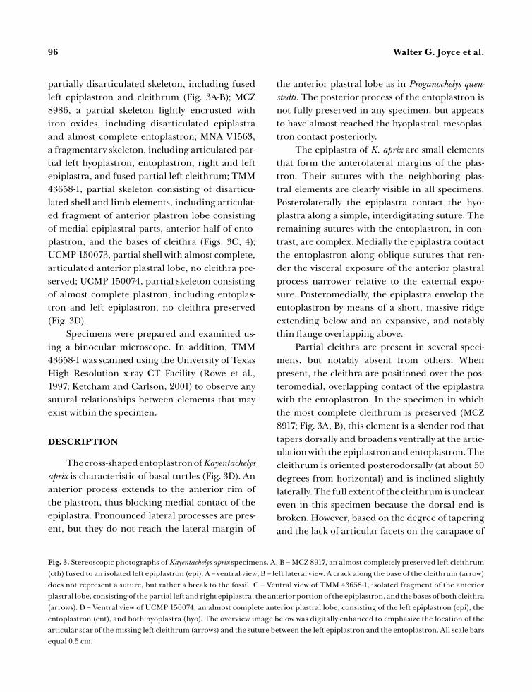
96 Walter G. Joyce et al.
partially disarticulated skeleton, including fused
left epiplastron and cleithrum (Fig. 3A-B); MCZ
8986, a partial skeleton lightly encrusted with
iron oxides, including disarticulated epiplastra
and almost complete entoplastron; MNA V1563,
a fragmentary skeleton, including articulated par-
tial left hyoplastron, entoplastron, right and left
epiplastra, and fused partial left cleithrum; TMM
43658-1, partial skeleton consisting of disarticu-
lated shell and limb elements, including articulat-
ed fragment of anterior plastron lobe consisting
of medial epiplastral parts, anterior half of ento-
plastron, and the bases of cleithra (Figs. 3C, 4);
UCMP 150073, partial shell with almost complete,
articulated anterior plastral lobe, no cleithra pre-
served; UCMP 150074, partial skeleton consisting
of almost complete plastron, including entoplas-
tron and left epiplastron, no cleithra preserved
(Fig. 3D).
Specimens were prepared and examined us-
ing a binocular microscope. In addition, TMM
43658-1 was scanned using the University of Texas
High Resolution x-ray CT Facility (Rowe et al.,
1997; Ketcham and Carlson, 2001) to observe any
sutural relationships between elements that may
exist within the specimen.
DESCRIPTION
The cross-shaped entoplastron of Kayentachelys aprix is characteristic of basal turtles (Fig. 3D). An
anterior process extends to the anterior rim of
the plastron, thus blocking medial contact of the
epiplastra. Pronounced lateral processes are pres-
ent, but they do not reach the lateral margin of
the anterior plastral lobe as in Proganochelys quen-stedti. The posterior process of the entoplastron is
not fully preserved in any specimen, but appears
to have almost reached the hyoplastral–mesoplas-
tron contact posteriorly.
The epiplastra of K. aprix are small elements
that form the anterolateral margins of the plas-
tron. Their sutures with the neighboring plas-
tral elements are clearly visible in all specimens.
Posterolaterally the epiplastra contact the hyo-
plastra along a simple, interdigitating suture. The
remaining sutures with the entoplastron, in con-
trast, are complex. Medially the epiplastra contact
the entoplastron along oblique sutures that ren-
der the visceral exposure of the anterior plastral
process narrower relative to the external expo-
sure. Posteromedially, the epiplastra envelop the
entoplastron by means of a short, massive ridge
extending below and an expansive, and notably
thin fl ange overlapping above.
Partial cleithra are present in several speci-
mens, but notably absent from others. When
present, the cleithra are positioned over the pos-
teromedial, overlapping contact of the epiplastra
with the entoplastron. In the specimen in which
the most complete cleithrum is preserved (MCZ
8917; Fig. 3A, B), this element is a slender rod that
tapers dorsally and broadens ventrally at the artic-
ulation with the epiplastron and entoplastron. The
cleithrum is oriented posterodorsally (at about 50
degrees from horizontal) and is inclined slightly
laterally. The full extent of the cleithrum is unclear
even in this specimen because the dorsal end is
broken. However, based on the degree of tapering
and the lack of articular facets on the carapace of
Fig. 3. Stereoscopic photographs of Kayentachelys aprix specimens. A, B – MCZ 8917, an almost completely preserved left cleithrum
(cth) fused to an isolated left epiplastron (epi): A – ventral view; B – left lateral view. A crack along the base of the cleithrum (arrow)
does not represent a suture, but rather a break to the fossil. C – Ventral view of TMM 43658-1, isolated fragment of the anterior
plastral lobe, consisting of the partial left and right epiplastra, the anterior portion of the epiplastron, and the bases of both cleithra
(arrows). D – Ventral view of UCMP 150074, an almost complete anterior plastral lobe, consisting of the left epiplastron (epi), the
entoplastron (ent), and both hyoplastra (hyo). The overview image below was digitally enhanced to emphasize the location of the
articular scar of the missing left cleithrum (arrows) and the suture between the left epiplastron and the entoplastron. All scale bars
equal 0.5 cm.

97The presence of cleithra in Kayentachelys aprix

98 Walter G. Joyce et al.
all known material of Kayentachelys aprix material,
it appears certain that the cleithrum did not pos-
sess a bony contact with the carapace, as seen in
Proganochelys quenstedti. In cross-section, the base
of the cleithrum has the shape of an acute triangle,
with its apex pointing anteriorly. The posterior,
short side of the triangle bears a shallow groove of
uncertain function. In all specimens in which the
cleithra are preserved, the posterior overlapping
contact of the cleithrum with the entoplastron
is clearly visible, but an anterior suture with the
epiplastra is absent, indicating that the cleithrum
and epiplastra are fully fused with one another in
this area. However, high resolution CT scans of
TMM 43658-1 (Figs. 3C) reveal the persistence of
a dense lamellar zone (arrows, Fig. 4), which repre-
sents the original cortical contact of the cleithrum
with plastral elements. Furthermore, the denser
cleithra are histologically distinct from the under-
lying elements.
The remaining specimens of Kayentachelys aprix, particularly UCMP 150074, provide a fur-
ther perspective on cleithral attachment. In these
specimens, cleithra are completely missing and
the plastron reveals a slightly depressed articu-
lar surface with no signs of breakage (Fig. 3D).
Furthermore, those parts of underlying epiplas-
tron that form much of this surface are fragile, but
intact, indicating that the cleithra disarticulated
easily.
DISCUSSION
The presence of cleithra in primitive turtles
has not always been controversial. Jaekel (1915)
was the fi rst to report well-developed cleithra for
the Upper Triassic turtle Stegochelys dux, a taxon
currently recognized as a junior synonym of
Proganochelys quenstedti (Gaffney, 1990). Although
not specifi cally stated, Jaekel’s identifi cation ap-
pears to have been primarily driven by the topo-
logical position of these structures anterior to the
scapulae and by comparisons with the pectoral
girdle of the primitive tetrapod Archegosaurus dech-eni (Goldfuss, 1847). Unfortunately, the accompa-
nying description is rather short, leaving unclear
whether Jaekel (1915) was aware of the detailed
morphology of these structures, and in particular
the ventral contact of the cleithra with the dorsal
surface of the plastron.
The presence of cleithra was not doubted in
Proganochelys quenstedti for most of the following
century until Gaffney (1990) carried out a com-
prehensive morphological review of this taxon.
Fig. 4. Computer-generated volumetric renderings of the
epiplastron and cleithra in Kayentachelys aprix (TMM 43658-1),
based on high-resolution X-ray CT imagery consisting of
135 horizontal slices gathered at interslice spacings («slice
thickness») of 0.0588 mm. A – horizontal slice, anterior
toward top of image; B – parasagittal section; C – coronal
section. The small arrows indicate the lamellar bone that
separate the cleithra from the plastron.

99The presence of cleithra in Kayentachelys aprix
Although Gaffney originally believed that cleithra
were present in P. quenstedti, he ultimately conclud-
ed that the structures were instead outgrowths of
the epiplastra. An explicit rational was not provid-
ed. However, given that well-developed ascending
processes of the clavicle occur in many primitive
tetrapods and that the clavicle is homologous with
the epiplastron, an interpretation of these struc-
tures as processes of the epiplastron appears plau-
sible.
A third possibility was put forward by Lee
(1996) who suggested that the vertical anterior
projections of the plastron of primitive turtles
represent the clavicles and that the epiplastra
are separate bones that must be considered neo-
morphs (see Table 1 for comparison with the other
hypotheses). Although an explicit rationale was
not provided, it is noteworthy that Lee’s (1997)
interpretation was formulated with explicit refer-
ence to Gaffney’s (1990) conclusion that cleithra
were absent.
An overwhelming amount of evidence indi-
cates that the epiplastra of extant hard-shelled
turtles derive partially or fully from embryologi-
cal precursors that must be interpreted as clavicles
(e.g., Zangerl, 1939, 1969; Walker, 1947; Williams
and McDowell, 1952; Cherepanov, 1984, 1997,
2005; Rieppel, 1993; Gilbert et al., 2001; Sheil
and Greenbaum, 2005). A crucial point is the de-
velopmental relationship of the processes to the
epiplastra. If the processes are outgrowths of the
epiplastra (i.e., the clavicles), the interpretation of
Gaffney (1990) is validated. However, if the pro-
cesses are proposed to be elements separate from
the epiplastra (Lee, 1996, 1997) and the epiplastra
are indeed formed by clavicular precursors, then
it is logically impossible for the projections to be
clavicles as well. Lee’s homology scheme can thus
be rejected. The two primary competing hypoth-
eses are therefore that these structures are either
cleithra (following Jaekel, 1915) or the ascending
processes of the epiplastra (following Gaffney,
1990).
The hypothesis that these elements are as-
cending clavicular processes would be supported
by the following interpretations or features: 1) they
are true outgrowths of the epiplastron, 2) they are
inseparate from the epiplastron, 3) postmortem
breakage would likely occur along the narrower
part of the process, not between the broad base
and the epiplastron, and 4) the character of the
bone of the process and epiplastron are indistin-
guishable. None of these features or interpreta-
tions is confi rmed by this study. In contrast, the
hypothesis that these elements are cleithra is di-
rectly supported by the observations that 1) the
structures are ontogenetically independent from
the epiplastra (clavicles), 2) even when fused
with the epiplastra, sutural evidence of their own
identity is present during later ontogeny, 3) the
structures separate along the ontogenetic sutural
contacts under postmortem mechanical stress,
and 4) the bone density differs from that of the
bordering epiplastra, and an abrupt transition
in bone density is seen at the cleithral-epiplastral
contact. Inasmuch as both the cleithrum and the
clavicle are dermal elements and positioned ante-
rior to the scapula, no additional information can
be gained from topological arguments.
Although not explicitly stated, Gaffney (1990)
observed some of these features when assessing the
identity of these structures in Proganochelys quenst-
TABLE 1. Comparison of primary homology assessments to the anterior plastral region of primitive turtles.
Jaekel (1915) Gaffney (1990) Lee (1996)
epiplastral process cleithrum clavicle clavicle
epiplastron clavicle clavicle neomorph
entoplastron interclavicle interclavicle interclavicle

100 Walter G. Joyce et al.
edti (Gaffney, pers. comm. WGJ, 2003). Gaffney’s
careful external analysis of all available P. quen-stedti material did not reveal any sutures along
the base of the processes and he consequently
concluded that they were part of the epiplastra.
A similar observation can be made for about half
of the available material for Kayentachelys aprix. In
addition, in the same specimens of K. aprix and
all available specimens of P. quenstedti, the process
tends to break well above its base, indicating a
fi rm connecting between the process and the epi-
plastron.
Although all of the material of Proganochelys quenstedti and some specimens of Kayentachelys aprix suggest that the processes represent out-
growths of the epiplastra, the remaining K. aprix
material provides multiple lines of evidence that
the processes should be regarded as independent
structures. We conclude that the structures in
question are cleithra that initially are independent
but fuse with the epiplastra during later ontogeny.
Unfortunately, the available K. aprix material does
not allow an assessment of the proposed ontoge-
netic fusion, because all specimens in this study
are approximately equal in size. Modest size dif-
ferences do exist, but these cannot be quantifi ed
rigorously because no two specimens possess com-
parable landmarks for measurement.
Considering that equivalent structures
can be found in other primitive turtles, such as
Proterochersis robusta and Palaeochersis talampayen-sis, our fi nding fi rmly establishes the presence
of cleithra in primitive turtles and indicates that
cleithra were lost in the turtle lineage indepen-
dently of the loss seen in the mammal stem lineage
and in saurian reptiles (Matsuoka et al., 2005).
The presence of cleithra in primitive tur-
tles has intriguing phylogenetic implications.
Placement of turtles within crown Sauria, a clade
that demonstrably lacks cleithra, requires the un-
parsimonious conclusion that cleithra reappeared
along the phylogenetic stem of turtles, only to be
subsequently lost again. An ad hoc hypothesis that
these structures should not be considered to have
been derived from girdle elements at all, but rath-
er are neomorphs, does not make a difference,
because the acquisition of neomorphs would also
add an additional step. Naturally, the presence of
cleithra (or equivalent neomorphs) in turtles is a
just a single character that may be rendered homo-
plastic by a comprehensive phylogenetic analysis.
The presence of cleithra in the primitive tur-
tle Kayentachelys aprix has interesting implications
regarding basal turtle evolution as well. When fi rst
described, this taxon was assessed to be the most
basal known representative of the cryptodiran
stem lineage (Gaffney et al., 1987). Considering
the absence of cleithra in all living pleurodires and
cryptodires, this would imply the loss of cleithra in
both lineages. A recent, comprehensive analysis of
basal turtles relationships, however, that used 136
osteological characters with 171 derived character
states for 45 fossil and 22 living species of turtles
(Joyce, In Press) placed K. aprix along the phylo-
genetic stem of crown turtles. Despite this reas-
sessment of the placement of K. aprix, this analysis
does not reveal the loss of cleithra to be an un-
ambiguous synapomorphy of crown Testudines.
Instead, the presence of remnant cleithra in the
unambiguous stem cryptodire taxa Baenidae and
Xinjiangchelys latimarginalis indicates that these
structures were lost independently along the stem
lineages of crown Pleurodira and Cryptodira.
Cleithra were thus lost at least twice in turtles
(Fig. 5).
CONCLUSIONS
A morphological review of all Kayentachelys aprix material reveals that this taxon possesses
cleithra. In addition to topological arguments,
this conclusion is primarily supported by observa-
tions that demonstrate this element to be develop-
mentally independent from the epiplastron and
hence not an outgrowth thereof. The equivalently
placed «epiplastral processes» of other primitive

101The presence of cleithra in Kayentachelys aprix
turtles are reinterpreted as cleithra that are fused
with the epiplastra during ontogeny. The pres-
ence of cleithra in primitive turtles is a character
that supports a basal placement of turtles within
Reptilia, rather than a placement within crown
Sauria, a clade that demonstrably lacks cleithra,
which would imply the reformation and subse-
quent loss of cleithra along their phylogenetic
stem. Finally, regardless of the phylogenetic posi-
tion of turtles within Amniota, fossil evidence re-
veals that cleithra were lost twice within the turtle
crown group.
Acknowledgements. We would like to thank
the Museum of Northern Arizona, the University of
California Museum of Paleontology, and the Staatliches
Museum für Naturkunde Stuttgart for providing us ac-
cess to specimens relevant to this study. Igor Danilov
and Randall Irmis provided thoughtful comments
that signifi cantly improved the quality of this manu-
script. We gratefully acknowledge Ahktar Zaman and
the Navajo Nation for access to conduct paleontologi-
cal studies. This study was supported by the National
Science Foundation (IIS-0208675).
REFERENCES
Baur G. (1887), «Ueber den Ursprung der Extremitaeten
der Ichthyopterygia,» Ber. Versamml. Oberrhein. Geol.
Ver., 20,17-20.
Bojanus L. H. (1819), Anatome Testudinis europaeae,
Josephus Zawadzkus, Vilna.
Carroll R. L. (1988), Vertebrate Paleontology and Evolution,
Freeman and Company, New York.
Cherepanov G. O. (1984), «On the nature of the anteri-
or elements of the plastron in turtles,» Zoologicheskii
Zhurnal, 63, 1529-1534. [in Russian with English
abstract]
Cherepanov G. O. (1997), «The origin of the bony shell
of turtles as a unique evolutionary model in rep-
tiles,» Russ. J. Herpetol., 4, 155-162.
Cherepanov G. O. (2005), «Morphogenesis of the
bony shell and a problem of Testudinata origin,»
Zoologicheskii Zhurnal, 84, 464-475. [in Russian
with English abstract]
Clack J. E. (2002), Gaining Ground: The Origin and
Evolution of Tetrapods. Indiana University Press,
Bloomington.
Clark J. M. and Fastovsky D. E. (1986), «Vertebrate bio-
stratigraphy of the Glen Canyon Group in north-
ern Arizona,» in: Padian K. (ed.), The Beginning
of the Age of Dinosaurs, Cambridge Univ. Press,
Cambridge, pp. 285-301.
Coates M. I., Jeffery J. E. and Ruta M. (2002), «Fins
to limbs: What the fossils say,» Evol. Devel., 4,
390-401.
Cope E. D. (1877), «On reptilian remains from the
Dakota Beds of Colorado,» Proc. Amer. Philos. Soc.,
17, 193-196.
deBraga M. and Rieppel O. (1997), «Reptile phylog-
eny and the interrelationships of turtles,» Zool. J.
Linn. Soc., 120, 281-354.
Duellman W. E. and Trueb L. (1986), Biology of
Amphibians, McGraw-Hill, New York.
Fig. 5. A phylogenetic hypothesis of basal turtles revealing
the independent loss of cleithra along the phylogenetic stem
of both crown Pleurodira and Cryptodira.

102 Walter G. Joyce et al.
Fraas E. (1913), «Proterochersis, eine pleurodire
Schildkroete aus dem Keuper,» Jahr. Ver. Vaterl.
Naturkd. Württ., 69, 13-90.
Gaffney E. S. (1990), «The comparative osteology of
the Triassic turtle Proganochelys,» Bull. Am. Mus.
Nat. Hist., 194, 1-263.
Gaffney E. S., Hutchison J. H. Jenkins F. A. and
Meeker L. J. (1987), «Modern turtle origins: The
oldest known cryptodire,» Science, 237, 289-291.
Gaffney E. S., Meylan P. A. and Wyss A. R. (1991),
«A computer assisted analysis of the relationships
of the higher categories of turtles,» Cladistics, 7,
313-335.
Gauthier J., Kluge A. G. and Rowe T. (1988), «Amniote
phylogeny and the importance of fossils,» Cladistics,
4, 105-209.
Gilbert S. F., Loredo G. A., Brukman A. and Burke
A. C. (2001), «Morphogenesis of the turtle shell:
the development of a novel structure in tetrapod
evolution, » Evol. Devel., 3, 47-58.
Goldfuss G. A. (1847), «Über das älteste der mit
Bestimmtheit erkannten Reptilien, einen
Krokodilier, und einige neue fossile Fische aus der
Steinkohlen-Formation,» N. Jb. Mineral. Geognos.
Geol., 1847, 400-404.
Jaekel O. (1915), «Die Wirbeltierfunde aus dem Keuper
von Halberstadt. Serie II. Testudinata. Teil 1.
Stegochelys dux n. g. n. sp.,» Palaeontol. Zeitsch., 2,
88-214.
Jarvik E. (1996), «The Devonian tetrapod Ichthyostega,»
Fossils and Strata, 40, 1 – 213.
Joyce W. G. (In Press), «Phylogenetic Relationships of
Basal Turtles,» Bull. Peabody Mus. Nat. Hist.
Ketcham R. A. and Carlson W. D. (2001), «Acquisition,
optimization and interpretation of X-ray comput-
ed tomographic imagery: applications to the geo-
sciences.» Comput. Geosci., 27, 381-400.
Laurin M. and Reisz R. R. (1995), «A reevaluation of
early amniote phylogeny,» Zool. J. Linn. Soc., 113,
165-223.
Lee M. S. Y. (1996), «The homologies and early evolu-
tion of the shoulder girdle in turtles,» Proc. R. Soc.
Lond. B, 263, 111-117.
Lee M. S. Y. (1997), «Pareiasaur phylogeny and the ori-
gin of turtles,» Zool. J. Linn. Soc., 120, 197–280.
Khosatzky L. I. (1997), «Big turtle of the late Cretaceous
of Mongolia,» Russ. J. Herpetol., 4, 148-154.
Kumazawa Y. and Nishida M. (1999), «Complete mito-
chondrial DNA sequences of the green turtle and
blue-tailed mole skink: Statistical evidence for ar-
chosaurian affi nity of turtles,» Mol. Biol. Evol., 16,
784-792.
Matsuoka T., Ahlberg P. E., Kessaris N., Iannarelli
P., Dennehy U., Richardson W. D., McMahon A.
P. and Koentges G. (2005), «Neural crest origins
of the neck and shoulder,» Nature, 436, 347-355.
Owen R. (1886), «Description of fossil remains of two
species of Megalania genus (Meiolania, Ow) from
Howe’s Island,» Proc. R. Soc. Lond., 40, 315.
Rieppel O. (1993), «Studies on skeleton formation
in reptiles: Patterns of ossifi cation in the skel-
eton of Chelydra serpentina (Reptilia, Testudines),»
J. Zool., 231, 487-509.
Romer A. S. (1956), Osteology of the Reptiles. Univ. of
Chicago Press, Chicago.
Rowe T., Kappelman J., Carlson W. D., Ketcham
R. A. and Denison, C. (1997), «High-Resolution
Computed Tomography: a breakthrough technol-
ogy for Earth scientists,» Geotimes, 42, 23-27.
Rougier G. W., Fuente M. S. de la and Arcucci A. B.
(1995), «Late Triassic turtles from South America,»
Science, 268, 855-858.
Sheil C. A. and Greenbaum E. (2005), «Reconsidera-
tion of skeletal development of Chelydra serpenti-
na (Reptilia: Testudinata: Chelydridae): Evidence
for intraspecifi c variation, » J. Zool., 265, 235-267.
Walker W. F. (1947), «The development of the shoulder
region of the turtle, Chrysemys picta marginata, with
special reference to the primary musculature,»
J. Morphol., 80, 195-249.
Williams E. E. and McDowell S. B. (1952), «The
plastron of soft-shelled turtles (Testudinata,
Trionychidae): A new interpretation,» J. Morphol.,
90, 263-279.

103The presence of cleithra in Kayentachelys aprix
Young C.-C. and Chow M.-C. (1953), «New fossil rep-
tiles from Szechuan China,» Acta Sci. Sinica, 2, 216-
243.
Zangerl R. (1939), «The homology of the shell elements
in turtles,» J. Morph., 65, 383-409.
Zangerl R. (1969), «The turtle shell,» in: Gans C.,
Bellairs A. and Parsons T. S. (eds.), The Biology of
the Reptilia, Volume 1, Academic Press, London, pp.
311-339
Zardoya R. and Meyer A. (2000), «Mitochondrial
evidence on the phylogenetic position of caeci-
lians (Amphibia: Gymnophiona),» Genetics, 155,
765-775.