fothy1 - clsgmbh.de · start the stretching process in fothy1 for the intended time period. ......
TRANSCRIPT
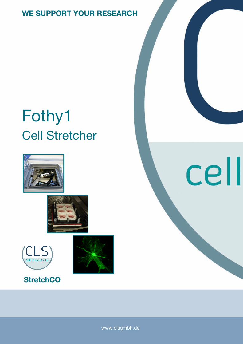
Fothy1Cell Stretcher
www.clsgmbh.de
StretchCO
WE SUPPORT YOUR RESEARCH
Page 2
Scientific BackgroundWith the development of special devices for studying the effect of mechanical force on cultured cells, it hasbecome clear that mechanical forces modulate important cell functions. These include differentiation andproliferation of cells, matrix and autacoid production, second messenger levels, and enzyme activities. Thecytoskeleton plays a key role not only in providing mechanical stability to increased load, but also in thesignal transduction of mechanical force. Such mechanical forces are applied by stretching the cells undervarious conditions according to the cellular properties.
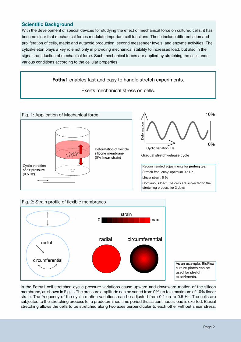
Cyclic variation of air pressure (0.5 Hz)
Deformation of flexible silicone membrane(5% linear strain)
Recommended adjustments for podocytes:Stretch frequency: optimum 0.5 HzLinear strain: 5 %Continuous load: The cells are subjected to thestretching process for 3 days.
radial circumferential
max0strain
radial
circumferential
Fig. 2: Strain profile of flexible membranes
Fothy1 enables fast and easy to handle stretch experiments.
Exerts mechanical stress on cells.
Fig. 1: Application of Mechanical force
In the Fothy1 cell stretcher, cyclic pressure variations cause upward and downward motion of the siliconmembrane, as shown in Fig. 1. The pressure amplitude can be varied from 0% up to a maximum of 10% linearstrain. The frequency of the cyclic motion variations can be adjusted from 0.1 up to 0.5 Hz. The cells aresubjected to the stretching process for a predetermined time period thus a continuous load is exerted. Biaxialstretching allows the cells to be stretched along two axes perpendicular to each other without shear stress.
10%
0%De
form
ation
Cyclic variation, Hz
Gradual stretch-release cycle
As an example, BioFlexculture plates can beused for stretchexperiments.
Page 3
Ordering information
Name of product / Product designation SizeCat. no.
Integral partsUnitFothy1 cell stretching device, automated complete system977021
977021-1 UnitFO1H Pressure chamber977021-2 UnitCX5 Manifold977021-3 UnitGasket, tubingSpare parts are available via CLS GmbH.
Due to the robustness and ease of technical set up the Fothy1 is very reliable,experiments are highly reproductive in terms of physical properties.
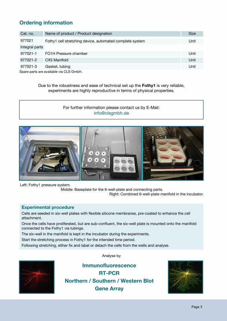
Experimental procedureCells are seeded in six-well plates with flexible silicone membranes, pre-coated to enhance the cellattachment.Once the cells have proliferated, but are sub-confluent, the six-well plate is mounted onto the manifoldconnected to the Fothy1 via tubings.The six-well in the manifold is kept in the incubator during the experiments.Start the stretching process in Fothy1 for the intended time period.Following stretching, either fix and label or detach the cells from the wells and analyse.
ImmunofluorescenceRT-PCR
Northern / Southern / Western BlotGene Array
For further information please contact us by E-Mail:[email protected]
Analyse by
Left: Fothy1 pressure system.Middle: Baseplate for the 6-well-plate and connecting parts.
Right: Combined 6-well-plate manifold in the incubator.
Page 4
Browse our website for more cell lines which may be of interest for your stretching experiments:www.clsgmbh.de
human cell lines - animal cell lines - primary cells - iPS cells
Mechanical stress can be applied onmany different cells of various tissue origins, e.g. kidney, skin, soft tissues,ligament, muscle, lung, bone, and many more.In HUVEC cells, mechanical stretch may promote inflammation (Jufri et al. 2015). Linke (2007) describes theinfluence of mechanical stress signals on cardiomyocytes resulting in changes of the contractile performanceand gene expression.
860015 500 mlPBS without calcium / magnesium100 mlAccutase detachment solution830100
Cell culture Supplements
840903-50 50 mlFetal Bovine Serum (FBS)12.5 mlhuman Platelet lysate, for your xenofree cell culture860065
Cells for StretchingName of product / Product designation SizeCat. no.
400494 cryovialE11 Murine podocyte cell line500 mlRPMI 1640 cell culture medium, ready-to-use, contains 10% FBS820700
300605 cryovialHUVEC Endothelial cells500 mlECGM, Endothelial cell growth medium, ready-to-use, with supplements820731
300703 cryovialhGF, human Gingival Fibroblasts500 mlDMEM:Ham's F12 (1:1, vol:vol), serum-free and xeno-free, with supplement820408-hpl
300493 cryovialHaCaT, human keratinocyte cell line500 mlDMEM, high glucose820300
400476 cryovialC2C12, murine muscle cell line500 mlRPMI 1640 cell culture medium, ready-to-use, contains 10% FBS820700
400101 cryovialNIH:3T3, Renal cell line500 mlDMEM:Ham's F12 (1:1, vol:vol), ready-to-use, contains 5% FBS820400
607264 cryovialLLC-PK1, Swine Renal carcinoma cell line500 mlDMEM:Ham's F12 (1:1, vol:vol), ready-to-use, contains 5% FBS820400
300238 cryovialRCC-FG2, Renal clear cell carcinoma cell line500 mlRPMI 1640 cell culture medium, serum-free and xeno-free, with supplement820708-hPL
300149 cryovialCaKi-1, Renal carcinoma cell line500 mlEMEM cell culture medium, serum-free and xeno-free, with supplement820108-hPL
For your concenience, the cell culture media recommended for each of the cell lines is indicated beyond each one.
cryovialSVI Murine podocyte cell line400495500 mlRPMI 1640 cell culture medium, ready-to-use, contains 10% FBS820700
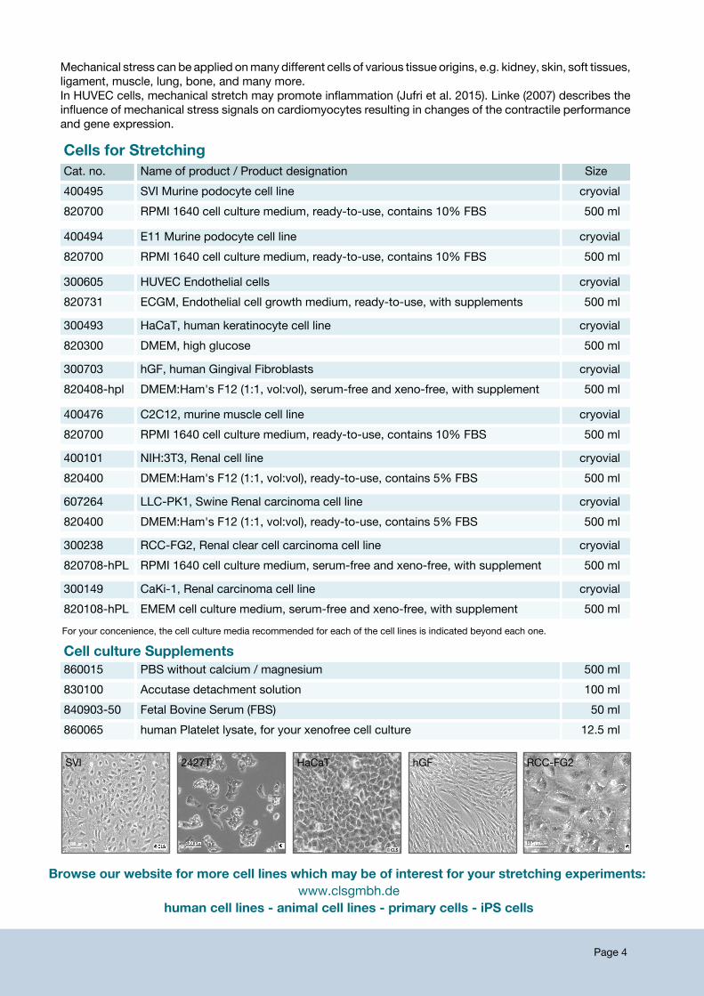
SVI 2427T HaCaT hGF RCC-FG2
Page 5
Applications (using the cell stretching device developed by StretchCo)
ReferencesEndlich N and Endlich K. The challenge and response of podocytes to glomerular hypertension. SeminNephrol. 2012 32(4):327-41. doi: 10.1016/j.semnephrol.2012.06.004.Jufri NF et al. Mechanical stretch: physiological and pathological implications for human vascularendothelial cells. Vascular Cell 7: 8, 2015.Linke WA. Sense and stretchability: The role of titin and titin-associated proteins in myocardial stress-sensing and mechanical dysfunction. Cardiovasc.Res. 77: 637-648, 2008.
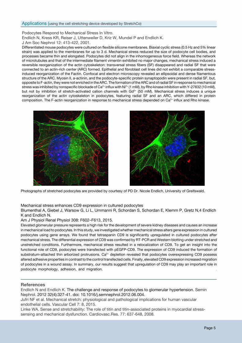
Podocytes Respond to Mechanical Stress In Vitro.Endlich N, Kress KR, Reiser J, Uttenweiler D, Kriz W, Mundel P and Endlich K.J Am Soc Nephrol 12: 413-422, 2001.Differentiated mouse podocytes were cultured on flexible silicone membranes. Biaxial cyclic stress (0.5 Hz and 5% linearstrain) was applied to the membranes for up to 3 d. Mechanical stress reduced the size of podocyte cell bodies, andprocesses became thin and elongated. Podocytes did not align in the inhomogeneous force field. Whereas the networkof microtubules and that of the intermediate filament vimentin exhibited no major changes, mechanical stress induced areversible reorganization of the actin cytoskeleton: transversal stress fibers (SF) disappeared and radial SF that wereconnected to an actin-rich center (ARC) formed. Epithelial and fibroblast cell lines did not exhibit a comparable stress-induced reorganization of the Factin. Confocal and electron microscopy revealed an ellipsoidal and dense filamentousstructure of the ARC. Myosin II, a-actinin, and the podocyte-specific protein synaptopodin were present in radial SF, but,opposite toF-actin, theywerenot enriched in theARC.The formationof theARCandof radial SF in response tomechanicalstresswas inhibited by nonspecific blockade ofCa2+ influxwithNi2+ (1mM), byRho kinase inhibitionwith Y-27632 (10mM),but not by inhibition of stretch-activated cation channels with Gd3+ (50 mM). Mechanical stress induces a uniquereorganization of the actin cytoskeleton in podocytes, featuring radial SF and an ARC, which differed in proteincomposition. The F-actin reorganization in response to mechanical stress depended on Ca2+ influx and Rho kinase.
Mechanical stress enhances CD9 expression in cultured podocytesBlumenthal A, Giebel J, Warsow G, Li L, Ummanni R, Schordan S, Schordan E, Klemm P, Gretz N,4 EndlichK and Endlich N.Am J Physiol Renal Physiol 308: F602–F613, 2015.Elevated glomerular pressure represents a high risk for the development of severe kidney diseases and causes an increaseinmechanical load topodocytes. In this study,we investigatedwhethermechanical stress alters geneexpression in culturedpodocytes using gene arrays. We found that tetraspanin CD9 is significantly upregulated in cultured podocytes aftermechanical stress. The differential expression of CD9was confirmed by RT-PCR andWestern blotting under stretched andunstretched conditions. Furthermore, mechanical stress resulted in a relocalization of CD9. To get an insight into thefunctional role of CD9, podocytes were transfected with pEGFP-CD9. The expression of CD9 induced the formation ofsubstratum-attached thin arborized protrusions. Ca2+ depletion revealed that podocytes overexpressing CD9 possessaltered adhesive properties in contrast to the control transfected cells. Finally, elevatedCD9expression increasedmigrationof podocytes in a wound assay. In summary, our results suggest that upregulation of CD9 may play an important role inpodocyte morphology, adhesion, and migration. .
Photographs of stretched podocytes are provided by courtesy of PD Dr. Nicole Endlich, University of Greifswald.
Certified ISO 9001:2008
CLS Cell Lines Service GmbHDr.Eckener-Strasse 869214 EppelheimGermanyPhone: +49(0)6221 700799Fax: +49(0)6221 [email protected]
C U L T U R E - C O N T R O L - C O N S E R V E
Murine podocytecell line SVI