fournier gangrene
TRANSCRIPT
Hindawi Publishing CorporationCase Reports in Emergency MedicineVolume 2012, Article ID 984195, 4 pagesdoi:10.1155/2012/984195
Case Report
Fournier’s Gangrene: Report of 2 Cases
Prasan Kumar Hota
Department of Surgery, Institute of Medical Sciences & SUM Hospital, SOA University, K-8, Kalinga Nagar,Orissa Bhubaneswar 751030, India
Correspondence should be addressed to Prasan Kumar Hota, [email protected]
Received 22 August 2011; Accepted 26 September 2011
Academic Editors: D. Elkharrat, P. Iannone, and W. Mauritz
Copyright © 2012 Prasan Kumar Hota. This is an open access article distributed under the Creative Commons Attribution License,which permits unrestricted use, distribution, and reproduction in any medium, provided the original work is properly cited.
Fournier’s gangrene is a very serious surgical emergency seen all over the world. With the newer advancement of surgical techniquesand critical care medicine, the mortality and morbidity of this disease has come down significantly over a period of time.An early diagnosis including evaluation of predisposing and etiological factors, metabolic and physiological parameters withprompt resuscitation, aggressive surgical debridement, broad-spectrum antibiotic coverage, and continuous monitoring of all theparameters is essential for a good outcome, therefore reducing the high mortality and morbidity of this condition. In this study,we report 2 different cases of Fournier gangrene. Our first case was a young, nondiabetic, and without any multiorgan failure, whowas managed successfully with good outcome. The second case was a 67-year-old man with diabetes and multiorgan dysfunctionwith extensive gangrene at presentation, who recovered well, but with a stormy postoperative period.
1. Introduction
Fournier’s gangrene (FG) is a serious surgical emergency.This clinical condition was first described by Jean AlfredFournier (1832–1914), a dermatologist and venereologist. Hehad first described this condition in 5 young male patients,who had presented with a rapidly progressing fulminatinginfection of the superficial tissues of scrotum and peniswithout any definite etiological factor [1, 2]. It is interestingto note that Bauriene in 1764 had described such a caseof scrotal gangrene due to traumatic injury from the hornof an ox, which was treated by multiple sittings of surgicaldebridement [3]. Over a period of time, the definition of FGwas broadened to have all the necrotizing infections of thegenitalia.
At present, FG is recognized as a subclassification of ne-crotizing fasciitis. Hence, FG is described as necrotizing softtissue infections originating from or limited to the genitaliaor perineum irrespective of sex. We report 2 cases of FG,presenting in two different scenarios and their outcome.
2. Case Presentation
2.1. Case 1. -year-old male patient reported to the hospitalwith complaints of painful swelling of the scrotum for 3 days
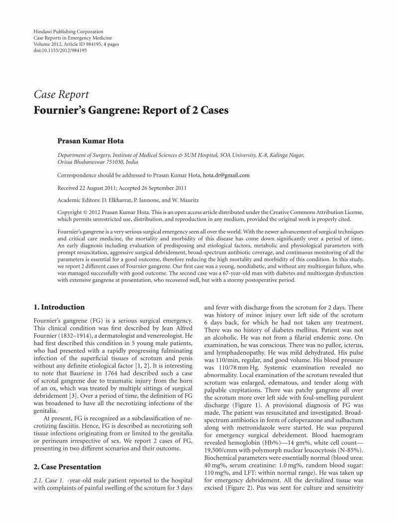
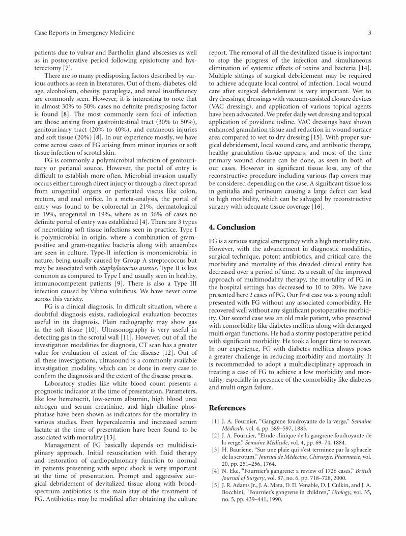
and fever with discharge from the scrotum for 2 days. Therewas history of minor injury over left side of the scrotum6 days back, for which he had not taken any treatment.There was no history of diabetes mellitus. Patient was notan alcoholic. He was not from a filarial endemic zone. Onexamination, he was conscious. There was no pallor, icterus,and lymphadenopathy. He was mild dehydrated. His pulsewas 110/min, regular, and good volume. His blood pressurewas 110/78 mm Hg. Systemic examination revealed noabnormality. Local examination of the scrotum revealed thatscrotum was enlarged, edematous, and tender along withpalpable crepitations. There was patchy gangrene all overthe scrotum more over left side with foul-smelling purulentdischarge (Figure 1). A provisional diagnosis of FG wasmade. The patient was resuscitated and investigated. Broad-spectrum antibiotics in form of cefoperazone and sulbactumalong with metronidazole were started. He was preparedfor emergency surgical debridement. Blood haemogramrevealed hemoglobin (Hb%)—14 gm%, white cell count—19,500/cmm with polymorph nuclear leucocytosis (N-85%).Biochemical parameters were essentially normal (blood urea:40 mg%, serum creatinine: 1.0 mg%, random blood sugar:110 mg%, and LFT: within normal range). He was taken upfor emergency debridement. All the devitalized tissue wasexcised (Figure 2). Pus was sent for culture and sensitivity

2 Case Reports in Emergency Medicine
Figure 1: Fournier’s gangrene.
Figure 2: FG after debridement.
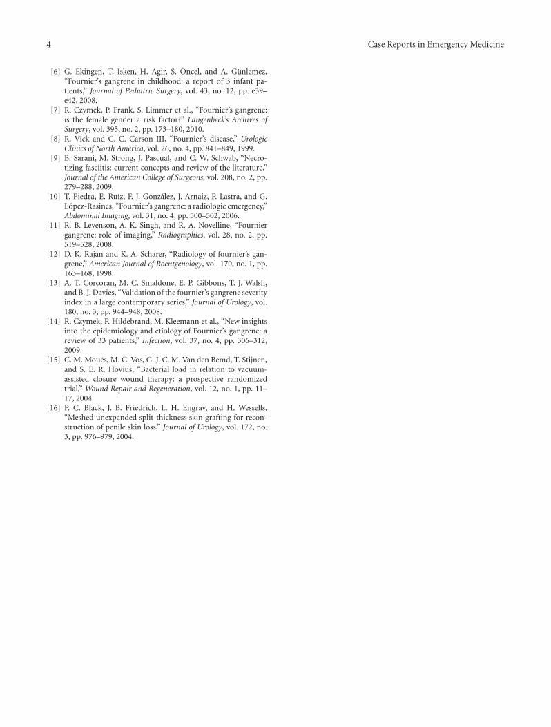
test. Postoperatively patient was managed with broad-spectrum antibiotics and wet dressing. Culture revealedStaph.aureus and E. coli sensitive to cefoperazone and sul-bactum. He responded to the treatment very well (Figure 3).Regular wet dressing was done along with topical applicationof povidone iodine. On 10th postoperative day, his woundwas reconstructed with secondary suturing (Figure 4). Hewas discharged on 22nd postoperative day. A review after sixweeks revealed the patient to be symptom free.
2.2. Case 2. Our second case was a 67-year-old male patient,a known diabetic on irregular treatment, reported withcomplaints of swelling, pain, and foul smelling dischargefrom the scrotum of 7 days duration. For this complaint,he was treated in a peripheral hospital, where his conditionwas deteriorated, for which he was brought to our hospital.On evaluation, he was found to have pallor, mild icterus,and moderate dehydration. His general condition was poor.His pulse was 130/min. Blood pressure was 100/50 mm Hg.He had tachypnoea. He was confused. Systemic examina-tion revealed no clinical abnormality. Local examinationrevealed that his scrotum was grossly edematous withmultiple discharging gangrenous patches all over. Scro-tum was tender with palpable crepitations all over. Thepatient was vigorously resuscitated and investigated. Broad-spectrum antibiotics in form of cefoperazone and sulbactumalong with clindamycin were started. An indwelling Foley’scatheterization was done, and only 50 mL of concentratedurine was drained. History revealed that he had passedonly 350 mL of high colored urine during previous 24hours. Blood haemogram revealed hemoglobin (Hb%):10 gm%, white cell count: 25,300/cmm with polymorph
Figure 3: 8th postop day.
Figure 4: postreconstruction.
nuclear leucocytosis (N-90%). Biochemical parameters werederanged (blood urea: 65 mg%, serum creatinine: 2.5 mg%,random blood sugar: 450 mg%, LFT: serum billirubin:2.8 mg%, AST: 75 IU/L, ALT: 65 IU/L, prothrombin time,and serum electrolyte—within normal range). ECG and X-ray chest revealed no abnormality. With the consultation ofthe endocrinologist, his high blood sugar was taken care of.He was taken up for emergency surgical debridement. Pusculture and sensitivity were asked for. Culture report showedE. coli and Staph. aureus sensitive to cefoperazone and sulbac-tum, cefotaxime and ciprofloxacin. Same antibiotic regimewas continued. He had a stormy postoperative period. Hedeveloped pneumonia, for which he was adequately treatedby the physician. His urine output became normal by 2ndpost op day. By 5th post op day, his blood urea and serumcreatinine came within normal range. His jaundice subsided,and by 7th postop day his liver function tests became normal.Blood sugar level was well controlled by insulin therapy.He required daily wet dressing and two more sittings ofdebridement. Healthy tissue was visible by 13th postop day.On 15th postop day, wound was reconstructed by secondarysuturing. He was discharged from the hospital on 25th post-op day.
3. Discussion
FG is a serious surgical problem with high mortality andmorbidity. Though there is a male predominance [4], thiscondition has been described in children also [5, 6]. Thoughwe have not come across a single case in females, a recentpublication shows a high incidence of 31.6% in female
Case Reports in Emergency Medicine 3
patients due to vulvar and Bartholin gland abscesses as wellas in postoperative period following episiotomy and hys-terectomy [7].
There are so many predisposing factors described by var-ious authors as seen in literatures. Out of them, diabetes, oldage, alcoholism, obesity, paraplegia, and renal insufficiencyare commonly seen. However, it is interesting to note thatin almost 30% to 50% cases no definite predisposing factoris found [8]. The most commonly seen foci of infectionare those arising from gastrointestinal tract (30% to 50%),genitourinary tract (20% to 40%), and cutaneous injuriesand soft tissue (20%) [8]. In our experience mostly, we havecome across cases of FG arising from minor injuries or softtissue infection of scrotal skin.
FG is commonly a polymicrobial infection of genitouri-nary or perianal source. However, the portal of entry isdifficult to establish more often. Microbial invasion usuallyoccurs either through direct injury or through a direct spreadfrom urogenital organs or perforated viscus like colon,rectum, and anal orifice. In a meta-analysis, the portal ofentry was found to be colorectal in 21%, dermatologicalin 19%, urogenital in 19%, where as in 36% of cases nodefinite portal of entry was established [4]. There are 3 typesof necrotizing soft tissue infections seen in practice. Type Iis polymicrobial in origin, where a combination of gram-positive and gram-negative bacteria along with anaerobesare seen in culture. Type-II infection is monomicrobial innature, being usually caused by Group A streptococcus butmay be associated with Staphylococcus aureus. Type II is lesscommon as compared to Type I and usually seen in healthy,immunocompetent patients [9]. There is also a Type IIIinfection caused by Vibrio vulnificus. We have never comeacross this variety.
FG is a clinical diagnosis. In difficult situation, where adoubtful diagnosis exists, radiological evaluation becomesuseful in its diagnosis. Plain radiography may show gasin the soft tissue [10]. Ultrasonography is very useful indetecting gas in the scrotal wall [11]. However, out of all theinvestigation modalities for diagnosis, CT scan has a greatervalue for evaluation of extent of the disease [12]. Out ofall these investigations, ultrasound is a commonly availableinvestigation modality, which can be done in every case toconfirm the diagnosis and the extent of the disease process.
Laboratory studies like white blood count presents aprognostic indicator at the time of presentation. Parameters,like low hematocrit, low-serum albumin, high blood ureanitrogen and serum creatinine, and high alkaline phos-phatase have been shown as indicators for the mortality invarious studies. Even hypercalcemia and increased serumlactate at the time of presentation have been found to beassociated with mortality [13].
Management of FG basically depends on multidisci-plinary approach. Initial resuscitation with fluid therapyand restoration of cardiopulmonary function to normalin patients presenting with septic shock is very importantat the time of presentation. Prompt and aggressive sur-gical debridement of devitalized tissue along with broad-spectrum antibiotics is the main stay of the treatment ofFG. Antibiotics may be modified after obtaining the culture
report. The removal of all the devitalized tissue is importantto stop the progress of the infection and simultaneouselimination of systemic effects of toxins and bacteria [14].Multiple sittings of surgical debridement may be requiredto achieve adequate local control of infection. Local woundcare after surgical debridement is very important. Wet todry dressings, dressings with vacuum-assisted closure devices(VAC dressing), and application of various topical agentshave been advocated. We prefer daily wet dressing and topicalapplication of povidone iodine. VAC dressings have shownenhanced granulation tissue and reduction in wound surfacearea compared to wet to dry dressing [15]. With proper sur-gical debridement, local wound care, and antibiotic therapy,healthy granulation tissue appears, and most of the timeprimary wound closure can be done, as seen in both ofour cases. However in significant tissue loss, any of thereconstructive procedure including various flap covers maybe considered depending on the case. A significant tissue lossin genitalia and perineum causing a large defect can leadto high morbidity, which can be salvaged by reconstructivesurgery with adequate tissue coverage [16].
4. Conclusion
FG is a serious surgical emergency with a high mortality rate.However, with the advancement in diagnostic modalities,surgical technique, potent antibiotics, and critical care, themorbidity and mortality of this dreaded clinical entity hasdecreased over a period of time. As a result of the improvedapproach of multimodality therapy, the mortality of FG inthe hospital settings has decreased to 10 to 20%. We havepresented here 2 cases of FG. Our first case was a young adultpresented with FG without any associated comorbidity. Herecovered well without any significant postoperative morbid-ity. Our second case was an old male patient, who presentedwith comorbidity like diabetes mellitus along with derangedmulti organ functions. He had a stormy postoperative periodwith significant morbidity. He took a longer time to recover.In our experience, FG with diabetes mellitus always posesa greater challenge in reducing morbidity and mortality. Itis recommended to adopt a multidisciplinary approach intreating a case of FG to achieve a low morbidity and mor-tality, especially in presence of the comorbidity like diabetesand multi organ failure.
References
[1] J. A. Fournier, “Gangrene foudroyante de la verge,” SemaineMedicale, vol. 4, pp. 589–597, 1883.
[2] J. A. Fournier, “Etude clinique de la gangrene foudroyante dela verge,” Semaine Medicale, vol. 4, pp. 69–74, 1884.
[3] H. Bauriene, “Sur une plaie qui s’est terminee par la sphacelede la scrotum,” Journal de Medecine, Chirurgie, Pharmacie, vol.20, pp. 251–256, 1764.
[4] N. Eke, “Fournier’s gangrene: a review of 1726 cases,” BritishJournal of Surgery, vol. 87, no. 6, pp. 718–728, 2000.
[5] J. R. Adams Jr., J. A. Mata, D. D. Venable, D. J. Culkin, and J. A.Bocchini, “Fournier’s gangrene in children,” Urology, vol. 35,no. 5, pp. 439–441, 1990.
4 Case Reports in Emergency Medicine
[6] G. Ekingen, T. Isken, H. Agir, S. Oncel, and A. Gunlemez,“Fournier’s gangrene in childhood: a report of 3 infant pa-tients,” Journal of Pediatric Surgery, vol. 43, no. 12, pp. e39–e42, 2008.
[7] R. Czymek, P. Frank, S. Limmer et al., “Fournier’s gangrene:is the female gender a risk factor?” Langenbeck’s Archives ofSurgery, vol. 395, no. 2, pp. 173–180, 2010.
[8] R. Vick and C. C. Carson III, “Fournier’s disease,” UrologicClinics of North America, vol. 26, no. 4, pp. 841–849, 1999.
[9] B. Sarani, M. Strong, J. Pascual, and C. W. Schwab, “Necro-tizing fasciitis: current concepts and review of the literature,”Journal of the American College of Surgeons, vol. 208, no. 2, pp.279–288, 2009.
[10] T. Piedra, E. Ruız, F. J. Gonzalez, J. Arnaiz, P. Lastra, and G.Lopez-Rasines, “Fournier’s gangrene: a radiologic emergency,”Abdominal Imaging, vol. 31, no. 4, pp. 500–502, 2006.
[11] R. B. Levenson, A. K. Singh, and R. A. Novelline, “Fourniergangrene: role of imaging,” Radiographics, vol. 28, no. 2, pp.519–528, 2008.
[12] D. K. Rajan and K. A. Scharer, “Radiology of fournier’s gan-grene,” American Journal of Roentgenology, vol. 170, no. 1, pp.163–168, 1998.
[13] A. T. Corcoran, M. C. Smaldone, E. P. Gibbons, T. J. Walsh,and B. J. Davies, “Validation of the fournier’s gangrene severityindex in a large contemporary series,” Journal of Urology, vol.180, no. 3, pp. 944–948, 2008.
[14] R. Czymek, P. Hildebrand, M. Kleemann et al., “New insightsinto the epidemiology and etiology of Fournier’s gangrene: areview of 33 patients,” Infection, vol. 37, no. 4, pp. 306–312,2009.
[15] C. M. Moues, M. C. Vos, G. J. C. M. Van den Bemd, T. Stijnen,and S. E. R. Hovius, “Bacterial load in relation to vacuum-assisted closure wound therapy: a prospective randomizedtrial,” Wound Repair and Regeneration, vol. 12, no. 1, pp. 11–17, 2004.
[16] P. C. Black, J. B. Friedrich, L. H. Engrav, and H. Wessells,“Meshed unexpanded split-thickness skin grafting for recon-struction of penile skin loss,” Journal of Urology, vol. 172, no.3, pp. 976–979, 2004.

Submit your manuscripts athttp://www.hindawi.com
Hindawi Publishing Corporationhttp://www.hindawi.com Volume 2013
Oxidative Medicine and Cellular Longevity
Hindawi Publishing Corporation http://www.hindawi.com Volume 2013Hindawi Publishing Corporation http://www.hindawi.com Volume 2013
The Scientific World Journal
International Journal of
EndocrinologyHindawi Publishing Corporationhttp://www.hindawi.com
Volume 2013
ISRN Anesthesiology
Hindawi Publishing Corporationhttp://www.hindawi.com Volume 2013
OncologyJournal of
Hindawi Publishing Corporationhttp://www.hindawi.com Volume 2013
PPARRe sea rch
Hindawi Publishing Corporationhttp://www.hindawi.com Volume 2013
OphthalmologyJournal of
Hindawi Publishing Corporationhttp://www.hindawi.com Volume 2013
ISRN AIDS
Hindawi Publishing Corporationhttp://www.hindawi.com Volume 2013
BioMed Research International
Hindawi Publishing Corporationhttp://www.hindawi.com Volume 2013
ObesityJournal of
Hindawi Publishing Corporationhttp://www.hindawi.com Volume 2013
ISRN Addiction
Hindawi Publishing Corporationhttp://www.hindawi.com Volume 2013
Hindawi Publishing Corporationhttp://www.hindawi.com Volume 2013
Computational and Mathematical Methods in Medicine
ISRN Allergy
Hindawi Publishing Corporationhttp://www.hindawi.com Volume 2013
Immunology ResearchHindawi Publishing Corporationhttp://www.hindawi.com Volume 2013
Journal of
Diabetes ResearchJournal of
Hindawi Publishing Corporationhttp://www.hindawi.com Volume 2013
Evidence-Based Complementary and Alternative Medicine
Volume 2013Hindawi Publishing Corporationhttp://www.hindawi.com
Hindawi Publishing Corporationhttp://www.hindawi.com Volume 2013
Gastroenterology Research and Practice
Hindawi Publishing Corporationhttp://www.hindawi.com Volume 2013
ISRN Biomarkers
Hindawi Publishing Corporationhttp://www.hindawi.com Volume 2013
MEDIATORSINFLAMMATION
of