fractional excretion of sodium as an early predictor of
TRANSCRIPT
I
FRACTIONAL EXCRETION OF SODIUM AS AN EARLY PREDICTOR
OF ACUTE KIDNEY INJURY IN TERM NEONATES WITH PERINATAL
ASPHYXIA AT KENYATTA NATIONAL HOSPITAL
A dissertation submitted in part fulfillment of the requirement of the University of Nairobi for the
award of the degree of Master of Medicine in Paediatrics and Child Health.
PRINCIPAL INVESTIGATOR
DR. ESTHER M. NJIRU (MBChB)
H58/75476/14
Department of Pediatrics and Child Health, University of Nairobi

II
DECLARATION
This dissertation is my original work and has not been presented for the award of a degree in any
other university
Signed.........................................................................Date.........................................................
Dr Esther M. Njiru (Principal Investigator)
MbChB Moi University
This dissertation proposal has been presented with full approval of supervisors
Signed.........................................................................Date........................................................
Dr. Bashir Admani,
Senior Lecturer,
Department of Paediatrics and Child Health, University of Nairobi
Signed.........................................................................Date.........................................................
Dr. Polly Okello, (Co – Investigator),Paediatric Nephrologist (KNH)
Signed........................................................................ Date.........................................................
Professor Dalton Wamalwa,
Associate Professor, Department of Paediatrics and Child Health, University of Nairobi
Signed........................................................................ Date.........................................................
Professor Rachel Musoke
Department of Paediatrics and Child Health, University of Nairobi

III
ACKNOWLEDGMENTS
I wish to express my sincere appreciation to:
Almighty God for His guidance throughout this course.
The Paediatrics department ,University of Nairobi. My supervisors for their guidance,
support, patience and valuable comments and criticism throughout the study.
The Department of Research and Programs, Kenyatta National Hospital
All the children and their caregivers for their willingness to participate and patience during
the study period.
My study assistants for working selflessly and helping me throughout the study period.
My husband, Gabriel, and daughter Natasha for their great support and encouragement.

IV
Table of contents
DECLARATION ............................................................................................................................ II
ACKNOWLEDGMENTS ............................................................................................................ III
LIST OF TABLES ........................................................................................................................ VI
LIST OF FIGURES ..................................................................................................................... VII
ABBREVIATIONS ................................................................................................................... VIII
DEFINITION OF TERMS ............................................................................................................ X
ABSTRACT .................................................................................................................................. 11
Introduction ................................................................................................................................... 11
INTRODUCTION ........................................................................................................................ 13
Perinatal asphyxia and Acute Kidney Injury............................................................................. 13
LITERATURE REVIEW ............................................................................................................. 16
Acute Kidney Injury in Perinatal Asphyxia .............................................................................. 16
Prevalence of AKI in asphyxia .............................................................................................. 16
Pathophysiology of AKI ......................................................................................................... 17
Diagnosis of Acute Kidney Injury ............................................................................................ 20
Limitations of serum creatinine ............................................................................................. 21
Novel biomarker .................................................................................................................... 21
FENa: A solution to the problem? ............................................................................................ 22
Validation studies .................................................................................................................. 23
Studies using FENA in asphyxia ............................................................................................ 26
Neonatal Renal Failure: Usefulness of Diagnostic Indices ........................................................... 27
FENa cut off <2.5% pre renal oliguria, >/= 2.5% renal failure ................................................... 27
STUDY JUSTIFICATION ........................................................................................................... 28
RESEARCH QUESTION ............................................................................................................. 29
OBJECTIVES ........................................................................................................................... 29
Primary Objective .................................................................................................................. 29
Specific objectives .................................................................................................................. 29
METHODOLOGY ....................................................................................................................... 30
Study design: ............................................................................................................................. 30
Study area: ................................................................................................................................. 30
Study population: ...................................................................................................................... 30
Study Tool: ................................................................................................................................ 30
Study period .............................................................................................................................. 30
V
Study personnel: ........................................................................................................................ 30
Inclusion criteria:....................................................................................................................... 31
Exclusion criteria: ..................................................................................................................... 31
Sampling technique: .................................................................................................................. 31
Sample size ................................................................................................................................ 32
Recruitment procedure .............................................................................................................. 33
Enrollment of participants............................................................................................................. 33
DATA MANAGEMENT AND ANALYSIS ............................................................................... 35
Table 5: Diagnostic utility of FENa .............................................................................................. 36
ETHICAL CONSIDERATIONS .................................................................................................. 37
RESULTS ..................................................................................................................................... 38
STUDY FLOW DIAGRAM ......................................................................................................... 38
DISCUSSION ............................................................................................................................... 46
STUDY STRENGTHS ................................................................................................................. 49
STUDY LIMITATIONS .............................................................................................................. 49
CONCLUSION ............................................................................................................................. 50
RECOMMENDATIONS .............................................................................................................. 50
REFERENCES ............................................................................................................................. 51
APPENDICES .............................................................................................................................. 54
Appendix 1: Questionnaire........................................................................................................ 54
Appendix II: pRIFLE Classification for AKI ........................................................................... 57
Appendix III: Ballard‟s maturational rating .............................................................................. 58
Appendix IV: Apgar score ........................................................................................................ 59
Appendix V: Definition and staging of perinatal asphyxia. ...................................................... 60
Appendix VI: Standard operating procedures for the measurement of weight, length and head
circumference ............................................................................................................................ 61
Appendix VII: Study budget ..................................................................................................... 62
Appendix VIII: Consent form for parents / guardians of participants ...................................... 63

VI
LIST OF TABLES
Table 1: Oliguric vs. Non oliguric AKI ........................................................................................ 20
Table 2: Interpretation of FENa .................................................................................................... 23
Table 3: Summary of validation studies ....................................................................................... 25
Table 4: Summary of FENa studies in AKI .................................................................................. 27
Table 5: Diagnostic utility of FENa .............................................................................................. 36
Table 6: Characteristics of the neonates on admission ................................................................. 39
Table 7: Characteristics of the mothers ........................................................................................ 40
Table 8: Prevalence of AKI using Creatinine and probable AKI by FENa .................................. 42
Table 9: Diagnostic Utility of FENa ............................................................................................. 42

VII
LIST OF FIGURES
Figure 1 Pathophysiology of Ischemic/ Intrinsic AKI .................................................................. 19
Figure 2: Maternal complications ................................................................................................. 41
Figure 3: HIE stage of the neonates on admission ........................................................................ 41
Fig 4: Graph of sensitivity vs specificity of FENa ....................................................................... 43
Figure 5: Receiver operating characteristic (ROC) curve for Fractional excretion of sodium
(FENa) on day 1 ............................................................................................................................ 44
Figure 6: Correlation between HIE Stage and AKI ...................................................................... 45
Figure 7: Progression of HIE Stage during first three days .......................................................... 45

VIII
ABBREVIATIONS
AKI - Acute kidney injury
AKIN - Acute Kidney Injury Network
ANC- Antenatal Clinic
APGAR - Appearance, Pulse, Grimace, Activity, Respiration
APH – Antepartum Hemorrhage
ARF – Acute Renal Failure
ATN – Acute Tubular Necrosis
ATP – Adenosine TriPhosphate
BP – Blood Pressure
BVM – Bag, valve and mask
Cl – Chloride
C/S – Caesarean Section
CM – Centimeter
DCT – Distal Convoluted Tubule
ECF - Extracellular Fluid
ERC – Ethics and Research Committee
FENa – Fractional Excretion of Sodium
FEUr – Fractional Excretion of Urea
GA – Gestational Age
GFR – Glomerular Filtration Rate
HIE – Hypoxic Ischemic Encephalopathy
KDHS – Kenya Demographics and Health Survey
KNH – Kenyatta National Hospital
MOD – Multi Organ Dysfunction
Na – Sodium
Na/K ATPase – Sodium/Potassium ATPase
NBU – New Born Unit
NGAL – Neutrophil gelatinase- associated lipocalin
NICU – Neonatal Intensive Care Unit
NPV – Negative Predictive Value

IX
PCT – Proximal Convoluted Tubule
PCr – Plasma Creatinine
PNa – Plasma Sodium
PO – Pre renal oliguria
PPV – Positive Predictive Value
PRA – Plasma Renin Activity
pRIFLE – pediatric Risk, Injury, Failure, Loss, End stage renal disease
RBF – Renal Blood Flow
RDS – Respiratory Distress Syndrome
RFI – Renal Failure Index
RRT – Renal replacement therapy
sCr – Serum creatinine
SPSS – Statistical Products and Services Solutions
TAL – Thick Ascending Loop of Henle
UCr – Urine Creatinine
UNa – Urine Sodium
UO – Urine Output
USA – United State of America
V/E – Vacuum Extraction
WHO – World Health Organisation
X
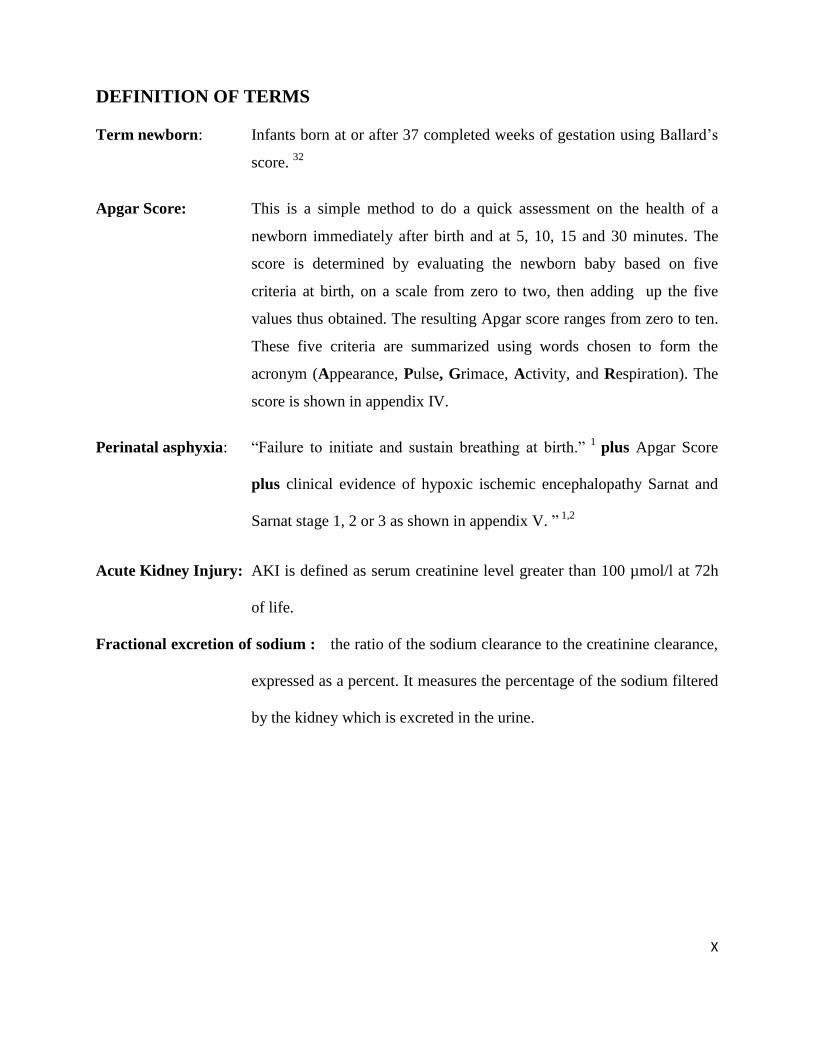
DEFINITION OF TERMS
Term newborn: Infants born at or after 37 completed weeks of gestation using Ballard‟s
score. 32
Apgar Score: This is a simple method to do a quick assessment on the health of a
newborn immediately after birth and at 5, 10, 15 and 30 minutes. The
score is determined by evaluating the newborn baby based on five
criteria at birth, on a scale from zero to two, then adding up the five
values thus obtained. The resulting Apgar score ranges from zero to ten.
These five criteria are summarized using words chosen to form the
acronym (Appearance, Pulse, Grimace, Activity, and Respiration). The
score is shown in appendix IV.
Perinatal asphyxia: “Failure to initiate and sustain breathing at birth.” 1
plus Apgar Score
plus clinical evidence of hypoxic ischemic encephalopathy Sarnat and
Sarnat stage 1, 2 or 3 as shown in appendix V. ” 1,2
Acute Kidney Injury: AKI is defined as serum creatinine level greater than 100 µmol/l at 72h
of life.
Fractional excretion of sodium : the ratio of the sodium clearance to the creatinine clearance,
expressed as a percent. It measures the percentage of the sodium filtered
by the kidney which is excreted in the urine.

11
ABSTRACT
Introduction
World Health Organization (WHO) defines perinatal asphyxia as “Failure to initiate and sustain
breathing at birth plus an Apgar score of less than 7 at 5minutes‟‟ (1)
. Newborns suffering from
perinatal asphyxia present with multiorgan dysfunction, with studies demonstrating that the
kidney is the most affected organ. AKI in asphyxia has been reported to have an incidence of 50
– 70%. Standard practice of diagnosis of AKI has been via measurement of serum creatinine and
urine output. In neonates these have several shortcomings: it reflects maternal creatinine in the
first 48 – 72 hrs of life, and it is a late marker of injury, and up to 80% of AKI is non oliguric.
This study therefore sought to determine whether fractional excretion of sodium (FENa), which
is deranged in acute tubular necrosis as is characteristic of AKI in asphyxia, could be used to
identify neonates with birth asphyxia who eventually suffer AKI on the first day of life.
Objective
To determine the diagnostic utility (sensitivity, specificity, PPV and NPV) of fractional
excretion of sodium (FENa) measured on day 1of life in the diagnosis of AKI on day three of life
in neonates with perinatal asphyxia in Kenyatta National Hospital.
Study design: This was a hospital based cross sectional design study.
Study methods
Newborns who had a diagnosis of perinatal asphyxia using the Apgar scoring and Sarnat and
Sarnat clinical hypoxic ischemic encephalopathy staging
whose parents consented to the study
were enrolled within 24 hours of birth. Urine sodium and creatinine and serum sodium and
creatinine were measured on the first day of life, and used to calculate FENa. On the third day of
life serum creatinine was measured and a diagnosis of AKI based on levels >100 μmol/l. Results
were analyzed for associations between deranged FENa on day 1 of life and AKI at three days of
life.

12
Results:
One hundred at eight neonates were admitted to KNH‟s NBU with perinatal asphyxia during the
study period, 79 of whom survived to the third day of life and were thus recruited, 3% of whom
had HIE stage I, 91% stage II and 6% stage III. The mean weight was 3289g (SD 478), and mean
length was 47cm (SD 3). There were 41 males and 38 females. Most (79%) of the neonates had
been delivered in KNH. Thirty out of seventy nine neonates met the criteria for diagnosis of AKI
on day 1 of life (>100 μmol/l) giving a prevalence of 38%. FENa was deranged (>2.5%) in 63%
of the neonates on day one of life. Twenty seven of the thirty neonates with AKI on the day three
of life had positive FENa on the first day of life giving us a sensitivity of 90%. On the other
hand, only 26/49 of the neonates without AKI had negative FENa on day 1 of life giving us a
specificity of 53%. Among the 50 neonates with positive FENa on day one of life, 27 had AKI
on day 3 of life giving us a PPV of 54%, while 26/29 neonates without AKI on the third day of
life had negative FENa thus giving us a high NPV of 90%. FENa as an early predictor of AKI
had a modest AUC of 0.715.
Conclusion :
Our study showed the prevalence of AKI to be high at 38% among neonates with perinatal
asphyxia with those with severe HIE stage being affected the most. FENa is an easy to do and
easily available test with a high sensitivity (90%) and a good positive likelihood ratio of 1.9
which makes it a good screening test. Our low sensitivity however means that it can falsely
identify neonates not having AKI as having AKI, but the benefits of preventative measures
outweigh the cost implication. Despite its modest PPV, it is a useful test since the benefit of early
treatment of AKI would outweigh the cost of undertaking the test.
13
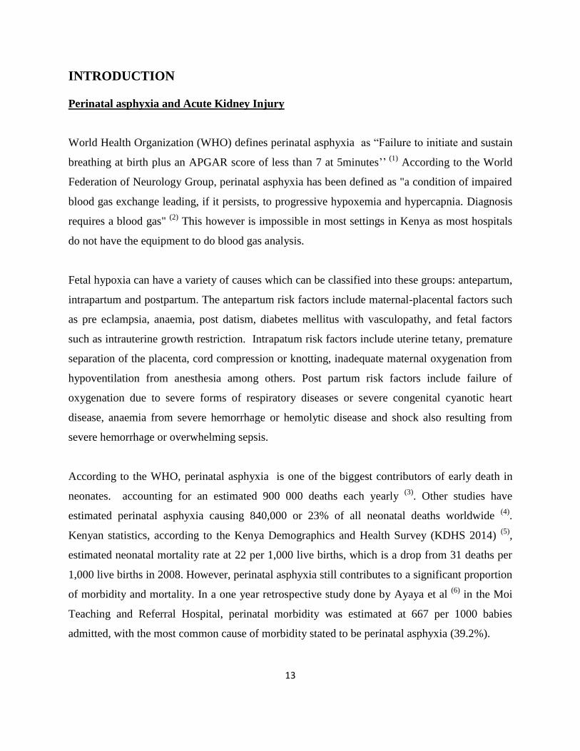
INTRODUCTION
Perinatal asphyxia and Acute Kidney Injury
World Health Organization (WHO) defines perinatal asphyxia as “Failure to initiate and sustain
breathing at birth plus an APGAR score of less than 7 at 5minutes‟‟ (1)
According to the World
Federation of Neurology Group, perinatal asphyxia has been defined as "a condition of impaired
blood gas exchange leading, if it persists, to progressive hypoxemia and hypercapnia. Diagnosis
requires a blood gas" (2)
This however is impossible in most settings in Kenya as most hospitals
do not have the equipment to do blood gas analysis.
Fetal hypoxia can have a variety of causes which can be classified into these groups: antepartum,
intrapartum and postpartum. The antepartum risk factors include maternal-placental factors such
as pre eclampsia, anaemia, post datism, diabetes mellitus with vasculopathy, and fetal factors
such as intrauterine growth restriction. Intrapatum risk factors include uterine tetany, premature
separation of the placenta, cord compression or knotting, inadequate maternal oxygenation from
hypoventilation from anesthesia among others. Post partum risk factors include failure of
oxygenation due to severe forms of respiratory diseases or severe congenital cyanotic heart
disease, anaemia from severe hemorrhage or hemolytic disease and shock also resulting from
severe hemorrhage or overwhelming sepsis.
According to the WHO, perinatal asphyxia is one of the biggest contributors of early death in
neonates. accounting for an estimated 900 000 deaths each yearly (3)
. Other studies have
estimated perinatal asphyxia causing 840,000 or 23% of all neonatal deaths worldwide (4)
.
Kenyan statistics, according to the Kenya Demographics and Health Survey (KDHS 2014) (5)
,
estimated neonatal mortality rate at 22 per 1,000 live births, which is a drop from 31 deaths per
1,000 live births in 2008. However, perinatal asphyxia still contributes to a significant proportion
of morbidity and mortality. In a one year retrospective study done by Ayaya et al (6)
in the Moi
Teaching and Referral Hospital, perinatal morbidity was estimated at 667 per 1000 babies
admitted, with the most common cause of morbidity stated to be perinatal asphyxia (39.2%).

14
A study done in Kenyatta National Hospital (KNH) showed that perinatal asphyxia accounted for
20% of the weekly admissions to the Newborn Unit and had a poor outcome with a mortality of
31.1% by day 7 (7)
. Data from monthly mortality audits in KNH NBU report perinatal asphyxia to
be amongst the leading three diagnosis for admission and death, the other two being prematurity
and respiratory distress syndrome(8)
.
Newborns with perinatal asphyxia are prone to multiorgan dysfunction, and studies have shown
that the worst affected organ in an asphyxiated term infant is the kidney (9)
. According to Acute
Kidney Injury Network (AKIN), AKI is defined as “an abrupt (within 48 hours) reduction in
kidney function, currently defined as an absolute increase in serum creatinine (SCr ) of at least
0.3 mg/dl (≥26.4μmol/l), a percentage increase in SCr of at least 50% (1.5-fold from baseline), or
a reduction in urine output (documented oliguria of less than 0.5 ml/kg per hour for more than 6
hours” (10)
.
There is however no global standard acceptable definition of AKI in neonates. Because of the
different definitions, AKI incidence post neonatal asphyxia is reported in up to 50 – 72% (11)
of
cases. The increasing incidence of AKI correlates with the severity of perinatal asphyxia(12)
.
AKI can either be pre – renal or intrinsic renal failure. Among newborns admitted to NICU,
approximately 6% to 8% have intrinsic ARF, with the most common cause cited as severe
perinatal asphyxia (11)
.
The fractional excretion of sodium (FENa) is the ratio of the clearance of sodium to the clearance
of creatinine, expressed as a percent. It measures, in percentage, how much sodium filtered by
the kidney is excreted in the patient‟s urine. It is obtained by measuring plasma and urine
sodium, rather than by urinary sodium concentration interpretation alone, as the sodium
concentration in urine differs with water reabsorption.
In clinical use, the FENa is useful in the evaluation of acute kidney failure in order to deduce
whether hypovolemia or reduced effective circulating plasma volume is contributing to the
kidney failure (low FENa values). Loss of sodium due to ATN or other causes of intrinsic kidney
failure can be suggested by higher FENa values.

15
It is obtained from the following formula:
in which UNa is the urinary concentration of sodium (mEq/L), Ucr is the urinary concentration
of creatinine (mg/dL), PNa is the plasma sodium concentration (mEq/L), and Pcr is the plasma
creatinine concentration (mg/dL).
FENa is deranged as early as day one of life in newborns with asphyxia who develop AKI (12)
,
and as such has the potential to be used for its diagnosis and thus early institution of appropriate
management.

16
LITERATURE REVIEW
Newborns suffering from perinatal asphyxia are prone to multiple organ dysfunction due to a
redistribution of cardiac output to sustain perfusion to critical organs e.g. adrenals, brain and
heart, while potentially leading to renal ischemia. This leads to damage to other organs, most
affected being the kidneys. (9)
Acute Kidney Injury in Perinatal Asphyxia
Prevalence of AKI in asphyxia
AKI incidence post neonatal asphyxia is reported in up to 50 – 72% in several studies (11)
. This
essentially underscores a huge burden of AKI in asphyxia. Studies have used different serum
creatinine cut offs to define AKI. Most of the prevalence studies investigating newborn AKI
report its existence to be between 11.7 and 70 % (12-19)
Shah et al (13)
in 2004 did a retrospective cohort study to assess multi organ dysfunction in infants
with post asphyxia HIE , renal dysfunction was the most prevalent with 91/130 (70%)
involvement. In another retrospective study by Leila (14)
et al to study multi organ dysfunction in
neonates with HIE, multi organ dysfunction (MOD) was diagnosed in 74 % (74/100), with renal
dysfunction being the most common, observed in 64% (47/74). In both these studies, renal
dysfunction was defined as oliguria/anuria or serum creatinine >1mg/dl (88 μmol/l).
Studies that used creatinine values >1.5 mg/dL (133 μmol/l) included a retrospective study by
Karlowicz (15)
in the US, which was aimed at determining the prevalence of AKI in moderate
and severe asphyxiated full-term neonates. Of the 33 neonates in the study, 20(61%) with severe
asphyxia had AKI, with none of 33 with moderate asphyxia having AKI (P<0.0001).
Essajee et al (16)
conducted a prospective cohort study in Kenya
using NGAL as an early marker
of AKI post asphyxia. AKI was found in 56% (60/108) of the study population. A prospective

17
cohort study done by Alaro et al (17)
in KNH found the prevalence of AKI to be 11.7% amongst
newborns with perinatal asphyxia.
Other studies defined AKI as serum creatinine >1mg/dl (88 μmol/l). This included Gupta et al (12)
who carried out a prospective case controlled study in India to determine the incidence of AKI in
perinatal asphyxia. The results showed that 33 of 70 (47.1%) had AKI.
Other studies used AKIN classification for AKI while others used pRIFLE classification. Kaur et
al (18)
did a prospective cohort study in newborns with perinatal asphyxia in India, and used
AKIN classification for AKI. The total incidence of AKI was 41.7%, with 9% (1/11) of
newborns with moderate asphyxia and 56% (12/25) with severe asphyxia developing AKI.
In Africa, a prospective cohort study done by Medani et al (19)
in Sudan aimed to determine the
pattern of AKI in asphyxiated neonates and its relation to the grade of hypoxic ischemic
encephalopathy (HIE). AKI was defined by pRIFLE and a prevalence rate of 54.1% (46/85) was
observed.
Pathophysiology of AKI
Perinatal asphyxia causes reduced blood flow to the kidneys secondary to hypotension and
hypovolemia , which can lead to impairment in both functions of the tubules and glomerular
filtration rate (GFR). AKI can either be pre – renal or intrinsic renal failure. Renal hypoperfusion
due to systemic hypotension causes prerenal failure and subsequently there is failure of
maintainance of renal blood flow due to loss of autoregulation. Severe or prolonged renal
hypoperfusion leads to renal parenchymal damage, a situation which leads to the evolution of pre
renal failure into intrinsic AKI.
The evolution of intrinsic AKI has three phases: initiation, maintenance, and recovery (Figure 1
below), with differing severity ranging from mild tubular dysfunction to ATN (with or without
oliguria and anuria), to infarction and corticomedullary necrosis with irreversible kidney
damage.

18
The initial period of renal hypoperfusion is the initiation phase (hours to days) , during which
there is evolving ischemic injury. The GFR declines because reduction in blood flow to the
kidneys reduces the glomerular ultrafiltration pressure due to an increase in vasoactive
mediators such as endothelin, plasma renin activity (PRA), adenosine, thromboxane and
reduction in nitric oxide, prostacyclin and natriuretic peptides which are vasodilatory. This leads
to vasoconstriction of afferent arterioles. Secondly, casts within the tubules composed of
epithelial cells and necrotic debris obstruct the flow of glomerular filtrate within tubules(20)
.
Subsequent increase in pericapilarry hydrostatic pressure in the Bowman‟s space leads to a
further decline in GFR. Finally, there is backleak of glomerular filtrate through injured tubular
epithelium.
The last medullary segment of the proximal tubule (S3 segment, pars recta) has the most
prominent ischemic injury, accompanied by the medullary portion of the TAL of the loop of
Henle(21)
. Tubule cells lose polarity with short periods of ischemia reperfusion and on prolonged
or severe ischemia the epithelial cells are damaged irreversibly, which leads to impairement in
re-absorption at the tubules resulting in increase in sodium excretion in the tubular fluid leading
to imbalance in electrolytes(22)
.
The maintenance phase (typically1 to 2 weeks) leads to injury of the renal cell. It entails
stability of GFR albeit at low levels. During the maintenance phase of ATN, the main factors are
nephronal and cellular. Programmed cell death (apoptosis) forms the main cellular injury, which
is then is followed by changes in intracellular calcium metabolism, phospholipid breakdown,
release of free oxygen radicals, altered cell polarity, loss of tight cell junctions between cells,
disruption of the cytoskeleton ,loss of the cell brush border, and loss of major cellular functions,
e.g., Na+/K+-ATPase, and cell swelling. All these processes are accompanied depletion of
cellular ATP.

19
Figure 1 Pathophysiology of Ischemic/ Intrinsic AKI
Renal parenchymal cell repair and return of GFR towards preillness levels characterizes the
recovery phase.
PERINATAL
ASPHYXIA
Redistribution of cardiac
output
Vasoconstriction of
afferent arteriole
NO, PGI2
Endothelin, PRA
GFR
Reduced blood flow and GFR
Loss of renal autoregulation
Ischemic injury to renal
parenchyma
Initiation
phase
phase
Tubule cell injury
Detachment with
obstruction by casts
Intratubular pressure
Tubular backleak
Tubular flow
Necrosis, Apoptosis of
tubular cells
Loss of polarity Increased distal Na delivery
Tubuloglomerular feedback
Release of adenosine, Calcium
Costriction of afferent arteriole, PRA
Sustained reduction in GFR
Maintainance
phase
Recovery of renal perfusion Regeneration of tubular epithelium
Recovery of GFR
Recovery phase

20
AKI in perinatal asphyxia is not without its consequences. Mortality rates in asphyxiated
newborns with AKI in various studies have been reported as 31.1% by day 7 (7)
, 71.4% by day
7(19)
and 14.1% by day 7 (12)
. In a study done by Gupta(12)
, those with perinatal asphyxia showed
changes in 5 (6.6%) cases noted as changes in echotexture, increased kidney size, and loss of
corticomedullary differentiation as assessed by renal sonography.
Diagnosis of Acute Kidney Injury
In spite of the available functional systems of classifying and diagnosing AKI (pRIFLE and
AKIN criteria), diagnosis in neonates has proven to be a challenge since the classification
systems are based on the patients level of serum creatinine (SCr) and urine output (UO). AKI in
neonates and more so in perinatal asphyxia has been shown in several studies to be non oliguric.
In a retrospective study by Karlowicz (15)
, he defined non oliguric renal failure as urine output >
1ml/kg/hour after day one of life. Medani et al (19)
carried out a prospective cohort study , in
which oliguric renal failure was diagnosed if the patient‟s urine output was less than 1
ml/kg/hour, while Gupta et al (12)
defined oliguria as urinary output of less than 0.5ml/kg/hr. The
outcome was as depicted table 1.
Table 1: Oliguric vs. Non oliguric AKI
Study Sample size Title Outcome
Medani (19)
, 2013
Prospective cohort
Country - Sudan
85 Acute kidney injury in
neonates with asphyxia
admitted to a tertiary
neonatal unit in Sudan
AKI in 54.1% (46/85). Non oliguric 65%
(30/46)
Gupta (12)
, 2009
Prospective Case
Control Study -
India
98 Case vs
control
(70/28)
Renal Failure in
Asphyxiated Neonates
47.1% with AKI, non-oliguric 78% cases
and oliguric type 22% of cases.

21
Generally, non oliguric AKI can be attributed to the greater total body water in newborns
compared to adults, especially in preterms with values as high as 80%, and, in addition,
immature tubular development leading to greater urine output. In asphyxia, injury to the tubular
cells leads to loss of the capacity to avidly reabsorb sodium thus leading to natriuresis and
consequently diuresis.
Limitations of serum creatinine
Traditionally, SCr has been used to diagnose AKI. However, it has several shortcomings in the
neonatal period which include:
SCr in the first 48 – 72 hours of life is usually a reflection of maternal creatinine values,
not the infants renal function
Normal nephrogenesis begins from 8 weeks GA and is complete by 34weeks. Depending
on maturity of the kidney, there is a steady improvement in GFR from 10-
20mls/min/1.73m2 by week 1 of life to 30 – 40mls/min/1.73m
2 by two weeks after
delivery, in line with alterations in RBF after which there is gradual steady improvement
over the first months of life. Due to this low GFR, normal serum creatinine levels are thus
distributed over a wide range of values depending on maturity and postnatal age.
Changes in SCr concentration may not be noted until there is loss of 25 to 50% of kidney
function.
Since SCr is secreted by tubular cells, even at low GFR SCr will still be low thus will
overestimate renal function.
Measurement of SCr does not differentiate between renal derangements that are
hemodynamically mediated and those that are not e.g. pre renal vs. intrinsic or
obstructive renal failure.
Novel biomarker
Incidence of AKI in asphyxia has been shown to be high through several studies, and outcomes
have remained poor, thus there has evolved a need to identify new biomarkers which would be
able to anticipate the diagnosis of AKI within hours or days prior to the reduction in the UO or

22
changes in SCr. Identification of a biomarker that can differentiate between the different causes
of AKI may change the approach to AKI management and lead to the implementation of
preventive interventions.
In the early acute setting, appropriate biomarkers for kidney injury should have several
characteristics which include:
New biomarkers e.g Serum cystatin C and NGAL have been shown by recent studies to be early
non invasive biomarkers. However, most of these biomarkers are not readily available in Kenya.
FENa: A solution to the problem?
FENa is the ratio of the clearance of sodium to the clearance of creatinine, expressed as a
percentage. FENa has historically been utilised to distinguish between pre renal kidney failure
and ischemic renal failure. The measurement of FENa is by urinary and serum creatinine and
sodium levels, and is independent of urine output. FENa is dependent on tubular function which
depends on autoregulation of blood flow to the kidneys.
Na_, K_-ATPase localized at the basolateral membrane aids in active sodium reabsorption.
Greater than 60% of the sodium that is filtered is reabsorbed in the proximal convoluted tubule
(PCT), 20% in the thick ascending limb (TAL) of the loop of Henle, while the remaining 10% of
Should be specific to the kidney, and be able to differentiate between pre-renal,
intrinsic and post-renal causes of kidney damage.
Should posses the ability for earlier detection of kidney injury.
Should be able to specifically isolate the cause of kidney injury
Should be able to pinpoint particular sites of damage in the kidneys and able to
provide information on disease changes in the primary location of damage.
Should be reliable, easily measurable, prompt and ideally non invasive.
Should be stable in its matrix
Lastly, should be an inexpensive, widely affordable and available marker to
measure

23
sodium (Na) and chloride (Cl) in the distal convoluted tubule (DCT). Absorption of sodium
proximally increases during development by three- to four fold (23)
. In the immature kidney, a
larger fraction of filtered Na is delivered the distal nephrons. During development of the kidney,
FENa decreases from as high as 13% in the fetus to about 3% in the premature neonates less than
30 weeks of gestation, to 2% in term neonates secondary to maturational process (24)
.
Due to the decreased ability to actively reabsorb sodium, FENa values in term newborns are
higher than those in older children (less than 2.5% percent versus less than 1 percent). There is
decreased ability to undergo anaerobic metabolism in the S3 segment during periods of
decreased oxygen tension such as in perinatal asphyxia, which leads to impaired reabsorption of
sodium thus higher values of FENa.
Table 2: Interpretation of FENa
FENa value Type of AKI Mechanism
<2% Pre renal AKI Almost all of the filtered sodium is
reabsorbed, which is an appropriate
response to hypovolemia.
>2.5% Intrinsic AKI in the absence of diuretics Impaired TAL and DCT absorption of Na
Validation studies
Several studies validate the importance of FENa in differentiating pre renal failure from intrinsic
renal failure. An adult prospective cohort study done by Carvounis et al (25)
analyzed the
sensitivity and specificity of both FENa and FEUr in differentiating prerenal failure from ATN.
Results showed low FENa (<1%) levels in patients with untreated plain prerenal failure, and
values >1% in those with both prerenal disease treated by diuretics and those with ATN. The
sensitivity and specificity of FENa was high at 91% and 82% respectively.
Another adult prospective cohort study by Pépin et al (26)
recruited patients in a tertiary care
centre, 99 patients in total, who developed AKI. In patients in whom diuretics was not
administered and in those who received diuretics, the specificity and sensitivity of FENa were
75% and 78% and 81% and 58% respectively.

24
A study done by Yassin (27)
looked at both FENa and FEUr in 40 adult patients with AKI who
had circulatory shock. The patients were grouped into 26 patients who had prerenal (group-1)
and 14 who were found to have renal azotemia (group-2). FENa cut off was at 1% pre renal and
>1% intrinsic AKI. Group – 1 had significantly lower FENa (0.99 ± 0.66) as opposed to group –
2 (2.57 ± 1.73, P <0.05). The sensitivity and specificity of FENa were 71.4% and 69.4%
respectively.
A pediatric study done by Hisayo et al (36)
recruited 74 patients (42 boys, 32 girls) in Japan, with
102 episodes of AKI. The children‟s mean age was 7.1 years, with an age range from 0 to 18
years. The study‟s aim was „To assess the utility of FEUr in the differential diagnosis of AKI in
children‟. FENa cut off was <1% for pre renal and >/=1% for ATN. The children were divided
into ATN (n=33), pre renal AKI (n=37), and pre renal AKI with furosemide (n=32) . Sensitivity
of FENa < 1% was53 % (17/32), 95 % (35/37) and 75 % (52/69) in all cases of prerenal AKI
with furosemide , prerenal AKI without furosemide and prerenal AKI respectively, with the
specificity shown to be 90.9% (30/33).

25
Table 3: Summary of validation studies
Study Sample size Title Outcome
Carvounis et al (25)
2002, USA
Prospective cohort
study
102 adult renal
cases. 50 pre
renal, 27 pre
renal treated
with diuretics
and 25 ATN.
„Significance of the
fractional excretion of urea
in the differential diagnosis
of acute renal failure‟.
FENa <1% pre renal
AKI, >/=1% in cases
treated with diuretics
and ATN.
Sensitivity 91% and
specificity 82%.
Pépin et al (26) 2007,
Canada
Prospective cohort
study
99 adult
patients
„Diagnostic Performance of
Fractional Excretion of Urea
and Fractional Excretion of
Sodium in the Evaluations of
Patients With Acute Kidney
Injury With or Without
Diuretic Treatment‟
Those without
diuretics, sensitivity
78%, specificity 75%
with PPV of 86% and
NPV of 64%
With diuretics:
sensitivity 58%,
specificity 81%
Yassin (27)
2011,
Egypt.
Prospective cohort
study
40 adult
patients
„Comparison between
fractional excretion of
sodium and fractional
excretion of urea in
differentiating prerenal from
renal azotemia in circulatory
shock‟
FENA cut off point
1.1% with sensitivity
71.4% and specificity
of 69.4%
Hisayo et al
(36),Tokyo, Japan.
Prospective cohort
study.
74 children „T o find out the utility of
FEUr in the differential
diagnosis of AKI in
children‟.
FENA cut off point
<1% for pre renal and
>/=1% for ATN, with
sensitivity 95% for
pre renal and
specificity of 90.9%

26
for ATN.
Studies using FENA in asphyxia
In a study done by Matthew et al (28)
, 42 neonates in NICU on treatment for RDS and or sepsis
were recruited, all of whom had oliguria, which was defined as urine output of less than
1ml/kg/hour for 12 hours or longer. After receiving either mannitol at 1g/kg or fluid challenge at
20mls/kg of normal saline, those with improved urine output were defined as having pre renal
oliguria (PO), while those with persistent oliguria and azotemia with no other evidence of renal
failure were defined as having renal failure. Sixteen neonates (38%) met the criteria for RF all of
whom had perinatal asphyxia. FENa based on creatinine was found to have a mean value of
0.95% in patients with PO as opposed to 4.25% in those who had renal failure (p<0.01). The
conclusion of the study was that sharp demarcation of the two groups was possible only when a
FENa >/= 2.5% was used and that FENa >/= 2.5% was seen to differentiate renal failure from
functional oliguria in the study.
Gupta et al (12)
did a prospective case controlled study to find out the incidence of AKI in
perinatal asphyxia. Serum urea, creatinine and electrolytes and sodium and creatinine in urine
were measured within 24 hours after delivery and on the third day of life. Neonates with
asphyxia were further divided into two groups, A1 without AKI and A2 with AKI. The control
groups were labeled as B. A statistically different mean FENa was 0.6 ±0.56% in-group A and
0.29 ± 0.27% in-group B was noted.
Kaur et al (18)
did a prospective cohort study in newborns asphyxia admitted to NICU in India.
Baseline serum electrolytes were done within 6 hours of birth, with daily repeats till four days of
life. Urine was collected between 24 to 36 hours and 72 to 96 hours of age. Mean FENa values at
24 – 36 hours in 15 neonates who developed AKI within that period was 5.59% as opposed to
FENa values of 1.18% in the 21 neonates without AKI during that period (P 0.007). At 72 to 96
hours, FENa values in 6 neonates with AKI had a mean of 9.42% as opposed to 1.26% FENa in
30 neonates without AKI at that point in time (P 0.004).
In a prospective case controlled study by Roberts et al (29)
21 babies of 34-41 weeks gestational
age with moderate to severe birth asphyxia were recruited. AKI was diagnosed by urinary retinol
27
binding protein: creatinine ratio. Fractional excretion of sodium was measured, results were
reported without administration of diuretics. AKI was diagnosed in 19% (4/21). Mean FENa
values were 31. 9% in those with AKI. That in control infants was below 1%.
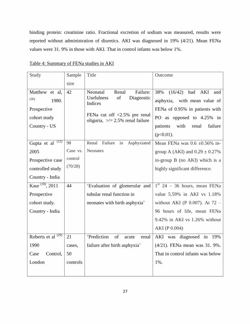
Table 4: Summary of FENa studies in AKI
Study Sample
size
Title Outcome
Matthew et al,
(26) 1980.
Prospective
cohort study
Country - US
42 Neonatal Renal Failure:
Usefulness of Diagnostic
Indices
FENa cut off <2.5% pre renal
oliguria, >/= 2.5% renal failure
38% (16/42) had AKI and
asphyxia, with mean value of
FENa of 0.95% in patients with
PO as opposed to 4.25% in
patients with renal failure
(p<0.01).
Gupta et al (12)
2005
Prospective case
controlled study
Country - India
98
Case vs.
control
(70/28)
Renal Failure in Asphyxiated
Neonates
Mean FENa was 0.6 ±0.56% in-
group A (AKI) and 0.29 ± 0.27%
in-group B (no AKI) which is a
highly significant difference.
Kaur (18)
, 2011
Prospective
cohort study.
Country - India
44 „Evaluation of glomerular and
tubular renal function in
neonates with birth asphyxia‟
1st 24 – 36 hours, mean FENa
value 5.59% in AKI vs 1.18%
without AKI (P 0.007). At 72 –
96 hours of life, mean FENa
9.42% in AKI vs 1.26% without
AKI (P 0.004)
Roberts et al (29)
1990
Case Control,
London
21
cases,
50
controls
„Prediction of acute renal
failure after birth asphyxia‟
AKI was diagnosed in 19%
(4/21). FENa mean was 31. 9%.
That in control infants was below
1%.
28
STUDY JUSTIFICATION
The incidence of AKI in neonatal asphyxia is reported in up to 50 – 72% (11)
of cases. A recent
study done in KNH found an 11.7% prevalence rate of AKI among newborns with perinatal
asphyxia (17)
, while another study by Essajee et al (16)
in KNH and Pumwani noted a prevalence
rate of 56%. With lack of an identifiable universally acceptable AKI definition in neonates and
the fact that its diagnosis in neonates is currently problematic as it relies on changes in serum
creatinine and oliguria, efforts are being made to evaluate better methods that can be used to
detect AKI before damage has been done to the kidneys.
Studies have reported high mortality rates amongst newborns with perinatal asphyxia who
subsequently develop AKI. Therefore there exists a need for identification of a biomarker that
can be used to diagnose AKI prior to derangements in creatinine, in order to institute appropriate
management strategies to prevent or treat AKI early in those affected.
FENa is an indicator of tubular damage in neonates with the test being historically used to
differentiate pre renal from intrinsic renal failure. Abnormalities in FENa can be picked up from
as early as 6 hours within birth; it is an easy to do and readily available test. Studies looking at
the sensitivity and specificity of FENa in differentiating pre renal from intrinsic AKI have values
as high as 91% and 82% respectively (25)
. However these studies have mostly been done in adults
and this study therefore sought to establish whether FENa can be used to predict AKI early in
perinatal asphyxia, and determine its sensitivity and specificity in our set up.

29
RESEARCH QUESTION
Is the fractional excretion of sodium (FENa) measured on day 1 of life an early predictor of AKI
in neonates with birth asphyxia at Kenyatta National Hospital‟s NBU?
OBJECTIVES
Primary Objective
To determine the diagnostic utility (sensitivity, specificity, PPV and NPV) of fractional excretion
of sodium (FENa) measured on day 1 of life in the diagnosis of AKI in neonates with perinatal
asphyxia in KNH. AKI will be diagnosed by serum creatinine level >100μmol/l on the third day
of life.
Specific objectives
1. To determine the sensitivity of FENa as an early diagnostic marker for AKI in neonates
with perinatal asphyxia.
2. To determine the specificity of FENa as an early diagnostic marker for AKI in neonates
with perinatal asphyxia.
3. To determine the PPV of FENa as an early diagnostic marker for AKI in neonates with
perinatal asphyxia.
4. To determine the NPV of FENa as an early diagnostic marker for AKI in neonates with
perinatal asphyxia.

30
METHODOLOGY
Study design:
This was a hospital based cross sectional study.
Study area:
The study was carried out at the Newborn Unit at the Kenyatta National Hospital (KNH), the
tertiary referral and teaching hospital for the College of Health Sciences, University of Nairobi.
It is also the main inpatient hospital for the low and middle-income society in Nairobi and its
environs. The newborn unit is a level III unit which admits all sick neonates born in KNH, and
also handles transfers from other hospitals. The unit admits between 160 and 200 neonates each
month, over 20% whom are term babies diagnosed with perinatal asphyxia (7)
.
Study population:
The study population was term newborns admitted in KNH NBU with a diagnosis of birth
asphyxia within 24hours of birth using the Apgar scoring (30)
and Sarnat and Sarnat clinical
staging of hypoxic ischemic encephalopathy (31)
outlined in appendices IV and V respectively.
Study Tool:
A standardized questionnaire was used for collecting data from the enrolled participants, which
was administered after obtaining informed consent from the parent/guardian. The questionnaire
was pre tested in the KNH New born unit among newborns with perinatal asphyxia. (Appendix
1)
Study period
The study ran for a period of four months (July 2016 to October 2016)
Study personnel:
1. The lead investigator was the supervisor in charge of the research team, whose role was
to ensure proper documentation and perform standard procedures on enrolled
participants. The lead investigator also ensured all the materials needed were available
and that all data collected was entered in to the computer systems daily.
2. Research assistants: with the help of two research assistants (clinical officers), data was
collected. They received training on standard ways of doing procedures for the study.

31
Inclusion criteria:
Term newborn (> 37 completed weeks) assessed by modified Ballard‟s exam (32)
as
outlined in appendix III
Age 0-24 hours at initial assessment
“Failure to initiate and sustain breathing at birth.” 1 plus Apgar Score less than or equal 7
at 5 minutes plus clinical evidence of hypoxic ischemic encephalopathy Sarnat and
Sarnat stage 1, 2 or 3 as shown in appendix V. ” The highest level of neurological
impairment was used to place the infant in the appropriate stage.
Consent by the parent or caregiver
Exclusion criteria:
• Newborns with major congenital anomalies
• Newborns who developed jaundice in the first three days of life.
• Those who die within three days of the study.
Sampling technique:
The sampling technique used was consecutive sampling.

32
Sample size
Sample size at the required absolute precision level for sensitivity and specificity was calculated
by Buderer's formula (33)
where n = required sample size,
S N = anticipated sensitivity, set at 0.98
S P = anticipated specificity, set at 0.98
α = size of the critical region (1 − α is the confidence level), set at 95% confidence interval
z 1-α/2 = standard normal deviate corresponding to the specified size of the critical region (α), and
L = absolute precision desired on either side (half-width of the confidence interval) of sensitivity
or specificity. Level of precision (set at±5%)
Prevalence = set at 56% using results from Farida Essajee et al‟s (18)
study
Using the above formula, sample size for a sensitivity of 98% was 54 neonates while a sample
size based on specificity of 98% was 71 neonates. Thus, minimal sample sizes of 68 neonates
were to be recruited to the study.

33
Recruitment procedure
Enrollment of participants
All term neonates aged 0-24 hours admitted at KNH NBU were assessed for perinatal asphyxia
using the Apgar scoring 30
and Sarnat and Sarnat clinical staging of hypoxic ischemic
encephalopathy 31
outlined in appendix II and III respectively. The most severe sign was used to
categorize the severity of the perinatal asphyxia. The gestational age was ascertained by Ballard
exam 32
All term newborns that met the criteria were consecutively enrolled within the first 24 hours of
life irrespective of day or night admission.
The parents or caregivers of the term neonates with perinatal asphyxia that satisfied the inclusion
criteria were requested to participate in the study. Only after explaining of the reason of the study
and its expected benefits and possible harms was informed consent obtained from the
parent/caregiver.
Newborns with malformations and those that did not survive to the third day of life were
excluded from the study.
Day 1: Determination of FENa
A single urine sample was obtained within 24 hours of birth from each of the study
subjects using in and out catheterization and for those in whom urine specimen wasn‟t
obtained at first try, urine collecting bags were left in place.
The urine collected was used to assay urine electrolytes i.e. urine sodium and creatinine.
Serum sodium and creatinine levels were measured by drawing 0.5 to 1ml of blood
sample into a microtainer on the first day by quick heel sampling.
The specimens were transported to the laboratories immediately after they were collected.
FENa was calculated by the following formula:
The cut off for a positive FENa was > 2.5% while a negative FENa was <2.5%

34
Day 3: Diagnosis of AKI
Serum creatinine levels were measured by drawing 0.5 to 1ml of blood sample into a
microtainer on the third day of life by quick heel sampling.
Definition of AKI was set at a serum creatinine level > 100μmol/l.
Diagnostic utility
FENa results obtained on day 1 of life were correlated with serum creatinine values on
day 3 of life to find out whether the neonates with a diagnosis of AKI on the third day of
life also had positive FENa (>2.5%) on day 1 of life. This was used to determine whether
FENa values on day 1 of life can be used as an early predictor of AKI in newborns with
perinatal asphyxia.
Laboratory analysis
The Ion Selective Electrodes (ISE) module of the COBAS INTEGRA systems was used for the
quantitative determination of sodium and creatinine in urine(34)
. The specimens were
automatically diluted 1:6 (l +5) by the instrument for readings on urine sodium levels while they
were automatically diluted by the machine to1:25 (1+24) in order to get the creatinine values.
The test principle for the creatinine estimation uses Buffered kinetic Jaffe reaction without
deproteinization. Creatinine reacts in alkaline solution with picrate to form a yellow-red adduct.
There is direct proportionality between the creatinine concentration in the specimen to the rate of
dye formation (color intensity). It is determined by measuring the increase in absorbance at 512
nm. Urine sodium and creatinine levels results were documented. Within 2 hours the serum
sample was centrifuged and analyzed using Cobas Integra machine using the compensated Jaffẻ
method (35)
. The Cobas Integra automatically calculated the analyte concentration of each
sample. The Cobas Integra uses the Precinorm U or Precinorm U plus for reference range
control, and the Precipath U or Precipath U plus for pathological range control. The control
interval was 24 hours. The machine was calibrated every seven days using deionised water as
zero calibrater according to the standard reference material guidelines. Serum and plasma

35
samples contain proteins which react non-specifically in the Jaffẻ method. For compensation of
serum and plasma results, values were automatically corrected by -18μmol/l.
DATA MANAGEMENT AND ANALYSIS
Data was collected using a standardized questionnaire and entered into a password protected
database. During entry, data collection forms were stored in a secured lockable cabinet to prevent
unauthorized access.
Data analysis was done using the IBM® SPSS Statistics software version 21. Data cleanliness
was ensured by comparing entered data with the hard copy forms after entry was complete.
On each day, neurologic exam was carried out and the infants categorized according to their HIE
Stage.
Categorical data such as neurological scores and AKI scores were presented using frequency
tables whereas continuous data for example length, birth weight, gestational age, head
circumference were summarized using measures of central tendency and dispersion.
Factors associated with the accurate diagnosis of AKI using FENa were determined using chi-
squared tests for categorical comparisons and t-tests for continuous comparisons. Independent
factors associated with accurate diagnosis of AKI using FENa were determined using binary
logistic regression methods.
FENa sensitivity was determined by calculating what proportion of neonates with AKI diagnosis
on the third day of life were positively identified by FENa levels >2.5% on day 1 of life.
Specificity was determined by calculating what proportion of neonates without a diagnosis of
AKI on the third day of life had a negative FENa on the first day of life.
The PPV was determined by calculating what proportions of neonates with positive FENa on day
1 of life truly had AKI on day 3 of life based on serum creatinine > 100μmol/l. The NPV was
determined by calculating what proportion of neonates with negative FENa on day 1 of life did
not have a diagnosis of AKI on day 3 of life.

36
Analysis is as depicted by Table 5 below
Table 5: Diagnostic utility of FENa
AKI by
sCR day 3
No AKI
on day 3
Total
FENA positive day 1 a B a + b
FENA negative d1 c D c + d
a + c b + d N
Sensitivity = a/(a+c)
Specificity = d/(d+b)
PPV = a/(a+b)
NPV = d/(c+d)

37
ETHICAL CONSIDERATIONS
This protocol, together with the informed consent document and any further modifications was
reviewed and approved by the KNH‟s Ethics, Research and Standards Committee, and a letter of
approval from the committee was obtained prior to commencement of the study.
Parents/caregivers were given full information about the study and a written consent was
obtained from them. No emergency/resuscitation measures were overlooked and they were given
priority to other procedures. No beneficial treatment was withheld from the patients. Subject
confidentiality was strictly upheld by the principal investigator, research assistants and other
supporting parties, and no information concerning the study or data collected was released to any
unauthorized person. Study details were given to the clinician taking care of the neonate.

38
RESULTS
General characteristics of all the mothers and neonates
During the study period, 108 neonates with perinatal asphyxia were admitted, but only seventy
nine babies survived to the third day of life, 41 (52%) of whom were male.
STUDY FLOW DIAGRAM
The diagram below summarises the recruitment procedure up to obtainment of desired outcomes.
Term neonates at KNH NBU assessed for perinatal asphyxia
(n = 118)
INFORMED CONSENT (n=108)
Urine electrolytes (sodium and creatinine) and creatinine in
serum measured on first day of life and creatinine in serum
measured on day 3 of life.
Daily follow up and assessment done for the
first 3 days of life. (n=79)_
Data entered and analyzed for primary and specific
objectives (n=79)
NO CONSENT
(n =10)
Died within 3 days of the
study (n = 29)

39
Table 6: Characteristics of the neonates on admission
Characteristics Frequency(%)/ Mean (SD)
Gender
Male 41 (52)
Female 38 (48)
Gestational age in weeks 39 (1)
Birth weight in grams 3289 (478)
Length in centimeters 47 (3)
Head circumference in centimeters 36 (2)
Place of delivery
KNH 61(79)
Other health facility 16(21)
Mode of delivery
Vertex vaginal 53(67)
C/S 26(33)
Apgar Score at 5 minutes
Mean 6 (1)
Mild (6-7) 53 (67)
Moderate (4-5) 23(29)
Severe (0-3) 3(4)
Resuscitation with BVM
Yes 31(39)
No 48(61)
Intubation+ mechanical ventilation
Yes
No
20 (25)
59 (75)
The mean weight was 3289 g (SD=478) and the mean length was 47 cm (SD=3) .The neonates
weight ranged from 2200 to 4600 g as depicted in table 6 above.
Majority of the deliveries took place in KNH (79%) by vertex vaginal mode (67%). Two
neonates did not have documentation of place of delivery. Of the neonates recruited, 39% were
resuscitated via bag valve and mask. The mean Apgar score was 6 (SD=1), with 68 % with mild,
29 % moderate and 3 % severe asphyxia on the day of admission as depicted in Table 6.

40
Table 7: Characteristics of the mothers
Variable Frequency (%)
Marital status
Single
Married
Widowed
7 (9)
71 (90)
1 (1)
Occupation
Salaried/formal employment
Informal employment
Self employed
Casual worker
Unemployed
6 (8)
2 (3)
12 (15)
16(20)
43(54)
Number of ANC visits
Twice
More than twice
1(1)
78 (99)
Parity
Primigravida
Para 1
Para 2 and above
6 (8)
46 (58)
27 (34)
Education level
Primary
Secondary not completed
Secondary completed
Tertiary and beyond
5 (6)
26 (33)
16 (20)
32 (41)
The mean age of the mothers was 24 (SD = 5) yrs, the youngest being 15 yrs and the oldest 38
years; 90% were married. Of the 79 mothers, 58 % were Para 1+0. The majority (54%) of the
mothers were unemployed, with 53% and 40 % having attained secondary and tertiary education
respectively. All mothers reported to have attended ANC, 98% reporting having attended more
than twice.
41
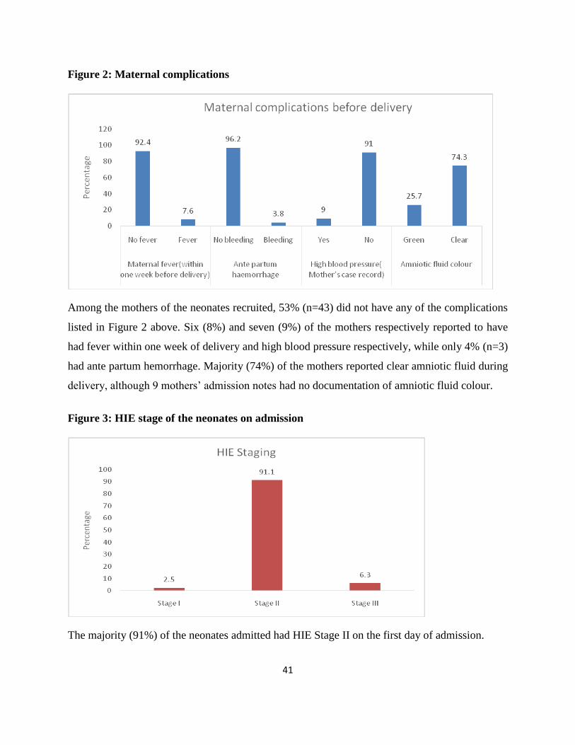
Figure 2: Maternal complications
Among the mothers of the neonates recruited, 53% (n=43) did not have any of the complications
listed in Figure 2 above. Six (8%) and seven (9%) of the mothers respectively reported to have
had fever within one week of delivery and high blood pressure respectively, while only 4% (n=3)
had ante partum hemorrhage. Majority (74%) of the mothers reported clear amniotic fluid during
delivery, although 9 mothers‟ admission notes had no documentation of amniotic fluid colour.
Figure 3: HIE stage of the neonates on admission
The majority (91%) of the neonates admitted had HIE Stage II on the first day of admission.

42
Table 8: Prevalence of AKI using Creatinine and probable AKI by FENa
Gender
Male=41 Female=38 Total
N % N % n % p-value
AKI(Serum Creatinine
>100μmol/l 21 51.2 9 23.7 30 38.0
0.012
Probable AKI (FENa
>2.5%) 27 65.9 23 60.5 50 63.3
0.624
Thirty out of seventy nine neonates had serum creatinine of >100μmol/l on day three of life
which translated to an AKI prevalence of 38%. Of the neonates with AKI, 51% were male and
24% female, which was statistically significant (p = 0.012).
FENa on day 1 of life was positive (>2.5%) in 50 neonates (63.3%) of which 27 (66%) were
males and 23 (60%) females. There was no statistical difference between males and females (p =
0.624)
Table 9: Diagnostic Utility of FENa
AKI by creatinine
AKI by FeNA
Serum
Creatinine
>100μmol/l
Serum
Creatinine
<100μmol/l Total
FeNA >2.5% 27 23 50
FeNA <2.5% 3 26 29
Total 30 49 79
AKI by creatinine (cut off 100 μmol/l)
Sensitivity 90% (95% CI 79% - 100%)
Specificity 53% (95% CI 39%- 68%)
Positive likelihood ratio 1.9
Negative likelihood ratio 0.19

43
Of the neonates recruited, 27/30 (90%) had a positive FENa (defined as FENa >/=2.5%) on day
one of life and AKI diagnosed on day 3 of life, thus setting our sensitivity at 90%. Amongst the
49 neonates who did not have AKI on day 3 of life, 26 neonates had negative FENa on day 1 of
life, thus setting the specificity of the test at 53%.
Of the 50 neonates who had positive FENa on day one, 27 of them had AKI diagnosed on day 3
of life thus setting the PPV at 54%. Conversely of the 29 neonates who had negative FENa on
day 1 of life, 26 of them had no AKI based on serum creatinine, thus giving a NPV of 90%.
Fig 4: Graph of sensitivity vs specificity of FENa
Figure 4 above plots the sensitivity and specificity of FENa at 90% vs 53% respectively at a
chosen cut-of off >/= 2.5%.

44
Figure 5: Receiver operating characteristic (ROC) curve for Fractional excretion of sodium
(FENa) on day 1
Figure 5 above shows a moderate efficacy (AUC 0.715) in AKI prediction.

45
Figure 6: Correlation between HIE Stage and AKI
Figure 6 above shows that AKI was highest in those with HIE Stage III (100%), with no AKI
documented in patients with HIE Stage I which is statistically significant (p value 0.008).
Figure 7: Progression of HIE Stage during first three days

46
Progressively, babies with HIE Stage II showed improvement by day three of life (as assesed by
Sanart and Sanart HIE stage) thus steadily increasing the number of neonates in Stage I by the
third day of life. Not much change was seen in neonates with Stage III HIE.
DISCUSSION
Most previous investigators used serum creatinine levels above133 µmol/ l(15, 17)
to diagnose
AKI. Studies by Shah et al (13)
, Leila (14)
and Gupta (12)
used levels of creatinine in serum of
greater than 1 mg/dL (88 μmol/l) at 72 hrs of life to diagnose AKI. So as to increase diagnostic
probability, serum creatinine level of 100 µmol / l at 72 hours of life was chosen.
The prevalance of AKI using serum creatinine levels of >100 μmol/l on the third day of life was
found to be 38% (30/79). Studies by Shah et al (13)
, Leila (14)
and Gupta (12)
used serum creatinine
of greater than 1 mg/dL (88 μmol/l) to diagnose AKI, which showed a prevalence of 70%, 64%
and 47.1% respectively. Studies that used serum creatinine of more than 1.5 mg/dL (133 μmol/l)
included those by Karlowicz (15)
and Alaro et al (17)
which had prevalence of 61% and 11.7 %
respectively. The prevalence of AKI in our study lies within the range found in other studies
(11.7% - 72%) which is reflective of the fact of AKI in asphyxia being a universal health
problem.
Our study showed a higher prevalence of AKI in boys than girls, with 51% male and 24%
females giving a male to female ratio of 2.33:1 which was statistically significant (p 0.012). This
is similar to what has been reported in other studies for example Esajee et al (16)
indicated a male-
to-female ratio of 2.3:1. Documented increase in susceptibility of perinatal disorders in the male
neonate could be an explanation for this finding (37)
.
We set out to assess the importance of FENa in the early diagnosis of AKI in neonates with
perinatal asphyxia since AKI post asphyxia has been associated with poor outcomes (7, 12, 17)
.
FENa has been studied in mostly adult population to differentiate between pre renal AKI and
ATN (which is the type of renal injury demonstrated in neonates with asphyxia) with sensitivities
and specificities ranging between 71.4% - 95% and 69.4% - 90% respectively (25,26,27,36)
.

47
However, very few studies have been done using FENa as a biomarker for AKI in neonates with
asphyxia.
In this study in Kenyan newborn infants, the specificity and sensitivity of detecting AKI as
indicated by serum creatinine levels of >100 μmol/l was 53% (95% CI 39% - 68%) and 90%
(95% CI 79% - 100%) respectively with a moderate efficacy (AUC 0.715) in prediction of AKI.
Our sensitivity of 90% is comparable to both Carvounis et al (25)
and Hisayo et al (36)
at 91% and
95% respectively, which makes FENa a good screening test for AKI. This would essentially
translate to the early diagnosis of AKI and thus earlier management strategies put in place in
terms of fluid and electrolyte balance, avoidance of nephrotoxic drugs, fluid restriction to
insensible loses and urine output in oliguric/anuric patients, and preparation for renal
replacement therapy as studies have shown high mortality rates in neonates with perinatal
asphyxia and AKI. (7,12,19)
Our modest specificity of 53% (95% CI 39% - 68%) means that the test, however, can also
falsely identify neonates without AKI on the third day of life as having positive FENa on day 1
of life. This means that one can incur expenses of costing and undergo the procedure of
obtaining the samples and end up having no AKI. However it should be put into consideration
that the severity of AKI in asphyxia and the implication of delayed management on mortality
should be weighed against the cost of obtaining false positive results, and that none of the
interventions undertaken are harmful.
Our study showed a modest PPV of 54 % but a high NPV of 90%. The low PPV essentially
would mean that slightly over half of the neonates who test positive for FENa will actually be
diagnosed as having AKI using serum creatinine on day 3 of life. However, it should be noted
that since creatinine only shows an estimate of renal function and not of injury, and since it is a
late marker of injury, then this ideally means that the neonate could be having AKI but there is
no change in serum creatinine until a substantial loss of kidney function occurs. On the other
hand, FENa as a predictor of AKI has a high NPV, meaning that the test is also quite useful in

48
that of those neonates who tested negative with FENa on day 1 of life, chances are quite high
that the neonates will not end up with AKI on the third day of life.
The positive and negative likelihood ratios for FENa >2.5% was 1.9 and 0.19 respectively. A
neonate with positive FENa test will therefore be almost twice as likely to end up with AKI than
one without abnormal FENa, the opposite holding true, i.e a neonate with negative FENa is
indicative of absence of AKI.
There is increasing incidence of AKI with increase in severity of AKI (12, 38)
. This can be
attributed to a redistribution of cardiac output to vital organs potentially leading to severe renal
ischemia. Our study showed AKI to be highest amongst neonates with HIE Stage III (100%) and
35% in neonates with HIE Stage II (p 0.008) with no AKI documented in patients with HIE
Stage I. Gupta et al. (12)
study showed significantly higher blood creatinine and urea levels in the
neonates with asphyxia as compared to that in the control group (P<0.001) and (P <0.05). Kaur
et al‟s (18)
study showed prevalence of 9.1% and 56% respectively in neonates with moderate and
sever asphyxia. Alaro et al‟s (17)
study showed AKI to be highest in neonates with HIE Stage III
(42.9%) and lowest in Stage I (4.8%). Our findings of 100% could be attributed to the small
sample size in newborns with HIE Stage III (n = 5).
49
STUDY STRENGTHS
The principal investigator and assistant recruited all the neonates and were able to follow
them up daily in the NBU with no difficulties.
The study received good support from the KNH laboratory staff with prompt analysis and
dispatch of results.
STUDY LIMITATIONS
Early perinatal death i.e. within three days of birth of the severely ill neonates gave us a
small sample size for babies with HIE Stage III which might have skewed some results
for example correlation between AKI and HIE Stage.
True specificity and sensitivity of FENa would be hampered by the use of serum
creatinine as the gold standard for the definition of AKI due to its intrinsic shortcomings.
Unavailability of urine sample during catheterization necessitating separate collection
times for urine and serum specimen from some study participants.
The Apgar score is user dependent.

50
CONCLUSION
FENa is an easy to do and readily available test with a high sensitivity (90%) and a good PLR of
1.9 which makes it a good screening test. Despite its modest PPV and specificity of 54 and 53%
respectively, it is a useful test since the benefit of early treatment of AKI would outweigh the
cost of undertaking the test.
RECOMMENDATIONS
FENa has shown to have high sensitivity and should thus be in cooperated into the initial
laboratory evaluation in neonates with asphyxia on the first day of life.
Further studies following up neonates for a longer follow up period to assess whether a
significant number of the patients initially deemed false positive using FENa would
actually fit the criteria for AKI using current creatinine values thus improving on our
specificity.

51
REFERENCES
1. World Health Organization (WHO). Guidelines on Basic Newborn Resuscitation. World
Heal Organ. 2012;1–61.
2. Bax M, Nelson KB. Birth asphyxia: a statement. World Federation of Neurology Group.
Dev Med Child Neurol 1993; 35:1022.
3. Lawn JE, Manandhar A, Haws R a, Darmstadt GL. Reducing one million child deaths
from birth asphyxia--a survey of health systems gaps and priorities. Health Res Policy
Syst . 2007;5:4.
4. Bryce J, Boschi-Pinto C, Shibuya K, Black RE. WHO estimates of the causes of death in
children. Lancet. 2005;365(9465):1147–52.
5. Kenya National Bureau of Statistics (KNBS) and ICF Macro. 2015. Kenya Demographic
and Health Survey 2014. Calverton, Maryland: KNBS and ICF Macro.
6. Ayaya SO, Esamai FO, Rotich J, Sidle J. Perinatal morbidity at the moi teaching and
referral hospital, Eldoret. East Afr Med J. 2001;78(10):544–9.
7. Maalim A, Wasunna A, et al. A study on the short term outcomes of term newborns
admitted with perinatal asphyxia in Kenyatta National Hospital Newborn Unit.
University of Nairobi, Masters in Paediatrics Thesis 2011.
8. Kenyatta National Hospital Newborn unit monthly mortality reports, 2015
9. Ira Adams-Chapman Barbara J. Stoll. Nervous system disorders. In Nelson textbook of
pediatrics: Kliegman R, Stanton B, St. Geme J, Schor N, Behrman R. (eds.) 18th ed.
Philadelphia: Elsevier Saunders; 2007. p. 718.
10. Mehta RL, Kellum J a, Shah S V, et al. Acute Kidney Injury Network: report of an
initiative to improve outcomes in acute kidney injury. Crit Care. 2007;11(2):1–8.
11. Stapleton FB, Jones DP, Green RS. Acute renal failure in neonates: Incidence, etiology
and outcome. Pediatr Nephrol. 1987;1(3):314–20.
12. Gupta BD, Sharma P, Bagla J, Parakh M, Soni JP, Pramod S. Renal Failure in
Asphyxiated Neonates. Indian Pediatr. 2005;42(9):928–34.
13. Shah P, Riphagen S, Beyene J, Perlman M. Multiorgan dysfunction in infants with post-
asphyxial hypoxic-ischaemic encephalopathy. Arch Dis Child Fetal Neonatal Ed.
2004;89(2):F152–5.

52
14. Mohammed LH, El-hussieny NA, Zaazou MH, Aly RM. Multi-Organ Dysfunction in
Neonates with Hypoxic-Ischemic Encephalopathy. Med. J. Cairo Univ., Vol. 78, No. 1,
December 461-467, 2010
15. Karlowicz MG, Adelman RD. Nonoliguric and oliguric acute renal failure in asphyxiated
term neonates. Pediatr Nephrol. 1995;9(6):718–22.
16. Essajee F, Were F, Admani B. Urine neutrophil gelatinase-associated lipocalin in
asphyxiated neonates: a prospective cohort study. Pediatr Nephrol. 2015;30(7):1189–96.
17. Alaro D, Bashir A, Musoke R, Wanaiana L. Prevalence and outcomes of acute kidney
injury in term neonates with perinatal asphyxia. Afr Heal Sci. 2014;14(3):682–8.
18. Kaur S, Jain S, Saha a, Chawla D, Parmar VR, Basu S, et al. Evaluation of glomerular
and tubular renal function in neonates with birth asphyxia. Ann Trop Paediatr.
2011;31(2):129–34.
19. Medani SA, Kheir AEM, Mohamed MB. Acute kidney injury in asphyxiated neonates
admitted to a tertiary neonatal unit in Sudan. Sudan J Paediatr. 2014;14(2):29–34.
20. Kon V, Ichikawa I: Research seminar: Physiology of acute renal failure. J Pediatr
105:351, 1984.
21. Drukker A, Guignard JP. Renal aspects of the term and preterm infant: a selective update.
Curr Opin Pediatr.2002;14(2):175–182
22. Lieberthal W. Biology of acute renal failure: therapeutic implications. Kidney Int 1997;
52:1102–15.
23. Celsi G, Larsson L, Aperia A. Proximal tubular reabsorption and Na-K-ATPase activity
in remnant kidney of young rats. Am J Physiol. 1986;251(4 Pt 2):F588–93.
24. Bueva a, Guignard JP. Renal function in preterm neonates. Pediatr Res. 1994;36(5):572–
7.
25. Carvounis CP, Nisar S, Guro-Razuman S. Significance of the fractional excretion of urea
in the differential diagnosis of acuterenal failure. Kidney Int 2002;62(6):2223–9.
26. Marie-Noëlle Pépin, MD, , Josée Bouchard, MD, Louis Legault, MD, et al Diagnostic
Performance of Fractional Excretion of Urea and Fractional Excretion of Sodium in the
Evaluations of Patients With Acute Kidney Injury With or Without Diuretic Treatment.
American journal of kidney disease 2007;50(4):566–573
27. Yassin AR, Sherif HM, Mousa AY, et al. Comparison between fractional excretion of
sodium and fractional excretion of urea in differentiating prerenal from renal azotemia in
circulatory shock. Egypt J Crit Care Med. 2013;1(2):69–77.
28. Mathew OP, Jones A, James E, Bland H, Groshong T. Neonatal renal failure: usefulness

53
of diagnostic indices. Pediatrics. 1980;65(1):57–60.
29. Roberts DS, Haycock GB, Dalton RN et al. Prediction of acute renal failure after birth
asphyxia. Arch Dis Child. 1990;65(10):1021–8.
30. Statement P. The APGAR score. Pediatrics. 2006;117(4):1444–7.
31. Sarnat HB, Sarnat MS. Neonatal encephalopathy following fetal distress. A clinical and
electroencephalographic study. Arch Neurol. 1976;33(10):696–705.
32. Ballard JL, Khoury JC, Wedig K. Maturational Assessment of Gestational Age (New
Ballard Score). J Pediatr. 1991;119:1.
33. Buderer NM. Statistical methodology: I. Incorporating the prevalence of disease into the
sample size calculation for sensitivity and specificity. Acad Emerg Med.
1996;3bu(9):895–900.
34. Integra C. ISE in Urine CE : :1–2.
35. Ise T, Integra C, Serum N. Integra 400/800 ise. 2008;3–5.
36. Hisayo Fujita1 & Masayoshi Shinjoh2 & Tomohiro Ishii 2. Utility of fractional excretion
of urea in the differential diagnosis of acute kidney injury in children. Pediatr Nephrol
2016;31(8):1349-1353.
37. Mortazavi F, Sakha SH, Nejati N. Acute kidney failure in neonatal period. Iran J
Kidney Dis 3(3):136–140
38. Martin-Ancel A, Garcia-Alix A, Gaya F et al. Multiple organ involvement in perinatal
asphyxia. J Pediatr 1995; 127(5): 786 -793.
39. Vaidya VS, Waikar SS, Ferguson MA, Collings FB, Sunderland K, Gioules C,Bradwin
G, et al. Urinary biomarkers for sensitive and spesific detection of acute kidney injury in
humans. Clinical and Translational Science 2008; 1(3):205-8.

54
APPENDICES
Appendix 1: Questionnaire
1.Questionnaire Serial
No.
Patient’s Hospital No. Date (dd/mm/yy)
2. 0 Personal details
2.1 Gender [_] Male [_] Female
2.2 Date of birth (dd/mm/yy)
2.3 Time of admission into NBU (24 hr clock)
2.4 Gestational age in weeks
2.5 Birth weight in grams
2.6 Length in centimeters
2.7 Head circumference in centimeters
2.8 Apgar score at 5 minutes
2.9. Resuscitation with
BVM
[_] No [_] Yes
2.9.1 If yes for 2.9, what was the duration
in minutes?
3.0 Intubation+ mechanical ventilation [_] No [_] Yes
4.0 Sarnat and Sarnat clinical staging of HIE
4.1 Level of consciousness : [_] alert/hyper alert [_] lethargic [_]coma
4.2 Muscle Tone : [_]normal [_] hypotonic [_]flaccid
4.3 Suck reflex : [_]active [_]weak [_] absent
4.4 Moro reflex : [_]exaggerated [_]incomplete [_]absent
4.5 Grasp reflex : [_] normal/exaggerated [_]weak [_]absent
4.6 HIE stage : [_] 1 Normal [_] 11 Abnormal [_] III Abnormal
5.0 Mother‟s Data
5.1 Date of birth :
(dd/mm/yy)

55
5.2 Relationship to the
newborn. If not mother
[_]Father
[_]Aunt
[_]Sibling
[_]Other relative
[_]Non relative
[_]Uncle
[_]Grand parent
5.3 Parity
5.4 Marital status [_]Single [_]Married
[_]separated [_]Widowed [_]Don‟t know
5.5 Occupation [_]Salaried/formal employment
[_]Informal employment
[_]Self employed
[_]Casual worker
[_]Unemployed
5.6 Level of education [_]none
[_]Not completed primary level
[_]Primary level completed
[_]Not completed secondary level
[_]Secondary level completed
[_]Tertiary and beyond
5.7 Antenatal clinic
visits
[_]No [_] Yes
5.7.1 If yes for 5.7 how many
times?
[_] Once [_]Twice [_]more than
twice
5.9 Place of delivery [_]Home [_]KNH [_]Other health facility [_]en route
to health facility
6.0 Mode of delivery [_]Vertex vaginal [_]Breech vaginal [_]C/S [_]V/E
6.1 Maternal fever(within one week before
delivery)
[_]No fever [_] Fever
6.2 Ante partum haemorrhage [_]No bleeding [_]Bleeding

56
6.3 High blood pressure( Mother‟s case
record)
[_] NO [_] YES
6.4 Convulsion during pregnancy [_] NO [_] YES
6.5 Other chronic diseases [_]NO [_] YES
6.5.1 If yes for 6.5 what disease
6.6 Duration of labour
6.7 Duration of rupture of membranes
6.8 Amniotic fluid colour [_] Green [_] Clear
6.0 DAILY CLINICAL
Time after initial
assessment
Day 1
Date:
Time:
Day 2
Date:
Time:
Day 3
Date:
Time:
6.10 Level of consciousness [_]alert[_] lethargic[_]coma
6.11 Muscle tone[_]normal [_]hypotonic [_]flaccid
6.12 Seizures [_]absent[_]focal/multifocal[_]generalized
6.13 Suck reflex [_]active[_]weak[_]absent
6.14 Moro reflex [_]normal/exaggerated[_]weak[_]absent
6.15 Grasp reflex: [_]normal[_]weak[_]absent
6.16 HIE Stage: [_]Normal [_] IIAbnormal [_]IIIAbnormal
6.20 Intubation+ mechanical ventilation [_]No [_]Yes
6.30 Mode of feeding in 24 hours [_]IV Fluids[_]Breastmilk[_]Formula[_]Mixed feeding
6.31 Amount of feed in 24 hours [_]Less[_]Adequate[_]More
6.40 Serum creatinine in μmol/l on day 1 and 3
6.50 Serum sodium in mmol/l on day1
6.60 Urine sodium in mmol/l on day 1
6.70 Urine creatinine in μmol/l on day 1
6.80 Medications administered
6.90 FENa [_]<2% [_]2-2.5% [_]>2.5%
7.00 [_]With AKI [_]Without AKI

57
Appendix II: pRIFLE Classification for AKI
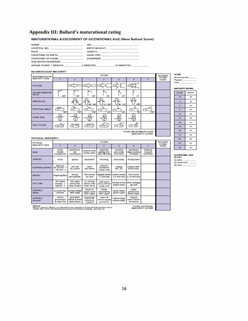
58
Appendix III: Ballard’s maturational rating

59
Appendix IV: Apgar score

60
Appendix V: Definition and staging of perinatal asphyxia.
“Failure to initiate and sustain breathing at birth.”1 PLUS clinical evidence of hypoxic ischemic
encephalopathy Sarnat and Sarnat stage 1, 2 or 3.
Sarnat and Sarnat Clinical Staging of Hypoxic Ischemic Encephalopathy
Variable Stage 1 Stage 2 Stage 3
Level of
consciousness
Alert/Hyperalert Lethargy Coma
Muscle tone Normal Hypotonia Flaccidity
Seizures Absent Focal or Multifocal Generalised
Reflexes
Suck Active Weak Absent
Moro Exaggerated incomplete Absent
Grasp Normal/
Exaggerated
Weak Absent

61
Appendix VI: Standard operating procedures for the measurement of weight, length and
head circumference
Weight: Babies was weighed in the NBU nude in a warm environment using a basin scale with
high sides to ensure baby‟s safety. Three weight readings was taken and an average taken to the
nearest 0.1grams (gm). The scale was checked against a standard weight of two kilograms (kgs)
at the beginning of each day and calibrated to zero.
Length: The length was measured with the help of an assistant using a stadiometer. Three supine
measurements was taken and the average recorded to the nearest 0.1 centimeter (cm).
Head circumference: The head circumference was measured using a tape measure. Three
occipitofrontal circumference measurements was taken and the average recorded to the nearest
0.1cm.
Fluid regimens given, infusions, transfusions, and medications given were recorded.

62
Appendix VII: Study budget
Category Remarks Units Unit cost (Kshs) Total (Kshs)
Proposal development Computer services 70hrs 1 5,000
Printing drafts 4500pgs 25 11,250
Proposal copies 600pgs 15 9,000
Binding 10 500 5,000
Data collection Training day 1 day 10,000
2 research assistants 40 days 1000 80,000
Study materials Study questionnaire 500pgs 10 5,000
Other stationery 20 50 1000
Study equipment Urinary catheters 70 300 21,000
Urine collecting bags 70 200 14,000
Laboratory tests FENa 70 2400 168,000
Creatinine serum 70 600 42,000
Data analysis Statistician 30,000
Thesis write up Comp services 50hrs 5,000
Printing thesis 200pgs 25 5,000
Binding 10 500 5,000
Contingency 50,000
Total 466,250

63
Appendix VIII: Consent form for parents / guardians of participants
TITLE: FRACTIONAL EXCRETION OF SODIUM AS AN EARLY PREDICTOR OF
AKUTE KIDNEY INJURY IN TERM NEONATES WITH PERINATAL ASPHYXIA AT
KENYATTA NATIONAL HOSPITAL
SCOPE: This informed consent is for enrolled participants in the study, and was read to them by
a qualified research assistant before answering the questionnaire.
SERIAL NO: DATE: SITE:
Investigator: Dr. Esther M. Njiru,
Tel 0721-363535
Email: [email protected]
Address- 87822 Mombasa, Kenya
Supervisor: Dr. Bashir Admani,
Tel 0721967818
Email: [email protected]
Part 1: Information Sheet Introduction : you are being invited to take part in this study to assess fractional excretion of
sodium as an early predictor of acute kidney injury in term neonates with perinatal asphyxia at
the Kenyatta National Hospital‟s Newborn Unit.
Perinatal asphyxia is a condition resulting from your new born baby failing to breathe
immediately after birth. This problem may be associated with injury to the kidney and its early
detection will help early initiation of appropriate care.
If you do not understand any words, kindly stop the research assistant as you go through the
information and they will take time to explain. In case of later queries, the investigator is
available on phone any time.
Purpose of the study: This study primarily aims to determine whether the amount of sodium that
your baby passes in urine can be used to find out early whether your baby is likely to develop
kidney injury as a result of being unable to breathe on their own immediately after birth. This
information will help us in making certain recommendations regarding service provision so as to
improve health care.
Procedures: You were provided with a questionnaire. The questionnaire can be answered
individually, or the questions read out to you as you provide the interviewer with the appropriate
response. You may skip any question you wish not to answer. The information is confidential, no
name will be written on the forms, only an identification number and only the research
investigators have access to your details.
Sampling: A urine sample will be collected from your baby by sterile insertion of a catheter
which will then be removed after the sample is obtained. A blood sample will be collected from

64
your baby as well within 24 hours of birth and again at the third day of life to measure the
functions of the kidney.
Risks: The specimens will be collected with utmost care to ensure that your child does not
develop infection in the bladder. The procedure to obtain the blood specimen will be slightly
uncomfortable for your baby. If possible the parent/ guardian or nurse will massage and talk to
the baby to distract from pain. If the child is able to breastfeed this was encouraged, or child
placed at skin to skin contact with the parent.
Benefits: Your baby will benefit from improved quality of services provided at the newborn unit
as a result of this study. You will not cater for the costs of the laboratory tests done.
Voluntariness: The study is fully voluntary. There is no financial rewards to your child for
participating in this study. One is free to participate or withdraw from the study at any point.
Refusal to participate will not affect the care given to your child.
Confidentiality: All information collected will be kept in strict confidence. No information
regarding you, your child or your family will be released to any person without your written
permission. Information will be shared by clinical teams taking care of your child, so that your
child can benefit from the possible interventions available.
Sharing results: We shall share the information that we get from this research with you. Results
will be published at completion of the study so that other interested people may benefit from the
research. However, we will not reveal the identity of your child during the publication of results.
Who to contact: If you have any questions about the study or the results, you can contact the
principal investigator, Dr. Esther M. Njiru on 0721363535
Any question on your rights as a research participant can be addressed by contacting Kenyatta
National Hospital Ethics and Research Committee by calling (254-020) 2726300 Ext 44355
Part II: Certificate of consent.
I, ________________________ guardian of _________________(name of child) has had the
information explained to me.
I AGREE/DISAGREE (cross out as appropriate) to participate/for my child to take part in the
study.
I understand that our participation is fully voluntary and that I can withdraw /withdraw my child
from the study at any point and this will not affect me/ my child‟s care in any way. I have been

65
given adequate opportunity to ask questions and seek clarification on the study and these have
been addressed satisfactorily.
Participant/Guardian‟s signature: _________________ Date: ____________
I, _______________________________________ declare that I have adequately explained to
the above participant/guardian the study procedure and risks and given him/her time to ask
questions and seek clarification regarding the study. I have answered the questions raised to the
best of my ability.
Investigator‟s signature: ________________________ Date: ____________
Only necessary if the parent/guardian cannot read:
I attest that the information concerning this research was accurately explained to and understood
by the caregiver and that informed consent was freely given by the parent/guardian.
Witness‟ signature: ________________________________ Date: _________
Witness‟ name: ___________________________________ Time:_________

66
FOMU YA KUPATA KIBALI CHA WAZAZI / WALEZI WA WASHIRIKI Kifunguo: Hii fomu ya kupata idhini ni kwa ajili ya watoto waliolazwa katika Hospitali ya Taifa
ya Kenyatta, ambao tunuwakaribisha kushiriki katika utafiti. Jina la mradi wa utafiti wetu ni
“Upimaji wa sehemu ya sodiamu kama ishara ya mapema ya kuumia papo hapo figo katika
watoto wanaozaliwa na ukosefu wa hewa”
Mimi ni Dk. Esther Njiru, mwanafunzi katika Chuo Kikuu cha Nairobi kutafuta masomo ya
utaalamu katika afya ya watoto. Mimi ninafanya utafiti juu ya ugonjwa wa kupumua kwa watoto
ambao wamekishazaliwa kwa muda usiozidi siku moja ambayo ni kawaida sana katika nchi hii.
Nitakupa taarifa na kukukaribisha kwa utafiti huu.
Kunaweza kuwa na baadhi ya maneno ambayo huelewi. Tafadhali uliza na mimi nitachukua
muda kueleza. Kama una maswali baadaye, unaweza bado kuniuliza
Sababu ya utafiti: Ugonjwa wa kupumua unaweza kusababisha magojwa mengine kwa kila
kiungo kwa watoto ambao wamekishazaliwa. Utafiti huu utazingatia ugonjwa wa figo kwa hawa
watoto ambao wamekishazaliwa na shida ya kupumua. Ugonjwa huu husababaisha maafa mengi
kwa watoto wachanga ndiposa umuhimu wa kuuangaza mapema na kuutibu mapema.
Maandalizi ya utafiti: Utafiti huu utahusu kupima damu na makojoo kwa mtoto wako ili kuweza
kupata ugonjwa wa figo mapema na kuitibu mapema. Utafiti utafanyiwa kwa njia ya kupitia
mahojiano ya moja kwa moja.
Kushiriki katika utafiti huu ni wa hiari kabisa. Bado utapokea huduma zote katika hospitali hii
hata usipochagua kushiriki. Unaweza kujiondoa katika utafiti huu wakati wowote.
Utafiti utafanyika katika kipindi cha siku tatu za kwanza za maisha ya mtoto wako. Wakati huo,
tutathmini afya ya mtoto wako kila siku.
Maadhara: Tutahakikisha usafi ili mtoto wako asipate maambukizi kwenye mkojo na tutajaribu
iwezekanavyo kuzuia motto asihisi uchungu.
Habari kukuhusu ambayo tutakusanya kutoka mradi wa utafiti huu utakuwa siri.
Mawasiliano: Maswali yoyote yanaweza ulizwa hivi sasa au baadaye, hata pia baada ya utafiti
imeanza. Baadaye ukitaka kuuliza maswali, wasiliana nami Esther M. Njiru kupitia nambari
hizi: 0721363535.
Nimesoma/ Nimesomewa maelezo haya na nimepewa nafasi ya kuuliza maswali kuhusu hayo
maelezo. Nimeidhini kushiriki katika utafiti huu kwa hiari.
Jina lako__________________
Sahihi ya Mshiriki ___________________
Tarehe_________________
Nina uhakika kuwa nimemsomea mwakilishi fomu hii, nilihakikisha kwamba mshiriki
ameelewa.
Nilithibitisha kuwa mshiriki alipewa nafasi ya kuuliza maswali kuhusu utafiti nakuyajibu vema
kwa kadri ya uwezo wangu. Mimi nathibitisha kwamba mwakilishi hakulazimishwa kutoa kibali
Jina la Mtafiti / Mtu kuchukua kibali________________________