fracture nasal bones
DESCRIPTION
FRACTURA NASALHUESOS NASALESHUESOS PROPIOS NARIZMANEJOTRANSCRIPT

ISSN 2250-0359 Volume 3 Issue 1.5 2013
Fracture Nasal Bones 1 Balasubramanian Thiagarajan 2 Venkatesan Ulaganathan
1 Stanley Medical College
2 Meenakshi Medical College
Abstract:
Nose is the most prominent part of the face, hence it is likely to be the most common
structure to be injured in the face. Although fractures involving the nasal bones are very
common, it is often ignored by the patient. Patients with fractures of nasal bone will
have deformity, tenderness, haemorrhage, edema, ecchymosis, instability, and
crepitation. These features may be present in varying combinations. This article discusses
the pathophysiology of these fractures, role of radiography and ultrasound in their diagnosis
and their management.
Introduction:
Nasal bone fractures are common because:
1. Nose happens to be the most prominent portion of the face
2. Increasing number of road traffic accidents1
3. Increasing incidence of domestic violence 2
4. Increase in the number of individuals taking part in contact sports 3
Anatomy:
Nasal bones are paired bones. Both these bones project like a tent on the frontal process of
maxilla. In the midline they articulate with one another. Just under this midline articulation
lies the nasal septum. Superiorly the nasal bones are thicker where it articulates with the
nasal process of frontal bone. This area is relatively stable and firm. Nasal bone fractures
commonly occur at the transition zone between the proximal thicker and distal thinner
portions. This zone precisely corresponds to the lower third of the nasal bone area.
Fractures involving nasal bones if not properly and promptly treated leads to:
1. Nasal deformities
2. Intranasal dysfunction like nasal block
Fracture nasal bone is known to cause higher incidence of morbidity and complications
when compared that of fractures involving other facial bones 4. In order to treat this
condition properly it is necessary to accurately diagnose this condition by:
1. Looking for crepitus and tenderness over the nasal bone area
2. Radiographic evaluation of nasal bones. Radiography helps in diagnosis and
classification of nasal bone fractures, and also in checking the adequacy of
reduction 5.
Clinicians are more interested in knowing:
1. Location of fracture site (like sidewall, dorsum, or the entire nasal bone)
2. To know whether the fracture involves the right nasal bone / left nasal bone or both
sides
3. Whether there is any displacement of the fractured fragments (medial / lateral),
presence of absence of comminution.
4. To identify the presence of concurrent fractures to other facial bones / nasal septum.
When there is the presence of fractures involving other facial bones / severe
fractures of nasal septum it is prudent to perform open reduction.
Pathophysiology:
The following points should be borne in mind before attempting to understand the
pathophysiological factors that lead to fractures involving nasal bones.
1. Nasal bones and underlying cartilage are susceptible for fracture because of their
more prominent and central position in the face.
2. These structures are also pretty brittle and poorly withstands force of impact.
3. The ease with which the nose is broken may help protect the integrity of the neck,
eyes, and brain. Thus it acts as a protective mechanism.
4. Nasal fractures occur in one of two main patterns- from a lateral impact or from a
head-on impact. In lateral trauma, the nose is displaced away from the midline on
the side of the injury, in head-on trauma, the nasal bones are pushed up and splayed
so that the upper nose (bridge) appears broad, but the height of the nose is
collapsed (saddle-nose deformity). In both cases, the septum is often fractured and
displaced.
5. The nasal bone is composed of two parts: A thick superior portion and a thin
inferior portion. The intercanthal line demarcates these two portions. Fractures
commonly occur below this line.
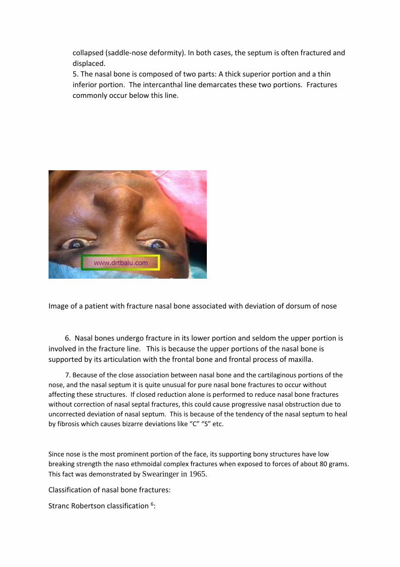
Image of a patient with fracture nasal bone associated with deviation of dorsum of nose
6. Nasal bones undergo fracture in its lower portion and seldom the upper portion is
involved in the fracture line. This is because the upper portions of the nasal bone is
supported by its articulation with the frontal bone and frontal process of maxilla.
7. Because of the close association between nasal bone and the cartilaginous portions of the
nose, and the nasal septum it is quite unusual for pure nasal bone fractures to occur without
affecting these structures. If closed reduction alone is performed to reduce nasal bone fractures
without correction of nasal septal fractures, this could cause progressive nasal obstruction due to
uncorrected deviation of nasal septum. This is because of the tendency of the nasal septum to heal
by fibrosis which causes bizarre deviations like “C” “S” etc.
Since nose is the most prominent portion of the face, its supporting bony structures have low
breaking strength the naso ethmoidal complex fractures when exposed to forces of about 80 grams.
This fact was demonstrated by Swearinger in 1965.
Classification of nasal bone fractures:
Stranc Robertson classification 6:
Stranc and Robertson suggested that lateral forces accounted for the majority of nasal bone
fractures. They also inferred that younger patients tend to have fracture dislocation
involving large segments while older patients tended to have comminuted fractures. In 1978
Stranc and Robertson came out with their classification of nasal bone fracture based on the
direction of impact and the associated damage. In this classification they also took into
consideration the degree of damage to nasal bones and the nasal septum. This classification
was based on the clinical examination of the nose and face. It did not take into account
radiological findings.
Type I injury:
Fractures due to this type of injury does not extend behind the imaginary line drawn from
the lower end of nasal bone to the anterior nasal spine In this type of injury the brunt of the
attack is borne by lower cartilaginous portion of the nasal cavity and the tip of the nasal
bones. This type of injury may cause avulsion of upper lateral cartilages, and occasionally
posterior dislocation of septal and alar cartilages.
Type II injury:
This type of injury involves the external nose, nasal septum and anterior nasal spine.
Patients with this type of injury manifest with gross deviations involving the dorsum of the
nose including splaying of nasal bones, flattening of dorsum of nose and loss of central
support of the nose.
Type III injury:
This injury involves orbit and intracranial structures.
Harrison’s classification:
Fractures involving nasal bones are divided into three categories depending on the degree of damage, and its management. Class I fractures: Very little force is sufficient to cause a fracture of nasal bone. It has been estimated to be as little as 25-75 pounds / sq inch. Class I fractures are mostly depressed fractures of nasal bones. The fracture line runs parallel to the dorsum of the nose and naso maxillary suture and joins at a point where the nasal bone becomes thicker. This point is about 2/3 of the way along its length. The fractured segment usually regains its position because of its attachment along its lower border to the upper lateral cartilage. The nasal septum is not involved in this particular injury. Class I fractures do not cause gross lateral
displacement of nasal bones, though a persistent depressed fragment may give it the appearance. In children these fractures could be of green stick variety and a significant nasal deformity may develop subsequently during puberty when nasal growth accelerates. Clinically this fracture will present as a depression over the nasal bone area. There may be tenderness and crepitus over the affected nasal bone. Radiological evidence may or may not be present. In fact class I fracture of nasal bone is purely a clinical diagnosis.
Class II fractures: These fractures cause a significant amount of cosmetic deformity. In this group not only the nasal bones are fractured, the underlying fronto nasal process of the maxilla is also fractured. The fracture line also involves the nasal septum. This condition must be recognised clinically because for a successful result both the nasal bones as well as the septum will have to be reduced. Since both the nasal bones and the fronto nasal process of maxilla would have absorbed a considerable amount of force, the ethmoidal labyrinth and the adjacent orbit should be intact. The precise nature of the deformity depends on the direction of the blow sustained. A frontal impact may cause comminuted fracture of nasal bones causing gross flattening and widening of the dorsum of the nose. A lateral blow of similar magnitude is likely to produce a high deviation of the nasal skeleton. The perpendicular plate of ethmoid is invariably involved in these fractures, and is characteristically C shaped (Jarjaway fracture of nasal septum).
Class III fractures: Are the most severe nasal injuries encountered. This is caused by high
velocity trauma. It is also known as naso orbital fracture / naso ethmoidal fracture. Recent
term to describe this class (Naso orbito ethmoid fracture) indicates the clinical importance
of orbital component in these injuries. These fractures are always associated with Le Fort
fracture of the upper face involving the maxilla also. In these fractures the nasal bone along
with the buttressing fronto nasal process of maxilla fractures, telescoping into the
ethmoidal labyrinth. Two types of naso ethmoidal fractures have been recognised:
Type I: In this group the anterior skull base, posterior wall of the frontal sinus and optic
canal remain intact. The perpendicular plate of ethmoid is rotated and the quadrilateral
cartilage is rotated backwards causing a pig snout deformity of the nose. The nose appears
foreshortened with anterior facing nostrils. The space between the eyes increase
(Telecanthus), the medial canthal ligament may be disrupted from the lacrimal crest.
Type II: Here the posterior wall of the frontal sinus is disrupted with multiple fractures
involving the roof of ethmoid and orbit. Sphenoid and parasellar regions may sometimes be
involved. Since the dura is adherent to the roof of ethmoid fractures in this region causes
tear in the dura causing csf rhinorrhoea. Pneumocranium and cerebral herniation may
complicate this type of injury.

Figure showing the three types of nasal bone fractures
Murray’s classification 7:
Murray etal after examining nearly 70 patients with fracture nasal bones classified them
into 7 types. This classification was based on damage suffered by the nasal septum. This is
actually a pathological classification.
Clinical pointers towards the diagnosis of fractures involving nasal bones:
1. Injuries involving middle third of face
2. History of bleeding from nose following injury
3. Oedema over dorsum of nose
4. Tenderness and crepitus over nasal bone area
5. Eyelid oedema
6. Subcutaneous emphysema involving eyelids
7. Periorbital ecchymosis
According to Sharp 8 X-rays of nasal bone fails to reveal fractures in nearly 50% of the
patients.
Clinical examination:
This should include careful examination to rule out deformities involving nose and middle
third of face. Clinical photograph of the patient should be taken in order to document the
deformity. Patient should be quizzed regarding the presence of deformities in the area prior
to injury. Acute injury photographs will help the surgeon to convince the patient that
fracture reduction has been done in an appropriate manner. Studies reveal that nearly 30%
of the patients 9 are not satisfied with the post reduction outcome.
Radiology:
X-ray of nasal bone has very minimal role in the diagnosis of fractures involving the nasal
bones. CT scan of nose and sinuses helps in identifying fractures involving other facial bones
and in Lefort II and Lefort III fractures. Ultrasound using 10 MHz probe gives a clear view of
the nasal bone area thereby facilitating easy identification of fractures. It also has the
advantage of nil radiation hazard to the patient. Many images can be taken without any
problem. It is also cost effective. According to Lee the accuracy of ultrasound in identifying
fracture nasal bone was close to 100% while for conventional radiographs it was close to
70%.

Plain X-ray nasal bone revealing fracture line
Axial CT of nose and sinuses showing buckling of nasal septum due to fracture
Management:
If fractures of nasal bones are left uncorrected it could lead to loss of structural integrity and
the soft tissue changes that follow may lead to both unfavourable appearance and
function. The management of nasal fractures is based solely on the clinical assessment of
function and appearance; therefore, a thorough physical examination of a decongested
nose is paramount.
Patients with fractures involving nose will have intense bleeding from nose making
assessment a little difficult. Bleeding must first be controlled by nasal packing. These
patients also have considerable amount of swelling involving the dorsum of the nose,
making assessment difficult. These patients must be conservatively managed for at least 3
weeks for the oedema to subside to enable precise assessment of bony injury. According to
Cummins Fracture reduction should be accomplished when accurate evaluation and
manipulation of the mobile nasal bones can be performed; this is usually within 5-10 days in
adults and 3-7 days in children 11. Reduction is ideally performed immediately after injury

before oedema sets in. If oedema has already set in it is prudent to wait for it to subside
because it is difficult to ascertain adequacy of reduction in the presence of oedema 12.
1. Closed reduction
2. Open reduction
3. Conservative management
Closed reduction:
This is the most preferred treatment modality in all acute phases of fractured nasal bones.
Even if large deviations are seen closed reduction can be attempted prior to rhinoplasty as
this would simplify the task of the plastic surgeon.
Indications for closed reduction according to Bailey:
1. Unilateral / Bilateral fracture of nasal bones
2. Fracture of nasal septal complex with nasal deviation of less than half of the width of
the nasal bridge
Closed reduction can be performed under local / general anaesthesia. This decision should
be made by the surgeon taking the patient into confidence. There is no difference in the
results produced between surgeries performed under local anaesthesia and general
anaesthesia 10. Patients seem to tolerate fracture reduction under local anaesthesia 14.
Preoperative profile photograph of the patient is a must. This will give a general idea about
adequacy of reduction.
Local anaesthesia:
This requires a thorough understanding of innervation of nose. Innervation of nose:
For effective administration of local anaesthesia a complete understanding of sensory
innervation of nose and nasal cavity is a must. Innervation of nose can be divided into:
1. Innervation of mucosa within the nasal cavity
2. Innervation of external nose and its skin covering
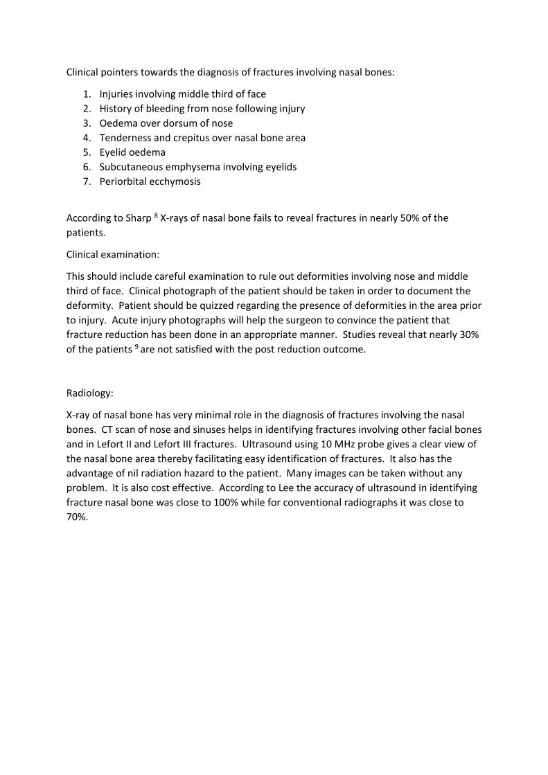
Sensory innervation of external nose:
External nose and its skin lining is innervated by ophthalmic and maxillary divisions of
trigeminal nerve.
Superior aspect of the nose is supplied by – Supratrochlear and Infratrochlear nerves
(branches of trigeminal nerve) and external nasal branch of anterior ethmoidal nerve.
Inferior and lateral parts of the nose – is supplied by infraorbital nerve.
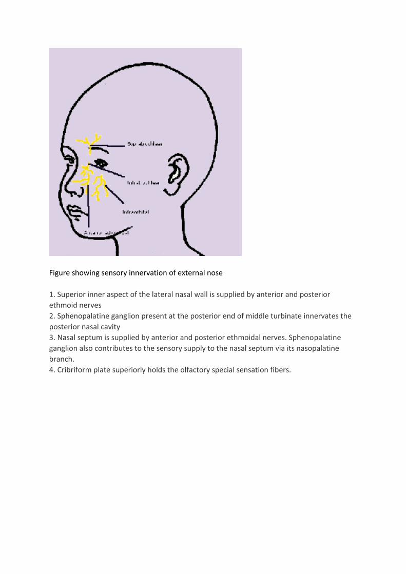
Figure showing sensory innervation of external nose
1. Superior inner aspect of the lateral nasal wall is supplied by anterior and posterior
ethmoid nerves
2. Sphenopalatine ganglion present at the posterior end of middle turbinate innervates the
posterior nasal cavity
3. Nasal septum is supplied by anterior and posterior ethmoidal nerves. Sphenopalatine
ganglion also contributes to the sensory supply to the nasal septum via its nasopalatine
branch.
4. Cribriform plate superiorly holds the olfactory special sensation fibers.
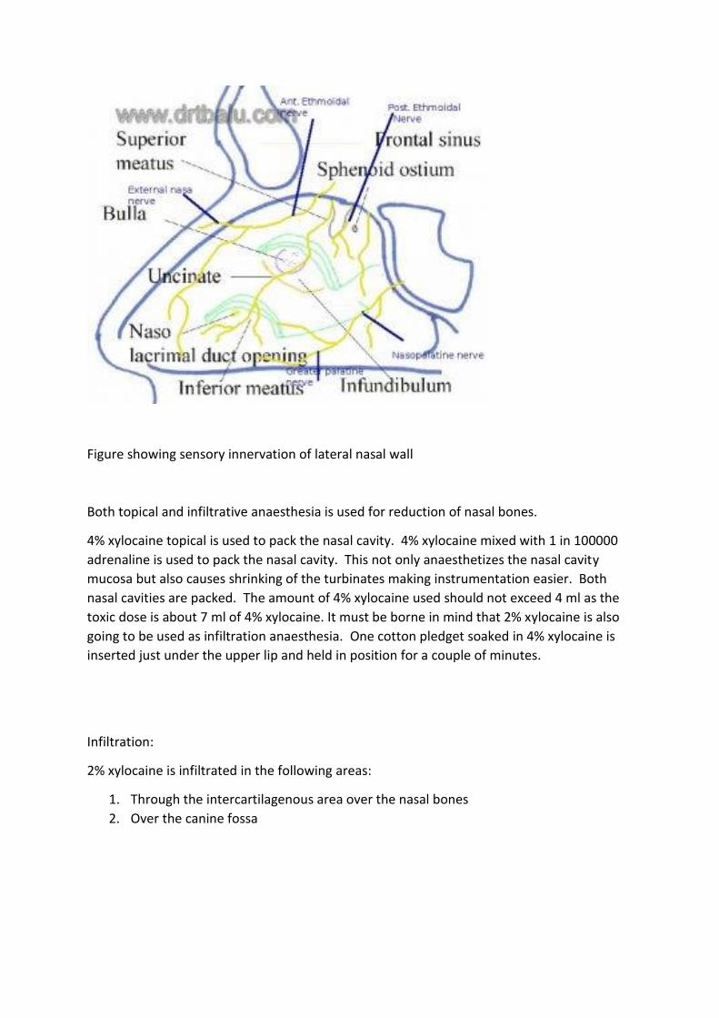
Figure showing sensory innervation of lateral nasal wall
Both topical and infiltrative anaesthesia is used for reduction of nasal bones.
4% xylocaine topical is used to pack the nasal cavity. 4% xylocaine mixed with 1 in 100000
adrenaline is used to pack the nasal cavity. This not only anaesthetizes the nasal cavity
mucosa but also causes shrinking of the turbinates making instrumentation easier. Both
nasal cavities are packed. The amount of 4% xylocaine used should not exceed 4 ml as the
toxic dose is about 7 ml of 4% xylocaine. It must be borne in mind that 2% xylocaine is also
going to be used as infiltration anaesthesia. One cotton pledget soaked in 4% xylocaine is
inserted just under the upper lip and held in position for a couple of minutes.
Infiltration:
2% xylocaine is infiltrated in the following areas:
1. Through the intercartilagenous area over the nasal bones
2. Over the canine fossa
Most of class I fractures can be reduced by closed reduction and immobilization using
Plaster of Paris cast. In majority of cases digital pressure alone is sufficient for the job.
Figure showing digital pressure being applied from the head end of the patient to reduce
the fractured nasal bone
If the fractured fragments are impacted then a Welsham's forceps will have to be used to
disimpact and reduce the fractured nasal bone. In the event of using Welsham's forceps to
disimpact the nasal bone, there will be extensive trauma to the nasal mucosa causing
epistaxis. The nasal cavity of these patients must be packed with roller gauze, with
application of an external splint to stabilise the bone. In these patients it is also imperative
to elevate the collapsed nasal septum using Ash forceps.
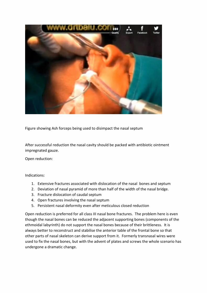
Figure showing Ash forceps being used to disimpact the nasal septum
After successful reduction the nasal cavity should be packed with antibiotic ointment
impregnated gauze.
Open reduction:
Indications:
1. Extensive fractures associated with dislocation of the nasal bones and septum
2. Deviation of nasal pyramid of more than half of the width of the nasal bridge.
3. Fracture dislocation of caudal septum
4. Open fractures involving the nasal septum
5. Persistent nasal deformity even after meticulous closed reduction
Open reduction is preferred for all class III nasal bone fractures. The problem here is even
though the nasal bones can be reduced the adjacent supporting bones (components of the
ethmoidal labyrinth) do not support the nasal bones because of their brittleness. It is
always better to reconstruct and stabilise the anterior table of the frontal bone so that
other parts of nasal skeleton can derive support from it. Formerly transnasal wires were
used to fix the nasal bones, but with the advent of plates and screws the whole scenario has
undergone a dramatic change.
Ellis procedure of management of Class III fractures:
Aims of the procedure include:
1. Provision of adequate surgical exposure to provide an unobstructed view of all
components of the fracture.
2. The medial canthal ligament should be identified. This is rarely avulsed and is usually
attached to a large fragment of bone. Once identified the ligament should be reattached
and secured to the lacrimal crest. This step will avoid the future development of
telecanthus.
3. Reduction and reconstruction of medial orbital rim. This can be achieved by use of
transnasal 26 gauge wires. If plates are used they should be very thin otherwise they will
become conspicuous once the wound has healed.
4. Reconstruction of medial orbital wall and floor with bone grafts
5. Realignment of nasal septum
6. Augmentation of dorsum of the nose by the use of bone grafts
7. Accurate soft tissue readaptation should be encouraged by placing splints.
Complications of nasal bone fracture:
1. Cosmetic deformity (saddle nose, pig snout deformity). This is actually common in
patients who have septal hematoma following injury to nasal bones.
2. Persistent septal deviation
3. CSF leak
4. Orbital oedema / complications
5. Nasal block / compromise of nasal functions
Nasal injuries in children:
Children’s nose is mostly cartilaginous in nature containing small bones that are soft and
more compliant more capable of absorbing forces due to injury. It is also a common fact
that birth trauma could be the cause for septal deviations in these patients. Septal
hematoma is more common in children 13. In children it is better to avoid open reduction
procedures and stick to closed manipulation techniques. Digital manipulation is the best
technique. While attempting to perform digital reduction manipulation the surgeon should
be aware that the feel of bone snapping back into place is not evident in children. Careful
visual assessment of the shape of the nose is a must to ascertain adequacy of reduction.
References:
1. Atighechi S, Karimi G: Serial nasal bone reduction: A new approach to the management of nasal bone fracture. J Craniofac Surg 20:49, 2009
2. Erdmann D, Follmar KE, Debruijn M, et al. A retrospective analysis of facial fracture etiologies. Ann Plast Surg. Apr 2008;60(4):398-403
3. Swenson DM, Yard EE, Collins CL, Fields SK, Comstock RD. Epidemiology of US high school sports-related fractures, 2005-2009. Clin J Sport Med. Jul 2010;20(4):293-9
4. Schultz RC: One thousand consecutive cases of major facial injury. Review Surg 27:394, 1970
5. Yabe T, Ozawa T, Sakamoto M, et al: Pre- and postoperative x-ray and computed tomography evaluation in acute nasal fracture. Ann Plast Surg 53:547, 2004
6. Stranc MF, Robertson GA: A classification of injuries of the nasal skeleton. Ann Plast Surg 2:468, 1979.
7. Murray JA, Maran AG, Busuttil A, et al: A pathological classification of nasal fractures. Injury 17:338, 1986.
8. Sharp JF, Denholm S. Routine x-rays in nasal trauma: the influence of audit of clinical practice. J Royal Soc Med 87:153-154, 1994
9. Mayell MJ (1973) Nasal fractures their occurrence, management and some late results. Journal of the Royal College of Surgeons of Edinburgh 18: 31-36.
10. Waldron J, Mitchell DB, Ford G. Reduction of fractured nasal bones; local versus
general anaesthesia. Clin Otolaryngol Allied Sci. Aug 1989;14(4):3579
11. Kerr AG. Scott-Brown's Otolaryngology. 6th. Oxford, England: Butterworth-
Heinemann; 1997:4/16/6-4/16/11.
12. Michael G Fractures of nasal skeleton Scott Brown’s otolaryngology 6th edition 13. Toriumi DM, Ries WM (1993) Innovative surgical management of the crooked nose.
Facial Plas Surg Clin North Am 1:63-77 14. Waldron J, Mitchell DB, Ford G (1989) Reduction of fractured nasal bones; local
versus general anaesthetic. Clinical Otolaryngology e. Otolaryngologic Clinics of North America 8: 663-677.
