fraser c. henderson sr, md ednf, baltimore, august 14th, 2015 · r s howard, f c henderson, n p...
TRANSCRIPT

Fraser C. Henderson Sr, MD
EDNF, Baltimore, August 14th, 2015

Complex neurological syndromes Chronic ,severe pain Minimal radiological findings Multiple systemic dysfunction multiplicity of overlapping syndromes

Lax ligaments result in abnormal bending
cranio-cervical junction and spine with the result of stretching and deformation of CNS
EDS may present with a variety of neurosurgical issues
Neurobiology Outcome of 2 studies of craniospinal fusion
for craniocervical instability Other neurological conditions associated with
EDS

Chiari Malformation
Atlanto-axial or cervical instability
TMJ Syndrome
Occipital neuralgia
Thrombosis, hypercoagulability
Hughes Syndrome , migranous TIAs
Pseudotumor cerebri
Orthostatic intolerance
Limbic encephalopathy, NeuroBehcet’s
Tethered cord syndrome
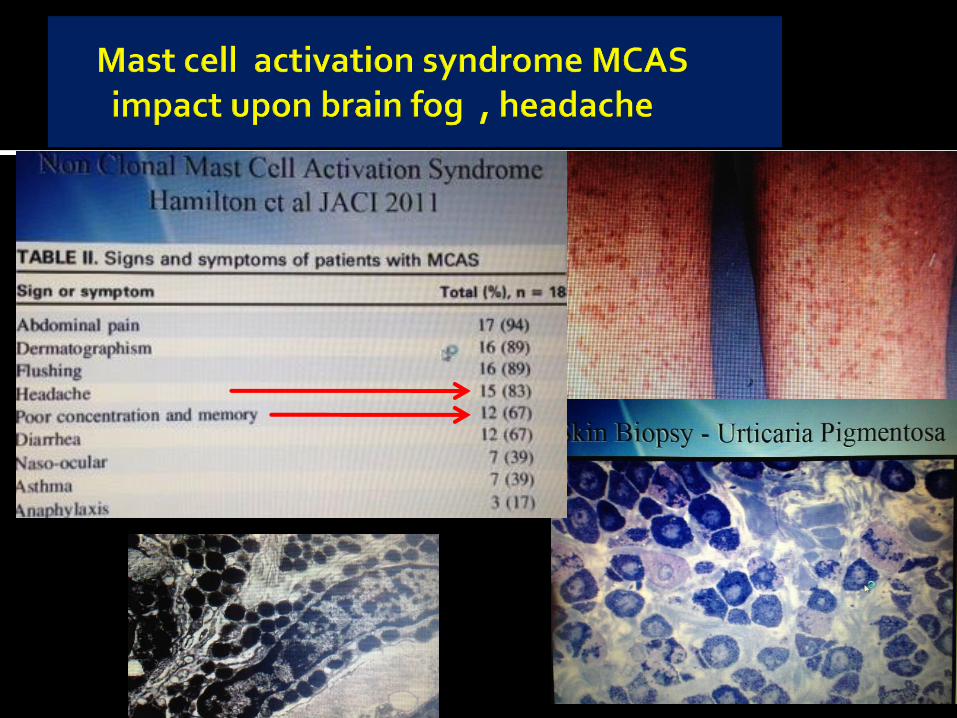
Mast Cell release Disorder
neck muscle spasm thoracic imbalance
Cranio-vertebral instability (floppy head)
Causes of Headache in EDS


12.7% of all Chiari
patients have connective tissue disorder
most EDS patients with Chiari malformation have CCI
Page 7
Milhorat TM, et al. Syndrome of occipitoatlantoaxial hypermobility, cranial settling, and Chiari malformation Type I in patients with hereditary disorders of connective tissue. J Neurosurg Spine 7:601-609, 200

Clumsiness, poor coordination ….the relatively high rate of dyslexia and dyspraxia suggests possibility of CNS involvement in this condition.
N. Adib, K. Davies, R. Grahame, P. Woo and K. J. N. Adib, and K. J. Murray1
Rheumatology 2005;44:744–750 Joint hypermobility syndrome in childhood

The neurobiology of
stretch injury

Ligamentous laxity results in deformation of the nervous system
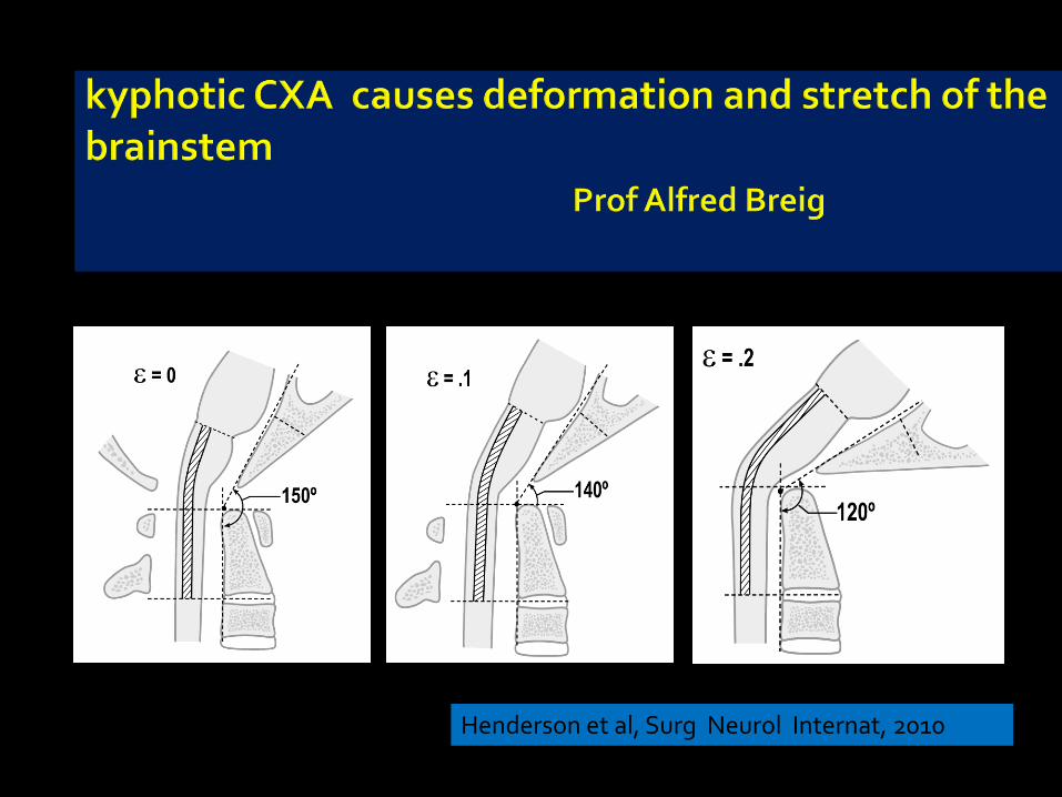
Henderson et al, Surg Neurol Internat, 2010
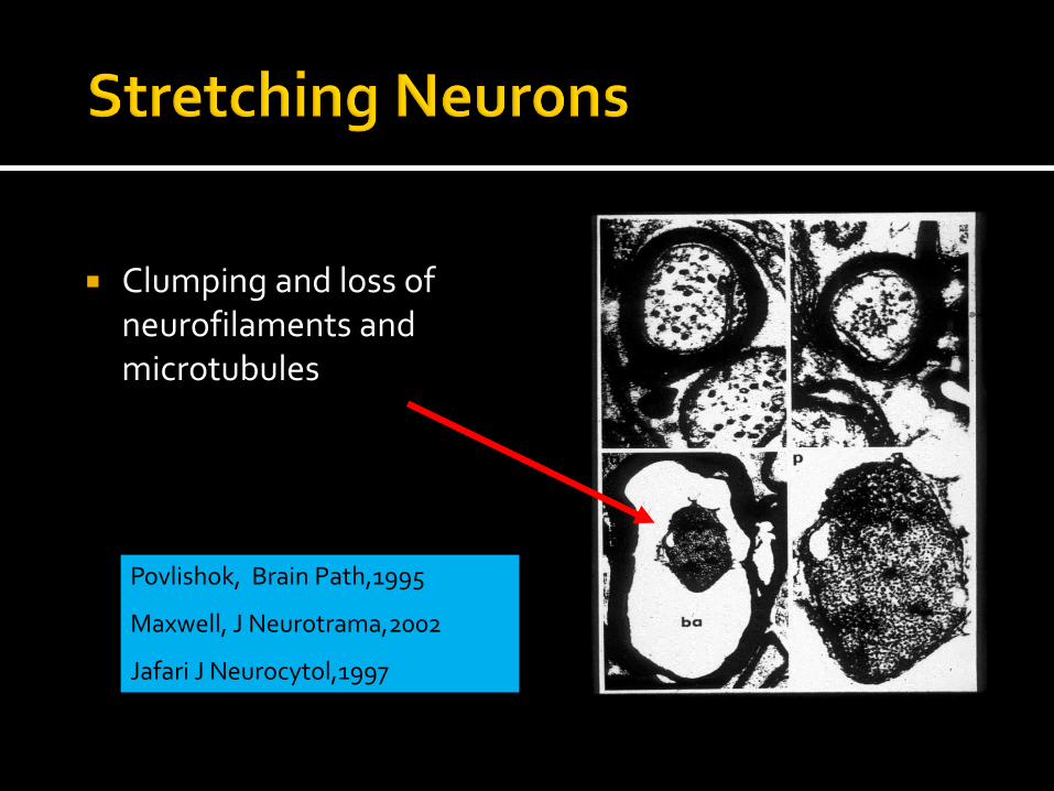
Clumping and loss of
neurofilaments and microtubules
Povlishok, Brain Path,1995
Maxwell, J Neurotrama,2002
Jafari J Neurocytol,1997

14
Mouse optic nerve stretched 2mm (ε = .2)
50 um
Saatman KE J Cereb Blood Flow Metab. 23(1):34-42, 2003

Calcium influx after stretch injury
Wolf et al, J Neurosci 2001
Distortion of
Na+ mechano-receptors
Reversal Na+ Ca++ gradient
Depolarizatof
voltage- gated Ca++
channels

16
Pre stretch Post-stretch
Calcium influx after stretch injury, blocked by TTX
Wolf, Sties, Lizard, Smith, J Neurosci 2001

17
Secondary injury Up-regulation of NMDA
receptors Vulnerability to nitrous
oxide and reactive oxygen species
Mitochondrial dysfunction and DNA fragmentation
Programmed cell death (apoptosis)
Arundine M et. al. J Neuroscience. 2004, 24(37): 8106-8123)

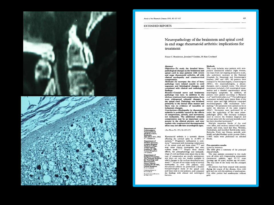
Basilar invagination causes sleep apnea
Reduction and stabilization in pts with RA resulted in resolution of sleep apnea, in all cases
Howard,Henderson et al. Ann Rheum Dis,1993 Menezes,J NSGY,1985

Ann Rheum Dis. 1994 February; 53(2): 134–136. PMCID: PMC1005266
Respiratory abnormalities due to craniovertebral junction compression in rheumatoid disease.
R S Howard, F C Henderson, N P Hirsch, J M Stevens, B E Kendall, and H A Crockard Harris Unit, National Hospital for Neurology and Neurosurgery, Queen Square, London, United Kingdom.
This article has been cited by other articles in PMC. Abstract OBJECTIVE--To assess the extent and severity of respiratory insufficiency
associated with severe rheumatoid atlantoaxial dislocation and its relation to compression of the neuraxis. METHODS--Twelve patients with severe atlantoaxial dislocation due to rheumatoid disease were studied. Detailed clinical, CT myelography and respiratory assessment including nocturnal oximetry, were performed on all patients

Mechanical forces modulate gene
expression and biochemical composition , directly effecting neurons, making them more sensitive and more vulnerable to injury.
This injury arising may underlie many of the
neurological deficits that we observe in “floppy head syndromes”
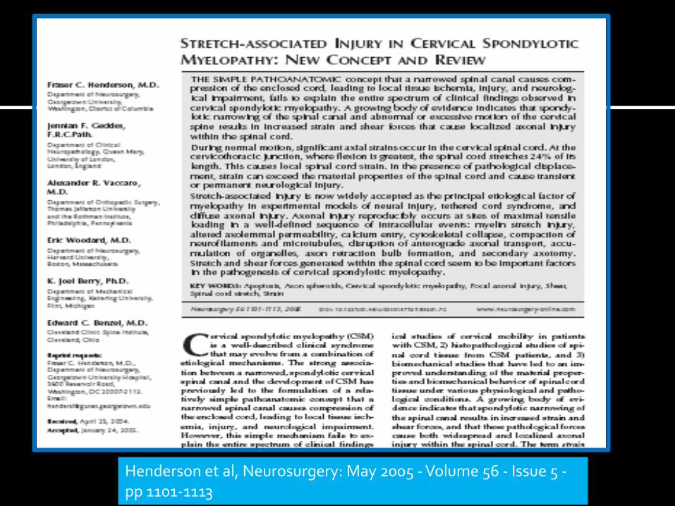
Henderson et al, Neurosurgery: May 2005 - Volume 56 - Issue 5 - pp 1101-1113


The Consensus Statement Chiari Syringomyelia Foundation Multi-disciplinary Colloquium for Craniocervical Hypermobility
San Francisco, October 19th, 2013 University College ,London, GB General Hospital of Chinese People’s Armed Police Forces, Beijing, China Johns Hopkins Cleveland Clinic UCLA Tufts Medical University of South Carolina Georgetown University University of Washington/ Seattle Children’s Hospital University of Utah Health Care Moriguchi -Ikuno MemorIal Hospital, Japan North Shore University Hospital The Canine Chiari Institute Greater Baltimore Medical Center University of Maryland Doctor’s Hospital, Maryland Advocate Children’s Hospital, Chicago

Headache, suboccipital or neck pain Diplopia , decreased or blurred vision Dizziness, vertigo, imbalance tinnitus or decreased hearing Dysautonomia, POTS, syncope or pre-syncope Dysarthria, dysphagia,choking Altered breathing and sleep architecture Weakness, clumsiness, spasticity, Altered sensation, paresthesia, dysesthesia Gait changes, urinary urgency or frequency
Chiari Syringomyelia Foundation Multi-disciplinary
The Consensus Statement Chiari Syringomyelia Foundation Multi-disciplinary Colloquium for Craniocervical Hypermobility Francisco, October 19th, 2013

• Clivo-axial angle
• Grabb-Mapstone-
Oakes measurement
• Harris’ measurement
The Consensus Statement Chiari Syringomyelia Foundation Multi-disciplinary
Colloquium Craniocervical Hypermobility Francisco, Oct
19th, 2013

130°
Chiari decompression
failed if the CXA < 135º
Kim, Rekate, Klopfenstein, Sonntag
2004
Chiari decompression
failed to improve
syringomyelia if CAA <
127º KUbota 2004
• ABNORMAL

Grabb Oakes Measurement

Harris Measurement
• Of 400 normal subjects, none had basion to PAL >12mm
• “In adults, the occipito-vertebral junction can be considered normal when both the basion axial interval and the basion dental interval are 12mm or less”
Harris JH, Carson GC, Wagner LK: Radiological
diagnosis of traumatic Occipitovertebr4al Dissociation

Harris measurement measures distraction and pathological translation

How do we treat craniocervical instability ?

• Multidisciplinary, centralized
• Surgery = the last option, after a thorough medical
vetting
• r/o other causes : co-morbid conditions, MS,
dystrophy, mitochondrial disorders, vitamin
deficiencies, Lymes etc
• brace, activity limitation
• physical therapy : isometrics, sagittal balance, core
strengthening, cardio
• Pain management : preop , postoperative plan
• Psychiatric evaluation where indicated

MCAS: aspirin 325 mg po tid
amitriptylline 25 mg po qhs
quercetin 500 mg po bid
zantac 150 mg twice daily
zyrtec 10 mg daily
valium 5 mg daily
Singulair 10 mg daily
cromolyn sodium I puff twice daily
Nutritional replacement : B12,
thiamine, pantothenate, vit D
Hydrocortisone 5 mg twice daily

• POTS : propranolol 20 mg twice daily for
tachycardia, improves sleep
fludrocortisone- volume
midodrine - vasoconstriction
Nunn salt tablets
Gastroparesis: pyridostigmine,
lubiprostone,
methyl-naltrexone
Hypercoagulability: full evaluation,
treatment with anticoagulants
Pseudotumor cerebri : Diamox 250 mg twice daily

primary care physician must be involved for ongoing
medical care - POTS, adrenal insufficiency,
gastroparesis, mal-absorption issues, poor sleep
architecture, dystonia
Confirm EDS diagnosis with geneticist before seeing the
surgeon
complete diagnosis of active surgical problems will
usually not be accomplished in the first appointment
Plan on several Follow up visits

EDS is one of the most complex disorders in medicine If you are willing to risk your life on the operating
table, then you will want the most precise diagnosis as to what is causing your most significant problem
It takes 13 years to train a neurosurgeon much longer time for the neurosurgeon to develop
the expertise to know when not to operate Very important to establish a relationship of mutual
trust and confidence in your neurosurgeon, and this trust begins with a careful diagnostic process

Flexion extension MRI
reveals craniocervical instability
assess the clivo-axial angle
assess instability or deformity of the cervical spine
CT Scan : rotation of the neck
to assess C1C2 subluxation
Scans may need to be repeated
Diagnostic tests may take a long time- but the surgeon
is trying to understand you and the problem
If the surgeon is on the fence about surgery, he will
need to trust you
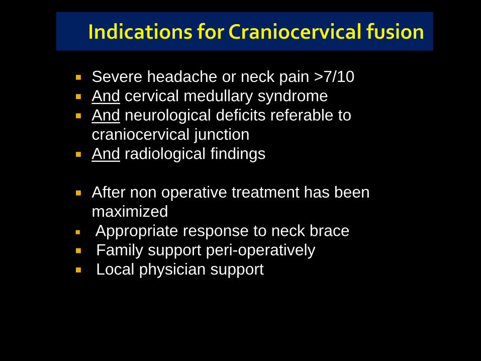
Severe headache or neck pain >7/10
And cervical medullary syndrome
And neurological deficits referable to
craniocervical junction
And radiological findings
After non operative treatment has been
maximized
Appropriate response to neck brace
Family support peri-operatively
Local physician support

> 7/10
After r/o
subaxial segmental instability
Pseudtotumor
Tmj dysfnc
Migraine
MCAS
Tethered cord
Intracranial thrombosis

Vertigo Headaches
Balance problems
Upper extremity numbness
Dizziness
Speech problems
Neck pain
Memory problems
Upper extremity weakness
Walking
Fainting
Hearing problems
Swallowing/choking problems
Lower extremity numbness
Visual problems
Numbness in back
Syncopal episodes
Photosensitivity , hyperacusis

Tender C1C2
Decreased gag reflex
Hypoesthesia to pinprick
Decreased vibratory sensation
Hyperreflexia
Dysdiadochokinesia
Romberg
Difficulty with heel to toe walking
Abnormal gait
Weakness
Loss of abdominal reflex
Babinski, Hoffman’s signs
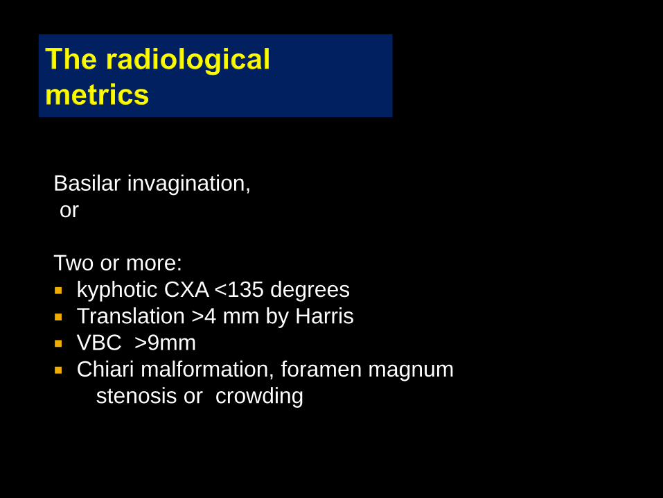
Basilar invagination,
or
Two or more:
kyphotic CXA <135 degrees
Translation >4 mm by Harris
VBC >9mm
Chiari malformation, foramen magnum
stenosis or crowding

• Positioned prone in head holder Fluoro CT unit
• Brainstem and spinal somato-
sensory evoked potentials • Very careful and precise
positioning of the cranio-cervical junction and cervical spine
• Gentle handling of soft tissue


10% PICA extracranial origination
2 % vertebral artery passing
beneath the posterior arch proatlantal variant 10% muscular branch wide
dissection may result in loss of collateral flow from occipital artery
risk of VA injury 2cm from midline, wherein the VA lying postero-lateral to the ring of C1

45
• 10% patients at risk for VA
injury
• Mandel Spine ,2000
•
EDS HAVE Small pedicles
• Most EDS have medially
placed VA

Pitfall: injury to occipital nerve
46

fflexion extextension
Traction reduction To reduce the basilar invagination

Open reduction to normalize the clivo-axial angle and relationship of the skull to spine
Clivoaxial angle <135°
Normal Clivoaxial angle
Modified from Kim, Rekate, Klopfenstein, Sonntag 2004

Clivo-axial angle
Basion to odontoid interval
Occipito-cervical angle
Mandible to C2 interval
Orbital Axial angle
Iterative process
Radiological Measurements

Fusion and Stabilization

Postoperative wound at 7 days and 1 month

5 weeks after fusion/ stabilization occiput to C1C2 Few patients complain of lost range of motion
Does cranios-pinal fusion relieve the pain and improve neurological deficits?
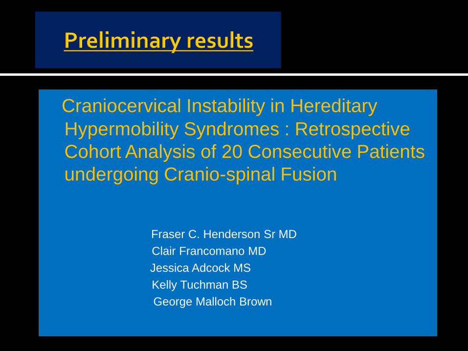
Craniocervical Instability in Hereditary
Hypermobility Syndromes : Retrospective
Cohort Analysis of 20 Consecutive Patients
undergoing Cranio-spinal Fusion
Fraser C. Henderson Sr MD
Clair Francomano MD
Jessica Adcock MS
Kelly Tuchman BS
George Malloch Brown

Headaches (100%)
Fatigue (100%)
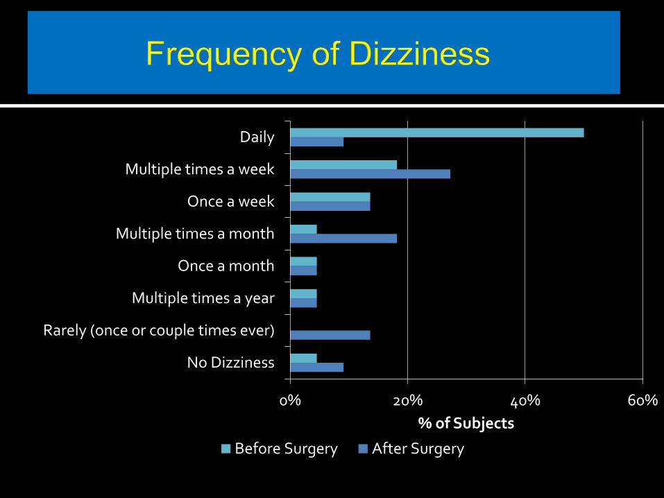
Dizziness (95.4%)
Muscle pain (95.4%)
Upper extremity weakness (90.9%)
Joint pain (86.3%)
Neck pain (83.3%)
Balance problems (82.8%)
Memory problems (81.8%)
Night awakenings (81.8%)
Upper extremity numbness (77.3%)
Walking problems (77.3%)
Hands and feet turning cold (72.7%)
Lower extremity numbness (72.7%)
Visual problems (72.7%)
Lower extremity weakness (63.6%)
Vertigo (59.1%)
Hearing problems (59.1%)
Speech problems (59.1%)
Frequent daytime urination (59.1%)
GERD (59.1%)
Swallowing/choking problems (54.5%)
Nocturia (54.5%)
IBS (50.0%)
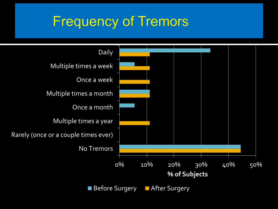
Tremors/ dystonia (45.5%)
Fainting (36.4%)
Numbness in back (31.8%)
Sleep apnea (22.7%)

Vertigo (92.3%)
Headaches (81.8%)
Balance problems (77.8%)
Upper extremity numbness (76.5%)
Dizziness (71.4%)
Speech problems (69.2%)
Neck pain (68.4%)
Memory problems (66.7%)
Upper extremity weakness (65.0%)
Walking (64.7%)
Fainting (62.5%)
Hearing problems (61.5%)
Swallowing/choking problems (58.3%)
Lower extremity numbness (56.3%)
Visual problems (56.3%)
Numbness in back (57.1%)
Lower extremity weakness (57.1%)
Tremors (50.0%)
Muscle pain (38.1%)
Frequent daytime urination (38.5%)
Hands and feet turning cold (37.5%)
Fatigue (36.4%)
Night awakenings (33.3%)
GERD (30.8%)
Nocturia (25.0%)
Sleep apnea (20.0%)
IBS (18.2%)
Joint pain (26.3%)

0%
10%
20%
30%
40%
50%
60%
70%
80%
StronglyAgree
Agree SomewhatAgree
SomewhatDisagree
Disagree StronglyDisagree
% o
f S
ub
ject
s
In looking back I would still choose to have the cranio-vertebral fusion surgery

0%
5%
10%
15%
20%
25%
30%
35%
40%
StronglyAgree
Agree SomewhatAgree
SomewhatDisagree
Disagree StronglyDisagree
% o
f S
ub
ject
s
The surgery improved my symptoms and decreased my limitations.

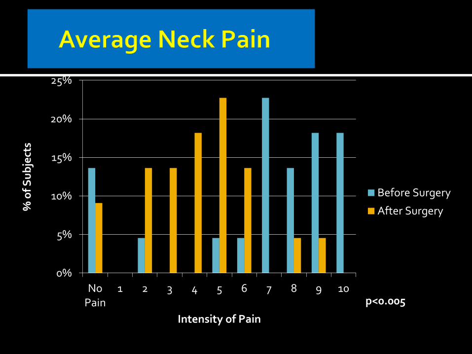
Participants reported headache before surgery (M = 8.18, SD = 1.62) after surgery significantly less headache pain (M =
4.50, SD = 1.82)
• t(21) = 6.532, p<0.001,
• (Mdiff = 3.68, SDdiff = 2.64),
• 95% CI [2.51-4.85]

0%
10%
20%
30%
40%
50%
60%
NoPain
1 2 3 4 5 6 7 8 9 10
% o
f S
ub
ject
s
Intensity of Pain
Before Surgery
After Surgery

Chiari malformation
Atlanto-axial instability
Vertebral instability
Pseudotumor cerebri
Occipital neuralgia
TMJ Syndrome
Intracranial thrombosis
Hypercoagulability ,Hughes Syndrome, migrainous
TIAs
Neuro-immunological disorders- Hashimoto’s
thyroiditis /encephalopathy, Anti-NMDA Ab, PANDAS,
neuro-Behcet’s
0%
5%
10%
15%
20%
25%
NoPain
1 2 3 4 5 6 7 8 9 10
% o
f S
ub
ject
s
Intensity of Pain
Before Surgery
After Surgery
p<0.005
0% 20% 40% 60%
No Dizziness
Rarely (once or couple times ever)
Multiple times a year
Once a month
Multiple times a month
Once a week
Multiple times a week
Daily
% of Subjects
Before Surgery After Surgery

0% 10% 20% 30% 40% 50% 60%
No Vertigo
Rarely (once or a couple times ever)
Multiple times a year
Once a month
Multiple times a month
Once a week
Multiple times a week
Daily
% of Subjects
Before Surgery After Surgery

0% 10% 20% 30% 40% 50%
No Walking Problems
Rarely (once or a couple times ever)
Multiple times a year
Once a month
Multiple times a month
Once a week
Multiple times a week
Daily
% of Subjects
Before Surgery After Surgery
p<0.03

0% 10% 20% 30% 40% 50% 60%
No Balance Problems
Rarely (once or a couple times ever)
Multiple times a year
Once a month
Multiple times a month
Once a week
Multiple times a week
Daily
% of Subjects
Before Surgery After Surgery
0% 10% 20% 30% 40% 50%
No Tremors
Rarely (once or a couple times ever)
Multiple times a year
Once a month
Multiple times a month
Once a week
Multiple times a week
Daily
% of Subjects
Before Surgery After Surgery

100% - Normal; no complaints; no evidence of disease. 90% - Able to carry on normal activity; minor signs or symptoms of disease. 80% - Normal activity with effort; some signs or symptoms of disease. 70% - Cares for self; unable to carry on normal activity or to do active work. 60% - Requires occasional assistance, but able to manage personal needs. 50% - Requires considerable assistance and frequent medical care. 40% - Disabled; requires special care and assistance. 30% - Severely disabled; hospital admission is indicated 20% - Very sick; hospital admission necessary for supportive treatment 10% - Moribund; fatal processes progressing rapidly. 0% - Dead
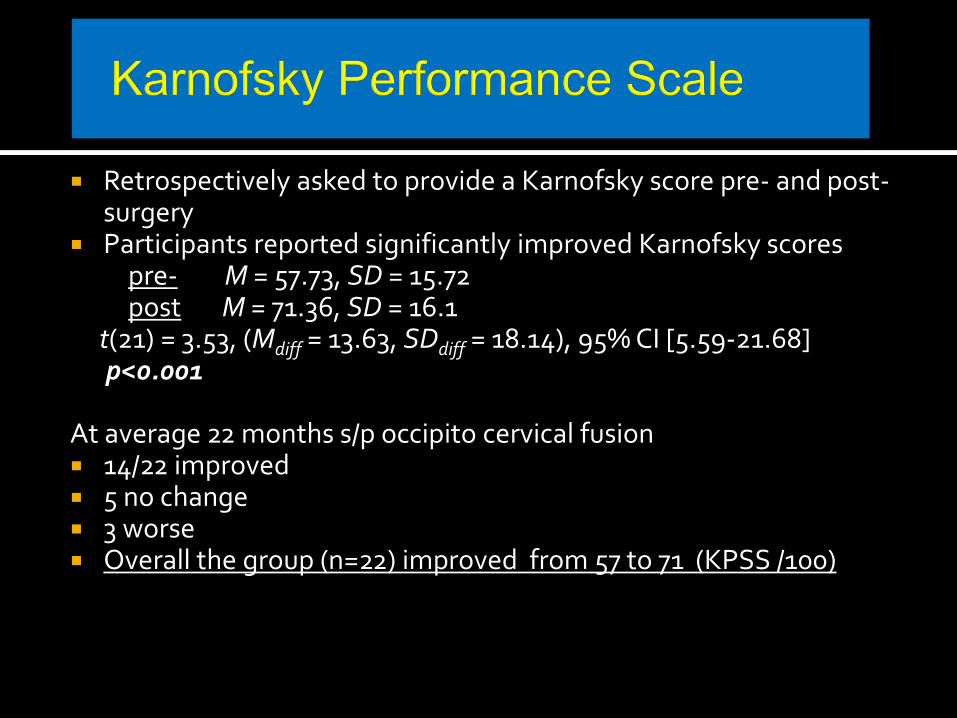
Retrospectively asked to provide a Karnofsky score pre- and post- surgery
Participants reported significantly improved Karnofsky scores pre- M = 57.73, SD = 15.72 post M = 71.36, SD = 16.1 t(21) = 3.53, (Mdiff = 13.63, SDdiff = 18.14), 95% CI [5.59-21.68] p<0.001 At average 22 months s/p occipito cervical fusion 14/22 improved 5 no change 3 worse Overall the group (n=22) improved from 57 to 71 (KPSS /100)

0.0%
5.0%
10.0%
15.0%
20.0%
25.0%
30.0%
35.0%
10 20 30 40 50 60 70 80 90 100
Pe
rce
nta
ge
of
Su
bje
cts
Karnofsky Score
Before Surgery
After Surgery
p<0.001
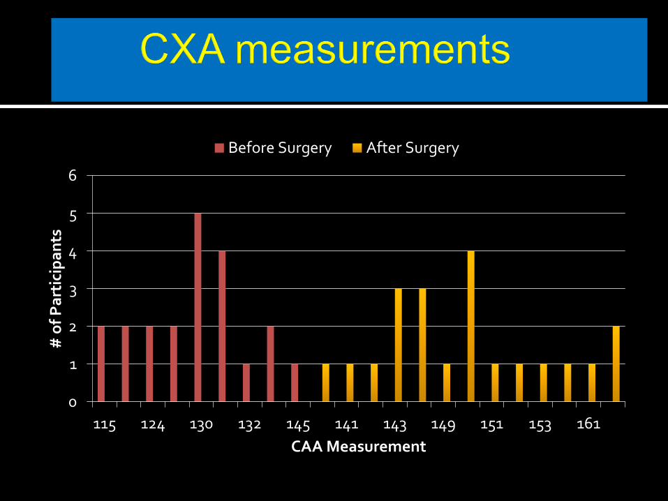
0
1
2
3
4
5
6
115 124 130 132 145 141 143 149 151 153 161
# o
f P
art
icip
an
ts
CAA Measurement
Before Surgery After Surgery
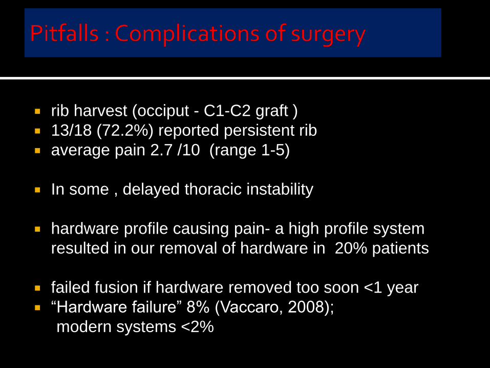
rib harvest (occiput - C1-C2 graft )
13/18 (72.2%) reported persistent rib
average pain 2.7 /10 (range 1-5)
In some , delayed thoracic instability
hardware profile causing pain- a high profile system
resulted in our removal of hardware in 20% patients
failed fusion if hardware removed too soon <1 year
“Hardware failure” 8% (Vaccaro, 2008);
modern systems <2%

Surgery is not a panacea Most of the symptoms related to the
underlying HCTD will not be improved Some symptoms will worsen Treatment requires ongoing pain
management and medical management of the non-surgical problems

74
Cranio-vertebral instability, Chiari I malformation s/p decompression, stabilization/fusion
Completely disabled , pain
10/10, syncope, unable to
stand or sit
Valedictorian
Worked through College in 3
yrs 3.98 average
Job
Married
Promotion
Several more neck surgeries


>44 degree
angle between
c1 and c2 on
full neck
rotation

Neck pain and headache, much worse when driving on bumpy roads
Nausea POTS, dysautonomia Pain with neck turning Pain over the C1/2 spinous process decreased pinprick sensation Hyper-reflexia Dysdiadochokinesia Improved with neck brace CT confirmation : pathological rotation of C1 upon C2


Complications : graft site pain - we are now using allograft Mobilize on day of surgery with neck brace for 3-4 weeks one failure of fusion, no hardware failure to date

discopathy is characteristic of EDS hyperangulation of the spine may manifest as a stretch myelopathy paresthesias of the hands and legs arm and leg weakness gait problems urinary urgency nausea, headache Pain between the shoulder blades
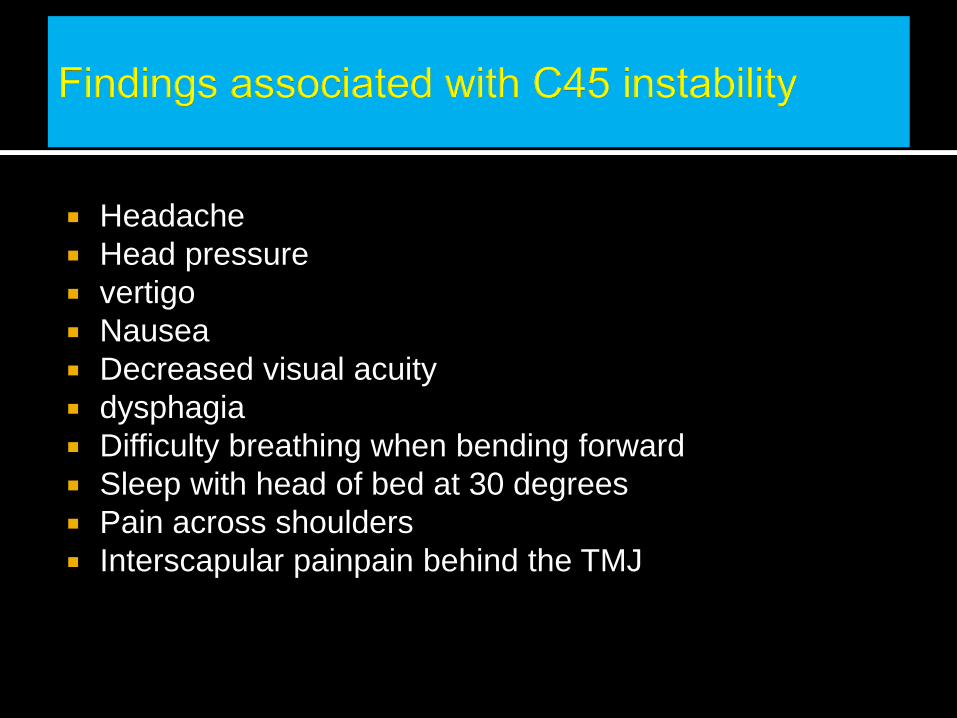
Headache
Head pressure
vertigo
Nausea
Decreased visual acuity
dysphagia
Difficulty breathing when bending forward
Sleep with head of bed at 30 degrees
Pain across shoulders
Interscapular painpain behind the TMJ


2 points each Anterior or posterior elements destroyed or unable to function
Positive stretch test
Dynamic Flexion-extension X-rays
* Total Sagittal plane translation > 3.5 mm or 20%
* Total Sagittal plane rotation > 20 degrees
Resting X-rays
Sagittal plane displacement > 3.5 mm or 20%
• Relative sagittal plane angulation > 11 degrees
• Spinal cord damage 1 pt each
Stenosis Sagittal diameter < 13 mm or Pavlov’s ratio < 0.8
Abnormal disc narrowing
Nerve root damage
Dangerous loading anticipated

The repetitive stretching of the spinal cord over the apex of the deformity
BENZEL E Neurosurgery Jan 2007 Suppl S1 P1

Neurosurgery: May 2005 - Volume 56 - Issue 5 - pp 1101-1113
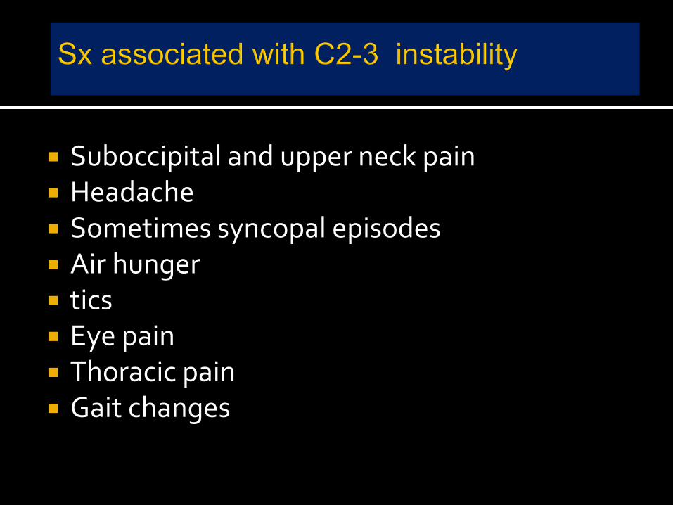
Suboccipital and upper neck pain Headache Sometimes syncopal episodes Air hunger tics Eye pain Thoracic pain Gait changes

Arm numbness and weakness
Eye pain
Nausea, vomiting
Occipital neuralgia
Poor balance
Twitching arms
Disturbed vision, conjunctival burning
Ears feel clogged


Intracranial thrombosis

Stretching of the spinal cord by any structure that anchors the spinal cord to the spine
In the EDS population this results from increased thickness and tightness of the filum terminale

Clinical diagnosis Weakness of the legs low back pain sensory loss, especially in the sacral dermatomes neurogenic bladder , urodynamics show large PVR,
sphinchter detrussor dysynergia h/o growing pains, multiple UTIs, enuresis, toe walking,
pidgeon toed, leg cramps, sleeping with knees bent, cannot walk uphill
flat feet scoliosis

Bowel incontinence
Headache
Stretch signs :severe back pain, tingling and nausea
Light shock waves going down the legs
Burning under the feet
Fullness of pharynx / dysphagia

Conus medullaris L 3
In EDS, usually “radiologically occult”
Fatty filum
Scoliosis Syringomyelia
Spinal bifida oculta
Cauda equina appears stretched
On prone MRI conus posterior
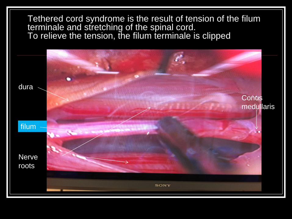
Tethered cord syndrome is the result of tension of the filum terminale and stretching of the spinal cord. To relieve the tension, the filum terminale is clipped
dura
Conus
medullaris
filum
Nerve
roots

0
2
4
6
8
10
12
NeurologicalChange
FunctionalChange
Quality of Life Pain Change
Improved
Worsened
No change
Preliminary results : Tethered cord release n=13

Every patient surveyed, with the exception of one, said they would do the surgery again if they had the choice and said they would recommend it to a friend or family member
Complications: subcutaneous hematoma, IV thrombosis, urethritis from catheter, pulmonary embolism
Other complications: pseudomeningocoele
is a clinical diagnosis, more often radiologically
occult in EDS patients
In well selected patients, sectioning the filum terminale improves pain, function and neurological deficit in more than 85% of patients

Problems of Brain Water

Hydrocephalus
Raised intracranial pressure with no evidence of “mass lesion”, hydrocephalus, infection or hypertensive encephalopathy
Etiology: obesity, hyper-vitaminosis, pregnancy, menarche,
menstrual irregularities, Addison’s disease, Fe++ def. anemia, polycythemia vera
Oral contraceptives, steroid withdrawal, tetracycline, nalidixic acid

Usually 3rd and 4th decades Women > men Headache (94%) visual obscurations/blurring (68%) Pulse synchronous tinnitus or whooshing noise (58%) Retro-orbital pain (44%) Diplopia (38%) Visual loss (30%) Normal radiological (CT/MRI) studies LP: CSF pressure greater than 25 cm H2O

Treat the underlying cause Weight loss Drugs- Acetazolamide - Thiazide diuretic Lumbo-peritoneal shunt Ventriculo-peritoneal shunt

Syringomyelia
fluid-filled cavity or syrinx within the spinal cord

Chiari malformation = most common cause Tethered cord causes small syrinx Other forms - Meningitis or arachnoiditis - Hemorrhage - Tumor - trauma
Treat the underlying cause - Chiari - Tethered cord syndrome Rarely a syringo-subarachnoid shunt
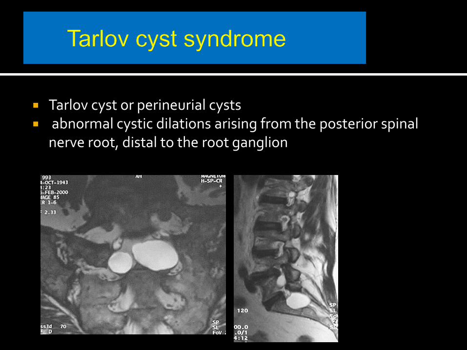
Tarlov cyst or perineurial cysts abnormal cystic dilations arising from the posterior spinal
nerve root, distal to the root ganglion

Pain: sacral>lumbar, buttocks Non-dermatomal Perineal pain: dyspareunia, proctalgia
exacerbated by standing, coughing, sneezing… Valsalva
pain worsened by standing or sitting,
Improved by lying flat Urinary: frequency, urgency, incontinence Bowel: constipation, rectal incontinence Sensory: paresthesias, sacral sensory loss

Pain Meds: temporary relief
Percutaneous aspiration 4/5 patients suffered recurrence of symptoms
AJNR 15: 293 -299, 1994

Sacral laminectomy, microsurgical stimulation to identify root fibers Resection of redundant cyst wall, fusion/laminoplasty +/- LP shunt
Treatment Tarlov Cyst
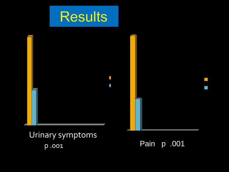
0
1
2
3
4
5
6
7
8
9
Preop
Postop
0
5
10
15
20
25
Preop
Postop
p .001
Urinary symptoms Pain p .Pain001
Pain p .001


headache and neurological deficits, in a patient with a
hypermobility connective tissue disorder, should prompt
consideration of cranio-cervical instability
(Consensus Statement, Oct 2013)
craniocervical instability, spinal instability and tethered
cord syndrome result in deformative stress of the
central nervous system
deformative stress of the CNS appears to underlie the
clinical manifestations of pain and neurological deficits

Patients with craniocervical instability should be treated
in a neck brace, physical therapy, activity limitation,
medication, and have other disorders ruled out before
consideration for surgery
Patients with severe headache, neck pain, neurological
deficits and appropriate radiological findings appear to
benefit from correction of the deformity, stabilization and
fusion
Co-morbid conditions may require continued follow up

Clair Francomano MD, JHI Peter Rowe MD,JHI Myles Koby MD, NIH, Doctors Hospital Rodney Grahame, Professor of Rheumatology Robert Gerwin MD Assoc Prof, JHI Ed Benzel MD- Prof, Chair Neurosurg ,CCI Alex Vaccaro MD- Prof Neurosurg Ortho, TJU Stephen Mott MD Assoc Prof Peds Neurol, Dartmouth Joel Berry PhD – Prof Chair Mech Eng, Kettering Univ Mark Alexander MD , Director Neuradiology Bethesda MRI Jonah Murdoch, MD, GUH,DCH Jessica Adcock BS,MS William Wilson IV, Yale Univ Rebecca Tuchmann, BSN