fried, christof koch and rodrigo quian quiroga carlos pedreira
TRANSCRIPT

103:97-107, 2010. First published Oct 28, 2009; doi:10.1152/jn.91323.2008 J NeurophysiolFried, Christof Koch and Rodrigo Quian Quiroga Carlos Pedreira, Florian Mormann, Alexander Kraskov, Moran Cerf, Itzhak
You might find this additional information useful...
for this article can be found at: Supplemental material http://jn.physiology.org/cgi/content/full/91323.2008/DC1
55 articles, 18 of which you can access free at: This article cites http://jn.physiology.org/cgi/content/full/103/1/97#BIBL
including high-resolution figures, can be found at: Updated information and services http://jn.physiology.org/cgi/content/full/103/1/97
can be found at: Journal of Neurophysiologyabout Additional material and information http://www.the-aps.org/publications/jn
This information is current as of January 13, 2010 .
http://www.the-aps.org/.American Physiological Society. ISSN: 0022-3077, ESSN: 1522-1598. Visit our website at (monthly) by the American Physiological Society, 9650 Rockville Pike, Bethesda MD 20814-3991. Copyright © 2005 by the
publishes original articles on the function of the nervous system. It is published 12 times a yearJournal of Neurophysiology
on January 13, 2010 jn.physiology.org
Dow
nloaded from

Responses of Human Medial Temporal Lobe Neurons Are Modulated byStimulus Repetition
Carlos Pedreira,1 Florian Mormann,2,3 Alexander Kraskov,2,4 Moran Cerf,2,3 Itzhak Fried,3,5 Christof Koch,2
and Rodrigo Quian Quiroga1,2,3
1Department of Engineering, University of Leicester, Leiscester, United Kingdom; 2Computation and Neural Systems, California Instituteof Technology, Pasadena, California; 3Department of Neurosurgery, David Geffen School of Medicine, and Semel Institute forNeuroscience and Human Behavior, University of California, Los Angeles, California; 4University College London, Institute of Neurology,Queen Square, London, United Kingdom; and 5Functional Neurosurgery Unit, Tel-Aviv Medical Center and Sackler Faculty of Medicine,Tel-Aviv University, Tel-Aviv, Israel
Submitted 15 December 2008; accepted in final form 20 October 2009
Pedreira C, Mormann F, Kraskov A, Cerf M, Fried I, Koch C,Quiroga RQ. Responses of human medial temporal lobe neurons aremodulated by stimulus repetition. J Neurophysiol 103: 97–107, 2010.First published October 28, 2009; doi:10.1152/jn.91323.2008. Recentstudies have reported the presence of single neurons with strongresponses to visual inputs in the human medial temporal lobe. Here weshow how repeated stimulus presentation—photos of celebrities andfamiliar individuals, landmark buildings, animals, and objects—mod-ulates the firing rate of these cells: a consistent decrease in the neuralactivity was registered as images were repeatedly shown duringexperimental sessions. The effect of repeated stimulus presentationwas not the same for all medial temporal lobe areas. These findingsare consistent with the view that medial temporal lobe neurons linkvisual percepts to declarative memory.
I N T R O D U C T I O N
The recognition of visual objects is processed along the ventralvisual pathway, extending from primary visual areas (V1) to theinferotemporal cortex (IT) (Logothetis and Sheinberg 1996;Tanaka 1996). IT cortex has direct projections to the medialtemporal lobe (MTL) (Lavenex and Amaral 2000; Saleem andTanaka 1996; Suzuki 1996), where single cell studies in monkeysreported visual responses, among others, by stimulus-selectiveneurons that were related to the learning and rehearsal of associ-ation between visual cues (Naya et al. 2001; Sakai and Miyashita1991; Wirth et al. 2003). In humans, it has been shown thatneurons in the MTL respond strongly to visual inputs (Fried et al.1997; Gelbard-Sagiv et al. 2008; Kreiman et al. 2000a,b, 2002;Quian Quiroga et al. 2005, 2008b, 2009). However, based onfindings with patient H.M.—and similar patients with lesions orresections of the hippocampus and other parts of the MTL—it iswidely accepted that the MTL is not necessary for visual recog-nition (but see Buckley and Gaffan 2006). Rather, the hippocam-pus is involved in declarative memory storage (Corkin 2002;Eichenbaum 2000; Rosenbaum et al. 2005; Scoville and Milner1957; Squire et al. 2004). This raises the question of why MTLneurons respond strongly to images if this area is not part of thevisual perception system. Based on 1) the well-established find-ings about the role of the MTL in memory storage, 2) therelatively long latency of MTL responses at �300 ms or longer(Mormann et al. 2008; Quian Quiroga et al. 2005) compared to
�120 ms in monkey IT (Hung et al. 2005), and 3) the fact thatMTL neurons encode abstract information and not particularvisual details (Quian Quiroga et al. 2005), it has been suggestedthat MTL neurons link visual perception to memory formation(Quian Quiroga et al. 2005, 2008a).
Brain imaging studies of stimulus repetition in humans showeda decrease in activity for different areas, including the MTL,which has been related to processes involving perception, atten-tion, learning, and memory (for reviews, see Grill-Spector et al.2006; Henson and Rugg 2003; Ranganath and Rainer 2003). Inaddition, experimental stimulus repetition paradigms induced re-sponse suppression patterns in monkey IT neurons (Li et al. 1993;Liu et al. 2009; Miller et al. 1991; Sawamura et al. 2006). Giventhese previous findings, here we set up to study whether a similarpattern of decreased responses with stimulus repetition was alsopresent in the visual responses in the human MTL. We hypothe-size—considering the abovementioned studies about MTL func-tion—that such a finding with neurons in this area may be becauseof its role in declarative memory.
M E T H O D S
Subjects and recordings
Subjects were 26 patients with pharmacologically intractable epi-lepsy (15 men; 22 right handed; 17–54 yr old). Extensive noninvasivemonitoring did not yield concordant findings corresponding to a singleresectable epileptogenic focus. Therefore patients were implantedwith chronic depth electrodes for typically 7–10 days to determine thefocus of the seizures for possible surgical resection (Fried et al. 1997).All studies conformed to the guidelines of the Medical InstitutionalReview Board at UCLA. The electrode locations were based exclu-sively on clinical criteria and were verified by MRI or CT co-registered to preoperative MRI. Here we report data from sites in thehippocampus, amygdala, entorhinal cortex, and parahippocampal cor-tex. Each electrode probe had a total of nine microwires at its end,eight active recordings channels, and one reference (Fried et al. 1997).The differential signal from the microwires was amplified using a64-channel Neuralynx system, filtered between 1 and 9,000 Hz, andsampled at 28 kHz. Each recording session lasted �30 min.
The data reported here were recorded during 44 experimental sessions.Subjects laid in bed facing a laptop computer on which pictures ofanimals, objects, landmark buildings and known and unknown faces wereshown. After image offset, subjects had to respond whether or not thepicture contained a human face, by pressing the Y and N keys, respec-tively. This simple task, on which performance was virtually flawless,required them to attend to the pictures (Quian Quiroga et al. 2005).
Address for reprint requests and other correspondence: R. Quian Quiroga,Dept. of Engineering, Univ. of Leicester, Leicester, UK LE1 7RH (E-mail:[email protected]).
J Neurophysiol 103: 97–107, 2010.First published October 28, 2009; doi:10.1152/jn.91323.2008.
970022-3077/10 $8.00 Copyright © 2010 The American Physiological Societywww.jn.org
on January 13, 2010 jn.physiology.org
Dow
nloaded from
Images covered �1.5° of the visual angle and were presented for 1 s atthe center of the screen, six times each in pseudo-random order. Themean number of images shown to the patient was 114.2 (range, 83–192).In a slightly different version of this paradigm, for 13 sessions, thepresentation time was 500 ms, and the key responses were omitted. Thesesessions were considered together with the 1-s presentation sessionsbecause there were no clear differences in the response patterns.
Of the 44 experimental sessions, 26 corresponded to the firstexperiment done with each of the 26 patients, so that the first trial foreach picture was the first time the patient saw the image at the UCLAward. The remaining 18 sessions corresponded to second sessionscollected from 18 of the 26 patients, carried out on a following day.All the pictures considered from the second sessions were alreadypresented in the first session. Because of the variability of spikeshapes, it was, in general, not possible to follow the activity of singleneurons across different experiments.
Data analysis
From the continuously recorded data, spikes were detected and sortedusing the Wave_clus software package (Quian Quiroga et al. 2004). Asin previous studies (Quian Quiroga et al. 2005, 2007), a response wasconsidered significant if it was larger than the mean plus 5 SD of thebaseline period (1,000 to 300 ms before stimulus onset) for all stimuli andhad at least two spikes in the time interval between 300 and 1,000 msafter the stimulus onset. For those pictures eliciting significant responses,we computed the total number of spikes between 300 to 2,000 ms afterstimulus onset for each trial. To account for the fact that different neuronshave different firing rates, the responses were normalized by dividing bythe maximum number of spikes across trials. The normalized number ofspikes across trials was statistically compared using a one-way ANOVA(test 1), where the independent variable was the trial number and therepeated measures were the normalized responses. This analysis wasperformed for the whole population of responses in the MTL and for eachof the four subregions separately.
For a further characterization of the response patterns, for each ofthe significant responses, we calculated a linear regression of thenumber of spikes with trial number. Differences in the slope values ofthese linear fits were compared for the different MTL areas andbetween the first and second experimental sessions using a two-wayANOVA (test 2). The independent variables were MTL area andsession number, and the repeated measures were the slopes of theresponses. Post hoc, we evaluated differences of the slope values tothe “zero slope” response pattern (i.e., a response with the samenumber of spikes for every stimulus presentation) for each areaseparately using a paired t-test (test 3).
To evaluate the time profile of the responses, the instantaneous firingrate was computed by convolving the normalized spike trains with aGaussian kernel (sampling period � 0.5 ms, � � 100 ms). From theaverage instantaneous firing rate (across all responses) for each trial, wedefined 1) the peak amplitude; 2) its latency; 3) the onset of the response,as the point where the instantaneous firing rate crossed 4 SD abovebaseline and stayed above for �100 ms; and 4) the duration, as the timeinterval between response onset and offset (Fig. S1).1 Offset was definedsimilarly to onset but crossing the 4 SD line downward and staying belowit for �100 ms. The effect of stimulus repetition on each of theseparameters was assessed using one-way ANOVA with independentvariable trial number (test 4). The repeated measures were the values ofthe corresponding parameters for each response.
R E S U L T S
In 26 first experimental sessions for each patient, we recordedfrom 1,210 MTL units (515 single units and 695 multiunits), withan average of 46.6 units per session. Of these 1,210 units, 262
(22%; 132 single units and 130 multiunits) had a statisticallysignificant response to a total of 725 pictures (an average of 2.77responses per unit). For the second experimental sessions, werecorded from a total of 745 units (328 single units and 417multiunits), with an average of 41.3 units per session. Of these 745units, 110 (15%; 57 single units and 53 multiunits) had a signif-icant response to a total of 289 pictures (2.63 responses per unit).The decrease in the responsiveness of the recorded units betweenexperimental sessions (22% for the 1st session against 15% for the2nd one) was significant (�2, P � 0.001).
Single cell responses
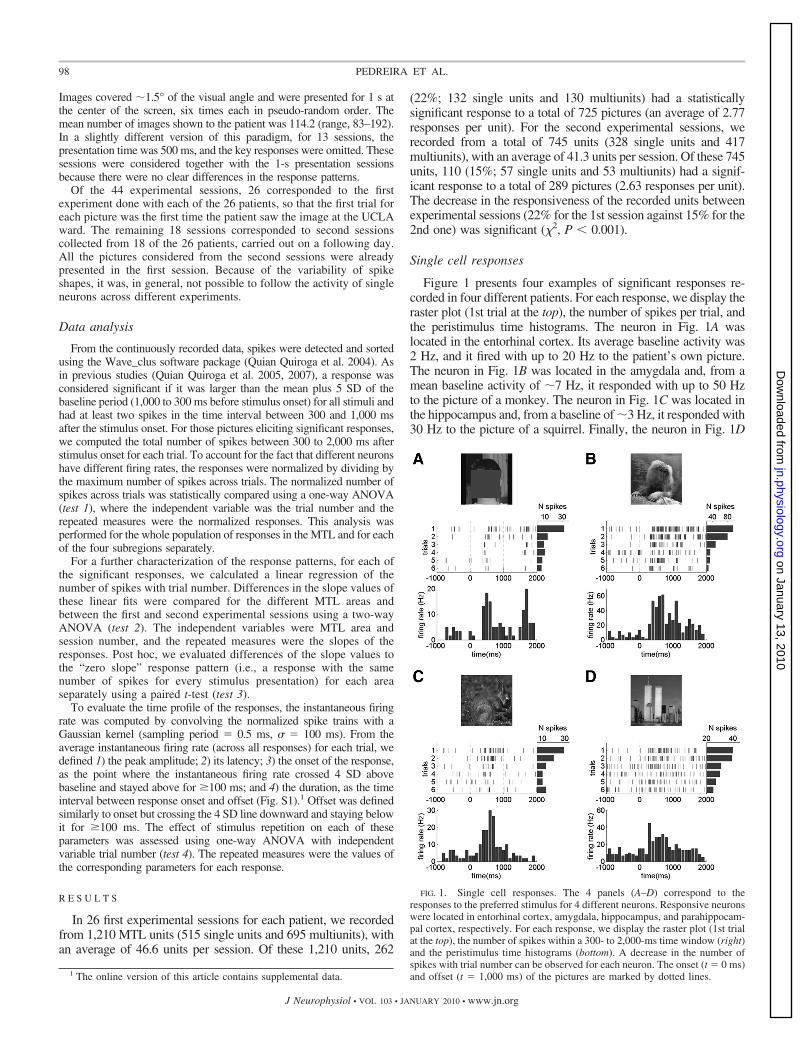
Figure 1 presents four examples of significant responses re-corded in four different patients. For each response, we display theraster plot (1st trial at the top), the number of spikes per trial, andthe peristimulus time histograms. The neuron in Fig. 1A waslocated in the entorhinal cortex. Its average baseline activity was2 Hz, and it fired with up to 20 Hz to the patient’s own picture.The neuron in Fig. 1B was located in the amygdala and, from amean baseline activity of �7 Hz, it responded with up to 50 Hzto the picture of a monkey. The neuron in Fig. 1C was located inthe hippocampus and, from a baseline of �3 Hz, it responded with30 Hz to the picture of a squirrel. Finally, the neuron in Fig. 1D
1 The online version of this article contains supplemental data.
FIG. 1. Single cell responses. The 4 panels (A–D) correspond to theresponses to the preferred stimulus for 4 different neurons. Responsive neuronswere located in entorhinal cortex, amygdala, hippocampus, and parahippocam-pal cortex, respectively. For each response, we display the raster plot (1st trialat the top), the number of spikes within a 300- to 2,000-ms time window (right)and the peristimulus time histograms (bottom). A decrease in the number ofspikes with trial number can be observed for each neuron. The onset (t � 0 ms)and offset (t � 1,000 ms) of the pictures are marked by dotted lines.
98 PEDREIRA ET AL.
J Neurophysiol • VOL 103 • JANUARY 2010 • www.jn.org
on January 13, 2010 jn.physiology.org
Dow
nloaded from
was in the parahippocampal cortex and it responded to a picture ofthe World Trade Center with �45 Hz from a baseline of 10 Hz.All these units increased their firing at least three times in responseto their preferred pictures. However, this change was not equallydistributed across the six trials. In fact, in the four examples, aclear decay in the number of spikes with trial number can beobserved, as shown by the spike counts for each trial.
Population results
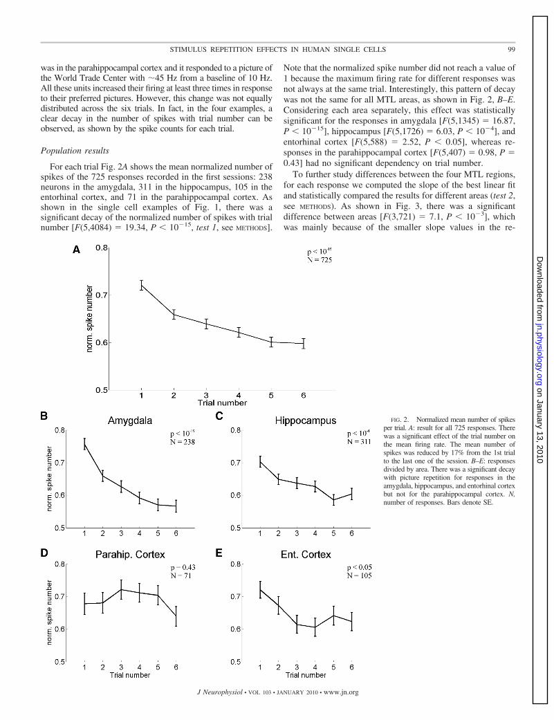
For each trial Fig. 2A shows the mean normalized number ofspikes of the 725 responses recorded in the first sessions: 238neurons in the amygdala, 311 in the hippocampus, 105 in theentorhinal cortex, and 71 in the parahippocampal cortex. Asshown in the single cell examples of Fig. 1, there was asignificant decay of the normalized number of spikes with trialnumber [F(5,4084) � 19.34, P � 10�15, test 1, see METHODS].
Note that the normalized spike number did not reach a value of1 because the maximum firing rate for different responses wasnot always at the same trial. Interestingly, this pattern of decaywas not the same for all MTL areas, as shown in Fig. 2, B–E.Considering each area separately, this effect was statisticallysignificant for the responses in amygdala [F(5,1345) � 16.87,P � 10�15], hippocampus [F(5,1726) � 6.03, P � 10�4], andentorhinal cortex [F(5,588) � 2.52, P � 0.05], whereas re-sponses in the parahippocampal cortex [F(5,407) � 0.98, P �0.43] had no significant dependency on trial number.
To further study differences between the four MTL regions,for each response we computed the slope of the best linear fitand statistically compared the results for different areas (test 2,see METHODS). As shown in Fig. 3, there was a significantdifference between areas [F(3,721) � 7.1, P � 10�3], whichwas mainly because of the smaller slope values in the re-
FIG. 2. Normalized mean number of spikesper trial. A: result for all 725 responses. Therewas a significant effect of the trial number onthe mean firing rate. The mean number ofspikes was reduced by 17% from the 1st trialto the last one of the session. B–E: responsesdivided by area. There was a significant decaywith picture repetition for responses in theamygdala, hippocampus, and entorhinal cortexbut not for the parahippocampal cortex. N,number of responses. Bars denote SE.
99STIMULUS REPETITION EFFECTS IN HUMAN SINGLE CELLS
J Neurophysiol • VOL 103 • JANUARY 2010 • www.jn.org
on January 13, 2010 jn.physiology.org
Dow
nloaded from
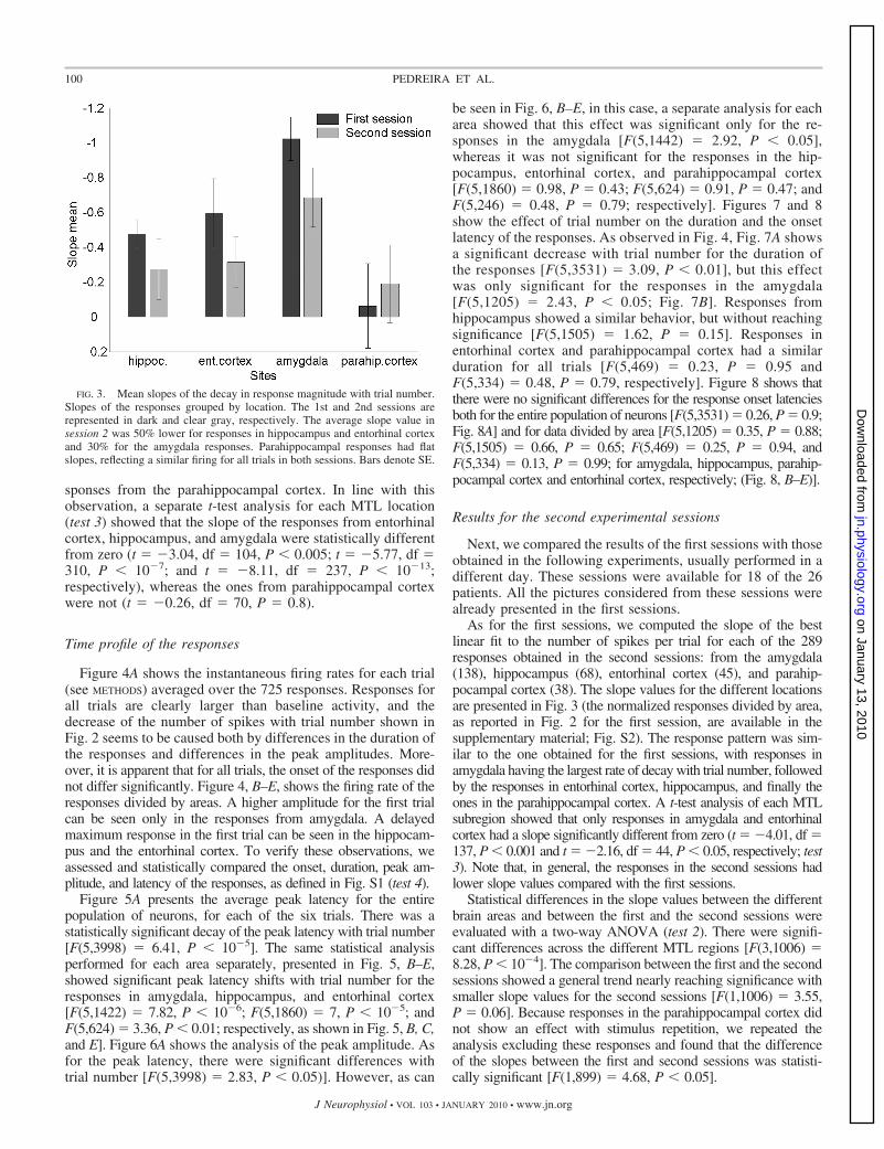
sponses from the parahippocampal cortex. In line with thisobservation, a separate t-test analysis for each MTL location(test 3) showed that the slope of the responses from entorhinalcortex, hippocampus, and amygdala were statistically differentfrom zero (t � �3.04, df � 104, P � 0.005; t � �5.77, df �310, P � 10�7; and t � �8.11, df � 237, P � 10�13;respectively), whereas the ones from parahippocampal cortexwere not (t � �0.26, df � 70, P � 0.8).
Time profile of the responses
Figure 4A shows the instantaneous firing rates for each trial(see METHODS) averaged over the 725 responses. Responses forall trials are clearly larger than baseline activity, and thedecrease of the number of spikes with trial number shown inFig. 2 seems to be caused both by differences in the duration ofthe responses and differences in the peak amplitudes. More-over, it is apparent that for all trials, the onset of the responses didnot differ significantly. Figure 4, B–E, shows the firing rate of theresponses divided by areas. A higher amplitude for the first trialcan be seen only in the responses from amygdala. A delayedmaximum response in the first trial can be seen in the hippocam-pus and the entorhinal cortex. To verify these observations, weassessed and statistically compared the onset, duration, peak am-plitude, and latency of the responses, as defined in Fig. S1 (test 4).
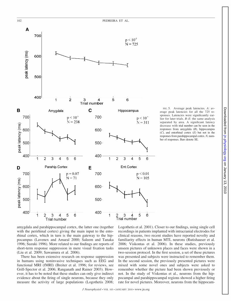
Figure 5A presents the average peak latency for the entirepopulation of neurons, for each of the six trials. There was astatistically significant decay of the peak latency with trial number[F(5,3998) � 6.41, P � 10�5]. The same statistical analysisperformed for each area separately, presented in Fig. 5, B–E,showed significant peak latency shifts with trial number for theresponses in amygdala, hippocampus, and entorhinal cortex[F(5,1422) � 7.82, P � 10�6; F(5,1860) � 7, P � 10�5; andF(5,624) � 3.36, P � 0.01; respectively, as shown in Fig. 5, B, C,and E]. Figure 6A shows the analysis of the peak amplitude. Asfor the peak latency, there were significant differences withtrial number [F(5,3998) � 2.83, P � 0.05)]. However, as can
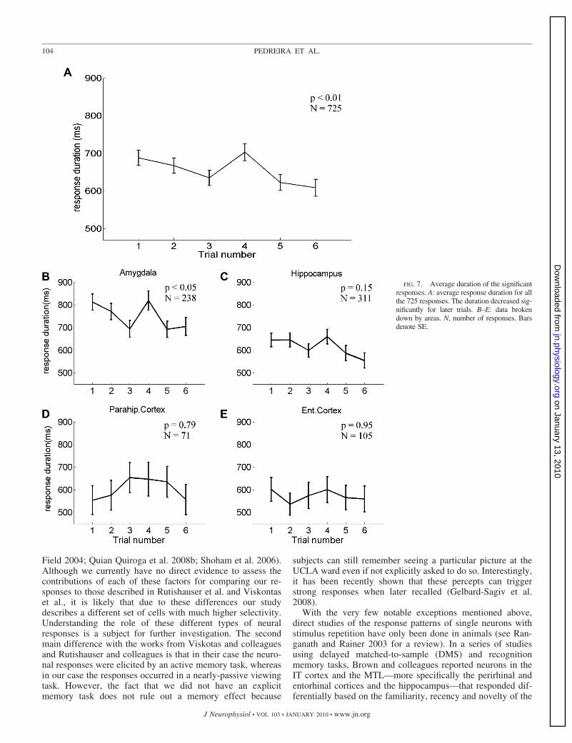
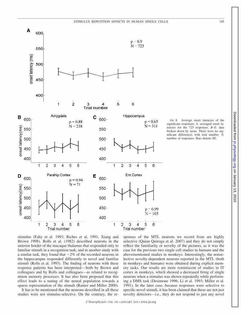
be seen in Fig. 6, B–E, in this case, a separate analysis for eacharea showed that this effect was significant only for the re-sponses in the amygdala [F(5,1442) � 2.92, P � 0.05],whereas it was not significant for the responses in the hip-pocampus, entorhinal cortex, and parahippocampal cortex[F(5,1860) � 0.98, P � 0.43; F(5,624) � 0.91, P � 0.47; andF(5,246) � 0.48, P � 0.79; respectively]. Figures 7 and 8show the effect of trial number on the duration and the onsetlatency of the responses. As observed in Fig. 4, Fig. 7A showsa significant decrease with trial number for the duration ofthe responses [F(5,3531) � 3.09, P � 0.01], but this effectwas only significant for the responses in the amygdala[F(5,1205) � 2.43, P � 0.05; Fig. 7B]. Responses fromhippocampus showed a similar behavior, but without reachingsignificance [F(5,1505) � 1.62, P � 0.15]. Responses inentorhinal cortex and parahippocampal cortex had a similarduration for all trials [F(5,469) � 0.23, P � 0.95 andF(5,334) � 0.48, P � 0.79, respectively]. Figure 8 shows thatthere were no significant differences for the response onset latenciesboth for the entire population of neurons [F(5,3531) � 0.26, P � 0.9;Fig. 8A] and for data divided by area [F(5,1205) � 0.35, P � 0.88;F(5,1505) � 0.66, P � 0.65; F(5,469) � 0.25, P � 0.94, andF(5,334) � 0.13, P � 0.99; for amygdala, hippocampus, parahip-pocampal cortex and entorhinal cortex, respectively; (Fig. 8, B–E)].
Results for the second experimental sessions
Next, we compared the results of the first sessions with thoseobtained in the following experiments, usually performed in adifferent day. These sessions were available for 18 of the 26patients. All the pictures considered from these sessions werealready presented in the first sessions.
As for the first sessions, we computed the slope of the bestlinear fit to the number of spikes per trial for each of the 289responses obtained in the second sessions: from the amygdala(138), hippocampus (68), entorhinal cortex (45), and parahip-pocampal cortex (38). The slope values for the different locationsare presented in Fig. 3 (the normalized responses divided by area,as reported in Fig. 2 for the first session, are available in thesupplementary material; Fig. S2). The response pattern was sim-ilar to the one obtained for the first sessions, with responses inamygdala having the largest rate of decay with trial number, followedby the responses in entorhinal cortex, hippocampus, and finally theones in the parahippocampal cortex. A t-test analysis of each MTLsubregion showed that only responses in amygdala and entorhinalcortex had a slope significantly different from zero (t � �4.01, df �137, P � 0.001 and t � �2.16, df � 44, P � 0.05, respectively; test3). Note that, in general, the responses in the second sessions hadlower slope values compared with the first sessions.
Statistical differences in the slope values between the differentbrain areas and between the first and the second sessions wereevaluated with a two-way ANOVA (test 2). There were signifi-cant differences across the different MTL regions [F(3,1006) �8.28, P � 10�4]. The comparison between the first and the secondsessions showed a general trend nearly reaching significance withsmaller slope values for the second sessions [F(1,1006) � 3.55,P � 0.06]. Because responses in the parahippocampal cortex didnot show an effect with stimulus repetition, we repeated theanalysis excluding these responses and found that the differenceof the slopes between the first and second sessions was statisti-cally significant [F(1,899) � 4.68, P � 0.05].
FIG. 3. Mean slopes of the decay in response magnitude with trial number.Slopes of the responses grouped by location. The 1st and 2nd sessions arerepresented in dark and clear gray, respectively. The average slope value insession 2 was 50% lower for responses in hippocampus and entorhinal cortexand 30% for the amygdala responses. Parahippocampal responses had flatslopes, reflecting a similar firing for all trials in both sessions. Bars denote SE.
100 PEDREIRA ET AL.
J Neurophysiol • VOL 103 • JANUARY 2010 • www.jn.org
on January 13, 2010 jn.physiology.org
Dow
nloaded from

D I S C U S S I O N
In this study, we showed a decrease of the number of spikesfired by neurons in the human MTL in response to repeatedpicture presentations. This effect was not homogeneous acrossthe different MTL areas. In particular, a decrease in theresponse peak amplitude with trial number was significant onlyfor the amygdala responses. Moreover, there were significantdecreases of the response peak latencies for the responses inthe amygdala, hippocampus, and enthorinal cortex (but not forparahippocampal cortex). Given that the onset of responses wasnot different for the different trials (see Fig. 8), the decrease inpeak latency can be attributed to a “time-sharpening”—i.e., responseswere more localized in time for the later trials—in agreement withthe pattern observed in the instantaneous firing rate curves
shown in Fig. 4. The fact that in our study the time-sharpeningof the responses was accompanied by a decrease in durationonly for the amygdala neurons can be attributed to the lessaccurate estimation of the response durations, which accumu-late inaccuracies in estimating both the onset and offset of theresponses. In agreement with the previous observations, therewas a decrease of the total number of spikes elicited inresponse to the stimulus for neurons in the amygdala, hip-pocampus, and entorhinal cortex.
Long-term response suppression effects have been reportedby recent studies in monkey IT cortex during visual fixationand stimulus classification tasks (Anderson et al. 2008; Freed-man et al. 2006). Interestingly, IT cortex has large projectionsto the MTL areas we record from, with direct projections to the
FIG. 4. Mean instantaneous firing ratefor each trial. A: average over the whole setof responses (725). Note that all 6 trialshad similar onset latencies. Responses forthe 1st trials, especially for trial 1, in darkblue, had a larger duration, as well as adelayed and slightly higher peak value.B–E: data broken down by areas. In B,responses from amygdala (238 responses)showed higher and later peak values for the1st presentations. In C, responses fromhippocampus (311) showed a late peakvalue for the 1st trial. D: no differences inthe responses from parahippocampal cor-tex (71) were observed for the differenttrials. E: responses from entorhinal cortex(105) showed a similar delay pattern asthose in hippocampus.
101STIMULUS REPETITION EFFECTS IN HUMAN SINGLE CELLS
J Neurophysiol • VOL 103 • JANUARY 2010 • www.jn.org
on January 13, 2010 jn.physiology.org
Dow
nloaded from
amygdala and parahippocampal cortex, the latter one (togetherwith the perirhinal cortex) giving the main input to the ento-rhinal cortex, which in turn is the main gateway to the hip-pocampus (Lavenex and Amaral 2000; Saleem and Tanaka1996; Suzuki 1996). More related to our findings are reports ofshort-term response suppression in mere visual fixation tasks(Liu et al. 2009; Sawamura et al. 2006).
There has been extensive research on response suppressionin humans using noninvasive techniques such as EEG andfunctional MRI (fMRI) (Breiter et al. 1996; for reviews, seeGrill-Spector et al. 2006; Ranganath and Rainer 2003). How-ever, it has to be noted that these studies can only give indirectevidence about the firing of single neurons, because they onlymeasure the activity of large populations (Logothetis 2008;
Logothetis et al. 2001). Closer to our findings, using single cellrecordings in patients implanted with intracranial electrodes forclinical reasons, two recent studies have reported novelty andfamiliarity effects in human MTL neurons (Rutishauser et al.2006; Viskontas et al. 2006). In these studies, previouslyunseen pictures of unknown places and faces were shown in atwo-session protocol. In the first session, a set of these pictureswas presented and subjects were instructed to remember them.In the second session, the previously presented pictures weremixed with some novel ones and subjects were asked toremember whether the picture had been shown previously ornot. In the study of Viskontas et al., neurons from the hip-pocampal and parahippocampal regions showed a higher firingrate for novel pictures. Moreover, neurons from the hippocam-
FIG. 5. Average peak latencies. A: av-erage peak latencies for all the 725 re-sponses. Latencies were significantly ear-lier for later trials. B–E: the same analysisseparated by area. A significant latencydecrease with trial number can be seen in theresponses from amygdala (B), hippocampus(C), and entorhinal cortex (E) but not in theresponses from parahippocampal cortex. N, num-ber of responses. Bars denote SE.
102 PEDREIRA ET AL.
J Neurophysiol • VOL 103 • JANUARY 2010 • www.jn.org
on January 13, 2010 jn.physiology.org
Dow
nloaded from

pus presented a decrease of their firing below the baselineactivity for subsequent presentations. Rutishauser et al. de-scribed two subsets of cells in hippocampus and amygdala: onegroup of cells increased their firing when the stimulus pre-sented was new and another one increased their firing when itwas shown few moments before (Rutishauser et al. 2006).Although related, there are two main differences between thesetwo studies and the one presented here. First, the neuronsdescribed by Rutishauser et al. (2006) and by Viskontas et al.(2006) were not stimulus selective, because their behavior wasindependent of the particular (either novel or familiar) pictureshown. This lack of selectivity compared to our study can beattributed to the fact that 1) we used familiar stimuli, which aremore likely to elicit responses (Viskontas et al. 2009); 2) we used
an optimal spike sorting algorithm that is particularly suited todetect sparsely firing neurons, which typically have very lowbaseline firing rates (Quian Quiroga et al. 2008b, 2009); and3) we recorded the continuous data and used optimal off-lineanalysis. Note that to avoid large data volumes many acquisi-tion systems detect spikes on-line based on amplitude thresh-olds set by hand. These thresholds may be set to nonoptimalvalues, especially if the experimenter is dealing with a rela-tively large number of channels. In particular, this approachmay miss or nonoptimally detect very selective neurons be-cause these may be silent when the thresholds are set. A similar“dark matter” problem arises when using movable electrodesbecause silent neurons may not be identified as the electrodepasses by, unless the right stimulus is shown (Olshausen and
FIG. 6. Normalized average peak ampli-tude of the significant responses. A: averagepeak amplitude for all the 725 responses.The peak amplitude was significantly higherfor the 1st trials. B–E: data broken down byareas. Only responses from amygdala showeda significant decrease trial number. N, num-ber of responses. Bars denote SE.
103STIMULUS REPETITION EFFECTS IN HUMAN SINGLE CELLS
J Neurophysiol • VOL 103 • JANUARY 2010 • www.jn.org
on January 13, 2010 jn.physiology.org
Dow
nloaded from
Field 2004; Quian Quiroga et al. 2008b; Shoham et al. 2006).Although we currently have no direct evidence to assess thecontributions of each of these factors for comparing our re-sponses to those described in Rutishauser et al. and Viskontaset al., it is likely that due to these differences our studydescribes a different set of cells with much higher selectivity.Understanding the role of these different types of neuralresponses is a subject for further investigation. The secondmain difference with the works from Viskotas and colleaguesand Rutishauser and colleagues is that in their case the neuro-nal responses were elicited by an active memory task, whereasin our case the responses occurred in a nearly-passive viewingtask. However, the fact that we did not have an explicitmemory task does not rule out a memory effect because
subjects can still remember seeing a particular picture at theUCLA ward even if not explicitly asked to do so. Interestingly,it has been recently shown that these percepts can triggerstrong responses when later recalled (Gelbard-Sagiv et al.2008).
With the very few notable exceptions mentioned above,direct studies of the response patterns of single neurons withstimulus repetition have only been done in animals (see Ran-ganath and Rainer 2003 for a review). In a series of studiesusing delayed matched-to-sample (DMS) and recognitionmemory tasks, Brown and colleagues reported neurons in theIT cortex and the MTL—more specifically the perirhinal andentorhinal cortices and the hippocampus—that responded dif-ferentially based on the familiarity, recency and novelty of the
FIG. 7. Average duration of the significantresponses. A: average response duration for allthe 725 responses. The duration decreased sig-nificantly for later trials. B–E: data brokendown by areas. N, number of responses. Barsdenote SE.
104 PEDREIRA ET AL.
J Neurophysiol • VOL 103 • JANUARY 2010 • www.jn.org
on January 13, 2010 jn.physiology.org
Dow
nloaded from
stimulus (Fahy et al. 1993; Riches et al. 1991; Xiang andBrown 1998). Rolls et al. (1982) described neurons in theanterior border of the macaque thalamus that responded only tofamiliar stimuli in a recognition task, and in another study witha similar task, they found that �2% of the recorded neurons inthe hippocampus responded differently to novel and familiarstimuli (Rolls et al. 1993). The finding of neurons with theseresponse patterns has been interpreted—both by Brown andcolleagues and by Rolls and colleagues—as related to recog-nition memory processes. It has also been proposed that thiseffect leads to a tuning of the neural population towards asparse representation of the stimuli (Rainer and Miller 2000).
It has to be mentioned that the neurons described in all thesestudies were not stimulus-selective. On the contrary, the re-
sponses of the MTL neurons we record from are highlyselective (Quian Quiroga et al. 2007) and they do not simplyreflect the familiarity or novelty of the pictures, as it was thecase for the previous two single cell studies in humans and theabovementioned studies in monkeys. Interestingly, the nonse-lective novelty-dependent neurons reported in the MTL (bothin monkeys and humans) were obtained during explicit mem-ory tasks. Our results are more reminiscent of studies in ITcortex in monkeys, which showed a decreased firing of singleneurons when a stimulus was shown repeatedly while perform-ing a DMS task (Desimone 1996; Li et al. 1993; Miller et al.1991). In the later case, because responses were selective tospecific novel stimuli, it has been claimed that these are not justnovelty detectors—i.e., they do not respond to just any novel
FIG. 8. Average onset latencies of thesignificant responses. A: averaged onset la-tencies for the 725 responses. B–E: databroken down by areas. There were no sig-nificant differences with trial number. N,number of responses. Bars denote SE.
105STIMULUS REPETITION EFFECTS IN HUMAN SINGLE CELLS
J Neurophysiol • VOL 103 • JANUARY 2010 • www.jn.org
on January 13, 2010 jn.physiology.org
Dow
nloaded from
stimuli—and instead, they act as adaptive mnemonic filtersproviding a signal of a novel stimulus deserving attention. Inaddition to this, recent single cell studies in monkey IT whilemonkeys performed repeated visual fixation and stimulus clas-sification tasks (Anderson et al. 2008 and Freedman et al. 2006,respectively) have shown a long-term decrease of neuronalfiring. Furthermore, two other studies in monkey IT during avisual fixation task reported neurons showing a time-localizedresponse suppression without a change in response onset, as inour case (Liu et al. 2009; Sawamura et al. 2006).
Even though response patterns in these studies are similar tothe ones reported here, the main difference is that the neuronsreported here were recorded in the human MTL. In contrast tomonkey IT cortex, converging evidence has shown that theMTL is part of the declarative memory system and is notnecessary for perception (Gazzaniga et al. 1998; Squire et al.2004). Given this role of MTL neurons, it is plausible to inferthat the novelty effect reported here is correlated to memoryformation processes, in agreement with our previous claim thatthese neurons are making the link between perception andmemory (Quian Quiroga et al. 2005, 2008a). In particular,decreased firing with stimulus repetition may reflect the decreaseof relevant information to be stored into memory after eachpresentation, because the amount of information is larger the firsttime the picture is seen than after several presentations. In otherwords, subjects may remember seeing a particular picture duringthe experiments, but after several repetitions, not much relevantinformation that could be stored in memory is added by anyfurther presentation of the same image. A mechanistic explanationof how these neurons know what the relevant information is goesbeyond what can be inferred with current data. However, it is inprinciple possible that the relevant information is selected byattention mechanisms in upstream areas or that the MTL neuronsdescribed here interact with the less selective MTL neuronsdescribed by Rutishauser et al. (2006) and Viskontas et al. (2006)to assess stimulus novelty or familiarity.
Our experimental design did not include control condi-tions—like showing some of the pictures for the first time halfway through the experiment—to rule out arousal effects. It istherefore possible that the actual arousal state of the patientsmay have contributed to the repetition effects described in ourstudy. However, it seems not likely that such pattern ofresponses can be attributed to an overall effect of tirednesswithin a recording session because 1) we found strongerrepetition effects for the first sessions compared with the onesobserved in sessions performed on following days and2) decreases in firing were not uniform for the different MTLareas. In particular, there was no stimulus repetition effect forthe neurons in the parahippocampal cortex, thus rendering ageneral “lack of arousal” explanation less plausible. In linewith these results showing different response patterns fordifferent MTL areas, we previously reported a lower selectivity(i.e., neurons fired to more images) and earlier responses inparahippocampal cortex compared with the other MTL areas(Mormann et al. 2008).
The role of different MTL areas in memory formation is stillunder discussion (Eichenbaum 2000; Gazzaniga et al. 1998;Moscovitch et al. 2006; Squire et al. 2004). For example, it hasbeen argued that the system formed by hippocampus andentorhinal cortex could provide support for establishing thelink between the different components of episodic memories
(Brown and Aggleton 2001; Eichenbaum et al. 2007). Mean-while, the amygdala has been correlated to emotional memo-ries (McGaugh 2004; Phelps and LeDoux 2005). Adding tothis evidence of functional specialization within the MTL, ourresults show a dissociation in the response pattern of neuronsin the parahippocampal cortex compared to the other MTLareas. In particular, parahippocampal neurons were the onlyones that did not show a decrease in firing with stimulusrepetition. Interestingly, the parahippocampal cortex has beenidentified as part of a system supporting stimulus familiarity(Brown and Aggleton 2001; Eichenbaum et al. 2007), a findingthat it is not in disagreement with our results because we usedfamiliar pictures—i.e., the persons or objects used were wellknown to the patients before the experiment. Considering theabovementioned explanation that MTL neurons may be pro-viding the link between perception and long-term memoryformation, the lack of a stimulus repetition effect in parahip-pocampal neurons may be showing that this area is not in-volved in such a process. However, this claim should be furthervalidated with future experiments analyzing the firing of thispopulation of selective neurons with memory related tasks.
A C K N O W L E D G M E N T S
We thank all the patients for participating in our study and M. J. Ison, J.Martinez, and S. Thill for discussions.
G R A N T S
This work was supported by grants from the Engineering and PhysicalSciences Research Council, Medical Research Council, National Institute ofNeurological Disorders and Stroke, National Institute of Mental Health,Defense Advanced Research Projects Agency, and the Mathers Foundation. F.Mormann acknowledges support from Marie Curie Outgoing InternationalFellowship from the European Commission.
R E F E R E N C E S
Anderson B, Mruczek REB, Kawasaki K, Sheinberg D. Effects of famil-iarity on neural activity in monkey inferior temporal lobe. Cereb Cortex 18:2540–2552, 2008.
Breiter HC, Etcoff NL, Whalen PJ, Kennedy WA, Rauch SL, BucknerRL, Strauss MM, Hyman SE, Rosen BR. Response and habituation of thehuman amygdala during visual processing of facial expression. Neuron 17:875–887, 1996.
Brown MW, Aggleton JP. Recognition memory: what are the roles of theperirhinal cortex and hippocampus? Nat Rev Neurosci 2: 51–61, 2001.
Buckley MJ, Gaffan D. Perirhinal cortical contributions to object perception.Trends Cogn Sci 10: 100–107, 2006.
Corkin S. What’s new with the amnesic patient H.M.? Nat Rev Neurosci 3:153–160, 2002.
Desimone R. Neural mechanisms for visual memory and their role in attention.Proc Natl Acad Sci USA 93: 13494–13499, 1996.
Eichenbaum H. A cortical-hippocampal system for declarative memory. NatRev Neurosci 1: 41–50, 2000.
Eichenbaum H, Yonelinas AP, Ranganath C. The medial temporal lobe andrecognition memory. Annu Rev Neurosci 30: 123–152, 2007.
Fahy FL, Riches IP, Brown MW. Neuronal activity related to visual recog-nition memory: long-term memory and the encoding of recency and famil-iarity information in the primate anterior and medial inferior temporal andrhinal cortex. Exp Brain Res 96: 457–472, 1993.
Freedman DJ, Riesenhuber M, Poggio T, Miller EK. Experience-dependentsharpening of visual shape selectivity in inferior temporal cortex. CerebCortex 16: 1631–1644, 2006.
Fried I, MacDonald KA, Wilson CL. Single neuron activity in humanhippocampus and amygdala during recognition of faces and objects. Neuron18: 753–765, 1997.
Gazzaniga MS, Ivry RB, Mangun GR. Cognitive Neuroscience: The Biologyof the Mind. New York: “WW” Norton, 1998.
Gelbard-Sagiv H, Mukamel R, Harel M, Malach R, Fried I. Internallygenerated reactivation of single neurons in human hippocampus during freerecall. Science 322: 96–101, 2008.
106 PEDREIRA ET AL.
J Neurophysiol • VOL 103 • JANUARY 2010 • www.jn.org
on January 13, 2010 jn.physiology.org
Dow
nloaded from
Grill-Spector K, Henson R, Martin A. Repetition and the brain: neuralmodels of stimulus-specific effects. Trends Cogn Sci 10: 14–23, 2006.
Henson RNA, Rugg MD. Neural response suppression, haemodynamic rep-etition effects, and behavioural priming. Neuropsychologia 41: 263–270,2003.
Hung CP, Kreiman G, Poggio T, DiCarlo JJ. Fast readout of object identityfrom macaque inferior temporal cortex. Science 310: 863–866, 2005.
Kreiman G, Fried I, Koch C. Single-neuron correlates of subjective vision inthe human medial temporal lobe. Proc Natl Acad Sci USA 99: 8378–8383,2002.
Kreiman G, Koch C, Fried I. Category-specific visual responses of singleneurons in the human medial temporal lobe. Nat Neurosci 3: 946–953,2000a.
Kreiman G, Koch C, Fried I. Imagery neurons in the human brain. Nature408: 357–361, 2000b.
Lavenex P, Amaral DG. Hippocampal-neocortical interaction: a hierarchy ofassociativity. Hippocampus 10: 420–430, 2000.
Li L, Miller EK, Desimone R. The representation of stimulus familiarity inanterior inferior temporal cortex. J Neurophysiol 69: 1918–1929, 1993.
Liu Y, Murray SO, Jagadeesh B. Time course and stimulus dependence ofrepetition-induced response suppression in inferotemporal cortex. J Neuro-physiol 101: 418–436, 2009.
Logothetis NK. What we can do and what we cannot do with fMRI. Nature453: 869–878, 2008.
Logothetis NK, Pauls J, Augath M, Trinath T, Oeltermann A. Neurophys-iological investigation of the basis of the fMRI signal. Nature 412: 150–157,2001.
Logothetis NK, Sheinberg DL. Visual object recognition. Annu Rev Neurosci19: 577–621, 1996.
McGaugh JL. The amygdala modulates the consolidation of memories ofemotionally arousing experiences. Annu Rev Neurosci 27: 1–28, 2004.
Miller EK, Li L, Desimone R. A neural mechanism for working andrecognition memory in inferior temporal cortex. Science 254: 1377–1379,1991.
Mormann F, Kornblith S, Quian Quiroga R, Kraskov A, Cerf M, Fried I,Koch C. Latency and selectivity of single neurons indicate hierarchicalprocessing in the human medial temporal lobe. J Neurosci 28: 8865–8872,2008.
Moscovitch M, Nadel L, Winocur G, Gilboa A, Rosenbaum RS. Thecognitive neuroscience of remote episodic, semantic and spatial memory.Curr Opin Neurobiol 16: 179–190, 2006.
Naya Y, Yoshida M, Miyashita Y. Backward spreading of memory-retrievalsignal in the primate temporal cortex. Science 291: 661–664, 2001.
Olshausen BA, Field DJ. Sparse coding of sensory inputs. Curr OpinNeurobiol 14: 481–487, 2004.
Phelps EA, LeDoux JE. Contributions of the amygdala to emotion process-ing: from animal models to human behavior. Neuron 48: 175–187, 2005.
Quian Quiroga R, Kraskov A, Koch C, Fried I. Explicit encoding ofmultimodal percepts by single neurons in the human brain. Curr Biol 19:1308–1313, 2009.
Quian Quiroga R, Kreiman G, Koch C, Fried I. Sparse but not ‘Grand-mother-cell’ coding in the medial temporal lobe. Trends Cogn Sci 12:87–91, 2008a.
Quian Quiroga R, Mukamel R, Isham EA, Malach R, Fried I. Humansingle-neuron responses at the threshold of conscious recognition. Proc NatlAcad Sci USA 105: 3599–3604, 2008b.
Quian Quiroga R, Nadasdy Z, Ben-Shaul Y. Unsupervised spike detectionand sorting with wavelets and super-paramagnetic clustering. Neural Com-put 16: 1661–1687, 2004.
Quian Quiroga R, Reddy L, Koch C, Fried I. Decoding visual inputs frommultiple neurons in the human temporal lobe. J Neurophysiol 98: 1997,2007.
Quian Quiroga R, Reddy L, Kreiman G, Koch C, Fried I. Invariant visualrepresentation by single neurons in the human brain. Nature 435: 1102–1107, 2005.
Rainer G, Miller EK. Effects of visual experience on the representation ofobjects in the prefrontal cortex. Neuron 27: 179–189, 2000.
Ranganath C, Rainer G. Neural mechanisms for detecting and rememberingnovel events. Nat Rev Neurosci 4: 193–202, 2003.
Riches IP, Wilson FAW, Brown MW. The effects of visual stimulation andmemory on neurons of the hippocampal formation and the neighboringparahippocampal gyrus and inferior temporal cortex of the primate. J Neu-rosci 11: 1763–1779, 1991.
Rolls ET, Cahusac PMB, Feigenbaum JD, Miyashita Y. Responses ofsingle neurons in the hippocampus of the macaque related to recognitionmemory. Exp Brain Res 93: 299–306, 1993.
Rolls ET, Perrett DI, Caan AW, Wilson FAW. Neuronal responses relatedto visual recognition. Brain 105: 611–646, 1982.
Rosenbaum RS, Kohler S, Schacter DL, Moscovitch M, Westmacott R,Black SE, Gao F, Tulving E. The case of K.C.: contributions of amemory-impaired person to memory theory. Neuropsychologia 43: 989–1021, 2005.
Rutishauser U, Mamelak AN, Schuman EM. Single-trial learning of novelstimuli by individual neurons of the human hippocampus-amygdala com-plex. Neuron 49: 805–813, 2006.
Sakai K, Miyashita Y. Neural organization for the long-term memory ofpaired associates. Nature 354: 152–155, 1991.
Saleem KS, Tanaka K. Divergent projections from the anterior inferotempo-ral area TE to the perirhinal and entorhinal cortices in the macaque monkey.J Neurosci 16: 4757–4775, 1996.
Sawamura H, Orban GA, Vogels R. Selectivity of neuronal adaptation doesnot match response selectivity: a single-cell study of the fMRI adaptationparadigm. Neuron 49: 307–318, 2006.
Scoville W, Milner B. Loss of recent memory after bilateral lesions. J NeurolNeurosurg Psychiatr 20: 11–21, 1957.
Shoham S, O’Connor DH, Segev R. How silent is the brain: is there a “darkmatter” problem in neuroscience? J Comp Physiol A Neuroethol SensNeural Behav Physiol 192: 777–784, 2006.
Squire LR, Stark CEL, Clark RE. The medial temporal lobe. Annu RevNeurosci 27: 279–306, 2004.
Suzuki WA. The anatomy, physiology and functions of the perirhinal cortex.Curr Opin Neurobiol 6: 179–186, 1996.
Tanaka K. Inferotemporal cortex and object vision. Annu Rev Neurosci 19:109–139, 1996.
Viskontas IV, Knowlton BJ, Steinmetz PN, Fried I. Differences in mne-monic processing by neurons in the human hippocampus and parahippocam-pal regions. J Cogn Neurosci 18: 1654–1662, 2006.
Viskontas I, Quian Quiroga R, Fried I. Human medial temporal lobeneurons respond preferentially to personally-relevant images. Proc NatlAcad Sci USA In press.
Wirth S, Yanike M, Frank LM, Smith AC, Brown EN, Suzuki WA. Singleneurons in the monkey hippocampus and learning of new associations.Science 300: 1578–1581, 2003.
Xiang J, Brown MW. Differential neuronal encoding of novelty, familiarityand recency in regions of the anterior temporal lobe. Neuropharmacology37: 657–676, 1998.
107STIMULUS REPETITION EFFECTS IN HUMAN SINGLE CELLS
J Neurophysiol • VOL 103 • JANUARY 2010 • www.jn.org
on January 13, 2010 jn.physiology.org
Dow
nloaded from