from cellulitis to crohn’s: the cutaneous clues that led ... cellulitis to... · from cellulitis...
TRANSCRIPT

From Cellulitis to Crohn’s: The Cutaneous Clues that Led Us
to the Correct Diagnosis
Pediatric Resident Grand Rounds January 28, 2015
Elizabeth L. Wisner, MD HO-III

Identification #1
http://www.uptodate.com/contents/image?imageKey=ID/82542&topicKey=ID%2F7670&source=outline_link&search=cellulitis&utdPopup=true
A) Eczema Superinfection B) Erysipelas C) Contact Dermatitis D) Vasculitis

Identification #2
A) Contact dermatitis B) Urticaria C) Severe atopic dermatitis D) Cellulitis
http://www.uptodate.com/contents/image?imageKey=PC/65407&topicKey=DERM%2F1729&source=outline_link&search=atopic+dermatitis&utdPopup=true

Identification #3
A) Eczema Superinfection B) Cellulitis with abscess formation C) Pyoderma gangrenosum D) HSP
http://www.uptodate.com/contents/image?imageKey=GAST%2F52528&topicKey=DERM%2F5571&rank=1%7E34&source=see_link&search=neutrophilic+dermatosis&utdPopup=true
What was the point of that?
• ALL are on the SKIN
• ALL look RED
• ALL look BAD
• So how do we diagnose them? • Back to the basics!

Case Presentation- HPI
• 14 yo female sent from clinic for evaluation of weeping leg lesions
• Red bump on back of left leg 3 months prior • Drained spontaneously à Spread to both legs
• Dermatology à Clindamycin
• 2 weeks prior to presentation, increase number of lesions • Became ulcerated
• BCx at OSH, Clinda + Orapred 5mg po qd
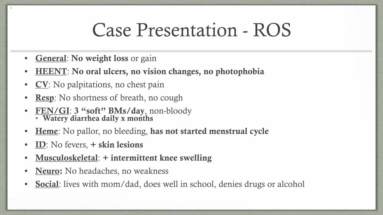
Case Presentation - ROS • General: No weight loss or gain
• HEENT: No oral ulcers, no vision changes, no photophobia
• CV: No palpitations, no chest pain
• Resp: No shortness of breath, no cough
• FEN/GI: 3 “soft” BMs/day, non-bloody • Watery diarrhea daily x months
• Heme: No pallor, no bleeding, has not started menstrual cycle
• ID: No fevers, + skin lesions
• Musculoskeletal: + intermittent knee swelling
• Neuro: No headaches, no weakness
• Social: lives with mom/dad, does well in school, denies drugs or alcohol

Case Presentation- PMHx • Heme/Onc: IDA of unknown etiology
• Iron supplementation: 120 mg elemental Fe BID • Alpha-thalassemia trait • BMA/Bx due to lack of improvement with Fe
• Allergy/Immunology: • Eczema: Tx with TAC • Persistent Asthma: Flovent, Albuterol prn • Allergies: Seafood
• Rheumatology: • Knee swelling, rash • Elevated inflammatory markers • +ANCA 1:80, Negative CT chest
• Perinuclear ANCA usually assoc. with IBD

Case Presentation- Family History
• Family History • Maternal great aunt- Lupus
• Maternal GM: Type II DM • Multiple family members: IDM
• Dad: Asthma
• Medications: • Orapred 5mg po qd, Albuterol prn
• Allergies: shellfish, PCN
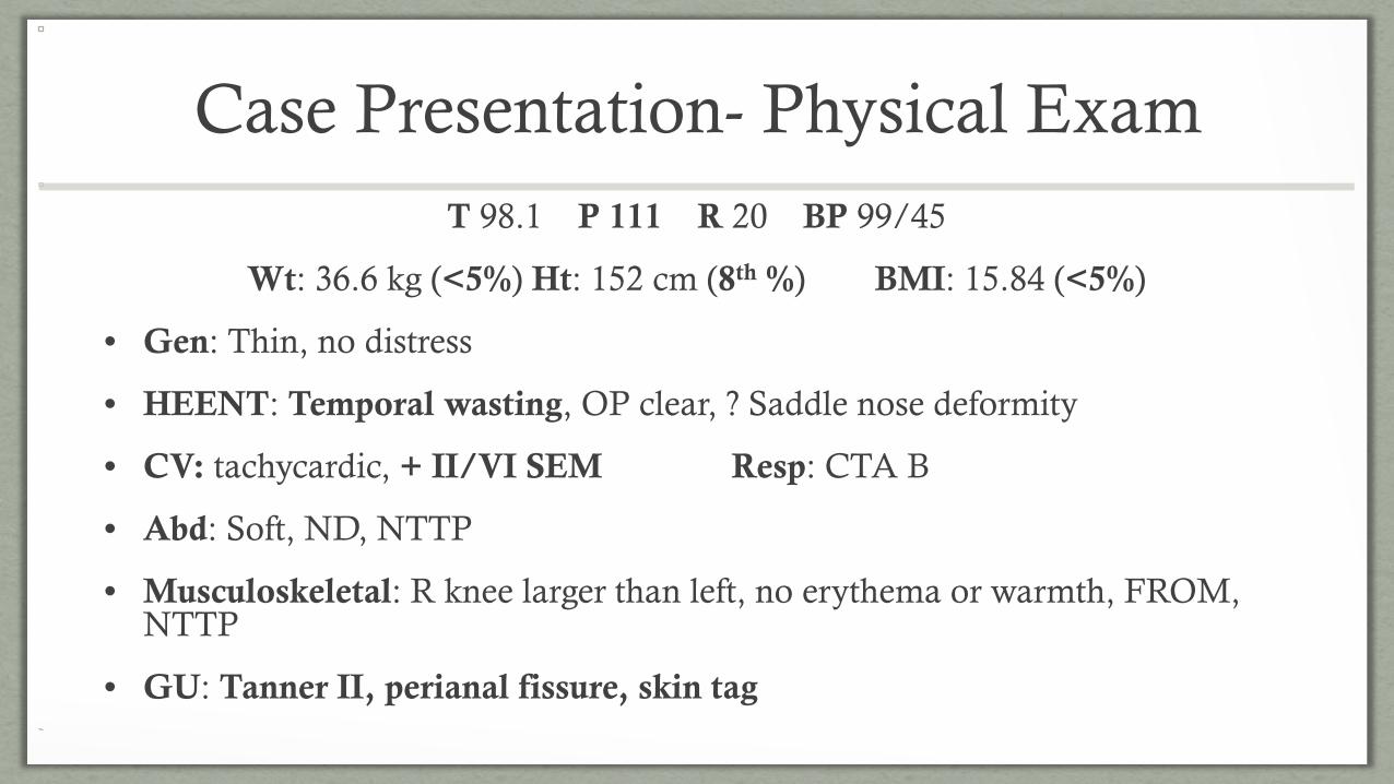
Case Presentation- Physical Exam T 98.1 P 111 R 20 BP 99/45
Wt: 36.6 kg (<5%) Ht: 152 cm (8th %) BMI: 15.84 (<5%)
• Gen: Thin, no distress
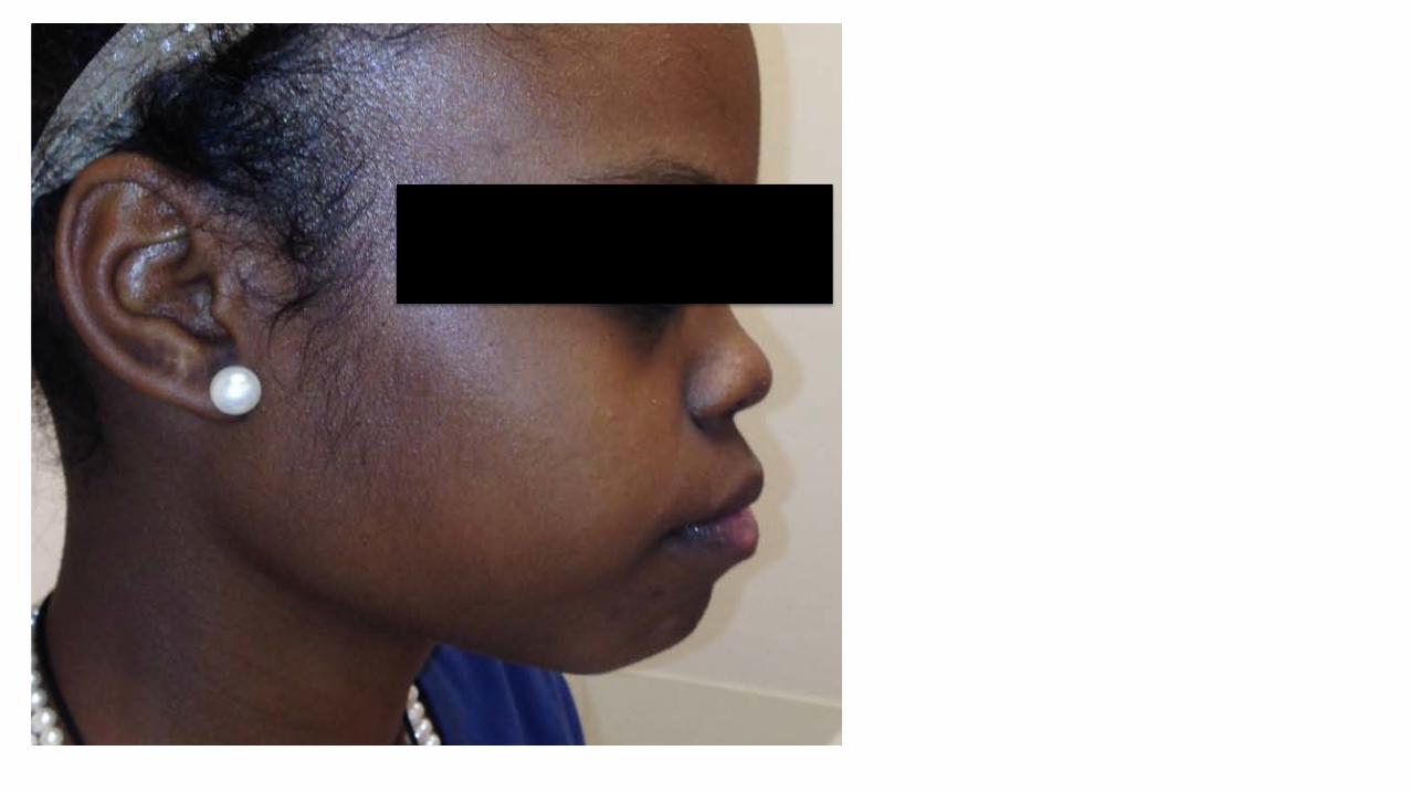
• HEENT: Temporal wasting, OP clear, ? Saddle nose deformity
• CV: tachycardic, + II/VI SEM Resp: CTA B
• Abd: Soft, ND, NTTP
• Musculoskeletal: R knee larger than left, no erythema or warmth, FROM, NTTP
• GU: Tanner II, perianal fissure, skin tag
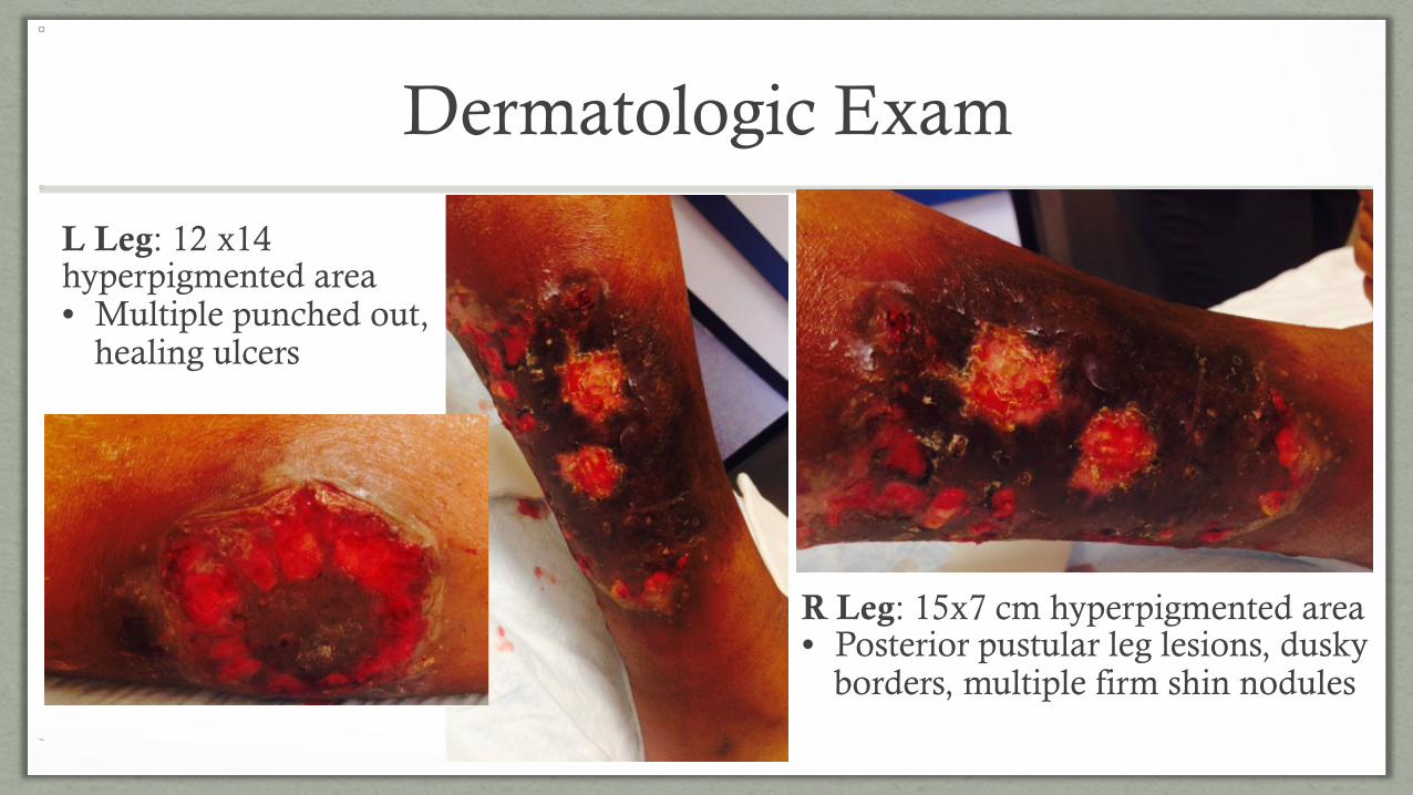
Dermatologic Exam
L Leg: 12 x14 hyperpigmented area • Multiple punched out,
healing ulcers
R Leg: 15x7 cm hyperpigmented area • Posterior pustular leg lesions, dusky
borders, multiple firm shin nodules

Initial Lab Evaluation
10.23
25.6
742
7.1 Retic 6.55% MCV 78.3
ESR 79 CRP 11.1 Fecal calprotectin 1323 ug/g
(<50 ug/g) Albumin 2.5 Blood Cx: No growth Wound Cx: No growth FOBT: +
N47 B30 L4 M14 E5

Dermatologic Evaluation
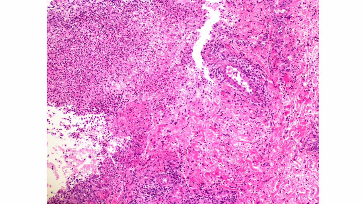
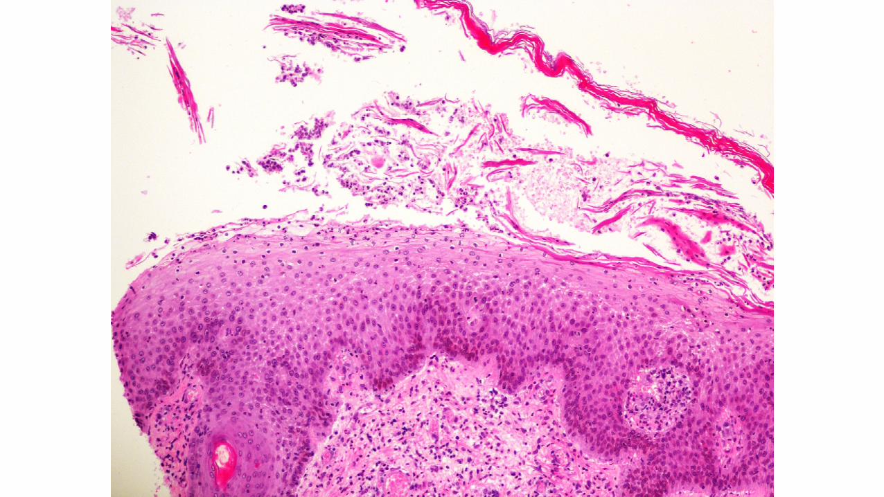
• Punch biopsies • Left: chronic active folliculitis with deep dermal granuloma
• Right: • Dermal necrotizing suppurative inflammation with chronic active vasculitis, mild • Diffuse infiltrate of neutrophils, histiocytes, and lymphocytes à pyoderma gangrenosum
• DDx: Sweet’s Syndrome; lacks nuclear destruction
• Epidermal neutrophilic invasion and bullae formation


GI Evaluation • EGD: normal • Colonoscopy:
• Right colon • Isolated site of erythematous, ulcerated colon amidst normal colon
• Transverse Colon • Decreased disease, mild erythema
• Rectum, Sigmoid, & Descending Colon • Erythema with friability, pus exudate, mucosal thickening
• Pathology: • Right and transverse colon- eosinophilia, lymphoid hyperplasia • Left colon- marked chronic active colitis with erosions and ulcerations

Colonoscopy Picture Examples
www.endoatlas.org www.endoatlas.org

Diagnosis/Treatment
• Diagnosis: IBD • Crohn’s Disease
• Pyoderma Gangrenosum
• Treatment: • IV corticosteroid, Remicade©, wound care

Objectives
• Become familiar with some extraintestinal manifestations of IBD
• Recognize some common neutrophilic dermatoses
• Know how to diagnose PG
• Understand treatment in PG

IBD
PCP
Ortho
GI
Surgery
Renal
Rheum
H/O
Ophtho
Derm
A/I
Extraintestinal Manifestions may find your
practice!

EIM
• Involve almost every organ system
• Affect 6-47% of patients • Possibly higher in pediatrics
• Pathogenesis not always clear • Immunologic
• Immunologic derangements à IBD
• IBD pts have increased risk of AI diseases
• Genetic
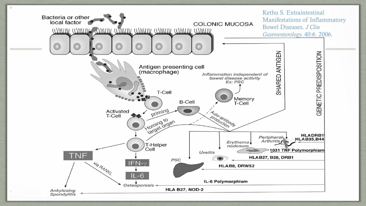
Kethu S. Extraintestinal Manifestations of Inflammatory Bowel Diseases. J Clin Gastroenterology. 40:6. 2006.

Growth Failure
• 15-40% of children • More common in CD than UC
• Z-score • Mean height for CD at diagnosis -0.54 • Mean weight for CD -1.06
• ~30% have Z-scores <3rd % • Mean weight for UC -0.32
• Why? • Discomfort eating • Proinflammatory cytokines à GH resistance and decr. IGF-1 production • Small bowel inflammation à PLE • Fat malabsorption & ADEK deficiency • High dose corticosteroids

Ophthalmology
• Ophtho conditions often seen in conjunction with arthritis & erythema nodosum
• Episcleritis • 2-5% of patients • Painless hyperemia of sclera and conjunctiva • No loss of vision • PE:
• Injection of ciliary vessels • Inflammation of episcleral tissues
• Tx: • Topical glucocorticoids
http://www.hopkinsmedicine.org/wilmer/conditions/episcleritis.html

Ophthalmology- Uveitis • 0.5-3%
• Bilateral, posterior to lens
• Insidious onset, chronic
• F >>>> M
• 75% associated arthritis
• Acute or subacute onset of eye pain, blurred vision, photophobia, headaches, iridospasm
• PE: slit-lamp exam: inflammation in anterior chamber, corneal clouding
• Tx: topical or systemic steroids, covering eye

Oral Cavity
Aphthous stomatitis
• 5-10% in UC, 20-30% in CD
• Lesions parallel disease activity
• Swelling, cobblestoning of mucosa, tag-like mucosal lesions
• Tx: systemic or local corticosteroids
!Hardy S, Fleming P, Rowland M, et al. A prospective study of the oral manefestations of Crohn's disease. Clin Gastroenterol Hepatol 2005; 3:886. Copyright © 2005 Elsevier.
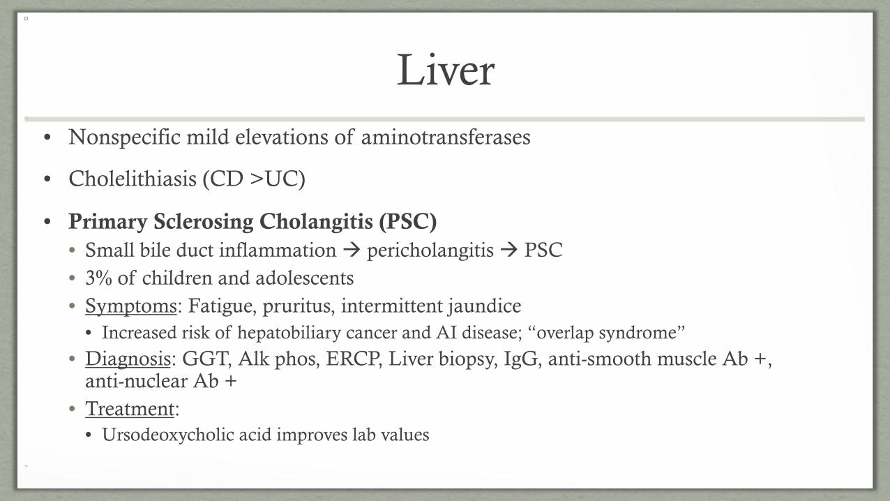
Liver • Nonspecific mild elevations of aminotransferases
• Cholelithiasis (CD >UC)
• Primary Sclerosing Cholangitis (PSC) • Small bile duct inflammation à pericholangitis à PSC • 3% of children and adolescents • Symptoms: Fatigue, pruritus, intermittent jaundice
• Increased risk of hepatobiliary cancer and AI disease; “overlap syndrome”
• Diagnosis: GGT, Alk phos, ERCP, Liver biopsy, IgG, anti-smooth muscle Ab +, anti-nuclear Ab +
• Treatment: • Ursodeoxycholic acid improves lab values
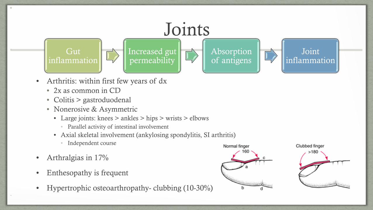
Joints
• Arthritis: within first few years of dx • 2x as common in CD • Colitis > gastroduodenal • Nonerosive & Asymmetric
• Large joints: knees > ankles > hips > wrists > elbows • Parallel activity of intestinal involvement
• Axial skeletal involvement (ankylosing spondylitis, SI arthritis) • Independent course
• Arthralgias in 17%
• Enthesopathy is frequent
• Hypertrophic osteoarthropathy- clubbing (10-30%)
Gut inflammation
Increased gut permeability
Absorption of antigens
Joint inflammation
Bones • Osteopenia 41% of children with CD and 25% in UC
• Interference with bone production • Poor diet • Hypogonadism (absent menses) • Inadequate calcium intake or malabsorption • Vitamin D deficiency • Excessive cytokine production • Corticosteroid use
• Acceleration of bone loss • Prolonged bed rest • Corticosteroid use
• Secondary hyperparathyroidism • Vit D and calcium deficiency

Hematology • Venous thromboembolism
• 1-2% of hospitalized patients • Increased risk with CVL, severe disease, older age, PN, oral contraceptives,
inherited thrombophilia • Prophylaxis for:
• Severe IBD + hx of thromboembolism • Severe IBD + CVL, h/o familial thromboembolism, immobility • All: adequate hydration, mobilization, compression stockings
• Anemia • 50% of patients • IDA, B12, FA, malnutrition, hemolysis, BM suppression (AZT), chronic
blood loss, anemia of chronic disease
• Thrombocytosis • 50% of patients

Cutaneous Manifestations
• There are many!
• We will discuss: • Erythema Nodosum
• 2 Neutrophilic Dermatoses • Sweet Syndrome
• Pyoderma Gangrenosum

Erythema Nodosum • CD > UC (3%)
• Reflects increasing bowel activity
• Single or multiple tender, red or purple-blue nodules on LE extensor surfaces • Pretibial area • 1-5cm
• Histology: • Neutrophilic perivascular reaction w/ dermal panniculitis • Normal epidermis
• 75% of pts develop arthritis
• Tx: treat the IBD

Erythema Nodosum
!
Courtesy of Lee T Nesbitt, Jr. The Skin and Infection: A Color Atlas and Text, Sanders CV, Nesbitt LT Jr (Eds), Williams & Wilkins, Baltimore. 1995.
Neutrophilic Dermatoses
• Histology: epidermal and/or dermal inflammatory infiltrates • Neutrophils
• No evidence of infection
• Classification based on: • Clinical and pathologic features
• Identification of associated diseases
• Pathogenesis: unknown; possibly immunologic • Respond to glucocorticoids and immunomodulatory therapies
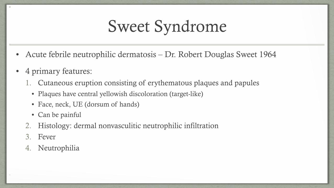
Sweet Syndrome
• Acute febrile neutrophilic dermatosis – Dr. Robert Douglas Sweet 1964
• 4 primary features: 1. Cutaneous eruption consisting of erythematous plaques and papules
• Plaques have central yellowish discoloration (target-like)
• Face, neck, UE (dorsum of hands)
• Can be painful
2. Histology: dermal nonvasculitic neutrophilic infiltration
3. Fever
4. Neutrophilia

Sweet Syndrome cont.
• Associated with: • IBD
• Malignancies (AML, solid tumors)
• Infections (URI & GI)
• Drugs
• Autoimmune diseases
• Pregnancy
• Tx: systemic/high-potency topical corticosteroids, potassium-iodide

Sweet Syndrome
!
!
http://www.uptodate.com
http://www.uptodate.com

Pyoderma Gangrenosum
• Rare: 3-10 cases/1 million people per year • Young and middle-aged adults, F > M
• IBD: <1% of patients; UC >CD
• >50% associated with underlying systemic disease • IBD, hematologic disorders or malignancies, arthritis, PAPA syndrome
• Pathophysiology: dysregulation of immune system
• DDx: antiphospholipid-Ab syndrome, venous stasis ulcers, Wegener’s, PAN

Pyoderma Gangrenosum Subtype #1
• Ulcerative (classic) • Erythematous pustule or noduleà spreads peripherally à degenerates
centrallyà ulcer w/ violaceous border & purulent base • Ulcers are sterile
• Ulcers extend to SQ tissue
• Extensor surfaces of LE
! !!

Pyoderma Gangrenosum Subtype #2
• Bullous (atypical) • Related to hematologic disease
• Rapid development of blue-gray, inflammatory bullae à erodeà ulcers • Arms and face
!Fitzpatrick TB, Johnson RA, Wolff K, et al. Color atlas and synopsis of clinical dermatology, 3rd ed, McGraw-Hill, New York 1997. Copyright © 1997 McGraw-Hill.

Pyoderma Gangrenosum Subtype #3
• Pustular PG • Acute exacerbations of IBD
• Rapid development of pustules surrounded by erythema
• Fever + arthralgias
!!

Pyoderma Gangrenosum Subtype #4
• Vegetative • “superficial granulomatous pyoderma”
• Localized, solitary, superficial form of PG
• Indolent, mildly painful nodule, plaque, or ulcer
• Verrucous quality
• Head and neck
!
www.uptodate.com

Pyoderma Gangrenosum Diagnosis
• Major criteria (must have both) • Rapid progression of painful, necrolytic, cutaneous ulcer
• Irregular, violaceous, undermined border
• 1-2 cm/day or 50% increase in size in 1 mo.
• Other causes of cutaneous ulceration excluded
• Minor criteria (must have two) • History suggestive of pathergy or finding of cribiform scarring
• Systemic disease associated w/ PG
• Histopathologic findings
• Treatment response (rapid response to systemic glucocorticoids)
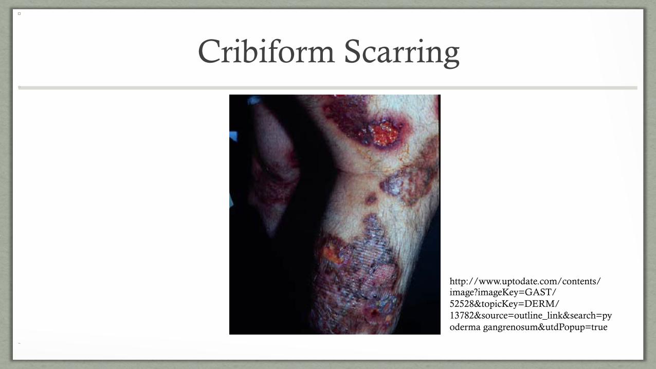
Cribiform Scarring
!
http://www.uptodate.com/contents/image?imageKey=GAST/52528&topicKey=DERM/13782&source=outline_link&search=pyoderma gangrenosum&utdPopup=true
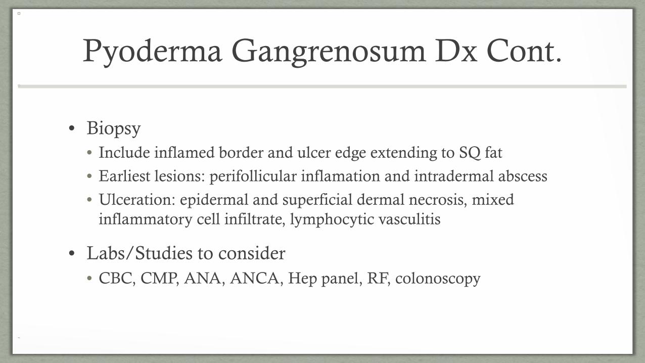
Pyoderma Gangrenosum Dx Cont.
• Biopsy • Include inflamed border and ulcer edge extending to SQ fat
• Earliest lesions: perifollicular inflamation and intradermal abscess
• Ulceration: epidermal and superficial dermal necrosis, mixed inflammatory cell infiltrate, lymphocytic vasculitis
• Labs/Studies to consider • CBC, CMP, ANA, ANCA, Hep panel, RF, colonoscopy

PG Treatment • Wound care; beware of pathergy!
• Mild disease: • Local corticosteroids or calcineurin inhibitor
• Extensive disease: • Systemic glucocorticoids or systemic cyclosporine
• 0.5-1.5mg/kg/day (max 60mg) oral prednisone or pulse IV steroids • Taper once progression has stopped + improvement; within 4-10 weeks
• + glucocorticoid-sparing agent: cyclosporine, azathioprine, infliximab
• Infliximab
• Severe, refractory disease • IVIG or alkylating agents
• Recurrent lesions 30%

Infliximab for the treatment of pyoderma gangrenosum: a randomised, double-blind placebo-
controlled trial • Brooklyn et. al
• Multicenter, randomized, placebo controlled trial
• 30 patients with PG; 19 with IBD
• Infliximab 5mg/kg or placebo infusions at week 0
• Physician & patient assessment of appearance of lesion at weeks 2, 4, & 6 • Reduction in size, depth, and degree of undermining • Week 2:
• 46% (6/13) of pts tx w/ infliximab had response compared to 6% (1/17) placebo • Subjects in both arms offered infliximab
• 69% (20/29) had positive response by week 6 • No difference in response between PG pts with underlying IBD
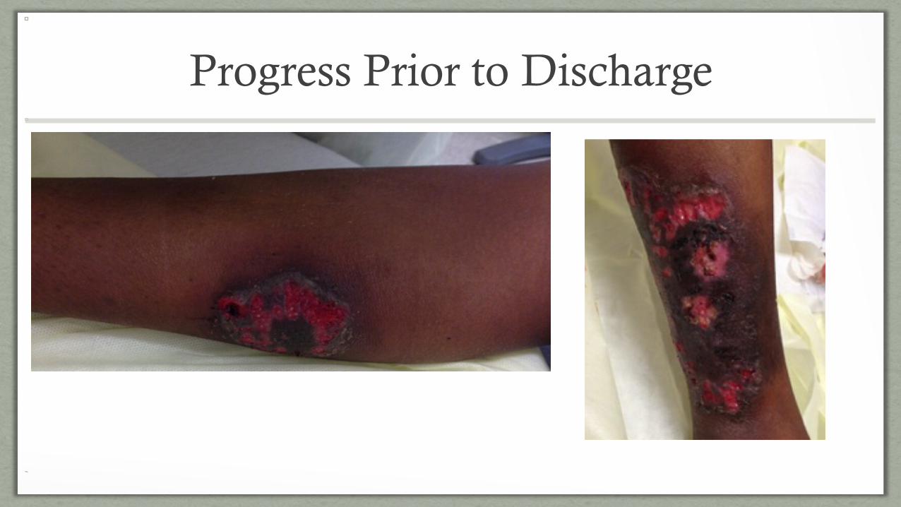
Progress Prior to Discharge

Continued improvement!!!

Take Home Points!
• Not all that’s red is cellulitis
• ALWAYS take a good history
• If you suspect a neutrophilic dermatosis, search for underlying disease

Special Thanks!
• Dr. Brown- Rheumatology
• Dr. Craver- Pathology
• Dr. Dimitriades- Rheumatology/A&I
• Dr. Malowitz- GI
• Dr. Sandlin- Hospitalist

References Brooklyn TN et al. Infliximab for the treatment of pyoderma gangrenosum: a randomised, double-blind placebo-controlled trial. Gut 2005.
Cohen P. Neutrophilic dermatoses: a review of current treatment options. Am J Clin Dermatol 2009:10. 301-312.
Glick S and Carvalho R. “Inflammatory Bowel Disease. Pediatrics in Review. Vol 32. No 1. Jan 1, 2011 pp 14-25.
Higuchi L and Bousvaros A. “Clinical presentation and diagnosis of inflammatory bowel disease in children and adolescents”. 2014. www.uptodate.com
Hyams J. “Extraintestinal Manifestations of Inflammatory Bowel Disease in Children. Journal of Pediatric Gastroenterology and Nutrition. 19:7-21. 1994.
Kethu S. Extraintestinal Manifestations of Inflammatory Bowel Diseases. J Clinical Gastroenterology. 40:6. 2006.
Jose F and Heyman M. “Extraintestinal Manifestations of Inflammatory Bowel Disease”. Journal of Pediatric Gastroenterology and Nutrition. . 46:124-133. 2008.
Moschella S. “Neutrophilic dermatoses”. 2014. www.uptodate.com.
Peppercorn M and Cheifetz A. “Skin and eye manifestations of inflammatory bowel disease”. 2014. www.uptodate.com
Schadt C. “Pyoderma gangrenosum: Pathogensis, clinical features, and diagnosis”. 2014. www.uptodate.com