from drug screening to target deconvolution: a target-based drug
TRANSCRIPT

From Drug Screening to Target Deconvolution: a Target-Based DrugDiscovery Pipeline Using Leishmania Casein Kinase 1 Isoform 2 ToIdentify Compounds with Antileishmanial Activity
Emilie Durieu,b* Eric Prina,d* Olivier Leclercq,a Nassima Oumata,f Nicolas Gaboriaud-Kolar,e Konstantina Vougogiannopoulou,e
Nathalie Aulner,c Audrey Defontaine,b Joo Hwan No,h Sandrine Ruchaud,b Alexios-Leandros Skaltsounis,e Hervé Galons,g
Gerald F. Späth,a Laurent Meijer,b* Najma Rachidib*
Institut Pasteur and INSERM U1201, Unité de Parasitologie moléculaire et Signalisation, Paris, Francea; Sorbonne Universités, UPMC Université de Paris 06, CNRS USR3151,Protein Phosphorylation and Human Diseases, Station Biologique, Roscoff, Franceb; Institut Pasteur, Imagopole, Paris, Francec; Institut Pasteur, Laboratoired’Immunophysiologie et Parasitisme, Paris, Franced; Department of Pharmacognosy and Natural Products Chemistry, School of Pharmacy, University of Athens, Athens,Greecee; ManRos Therapeutics, Centre de Perharidy, Roscoff, Francef; Université Paris-Descartes, Laboratoire de Chimie Organique 2, INSERM U 648, Paris, Franceg; InstitutPasteur Korea, Leishmania Research Laboratory, Seongnam-si, Republic of Koreah
Existing therapies for leishmaniases present significant limitations, such as toxic side effects, and are rendered inefficient by par-asite resistance. It is of utmost importance to develop novel drugs targeting Leishmania that take these two limitations into con-sideration. We thus chose a target-based approach using an exoprotein kinase, Leishmania casein kinase 1.2 (LmCK1.2) that wasrecently shown to be essential for intracellular parasite survival and infectivity. We developed a four-step pipeline to identifynovel selective antileishmanial compounds. In step 1, we screened 5,018 compounds from kinase-biased libraries with Leishma-nia and mammalian CK1 in order to identify hit compounds and assess their specificity. For step 2, we selected 88 compoundsamong those with the lowest 50% inhibitory concentration to test their biological activity on host-free parasites using a resaz-urin reduction assay and on intramacrophagic amastigotes using a high content phenotypic assay. Only 75 compounds showedantileishmanial activity and were retained for step 3 to evaluate their toxicity against mouse macrophages and human cell lines.The four compounds that displayed a selectivity index above 10 were then assessed for their affinity to LmCK1.2 using a targetdeconvolution strategy in step 4. Finally, we retained two compounds, PP2 and compound 42, for which LmCK1.2 seems to bethe primary target. Using this four-step pipeline, we identify from several thousand molecules, two lead compounds with a selec-tive antileishmanial activity.
The protozoan parasite Leishmania is the causative agent ofleishmaniasis, a potentially fatal disease with worldwide
distribution. Depending on the species, three clinical forms ofthe disease can be distinguished, cutaneous leishmaniasis (CL;e.g., Leishmania major), mucocutaneous leishmaniasis (MCL;e.g., Leishmania braziliensis), and fatal visceral leishmaniasis(VL; e.g., Leishmania donovani) (1). Several treatment options areavailable, which either show important side effects or are unaf-fordable. In all regions where these infections are endemic, thefirst line of treatment is pentavalent antimonials, despite theirimportant side effects and the appearance of parasite resistance.Although their target has not been identified, one member, so-dium stibogluconate was shown to inhibit the energy metabolismand macromolecule biosynthesis (2, 3). The second line of treat-ment is amphotericin B, a very potent but highly toxic antifungaldrug. This compound creates pores targeting ergosterol only pres-ent in the plasma membrane of parasites but not in that of mam-malian cells. Its less toxic lipid formulation is extremely expensiveand thus incompatible with treatment in developing countries (3,4). Among the other drugs that have been recently developed,miltefosine was a major breakthrough in leishmaniasis therapy asthis anticancer drug is the first oral treatment against VL. Unfor-tunately, its teratogenicity excludes the treatment of pregnantwomen and its slow turnover could promote the emergence ofclinical parasite resistance (4). Miltefosine plays a role in the per-turbation of the lipid metabolism and the induction of apoptosis-like cell death and has immunostimulatory effects; however, its
mode of action has not been precisely identified (5). The activity ofmiltefosine is due to its accumulation inside the parasite (6),which is prevented in resistant lines generated in vitro by the over-expression of members of the ABC (ATP-binding cassette) trans-porter family and/or mutation of the flippase LdMT (6, 7). Threemore drugs complete the list of available treatments for leishman-iasis: (i) pentamidine, which has been used for VL, CL, and MCLtreatment, induces the inhibition of polyamine biosynthesis and a
Received 5 January 2016 Returned for modification 16 January 2016Accepted 18 February 2016
Accepted manuscript posted online 22 February 2016
Citation Durieu E, Prina E, Leclercq O, Oumata N, Gaboriaud-Kolar N,Vougogiannopoulou K, Aulner N, Defontaine A, No JH, Ruchaud S, Skaltsounis A-L,Galons H, Späth GF, Meijer L, Rachidi N. 2016. From drug screening to targetdeconvolution: a target-based drug discovery pipeline using Leishmania caseinkinase 1 isoform 2 to identify compounds with antileishmanial activity.Antimicrob Agents Chemother 60:2822–2833. doi:10.1128/AAC.00021-16.
Address correspondence to Najma Rachidi, [email protected].
* Present address: Emilie Durieu and Laurent Meijer, ManRos Therapeutics, Centrede Perharidy, Roscoff, France; Eric Prina and Najma Rachidi, InstitutPasteur/INSERM U1201, Unité de Parasitologie Moléculaire et Signalisation, Paris,France.
E.D. and E.P. contributed equally to this article.
Supplemental material for this article may be found at http://dx.doi.org/10.1128/AAC.00021-16.
Copyright © 2016, American Society for Microbiology. All Rights Reserved.
crossmark
2822 aac.asm.org May 2016 Volume 60 Number 5Antimicrobial Agents and Chemotherapy
on February 21, 2018 by guest
http://aac.asm.org/
Dow
nloaded from

decrease of the mitochondrial inner membrane potential; (ii) theaminoglycosidic antibiotic paromomycin, which is restricted toareas where such infections are endemic, cures both VL and CLefficiently by targeting mitochondria; and (iii) sitamaquine, an8-aminoquinoline, which intercalates within biological mem-branes to accumulate in Leishmania cytosolic acidic compart-ments (4).
Despite the various drugs available, none of these treatmentsare ideal because of two main aspects: (i) their side effects, duemainly to off-target effects that cannot be eliminated by drug op-timization as the target responsible for the antileishmanial effect isunknown, and (ii) the emergence of parasite resistance, due to theplasticity of the parasite. Therefore, there is an urgent need todiscover new molecules and to develop new drug discovery pipe-lines that take these two aspects into consideration. First, the useof known validated targets for drug screening represents a majoradvantage as the compound could be optimized to fit exclusivelythe target, avoiding off-target effects mainly responsible for sideeffects. Protein kinases are among the best candidates as drugtargets for leishmaniasis because: (i) kinase inhibitors are one ofthe most important group of U.S. Food and Drug Administration-approved drugs for the treatment of diseases such as cancer orAlzheimer’s disease; (ii) they are considered valid targets for dis-eases caused by unicellular parasites, such as malaria; and (iii)kinases regulate many key processes, such as cell cycle or signaltransduction, and thus the inhibition of their activity decreasescell viability (8, 9–14). Second, targeting proteins secreted by theparasite could delay the emergence of drug resistance. Indeed, ithas been recently shown that parasitic proteins could be exported,via exosomes, into the host cell to modify its biology or its innateimmune response (15, 16). To perform their function in the host,these proteins need to interact with host proteins, and thus anymutations could abrogate their functions in the host cell, whichcould be detrimental for the intracellular parasite survival.
Among the proteins identified in the recent proteomic analysisof Leishmania exosomes, 13 could qualify as good drug targets asdefined above because they are excreted kinases. Most of thesekinases are involved in purine or glucose metabolism, and onlyone is involved in signal transduction, casein kinase 1 (CK1). Amember of the highly conserved Ser/Thr protein kinase family(17), CK1 contains six isoforms in Leishmania (15, 18–20).LmjF35.1010 (LmCK1.2), the major isoform, has been validatedpharmacologically as a drug target based on the findings that theinhibition of CK1 activity by the specific inhibitor D4476 stronglycompromises axenic amastigote viability and decreases the per-centage of infected macrophages (21). We hypothesize that thecapacity of LmCK1.2 to recognize and phosphorylate host pro-teins could allow the parasite to regulate essential host cell pro-cesses (22) and therefore to survive. This hypothesis is based onour previous findings showing that (i) the protein sequence ofLmCK1.2 kinase domain is 100% identical in all sequenced Leish-mania species (except the lizard-isolated Leishmania tarentolaeand unclassified Leishmania sp. strain MAR LEM2494), suggest-ing that there is a selection pressure to maintain the integrity of theprotein sequence, and (ii) LmCK1.2 is the most closely relatedkinase to its human orthologs in Leishmania. These two elementssuggest that LmCK1.2 cannot be mutated without compromis-ing the survival of the intracellular parasite, which would ren-der the emergence of drug-resistant parasites expressing mu-tated LmCK1.2 unlikely (21).
We present here a four-step pipeline that allows the discoveryof novel lead compounds. First, we generated an active recombi-nant LmCK1.2 and purified mammalian CK1 from porcine brain(SsCK1 [23]). We developed an enzymatic assay to screen 4,030compounds from kinase-biased and focused libraries, as well as988 analogs with both Leishmania and mammalian kinases, inorder to identify hit compounds and assess their specificity. Weselected 88 compounds with a 50% inhibitory concentration(IC50) below 10 �M. Second, we tested the antileishmanial effectof these compounds on cultured parasites using a rezasurin-basedassay, as well as on intracellular parasites using a high-contentphenotypic screen. We retained 75 compounds with an antileish-manial activity. Third, after evaluation of the toxicity of the se-lected antileishmanial compounds against mouse macrophagesand human cell lines, only four compounds had a selectivity index(SI) greater than 10. Fourth, the affinity for LmCK1.2 for thesecompounds was tested using a target deconvolution approach.Two compounds for which LmCK1.2 seems to be the primarytarget were eventually selected. The identification of these twolead compounds validates our pipeline, which will be used toscreen diversified libraries to identify more lead compounds.
MATERIALS AND METHODSL. donovani culture and axenic amastigote differentiation. L. donovani1S2D (MHOM/SD/62/1S-CL2D), clone LdB, was obtained from SteveBeverley, Washington University School of Medicine, St. Louis, MO, andcultured as described previously (24–26).
Parasite growth inhibition assay. L. donovani promastigotes and ax-enic amastigotes (2 � 106 cells/ml) in their respective media were distrib-uted in 96-well plates (125 �l/well). An equal volume of medium contain-ing inhibitor at the indicated concentrations (in 1% dimethyl sulfoxide[DMSO]) was added. After 24 h of incubation in the dark at 26°C (pro-mastigotes) or 37°C (amastigotes), 25 �l of resazurin solution at 0.001%was added, and the plates were incubated for an additional 24 h in the darkat appropriate temperatures. The plates were read (excitation, 544 nm;emission, 590 nm) using a fluorescent microplate reader (Safas XeniusXML) (27).
Human cell line MTT assay. HFF1, SH-SY5Y, and U-2 OS cells werecultured in Dulbecco modified Eagle medium (DMEM); hTERT RPE-1cells were cultured in DMEM/F-12 medium. All media were supple-mented with 10% fetal calf serum, 2 mM L-glutamine, and 50 IU of pen-icillin and streptomycin. Cell viability was assayed using a Promega Cell-Titer96 AQueous nonradioactive cell proliferation assay according to themanufacturer’s instructions.
Macrophage infection and assessment of intracellular parasite sur-vival. A high-content, biologically relevant, cell-based assay was used todetermine the antileishmanial activity of D4476 as previously described(21, 28). Briefly, the assay combines (i) the use of primary bone marrow-derived mouse macrophages as natural host cells and DsRed2-expressingL. amazonensis (MPRO/BR/1972/M1841) amastigotes, the clinically rele-vant parasite stage of Leishmania, with (ii) the detection of fluorescentmarkers as reporter molecules. A total of 10,000 macrophages werecounted per well depending on the number of replicates per tested com-pound.
Ethics statement. All animals were housed in our A3 animal facilitiesin compliance with the guidelines of the A3 animal facilities at the InstitutPasteur, which is a member of Committee 1 of the Comité d’Ethique pourl’Expérimentation Animale (CEEA), Ile de France, France. Animal hous-ing conditions and the protocols used in the work described here wereapproved by the Direction des Transports et de la Protection du Public,Sous-Direction de la Protection Sanitaire et de l’Environnement, PoliceSanitaire des Animaux, B75-15-27 and B75-15-28, in accordance with theEthics Charter of animal experimentation that includes appropriate pro-
Leishmania CK1.2 Drug Screening
May 2016 Volume 60 Number 5 aac.asm.org 2823Antimicrobial Agents and Chemotherapy
on February 21, 2018 by guest
http://aac.asm.org/
Dow
nloaded from

cedures to minimize pain and animal suffering. G.S. and E.P. are autho-rized to perform experiments on vertebrate animals (licenses B75-1159and 75-1265, respectively) issued by the Direction Départementale de laProtection des Populations de Paris and were responsible for all experi-ments conducted personally or under supervision as governed by the lawsand regulations relating to the protection of animals.
Automated microtiter plate CK-S kinase assay. A mixture of nativeCK1 isoforms (essentially CK1� and CK1ε) was extracted from porcinebrain (SsCK1) and purified by affinity chromatography on immobilizedaxin (23). LmCK1.2 was produced and purified as previously described(21). Both SsCK1 and recombinant LmCK1.2 were assayed, with 27 �MCK-specific peptide substrate CK-S (RRKHAAIGpSAYSITA) synthesizedby Proteogenix (Oberhausbergen, France) in buffer C (pH 7; 60 mM�-glycerophosphate, 30 mM p-nitrophenyl phosphate, 25 mM morpho-linepropanesulfonic acid, 5 mM EGTA, 15 mM MgCl2, 1 mM dithiothre-itol [DTT], 0.1 mM sodium vanadate), with 15 �M [�-33P]ATP in a finalvolume of 30 �l. After 30 min of incubation at 30°C, 30-�l aliquots werefiltered onto Whatman P81 phosphocellulose paper. The filters werewashed with a solution of 1% phosphoric acid and then counted in thepresence of 20 �l of scintillation fluid per well. Blank values were sub-tracted and activities calculated as the pmol of phosphate incorporatedduring 30 min of incubation. The activities were expressed as the percent-ages of maximal activity, i.e., in the absence of inhibitors. Controls wereperformed with appropriate dilutions of DMSO.
ATP depletion and competition. The axenic amastigote total proteinextract (7 mg) was dialyzed overnight at 4°C in dialysis solution (1�phosphate-buffered saline, 1 mM EDTA, 1 mM DTT) using a Slide-A-Lyzer 10kD dialysis cassette (Pierce) to eliminate free ATP. Then, 1 mg ofdialyzed extract per condition was mixed with the binding solution (1�binding solution, 1 mM DTT, 1� protease inhibitor) of the ATP affinitytest kit (Jena Bioscience) and 500 �M inhibitor. The samples were incu-bated 30 min at 4°C and added to a mixture containing 12.5 �l of eachATP agarose (ATP affinity test kit from Jena Bioscience). ATP bindingproteins from the assay and competition samples were enriched accordingto the manufacturer’s instructions. Finally, eluted samples were concen-trated using Amicon Ultra-10K centrifugal filters (Millipore) to a finalvolume of approximately 100 �l.
Next, 12.5 �l of the flowthrough and 30 �l of the eluate were separatedon Novex NuPAGE 4 to 12% Bis-Tris gel (Life Technologies) from boththe assay and the competition. The gel was stained with SYPRO Ruby (LifeTechnologies) according to the manufacturer’s instructions and revealedusing a Typhoon scanner. Alternatively, the proteins separated by SDS-PAGE were transferred onto a polyvinylidene difluoride membrane andprobed with SY3535 antibody (21). Signals were revealed by SuperSignal-ECL (Pierce).
Compound libraries. We screened 5,018 compounds from theRoscoff library including 588 purine derivatives (29) and 400 indirubinderivatives (30, 31, 32).
Homology modeling and structural alignment. The amino acid se-quence of L. major CK1.2 was retrieved from the NCBI database (acces-sion number XP_003722496) in FASTA format. The homology modelingof the sequence was performed by Swiss-Model program (33), and theprotein with PDB code 3SV0 was selected as a template. The PyMOLprogram (34) was used for the structural alignment of Schizosaccharomy-ces pombe CK1 in complex with the specific CK1 inhibitor, IC261 (PDBcode 1EH4) and Homo sapiens CK1� (PDB code 4KB8) to the generatedL. major CK1.2 homology model. The figures were also prepared usingPyMOL (34).
RESULTS
We present below a comprehensive drug discovery pipeline en-compassing four steps.
Primary screening comparing SsCK1 and LmCK1.2 to iden-tify specific LmCK1.2 inhibitors. Primary screening comparingSsCK1 and LmCK1.2. We purified recombinant LmCK1.2-V5-
His6 from Escherichia coli and a mixture of Sus scrofa CK1� andCK1ε (SsCK1) from porcine brain (21, 23). The conditions usedpreviously for the manual kinase assay were adapted to an auto-mated 96-well plate format (21, 23). We used CK-S as the sub-strate for both kinases and 15 �M [�-33P]ATP for the assay (21,23). We screened 4,030 compounds at 10 �M from a kinase-bi-ased library that has been previously tested on mammalian ki-nases, such as cyclin-dependent kinases (29). For each compound,the percent inhibition of LmCK1.2 versus that of SsCK1 is pre-sented in Fig. 1A. We classified as hit compounds those that de-creased the kinase activity by �40% (Fig. 1Aa and b). We obtainedtwice as many hit compounds for the mammalian CK1 than forLmCK1.2 (Fig. 1B). Indeed, we identified 245 hit compoundsagainst SsCK1 (6.1% hit rate) and 128 against LmCK1.2 (3.2% hitrate), with only 37 compounds with a similar potency against bothkinases. This finding, which is surprising considering the highsimilarity between the protein sequences of LmCK1.2 and themammalian CK1s (ca. 70% [21]), suggests that the ATP bindingpocket of the two kinases is sufficiently divergent to accommodatedifferent inhibitors. We next classified the compounds accordingto their potency and specificity (class 1, compounds that inhibitthe kinase activity between 80 and 100%; class 2, compounds thatinhibit the kinase activity between 60 and 80%; and class 3, com-pounds that inhibit the kinase activity between 40 and 60%; Fig.1C). We obtained a similar number and distribution of hit com-pounds active on SsCK1 (30 compounds) and on LmCK1.2 (31compounds) in class 1. In contrast, we observed an increasednumber of compounds inhibiting specifically SsCK1 in classes 2and 3 (Fig. 1C). This could suggest that the ATP binding pocket ofSsCK1 could be more permissive than that of LmCK1.2. We se-lected 45 compounds either belonging to class 1 that inhibitedLmCK1.2 activity by �90% or belonging to class 2 and were spe-cific to LmCK1.2 to determine their IC50. As shown in Fig. 2, allthe compounds with an IC50 below 1.3 �M (most potent) werenonspecific, whereas all the specific compounds had an IC50 above1.3 �M.
Among the 45 compounds that were potent against LmCK1.2,we identified several inhibitors described to have antileishmanialactivity, including known CK1 inhibitors, such as anthraquinone(35–37), or compounds for which we revealed CK1 as a new tar-get, such as gossypol, purpurogallin, and some flavonoids (38–42,43). These compounds, identified from several libraries, werefound at least twice with similar IC50s, indicating that our assay isreproducible (data not shown). Altogether, these data demon-strate the efficiency of using CK1 as a target to identify compoundswith antileishmanial activities and confirm LmCK1.2 as a validdrug target. However, we did not retain these compounds forsubsequent characterization since they have been already exten-sively studied.
Secondary screening of purine and indirubin libraries. Ofthe 45 compounds for which we determined the IC50, we selectedtwo compounds with low IC50s but only moderate specificity to-ward LmCK1.2, purvalanol B, and indirubin-3=-monoxime, andwe tested analog libraries to perform structure-activity relation-ship (SAR) analysis in order to identify more specific compounds.We chose purvalanol B because we showed previously that Leish-mania CK1.2 binds to purvalanol B better than its mammaliancounterpart, suggesting that the sensitivity to purvalanol B couldbe higher for LmCK1.2 than for SsCK1 (44). Confirming this find-ing, the IC50 of purvalanol B toward LmCK1.2 (2 � 0.3 �M) is
Durieu et al.
2824 aac.asm.org May 2016 Volume 60 Number 5Antimicrobial Agents and Chemotherapy
on February 21, 2018 by guest
http://aac.asm.org/
Dow
nloaded from

slightly lower than that toward SsCK1 (2.9 � 1.2 �M). We testedthe potency of 588 purine analogs (Fig. 3A) at 10 �M againstLmCK1.2 and SsCK1 to identify compounds with better potencyand/or specificity. As shown in Fig. 3B and C, most compounds
were more potent against mammalian CK1 than against LmCK1.2(below the black line in Fig. 3B and dark gray in Fig. 3C). Next, wedetermined the IC50s of the 21 most potent purine derivativesagainst LmCK1.2 (Fig. 3D). For all of the compounds except onethe IC50 was systematically higher against LmCK1.2 (0.44 to 2.4�M) than against mammalian CK1 (0.081 to 1.6 �M). Only com-pound 26 had a lower IC50 against LmCK1.2 (1 � 0.4 �M) thanagainst SsCK1 (3.7 � 1.2 �M), which could be due to the presenceof a long carbon chain, a unique feature compared to the otherderivatives. Thus, although the purines were very potent towardLmCK1.2, they present a higher affinity for SsCK1.
The second family of compounds we screened are the indiru-bins (Fig. 4A). We showed that the IC50 of indirubin-3=-mon-oxime is lower against LmCK1.2 (0.13 � 0.03; see Table S1 in thesupplemental material) than against mammalian CK1 (0.39 �0.08 �M; see Table S1 in the supplemental material). To identifybetter compounds with higher selectivity, we tested 400 indirubinderivatives (Fig. 4B). In contrast to the purine analogs, the indiru-bins were globally more potent against LmCK1.2 than againstSsCK1 (Fig. 4B). For instance, in class 1 a total of 46 compoundswere more specific toward LmCK1.2 than SsCK1, whereas only 9
FIG 1 Differential target-based screen of 4,030 compounds from various libraries. (A) Representation of the percentage of inhibition toward LmCK1.2 activityversus the percentage of inhibition toward SsCK1 activity. The compounds in sectors a and b are potent toward LmCK1.2 since they show more than 40%inhibition, whereas the compounds in sectors b and d are potent toward SsCK1. (B) A total of 336 hit compounds were identified in the screen, of which 245inhibit SsCK1 (6.1% hit rate) and 128 inhibit LmCK1.2 (3.2% hit rate). Only 37 compounds showed equal potency against both CK1s. (C) Compounds wereclassified according to their specificity: compounds only potent against SsCK1 (only SsCK1), more potent against SsCK1 than LmCK1.2 (SsCK1 � LmaCK1.2),equally potent on both kinases (SsCK1 LmCK1.2), more potent against LmCK1.2 than SsCK1 (SsCK1 LmaCK1.2), and only potent on LmCK1.2 (onlyLmCK1.2). Compounds were also classified according to their percent inhibition: class 1 corresponds to compounds that inhibit the kinase activity between 80and 100%, class 2 corresponds to compounds that inhibit the kinase activity between 60 and 80%, and class 3 corresponds to compounds that inhibit the kinaseactivity between 40 and 60%. A total of 23 compounds were more potent toward LmCK1.2 than SsCK1, and 68 compounds were specific to LmCK1.2 (thenumbers in the histograms indicate the percentage of compounds in each category).
FIG 2 Determination of the IC50 of the 45 compounds belonging to class 1that have a percent inhibition above 90%. Each point represents the IC50 of aparticular compound toward LmCK1.2. Nonspecific compounds have a po-tency below 10 �M toward both kinases, whereas specific compounds have apotency below 10 �M only toward LmCK1.2.
Leishmania CK1.2 Drug Screening
May 2016 Volume 60 Number 5 aac.asm.org 2825Antimicrobial Agents and Chemotherapy
on February 21, 2018 by guest
http://aac.asm.org/
Dow
nloaded from

were more specific toward SsCK1 than LmCK1.2 (Fig. 4C). In-deed, the most active compounds were the most selective towardLmCK1.2, whereas compounds with lower potency (class 3) weremore specific toward SsCK1 (Fig. 4C). We selected the 55 mostpotent compounds to measure their IC50s. The IC50s were be-tween 0.08 and 10 �M, and for almost all the compounds the IC50
against LmCK1.2 was systematically lower than that against mam-malian CK1 (Fig. 4D), suggesting that the indirubin compoundshave more affinity toward LmCK1.2 than toward SsCK1.
The differences in specificity observed with the purine and in-dirubin compound families confirm that important differencesexist between the ATP binding pocket of both kinases (21). It alsosuggests that due to the strong affinity of the purine for the mam-malian CK1, it is likely that these compounds will be toxic for thehost cell.
Among all the compounds identified in the primary and sec-ondary screenings, we eliminated all the compounds for which thechemical optimization was unfeasible and selected 12 compoundsfrom the main library, 21 compounds from the purine library, and55 compounds from the indirubin library to assess their antileish-manial activity.
(Step 2) Evaluation of the antileishmanial activity of selectedcompounds. We evaluated the antileishmanial activity of the 88
compounds selected in step 1 on cultured L. donovani promasti-gotes and axenic amastigotes by measuring the percentage of met-abolically active parasites in liquid culture using a resazurin-basedassay (27) (see Table S1 in the supplemental material). As a posi-tive control, we treated the parasites with a 1 �M concentration ofthe antileishmanial reference drug, AMB, and obtained growthinhibitions of 90.4% � 1.5% and 79.9% � 1.3% for promastigotesand amastigotes, respectively, with excellent reproducibility, asreported by the small standard deviation (SD) values.
We then tested the compounds against intracellular L. ama-zonensis using a visual high-content phenotypic assay (21, 28). Wemeasured three parameters: (i) the percentage of cells remainingafter treatment compared to the vehicle control (DMSO) to eval-uate cell detachment (total macrophages), (ii) the percentage ofhealthy cells compared to the total number of cells remaining aftertreatment to evaluate cell mortality (viability index [VI]), and (iii)the percentage of parasitophorous vacuoles per healthy cell toevaluate the parasite burden (PB). We used AMB at 0.5 �M andcycloheximide at 150 �M as antileishmanial and cytotoxic controlcompounds, respectively. Figure S1 in the supplemental materialpresents the data for all of the controls performed during thescreening campaign. As expected, AMB reduced the parasite bur-den without affecting the number of macrophages or their viabil-
FIG 3 Screening of the purine derivative library. (A) Structure of the purine backbone. R1, R2, and R3 represent different substitutions of the purines. (B) Weperformed a target-based screening of 588 derivatives. Each point represents the percent inhibition toward LmCK1.2 activity versus the percent inhibition towardSsCK1 activity of each compound. The compounds in the top left are more potent toward LmCK1.2, whereas the compounds in the bottom right are more potenttoward SsCK1. (C) Compounds were classified according to their specificity: only potent on SsCK1, more potent on SsCK1 than LmCK1.2 (SsCK1 � LmCK1.2),equally on both kinases (SsCK1 LmCK1.2), more potent on LmCK1.2 than SsCK1 (SsCK1 LmCK1.2), and only potent on LmCK1.2. Compounds were alsoclassified according to their % of inhibition: class 1 corresponds to compounds that inhibit kinases between 80 and 100%, class 2 corresponds to compounds thatinhibit kinases between 60 and 80%, and class 3 corresponds to compounds that inhibit kinases between 40 and 60%. Only 4% of the compounds are more potenttoward LmCK1.2 than SsCK1 or specific to LmCK1.2. (D) We determined the IC50 of the 21 compounds belonging to class 1 that have a percent inhibition above90%. Each point represents the IC50 of a particular compound toward LmCK1.2 versus SsCK1. The IC50 values are lower toward SsCK1 than LmCK1.2.
Durieu et al.
2826 aac.asm.org May 2016 Volume 60 Number 5Antimicrobial Agents and Chemotherapy
on February 21, 2018 by guest
http://aac.asm.org/
Dow
nloaded from

ity, whereas cycloheximide did not significantly affect the numberof macrophages but decreased dramatically the viability, since it isextremely toxic to macrophages. The data were reproducible, asjudged by the small SD values (see Fig. S1 in the supplementalmaterial).
For each of the 88 compounds, we plotted the percentages ofmetabolically active promastigotes or amastigotes at 10 �M versusthe percentages of parasite burden at 10 �M (Fig. 5). We consid-ered efficient any compounds that decreased the percentages ofmetabolically active parasites or the percentages of parasite bur-den by 40%. We eliminated compounds that had no effect on thepercentage of intracellular parasites; remarkably, these com-pounds were also mainly inefficient on cultured parasites (Fig. 5band d). Among the 65 compounds that were potent toward theintracellular parasites, we identified two categories: compoundsequally efficient on cultured and intracellular parasites (Fig. 5c)and compounds that were only efficient on intracellular parasites,which represent the majority of the compounds (43 of 65 for pro-mastigotes and 57 of 65 for amastigotes, Fig. 5a). This findingsuggests that the exclusion of compounds based on their lack ofefficacy on cultured parasites could lead to the elimination ofcompounds that are very efficient on intracellular parasites, in-
cluding inhibitors that indirectly kill parasites by targeting hostcell proteins.
Main library. We selected 7 of 12 compounds from the mainlibrary, including (i) rottlerin, NSC146771, gefitinib, and sunitinib(all potent against intracellular parasites at 10 �M) and (ii) 5=ITu,PP2 [1-tert-butyl-3-(4-chlorophenyl)-1 h-pyrazolo[3,4-d]pyri-midin-4-amine (45)] and NSC699479, which were efficientagainst intracellular parasites at 10 and 1 �M, as well as againstcultured parasites (see Table S1 in the supplemental material).5=ITu, which is described as a general kinase inhibitor (46), ispotent against promastigotes and axenic amastigotes, with EC50sof 0.4 � 0.1 and 5.4 � 1.8 �M, respectively (see Table S1 in thesupplemental material). It has also a strong effect at 10 and 1 �Mon intracellular parasites with only 10% � 1% and 13 � 4.5%remaining PB, respectively, an observation similar to that for 0.5�M AMB. PP2 is potent against intracellular amastigotes at 1 �M(54% � 3% PB; see Table S1 in the supplemental material).NSC699479 {4-[(E)-[2-(4-chlorophenyl)-1-methylpyrazolo[1,5-a]indol-1-ium-4-ylidene]methyl]-N,N-dimethylaniline-trifluoro-methane sulfonate} is known for anticancer activity and has beenshown to target a wide range of proteins, including the DNA poly-merase iota (47). It is extremely potent against promastigotes and
FIG 4 Screening of the indirubin derivative library. (A) Structure of the indirubin backbone. (B) Target-based screening of 400 derivatives. Each point representsthe percent inhibition toward LmCK1.2 activity versus the percent inhibition toward SsCK1 activity for each compound. The compounds in the top left are morepotent toward LmCK1.2, whereas the compounds in the bottom right are more potent toward SsCK1. (C) Compounds were classified according to theirspecificity: only potent on SsCK1, more potent on SsCK1 than LmCK1.2 (SsCK1 � LmaCK1.2), equally on both kinases (SsCK1 LmCK1.2), more potent onLmCK1.2 than SsCK1 (SsCK1 LmaCK1.2), and only potent on LmCK1.2. Compounds were also classified according to their percent inhibition: class 1corresponds to compounds that inhibit the kinases between 80 and 100%, class 2 corresponds to compounds that inhibit the kinases between 60 and 80%, andclass 3 corresponds to compounds that inhibit the kinases between 40 and 60%. Fifty-seven percent of the compounds are more potent toward LmCK1.2 thanSsCK1, and 46% are specific to LmCK1.2. (D) IC50s of the 55 compounds that are specific to LmCK1.2 or that belong to class 1 with a percent inhibition above90%. Each point represents the IC50 of a particular compound toward LmCK1.2 versus SsCK1. The IC50 are lower against LmCK1.2 than SsCK1.
Leishmania CK1.2 Drug Screening
May 2016 Volume 60 Number 5 aac.asm.org 2827Antimicrobial Agents and Chemotherapy
on February 21, 2018 by guest
http://aac.asm.org/
Dow
nloaded from

axenic amastigotes, with an EC50 below 1 �M, as well as intracel-lular parasites at 10 �M (3.6% � 1.9% PB) and 1 �M (11% �1.4% PB; see Table S1 in the supplemental material), an activitythat is comparable to that of AMB.
Purine library. We selected 13 of 21 purine compounds thatwere able to kill efficiently intracellular parasites at either 1 or 10�M (compounds 21 to 30, see Table S1 in the supplemental ma-terial). Consistent with their high potency against recombinantLmCK1.2, most compounds were active against intracellular par-asites. Surprisingly, the purine derivatives were not very potentagainst promastigotes and axenic amastigotes. With the exceptionof compounds 22 and 30, which present EC50s of 0.72 � 0.03 �Mand 6.2 � 0.8 �M, respectively, against promastigotes, most of thecompounds were weakly active against promastigotes and inactiveagainst axenic amastigotes at 50 �M (see Table S1 in the supple-mental material). This lack of potency against cultured parasitescannot be explained by cell permeability since these compoundsefficiently decrease the parasite burden of infected macrophages.
Indirubin library. A total of 55 indirubins were tested againstpromastigotes and axenic amastigotes. Twenty-one compoundsshowed an EC50 below 10 �M against promastigotes (range, 0.4 to2 �M), whereas only 6 showed an EC50 below 10 �M againstamastigotes (range, 3.5 to 8 �M). These compounds were allmembers of a subfamily of indirubins, containing a diethanol-amine substitution at position 3=, suggesting that the presence ofthis substitution could be important for their antileishmanial ac-tivity against cultured parasites. It is remarkable that the EC50sagainst promastigotes were systematically lower than those againstaxenic amastigotes (21). We next tested all the indirubin deriva-tives against intracellular parasites. In contrast to purine deriva-tives, all indirubin compounds were efficient against intracellularparasites at 10 �M, with 9 also being efficient at 1 �M (see Table S1
in the supplemental material). The most efficient indirubin wascompound 42, with a remaining 22% � 5% PB, corresponding toa decrease of 78% compared to the DMSO-treated controls. Alto-gether, these data confirm what we observed with recombinantLmCK1.2 (Fig. 4C and D) that the indirubin compound family,which has a stronger affinity for LmCK1.2, also shows a greaterantileishmanial activity.
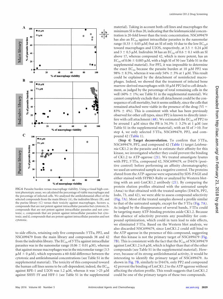
(Step 3) Evaluation of the toxicity of the compounds: cyto-toxicity against mouse bone marrow-derived macrophages. Wefirst assessed toxicity toward mouse bone marrow-derived mac-rophages of 75 compounds that displayed antileishmanial activityagainst intracellular parasites (see Table S1, column VI%, in thesupplemental material). We plotted the percentage of parasiteburden versus the percentage of viable macrophages for each ofthe three libraries (Fig. 6). As shown in Fig. 6A, three compoundsof the seven selected from the main library were toxic towardmacrophages at 10 �M (Fig. 6Ac, black dots), but none were toxicat 1 �M (Fig. 6Ab and d, gray dots). However, decreasing theirconcentration from 10 to 1 �M to prevent cytotoxicity led, insome cases (such as with sunitinib), to a decrease in potencyagainst intracellular parasites. Nevertheless, we identified com-pounds that were not toxic and were able to efficiently decrease PB(Fig. 6Ad). We obtained a similar result with the indirubin deriv-atives (Fig. 6Bb and d) since we identified compounds with anti-leishmanial activity and no toxicity against macrophages: 35 com-pounds at 10 �M and 9 compounds at 1 �M (Fig. 6Bd). Theseresults are in contrast to the results obtained for the purine library,since most of the 21 purine derivatives that we tested led to celldeath, preventing proper analysis of their effects on intracellularparasites. Indeed, we did not identify any compound that de-creased the percentage of the parasite burden without cytotoxicity(Fig. 6Cd). To investigate whether these derivatives could be effi-cient toward intracellular parasites at lower concentrations, wetested compounds 16, 22, and 30 at 0.01 and 0.1 �M (data notshown). These compounds were no longer toxic to host cells at0.1, 0.1, and 0.01 �M, respectively, and were no longer activeagainst intracellular parasites. It seems that the efficient concen-tration to kill intracellular parasites could be similar to the cyto-toxic concentration. This finding could be explained by the higheraffinity of this compound family for the mammalian CK1 com-pared to Leishmania CK1.2 (Fig. 3B and C).
Based on these results, we eliminated the sunitinib from themain library, all the remaining compounds from the purine li-brary, and 11 compounds from the indirubin library because oftheir toxicity against macrophages. We thus retained six com-pounds from the main library and seven indirubins from thosewith antileishmanial activity at 1 �M and without cytotoxicityagainst BMDM.
Cytotoxicity against human cell lines. To establish the selec-tivity index (SI; this unit corresponds to the ratio between the EC50
against intracellular parasites and the EC50 against mammaliancells), we determined the EC50s against intracellular parasites,macrophages, and human cell lines (RPE-1, SHSY-5Y, HFF-1, andU2OS) for the remaining 13 compounds (see Table S1 in the sup-plemental material). The SIs ranged from 0.15 to 50, which isconsistent with small molecules being able to discriminate be-tween Leishmania CK1.2 and mammalian CK1, as we identifiedcompounds that show leishmanicidal activity without cytotoxicity(see Table S1 in the supplemental material). We eliminated all thecompounds with an SI below 10, since they were likely to lead
FIG 5 Comparison of the antileishmanial activity of compounds on culturedand intracellular parasites. We performed a screening of 88 compounds fromthe main, the purine and the indirubin libraries on cultures promastigotes,axenic amastigotes, and intracellular parasites. Each point represents the per-centage of metabolically active promastigotes or amastigotes at 10 �M versusthe percentage of parasite burden at 10 �M for each compound. Black squarescorrespond to the percentage of metabolically active promastigotes at 10 �Mversus the percentage of parasite burden at 10 �M, and gray dots correspond tothe percentage of metabolically active amastigotes at 10 �M versus the per-centage of parasite burden at 10 �M. Sectors: a, compounds that are potentagainst intracellular parasites but not against cultured parasites; b, compoundsthat are not potent against intracellular and cultured parasites; c, com-pounds that are potent against intracellular and cultured parasites; and d,compounds that are not potent against intracellular parasites but potentagainst cultured parasites.
Durieu et al.
2828 aac.asm.org May 2016 Volume 60 Number 5Antimicrobial Agents and Chemotherapy
on February 21, 2018 by guest
http://aac.asm.org/
Dow
nloaded from
to side effects, retaining only five compounds: 5=ITu, PP2, andNSC699479 from the main library and compounds 38 and 42from the indirubin library. The EC50 of 5=ITu against intracellularparasites was in the nanomolar range (0.06 � 0.01 �M), whereasthat against mouse macrophages was in the micromolar range (3.5�M � 0 �M), which represents a 60-fold difference between thecytotoxic and antileishmanial concentrations (see Table S1 in thesupplemental material). The toxicity of this compound towardthe human cell lines seems to be cell-dependent; indeed, the EC50
against RPE-1 and U2OS was 1.2 �M, whereas it was �25 �Magainst SHSY-5Y and HFF-1 (see Table S1 in the supplemental
material). Taking in account both cell lines and macrophages theminimum SI is thus 20, indicating that the leishmanicidal concen-tration is 20-fold lower than the toxic concentration. NSC699479has also an EC50 against intracellular parasites in the nanomolarrange (0.33 � 0.05 �M) but an SI of only 10 due to the low EC50stoward macrophages and U2OS, respectively, at 3.5 � 0.24 �Mand 3 � 0.5 �M. Indirubin 38 has an EC50 of 0.6 � 0.1 with an SIabove 17, whereas compound 42, which is more potent, has anEC50 of 0.06 � 0.005 �M, with a high SI of 50 (see Table S1 in thesupplemental material). For PP2, it was impossible to determinethe exact EC50 because the parasite burden at 10 �M PP2 was98% � 8.5%, whereas it was only 54% � 3% at 1 �M. This resultcould be explained by the detachment of noninfected macro-phages. Indeed, we showed that the treatment of infected bonemarrow-derived macrophages with 10 �M PP2 led to cell detach-ment, as judged by the percentage of total remaining cells in thewell (60% � 1%; see Table S1 in the supplemental material). Wecannot completely exclude that cell detachment could be the con-sequence of cell mortality, but it seems unlikely, since the cells thatremained attached were viable in the presence of the drug (VI 83% � 4%). This is consistent with what has been previouslyobserved for other cell types, since PP2 is known to directly inter-fere with cell attachment (48). We estimated the EC50 of PP2 tobe around 1 �M since the PB is 54.3% � 3.2% at 1 �M (seeTable S1 in the supplemental material), with an SI of �10. Forstep 4, we only selected 5=ITu, NSC699479, PP2, and com-pound 42 (Table 1).
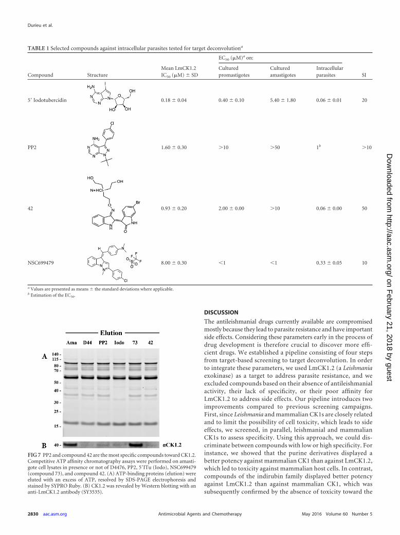
(Step 4) Target deconvolution. To confirm that 5=ITu,NSC699479, PP2, and compound 42 (Table 1) target Leishma-nia CK1.2 in the parasite and to estimate their affinity for thiskinase, we investigated whether they could prevent the bindingof CK1.2 to ATP-agarose (21). We treated amastigote lysateswith PP2, 5=ITu, compound 42, NSC699479, or D4476 (posi-tive control) before performing an affinity chromatography;we used an untreated sample as a negative control. The proteinseluted from the ATP-agarose were separated by SDS-PAGE andeither stained with SYPRO-Ruby or analyzed by Western blot-ting with an anti-LmCK1.2 antibody (21). By comparing theprotein elution profiles obtained with the untreated sample(Ama) to that obtained with the treated samples (D4476, PP2,Iodo, 73, and 42), we were able to assess compound selectivity(Fig. 7A). Most of the treated samples showed a profile similarto that of the untreated sample, except for the 5=ITu (Fig. 7A).As judged by the disappearance of several bands, 5=ITu couldbe targeting many ATP-binding proteins aside CK1.2. Becausethis absence of selectivity prevents any possibility for com-pound optimization, which could in turn lead to side effects,we eliminated 5=ITu. Based on the Western blot analysis, wealso discarded NSC699479, since LmCK1.2 could still bind tothe ATP agarose in the presence of this compound, suggestingthat this kinase is not the primary target of NSC699479 (Fig.7B). This is consistent with the fact that the IC50 of NSC699479against LmCK1.2 is 8 �M, which is higher than that of the othercompounds (see Table S1 in the supplemental material). How-ever, because of its strong antileishmanial activity, it would beinteresting to identify the primary target of NSC699479. Asshown in Fig. 7B, similarly to D4476, only PP2 and compound42 prevent the binding of LmCK1.2 to the ATP agarose withoutaffecting the elution profile. This result suggests that LmCK1.2could be one of the primary targets of these two compounds.
FIG 6 Parasite burden versus macrophage viability. Using a visual high-con-tent phenotypic assay, we calculated the percentage of viable macrophages andthe percentage of infected cells. We analyzed the antileishmanial effect of theselected compounds from the main library (A), the indirubin library (B), andthe purine library (C) versus their toxicity against macrophages. Sectors: a,compounds that are not potent against intracellular parasites but cytotoxic; b,compounds that are not potent against intracellular parasites and not cyto-toxic; c, compounds that are potent against intracellular parasites but cyto-toxic; and d, compounds that are potent against intracellular parasites and notcytotoxic.
Leishmania CK1.2 Drug Screening
May 2016 Volume 60 Number 5 aac.asm.org 2829Antimicrobial Agents and Chemotherapy
on February 21, 2018 by guest
http://aac.asm.org/
Dow
nloaded from
DISCUSSION
The antileishmanial drugs currently available are compromisedmostly because they lead to parasite resistance and have importantside effects. Considering these parameters early in the process ofdrug development is therefore crucial to discover more effi-cient drugs. We established a pipeline consisting of four stepsfrom target-based screening to target deconvolution. In orderto integrate these parameters, we used LmCK1.2 (a Leishmaniaexokinase) as a target to address parasite resistance, and weexcluded compounds based on their absence of antileishmanialactivity, their lack of specificity, or their poor affinity forLmCK1.2 to address side effects. Our pipeline introduces twoimprovements compared to previous screening campaigns.First, since Leishmania and mammalian CK1s are closely relatedand to limit the possibility of cell toxicity, which leads to sideeffects, we screened, in parallel, leishmanial and mammalianCK1s to assess specificity. Using this approach, we could dis-criminate between compounds with low or high specificity. Forinstance, we showed that the purine derivatives displayed abetter potency against mammalian CK1 than against LmCK1.2,which led to toxicity against mammalian host cells. In contrast,compounds of the indirubin family displayed better potencyagainst LmCK1.2 than against mammalian CK1, which wassubsequently confirmed by the absence of toxicity toward the
TABLE 1 Selected compounds against intracellular parasites tested for target deconvolutiona
Compound StructureMean LmCK1.2IC50 (�M) � SD
EC50 (�M)a on:
SICulturedpromastigotes
Culturedamastigotes
Intracellularparasites
5= Iodotubercidin 0.18 � 0.04 0.40 � 0.10 5.40 � 1.80 0.06 � 0.01 20
PP2 1.60 � 0.30 �10 �50 1b �10
42 0.93 � 0.20 2.00 � 0.00 �10 0.06 � 0.00 50
NSC699479 8.00 � 0.30 1 1 0.33 � 0.05 10
a Values are presented as means � the standard deviations where applicable.b Estimation of the EC50.
FIG 7 PP2 and compound 42 are the most specific compounds toward CK1.2.Competitive ATP affinity chromatography assays were performed on amasti-gote cell lysates in presence or not of D4476, PP2, 5=ITu (Iodo), NSC699479(compound 73), and compound 42. (A) ATP-binding proteins (elution) wereeluted with an excess of ATP, resolved by SDS-PAGE electrophoresis andstained by SYPRO Ruby. (B) CK1.2 was revealed by Western blotting with ananti-LmCK1.2 antibody (SY3535).
Durieu et al.
2830 aac.asm.org May 2016 Volume 60 Number 5Antimicrobial Agents and Chemotherapy
on February 21, 2018 by guest
http://aac.asm.org/
Dow
nloaded from

mammalian host cell at 1 �M. This finding suggests a strongcorrelation between specificity toward the target and the sub-sequent effect on intracellular parasite survival. Moreover, ourresults also confirm that LmCK1.2 has an ATP binding pocketsufficiently divergent from that of mammalian CK1 to identifydiscriminating compounds (21). Indeed, more than 70% of thesmall molecules that we tested showed a differential potencyagainst both kinases. We modeled the structure of LeishmaniaCK1.2 based on existing crystal structures of CK1s found inProtein Data Bank and noticed a few differences between theLmCK1.2 model (green; see Fig. S2 in the supplemental mate-rial) and the crystal structure of human CK1� or of Schizosac-charomyces pombe CK1 (magenta or cyan [SpCK1], respectively;see Fig. S2 in the supplemental material) that could account forthe specificity of LmCK1.2 toward certain compounds. Indeed,residues in the active site of LmCK1.2, such as F22 and K40,could be positioned differently, which could overall change theshape of the active site. Moreover, the structural alignment ofLmCK1.2, human CK1�, and SpCK1 (49), which is in complexwith IC261 (a specific CK1 inhibitor), shows that the positionof K40 in the active site of LmCK1.2 compared to that of K41 orK38 in the active sites of CK1� and SpCK1, respectively, mayaccount for the differential response to compound inhibition.Indeed, in contrast to K38 and K41, K40 could lead to conforma-tional clash with IC261. This finding supports our previous resultsshowing that the IC50 of IC261 toward LmCK1.2 is �10 �M,whereas it is 0.47 �M toward mammalian CK1 (21). Our resultsdemonstrate that Leishmania CK1.2, which would have been re-jected based on the strong identity to its mammalian orthologs, isa good drug target.
A second improvement was to assess whether compoundshave multiple targets or low affinity for LmCK1.2. To addressthis issue, we performed target deconvolution. This strategy,which depends on affinity purification and competition, al-lowed the elimination of compounds based on their lack ofspecificity or their lack of affinity toward LmCK1.2. Indeed, weexcluded 5=ITu that could be targeting many proteins, as re-vealed by their depletion after competition assay and ATP-affinity chromatography. This finding is consistent with recentpublications showing that 5=ITu is a general kinase inhibitordue to its broad inhibitory activity (46). Although this com-pound could be a good lead compound purely based on its SI,its optimization could be difficult since the target responsiblefor the leishmanicidal activity is unknown. We also excludedNSC699479 because of its weak affinity toward LmCK1.2, sug-gesting that this kinase might not be its primary target. Basedon previous results in mammalian cells, the primary target ofNSC699479 could be topoisomerases in Leishmania, enzymesalready known as good drug targets (50, 51, 52).
Using this pipeline, we screened 5,018 compounds in totaland identified two lead compounds, PP2 and compound 42.PP2 is an inhibitor for which no antileishmanial activity againstintracellular parasites in THP-1 cells in vitro has been previ-ously described but appears to be efficient in an animal model(53). It has an EC50 on intracellular parasites of �1 �M but SIsof �10 for murine macrophages and �25 for human cell lines.The known targets of PP2 are tyrosine kinases, Src, Lck, Csk,Rip2, and Gak, for which there are no orthologs identified inLeishmania (54, 55). The two other kinases that are targeted byPP2 are p38 (52% inhibition at 1 �M [54]) and CK1� (93%
inhibition at 1 �M [54]), suggesting that the antileishmanialactivity of PP2 is more likely mediated by the inhibition ofCK1.2 in Leishmania. This finding is consistent with our experi-mental data showing that Leishmania CK1.2 is one of the primarytargets of PP2. However, treatment with high concentrationsof PP2 leads to macrophage detachment (40% at 10 �M) sim-ilar to what has been demonstrated previously for other celltypes (48). Our results seem to indicate that most of the mac-rophages that detach from the slides are those that are notinfected by Leishmania. This hypothesis is supported by a reportby Tejle et al., which showed that the presence of L. donovani affectthe detachment of monocyte-derived dendritic cells, suggest-ing that the presence of the parasite could promote cell adhe-sion (56).
Indirubins are particularly potent against both cultured andintracellular parasites and, among the 55 indirubin derivativesshowing leishmanicidal activity at 10 �M, only 37% were cy-totoxic against macrophages. Compound 42 is our best leadcompound, with an EC50 on intracellular parasites of 60 � 5nM and an SI of 50. Although several authors have alreadydescribed the antileishmanial effect of indirubins (57–59), thisparticular derivative has not been previously tested on intra-cellular parasites. From previous published work, we alreadyknow some of the targets of the indirubins, such as LeishmaniaCRK3 or GSK3 (57, 58). In our study, we reveal for the firsttime CK1.2 as a novel target for this family of compounds. Thisis particularly striking since in higher eukaryotes GSK3 andCK1 are often involved in similar signaling pathways, such asthe Wnt/�-catenin or the Hedgehog pathways, where they actas priming kinases for one another (60–63). Using affinity pu-rification, we found that GSK-3 is also a target of compound 42(data not shown). We will determine precisely, using biochem-ical approaches, whether this compound targets other kinasesand which one causes the antileishmanial effect.
In conclusion, we have established a comprehensive pipelinethat identifies and selects LmCK1.2 inhibitors based on their spec-ificity, antileishmanial activity, absence of cytotoxicity, and selec-tivity. As a proof of principle, we identified two lead compounds,PP2 and compound 42, that will be studied further to understandtheir mode of action and could also be used as pharmacologicaltools to study parasite-specific signal transduction. We will usethis pipeline to screen diversified libraries that have not yet beenscreened against Leishmania kinases in order to identify lead com-pounds.
ACKNOWLEDGMENTS
This study was supported by the 7th Framework Program of the Eu-ropean Commission through grants to the LEISHDRUG project(223414), by ANR-11-RPIB-0016 TRANSLEISH, and by the FrenchGovernment’s Investissements d’Avenir Program Laboratoired’Excellence Integrative Biology of Emerging Infectious Diseases(grant ANR-10-LABX-62-IBEID). The Imagopole-CiTech is part ofthe FranceBioImaging infrastructure supported by the French Na-tional Research Agency (ANR-10-INSB-04-01, Investments for theFuture) and is supported by the Conseil de la Region Ile-de-France(program Sesame 2007, project Imagopole, S. Shorte) and the Fonda-tion Française pour la Recherche Médicale (Programme GrandsEquipements [N.A.]).
Leishmania CK1.2 Drug Screening
May 2016 Volume 60 Number 5 aac.asm.org 2831Antimicrobial Agents and Chemotherapy
on February 21, 2018 by guest
http://aac.asm.org/
Dow
nloaded from
FUNDING INFORMATIONThis work, including the efforts of Emilie Durieu, Eric Prina, OlivierLeclercq, Nathalie Aulner, Gerald F. Späth, Laurent Meijer, and NajmaRachidi, was funded by European Commission (EC) (LeishDrug 223414).This work, including the efforts of Emilie Durieu, Eric Prina, OlivierLeclercq, Nassima Oumata, Audrey Defontaine, Sandrine Ruchaud,Hervé Galons, Gerald F. Späth, Laurent Meijer, and Najma Rachidi, wasfunded by Agence Nationale de la Recherche (ANR) (ANR-11-RPIB-0016). This work, including the efforts of Eric Prina, Olivier Leclercq,Gerald F. Späth, and Najma Rachidi, was funded by Agence Nationale dela Recherche (ANR) (ANR-10-LABX-62-IBEID). This work, includingthe efforts of Nathalie Aulner, was funded by Agence Nationale de laRecherche (ANR) (ANR-10-INSB-04-01). This work, including the ef-forts of Nathalie Aulner, was funded by Fondation pour la RechercheMédicale (FRM) (Grands Equipement). This work, including the effortsof Nathalie Aulner, was funded by Conseil Régional, Ile-de-France (Ile-de-France Regional Council) (sesame 2007).
We thank all members of the FP7 LEISHDRUG consortium for fruitfuldiscussions and, in particular, Geneviève Milon. We also thank OlivierHelynck and Hélène Munier-Lehmann for providing access to theTECAN Freedom EVOware platform for the automatic distribution ofcells, parasites, and chemicals in a biosafety level 2 facility.
REFERENCES1. Pace D. 2014. Leishmaniasis. J Infect 69(Suppl 1):S10 –S18. http://dx.doi
.org/10.1016/j.jinf.2014.07.016.2. Hussain H, Al-Harrasi A, Al-Rawahi A, Green IR, Gibbons S. 2014.
Fruitful decade for antileishmanial compounds from 2002 to late 2011.Chem Rev 114:10369 –10428. http://dx.doi.org/10.1021/cr400552x.
3. Croft SL, Sundar S, Fairlamb AH. 2006. Drug resistance in leishmaniasis.Clin Microbiol Rev 19:111–126. http://dx.doi.org/10.1128/CMR.19.1.111-126.2006.
4. Singh N, Kumar M, Singh RK. 2012. Leishmaniasis: current status ofavailable drugs and new potential drug targets. Asian Pac J Trop Med5:485– 497. http://dx.doi.org/10.1016/S1995-7645(12)60084-4.
5. Dorlo TP, Balasegaram M, Beijnen JH, de Vries PJ. 2012. Miltefosine: areview of its pharmacology and therapeutic efficacy in the treatment ofleishmaniasis. J Antimicrob Chemother 67:2576 –2597. http://dx.doi.org/10.1093/jac/dks275.
6. Perez-Victoria FJ, Sanchez-Canete MP, Castanys S, Gamarro F. 2006.Phospholipid translocation and miltefosine potency require both Leish-mania donovani miltefosine transporter and the new protein LdRos3 inLeishmania parasites. J Biol Chem 281:23766 –23775. http://dx.doi.org/10.1074/jbc.M605214200.
7. Perez-Victoria JM, Bavchvarov BI, Torrecillas IR, Martinez-Garcia M,Lopez-Martin C, Campillo M, Castanys S, Gamarro F. 2011. Sitama-quine overcomes ABC-mediated resistance to miltefosine and antimonyin Leishmania. Antimicrob Agents Chemother 55:3838 –3844. http://dx.doi.org/10.1128/AAC.00065-11.
8. Cohen P. 2002. Protein kinases–the major drug targets of the twenty-firstcentury? Nat Rev Drug Discov 1:309 –315. http://dx.doi.org/10.1038/nrd773.
9. Doerig C. 2004. Protein kinases as targets for anti-parasitic chemother-apy. Biochim Biophys Acta 1697:155–168. http://dx.doi.org/10.1016/j.bbapap.2003.11.021.
10. Weinmann H, Metternich R. 2005. Drug discovery process for kinaseinhibitors. Chembiochem 6:455– 459. http://dx.doi.org/10.1002/cbic.200500034.
11. Eglen RM, Reisine T. 2009. The current status of drug discovery againstthe human kinome. Assay Drug Dev Technol 7:22– 43. http://dx.doi.org/10.1089/adt.2008.164.
12. Eglen R, Reisine T. 2011. Drug discovery and the human kinome: recenttrends. Pharmacol Ther 130:144–156. http://dx.doi.org/10.1016/j.pharmthera.2011.01.007.
13. Carvalho TG, Doerig C, Reininger L. 2013. Nima- and Aurora-relatedkinases of malaria parasites. Biochim Biophys Acta 1834:1336 –1345. http://dx.doi.org/10.1016/j.bbapap.2013.02.022.
14. Yang SH, Sharrocks AD, Whitmarsh AJ. 2013. MAP kinase signalingcascades and transcriptional regulation. Gene 513:1–13. http://dx.doi.org/10.1016/j.gene.2012.10.033.
15. Silverman JM, Clos J, de’Oliveira CC, Shirvani O, Fang Y, Wang C,Foster LJ, Reiner NE. 2010. An exosome-based secretion pathway isresponsible for protein export from Leishmania and communication withmacrophages. J Cell Sci 123:842– 852. http://dx.doi.org/10.1242/jcs.056465.
16. Silverman JM, Clos J, Horakova E, Wang AY, Wiesgigl M, Kelly I, LynnMA, McMaster WR, Foster LJ, Levings MK, Reiner NE. 2011. Leishma-nia exosomes modulate innate and adaptive immune responses througheffects on monocytes and dendritic cells. J Immunol 185:5011–5022. http://dx.doi.org/10.4049/jimmunol.1000541.
17. Knippschild U, Gocht A, Wolff S, Huber N, Lohler J, Stoter M. 2005.The casein kinase 1 family: participation in multiple cellular processes ineukaryotes. Cell Signal 17:675– 689. http://dx.doi.org/10.1016/j.cellsig.2004.12.011.
18. Allocco JJ, Donald R, Zhong T, Lee A, Tang YS, Hendrickson RC,Liberator P, Nare B. 2006. Inhibitors of casein kinase 1 block the growthof Leishmania major promastigotes in vitro. Int J Parasitol 36:1249 –1259.http://dx.doi.org/10.1016/j.ijpara.2006.06.013.
19. Silverman JM, Chan SK, Robinson DP, Dwyer DM, Nandan D, FosterLJ, Reiner NE. 2008. Proteomic analysis of the secretome of Leishmaniadonovani. Genome Biol 9:R35. http://dx.doi.org/10.1186/gb-2008-9-2-r35.
20. Paape D, Barrios-Llerena ME, Le Bihan T, Mackay L, Aebischer T.2010. Gel-free analysis of the proteome of intracellular Leishmania mexi-cana. Mol Biochem Parasitol 169:108 –114. http://dx.doi.org/10.1016/j.molbiopara.2009.10.009.
21. Rachidi N, Taly JF, Durieu E, Leclercq O, Aulner N, Prina E, PescherP, Notredame C, Meijer L, Spath GF. 2014. Pharmacological assessmentdefines the Leishmania donovani casein kinase 1 as a drug target and re-veals important functions in parasite viability and intracellular infection.Antimicrob Agents Chemother 58:1501–1515. http://dx.doi.org/10.1128/AAC.02022-13.
22. Liu J, Carvalho LP, Bhattachariya S, Carbone CJ, Kumar KG, Leu NA,Yau PM, Donald RG, Weiss MJ, Baker DP, McLaughlin KJ, Scott P,Fuchs SY. 2009. Mammalian casein kinase 1� and its leishmanial orthologregulate stability of IFNAR1 and type I interferon signaling. Mol Cell Biol29:6401– 6412. http://dx.doi.org/10.1128/MCB.00478-09.
23. Reinhardt J, Ferandin Y, Meijer L. 2007. Purification of CK1 by affinitychromatography on immobilised axin. Protein Expr Purif 54:101–109.http://dx.doi.org/10.1016/j.pep.2007.02.020.
24. Saar Y, Ransford A, Waldman E, Mazareb S, Amin-Spector S, PlumbleeJ, Turco SJ, Zilberstein D. 1998. Characterization of developmentallyregulated activities in axenic amastigotes of Leishmania donovani. MolBiochem Parasitol 95:9 –20. http://dx.doi.org/10.1016/S0166-6851(98)00062-0.
25. Goyard S, Segawa H, Gordon J, Showalter M, Duncan R, Turco SJ,Beverley SM. 2003. An in vitro system for developmental and geneticstudies of Leishmania donovani phosphoglycans. Mol Biochem Parasitol130:31– 42. http://dx.doi.org/10.1016/S0166-6851(03)00142-7.
26. Morales MA, Watanabe R, Laurent C, Lenormand P, Rousselle J-C,Namane A, Späth GF. 2008. Phosphoproteomic analysis of Leishmaniadonovani pro- and amastigote stages. Proteomics 8:350 –363. http://dx.doi.org/10.1002/pmic.200700697.
27. Shimony O, Jaffe CL. 2008. Rapid fluorescent assay for screening drugson Leishmania amastigotes. J Microbiol Methods 75:196 –200. http://dx.doi.org/10.1016/j.mimet.2008.05.026.
28. Aulner N, Danckaert A, Rouault-Hardoin E, Desrivot J, Helynck O,Commere PH, Munier-Lehmann H, Spath GF, Shorte SL, Milon G,Prina E. 2013. High content analysis of primary macrophages hostingproliferating Leishmania amastigotes: application to anti-leishmanialdrug discovery. PLoS Negl Trop Dis 7:e2154. http://dx.doi.org/10.1371/journal.pntd.0002154.
29. Oumata N, Bettayeb K, Ferandin Y, Demange L, Lopez-Giral A, God-dard ML, Myrianthopoulos V, Mikros E, Flajolet M, Greengard P,Meijer L, Galons H. 2008. Roscovitine-derived, dual-specificity inhibi-tors of cyclin-dependent kinases and casein kinases 1. J Med Chem 51:5229 –5242. http://dx.doi.org/10.1021/jm800109e.
30. Polychronopoulos P, Magiatis P, Skaltsounis A-L, Myrianthopoulos V,Mikros E, Tarricone A, Musacchio A, Roe SM, Pearl L, Leost M,Greengard P, Meijer L. 2004. Structural basis for the synthesis of indiru-bins as potent and selective inhibitors of glycogen synthase kinase-3 andcyclin-dependent kinases. J Med Chem 47:935–946. http://dx.doi.org/10.1021/jm031016d.
Durieu et al.
2832 aac.asm.org May 2016 Volume 60 Number 5Antimicrobial Agents and Chemotherapy
on February 21, 2018 by guest
http://aac.asm.org/
Dow
nloaded from

31. Vougogiannopoulou K, Ferandin Y, Bettayeb K, Myrianthopoulos V,Lozach O, Fan Y, Johnson CH, Magiatis P, Skaltsounis A-L, Mikros E,Meijer L. 2008. Soluble 3=,6-substituted indirubins with enhanced selec-tivity toward glycogen synthase kinase-3 alter circadian period. J MedChem 51:6421– 6431. http://dx.doi.org/10.1021/jm800648y.
32. Ferandin Y, Bettayeb K, Kritsanida M, Lozach O, Polychronopoulos P,Magiatis P, Skaltsounis A-L, Meijer L. 2006. 3=-Substituted 7-halog-enoindirubins, a new class of cell death inducing agents. J Med Chem49:4638 – 4649. http://dx.doi.org/10.1021/jm060314i.
33. Schwede T, Kopp J, Guex N, Peitsch MC. 2003. Swiss-Model: an auto-mated protein homology-modeling server. Nucleic Acids Res 31:3381–3385. http://dx.doi.org/10.1093/nar/gkg520.
34. Delano W. 2009. The PyMOL molecular graphics system, v1.01. DeLanoScientific, Palo Alto, CA. http://www.pymol.sourceforge.net.
35. Schnur L, Bachrach U, Bar-Ad G, Haran M, Tashma Z, Talmi M,Katzhendler J. 1983. The effect of diaminoalkyl-anthraquinone deriva-tives on the growth of the promastigotes of Leishmania tropica minor, L.major, L. donovani, and L. aethiopica. Biochem Pharmacol 32:1729 –1732.http://dx.doi.org/10.1016/0006-2952(83)90117-X.
36. Sittie AA, Lemmich E, Olsen CE, Hviid L, Kharazmi A, Nkrumah FK,Christensen SB. 1999. Structure-activity studies: in vitro antileishmanialand antimalarial activities of anthraquinones from Morinda lucida. PlantaMed 65:259 –261. http://dx.doi.org/10.1055/s-2006-960473.
37. Bolognesi ML, Lizzi F, Perozzo R, Brun R, Cavalli A. 2008. Synthesis ofa small library of 2-phenoxy-1,4-naphthoquinone and 2-phenoxy-1,4-anthraquinone derivatives bearing anti-trypanosomal and anti-leishmanial activity. Bioorg Med Chem Lett 18:2272–2276. http://dx.doi.org/10.1016/j.bmcl.2008.03.009.
38. Conners R, Schambach F, Read J, Cameron A, Sessions RB, Vivas L,Easton A, Croft SL, Brady RL. 2005. Mapping the binding site for gos-sypol-like inhibitors of Plasmodium falciparum lactate dehydrogenase.Mol Biochem Parasitol 142:137–148. http://dx.doi.org/10.1016/j.molbiopara.2005.03.015.
39. Montamat EE, Burgos C, Gerez de Burgos NM, Rovai LE, Blanco A,Segura EL. 1982. Inhibitory action of gossypol on enzymes and growthof Trypanosoma cruzi. Science 218:288 –289. http://dx.doi.org/10.1126/science.6750791.
40. Padmanabhan PK, Mukherjee A, Singh S, Chattopadhyaya S, GowriVS, Myler PJ, Srinivasan N, Madhubala R. 2005. Glyoxalase I fromLeishmania donovani: a potential target for anti-parasite drug. BiochemBiophys Res Commun 337:1237–1248. http://dx.doi.org/10.1016/j.bbrc.2005.09.179.
41. Tasdemir D, Kaiser M, Brun R, Yardley V, Schmidt TJ, Tosun F, RuediP. 2006. Antitrypanosomal and antileishmanial activities of flavonoidsand their analogues: in vitro, in vivo, structure-activity relationship, andquantitative structure-activity relationship studies. Antimicrob AgentsChemother 50:1352–1364. http://dx.doi.org/10.1128/AAC.50.4.1352-1364.2006.
42. Sen G, Mukhopadhyay S, Ray M, Biswas T. 2008. Quercetin interfereswith iron metabolism in Leishmania donovani and targets ribonucleotidereductase to exert leishmanicidal activity. J Antimicrob Chemother 61:1066 –1075. http://dx.doi.org/10.1093/jac/dkn053.
43. da Silva ER, Maquiaveli Cdo C, Magalhaes PP. 2012. The leishmanicidalflavonols quercetin and quercitrin target Leishmania (Leishmania) ama-zonensis arginase. Exp Parasitol 130:183–188. http://dx.doi.org/10.1016/j.exppara.2012.01.015.
44. Knockaert M, Gray N, Damiens E, Chang YT, Grellier P, Grant K,Fergusson D, Mottram J, Soete M, Dubremetz JF, Le Roch K, Doerig C,Schultz P, Meijer L. 2000. Intracellular targets of cyclin-dependent kinaseinhibitors: identification by affinity chromatography using immobilisedinhibitors. Chem Biol 7:411–422. http://dx.doi.org/10.1016/S1074-5521(00)00124-1.
45. Hanke JH, Gardner JP, Dow RL, Changelian PS, Brissette WH, Wer-inger EJ, Pollok BA, Connelly PA. 1996. Discovery of a novel, potent, andSrc family-selective tyrosine kinase inhibitor: study of Lck- and FynT-dependent T cell activation. J Biol Chem 271:695–701.
46. Massillon D, Stalmans W, van de Werve G, Bollen M. 1994. Identifi-cation of the glycogenic compound 5-iodotubercidin as a general proteinkinase inhibitor. Biochem J 299(Pt 1):123–128. http://dx.doi.org/10.1042/bj2990123.
47. Bento AP, Gaulton A, Hersey A, Bellis LJ, Chambers J, Davies M,Kruger FA, Light Y, Mak L, McGlinchey S, Nowotka M, Papadatos G,Santos R, Overington JP. 2014. The ChEMBL bioactivity database: anupdate. Nucleic Acids Res 42:D1083–D1090. http://dx.doi.org/10.1093/nar/gkt1031.
48. Hishiki T, Saito T, Sato Y, Mitsunaga T, Terui E, Matsuura G, Saito E,Shibata R, Mise N, Yokoyama Y, Yoshida H. 2011. Src kinase familyinhibitor PP2 induces aggregation and detachment of neuroblastoma cellsand inhibits cell growth in a PI3 kinase/Akt pathway-independent man-ner. Pediatr Surg Int 27:225–230. http://dx.doi.org/10.1007/s00383-010-2775-2.
49. Mashhoon N, DeMaggio AJ, Tereshko V, Bergmeier SC, Egli M, Hoek-stra MF, Kuret J. 2000. Crystal structure of a conformation-selectivecasein kinase-1 inhibitor. J Biol Chem 275:20052–20060. http://dx.doi.org/10.1074/jbc.M001713200.
50. Balana-Fouce R, Alvarez-Velilla R, Fernandez-Prada C, Garcia-EstradaC, Reguera RM. 2014. Trypanosomatids topoisomerase re-visited: newstructural findings and role in drug discovery. Int J Parasitol Drugs DrugResist 4:326 –337. http://dx.doi.org/10.1016/j.ijpddr.2014.07.006.
51. Katayama H, Kiryu Y, Kaneko K, Ohshima R. 2000. Anti-cancer activ-ities of pyrazolo[1,5-a]indole derivatives. Chem Pharm Bull 48:1628 –1633. http://dx.doi.org/10.1248/cpb.48.1628.
52. Das BB, Ganguly A, Majumder HK. 2008. DNA topoisomerases ofLeishmania: the potential targets for anti-leishmanial therapy. Adv ExpMed Biol 625:103–115. http://dx.doi.org/10.1007/978-0-387-77570-8_9.
53. Sanderson L, Yardley V, Croft SL. 2014. Activity of anti-cancer proteinkinase inhibitors against Leishmania spp. J Antimicrob Chemother 69:1888 –1891. http://dx.doi.org/10.1093/jac/dku069.
54. Bain J, Plater L, Elliott M, Shpiro N, Hastie CJ, McLauchlan H,Klevernic I, Arthur JS, Alessi DR, Cohen P. 2007. The selectivity ofprotein kinase inhibitors: a further update. Biochem J 408:297–315. http://dx.doi.org/10.1042/BJ20070797.
55. Parsons M, Worthey EA, Ward PN, Mottram JC. 2005. Comparativeanalysis of the kinomes of three pathogenic trypanosomatids: Leishmaniamajor, Trypanosoma brucei, and Trypanosoma cruzi. BMC Genomics6:127. http://dx.doi.org/10.1186/1471-2164-6-127.
56. Tejle K, Lindroth M, Magnusson KE, Rasmusson B. 2008. Wild-typeLeishmania donovani promastigotes block maturation, increase integrinexpression and inhibit detachment of human monocyte-derived dendriticcells–the influence of phosphoglycans. FEMS Microbiol Lett 279:92–102.http://dx.doi.org/10.1111/j.1574-6968.2007.01013.x.
57. Grant KM, Dunion MH, Yardley V, Skaltsounis AL, Marko D, Eisen-brand G, Croft SL, Meijer L, Mottram JC. 2004. Inhibitors of Leishmaniamexicana CRK3 cyclin-dependent kinase: chemical library screen and an-tileishmanial activity. Antimicrob Agents Chemother 48:3033–3042. http://dx.doi.org/10.1128/AAC.48.8.3033-3042.2004.
58. Xingi E, Smirlis D, Myrianthopoulos V, Magiatis P, Grant KM, MeijerL, Mikros E, Skaltsounis AL, Soteriadou K. 2009. 6-Br-5methylindirubin-3=oxime (5-Me-6-BIO) targeting the leishmanial gly-cogen synthase kinase-3 (GSK-3) short form affects cell-cycle progressionand induces apoptosis-like death: exploitation of GSK-3 for treating leish-maniasis. Int J Parasitol 39:1289 –1303. http://dx.doi.org/10.1016/j.ijpara.2009.04.005.
59. Efstathiou A, Gaboriaud-Kolar N, Smirlis D, Myrianthopoulos V, Vou-gogiannopoulou K, Alexandratos A, Kritsanida M, Mikros E, Soteria-dou K, Skaltsounis AL. 2014. An inhibitor-driven study for enhancing theselectivity of indirubin derivatives towards leishmanial glycogen synthasekinase-3 over leishmanial cdc2-related protein kinase 3. Parasit Vectors7:234. http://dx.doi.org/10.1186/1756-3305-7-234.
60. Harwood AJ. 2002. Signal transduction in development: holding the key.Dev Cell 2:384 –385. http://dx.doi.org/10.1016/S1534-5807(02)00156-9.
61. Niehrs C, Shen J. 2010. Regulation of Lrp6 phosphorylation. Cell Mol LifeSci 67:2551–2562. http://dx.doi.org/10.1007/s00018-010-0329-3.
62. Knippschild U, Kruger M, Richter J, Xu P, Garcia-Reyes B, Peifer C,Halekotte J, Bakulev V, Bischof J. 2014. The CK1 family: contribution tocellular stress response and its role in carcinogenesis. Front Oncol 4:96.http://dx.doi.org/10.3389/fonc.2014.00096.
63. Beurel E, Grieco SF, Jope RS. 2015. Glycogen synthase kinase-3 (GSK3):regulation, actions, and diseases. Pharmacol Ther 148:114 –131. http://dx.doi.org/10.1016/j.pharmthera.2014.11.016.
Leishmania CK1.2 Drug Screening
May 2016 Volume 60 Number 5 aac.asm.org 2833Antimicrobial Agents and Chemotherapy
on February 21, 2018 by guest
http://aac.asm.org/
Dow
nloaded from