from training to practice
TRANSCRIPT
FROM TRAINING TO PRACTICE: Development of a Duplex Ultrasound Simulator
R. Eugene Zierler, MD Department of Surgery, Division of Vascular Surgery
University of Washington

DISCLOSURES
▪ No financial or commercial conflicts ▪ Funding
This work is supported by funding from the National Institute of Biomedical Imaging and Bioengineering and the National Institute of Environmental Health Sciences, National Institutes of Health, Bethesda MD Florence H. Sheehan, MD (Principal Investigator) Department of Medicine, Division of Cardiology University of Washington
R. Eugene Zierler, MD

SIMULATION IN ULTRASOUND
▪ Training is essential for healthcare providers who use ultrasound to develop the necessary technical and interpretive skills
▪ Assessment of competence is a critical component of training that documents a learner’s progress ✓ Competency-based education ✓ Specialty certification or credentialing
▪ Tools are currently available to evaluate knowledge and judgement (cognitive skills)
▪ No standardized method is widely available for assessment of technical (psychomotor) skills

SIMULATION IN ULTRASOUND
▪ Use of simulation for training is appealing since it incorporates both cognitive and psychomotor domains ✓ Eliminates need for live patients (safety, comfort, privacy) ✓ Provides exposure to uncommon conditions ✓ Enables repetitive practice of basic skills ✓ Provides immediate feedback to improve proficiency ✓ Preserves faculty time for advanced training
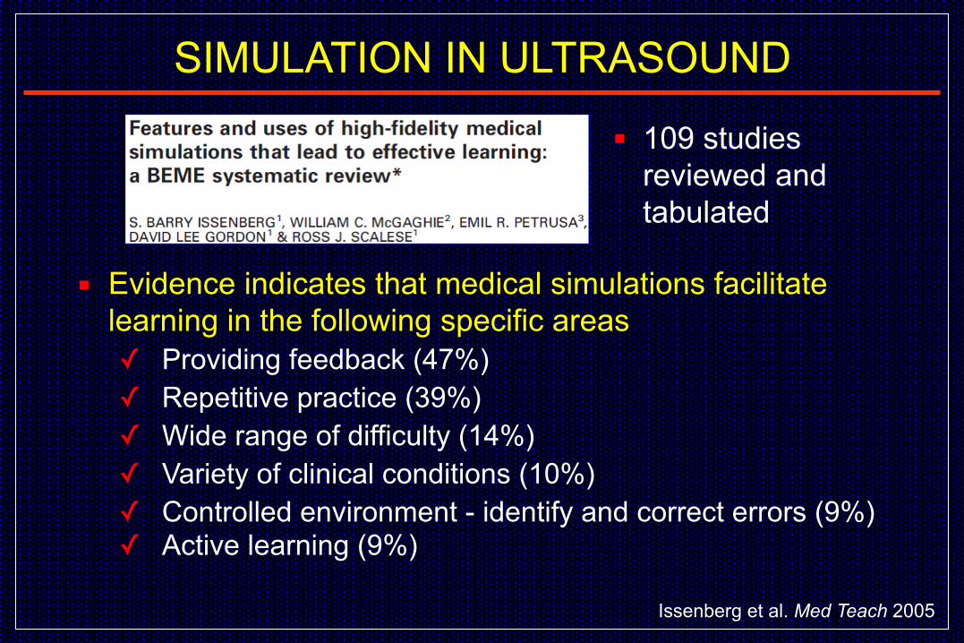
▪ Evidence indicates that medical simulations facilitate learning in the following specific areas ✓ Providing feedback (47%) ✓ Repetitive practice (39%) ✓ Wide range of difficulty (14%) ✓ Variety of clinical conditions (10%) ✓ Controlled environment - identify and correct errors (9%) ✓ Active learning (9%)
Issenberg et al. Med Teach 2005
▪ 109 studies reviewed and tabulated
SIMULATION IN ULTRASOUND
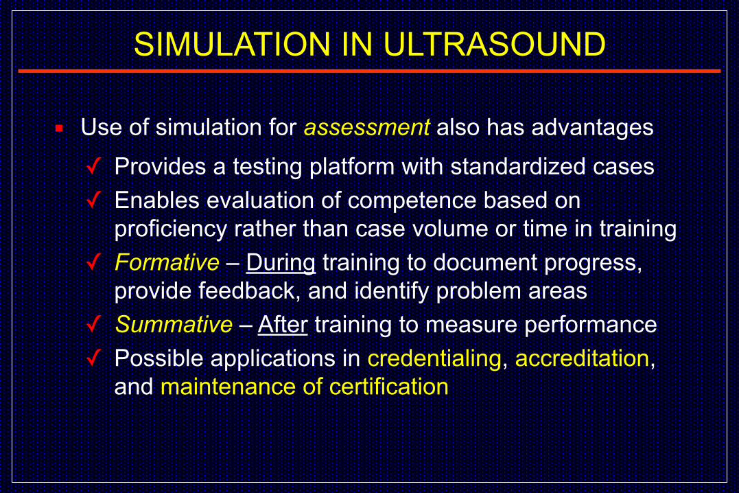
▪ Use of simulation for assessment also has advantages ✓ Provides a testing platform with standardized cases ✓ Enables evaluation of competence based on
proficiency rather than case volume or time in training ✓ Formative – During training to document progress,
provide feedback, and identify problem areas ✓ Summative – After training to measure performance ✓ Possible applications in credentialing, accreditation,
and maintenance of certification
SIMULATION IN ULTRASOUND

▪ Outcome measures: Procedural time, instructor ratings of technique, and rate of procedural success or complications ▪ All studies showed that higher simulator scores were
associated with higher performance in clinical practice
Cook et al. Acad Med 2013
▪ 28 studies evaluated associations between simulation-based performance and performance with real patients
SIMULATION IN ULTRASOUND

▪ Successful application of simulation in ultrasound requires: ✓ Realistic, full-featured simulators ✓ Standardized metrics for competency testing
▪ Simulators are available for a variety of ultrasound applications: ✓ Echocardiography ✓ Duplex Ultrasound
SIMULATION IN ULTRASOUND

▪ Simulators for echocardiography have been available since the 1990s and are currently more advanced than those for duplex (peripheral vascular) ultrasound
▪ Computer-based echocardiography simulators have 3D graphic displays that provide feedback to the learner and metrics for assessment of technical skill that are based on transducer tracking data
▪ Such metrics provide a motion-based or kinematic analysis of skill in performing cardiac ultrasound
SIMULATION FOR ECHOCARDIOGRAPHY
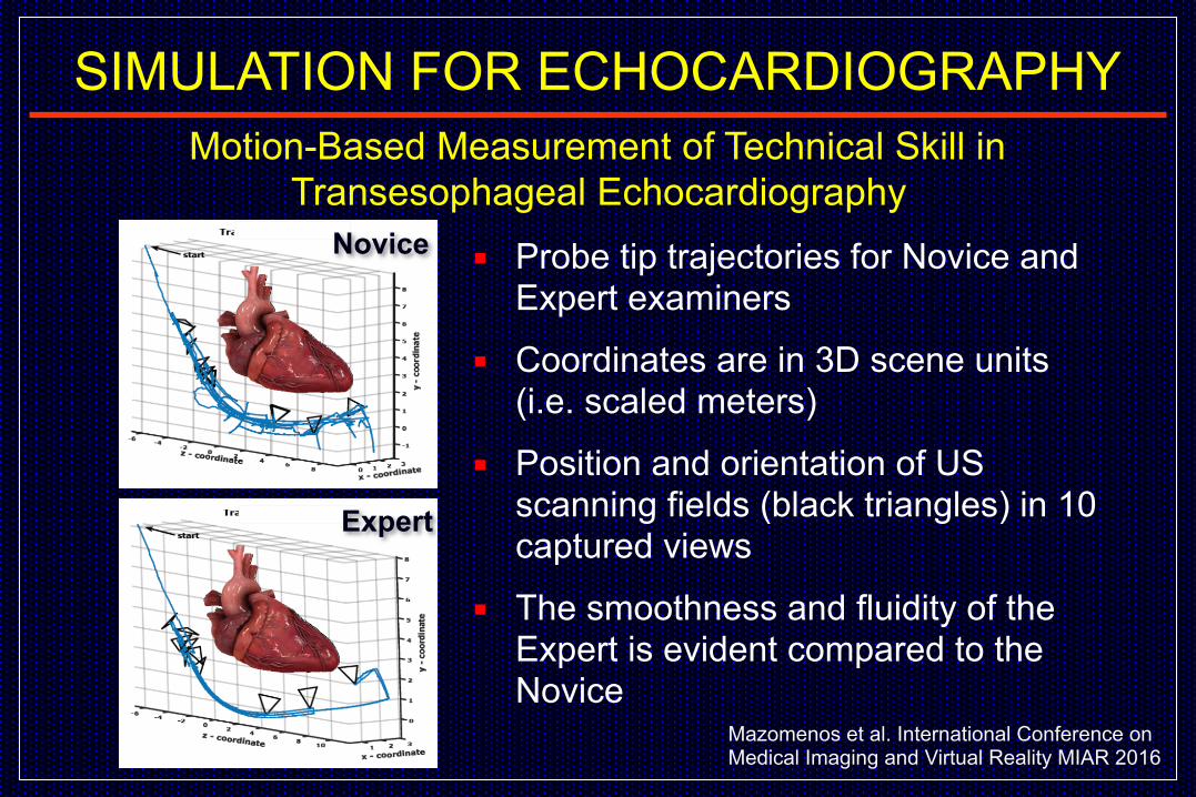
Motion-Based Measurement of Technical Skill in Transesophageal Echocardiography
▪ Probe tip trajectories for Novice and Expert examiners
▪ Coordinates are in 3D scene units (i.e. scaled meters)
▪ Position and orientation of US scanning fields (black triangles) in 10 captured views
▪ The smoothness and fluidity of the Expert is evident compared to the Novice
Novice
Expert
Mazomenos et al. International Conference on Medical Imaging and Virtual Reality MIAR 2016
SIMULATION FOR ECHOCARDIOGRAPHY

Transesophageal Echo Training with Motion Analysis▪ 3D path length of a Trainee at week
1 and week 4 vs. an Expert ▪ Movement of the probe from the
mid-esophageal 4-chamber view to the mid-esophageal long-axis view
▪ First session shows a high number of misdirected probe movements and a long path length
▪ Fourth session shows more efficient probe movement with shorter path length that approximates the movement patterns of the Expert
Matyal et al. Anesthesiology 2014
Probe Motion
SIMULATION FOR ECHOCARDIOGRAPHY
▪ Duplex ultrasound is the primary direct noninvasive method for diagnosis of vascular disease ✓ B-mode imaging ✓ Color-flow Doppler ✓ Doppler spectral waveforms
▪ Classification of stenosis severity is based primarily on blood flow velocities obtained from Doppler spectral waveforms
▪ Real-time simulation of color-flow Doppler and spectral waveforms presents unique technical challenges
▪ A duplex ultrasound simulator is being developed at UW
SIMULATION FOR DUPLEX ULTRASOUND

Transducer with magnetic
tracker
The simulator consists of a mannequin, computer, and magnetic field system to track the location of a mock transducer
Components
SIMULATION FOR DUPLEX ULTRASOUND
Mannequin
Transducer with magnetic
trackerControl panel with B-mode image, color
Doppler and spectral waveform display

▪ Cases are built from B-mode images acquired from patients using a 3-dimensional tracking device
▪ The arteries are reconstructed as a 3-dimensional surface model
▪ Computational flow modeling is used to calculate the spatial and temporal distribution of blood flow velocities in the surface model based on vessel geometry and boundary conditions (and one Doppler spectral waveform at the model inlet)
Case Creation
SIMULATION FOR DUPLEX ULTRASOUND

Computed Flow in a Carotid Artery Model
▪ Doppler shifts are calculated based on the velocity vectors within the sample volume
▪ Accounts for beam angle and other user-defined Doppler parameters
SIMULATION FOR DUPLEX ULTRASOUND

Computed Flow in a Dialysis Fistula Model
▪ 900 2-dimensional B-mode images are used to create a 3-dimensional surface model of the inflow artery, anastomosis, and outflow vein
▪ Computed flow velocity “pathlines” are shown for peak systole from the arterial inlet to the venous outlet
SIMULATION FOR DUPLEX ULTRASOUND

Duplex display showing B-mode image with color Doppler and Doppler spectral waveforms
Controls for the ultrasound display mode and Doppler settings
3-dimensional display shows the transducer location (blue cone)
PSV measurement
Probe location and scan plane
Duplex Simulator Examiner Interface
SIMULATION FOR DUPLEX ULTRASOUND

Peak systolic Velocity (PSV) in Carotid Artery Models
▪ Accuracy was assessed by comparing PSV measured from spectral waveforms with the “true” calculated PSV at the sample volume site
▪ A total of 36 carotid PSV measurements were made by 3 “expert” examiners in two carotid artery models
▪ Overall Accuracy: The PSV selected by the examiner deviated from the true (calculated) PSV by 8 ± 5%
▪ Accuracy was similar between the normal and stenotic segments and for the CCA, ICA, and ECA
SIMULATION FOR DUPLEX ULTRASOUND
Zierler et al. Vasc Endovasc Surg 2016
▪ A total of 43 PSV measurements were made by 4 “expert” examiners in the 2 fistula models
▪ 24 arterial segments and 19 venous segments ▪ Overall Accuracy: The PSV selected by the examiner
deviated from the true (calculated) PSV by 8 ± 6% ▪ Accuracy was similar between examiners and for
arterial and venous segments
SIMULATION FOR DUPLEX ULTRASOUNDPeak systolic Velocity (PSV) in Dialysis Fistula Models
Leotta et al. Ultrasound Med Biol 2018
▪ 17 Experts (≥5 years experience) and 16 Novices (sonography student or surgery resident) scanned carotid artery, femoral artery, and dialysis fistula models and made 657 PSV measurements
▪ Error was calculated from the mean absolute deviation of the PSV from the true (computed) velocity at the sample site
▪ Overall error was lower for experts (16±17%) vs. novices (25±25%)
▪ Error was also lower for experts vs. novices when the case types were analyzed separately
SIMULATION FOR DUPLEX ULTRASOUNDAssessment of Skill – Experts vs. Novices
Sheehan et al. Unpublished data 2019

Acquire transverse view, rotate to longitudinal view and position sample volume
Duplex Ultrasound – Carotid Timed Skill Study
SIMULATION FOR DUPLEX ULTRASOUND

▪ Simulation offers numerous advantages for training and assessment in vascular ultrasound
▪ Simulators are available for echocardiography that provide feedback and objective metrics for technical skill
▪ A duplex simulator is being developed which can provide feedback on scan performance, interpretation training, and assessment of technical skill (expert vs. novice)
▪ Could provide a practical approach to assessment of technical proficiency for vascular ultrasound certification
Conclusions
SIMULATION FOR DUPLEX ULTRASOUND