fully automatic segmentation of stained histological...
TRANSCRIPT

POSTER 2013, PRAGUE MAY 16 1
Fully automatic segmentation of stained histological cuts
Jirı BOROVEC ∗
Center for Machine Perception, Faculty of Electrical Engineering,Czech Technical University in Prague, Czech Republic
Abstract. The paper describes an automatic unsupervisedsegmentation of stained histological sections, which wouldbe suitable for further registration of series of stained con-secutive histological cuts. We combine some already exist-ing methods – Gaussian Mixture model above colour his-togram, superpixels to increase the robustness and speed andthe Graph Cut method to obtain compact segmentation. Weshow the experimental results and segmentation precision onboth synthetic and real histological images. For syntheticimages we reach mean classification error for 4-class seg-mentation of about 3%. The unsupervised segmentation onreal images shows us always reasonable object, which is im-portant for future segmentation-based registration.
KeywordsSuperpixel, segmentation, GMM, Graph Cut, super-pixels, histological sections, stains.
1. IntroductionThe technologies for capturing microscopy images (see
Fig.1a,b,c) of nowadays machines are capable to capturevery large images with high resolution. Typically thecoloured histological images we work with have mean im-age size of around 40.000 × 40.000 pixels. In this paperwe deal with series of stained histological images stained byvarious dyes, such as H&E, Pro-SPC, CC10, Ki67, CD31,etc.
Image segmentation as well as registration are fre-quently used in medical imaging. For the last decades theimage registration is rapidly growing and many interestingmethods were developed [2] for various applications such asmicroscopy, US, MRI and CT images. We found that themain standard feature and intensity based registration tech-niques fail on large stained histological images. In featurebased registration, finding the correct match between de-tected features is hard because of repetitive texture (in de-tail). The intensity based registration for such large imagesis very time demanding without any special sampling strat-
∗ This paper is partial short-cut and extension of [1].
egy which could be more sensitive to falling into local min-ima. In general segmentation as well as registration for suchlarge images is very hard to use because of time efficiencyand available computation resources.
We assume that if we are able to segment the imagesinto a few classes we would not lose much spatial infor-mation comparing to the original images. Furthermore, theregistration on the segmented images would be more robustthan the state-of-the-art methods and also faster when usingsimple criteria. The aim of this paper is develop a fully auto-matic segmentation estimating a few compact classes whichcould appear in whole series of histological cuts using othersegmentation techniques. We assume the biological meaningof the segmented objects/regions is not very important.
Segmentation has been used in medical imaging for along time and many segmentation techniques were published[3, 4, 5]. In the last few years several interesting articlesabout semi-automatic [6] and automatic [7, 8, 9, 10] seg-mentation of histological images were introduced.
Image segmentation models such as Bayesian classifi-cation together with Markov Random Field (MRF) were firstintroduced in [11]. This approach was recently applied onhistological images as a supervised image segmentation [6]which uses the Metropolis algorithm. Furthermore for largeimages Monaco proposed a growing region procedure to de-crease dimensionality and extract more robust features andthen he optimised created MRF using Dhull algorithm [10].
This developed segmentation takes inspiration fromboth methods [10, 6]. We use Bayesian and MRF segmen-tation model. The proposed pipeline is: (1) SLIC superpixelsegmentaion [12] to decrease the complexity and estimatereasonable region; (2) computing colour descriptors on ex-tracted superpixels; (3) unsupervised learning of the prob-abilistic models of expected classes using the Expectation-Maximisation (EM) algorithm [13] initialised by K-means[14] for Gaussian mixture models (GMM) [15]; (4) GraphCut [16] segmentation to obtain compact segmentation. An-other reason for using superpixels is the very extreme timecomplexity for applying Graph Cut segmentation [17] onpixel grid for large images (more than 5.000.000 pixels).
For experimental evaluation we created a dataset ofsynthetic images which simulates the structures and colours
2 BOROVEC, Segmentation of stained histological sections
(a) (b) (c)
(d) (e) (f)
(g) (h) (i)
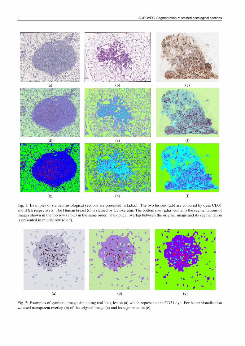
Fig. 1: Examples of stained histological sections are presented in (a,b,c). The two lesions (a,b) are coloured by dyes CD31and H&E respectively. The Human breast (c) is stained by Cytokeratin. The bottom row (g,h,i) contains the segmentations ofimages shown in the top row (a,b,c) in the same order. The optical overlap between the original image and its segmentationis presented in middle row (d,e,f).
(a) (b) (c)
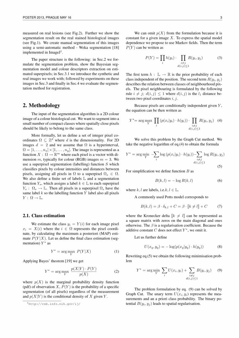
Fig. 2: Examples of synthetic image simulating real long-lesion (a) which represents the CD31 dye. For better visualisationwe used transparent overlap (b) of the original image (a) and its segmentation (c).
POSTER 2013, PRAGUE MAY 16 3
measured on real lesions (see Fig.2). Further we show thesegmentation result on the real stained histological images(see Fig.1). We create manual segmentation of this imagesusing a semi-automatic method - Weka segmentation [18]implemented in ImageJ1.
The paper structure is the following: in Sec.2 we for-mulate the segmentation problem, show the Bayesian seg-mentation model and colour descriptors extraction on esti-mated superpixels; in Sec.3.1 we introduce the synthetic andreal images we work with; followed by experiments on theseimages in Sec.3 and finally in Sec.4 we evaluate the segmen-tation method for registration.
2. MethodologyThe input of the segmentation algorithm is a 2D colour
image of a colour histological cut. We want to segment into asmall number of compact classes where spatially close pixelsshould be likely to belong to the same class.
More formally, let us define a set of integer pixel co-ordinates Ω ⊆ Zd where d is the dimensionality. For 2Dimages d = 2 and we assume that Ω is a hyperinterval,Ω = [1, . . . , nx]× [1, . . . , ny]. The image is represented as afunction X : Ω→ Rm where each pixel is a vector with di-mension m, typically for colour (RGB) images m = 3. Weuse a superpixel segmentation (labelling) function S whichclassifies pixels by colour intensities and distances betweenpixels, assigning all pixels in Ω to a superpixel Ωs ∈ Ω.We also define a finite set of labels L and a segmentationfunction Ys, which assigns a label k ∈ L to each superpixelYs : Ωs → L. Then all pixels in a superpixel Ωs have thesame label k so the labelling function Y label also all pixelsY : Ω→ L.
2.1. Class estimation
We estimate the class yi = Y (i) for each image pixelxi = X(i) where the i ∈ Ω represents the pixel coordi-nate, by calculating the maximum a posteriori (MAP) esti-mate P (Y |X). Let us define the final class estimation (seg-mentation) Y ∗ as
Y ∗ = arg maxY
P (Y |X) (1)
Applying Bayes’ theorem [19] we get
Y ∗ = arg maxY
p(X|Y ) · P (Y )p(X)
(2)
where p(X) is the marginal probability density function(pdf) of observation X , P (Y ) is the probability of a specificsegmentation (of all pixels) regardless of the measurementand p(X|Y ) is the conditional density of X given Y .
1http://rsb.info.nih.gov/ij/
We can omit p(X) from the formulation because it isconstant for a given image X . To express the spatial modeldependence we propose to use Markov fields. Then the termP (Y ) can be written as
P (Y ) =∏
i
h(yi) ·∏i 6=j
d(i,j)≤1
R(yi, yj) (3)
The first term h : L → R is the prior probability of eachclass independent of the position. The second termR(yi, yj)describes the relation between classes of neighbourhood pix-els. The pixel neighbouring is formulated by the followingrule i 6= j; d(i, j) ≤ 1 where d(i, j) is the l1 distance be-tween two pixel coordinates i, j.
Because pixels are conditionally independent given Y ,the equation can be then written as
Y ∗= arg maxY
∏i
(p(xi|yi) · h(yi)) ·∏i 6=j
d(i,j)≤1
R(yi, yj) (4)
We solve this problem by the Graph Cut method. Wetake the negative logarithm of eq.(4) to obtain the formula
Y ∗ = arg minY−∑
i
log (p(xi|yi) · h(yi))−∑i6=j
d(i,j)≤1
logR(yi, yj)
(5)For simplification we define function B as
B(k, l) = − logR(k, l) (6)
where k, l are labels, i.e.k, l ∈ L.
A commonly used Potts model corresponds to
B(k, l) = β · δk,l + C = β · Jk 6= lK + C (7)
where the Kronecker delta Jk 6= lK can be represented asa square matrix with zeros on the main diagonal and onesotherwise. The β is a regularisation coefficient. Because theadditive constant C does not effect Y ∗, we omit it.
Let us further define
U(xq, yq) = − log(p(xq|yq) · h(yq)) (8)
Rewriting eq.(5) we obtain the following minimisation prob-lem
Y ∗ = arg minY
∑i
U(xi, yi) +∑i6=j
d(i,j)≤1
B(yi, yj) (9)
The problem formulation by eq. (9) can be solved byGraph Cut. The unary term U(xi, yi) represents the mea-surements and an a priori class probability. The binary po-tential B(yi, yj) leads to spatial regularisation.
4 BOROVEC, Segmentation of stained histological sections
(a)
(b)
(c)
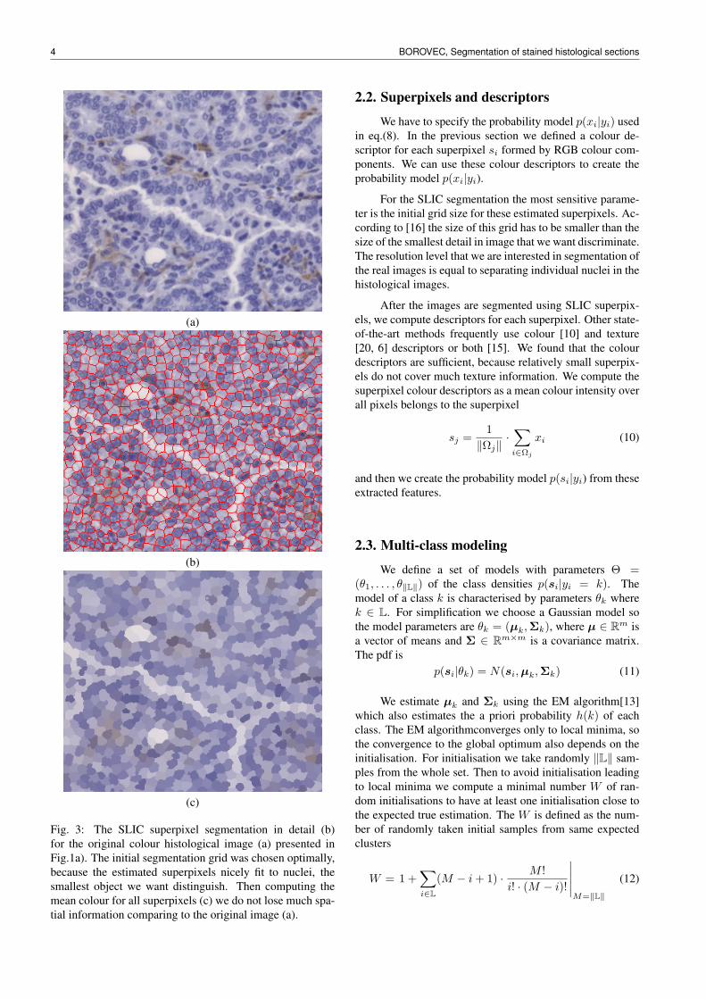
Fig. 3: The SLIC superpixel segmentation in detail (b)for the original colour histological image (a) presented inFig.1a). The initial segmentation grid was chosen optimally,because the estimated superpixels nicely fit to nuclei, thesmallest object we want distinguish. Then computing themean colour for all superpixels (c) we do not lose much spa-tial information comparing to the original image (a).
2.2. Superpixels and descriptors
We have to specify the probability model p(xi|yi) usedin eq.(8). In the previous section we defined a colour de-scriptor for each superpixel si formed by RGB colour com-ponents. We can use these colour descriptors to create theprobability model p(xi|yi).
For the SLIC segmentation the most sensitive parame-ter is the initial grid size for these estimated superpixels. Ac-cording to [16] the size of this grid has to be smaller than thesize of the smallest detail in image that we want discriminate.The resolution level that we are interested in segmentation ofthe real images is equal to separating individual nuclei in thehistological images.
After the images are segmented using SLIC superpix-els, we compute descriptors for each superpixel. Other state-of-the-art methods frequently use colour [10] and texture[20, 6] descriptors or both [15]. We found that the colourdescriptors are sufficient, because relatively small superpix-els do not cover much texture information. We compute thesuperpixel colour descriptors as a mean colour intensity overall pixels belongs to the superpixel
sj =1‖Ωj‖
·∑i∈Ωj
xi (10)
and then we create the probability model p(si|yi) from theseextracted features.
2.3. Multi-class modeling
We define a set of models with parameters Θ =(θ1, . . . , θ‖L‖) of the class densities p(si|yi = k). Themodel of a class k is characterised by parameters θk wherek ∈ L. For simplification we choose a Gaussian model sothe model parameters are θk = (µk,Σk), where µ ∈ Rm isa vector of means and Σ ∈ Rm×m is a covariance matrix.The pdf is
p(si|θk) = N(si,µk,Σk) (11)
We estimate µk and Σk using the EM algorithm[13]which also estimates the a priori probability h(k) of eachclass. The EM algorithmconverges only to local minima, sothe convergence to the global optimum also depends on theinitialisation. For initialisation we take randomly ‖L‖ sam-ples from the whole set. Then to avoid initialisation leadingto local minima we compute a minimal number W of ran-dom initialisations to have at least one initialisation close tothe expected true estimation. The W is defined as the num-ber of randomly taken initial samples from same expectedclusters
W = 1 +∑i∈L
(M − i+ 1) · M !i! · (M − i)!
∣∣∣∣∣M=‖L‖
(12)

POSTER 2013, PRAGUE MAY 16 5
(a) (b) (c) (d)
(e) (f) (g) (h)
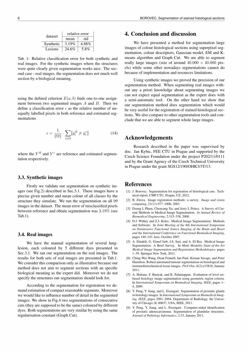
Fig. 4: Illustration of how the chosen Graph Cut regularisation influence the segmentation compactness and also the level ofdetail. We estimated one classification model and segmented this model by Graph Cut with various regularisation constants[1000, 10000, 30000, 100000] shown in rows (a,b,c,d) and (e,f,g,h) respectively. The two different stained lesions (CD31 inthe top row and H&E in bottom row) for the same regularisation constant containa similar amount of detail.
3. ExperimentsIn this section, we introduce both, the created synthetic
and the real images we work with (see Fig.1). Then wepresent the experimentally measured precision of our seg-mentation method and illustrate the dependency of chosenregulation constant in Graph Cut to the segmentation consis-tency (see Fig.4).
According to the biological meaning of the real his-tological images we fix the number of estimated classesM = ‖L‖ = 4 for all images.
3.1. Material
We will evaluate both algorithm on synthetic imagesand also real histological cuts of lesion. First we present foursynthetic datasets each consisting of 99 image pairs with var-ious transformations between them. Then we introduce twoconsecutive sequences of the lesions coloured by 5 differentstains each.
Synthetic images. We create a compact 4-class segmen-tation. Then, we assign to each class a colour such that allcreated images X have the same colours representation asone of possible stains in real histological images. To get tex-ture pattern we add 5% white Gaussian noise. The size ofthese images are 1600× 1600 pixels.
Real images. We have used material extracted from long-term urethane. For now, two nodules (adenoma or adeno-
carcinomas) were acquired with a Zeiss Axio Imager M1microscope with a 40x dry objective. Consecutive sectionswere stained with: H&E (Hematoxylin and Eosin), Pro-SPC(pulmonary pro-surfactant protein C segregated by type 2pneumocytes), CC10 (Clara Cells 10 protein), Ki67 (cancerantign that is found in growing dividing cells but is absent inthe resting phase of cell growth), CD31 (Platelet endothelialcell adhesion molecule-1. It is a protein expressed at highlevels on early and mature endothelial cells, platelets, andmost leukocyte sub-populations).
For evaluation, an expert created reference segmenta-tions by Weka segmentation plugin [18] implemented in Im-ageJ. He segmented 4 biological structures in each image.Because of implementation and resources limitation we werenot able to segment whole images but only individual partsand then we compose them together.
3.2. Assignment problem
The segmentation of each image is made independentlyso that identical objects in two different images might bedenoted by different labels k. To evaluate our segmentationwe need to find a correspondence between the class labels inboth reference and estimated segmentation.
Let us define the consistency error
E(a, b) =1‖Ω‖
·∑i∈Ω
Jyai 6= yb
i K (13)
where the a, b ∈ L are classes in two different images A andB respectively. We use the Hungarian algorithm [21] which
6 BOROVEC, Segmentation of stained histological sections
dataset relative errormean std
Synthetic 3.19% 4.88%Lesions 24.6% 5.8%
Tab. 1: Relative classification error for both synthetic andreal images. For the synthetic images where the structureswere quite clearly given segmentation works nice. The sec-ond case - real images, the segmentation does not much wellsection by a biological meaning.
using the defined criterion E(a, b) finds one-to-one assign-ment between two segmented images A and B. Then wedefine a classification error ε as the relative number of un-equally labelled pixels in both reference and estimated seg-mentations
ε =1‖Ω‖
·∑i∈Ω
JyRi 6= y∗i K (14)
where the Y R and Y ∗ are reference and estimated segmen-tation respectively.
3.3. Synthetic images
Firstly we validate our segmentation on synthetic im-ages (see Fig.2) described in Sec.3.1. These images have aprecise given number and mean colour of all classes by thestructure they simulate. We run the segmentation on all 99images in the dataset. The mean error of misclassified pixelsbetween reference and obtain segmentation was 3.19% (seeTab.1).
3.4. Real images
We have the manual segmentation of several lung-lesion, each coloured by 5 different dyes presented inSec.3.1. We ran our segmentation on the real images. Theresults for both sets of real images are presented in Tab.1.We consider this comparison only as illustrative because ourmethod does not aim to segment sections with an specificbiological meaning as the expert did. Moreover we do notspecify the structures our segmentation should look for.
According to the segmentation for registration we de-mand estimation of compact reasonable segments. Moreoverwe would like to influence number of detail in the segmentedimages. We show in Fig.4 two segmentations of consecutivecuts (they are supposed to be the same) coloured by differentdyes. Both segmentations are very similar by using the sameregularisation constant (Graph Cut).
4. Conclusion and discussionWe have presented a method for segmentation large
images of colour histological sections using superpixel seg-mentation, colour descriptors, Gaussian model, EM and K-means algorithm and Graph Cut. We are able to segmentreally large images (size of around 40.000 × 40.000 pix-els) while some other nowadays segmentations cannot dobecause of implementation and resources limitations.
Using synthetic images we proved the precision of oursegmentation method. When segmenting real images with-out any a priori knowledge about segmenting images wecan not expect equal segmentation as the expert does witha semi-automatic tool. On the other hand we show thatour segmentation method does segmentation which wouldbe very useful for the registration of stained histological sec-tions. We also compare to other segmentation tools and con-clude that we are able to segment whole large images.
AcknowledgementsResearch described in the paper was supervised by
doc. Jan Kybic, FEE CTU in Prague and supported by theCzech Science Foundation under the project P202/11/0111and by the Grant Agency of the Czech Technical Universityin Prague under the grant SGS12/190/OHK3/3T/13.
References[1] J. Borovec. Segmentation for registration of histological cuts. Tech-
nical report, CMP CTU, Prague, CZ, 2012.[2] B. Zitova. Image registration methods: a survey. Image and vision
computing, 21(11):977–1000, 2003.[3] Dzung L Pham, Chenyang Xu, and Jerry L Prince. A Survey of Cur-
rent Methods in Medical Image Segmentation. In Annual Review ofBiomedical Engineering, 2:315–338, 2000.
[4] D.J. Withey and Z.J. Koles. Medical Image Segmentation: Methodsand Software. In Joint Meeting of the 6th International Symposiumon Noninvasive Functional Source Imaging of the Brain and Heartand the International Conference on Functional Biomedical Imaging,pages 140–143. Ieee, October 2007.
[5] A. Elnakib, G. Gimel’farb, J.S. Suri, and A. El-Baz. Medical ImageSegmentation: A Brief Survey. In Multi Modality State-of-the-ArtMedical Image Segmentation and Registration Methodologies, pages1–39. Springer New York, 2011.
[6] Ching-Wei Wang, Dean Fennell, Ian Paul, Kienan Savage, and PeterHamilton. Robust automated tumour segmentation on histological andimmunohistochemical tissue images. PloS One, 6(2):e15818, January2011.
[7] A. Hafiane, F. Bunyak, and K. Palaniappan. Evaluation of level set-based histology image segmentation using geometric region criteria.In International Symposium on Biomedical Imaging, IEEE, pages 1–4, 2009.
[8] Y. Peng, Y. Jiang, and L. Eisengart. Segmentation of prostatic glandsin histology images. In International Symposium on Biomedical Imag-ing, IEEE, pages 2091–2094. Department of Radiology, the Univer-sity of Chicago, IL 60637, USA, IEEE, 2011.
[9] Y. Peng, Y. Jiang, and L. Eisengart. Computer-aided identificationof prostatic adenocarcinoma: Segmentation of glandular structures.Journal of Pathology Informatics, 2:33, January 2011.
POSTER 2013, PRAGUE MAY 16 7
[10] J.P. Monaco and J.E. Tomaszewski. High-throughput detection ofprostate cancer in histological sections using probabilistic pairwiseMarkov models. Medical Image Analysis, 14(4):617–29, August2010.
[11] M. Berthod, Z. Kato, S. Yu, and J. Zerubia. Bayesian image classi-fication using Markov random fields. Image and Vision Computing,14(4):285–295, 1996.
[12] R. Achanta and A. Shaji. SLIC Superpixels Compared to State-of-the-art Superpixel Methods. Pattern Analysis and Machine Intelligence,IEEE, 34(11):2274 – 2282, 2012.
[13] G. Xuan and W. Zhang. EM algorithms of Gaussian mixture modeland hidden Markov model. Image Processing, 2001., 1:145–148,2001.
[14] J.A. Hartigan and M.A. Wong. Algorithm AS 136: A K-means clus-tering algorithm. Journal of the Royal Statistical Society. Series C(Applied Statistics), 28(1):100–108, October 1979.
[15] H. Permuter, J. Francos, and I. Jermyn. A study of Gaussian mix-ture models of color and texture features for image classification andsegmentation. Pattern Recognition, 39(4):695–706, April 2006.
[16] A. Lucchi, K. Smith, and R. Achanta. Supervoxel-Based Segmenta-tion of Mitochondria in EM Image Stacks With Learned Shape Fea-tures. Medical Imaging, IEEE, 31(2):474 – 486, 2012.
[17] Y. Boykov and O. Veksler. Fast approximate energy minimizationvia graph cuts. Pattern Analysis and Machine Intelligence, IEEE,23(11):1222–1239, 2001.
[18] M. Hall, E. Frank, and G. Holmes. The WEKA data mining software:an update. SIGKDD Explorations, 11(1), 2009.
[19] R.O. Duda, P.E. Hart, and D.G. Stork. Pattern Classification andScene Analysis. John Wiley & Sons, 1973.
[20] M. Unser. Texture classification and segmentation using waveletframes. Image Processing, IEEE, 4(11):1549–1560, 1995.
[21] H. W. Kuhn. The Hungarian method for the assignment problem.Naval Research Logistics Quarterly, 2(1-2):83–97, March 1955.
About Authors. . .
Jirı Borovec received a MSc double-degree from IUP Sys-tem Intelligent on Universite Paul Sabatier, France, and Fac-ulty of Mechatronics at Technical University of Liberec,Czech Republic. He is currently pursuing a PhD degreeat Faculty of Electrical Engineering of the Czech Techni-cal University in Prague. His research interests include im-age registration and segmentation of stained histological sec-tions, medical imaging and robotic.