fumagillin and dicyclohexylamine in apiculture
TRANSCRIPT

Fumagillin and Dicyclohexylamine in Apiculture
by
Johan Philip van den Heever
A thesis submitted in partial fulfillment of the requirements for the degree of
Doctor of Philosophy
in
Food Science and Technology
Department of Agricultural, Food and Nutritional Science
University of Alberta
© Johan Philip van den Heever, 2015

ii
ABSTRACT
Nosema disease of the Western honey bee (Apis mellifera L.), is caused by two distinct
microsporidian fungal species, Nosema ceranae Fries et al. and Nosema apis Zander. N. apis
infection of A. mellifera was first documented in 1909, while N. ceranae infection of A. mellifera
was described in 1996. N. ceranae infection has been implicated in colony collapse disorder
(CCD) and decreased survival of overwintered colonies. There is currently only one registered
chemical treatment available to control Nosema disease in apiculture in Canada, namely
Fumagilin-B®, a potent fungal metabolite first isolated from Aspergillus fumigatus Fres.
Fumagilin-B® (and the equivalent Fumidil-B®) has been extensively used against N. apis since
its discovery in the early 1950’s, and has more recently been used to control N. ceranae
infections.
The toxicity of fumagillin, which has limited its use in human medicine, is also of concern for
beekeeping, since any residues of fumagillin remaining in hive products pose a direct risk to the
consumer. All analytical methods published to date measure only fumagillin and its
decomposition products in honey, but overlook the fact that fumagillin is present in a 1:1
stoichiometric ratio with its dicyclohexylamine (DCH) counter ion in the commercial salt
formulations (Fumagilin-B® and Fumidil-B®). DCH is almost five times more toxic than
fumagillin to rats, and also exhibits genotoxic and tumorigenic properties. A reversed phase (RP)
liquid chromatography tandem mass spectrometric (LC-MS/MS) method was developed to
confirm and quantitate trace levels of fumagillin and DCH residues in honey. A labelled d10-
DCH internal standard was also synthesized and used to compensate for observed matrix affects
when quantitating DCH in honey from different floral origins. While analyzing domestically
iii
produced honey samples fumagillin was seldom detected at levels above 10 ng g-1 (method limit
of quantitation), while DCH was detected in almost all of the samples at concentrations above 10
ng g-1. The frequency and concentrations of DCH detected, even in the absence of any detectable
amounts of fumagillin or its known degradation products, led to the design of an experiment to
evaluate the relative stability of fumagillin and DCH in honey under a range of time-temperature
exposures. During this experiment it was observed that DCH was significantly more stable in
honey than fumagillin highlighting DCH as an important potential contaminant of honey. This
further emphasizes the importance of evaluating all of the potentially active ingredients that may
be present in a pharmaceutical formulation, since the latter may be more important than
immediately apparent.
Finding alternative chemical treatments to fumagillin is necessary, as the sustained usage of this
drug in apiculture for six decades may lead to the development of resistance in Nosema spp. In
human medicine fumagillin has been tested against a variety of diseases, including cancer.
Fumagillin inhibits angiogenesis (the formation of new blood vessels around a cancerous tumor)
through covalently bonding to the methionine aminopeptidase 2 (MetAP-2) enzyme. This
enzyme occurs ubiquitously in humans, honey bees and in both N. apis and N. ceranae. I
hypothesized that the MetAP-2 structure-activity relationships discovered in human medicine
could be extrapolated to apiculture. Several semi-synthetic and purely synthetic compounds were
designed and synthesized to mimic this mode of action, and were subsequently tested on N.
ceranae-infected bees in cage trial assays. Fumagillol, the basic hydrolysis product of fumagillin,
as well as two semisynthetic fumagillin analogues and four other synthetic compounds exhibited
activity against N. ceranae-infected caged bees. None of these compounds were, however, as
effective as Fumagilin-B®. Commercially available thymol and enilconazole also exhibited
iv
activity against N. ceranae, with thymol being the most promising chemical treatment other than
Fumagilin-B®. In addition, high bee mortality was observed while evaluating Fumagilin-B®, and
a series of cage trial experiments were thus conducted to evaluate the effect of fumagillin,
Fumagilin-B® (fumagillin and DCH) and DCH on N. ceranae-infected bees. From these
experiments it was observed that orally ingested DCH caused a statistically significant risk of
increased bee mortality in N. ceranae-infected bees.

v
PREFACE
Some of the research for this thesis were conducted in collaboration with Dr. S.F. Pernal of
Agriculture and Agri-Food Canada (AAFC) at the Beaverlodge Research Farm. He was assisted
by Dr. A. Ibrahim, who was responsible for conducting the cage trial assays. All other research
and activities toward this thesis are my own original work.
Dr. S.F. Pernal was my co-supervisor, with my main academic supervisor being Dr. J.M. Curtis.
Dr. T.S. Thompson is a colleague who provided general advice and reviewed and critiqued all
manuscripts that I wrote, prior to sending them out for review by the other co-authors.
Chapter 2 of this thesis has been published as:
van den Heever, J.P., Thompson, T.S., Curtis, J.M., Ibrahim, A., Pernal, S.F.
Fumagillin: An overview of recent scientific advances and their significance for
apiculture, J. Agric. Food Chem. 2014, 62, 2728-2737.
I planned and wrote the entire manuscript, with the co-authors being responsible for review and
approval of the manuscript before submission for publication.
Chapter 3 of this thesis has been published as:
van den Heever, J.P., Thompson, T.S., Curtis, J.M., Pernal, S.F. (2015)
Determination of dicyclohexylamine and fumagillin in honey by LC-MS/MS.
Food Anal. Methods 2015, 8, 767-777.
I developed and validated the method as well as designed and synthesized the deuterated internal
standard for DCH. I planned and wrote the entire manuscript, with the co-authors being
responsible for the review and approval of the manuscript before submission for publication.
Chapter 4 of this thesis has been published as:
van den Heever, J.P., Thompson, T.S., Curtis, J.M., Pernal, S.F. Stability of
dicyclohexylamine and fumagillin in honey. Food Chem. 2015, 179, 152-158.
I designed and conducted the stability study experiment, with assistance in sample preparation
from the following lab technicians: D. Tang, M. Siegenthaler, J. Kormanicki and R. Limanowka
in the Chemistry Laboratory of the Agri-Food Laboratories Branch of Alberta Agriculture and

vi
Rural Development in Edmonton. I planned and wrote the entire manuscript, with the rest of the
co-authors being responsible for the review and approval of the manuscript before submission
for publication.
Chapter 5 of this thesis is presented as originally submitted to Apidologie on 05 May 2015 for
publication. The manuscript was subsequently divided into two parts, at the request of the
journal, and accepted for publication as:
van den Heever, J.P., Thompson, T.S., Otto, S.J.G., Curtis, J.M., Ibrahim, A.,
Pernal, S.F. Evaluation of Fumagilin-B® and other potential alternative
chemotherapies against Nosema ceranae-infected honey bees (Apis mellifera) in
cage trial assays. 2015, in press
van den Heever, J.P., Thompson, T.S., Otto, S.J.G., Curtis, J.M., Ibrahim, A.,
Pernal, S.F. The effect of dicyclohexylamine and fumagillin on Nosema ceranae-
infected honey bee (Apis mellifera) mortality in cage trial assays. 2015, in press
All cage trials, as well as preliminary statistical analysis, were conducted by Dr. S.F. Pernal and
Dr. A. Ibrahim and their team at AAFC’s Beaverlodge Research Farm, while the design of the
experiments and synthesis of the semisynthetic and synthetic compounds were done by myself.
The evaluation of the toxicity of DCH was also self-initiated, and I also supplied the chemicals
required for this cage trial experiment. I planned and wrote the entire manuscript, while the final
statistical analysis (Chapter 5) was conducted by Dr. S.J.G. Otto. The remaining co-authors
being responsible for the review and approval of the two manuscripts before submission for
publication.

vii
ACKNOWLEDGEMENTS
I wish to acknowledge the following people and organizations for their contributions towards
this project:
My committee for their time and encouragement over the past six years: The late Dr.
Lloyd Dosdall (1952-2014) for his advice and encouragement, as well as Dr. Jonathan
Curtis (main supervisor), Dr. Stephen Pernal (co-supervisor) and Dr. Andreas Schieber.
Dr. Tom Thompson, my colleague, for his invaluable advice and support.
The late Mr. Willy Baumgartner (1928-2010), former owner of Medivet Pharmaceuticals
Ltd, High River, Alberta, for his generous contribution of enough technical Fumagilin-B®
to complete this project, and for some future work as well.
My current employment supervisor, Mrs. Norine Best, and my former supervisor, Mr.
Don Noot, for their support and commitment to my research.
Alberta Agriculture and Rural Development, Animal Health and Assurance Division,
Agri-Food Laboratories Branch for the use of their laboratories, equipment, time and
consumables to conduct this research.
The Foundation for the Preservation of Honey Bees, Inc., a non-profit corporation that
awarded me with a scholarship to attend the 2012 North American Beekeeping
Conference held in Las Vegas, Nevada on January 10-14, 2012.
Agriculture and Agri-Food Canada, Beaverlodge Research Farm, specifically my co-
supervisor Dr. S.F. Pernal, as well as Dr. A Ibrahim and their team for conducting the
cage trial assays.
Desmond Tang, Marlise Siegenthaler, Joy Kormanicki and Renata Limanowka for their
help in preparing samples for the stability study and analytical work.
Table of Contents
Chapter 1 General Introduction .....................................................................................................1
Chapter 2 Fumagillin: An Overview of Recent Scientific Advances, and their
Significance for Apiculture .........................................................................................10
2.1 Introduction .................................................................................................... 10
2.2 Nosema disease in beekeeping ...................................................................... 10
2.3 Fumagillin discovery and usage in apiculture, human medicine and in
other agriculture ............................................................................................. 12
2.4 Toxicity of fumagillin .................................................................................... 17
2.5 Chemical analysis and stability of fumagillin in the field and under
laboratory conditions ..................................................................................... 22
2.6 Future outlook ................................................................................................ 25
Chapter 3 Determination of dicyclohexylamine and fumagillin in honey by LC-MS/MS .........28
3.1 Introduction .................................................................................................... 28
3.2 Materials and Methods .................................................................................. 31
3.2.1 Reagents and Materials .................................................................................. 31
3.2.2 d10-DCH synthesis ......................................................................................... 32
3.2.3 Preparation of Standard solutions .................................................................. 34
3.2.4 LC-MS/MS Equipment .................................................................................. 35
3.2.5 Mobile phase preparation .............................................................................. 35
3.2.6 HPLC conditions ........................................................................................... 35
3.2.7 MS conditions ................................................................................................ 36
3.2.8 Sample Extraction .......................................................................................... 38
3.2.9 Matrix matched calibration standards, negative and positive control
standards ........................................................................................................ 39
3.2.10 Method validation .......................................................................................... 40
3.3 Results............................................................................................................ 41
3.4 Discussion ...................................................................................................... 42
3.5 Conclusions.................................................................................................... 47
Chapter 4 Stability of dicyclohexylamine and fumagillin in honey ............................................49
4.1 Introduction .................................................................................................... 49
4.2 Materials and Methods .................................................................................. 53
4.2.1 Reagents and Materials .................................................................................. 53
4.2.2 LC-MS/MS Equipment .................................................................................. 53
4.2.3 Preparation of standards and stability samples .............................................. 54
4.3 Results............................................................................................................ 55
4.4 Discussion ...................................................................................................... 61
4.5 Conclusions.................................................................................................... 63
Chapter 5 Evaluation of Fumagilin-B® and other potential alternative chemotherapies
against Nosema ceranae-infected honey bees (Apis mellifera) in cage trial
assays ..........................................................................................................................65
5.1 Introduction .................................................................................................... 65
5.2 Materials and Methods .................................................................................. 67
5.2.1 Reagents and materials .................................................................................. 67
5.2.2 General synthetic methodology ..................................................................... 68
5.2.3 Ester synthesis ............................................................................................... 70
5.2.4 Epoxide formation from ketones ................................................................... 70
5.2.5 Cage assays – general procedures .................................................................. 71
5.2.6 Determination of spore levels ........................................................................ 72
5.2.7 Nosema spp. Identification ............................................................................ 72
5.2.8 Testing of semisynthetic, synthetic and commercially available
compounds ..................................................................................................... 74
5.2.9 Testing of DCH in the commercial Fumagilin-B® ........................................ 76
5.2.10 Statistical Analysis ......................................................................................... 76
5.3 Results............................................................................................................ 78
5.3.1 Potential alternative chemotherapies against N. ceranae .............................. 78
5.3.2 Toxicity of DCH in the commercial formulation Fumagilin-B® ................... 79
5.4 Discussion ...................................................................................................... 85
5.5 Conclusions.................................................................................................... 89
Chapter 6 General discussion and conclusions ...........................................................................91
6.1 Significance of this research .......................................................................... 91
6.2 Conclusions and Future Prospects ................................................................. 93

List of Tables
Table 1 Current lowest Canadian AMRL’s or MRL’s for antibiotics in
meat, as well as the corresponding extrapolated WRL values for
honey (Health Canada 2015a). ........................................................................ 4
Table 2 A summary of Canadian pesticide MRL’s in honey and related
hive products (Health Canada 2015c). ............................................................. 5
Table 3 Details of the MRM MS/MS parameters. ...................................................... 36
Table 4 Intra- and inter day accuracy and precision data for the validation
of fumagillin and DCH in fortified honey samples. ...................................... 40
Table 5 Results of 16 random domestic honey samples analyzed for
fumagillin and DCH ...................................................................................... 42
Table 6 Results for the study simulating bulk storage (21 oC in darkness),
shelf (21 oC exposed to light) and hive conditions (34 oC in
darkness) for both fumagillin and DCH in fortified honey
samples. ......................................................................................................... 57
Table 7 Results for the linear regression analysis of fumagillin and DCH
for data recorded under simulated shelf (21 oC exposed to light),
bulk storage (21 oC in darkness) and hive (34 oC in darkness)
conditions. ...................................................................................................... 61
Table 8 Confirmation of the identity of semisynthetic and synthetic
compounds that were tested against N. ceranae by high resolution
mass spectrometric analysis. .......................................................................... 70
Table 9 Results of the negative binomial models to assess the relationship
between test compound concentration and average N. ceranae
spore count at 17 days post-infection after feeding each
compound ad libitum in 60% sugar solution. ................................................ 80
Table 10 Results of the zero-inflated negative binomial models to assess
the relationship between test compound concentration and
average N. ceranae spore count at 17 days post-infection after
feeding each compound ad libitum in 60% sugar solution. ........................... 81
Table 11 Cumulative mortalities for bees infected with N. ceranae that
were fed varying concentrations of test compounds ad libitum in
60% sugar solution. Only compounds that had a statistically
significant impact on spore counts are included. ........................................... 82

Table 12 Results of the complimentary log-log, Cox proportional hazards
survival model for the cage trial to assess toxicity of
dicyclohexylamine (DCH), fed ad libitum at a concentration of
40 μM in 60% sucrose solution, to bees infected with N. ceranae
over 17 days of treatment. ............................................................................. 84
Table 13 Results of linear regression model for the cage trial to assess the
effects of various treatment preparations with or without
dicyclohexylamine (DCH), fed ad libitum at a concentration of
40 μM in 60% sugar solution, on spore counts in bees infected
with N. ceranae over 17 days of treatment. ................................................... 85

List of Figures
Figure 1 The chemical structures of fumagillin (1), dicyclohexylamine (2),
5-nitrofuran (3), tetracycline (4), chloramphenicol (5), penicillin
(6), sulphonamide drugs (7), chlorotetracycline (8), tylosin (9),
oxytetracycline (10), streptomycin (11), erythromycin (12) and
lincomycin (13). ............................................................................................... 3
Figure 2 The chemical structures of tau-fluvalinate (14), amitraz (15) and
coumaphos (16), the allowable pesticide residues in Canadian
honey. ............................................................................................................... 5
Figure 3 Fumagillin (1), UV-decomposed fumagillin (2), fumagillol (3),
thermally decomposed fumagillin (4) and TNP-470 (5). .............................. 13
Figure 4 Fumagillin, CAS 23110-15-8 (6) as the DCH CAS 101-83-7 (7)
salt. ................................................................................................................. 14
Figure 5 Chemical structure representation of fumagillin (1) and its
biologically active UV decomposition (2) and semisynthetic
hydrolysis product, fumagillol (3), as well as the biologically
inactive thermal decomposition product (4). ................................................. 29
Figure 6 The current internal standard for fumagillin analysis,
roxithromycin (5), TNP-470 (6), and the salt of fumagillin (1)
with its counter ion, DCH (7) as found in the commercial
formulation, Fumagilin-B®. ........................................................................... 30
Figure 7 Schematic representation of the chemical synthesis of d10-DCH
(10), d11-DCH (13) and d21-DCH (14) from commercially
available starting materials cyclohexylamine (8), d10-
cyclohexanone (9), cyclohexanone (11) and d11-cyclohexylamine
(12). ................................................................................................................ 33
Figure 8 LC-MS/MS chromatograms at normal working concentration of
300 ng g-1, showing that no detectable amounts of non-deuterated
DCH (7) contamination is present in the synthesised d10-DCH
(10), indicating that it is suitable for use as an internal standard. ................. 34
Figure 9 Unprocessed LC-MS/MS chromatograms at 10 ng g-1 for the
analytes (not all to the same scale) that show the elution of
fumagillin (1), UV decomposed fumagillin (2) and DCH (7). The
internal standards d10-DCH (10) and roxithromycin (5) are shown
at operational concentrations. ........................................................................ 37

Figure 10 Schematic representation of fumagillin (1) as its
dicyclohexylamine (DCH) salt (2) in the commercial formulation
Fumagilin-B®. Meglumine (3), an alternative compound that
could potentially be utilized as a replacement for DCH is also
shown. ............................................................................................................ 50
Figure 11 Schematic representation of the biologically inactive thermally
decomposed fumagillin (4) and it’s two other biologically active
breakdown products, namely the UV decomposition products (5),
and the semi-synthetic potential hydrolysis product, fumagillol
(6). .................................................................................................................. 51
Figure 12 Graphical representation of data recorded for fumagillin and
DCH under simulated shelf conditions (21 oC exposed to light)
indicating that no fumagillin or UV decomposed fumagillin is
present after 30 days. ..................................................................................... 58
Figure 13 Graphical representation of data recorded for fumagillin and
DCH under simulated bulk storage conditions (21 oC in
darkness). ....................................................................................................... 59
Figure 14 Graphical representation of data recorded for fumagillin and
DCH under simulated hive conditions (34oC in darkness) ............................ 60
Figure 15 The commercial Fumagilin-B®, consisting of fumagillin (1) as
the dicyclohexylamine (2) salt, which can be hydrolyzed to afford
the alcohol, fumagillol (3). ............................................................................ 66
Figure 16 Coupling of fumagillol (3) with aspirin (4) or piperonylic acid (6)
respectively, under Steglich reaction conditions using N,N'-
dicyclohexylcarbodiimide (DCC) and dimethylaminopyridine
(DMAP), to afford the aspirin analogue (5) and piperonylic acid
analogue (7) of fumagillin. ............................................................................ 68
Figure 17 Purely synthetic compounds prepared by using the Corey-
Chaykovsky epoxidation reaction to afford the cyclohexanone
derivative (8), menthol derivative (9) as well as other synthetic
analogues (10, 11, 12, 13, 14). ....................................................................... 69
Figure 18 Commercially available compounds evaluated included
carbendazim (15), toltrazuril (16), thiabendzole (17), thymol (18)
and enilconazole (19). Nozevit, another commercially available
product tested, is a bee supplement consisting of a mixture of
unspecified polyphenols and other compounds. ............................................ 75

Figure 19 The predicted hazards from the complimentary log-log, Cox
proportional hazards survival model for the cage trial to assess
toxicity of dicyclohexylamine (DCH), fed ad libitum at a
concentration of 40 μM in 60% sugar solution, to bees infected
with N. ceranae over 17 days of treatment. ................................................... 83
Figure 20 Comparison of the chemical structures of fumagillin (1), UV-
decomposed fumagillin diastereomeric compounds (20) and the
naturally occurring chlorohydrin, ligerin (21), which is reportedly
equally or more biologically active against osteosarcoma cell
lines than fumagillin, in spite of the fact that the intact spiro-
epoxide (which is reportedly responsible for biological activity of
fumagillin analogues) is not present in this compound. ................................ 87

1
Chapter 1 General Introduction
Honey has been a prized commodity since ancient times, with the oldest recorded evidence of
honey collection being found in rock paintings from caves in Altamira in Northern Spain, dated
between 8000 to 2000 BCE (Crane 1983). The earliest recorded evidence of beekeeping
practices have been found on a stone bas-relief in the sun temple of Neuserre, situated in Abu
Ghorab on the bank of the Nile River in Egypt, dated to 2400 BCE. This relief depicts honey
being harvested and placed into containers (Crane 1983). The oldest known bee hives (1000 to
900 BCE) discovered during an excavation in the ancient town of Tel Rehov in Israel, were
made from clay pots that formed part of a large apiary (Bloch et al. 2010). Modern commercial
beekeeping however owes its existence to the development of the removable wooden frame that
was developed by the Rev. L.L. Langstroth in 1851 (Johansson and Johansson 1967).
In addition to honey being a natural sweetener, it also possesses additional beneficial health and
medicinal properties (Crane 1975). A recent example of a honey with proven in vitro
antibacterial properties (ascribed to the chemical methylglyoxal) is Mānuka honey, which
originates from the nectar of the Mānuka tree that is native to New Zealand and Southern
Australia (Mavric et al. 2008). Mānuka honey was also approved for general wound management
by the US Food and Drug Administration (FDA) in 2007 (Pieper 2009).
Current apicultural techniques have changed significantly since ancient times, keeping pace with
scientific advances in chemistry and genetics, to name but a few. The bulk of honey produced
today is through large-scale commercial beekeeping. Canada had 8,777 beekeepers with a
combined number of 694,217 colonies. Canada produced 36,993,179 kg (81,556,000 lb) of
honey during 2014 (Darrach and Page 2015). The province of Alberta was the largest honey
producer, producing 41% of all of the honey in Canada. The total monetary value of honey alone
amounted to CA $ 201,620,000 while the value of pollination is estimated at CA $ 4.4 billion.
Canada was a net exporter of honey in 2014 with a value of CA $ 18,641,000. In total 64% of
exported Canadian honey to the United States, followed by 33% to Japan and 3% to China
(Darrach and Page 2015). The economic benefit of Canadian apiculture, including pollination, is
therefore significant.
2
Current large-scale commercial apicultural practices have also created new challenges with
regard to the control of disease outbreaks, which have the potential for significant financial loss
to the beekeeper, as well as impacting the local economy. One important method used in
controlling disease is through the use of pharmaceuticals. The major pharmaceutical treatments
used in beekeeping worldwide, and the diseases they are used against, are summarized in recent
research (Mullin et al. 2010; Reybroeck et al. 2012; Johnson et al. 2013). Chemical control of
disease unfortunately also leads to the potential of residues of the applied chemicals being
present in hive products, including honey. This is of concern to the consumer with regard to
human health and the perceived quality of the honey.
The amount of chemical residues that are allowed to remain in a food product destined for
human consumption, posing no adverse effects when consumed daily over the lifetime of an
individual, is regulated by individual countries. Most, if not all countries have regulatory bodies
that set maximum residue limits (MRL’s) for known traces of agricultural pharmaceuticals and
chemical contaminants in different food commodities. In Canada, MRL’s are set by Health
Canada, and are listed in parts per million (ppm). For consistency throughout, all MRL numbers
were converted to ng g-1, or parts per billion (ppb), to allow for ease of comparison. Health
Canada also sets administrative maximum residue limits (AMRL’s) for chemical contaminants
that have been scientifically evaluated and for which a MRL has been established, but for which
the official publication of the MRL has not yet been completed. Once the approved MRL is
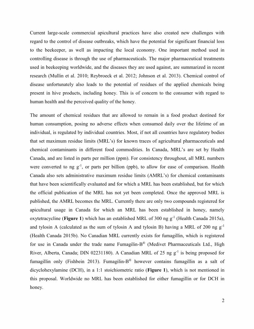
published, the AMRL becomes the MRL. Currently there are only two compounds registered for
apicultural usage in Canada for which an MRL has been established in honey, namely
oxytetracycline (Figure 1) which has an established MRL of 300 ng g-1 (Health Canada 2015a),
and tylosin A (calculated as the sum of tylosin A and tylosin B) having a MRL of 200 ng g-1
(Health Canada 2015b). No Canadian MRL currently exists for fumagillin, which is registered
for use in Canada under the trade name Fumagilin-B® (Medivet Pharmaceuticals Ltd., High
River, Alberta, Canada; DIN 02231180). A Canadian MRL of 25 ng g-1 is being proposed for
fumagillin only (Fishbein 2013). Fumagilin-B® however contains fumagillin as a salt of
dicyclohexylamine (DCH), in a 1:1 stoichiometric ratio (Figure 1), which is not mentioned in
this proposal. Worldwide no MRL has been established for either fumagillin or for DCH in
honey.

3
O
O
O
O
O
OOMe
N
S
COOHO
HH
O
R
O2NHN
OH OH
O
Cl
ClR'
SN
R''O O
R'''
O
O2N
RHO O OH O
NH2
O
OH
HO H HN
OH
HO O OH O
NH2
O
OH
HOH H
N
OH
Cl
O
O
OH
O OHO
N
O
O
OO
OCH3H3CO
HO
O
O
OH
OH
O
O OHO
N
O
OH
O OH
OCH3O
O
HOOHO
O
OOH
CHO
HO N
NH
NH2
HOH
HN
NHH2N
ON
H
HO
HO
HO
O S
OH
OH
HO
HO
NH
O
N
NH H
(1) (2) (3) (4)
HO O OH O
NH2
O
OH
HO H HN
OH
OH
(5) (6) (7)
(9) (10)
(8)
(11) (12) (13)
Figure 1 The chemical structures of fumagillin (1), dicyclohexylamine (2), 5-nitrofuran (3),
tetracycline (4), chloramphenicol (5), penicillin (6), sulphonamide drugs (7),
chlorotetracycline (8), tylosin (9), oxytetracycline (10), streptomycin (11),
erythromycin (12) and lincomycin (13).
The concept of a Working Residue Limit (WRL) for chemical residues is a uniquely Canadian
designation used for honey only (CFIA 2014). WRL’s are established in collaboration with the
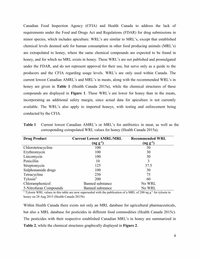
4
Canadian Food Inspection Agency (CFIA) and Health Canada to address the lack of
requirements under the Food and Drugs Act and Regulations (FDAR) for drug submissions in
minor species, which includes apiculture. WRL’s are similar to MRL’s, except that established
chemical levels deemed safe for human consumption in other food producing animals (MRL’s)
are extrapolated to honey, where the same chemical compounds are expected to be found in
honey, and for which no MRL exists in honey. These WRL’s are not published and promulgated
under the FDAR, and do not represent approval for their use, but serve only as a guide to the
producers and the CFIA regarding usage levels. WRL’s are only used within Canada. The
current lowest Canadian AMRL’s and MRL’s in meats, along with the recommended WRL’s in
honey are given in Table 1 (Health Canada 2015a), while the chemical structures of these
compounds are displayed in Figure 1. These WRL’s are lower for honey than in the meats,
incorporating an additional safety margin, since actual data for apiculture is not currently
available. The WRL’s also apply to imported honeys, with testing and enforcement being
conducted by the CFIA.
Table 1 Current lowest Canadian AMRL’s or MRL’s for antibiotics in meat, as well as the
corresponding extrapolated WRL values for honey (Health Canada 2015a).
Drug Product Current Lowest AMRL/MRL
(ng g-1)
Recommended WRL
(ng g-1)
Chlorotetracycline 100 30
Erythromycin 100 30
Lincomycin 100 30
Penicillin 10 3
Streptomycin 125 37.5
Sulphonamide drugs 100 30
Tetracycline
Tylosin2
250
200
75
60
Chloramphenicol Banned substance No WRL
5-Nitrofuran Compounds Banned substance No WRL 2 Tylosin WRL values in this table are now superseded with the publication of a MRL of 200 ng g-1 for tylosin in
honey on 28 Aug 2015 (Health Canada 2015b)
Within Health Canada there exists not only an MRL database for agricultural pharmaceuticals,
but also a MRL database for pesticides in different food commodities (Health Canada 2015c).
The pesticides with their respective established Canadian MRL’s in honey are summarized in
Table 2, while the chemical structures graphically displayed in Figure 2.

5
Table 2 A summary of Canadian pesticide MRL’s in honey and related hive products (Health
Canada 2015c).
Pesticide Name Food Commodity MRL
(ng g-1)
tau-Fluvalinate Honey 20
Amitraz Honey 100
Coumaphos Honey 20
Honeycomb 100
Beeswax 1000
(14) (15)
(16)
O
C
O
N
O
N
H
Cl
F3C
N N N
OO
Cl
OP
O
S
O
Figure 2 The chemical structures of tau-fluvalinate (14), amitraz (15) and coumaphos (16), the
allowable pesticide residues in Canadian honey.
Pesticides in honey and in other commodities are regulated under the Pest Control Products Act
(PCPA), where the term Proposed MRL (PMRL) was previously used as the equivalent of the
AMRL for the agricultural pharmaceutical residues that had established MRL values that have
6
not yet been officially published. PMRL’s, upon being published then become Established
MRL’s (EMRL’s). The term PMRL is still used, but the term EMRL was replaced with MRL.
The different acts and terminology used, even within Health Canada, makes finding and
evaluating residue information quite tedious and exceedingly difficult for producers and the
public. The list of compounds that can be reasonably expected to occur as contaminants in honey
is also not complete, which is understandable when considering the amount of work required to
conduct risk assessments and to establish MRL’s. The lack of suitable MRL’s that encompass all
possible chemical residues presents a significant problem for many agricultural commodities.
Health Canada therefore employs a general MRL of 100 ng g-1 for agricultural residues that are
not specifically defined for a given food commodity. Other countries such as Japan adopted a
similar approach, with a general MRL value of 10 ng g-1 being used for undefined residues. It
should be noted that this value is ten times lower than what is established for Canada. The EU
does not have such a general MRL provision, although certain select compounds will have a
general MRL specified in commodities not specifically mentioned. However, if a compound
does not have a MRL in the honey, then that compound is considered to be a violation, and the
honey may not be imported into the EU. Some countries, such as Japan, also have a MRL of 300
ng g-1 (The Japan Food Chemical Research Foundation 2014) for a combined class of
compounds like the tetracyclines (tetracycline, oxytetracycline and chlorotetracycline).This
practice is however not universally accepted, and the detection of an undefined or unknown
chemical contaminant may lead to a product being refused entry into a jurisdiction that has a
resulting “zero tolerance” policy. The recent change to a “zero tolerance” policy for tylosin-A
and tylosin-B (desmycosin) residues in honey imported into Japan (The Japan Food Chemical
Research Foundation 2014), as opposed to the previously acceptable limit of below 10 ng g-1 for
combined tylosin-A and tylosin-B residues, is a good example this. This change effectively
closes the Japanese market to any producer having detectable amounts of either tylosin-A or
desmycosin in his honey.
The increasing sensitivity of analytical instrumentation used to detect trace levels of
contaminants amplifies the problem of “zero tolerance” approaches, which may lead to artificial
trade barriers that could unfairly exclude trade with other countries. This may then serve as a
mechanism to help protect local industries. The difference in MRL requirements for jurisdictions

7
such as the European Union (Commission Regulation (EU) No 37/2010 2009), Canada, the USA
and others complicate matters, because what is acceptable in one country may be in violation of
another country’s requirements.
The detection of any prohibited chemical contaminant in honey has serious long-term trade
implications, as was witnessed by the suspension of honey imports from China into the European
Union (EU) in 2002, upon the discovery of chloramphenicol and streptomycin residues resulting
from their attempts to control American foulbrood disease (Reybroeck et al. 2012). The use of
chloramphenicol and streptomycin in food producing animals was previously banned by the EU
in 1994, thereby rendering imported honey containing these contaminants unsuitable for sale in
the EU. The reputation of Chinese honey was severely damaged by the detection of these
residues. Argentinian honey trade with the EU and North America also suffered a similar fate
following the detection of 5-nitrofuran residues in Argentinian honey destined for the EU market
(FAO/WHO 2004). In addition to antibiotics, pesticides like amitraz (100 ng g-1 Canadian MRL)
are also applied against Varroa destructor Anderson and Trueman mites. There is also the
possibility of environmental contamination of honey resulting from other agricultural or
industrial sources. Testing for chemical contaminants in honey is therefore routinely conducted
in almost all international trade in honey.
Another complication when monitoring chemical contamination of food commodities, including
honey, is the importance of examining honey for the presence of other potentially bioactive
degradation products or metabolites that may result from the application of the initial
pharmaceutical formulation. The main biologically active compound of a formulated product
may in some cases be unstable in the acidic honey, however their biologically active degradation
products may be significantly more stable, and might be detectable for longer time periods. An
example of this is tylosin-A in honey, where the degradation product, called desmycosin
(tylosin-B), was reported to be more stable than tylosin-A in honey (Thompson et al. 2007a).
Tylosin-B is reportedly detectable in honey, even when no trace of tylosin-A can be detected.
Similarly, erythromycin reportedly rapidly decomposes in honey, affording several stable
biologically active degradation products that can be detected in honey long after any trace of the
parent compound is detectable (Thompson and van den Heever 2012). Recognizing the
significance of biologically active degradation products, metabolites or pharmaceutical

8
excipients related to the commercial formulation of an applied product is therefore extremely
important, as the resulting residues may have the potential to be even more significant than the
main active ingredient in the commercial product (Kirkby et al. 1972). The synergistic effect of
different chemical contaminants should also not be overlooked (Johnson et al. 2013; Zhu et al.
2014).
Even though there may be negative perceptions relating to the use and fate of agricultural
pharmaceuticals in beekeeping and in other commodities, these compounds are indispensable to
the success of modern apiculture. The main focus of my research was only on one of the many
current bee diseases prevalent in apiculture, namely Nosema disease. Fumagilin-B® is currently
the only effective treatment used to control Nosema disease, which is a microsporidian fungal
infection of Apis mellifera L. caused by two distinct single cellular pathogens, namely Nosema
apis and Nosema ceranae. It is important realize that fumagillin has been extensively used to
control Nosema disease since its discovery in the 1950’s. This extended usage of fumagillin
makes the development of alternative chemical treatments desirable, should resistance to
fumagillin develop in N. apis or N. ceranae. This inspired me to investigate alternative chemical
treatments that could be used if fumagillin should fail.
An overview of the usage of fumagillin, as well as the importance of its related metabolites,
degradation products and formulation is discussed in Chapter 2. While reviewing the available
literature on fumagillin, I recognized the significance of the counter ion of the fumagillin salt,
dicyclohexylamine (DCH), which is widely used in the commercial formulations (Fumagilin-B®
and Fumidil-B®). This realization stimulated the further development of a LC-MS/MS method to
detect DCH and fumagillin residues in honey, as described in Chapter 3. During subsequent
analysis of domestically produced honey samples I observed that DCH appeared to be more
stable than fumagillin in honey, which is evidenced by the higher concentration of DCH residues
detected in honey compared to that of fumagillin. Both fumagillin and DCH are applied in equal
amounts when using the commercial products. In order to investigate this, I designed and
conducted an experiment to determine the relative stabilities of fumagillin and DCH in honey
under a variety of conditions simulating storage, hive and shelf conditions, which is described in
Chapter 4. I also attempted to find alternative chemical treatments against Nosema disease, as

9
described in Chapter 5. These alternative compounds were designed to mimic the mode of
action of fumagillin against the MetAp-2 enzyme, known from human medicine.

10
Chapter 2 Fumagillin: An Overview of Recent Scientific Advances, and their
Significance for Apiculture
2.1 Introduction
Fumagillin is a potent antibiotic isolated from Aspergillus fumigatus Fres. that has been applied
against microsporidian infections and diseases in apiculture and in human medicine. In this
review, in order to examine the importance and current usage of fumagillin in apiculture, it is
necessary to first give a brief introduction to Nosema disease, the reason for the usage of
fumagillin in beekeeping. The possible residues of fumagillin, its metabolites, degradation
products and additives remaining in hive products destined for human consumption will then be
discussed as they may have a significant impact on the health and safety of the consumer, as well
as to apiculturists through occupational exposure. Furthermore, examination of the use of
fumagillin and its analogues in human medicine reveals certain activities that may have
relevance to apiculture. Finally, an overview of the toxicity and published analytical techniques
for fumagillin will be presented to illustrate their importance to beekeeping, and possible
deficiencies will be discussed.
2.2 Nosema disease in beekeeping
Nosema disease is one of the most prevalent diseases encountered in apiculture (Ellis and Munn
2005) and is now known to be caused by two species of single-cellular microsporidian parasites,
Nosema apis Zander and Nosema ceranae Fries et al. (Genersch 2010). The phylum
Microsporidia is comprised of more than 160 genera and about 1300 different species (Didier
and Weiss 2008; Franzen 2008). Based on molecular evidence, microsporidia are now
considered to be highly specialized parasitic fungi (Weiss et al. 1999; Sina et al. 2005).
The effects of N. apis are well documented (Fries 1993), with the organism first reported over a
century ago (Zander 1909). In contrast, N. ceranae was described as a parasite of the Asian
honey bee, Apis cerana Fab. as recently as 1996 (Fries et al. 1996). N. ceranae was suggested to
be able to infect other Apis spp. (Fries 1997), with the first genetic confirmation of this reported
when a N. ceranae isolate was obtained from Apis mellifera L. (Huang et al. 2007). The absence
of host species boundaries for N. ceranae was further postulated after a study that compared

11
Nosema spp. isolates obtained from A. mellifera and from A. cerana across different
geographical locations in Taiwan (Huang et al. 2008). While the earliest record of N. ceranae in
A. mellifera appears to be from Africanized honey bees in Brazil (Weinstein Teixeira et al.
2013), discovery of this parasite in A. mellifera populations outside of Asia was first associated
with samples collected in Spain in 2004 and 2005 (Higes et al. 2006). The parasite is now known
to be commonly and widely distributed (Klee et al. 2007; Fries 2010; Higes et al. 2010; Martín-
Hernández et al. 2012; Medici et al. 2012), and has subsequently been found in European
samples of A. mellifera from as early as 1998 (Paxton et al. 2007). N. ceranae remained
undetected in North America for several years after initial finds in Europe, however it has since
been identified in analysis of historical samples dating from 1994 to 2007 (Klee et al. 2007;
Chen et al. 2008; Currie et al. 2010) indicating that it may have been widespread and present
much longer in this continent than first realized. Importantly, this “new” form of Nosema
disease has been implicated in the large-scale loss of bee colonies not only in Europe, but also in
North and South America (Cox-Foster et al. 2007; Martín-Hernández et al. 2007; Higes et al.
2008, 2009a, 2009b, 2009c; vanEngelsdorp et al. 2009; Botías et al. 2012a, 2012b; Martínez et
al. 2012).
N. ceranae infections have also been detected in honey bee species other than A. cerana and A.
mellifera. It was recently reported to occur in the dwarf Asian honey bee (Apis florea Fab.) and
giant Asian honey bee (Apis dorsata Fab.) in Thailand (Chaimanee et al. 2010), as well as in A.
koschevnikovi Enderlein in Borneo (Botías et al. 2012a). N. ceranae has also been detected in
three native South American bumble bee species (Hymenoptera: Apidae), demonstrating its high
virulence and ability to infect multiple bee species (Plischuck et al. 2009). Recent literature
reports confirm the ubiquitous geographical presence of this invasive pathogen, its presence now
being confirmed not only in Europe, Asia and North America (Williams et al. 2008b), but also in
more remote geographical locations such as Australia (Giersch et al. 2009), South America
(Plischuck et al. 2009) and North Africa (Higes et al. 2009b). Evidence suggests that N. apis is
gradually being replaced by N. ceranae in A. mellifera (Martín-Hernández et al. 2012), though N.
apis remains more prevalent in colder climates (Fries 2010; Forsgren and Fries 2013).
Controversy regarding the greater relative virulence of N. apis compared with N. ceranae
remains unresolved, with different results being obtained by different research groups

12
(Stevanović et al. 2010; Forsgren and Fries 2010, 2013; Martín-Hernández et al. 2012). Co-
infection of N. apis and N. ceranae commonly occurs in A. mellifera (Paxton et al. 2007; Fries
2010) and has also been reported in A. cerana (Chen et al. 2009a).
N. ceranae infects the bee digestive tract midgut epithelial cells (ventriculus) of adult workers
and queens (Higes et al. 2010; Traver and Fell 2012). Similar to N. apis, the mature spores burst
forth by rupturing of the epithelial cells and spill into the midgut lumen, followed by defecation
with the fecal matter. Infection spreads via fecal-oral route, where adult worker bees contract the
infection while cleaning up fecal material originating from infected bees, or through trophallaxis
of contaminated food (Higes et al. 2009a; Smith 2012). Infection with N. ceranae has been
shown to increase precocious foraging in worker bees, resulting in reduced life expectancy by 9
days on average in cage trials when compared to control groups (Goblirsch et al. 2013a). Though
outward signs of Nosema spp. infection cannot typically be seen, the inhibition of pollen
digestion caused by the parasites leads to poor nourishment, smaller populations, reduced honey
production and higher winter colony mortality (Bailey and Ball 1991). For more information on
the biology of N. ceranae as honey bee pathogen, the reader is directed to recently published
reviews (Fries 2010; Higes et al. 2010, 2013).
2.3 Fumagillin discovery and usage in apiculture, human medicine and in
other agriculture
Fumagillin is a naturally occurring antibiotic compound that was first isolated in 1949 from an
Aspergillus species, designated H-3 (Hanson and Eble 1949), later identified as Aspergillus
fumigatus (Eble and Hanson 1951). The drug was also found to be a potent amebicide
(McCowan et al. 1951). The structure of fumagillin was eventually elucidated (Tarbell et al.
1961) through an extensive series of chemical manipulations, including the hydrolysis of
fumagillin to yield the alcohol, fumagillol (Figure 3).

13
O
O
OMe
O
O
OH
O
O
O
OMe
O
O
OHO
O
OH
OMe
O
OH
O
OMe
O
O
OH
O
HO
HO
UV
O
O
OMe
O
HN
Cl
O
(1)
(2)
(3) (4)
O
(5)
CouplingReaction
Figure 3 Fumagillin (1), UV-decomposed fumagillin (2), fumagillol (3), thermally decomposed
fumagillin (4) and TNP-470 (5).
14
The importance of fumagillin as a treatment against the microsporidian fungal disease N. apis
plaguing the European honey bee (A. mellifera) was soon recognized (Katznelson and Jamieson
1952; Bailey 1953). Fumagillin is also currently the only effective chemical treatment available
to control N. ceranae (Williams et al. 2008a, 2011; Higes et al. 2011). The commercial
formulation of fumagillin consists of the dicyclohexylamine (DCH) salt of fumagillin (Figure 4).
The significance of the usage of fumagillin in the “salt” form is described in the section relating
to the toxicity of fumagillin. Based on experimentation using cage bioassays it was nevertheless
purported that fumagillin provides only short term suppression of N. ceranae, compared with N.
apis, and that hyperproliferation of spores of the former results at specific and much degraded
concentrations of the drug (Huang et al. 2013). Fumagillin was also tested for the treatment of
microsporidian infections in fish (El-Matbouli and Hoffmann 1991; Kent and Dawe 1994;
Molnar 1994; Rigos et al. 2000).
O
O
OMe
O
O
O
O
N
H
H
(6) (7)
Figure 4 Fumagillin, CAS 23110-15-8 (6) as the DCH CAS 101-83-7 (7) salt.
In human medicine, fumagillin is used as an inhibitor of microsporidian infections in patients
with compromised immune systems due to acquired immunodeficiency syndrome (AIDS), or to
relieve symptoms of intestinal microsporidiosis that may occur after organ transplant procedures
(Molina et al. 2000, 2002; Lanternier et al. 2009). More interestingly though, fumagillin and its
analogues are used to treat various cancers by inhibiting the formation of new blood vessels
around growing tumors (angiogenesis) thereby limiting their blood supply (Ingber et al. 1990).
15
The treatment of cancer tumors by inhibition of angiogenesis was first proposed in 1985
(Folkman 1985; Folkman and Shing 1992). The discovery that fumagillin inhibits angiogenesis
led to a renewed interest in fumagillin as a therapeutic drug (Ingber et al. 1990). The mechanism
responsible for this activity was not clear, until the discovery of fumagillin binding to the
methionine aminopeptidase type 2 (MetAP-2) protein (Griffith et al. 1997; Sin et al. 1997). The
exact binding of fumagillin to the MetAP-2 enzyme was determined by crystallizing the enzyme
with fumagillin covalently bound to the enzyme active site (Liu et al. 1998). A crystallographic
study then revealed that fumagillin covalently binds to a histidine moiety (His231) of the enzyme,
resulting in an irreversible opening of the spiro-epoxide on the cyclohexane core skeleton of
fumagillin (Sin et al. 1997; Liu et al. 1998; Zhang et al. 2002).
This result prompted numerous research efforts aimed at modulating the mode of action (Han et
al. 2000; Weiss et al. 2003) by preparing analogues of fumagillin via modification of the alkene
side chain. First, fumagillin was hydrolyzed with a suitable base, thereby removing the side
chain to yield fumagillol (Figure 3). Subsequently, a new chemical moiety could be coupled to
fumagillol (Han et al. 2000; Baldwin et al. 2002; Lee et al. 2007), resulting in analogues like
TNP-470 (Figure 3), which has shown promise as a potential new treatment for malaria (Zhang
et al. 2002; Chen et al. 2009b). Another analogue of fumagillin, named fumarranol, with the
spiro-epoxide opened, exhibited an 80-100 fold lower activity than TNP-470 against malaria
(Chen et al. 2009b). The low activity of fumarranol indicates the importance of the intact
epoxide for biological activity of the fumagillin family of analogues. A 1000-fold decrease of
MetAP-2 inhibition by fumagillin upon the opening of this cyclohexane ring spiro-epoxide was
observed (Griffith et al. 1997, 1998; Lu et al. 2006).
Fumagillin binds only to the MetAP-2 enzyme via the epoxide group located on the cyclohexane
core ring structure, and not to the MetAP-1 enzyme (Sin et al. 1997). The other remaining
epoxide on the molecule is not crucial for the binding to take place, and is therefore considered
dispensable (Griffith et al. 1998).
MetAP-2 enzymes are found ubiquitously in all organisms (Zhang et al. 2002). Fumagillin acts
against microsporidian as well as the mammalian MetAP-2 enzyme, and the low selectivity of
fumagillin between human and microsporidian MetAP-2 is the cause of its toxicity to humans, as

16
it also inhibits the human MetAP-2 enzyme necessary in protein maturation and post translation
processes (Drahl et al. 2005; Huang et al. 2013). A similar observation was recently reported
where it was shown that fumagillin is active against honey bee MetAP-2 at low concentrations
while it has no therapeutic activity at those concentrations against N. ceranae MetAP-2 (Huang
et al. 2013). The fact that fumagillin acts against both the disease and bee MetAP-2 may explain
the significant bee mortality associated with fumagillin usage (Rada et al. 1997). An earlier study
supports this increased mortality associated with fumagillin usage (Furgala and Boch 1970). In
this study it was observed that fumagillin does not influence the mortality of healthy bees during
cage trials, but when N. apis-infected caged bees are treated with fumagillin, at concentrations of
12.6-50 mg L-1 in sugar syrup, increased bee mortality was observed. Even though fumagillin
degrades over time (Kochansky and Nasr 2004; Nozal et al. 2008), the repeated seasonal
treatment with fumagillin that is required to control reestablishment of N. ceranae infections (not
necessary for N. apis control) ensures that multiple generations of honey bees are exposed to low
levels of fumagillin (Higes et al. 2011; Huang et al. 2013). It is postulated that this constant low
level of fumagillin in the hive creates conditions conducive to hyperproliferation of Nosema spp.
(Huang et al. 2013). In comparing the MetAP-2 fumagillin binding site and coordination site
amino acid sequences among humans, honey bees, N. apis, N. ceranae and Nosema bombi
(Nosema infection of bumble bees – Bombus spp.), sequences were found to be identical for
honey bee and human MetAP-2, and were identical amongst the microsporidia . Nosema spp
sequences differed from honey bees and humans at only two binding site amino acids (Huang et
al. 2013). The authors also speculate that the MetAP-2 enzymes may not be the only factor
influencing response to fumagillin.
Semisynthetic analogues of fumagillin exhibit different properties from fumagillin regarding
their potency, selectivity and toxicity, and it has been shown that the potency of fumagillin
against MetAP-2 depends not only on the covalent interaction with the spiro-epoxide, but also on
more complex non-covalent interactions necessary for molecular recognition of the target drug
by the enzyme (Arico-Muendel et al. 2009).

17
2.4 Toxicity of fumagillin
It is important to note that the key purpose in creating new analogues of fumagillin has been the
need to limit the human toxicity associated with fumagillin, while retaining or enhancing the
beneficial properties of the parent compound (Killough et al. 1952; Ingber et al. 1990; Didier
1997, Didier et al. 2006; Conteas et al. 2000).
Several contradictory findings have been reported in the literature regarding the toxicity of
fumagillin. It is important to know that the commercial formulations of fumagillin
(Fumagilin-B® and Fumidil-B®) contains the dicyclohexylamine (DCH, DCHA) salt of
fumagillin in a 1:1 stoichiometric ratio with fumagillin. When evaluating the possible adverse
effects of a compound, any additive or contaminant should also be taken into account, regardless
of its concentration, as the contribution to properties such as toxicity or mutagenicity from the
additive may be more significant than what is expected (Kirkby et al. 1972). The cited study
actually used the potential carcinogenicity of DCH contamination in a cyclamate study as an
example of such an effect.
It is also interesting to note that most studies on the toxicity of fumagillin itself were conducted
using the fumagillin DCH salt as reagent, thereby introducing not only one, but two potentially
toxic compounds (Didier et al. 2006; Stanimirović et al. 2006, 2007a, 2007b , 2010; Stevanović
et al. 2006; 2008; Kulić et al. 2009). Experiments using the fumagillin analogue TNP-470
(AGM-1470) (Figure 3), appear to use only the pure TNP-470 in toxicity testing with no DCH
included (Yamaoka et al. 1993a; Yanase et al. 1993; Didier 1997; Didier et al. 2006). In other
studies it is unclear whether fumagillin was used as the DCH salt, free acid, or as an alternative
formulation (Liu 1990; Kusaka et al. 1991; Rada et al. 1997), although one study was found that
refers to the use of purified fumagillin with no DCH present (Ingber et al. 1990). It is important
to realize this fact when the reported data on the toxicity of fumagillin, or the more frequently
employed fumagillin DCH salt, is evaluated.
The toxicity of DCH in isolation has been investigated (Stoltz et al. 1970) and DCH was found to
induce chromosomal damage in human leucocyte cultures at low concentrations of
10-3 to 10-5 M, over periods ranging from 5 to 25 hours. Stoltz et al. (1970) also reported that N-
hydroxycyclohexylamine, a metabolite of DCH, exhibited equivalent toxicity. A study using a

18
Salmonella enterica serovar Typhimurium strain (TA1535/pSK1002) to verify the genotoxicity
of DCH yielded negative results (Heil et al. 1996). Nevertheless, the same study yielded positive
results when using an in vivo alkaline filter elution test (AFE) with freshwater clams as test
subject to detect DNA damage. Salmonella mutagenicity tests also proved negative for DCH in
another study (Mortelmans et al. 1986). Similar negative results were reported where DCH only
exhibited a positive carcinogenic response in one out of six tests (Purchase et al. 1978). DCH
was also reported to be a powerful inhibitor of spermidine synthesis catalyzed by extracts from
E. coli and P. aeruginosa in vitro (Pegg et al. 1983). A national screening program in Sweden
determined that DCH is toxic to rats by several modes of action, but no mutagenic properties
were reported (Woldegiorgis et al. 2007). According to the MSDS (Sigma-Aldrich product
185841 v5.0 07/24/2012), DCH exhibits serious immediate toxic effects on rats with an LD50 of
373 mg kg-1 orally. Similar results were reported with a LD50 of 200-373 mg kg-1 (Greim et al.
1998). According to the MSDS, DCH is also extremely toxic to aquatic life and should not be
released into the water system. It is also tumorigenic causing gastrointestinal and liver tumors
(orally) as well as being subcutaneously carcinogenic in mice, causing tumors at the site of
application (Pliss 1958). In contrast, the MSDS of pure fumagillin (Sigma-Aldrich product
F6771 v4.5 08/04/2014), states a LD50 of 2000 mg kg-1 orally in mice, making DCH at least five
times more toxic than fumagillin.
Fumagillin toxicity was extensively examined by Stanimirović et al. (Stanimirović et al. 2006,
2007a, 2007b, 2010; Stevanović et al. 2006, 2008, 2010; Kulić et al. 2009). In these reports it is
sometimes unclear whether fumagillin DCH, or DCH alone was used in their experiments as
their use of the terms fumagillin and dicyclohexylamine appear to be equivalent in earlier
publications (Stanimirović et al. 2006; Stevanović et al. 2006). The authors also ascribed the
dicyclohexylamine (DCH) toxicity data (Yamaoka et al. 1993b) as being that of fumagillin
(Stanimirović et al. 2006; Stevanović et al. 2006; Kulić et al. 2009), while Stoltz et al. (1970)
clearly only examined DCH, with no mention of fumagillin ever being made. These
inconsistencies, however, should not detract from the value of their research, which is briefly
summarized below.
Fumagillin was observed to exhibit significant negative chromosomal aberration effects at 50-75
mg kg-1 bodyweight in mice. Concentrations of 25, 50 and 75 mg kg-1 bodyweight were
19
administered by gavage (Stanimirović et al. 2006). All experimental dosages listed above
induced significant antiproliferative and genotoxic potential in mice (Stevanović et al. 2006).
Fumagillin exhibited clastogenic activity in human lymphocites at concentrations equivalent to
the therapeutic dose in beekeeping (Stevanović et al. 2008). Genotoxicity to mouse bone marrow
cells at concentrations of 10-20 mg kg-1 bodyweight administered in vivo to mice by gastric
probe (5, 10 and 20 mg kg-1 concentrations tested), as compared to a control group was also
observed (Kulić et al. 2009). These results were confirmed with fumagillin-induced
chromosomal aberrations at 10–20 mg kg-1 in mouse bone marrow cells (Stanimirović et al.
2010). In summary, the Stanimirović group concluded that fumagillin DCH is a mutagenic
formulation, both in vitro and in vivo.
Fumagillin was evaluated in the United Kingdom by the Committee on Mutagenicity in 1999,
with the aim of establishing a maximum residue limit (MRL) in honey, after a submission by the
patent holder, CEVA Animal Health (COM 2009a, 2009b). No MRL recommendation could be
made by the Committee on Veterinary Medicinal Products because the available toxicity data at
that time was considered to be insufficient to draw any conclusions on the risk. In 2011, it was
concluded that fumagillin should be considered as an in vitro mutagen, but not an in vivo
mutagen (COM 2011). Fumagillin is still not licensed for general use in beekeeping in Europe,
except in exceptional circumstances where a temporary authorization to use it under veterinary
supervision will be allowed (Higes et al. 2011). Fumagillin is registered for use in the USA and
Canada to treat Nosema disease. Temporary authorization has been issued in exceptional
conditions to use fumagillin in the UK, Spain, Belgium, Greece, Hungary and in Romania under
veterinary supervision (Higes et al. 2011).
Although fumagillin is an extremely beneficial compound in human medicine and in apiculture,
some undesirable side-effects cannot be ignored (Ingber et al. 1990; Yanase et al. 1993; Didier
1997; Conteas et al. 2000; Molina et al. 2000, 2002; Didier et al. 2006). Extended usage of
fumagillin over prolonged periods of time, as required by chemotherapy, caused severe body
weight loss of over 15% from the starting weight in the human test subjects (Yanase et al. 1993).
In 1952, it was reported that fumagillin was essentially non-toxic to humans at oral doses of up
to 50 mg daily for durations of two weeks of treating intestinal amebiasis (Killough et al. 1952),
although no weight loss was observed in test subjects. A more recent study administered

20
fumagillin orally up to 60 mg daily for two weeks to treat microsporidiosis in patients with HIV
infection (Molina et al. 2000). The authors acknowledged significant bone marrow toxicity of
fumagillin, with 4 patients out of a group of 11 developing severe toxic side effects at the highest
dosage administered (60 mg). These effects ceased within days of the treatment being
terminated.
A common side-effect in human trials where fumagillin is administered orally is gastrointestinal-
related cramping, diarrhea and significant loss of body weight (Yanase et al. 1993). This
undesirable weight loss side effect prompted recent, perhaps ethically questionable, trial use of
fumagillin as a chemical mitigation for obesity (Lijnen et al. 2010; Scroyen et al. 2010).
In beekeeping, potential toxic and undesirable consequences of fumagillin treatment have also
been examined, with a limited number of effects being documented. For example, unique
changes in the ultrastructure of the hypopharyngeal glands in worker bees have been observed
after infection with N. apis followed by subsequent treatment with fumagillin (Liu 1990). Such
alterations may influence protein secretions from these glands, though the role of fumagillin
treatment on gland structure and function remain speculative from this descriptive study.
Fumagillin was also noted to have a significant influence on bee mortality during a cage trial
experiment where honey bees (150–200 bees per cage) were fed a sugar syrup solution for 7 days
using a total dose of 140 mg of fumagillin per cage, compared with unmedicated controls
(Rada et al. 1997). In contrast, a later study indicated few to no deleterious effects of fumagillin
(50 mg L-1 fumagillin in sugar syrup) for caged N. apis-infected bees, also over a seven day
period (Webster 1994). This concentration is also double that currently recommended for the
only commercial formulation of the drug registered in North America for apiculture
(Fumagilin-B®, Medivet Pharmaceuticals Ltd., DIN 02231180). Label directions for this product
prescribe 9.5 g fumagillin DCH base to prepare 380 litres of syrup (25 mg L-1) which is
sufficient to treat 100-110 colonies (86-95 mg per colony) in the spring, or 50 overwintering
colonies (190.5 mg per colony) in the fall.
The majority of other reported cage experiments that evaluated bee mortality associated with
fumagillin usage has found few effects. No statistically observable increase in mortality was
observed when feeding sugar syrup at concentrations of 12.6, 25.2 and 50.4 mg L-1 ad libitum

21
over 17 days to bees not infected by N. apis (Furgala and Boch 1970). Field trials on
overwintering bee colonies infected with N. apis in Ontario, Canada showed that fumagillin
treatments in the fall (25 mg L-1, 440 mg per colony) significantly increased colony survival, and
did not harm bees at the colony level (Furgala and Boch 1970). In a Polish study, fumagillin
(56.8 mg L-1, 71 mg per colony) increased the unit honey productivity (+19%), brood production
(+20%), surplus honey production (+58%) and the lifespan of worker honey bees (+20%), with
no observable negative effects for N. apis-infected colonies in the spring (Woyke 1984). A study
from Alberta, Canada recommended fall treatments with fumagillin at increased amounts of
fumagillin (22.2 mg L-1, 300-400 mg per colony), to effectively treat N. apis infection in the fall
with no noted harmful side-effects (Szabo and Heikel 1987). Improvements in colony survival
were similarly noted for fall sugar syrup treatments containing fumagillin at lower dosages
(26.4 mg L-1, 200 mg per colony), also under Canadian wintering conditions (Furgala and
Gochnauer 1969a). Sugar syrup applications of the drug require a longer period for bees to
consume, and hence have a longer duration of treatment, compared with dusting or pollen patty
applications which are more suitable as spring treatments (Furgala and Gochnauer 1969a).
Though previous studies have concluded that fumagillin has suppressed infections and reduced
mortality of Nosema spp. infected bees in cages (Furgala and Boch 1970; Webster 1994) and
colonies (Furgala and Gochnauer 1969b; Furgala and Boch 1970; Woyke 1984; Szabo and
Heikel 1987) a recent report suggests that fumagillin has negative effects on bee health and leads
to the hyperproliferation of N. ceranae spores (Huang et al. 2013). It is difficult to directly
compare this with older studies because of differences in methods used to evaluate the effects of
fumagillin on the parasites and bees. Previous research has evaluated bee mortality specifically at
high therapeutic concentrations of fumagillin using spore counts as an indicator of the efficacy of
treatment, while harmful effects on bees have been measured as differential survival of bees or
colonies compared with infected, untreated controls. In Huang et al. (2013), deleterious effects
on bees were confirmed by alterations in structural and metabolic midgut proteins, notably at
concentrations that did not suppress microsporidia reproduction. Though the study did use spore
production as a measure of fumagillin efficacy, concentrations at which hypoproliferation of
spores were seen were at very low, sub-therapeutic levels of fumagillin that were not examined
in other studies. Such levels were estimated to represent the degraded concentrations of

22
fumagillin 2 to 5.5 months after cessation of treatment to a colony (Huang et al. 2013). Previous
work examining the effects of degraded fumagillin-medicated syrup, after 44 months of storage,
showed clear suppression of N. apis infections in caged bees with no significant deleterious
effects on bee mortality (Furgala and Gochnauer 1969a).
The difference between test methods employing controlled environments as in cage trials, and
field trials where colonies are exposed to other environmental influences, also make direct
comparison amongst studies difficult. Field trial studies are more prone to the effects of
environmental contamination by pesticides for example, making bees more susceptible to N.
ceranae infection (Pettis et al. 2013). Furthermore, synergistic effects exhibited by some
pesticides on fumagillin (Johnson et al. 2013) have been observed. The effect of other stress
factors (Williams et al. 2011) and co-infection by pathogens, such as the deformed wing virus,
further complicates the interpretation of results (Martin et al. 2013). A recent publication that
details a comprehensive standard approach to Nosema disease research is of interest, as it will
simplify the comparison of results obtained by different research groups (Fries et al. 2013).
2.5 Chemical analysis and stability of fumagillin in the field and under
laboratory conditions
Mammalian toxicity and mutagenicity of fumagillin is well reported, and therefore it is important
that the amount of fumagillin residues in honey intended for human consumption be established.
The proposed structures of the main thermal and UV degradation products of fumagillin are
shown in Figure 3. UV degradation products involve isomerization and cyclization of the alkene
side chain, while the epoxide on the main cyclohexane skeleton of the active moiety remains
intact (Kochansky and Nasr 2004; Nozal et al. 2008). Thermal degradation results in the opening
of the epoxide situated on the cyclohexane ring. A combination of thermal and UV degradation
is likely to occur during use. It is important to note that the UV decomposition products of
fumagillin retain their biological activity (Kochansky and Nasr 2004), while the thermally
degraded fumagillin does not (Higes et al. 2011). The hydrolyzed product (fumagillol) also
retains some biological activity, albeit only about 10% of that of fumagillin (Gochnauer and
Furgala 1962).
23
Several analytical methods for the detection of fumagillin have been reported. In 1988 a
reversed-phase HPLC-UV method was reported to analyze fumagillin in acetonitrile solutions
using a UV detection wavelength of 351 nm, which corresponds to the maximum UV absorption
of the alkene side chain of the molecule, and a detection concentration range of
0.000-0.035 mg mL-1 (Brackett et al. 1988). Interestingly, it was found that 254 nm UV light
does not cause fumagillin to degrade, but that 336 nm and 351 nm UV light
(fumagillin max 351 nm) and fluorescent room lights does cause degradation. Samples in that
study were prepared in acetonitrile, and it is not clear if pure fumagillin, or a salt form of
fumagillin was used. An ELISA method with a limit of detection (LOD) of 20 ng g-1 and a
HPLC UV method (350 nm detection wavelength) with a LOD of 100 ng g-1 for quantitation of
fumagillin and its degradation products in honey were developed (Assil and Sporns 1991).
Honey samples were prepared by dissolving 5.0 g of honey in equal amounts first of water
(25 mL), and then dilution with acetonitrile (25 mL). Assil and Sporns (1991) also describe the
isolation of pure fumagillin with the elimination of the DCH and other formulation compounds
from the commercial product. The authors also identify the UV-cyclized product which they
called “neofumagillin”, as a further potential contaminant of honey.
Fumagillin was quantitated in rainbow trout muscle tissue, where it was tested to control
Myxosporidiae, by using ion-pairing liquid chromatography with UV (351 nm detection
wavelength) detection (Guyonnet et al. 1995). Fish muscle tissue (1 g) was macerated in the
presence of 8mL acetonitrile-water mixture (2:6 v/v) with sample cleanup and concentration
done using Bond Elut Octyl C8 cartridge columns to give a method that has linear range of 20 –
1000 ng g-1.
The photostability of fumagillin was investigated, and a HPLC method reported using a UV
method for detection at 350 nm (Kochansky and Nasr 2004). The investigators found fumagillin
to be extremely unstable when exposed to sunlight or fluorescent laboratory lighting in 50%
ethanol solutions, with half-lives of fumagillin determined as being in the range of seconds to
minutes. Nevertheless, the activity of UV-irradiated fumagillin in syrup solutions did not reduce
its efficacy against Nosema disease significantly, with N. apis being identified as the
microsporidian used in the efficacy experiments.

24
Fumagillin and its UV and thermal decomposition products were quantitated by LC-diode array-
electrospray ionization mass spectrometry (Nozal et al. 2008). The method that the researchers
developed to purify and concentrate fumagillin from the honey matrix using a polymeric solid
phase extraction (SPE) technique forms the basis for most of the subsequent published analytical
methods (Higes et al. 2011). Method detection limits ranging from 1 – 24 ng g-1 (for light
coloured honeys), to 4 – 45 ng g-1 for darker coloured honeys are reported. The researchers also
note that honey samples to be analyzed for fumagillin can be stored at room temperature for one
month in the dark without influencing the fumagillin results significantly.
A multiclass LC-MS/MS method to determine a variety of antibiotics in honey, including
fumagillin, was developed (Lopez et al. 2008). It was noted that fluorescent lights and sunlight
accelerate fumagillin decomposition, and that sodium laboratory lights were preferred for
fumagillin analysis. The method LOD for fumagillin was reported as 60 ng g-1, and was used to
determine several other classes of compounds including tetracyclines, fluoroquinolones,
macrolides, lincosamides, aminoglycosides, sulfonamides and phenicols. Sample cleanup was
done using the same SPE cartridges described earlier (Nozal et al. 2008), but sample elution
consisted of a very laborious process with multiple elutions of the samples off the SPE
cartridges, followed by concentration and reconstitution steps, making this very time consuming.
A rapid LC-MS/MS method quantitating fumagillin using a QuEChERS (quick, easy, cheap,
effective, rugged and safe) sample preparation technique in conjunction with an Oasis mixed-
mode weak anion-exchange SPE cartridge for concentrating fumagillin was reported, with a
reported LOQ of 0.1 ng g-1 (Kanda et al. 2011). The additional QuEChERS step adds 30 minutes
to the extraction, since it is still followed by an SPE extraction.
The stability of fumagillin under various laboratory and field conditions pertaining to its thermal
and UV degradation was reported (Higes et al. 2011). It was shown that higher temperatures
(40 oC) caused rapid thermal degradation of fumagillin in sugar syrup application solutions, with
resulting loss of its activity. Fumagillin was undetectable in sugar syrup solutions after 20 days at
this temperature. A stability study of fumagillin under various temperature conditions in the
presence and absence of light was also conducted, confirming the sensitivity of fumagillin to
light that is amplified by higher temperatures (Higes et al. 2011).
25
The most recent published method of fumagillin analysis in honey by LC-MS/MS (Dmitrovic
and Durden 2013) is an optimized version of a previously published method (Nozal et al. 2008).
This study provides useful information for laboratory analysis, demonstrating that fumagillin
solutions stored in acetonitrile at 4 oC are stable for 6 months (Dmitrovic and Durden 2013). The
authors report a LOD of 0.368 – 0.522 ng g-1, and a LOQ of 1.564 – 1.112 ng g-1 in honey,
depending on the brand of analytical instrumentation used. They also observed that performing
the SPE cleanup and sample concentration step under gravity elution yielded better sample
recoveries.
None of the analytical methods for fumagillin published to date recognize the importance of the
presence of DCH as a contaminant in honey or other hive products (like wax), although the
presence of DCH in solvent samples originating from the formulation is recognized in a recent
report (Dmitrovic and Durden 2013).
2.6 Future outlook
It could be hypothesized that the mode of action of fumagillin against the two Nosema spp. in
question is similar to that observed in humans against the MetAP-2 enzyme. It is important to
note that fumagillin will also act in a similar fashion on the MetAP-2 enzyme of the host honey
bee (Huang et al. 2013). The intact epoxide on the cyclohexane ring of fumagillin is crucial for
its observed activity, but the rest of the molecule is also important for enzymatic recognition.
This observation is supported by the reports in the beekeeping literature (Kochansky and Nasr
2004; Higes et al. 2011). A potential strategy for the design of simpler and more cost effective
compounds that mimic the chemical behavior of fumagillin (Balthaser et al. 2011) using the
cyclohexane spiro-epoxide as a scaffold should be considered. The development of new
compounds to treat Nosema disease in apiculture is of great importance, owing to the fact that
fumagillin has been extensively employed in human medicine and in apiculture almost since its
discovery in the early 1950s. Although there is no proof in apiculture to indicate that Nosema
spp. have developed resistance to fumagillin, it has been reported that Nosema spp. infecting
diamondback moth, Pluetella xylostella L., have developed resistance to fumagillin
(Fumidil-B®), although the identity of the Nosema spp. was not established (Idris et al. 2000,
2001). Given this finding, it can be speculated that Nosema spp. resistance to fumagillin in

26
apiculture may develop in the future, owing to the prolonged and intensive utilization of
fumagillin dating back to the 1950’s.
Numerous different fumagillin toxicity evaluations, employing direct in vitro testing against
isolated cancer cell lines and in vivo testing using test organisms such as mice and rabbit with the
DCH salt form of fumagillin have been reported, as discussed in the section relating to the
toxicity of fumagillin. In early human trials no observable side-effects are noted in some studies,
while significant weight loss and toxic effects were observed in others. A recent report (Huang et
al. 2013) proposes that fumagillin may influence the MetAP-2 enzymes in the host and in the
pathogen differently due to the slight differences in the amino acid sequence of the binding site
portion of the enzymes in Nosema spp. as compared to that in honey bees. It remains unclear
whether the reduced toxicity of fumagillin and its analogues is due to inherent properties of these
analogues themselves, or to the fact that the toxicity contribution arising from the DCH is not
accounted for in reports to date.
It is also important in the evaluation of toxicity studies to take into account the inter-species
differences when extrapolating animal and other data to humans (Bajić et al. 2004). In vitro
testing is also not equivalent to in vivo human testing when using human and other cell cultures
or bacteria such as Salmonella spp. When all the currently available information is evaluated, it
is evident that fumagillin is indeed cytotoxic and mutagenic in vitro, and that it has undisputed
negative effects in humans such as short term toxicity, weight loss and diarrhea. The
establishment of a MRL for fumagillin and DCH in honey and other hive products is needed.
Caution should be exercised by beekeepers during fumagillin sugar syrup solution preparation,
since it could lead to unnecessary exposure of beekeepers to elevated levels of fumagillin and
DCH through dermal contact, and through breathing of airborne fumagillin dust.
However, a significant concern is not only the fumagillin toxicity, but also that of DCH, which is
present in equimolar amounts to fumagillin in the commercial products. As discussed in the
fumagillin toxicity section, it is evident that DCH is at least five times as toxic as fumagillin,
comparing only the LD50 values, and disregarding any additional negative effects. All published
analytical methods to date only evaluate the presence of fumagillin, and in a few instances one or
two of the major metabolites or degradation products, in honey. It is known that fumagillin

27
degrades within days to weeks in honey (Higes et al. 2011), and that there is a low likelihood of
fumagillin being detected in significant quantities in honey if it is applied well before the
extraction process.
The contribution of DCH present in the commercial fumagillin formulation to the overall toxicity
and resulting food safety implications in products destined for human consumption is definitely
not negligible (Kirkby et al. 1972). None of the analytical methods for analyzing fumagillin and
metabolites in honey published to date even mention the presence of DCH as an analyte of
concern. This is a significant oversight, since the potential presence of DCH in hive products is
clearly of significance for human health and food safety.
The industrial use of DCH as a corrosion inhibitor and in the rubber industry (vehicle tires) could
result in a background level of this compound being present in hive products, especially in hives
located close to main roads and areas of higher vehicle use (Woldegiorgis et al. 2007). This
potential background level of DCH contamination needs to be further investigated to determine
whether it is a real cause of concern. More importantly, the stability and presence of DCH in bee
products such as honey and wax also needs to be investigated. There is a clear need for the
inclusion of the analysis of DCH relating to usage of fumagillin in hive products.
Despite all the potential shortcomings and detrimental aspects relating to fumagillin’s usage in
beekeeping, and their resulting implications for bee and human health, it is important to note that
this is still the only effective chemical treatment currently available to treat Nosema disease. The
potential contamination by DCH of hive products destined for human consumption could be
eliminated by exploring alternative formulation methods of fumagillin, or through the
development of alternative chemical treatments utilizing the current knowledge regarding the
proposed mode of action of fumagillin against Nosema spp.

28
Chapter 3 Determination of dicyclohexylamine and fumagillin in honey by
LC-MS/MS
3.1 Introduction
Fumagillin (Figure 5) is a fungal metabolite first isolated from Aspergillus fumigatus Fres.
(Hanson and Eble 1949). Soon after its discovery, fumagillin was successfully used to control the
microsporidian fungal disease caused by Nosema apis Zander infections (Katznelson and
Jamieson 1952; Bailey 1953) plaguing the European honey bee (Apis mellifera L.). A related,
emerging, but epidemiologically different disease caused by Nosema ceranae Fries et al. is
implicated in the large-scale loss of bee colonies worldwide (Cox-Foster et al. 2007; Higes et al.
2008, 2009c; vanEngelsdorp et al. 2009; Fries 2010; Higes et al. 2010; Botías et al. 2012b;
Martínez et al. 2012). Infections by N. ceranae and N. apis are collectively referred to as Nosema
disease. Fumagillin is the only currently available effective chemical treatment against this
parasite (Williams et al. 2008a, 2011; Higes et al. 2011).
The commercial formulation of fumagillin, Fumagilin-B® (Medivet Pharmaceuticals Ltd., High
River, Alberta, Canada; DIN 02231180), consists of a salt of fumagillin, the counter ion of this
salt being dicyclohexylamine (DCH). DCH is therefore applied in a 1:1 stoichiometric ratio with
fumagillin (Figure 6). The importance of recognizing DCH as a possible contaminant having
potential human health and food safety implications for consumers of honey was recently
recognized in Chapter 2 of this thesis, with DCH reportedly being at least five times more toxic
than fumagillin when tested on rats (van den Heever et al. 2014). The DCH salt of fumagillin
reportedly has genotoxic (clastogenic) potential to mammalian cells in vivo, and is reportedly
clastogenic and cytotoxic to cultured human lymphocytes (Stanimirović et al. 2006, 2007;
Stevanović et al. 2006, 2008, 2010; Kulić et al. 2009). There is currently no maximum residue
limit (MRL) set for fumagillin in Canada (Dmitrovic and Durden 2013) or any other jurisdiction,
but a proposed MRL of 25 ng g-1 is being considered by Health Canada, although this number is
not official and might be subject to change (Fishbein 2013). To the best knowledge of the author
no MRL exists for DCH in honey or other hive products.

29
Figure 5 Chemical structure representation of fumagillin (1) and its biologically active UV
decomposition (2) and semisynthetic hydrolysis product, fumagillol (3), as well as the
biologically inactive thermal decomposition product (4).
Analytical methods to date describe the analysis of fumagillin and its UV degradation products
in honey and other commodities such as fish, but none recognize the importance of DCH as an
analyte (Gochnauer and Furgala 1962; Brackett et al. 1988; Assil and Sporns 1991; Guyonnet et
al. 1995; Kochansky and Nasr 2004; Lopez et al. 2008; Nozal et al. 2008; Higes et al. 2011;
Kanda et al. 2011; Dmitrovic and Durden 2013). Fumagillin can undergo decomposition in light,
to yield biologically active UV decomposition products with similar activity as compared to that
of fumagillin (Kochansky and Nasr 2004), or undergo hydrolysis under basic conditions in the
O
O
OMe
O
O
OH
O
O
O
OMe
O
O
OHO
O
OH
OMe
O
OH
O
OMe
O
O
OH
O
(1) (2)
(3) (4)
HO
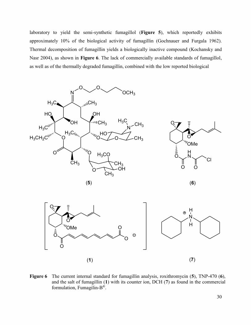
30
laboratory to yield the semi-synthetic fumagillol (Figure 5), which reportedly exhibits
approximately 10% of the biological activity of fumagillin (Gochnauer and Furgala 1962).
Thermal decomposition of fumagillin yields a biologically inactive compound (Kochansky and
Nasr 2004), as shown in Figure 6. The lack of commercially available standards of fumagillol,
as well as of the thermally degraded fumagillin, combined with the low reported biological
O
O
OMe
O
O
O
O
O
NO O
OCH3
H3C
HO
CH3
OH
H3C
H3CH2C
O
O
CH3
CH3
O
OH
H3CO
O
CH3
NCH3
H3C
H3CO
CH3OH
CH3
HO
N
H
H
(1) (7)
(5)
O
O
OMe
O
HN
Cl
O O
(6)
Figure 6 The current internal standard for fumagillin analysis, roxithromycin (5), TNP-470 (6),
and the salt of fumagillin (1) with its counter ion, DCH (7) as found in the commercial
formulation, Fumagilin-B®.
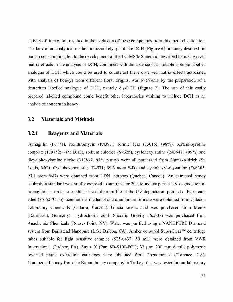
31
activity of fumagillol, resulted in the exclusion of these compounds from this method validation.
The lack of an analytical method to accurately quantitate DCH (Figure 6) in honey destined for
human consumption, led to the development of the LC-MS/MS method described here. Observed
matrix effects in the analysis of DCH, combined with the absence of a suitable isotopic labelled
analogue of DCH which could be used to counteract these observed matrix effects associated
with analysis of honeys from different floral origins, was overcome by the preparation of a
deuterium labelled analogue of DCH, namely d10-DCH (Figure 7). The use of this easily
prepared labelled compound could benefit other laboratories wishing to include DCH as an
analyte of concern in honey.
3.2 Materials and Methods
3.2.1 Reagents and Materials
Fumagillin (F6771), roxithromycin (R4393), formic acid (33015; ≥98%), borane-pyridine
complex (179752; ~8M BH3), sodium chloride (S9625), cyclohexylamine (240648; ≥99%) and
dicyclohexylamine nitrite (317837; 97% purity) were all purchased from Sigma-Aldrich (St.
Louis, MO). Cyclohexanone-d10 (D-571; 99.3 atom %D) and cyclohexyl-d11-amine (D-6305;
99.1 atom %D) were obtained from CDN Isotopes (Quebec, Canada). An extracted honey
calibration standard was briefly exposed to sunlight for 20 s to induce partial UV degradation of
fumagillin, in order to establish the elution profile of the UV degradation products. Petroleum
ether (35-60 oC bp), acetonitrile, methanol and ammonium formate were obtained from Caledon
Laboratory Chemicals (Ontario, Canada). Glacial acetic acid was purchased from Merck
(Darmstadt, Germany). Hydrochloric acid (Specific Gravity 36.5-38) was purchased from
Anachemia Chemicals (Rouses Point, NY). Water was purified using a NANOPURE Diamond
system from Barnstead Nanopure (Lake Balboa, CA). Amber coloured SuperClearTM centrifuge
tubes suitable for light sensitive samples (525-0437; 50 mL) were obtained from VWR
International (Radnor, PA). Strata X (Part 8B-S100-FCH; 33 μm; 200 mg; 6 mL) polymeric
reversed phase extraction cartridges were obtained from Phenomenex (Torrence, CA).
Commercial honey from the Buram honey company in Turkey, that was tested in our laboratory
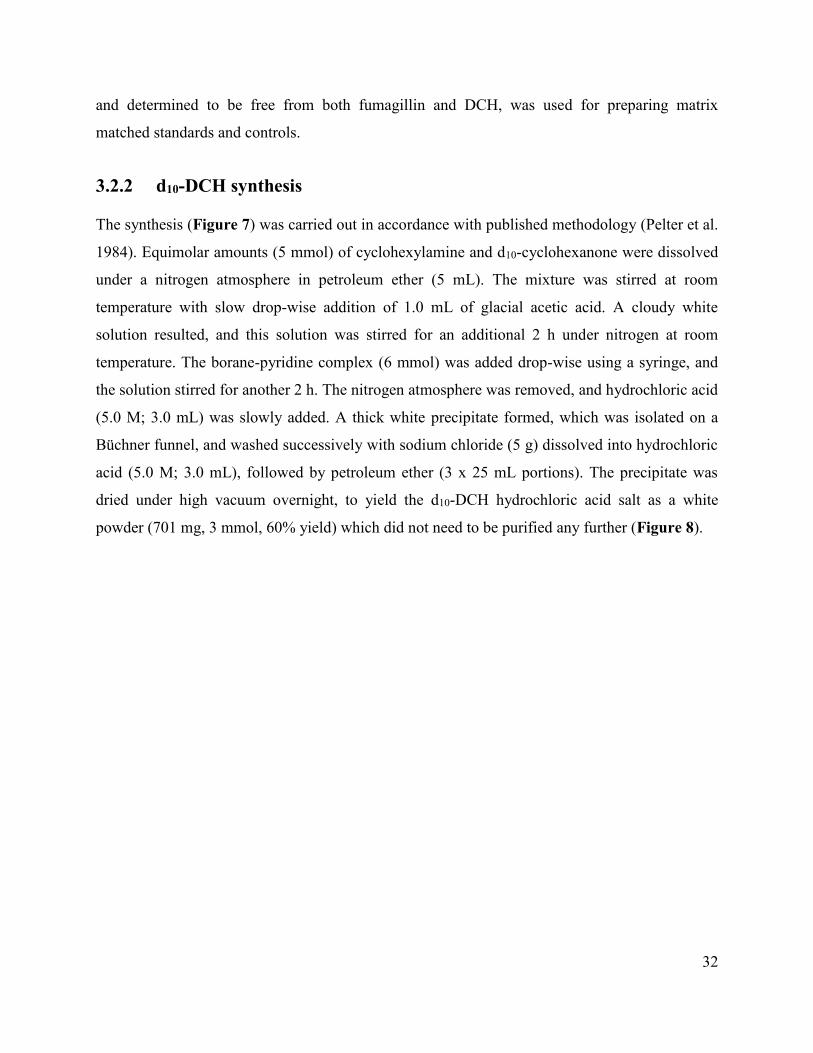
32
and determined to be free from both fumagillin and DCH, was used for preparing matrix
matched standards and controls.
3.2.2 d10-DCH synthesis
The synthesis (Figure 7) was carried out in accordance with published methodology (Pelter et al.
1984). Equimolar amounts (5 mmol) of cyclohexylamine and d10-cyclohexanone were dissolved
under a nitrogen atmosphere in petroleum ether (5 mL). The mixture was stirred at room
temperature with slow drop-wise addition of 1.0 mL of glacial acetic acid. A cloudy white
solution resulted, and this solution was stirred for an additional 2 h under nitrogen at room
temperature. The borane-pyridine complex (6 mmol) was added drop-wise using a syringe, and
the solution stirred for another 2 h. The nitrogen atmosphere was removed, and hydrochloric acid
(5.0 M; 3.0 mL) was slowly added. A thick white precipitate formed, which was isolated on a
Büchner funnel, and washed successively with sodium chloride (5 g) dissolved into hydrochloric
acid (5.0 M; 3.0 mL), followed by petroleum ether (3 x 25 mL portions). The precipitate was
dried under high vacuum overnight, to yield the d10-DCH hydrochloric acid salt as a white
powder (701 mg, 3 mmol, 60% yield) which did not need to be purified any further (Figure 8).

33
D2C
D2C
CD2
CD2
CD2
ONH2
HN
D2C
CD2
CD2
CD2
D2C
D2C
D2C
CD2
CD2
D2C
HN
D2C
CD2
CD2
CD2
D2C
H
H
D
CD2
CD2
CD2
D2C
D2C
HN
DH
O
D2C
D2C
CD2
CD2
CD2
NH2
D
D2C
D2C
CD2
CD2
CD2
O
D2C
D2C
CD2
CD2
CD2
NH2
D
H
+
(8) (9) (10)
(11) (12) (13)
+
+
(9) (12) (14)
Borane-Pyridine
Acetic Acid
Borane-Pyridine
Acetic Acid
Borane-Pyridine
Acetic Acid
Figure 7 Schematic representation of the chemical synthesis of d10-DCH (10), d11-DCH (13) and
d21-DCH (14) from commercially available starting materials cyclohexylamine (8), d10-
cyclohexanone (9), cyclohexanone (11) and d11-cyclohexylamine (12).

34
Figure 8 LC-MS/MS chromatograms at normal working concentration of 300 ng g-1, showing
that no detectable amounts of non-deuterated DCH (7) contamination is present in the
synthesized d10-DCH (10), indicating that it is suitable for use as an internal standard.
3.2.3 Preparation of Standard solutions
A fumagillin stock solution was prepared at 1000 μg mL-1 by dissolving 5 mg of fumagillin into
5 mL of acetonitrile. An intermediate solution was then prepared at a concentration of
50 μg mL-1 by dilution with acetonitrile. The absence of any DCH in this material (F6771;
Sigma-Aldrich; (St. Louis, MO) was established by testing using the method described in this
paper. A working solution was prepared at a concentration of 5 μg mL-1 by further dilution with
acetonitrile. A DCH stock solution at 1000 μg mL-1 was prepared by dissolving 5 mg of DCH
into 5 mL of methanol, after correction for using the nitrite salt form. The nitrite salt form of
DCH was used, since pure DCH is a liquid that is volatile and difficult to handle safely and
35
accurately in the laboratory when used as an analytical standard. Then an intermediate solution
was prepared at a concentration of 50 μg mL-1 by dilution with methanol, and a working solution
was prepared at a concentration of 5 μg mL-1 by further 10 fold dilution with methanol.
Internal standard stock solutions were prepared at concentrations of 1000 μg mL-1 in acetonitrile
for roxithromycin, and at 1000 μg mL-1 for d10-DCH in methanol. A combined internal standard
working solution consisting of roxithromycin (20 μg mL-1) and d10-DCH (300 μg mL-1) was then
prepared by dilution with acetonitrile.
3.2.4 LC-MS/MS Equipment
The liquid chromatography system used was a Waters 2695 separations module in tandem with a
Waters Quattro Ultima Pt mass spectrometer. The column used for this analysis was a Waters
Xterra MS-C18 with column internal dimensions of 4.6x100 mm with 3.5 μm packing particle
size (Waters; Milford; MA), protected by a matching 4x2.0 mm guard column (Phenomenex;
Torrance; CA ).
3.2.5 Mobile phase preparation
A 1.0 M ammonium formate solution was prepared by dissolving ammonium formate (15.77 g,
0.25 mol) into distilled water (250 mL). Mobile phase A was prepared by adding 2.0 mL of the
1.0 M ammonium formate solution and 100 μL of formic acid into a 1000 mL volumetric flask,
and diluting to volume with distilled water. Mobile phase B was prepared by adding 2.0 mL of
the 1.0 M ammonium formate solution, 100 μL of formic acid and 100 mL of methanol into a
1000 mL volumetric flask, followed by dilution to volume with acetonitrile.
3.2.6 HPLC conditions
The mobile phase flow was kept constant at 0.250 mL min-1 with the column temperature
maintained at 30 oC. The initial conditions were 90% mobile phase A (10% mobile phase B),
linearly changing to 25% A (75% B) over 5 min. This condition was maintained for a further 15
min, followed by a linear return to 90% A over 5 min. The final condition was held for 5 min, for
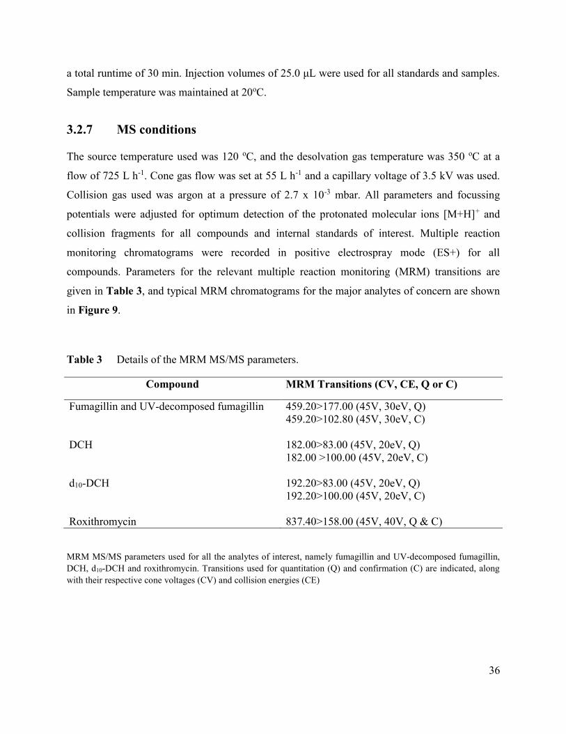
36
a total runtime of 30 min. Injection volumes of 25.0 μL were used for all standards and samples.
Sample temperature was maintained at 20oC.
3.2.7 MS conditions
The source temperature used was 120 oC, and the desolvation gas temperature was 350 oC at a
flow of 725 L h-1. Cone gas flow was set at 55 L h-1 and a capillary voltage of 3.5 kV was used.
Collision gas used was argon at a pressure of 2.7 x 10⁻³ mbar. All parameters and focussing
potentials were adjusted for optimum detection of the protonated molecular ions [M+H]+ and
collision fragments for all compounds and internal standards of interest. Multiple reaction
monitoring chromatograms were recorded in positive electrospray mode (ES+) for all
compounds. Parameters for the relevant multiple reaction monitoring (MRM) transitions are
given in Table 3, and typical MRM chromatograms for the major analytes of concern are shown
in Figure 9.
Table 3 Details of the MRM MS/MS parameters.
Compound MRM Transitions (CV, CE, Q or C)
Fumagillin and UV-decomposed fumagillin 459.20>177.00 (45V, 30eV, Q)
459.20>102.80 (45V, 30eV, C)
DCH 182.00>83.00 (45V, 20eV, Q)
182.00 >100.00 (45V, 20eV, C)
d10-DCH 192.20>83.00 (45V, 20eV, Q)
192.20>100.00 (45V, 20eV, C)
Roxithromycin 837.40>158.00 (45V, 40V, Q & C)
MRM MS/MS parameters used for all the analytes of interest, namely fumagillin and UV-decomposed fumagillin,
DCH, d10-DCH and roxithromycin. Transitions used for quantitation (Q) and confirmation (C) are indicated, along
with their respective cone voltages (CV) and collision energies (CE)

37
Figure 9 Unprocessed LC-MS/MS chromatograms at 10 ng g-1 for the analytes (not all to the
same scale) that show the elution of fumagillin (1), UV decomposed fumagillin (2) and
DCH (7). The internal standards d10-DCH (10) and roxithromycin (5) are shown at
operational concentrations.

38
MRM parameters were established by infusion, followed by optimization of all other
instrumental parameters using injection of analytes dissolved in acetonitrile, and elution by the
using the stated liquid chromatographic conditions. The UV degradation products of fumagillin
were not validated for analysis, owing to the lack of commercially available standards and the
difficulty in producing these compounds in house in sufficient purity and quantities, combined
with the observed non-detection of these compounds in samples and standards during method
development. The chromatographic elution profile of the UV decomposed fumagillin products
relative to that of fumagillin was established by spiking fumagillin into a honey sample, followed
by partial decomposition of fumagillin under fluorescent light, before sample extraction.
Subsequent sample extraction and analysis enabled detection of the UV decomposition products
eluting as a single peak slightly later than fumagillin (Figure 9).
This allowed the detection UV decomposed fumagillin for screening purposes. It was previously
reported that photodecomposition products were rarely observed in honey-sugar mixtures, even
when there is a notable decrease in fumagillin concentration resulting from induced photo-
decomposition (Higes et al. 2011). The authors also noted that no other relevant decomposition
peaks appear in the chromatograms, and speculated that this explained the non-detection of
fumagillin and its decomposition products in bee hives treated with fumagillin as reported in
another study (Assil and Sporns 1991). During our method development, the presence of the UV
decomposition products of fumagillin could not be detected after 30 days in honey spiked with
fumagillin, and then placed under fluorescent laboratory light conditions. Based on this
observation, and on other research (Assil and Sporns 1991; Kochansky and Nasr 2004; Nozal et
al. 2008; Higes et al. 2011; Kanda et al. 2011) the method described in this paper was not
validated for analysis of the fumagillin UV degradation products, but only allows for screening
to detect the presence of these compounds in honey. Roxithromycin was used as the internal
standard to quantitate fumagillin, while the synthesized d10-DCH was used as the internal
standard to quantitate DCH.
3.2.8 Sample Extraction
The sample extraction procedure is based on previously published methods (Lopez et al. 2008;
Nozal et al. 2008; Dmitrovic and Durden 2013). Care was taken not to expose the samples or

39
standards to fluorescent or other light sources that would cause or accelerate decomposition of
fumagillin (Lopez et al. 2008; Nozal et al. 2008; Dmitrovic and Durden 2013). A sample
consisting of 5 g of honey was weighed into an amber coloured 50 mL centrifuge tube, and
50 μL of the internal standard working solution was added. After addition of 10 mL of distilled
water, the tube was vortexed for approximately 20 s to loosen the honey, followed by shaking on
a mechanical shaker for one hour. The tubes were briefly centrifuged at 3500 rpm (2964 rcf) for
5 min in order to improve the flow through the SPE cartridge by isolating any suspended solid
particles in the diluted honey. The Strata SPE cartridges were conditioned by sequential elution
with methanol (5 mL) and water (5 mL) at a flow rate of 1 mL min-1. The contents of the amber
centrifuge tubes were then run through the SPE cartridges at a flow rate of 1 mL min-1, with the
waste being discarded. The SPE tubes were then washed once with 10 mL of a water/methanol
solution (7:3 v/v) at a flow rate of 1 mL min-1. The filtrate was discarded, and the SPE tubes
were dried by pulling air through them for 5 min at a higher vacuum than that used for elution.
Clean collection tubes were then placed in the vacuum manifold and the analytes of interest were
eluted off the SPE cartridges by using 2 mL of acetonitrile containing 5% of formic acid (using
vacuum to remove all possible eluent from the SPE tubes). The eluent was transferred into an
amber 1.5 mL sample vial for analysis, without any further processing.
3.2.9 Matrix matched calibration standards, negative and positive control
standards
Honey samples (5 g) free from both fumagillin and DCH were weighed into amber coloured 50
mL centrifuge tubes, and 50 μL of the internal standard working solution was added. A positive
control sample was spiked at 200 ng g-1 with both DCH and fumagillin, while a negative control
was only spiked with the internal standard working solution. Calibration standards were spiked at
10, 20, 50, 100 and 500 ng g-1 respectively. All standards were processed in the same manner, as
described for the sample preparation above, by adding 10 mL of water to all and following the
remainder of the procedure as described.

40
3.2.10 Method validation
The method validation entailed analysis of three replicate analyte concentrations (fortified at 10,
100 and 500 ng g-1 respectively), each fortified concentration comprised of six different samples
being analyzed on three different occasions, using matrix matched calibration standards. The
matrix matched calibration standards had a linear dynamic range of 10-500 ng g-1 (10, 20, 50,
100 and 500 ng g-1 respectively) with observed correlation coefficients of greater than 0.995 for
both fumagillin and DCH being observed on all 3 days. The accuracy and precision data from the
analysis of the fortified samples are presented in Table 4. The Limit of Detection (LOD) values
were calculated using a signal-to-noise (s/n) ratio of 3:1 as criteria to analyze the combined 18
replicate honey samples fortified at the lowest concentration of 10 ng g-1. The Limit of
Quantitation (LOQ) values were calculated using a 10:1 s/n ratio as criteria. No interference was
observed for either DCH, fumagillin or its associated UV degradation products in any sample or
matrix matched standard during the entire method validation and development, confirming the
specificity of the LC-MS/MS analysis for all these compounds. The validated method was
applied to the analysis of 16 domestically produced honey samples available in our laboratory
(Table 5).
Table 4 Intra- and inter day accuracy and precision data for the validation of fumagillin and
DCH in fortified honey samples.
Compound
Fortification
level (ng g-1)
% Recovery (% RSD)
Day 1
(n=6)
Day 2
(n=6)
Day 3
(n=6)
Day 1 to 3
(n=18)
Fumagillin
10
100
500
108.5 (8.6)
100.3 (6.4)
101.4 (3.3)
109.2 (8. 9)
108.9 (7.9)
115.2 (4.6)
99.5 (13.8)
93.2 (6.4)
96.3 (3.6)
105.7 (10.8)
100.8 (9.3)
104.3 (8.7)
Dicyclohexylamine
10
100
500
105.5 (5.0)
101.4 (3.5)
103.2 (4.9)
102.2 (8.3)
102.3 (5.7)
112.9 (7.7)
104.3 (4.4)
91.0 (4.2)
95.8 (8.4)
104.0 (5.9)
98.3 (6.9)
104.0 (9.7)
41
3.3 Results
Sample extraction was achieved using reversed phase polymeric solid phase extraction (SPE)
with recoveries (±RSD%) at 10, 100 and 500 ng g-1 for 18 replicates at each respective
concentration calculated as 105.7±10.8, 100.8±9.3 and 104.3±8.7 for fumagillin and as
104.0±5.9, 98.3±6.9 and 104.0±9.7 for DCH (Table 4).
The linearity fit (R2) was observed to be greater than 0.995 for both DCH and fumagillin over the
linear range of 10-500 ng g-1 for all instances. Limits of detection (LOD) were calculated as
1.2 ng g-1 for fumagillin and as 0.24 ng g-1 for DCH, using the s/n criteria of 3:1. A quantitation
limit (LOQ) of 10 ng g-1 was chosen, based on the lowest matrix-matched calibration standard,
which is well above the calculated LOQ of 4.03 ng g-1 for fumagillin and 0.79 ng g-1 for DCH.
A total of 16 domestically produced honey samples were analyzed for fumagillin and DCH,
while the samples were screened to detect the presence of the UV degraded fumagillin residues
(Table 5). The UV decomposition products of fumagillin elute as one peak under the
chromatographic conditions used (Figure 9). No UV decomposed fumagillin could be detected
during screening of the 16 samples. Fumagillin was detected and quantitated in 2 samples at
levels above the LOQ at concentrations of 11.9 and 11.6 ng g-1 respectively. No traces of
fumagillin were detected in 5 samples, however it was detected at concentrations below 10 ng g-1
in 9 samples. DCH was detected in all of the 16 samples, with only 1 sample having a
concentration lower than 10 ng g-1. Observed DCH concentrations higher than 10 ng g-1 ranged
from 20.0 to 234.6 ng g-1, with a median value of 49.4 ng g-1.

42
Table 5 Results of 16 random domestic honey samples analyzed for fumagillin and DCH
Sample
Number
Fumagillin
(ng g-1)
DCH
(ng g-1)
UV-decomposed fumagillin
(ng g-1)
1 11.6 234.6 ND 2 <LOD 20.0 ND 3 Trace 116.4 ND 4 Trace 72.8 ND 5 11.9 124.4 ND 5 Trace 76.8 ND 7 Trace 116.0 ND 8 <LOD Trace ND 9 Trace 59.6 ND 10 <LOD 21.7 ND 11 <LOD 39.1 ND 12 <LOD 28.8 ND 13 Trace 64.5 ND 14 Trace 28.9 ND 15 Trace 27.5 ND 16 Trace 25.9 ND
Limits of quantitation (LOQ) were set as 10 ng g-1 and higher for both fumagillin and DCH,
while limits of detection (LOD) was calculated as 1.2 ng g-1 for fumagillin, and as 0.24 ng g-1
for DCH. The presence or absence resulting from the screening for the UV decomposed
fumagillin is indicated as detected (D) or not detected (ND), while “Trace” denotes levels
greater than the LOD but below the LOQ for fumagillin and DCH.
3.4 Discussion
LC-MS/MS is the analytical method of choice for trace level analysis of agricultural
pharmaceutical and other organic contaminants in honey, owing to its high specificity and
sensitivity. A complication associated with this technique is however the observed matrix effects
for certain compounds, due to the presence of co-eluting compounds or impurities that are not
observed owing to the specificity of the LC-MS/MS technique when operating in the MRM
mode. The consequence of these co-eluting “invisible” compounds may be either signal
suppression or enhancement of the LC-MS/MS detector response. These matrix effects are
usually analyte specific (Chambers et al. 2007). Matrix effects can be assessed by various post-
column infusion or post-extraction spike methods, and can sometimes be reduced by optimizing
the sample extraction, or by modifying the chromatography to alter elution times. The use of
stable isotopically labelled analogues of the analytes of interest (Chambers et al. 2007; Van
Eeckhaut et al. 2009) is preferred, but is often limited by their commercial availability.
43
Current methods for fumagillin analysis have employed some of these mitigation strategies
mentioned above to limit matrix effects when performing LC-MS/MS analysis, for example the
use of an improved sample preparation procedure to successfully limit matrix effects (Kanda et
al. 2011). This approach was successful, but it resulted in the addition of a time consuming
sample pre-purification technique using the QuEChERS technique (quick, easy, cheap, effective,
rugged and safe), followed by SPE extraction. The use of roxithromycin as an internal standard
(Nozal et al. 2008; Dmitrovic and Durden 2013) was shown to be suitable to overcome the
matrix effects related to fumagillin analysis. The choice of roxithromycin as an internal standard
for fumagillin is not ideal since its chemical structure is very different from that of fumagillin
(Figure 6). Roxithromycin also elutes at a different retention time than fumagillin (Figure 9),
which could make it susceptible to different matrix effects than those fumagillin is subject to. An
attempt to use a structurally more closely related analogue of fumagillin as an internal standard,
namely the commercially available TNP-470 (T1455, Sigma-Aldrich, St. Louis; MO), proved to
be unsuccessful owing to the instability of this compound in extracted honey samples, as
observed during this method development. The lack of a commercially available stable
isotopically-labelled fumagillin, or of a closely related structural analogue of fumagillin,
necessitates the use of roxithromycin. No matrix effects associated with the use of roxithromycin
as an internal standard to analyze fumagillin were however reported (Nozal et al. 2008;
Dmitrovic and Durden 2013), nor did we observe any detrimental matrix effects relating to
fumagillin analysis using roxithromycin as an internal standard. Matrix effects were evaluated by
spiking a known quantity of fumagillin and DCH (after samples were extracted and analyzed)
into a processed sample of domestic honey, as well as into a Buram honey sample. Both samples
were analyzed again, and the analyte recovery for fumagillin was observed to be similar in both
Buram and the domestic honey using roxithromycin as internal standard, while a recovery of 20-
30% lower than expected was observed for DCH in the domestic honey, but not in the Buram
honey, when using roxithromycin as the internal standard. Roxithromycin did therefore not
compensate for the difference between the two honey matrices when analyzing for DCH, and a
more suitable internal standard was required to perform DCH analysis.
The lack of a commercially available stable isotopically labelled analogue of DCH, combined
with the fact that roxithromycin was not able to compensate for the observed matrix effects (20-
44
30% signal suppression), necessitated the in house development of a labelled analogue of DCH.
The preferred internal standard for use in LC-MS/MS methodology is a 13C analogue of that
compound, having a different molecular mass, but behaving in all other aspects in same manner
as the analyte with respect to chromatography and sample extraction. This makes the internal
standard subject to the same matrix effects as the analyte. No readily available potential 13C
starting materials for the synthesis of labelled DCH could be commercially obtained at a
reasonable cost within the time frame of our method development, but several deuterium labelled
precursors to DCH were readily available.
A d21-DCH deuterium labelled analogue of DCH was first synthesized (Figure 7) in a one pot
reaction from readily available starting materials, based on established methodology (Pelter et al.
1984). However when this synthesized d21-DCH was used as an internal standard to analyze
DCH in honey samples, a slow decrease in the area counts with time was observed for the d21-
DCH both in standards and in samples. After two days no d21-DCH could be detected in the
extracts of any samples or standards, while fumagillin, DCH and roxithromycin could still be
detected at levels comparable to the originally observed values. This change in signal response
with time for d21-DCH was not acceptable, since it could lead to inaccurate results being
generated during an extended analysis sequence. Examination of the chemical structure of d21-
DCH indicated the presence of a labile deuterium atom on the carbon atom adjacent to the
nitrogen atom. This deuterium atom readily undergoes exchange with protons from the acidic
sample matrix, leading to a net reduction in molecular mass by one atomic mass unit, translating
into a decrease in signal response when using LC-MS/MS in the MRM mode. A d11-DCH
analogue, which was not synthesized, would also be subject to the same proton-deuterium
exchange, making this potential compound unsuitable for use as a “stable” internal standard as
well.
Another analogue, d10-DCH, was therefore synthesized, employing the same methodology as
described for the initial d21-DCH synthesis (Figure 7). The d10-DCH was observed to be stable in
the acidic sample matrix, since it is not subject to deuterium-proton exchange, owing to it having
no labile deuterium atom adjacent to the nitrogen atom. In addition to its stability to deuterium-
proton exchange, it was also found to be free from any observable residues of unlabeled DCH
(Figure 8) that may result from the small amount of non-deuterated cyclohexanone residue being

45
present in the d10-cyclohexanone (99.3% atom %D). This residue of cyclohexanone is also
subsequently converted to unlabeled DCH during the synthesis of the labelled DCH. Using this
synthesized d10-DCH as an internal standard, we were able to successfully counteract the
observed matrix effects associated with the DCH analysis in honey when honeys of different
floral origins were employed as matrix matched calibration standards as compared to the samples
being analyzed.
Fumagillin (and the associated DCH) is registered for, and extensively used in Canada
(Fumagilin-B®; Medivet Pharmaceuticals Ltd) and in the United States. Even though there are
reports that fumagillin is allowed for emergency use under veterinary supervision in several
European countries including the United Kingdom, Spain, Belgium, Greece, Hungary and
Romania (Higes et al. 2011), there is currently no maximum residue limit (MRL) listed for
fumagillin in any animal species in Europe, and its use is therefore not allowed. Other countries
like Argentina, Australia, Korea, Libya and Uruguay do permit the use of fumagillin (WHO
2010).
In a previous study, no fumagillin or related biologically active degradation products could be
detected in any commercial honey samples at levels above 100 ng g-1 using a HPLC-UV method
(Assil and Sporns 1991). During a more recent analysis of 20 commercial honey samples from
Japan, no fumagillin was detected above the LOQ of 0.1 ng g-1 (Kanda et al. 2011). In the most
recent report, 30 honey samples imported into Canada were analyzed for fumagillin by the
Canadian Food Inspection Agency, using a LC-MS/MS method with a LOD of 0.368 ng g-1.
Only 2 samples contained fumagillin, but at levels below 1.12 ng g-1 (Dmitrovic and Durden
2013). Other methods describing the analysis of fumagillin in honey focused on either the
stability of fumagillin under laboratory and field conditions, where fumagillin was applied at
different concentrations using different application techniques, and did not report the analysis of
incurred honey samples destined for human consumption (Kochansky and Nasr 2004; Nozal et
al. 2008). No detectable fumagillin residues below a method detection limit of 1 ng g-1 were
observed in a study where samples were analyzed one year after treatment at 120 mg fumagillin
per bee colony (Higes et al. 2011).
46
In the time frame at our disposal we could not find any domestically produced raw honey
samples on hand in our laboratory that were completely free from traces of both fumagillin and
DCH. Our search for a suitable honey to be used for preparation of calibration standards was
expanded to include off the shelf retail honey, and was stopped as soon as the honey from the
Buram honey company in Turkey was sourced and tested to be free from both fumagillin and
DCH. A few other commercially available honeys were tested and found to contain trace levels
of DCH, making them unsuitable for preparation of matrix matched calibration standards. The
Buram honey was also available for purchase in the quantity we required for method validation,
and was therefore used for all of the method development and validation. The detection of DCH
in quantifiable amounts in all but one of the incurred domestic samples at our disposal, as well as
the difficulty in finding honey that was completely free from both fumagillin and DCH, highlight
the significance of including DCH as an analyte of concern in honey destined for human
consumption. DCH in honey could be of real concern, owing to its reported tumorigenic and
genotoxic and properties (Pliss 1958; Stoltz et al. 1970; Purchase et al. 1978; Pegg et al. 1983;
Mortelmans et al. 1986; Heil et al. 1996; Greim et al. 1998; Woldegiorgis et al. 2007). In
Chapter 2 it was concluded that DCH is at least five times more toxic than fumagillin to rats
(van den Heever et al. 2014).
A potential environmental source of DCH contamination could result from its usage in
aquaculture (El-Matbouli and Hoffmann 1991; Kent and Dawe 1994; Molnar 1994). The use of
the commercial formulation of fumagillin is however not the only possible source of DCH
residues in honey and other agricultural products. Other potential sources of DCH in honey could
be related to environmental contamination associated with industrial activity (vehicle antifreeze,
corrosion inhibitor, rubber additive, oil refineries), and needs to be investigated in order to
establish whether this is of any real concern for human or bee health (Woldegiorgis et al. 2007;
Brorström-Lundén et al. 2011). If environmental contamination with DCH is indeed present in
quantities that could be of concern, it could have an impact on apicultural and other agricultural
activities located in close proximity to busier major roadways and industrial centers, including
oil refineries (Brorström-Lundén et al. 2011). The effect of DCH on honey bees at various stages
of bee development also needs to be studied, because the lipophilic nature of DCH, having an
estimated log Kow value of between 3.5 to 4.37 and solubility of 800 mg L-1 in water at 25 oC

47
(Brorström-Lundén et al. 2011), indicates that there is a probability that DCH could accumulate
in wax comb. The close proximity of developing bee larvae to the comb wax may then make
them more susceptible to the genotoxic effects of DCH.
3.5 Conclusions
The relevance of including DCH as an analyte in honey destined for human consumption is
evident from the results obtained for the 16 domestic honey samples that were tested, where 15
samples had quantifiable residues of DCH (Table 5), even in the absence of fumagillin. The SPE
procedure described in this method is compatible with those used in other published methods
(Higes et al. 2011; Kanda et al. 2011; Dmitrovic and Durden 2013), and these existing methods
could be easily adapted to include DCH as an analyte. DCH is present in at least a 1:1 molar ratio
with fumagillin in the commercial formulations (Fumagilin-B® and Fumidil-B®), and was
observed to be significantly more stable than fumagillin in honey during our method
development. This relative stability of DCH was reflected by the difficulty in finding honey that
was completely free from trace levels of both fumagillin and DCH intended for preparation of
matrix matched calibration standards and controls. The long term stability of DCH relative to
that of fumagillin in honey however still needs to be determined.
The analysis of honey from different floral origins present a challenge, since different matrices
may influence detector response differently, necessitating the use of suitably isotopic labelled
internal standards to mitigate these effects. The limited commercial availability of suitably
labelled internal standards remains a challenge for LC-MS/MS methodology. We were able to
overcome this problem for the DCH analysis, by using the described simple one-pot synthesis to
afford d10-DCH in acceptable yield and purity. However, as we have demonstrated care should
be taken when using deuterium labelled compounds as internal standards, since the chemical
structure of a compound may cause some of the deuterium atoms to be labile and subject to
exchange with protons from the matrix, which could lead to errors in quantitation. Although 13C-
labelled internal standards are preferred for this reason, it does not exclude the use of suitable
stable deuterium labelled compounds.

48
The stability of fumagillin in solvent or sugar syrup solutions is well studied, but surprisingly
limited information is currently available regarding the prevalence of fumagillin in commercially
incurred honey samples destined for consumption. The prevalence of DCH in honey is not
established at all, even though DCH is clearly a significantly better and more relevant marker
residue when evaluating fumagillin usage in beekeeping. DCH is also of greater concern for
human and bee health than fumagillin or any of its degradation products.
No maximum residue limit (MRL) is currently set for fumagillin in any jurisdiction that allows
its use in apiculture, but a MRL of 25 ng g-1 has been proposed for Canada (Fishbein 2013). It is
imperative that any future MRL for fumagillin should also take into account the potential
presence of DCH when conducting a risk assessment. Comprehensive statistically valid surveys
needs to be conducted to determine the frequency and concentration of DCH as well as
fumagillin in honey destined for human consumption, especially in jurisdictions that allow the
use of fumagillin to control Nosema disease. The environmental occurrence of DCH also needs
to be investigated in order to determine if it is indeed of concern for beekeeping. An alternative
formulation of fumagillin that does not include DCH would be beneficial to limit unwanted
residues in honey destined for human consumption.

49
Chapter 4 Stability of dicyclohexylamine and fumagillin in honey
4.1 Introduction
Fumagillin is a potent fungal metabolite that was first isolated from Aspergillus fumigatus Fres.
(Hanson and Eble 1949) and is extensively employed to control the well-known microsporidian
fungal diseases caused by Nosema apis Zander (Katznelson and Jamieson 1952; Bailey 1953;
Fries 1993) and the more recent Nosema ceranae Fries et al. infections plaguing the European
honey bee (Apis mellifera L.) (Cox-Foster et al. 2007; Martín-Hernández et al. 2007; Higes et al.
2008, 2009a, 2010; vanEngelsdorp et al. 2009; Fries 2010; Botías et al. 2012b; Martínez et al.
2012). The two epidemiologically and etiologically distinct diseases are collectively referred to
as Nosema disease. Fumagillin is currently the only registered chemical treatment against
Nosema disease (Williams et al. 2008a, 2011), and has been extensively used in apiculture since
its discovery in the 1950’s. For a more detailed review on the history, usage and properties of
fumagillin and DCH, the reader is referred to Chapter 2 of this thesis (van den Heever et al.
2014).
A problem with compounds such as fumagillin, which have been in use for several decades, is
that the initial residue and toxicity investigations during the product’s commercial development
were performed using analytical methodology that was relatively insensitive compared with
currently available techniques. Consequently, higher limits of detection (LOD) and limits of
quantitation (LOQ) were established during initial assessments of the drug. Furthermore, the
significance and biological activity of metabolites or excipients used in the product’s formulation
may have been unintentionally overlooked due to the lack of available information at the time of
commercialization. It is therefore important that compounds that have been in use for long
periods of time be re-evaluated to incorporate all information obtained since the initial
commercialization. A critical re-evaluation of fumagillin reveals that the commercial formulation
(Fumagilin-B®) contains fumagillin as a salt (Figure 10), the counter ion being
dicyclohexylamine (DCH). The importance of DCH as a chemical contaminant in hive products
with potential human health and food safety implications is extensively described in Chapter 2
of this thesis (van den Heever et al. 2014). Previous studies report that DCH is five times more

50
toxic than fumagillin in rats, and that it also exhibits genotoxic properties, making it an important
possible contaminant of honey and other hive products.
O
O
OMe
O
O
O
O
N
H
H
(1)
(2)
NOH
H OH
OH
OH
OH
(3)
Figure 10 Schematic representation of fumagillin (1) as its dicyclohexylamine (DCH) salt (2) in
the commercial formulation Fumagilin-B®. Meglumine (3), an alternative compound
that could potentially be utilized as a replacement for DCH is also shown.
Current analytical methods to detect fumagillin in honey only recognize fumagillin and its UV,
thermal and hydrolysis degradation products (Figure 11), without recognizing the importance of
DCH (Gochnauer and Furgala 1962; Brackett et al. 1988; Assil and Sporns 1991; Guyonnet et al.
1995; Kochansky and Nasr 2004; Lopez et al. 2008; Nozal et al. 2008; Higes et al. 2011; Kanda
et al. 2011; Dmitrovic and Durden 2013). DCH is, however, briefly mentioned as a component
of the commercial formulation Fumgilin-B®, that is detectable by LC-MS/MS methodology, but
without recognizing its importance as a potential chemical contaminant in honey (Dmitrovic and
Durden 2013). Several cis-trans as well as E-Z diastereoisomeric UV-degradation products exist
(Nozal et al. 2008), as indicated (Figure 11). These diastereoisomers have the same molecular
mass, and are observed to elute as one peak under the chromatographic conditions used in this
analysis, using a recently described LC-MS/MS method that allows accurate identification and

51
quantification of both fumagillin and DCH in honey destined for human consumption (van den
Heever et al. 2015a).
O
O
OMe
O
O
OHOO
OH
OMe
O
OH
O
OMe
O
O
HO
(4)
(5)
(6)
OH
O
Figure 11 Schematic representation of the biologically inactive thermally decomposed fumagillin
(4) and it’s two other biologically active breakdown products, namely the UV
decomposition products (5), and the semi-synthetic potential hydrolysis product,
fumagillol (6).
The stability of fumagillin under the influence of light has been extensively examined in
solutions such as sugar syrup (50% w/w) and 50% aqueous ethanol, with the half-life determined
to be in the order of seconds to minutes (Kochansky and Nasr 2004). Earlier studies also focused
on the stability of fumagillin to light in ethanol/water solutions (Eble and Garrett 1954; Garrett
and Eble 1954) as well as the thermal degradation of the molecule (Garrett 1954).
Recently, a study examining the best method for applying fumagillin to honey bee colonies to
ensure that the maximum amount of active fumagillin reached infected adult bees (with a
minimum of decomposition) was reported (Higes et al. 2011). The investigators examined the
52
stability of fumagillin in 50% sugar syrup spiked with fumagillin at three concentrations of 1, 1.5
and 2.5 g Fumidil-B® in 250 mL of sugar syrup. The stability of fumagillin was determined at
each concentration in the presence and absence of light at 4, 22, 30 and 40 oC. Fumagillin was
observed to rapidly decompose upon exposure to light, and this decomposition was accelerated
by elevated temperatures, with 30% decomposition being observed at 4oC, 60% decomposition at
22 oC, and 65% decomposition at 30 oC after 70 days for all samples. At 40 oC, when exposed to
light, no fumagillin could be detected past 20 days. In the absence of light (amber vials)
fumagillin was observed to be more stable at lower temperatures, with only 12% decomposition
at 4 oC being observed after 70 days. Exposure to UV irradiation, even when amber vials were
used, resulted in complete decomposition of fumagillin after 40 days, with the UV-decomposed
fumagillin products only being observed for up to 60 days. Similar observations were made
when evaluating the stability of fumagillin in sucrose/honey patties consisting of 1 part sucrose
and one part honey, spiked with fumagillin. Fumagillin was however observed to be more stable
in the sucrose/honey patties than in the 50% sugar syrup solution. The presence of fumagillin in
honey after treatment of a hive was reported as ranging from 66 ng g-1 to below their method
limit of detection (LOD) of 1 ng g-1, but no other details are given regarding fumagillin
degradation products in these incurred samples (Higes et al. 2011). The susceptibility of
fumagillin to photolytic degradation was also suggested as the reason for the observed absence of
fumagillin residues in commercial honey samples originating from hives that have been treated
with Fumidil-B® (Nozal et al. 2008).
The stability of fumagillin under the influence of light and temperature variations in sugar syrup
and in organic solvents is therefore well documented. However, the stability of fumagillin in
honey under hive (34 oC in darkness) and typical storage conditions (21 oC in either darkness or
when exposed to light) has not been reported, and neither has the stability and presence of DCH
in honey. This study was conducted in order to establish the relative stabilities of both fumagillin
as well as DCH in honey under simulated hive and storage conditions, both in the presence and
in the absence of light. The observed half-lives will provide apiculturists with information that
could be used with caution to estimate the levels of fumagillin and DCH in honey destined for
human consumption, based on the amounts of Fumagilin-B® being applied. This estimate could
be used as a general guideline, as the actual fate of DCH in honey destined for human

53
consumption should be further examined by field dosage experiments and subsequent analysis of
the harvested honeys.
4.2 Materials and Methods
4.2.1 Reagents and Materials
Fumagillin as the free acid (F6771), roxithromycin (R4393), dicyclohexylamine nitrite (317837,
97% purity) and formic acid (≥98 %; 33015) were all obtained from Sigma-Aldrich (St. Louis;
MO). Acetonitrile, methanol and ammonium formate were sourced from Caledon Laboratory
Chemicals (Ontario, Canada). Purified water was obtained using a Barnstead NANOPURE
Diamond system. Amber coloured 50mL SuperClearTM centrifuge tubes (525-0437) for use with
light sensitive samples were bought from VWR International (Edmonton, AB). Strata X (33 μm,
200 mg, 6 mL, 8B-S100-FCH) polymeric reversed phase extraction cartridges were from
Phenomenex (Torrence, CA). Honey from the Buram honey company in Turkey was used to
prepare all matrix matched standards, controls and samples for this stability study. The d10-DCH
internal standard used for DCH analysis (to limit the observed matrix effects) was prepared
according to published methodology (Pelter et al. 1984; van den Heever et al. 2015a).
4.2.2 LC-MS/MS Equipment
The liquid chromatography system used was a Waters 2695 separations module in tandem with a
Waters Quattro Ultima Pt mass spectrometer. The column used for this analysis was a Waters
Xterra MS-C18 with internal column dimensions of 4.6 x 100 mm with 3.5 μm packing particle
size, protected by a matching 4 x 2.0 mm guard column (Phenomenex, Torrence, CA). The mass
spectrometer was operated in the multiple reaction monitoring (MRM) mode using positive
electrospray ionisation (ES+). The two mobile phases consisted of acetonitrile/methanol
(9:1, v:v) and water, both buffered with ammonium formate/formic acid. For a complete
description of all the instrumental and other parameters see Chapter 3 (van den Heever et al.
2015a).

54
4.2.3 Preparation of standards and stability samples
A combined internal standard working solution consisting of roxithromycin (20 μg mL-1) and
d10-DCH (300 μg mL-1) was prepared in acetonitrile. Honey matrix matched calibration
standards, free from both fumagillin and DCH, were prepared by weighing honey (5 g) into
amber coloured 50 mL centrifuge tubes, followed by addition of 50 μL of the internal standard
working solution. Positive control samples were fortified at 200 ng g-1 with both DCH and
fumagillin as the free acid, while the negative controls were only spiked with the internal
standard working solution. Calibration standards were fortified at 10, 20, 50, 100 and 500 ng g-1
respectively for each analyte.
Samples for the stability study were also weighed (5 g) into either amber coloured or clear
50 mL centrifuge tubes. The samples were fortified at 500 ng g-1 with both fumagillin (free acid
form) and with DCH, followed by shaking for 30 min on a mechanical shaker to homogenize the
samples as well as possible. Simulated shelf condition stability samples (n=93) were weighed
into clear 50 mL centrifuge tubes to enable exposure of the honey to ambient fluorescent light.
Amber-coloured centrifuge tubes were divided into two sets consisting of 93 tubes per set, with
one set being stored in an incubator (Thelco Precision Scientific, Cat. # 31485, Chicago, IL) at
34oC in darkness, to simulate hive conditions, and the other set kept at 21oC in the dark,
simulating bulk storage conditions in drums. The temperatures for all storage conditions were
monitored and recorded using HOBO H8 Pro Series temperature data loggers (Onset Computer
Corporation, Bourne, MA).
All standards, control samples and stored honey samples were then processed in the manner
previously described in Chapter 3 (van den Heever et al. 2015a), by dilution with water, shaking
and brief centrifugation, followed by percolation through preconditioned Strata X polymeric
(Phenomenex Part 8B-S100-FCH; 33μm; 200 mg; 6 mL; Torrence; CA) solid phase extraction
(SPE) cartridges. Analytes were eluted off the cartridges using acetonitrile acidified with 5%
formic acid. Sampling proceeded with 3 random samples from each of the three sets of samples
being analyzed at 3 to 4 day intervals after fortification (up to day 48). The sampling period was
then extended to once a week up to day 112, and samples were then subsequently analyzed once
every three weeks.

55
Linear regression analysis was conducted using Excel 2010 (Microsoft Corporation, Redmond,
WA, USA), and analyte half-lives were calculated by substituting 250 ng g-1 (half of the
formulated analyte concentration) into the formulas obtained for the respective regression
analyses, and solving the equation to obtain the value of x (the time needed for the respective
analytes to decompose to 50% of their original formulated values).
4.3 Results
Temperature was recorded every hour over the entire duration of the study for both the incubator
(hive temperature study) and the room temperature study using data loggers. The average
incubator temperature was 33.9 ± 4.1(SD) oC, while the room temperature averaged 21.2 ±
1.5(SD) oC. The data obtained during this stability study is summarized in Table 6, and
graphically portrayed in Figure 12, 13 and 14.
From Figure 12 it is evident that fumagillin and its UV degradation products are highly unstable
upon exposure to light. This rapid degradation of fumagillin and its UV decomposition products
in light is contrasted by the relative stability observed for DCH under the same conditions. In the
absence of UV decomposed fumagillin standards, the calibration curve of fumagillin was used to
quantitate the UV decomposed fumagillin. DCH was also observed to be significantly more
stable than fumagillin at hive and room temperatures, in the absence of light.
Linear regression analysis of the observed data was used to estimate the half-lives for both DCH
and fumagillin under all the conditions examined (Table 7). Due to the rapid degradation of both
fumagillin and its biologically active UV degradation products when exposed to fluorescent light
at 21oC, the half-life of fumagillin and UV decomposed fumagillin was estimated to be
approximately 3 days for both, because there were insufficient data points to enable accurate
regression analysis to be conducted for the short time interval it took to reach 50%
decomposition for these compounds. No fumagillin or its UV decomposition products could be
detected in the honey 30 days when exposed to light. No UV-decomposition products of
fumagillin were detected in honey stored in darkness at either 21 oC or 34 oC. The observed half-
lives for DCH ranged from a minimum of 368 days (34 oC in darkness) to a maximum of 852
days (21 oC in darkness), compared to that of fumagillin ranging from 141 days (34 oC in
56
darkness) to 246 days (21 oC in darkness) when determined over a 6 month time period. At
room temperature upon exposure to light, fumagillin has an estimated half-life of 3 days,
compared to that of 829 days for DCH. Exposure to light therefore appears to have little or no
effect on the rate of degradation of DCH in honey. All of the observations indicate that DCH is
more stable than fumagillin in honey under all the conditions studied.
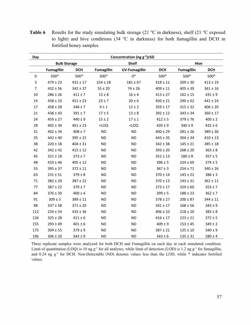
57
Table 6 Results for the study simulating bulk storage (21 oC in darkness), shelf (21 oC exposed
to light) and hive conditions (34 oC in darkness) for both fumagillin and DCH in
fortified honey samples.
Day Concentration (ng g-1)±SD
Bulk Storage Shelf Hive
Fumagillin DCH Fumagillin UV-Fumagillin DCH Fumagillin DCH
0 500* 500* 500* 0* 500* 500* 500*
3 479 ± 23 431 ± 17 154 ± 18 181 ± 67 418 ± 11 269 ± 30 413 ± 19
7 432 ± 56 342 ± 37 55 ± 20 74 ± 26 409 ± 11 405 ± 39 361 ± 16
10 286 ± 26 411 ± 7 13 ± 8 16 ± 4 413 ± 27 162 ± 15 431 ± 9
14 458 ± 10 451 ± 23 23 ± 7 20 ± 6 450 ± 21 290 ± 62 441 ± 24
17 458 ± 28 348 ± 7 9 ± 1 12 ± 2 359 ± 17 315 ± 32 406 ± 20
21 438 ± 43 391 ± 7 17 ± 5 13 ± 8 392 ± 12 343 ± 34 360 ± 17
24 459 ± 27 440 ± 9 15 ± 2 17 ± 1 412 ± 5 379 ± 76 400 ± 2
28 402 ± 34 461 ± 23 <LOQ <LOQ 420 ± 9 340 ± 9 432 ± 6
31 402 ± 34 408 ± 7 ND ND 400 ± 29 281 ± 26 389 ± 36
35 442 ± 40 395 ± 22 ND ND 443 ± 35 304 ± 34 410 ± 13
38 220 ± 18 404 ± 31 ND ND 342 ± 38 145 ± 21 385 ± 18
42 342 ± 42 415 ± 12 ND ND 393 ± 20 268 ± 20 363 ± 8
45 321 ± 18 373 ± 7 ND ND 352 ± 13 180 ± 8 357 ± 5
48 410 ± 46 405 ± 12 ND ND 396 ± 5 234 ± 69 374 ± 5
55 395 ± 37 372 ± 11 ND ND 367 ± 3 254 ± 71 340 ± 26
63 231 ± 51 379 ± 8 ND ND 370 ± 14 143 ± 22 386 ± 3
71 382 ± 20 387 ± 22 ND ND 370 ± 13 143 ± 31 362 ± 11
77 387 ± 22 379 ± 7 ND ND 373 ± 17 329 ± 60 353 ± 7
84 376 ± 30 400 ± 4 ND ND 399 ± 5 188 ± 23 362 ± 7
91 309 ± 5 389 ± 11 ND ND 378 ± 27 200 ± 87 344 ± 11
98 337 ± 58 371 ± 20 ND ND 341 ± 17 168 ± 56 343 ± 9
112 234 ± 54 435 ± 36 ND ND 406 ± 10 218 ± 20 383 ± 8
126 325 ± 28 411 ± 6 ND ND 416 ± 17 223 ± 21 372 ± 5
155 293 ± 49 401 ± 6 ND ND 409 ± 9 153 ± 45 349 ± 2
175 304 ± 55 379 ± 9 ND ND 387 ± 21 135 ± 10 340 ± 9
196 306 ± 20 343 ± 9 ND ND 343 ± 6 135 ± 31 280 ± 4
Three replicate samples were analyzed for both DCH and Fumagillin on each day at each simulated condition.
Limit of quantitation (LOQ) is 10 ng g-1 for all analytes, while limit of detection (LOD) is 1.2 ng g-1 for fumagillin,
and 0.24 ng g-1 for DCH. Non-Detectable (ND) denotes values less than the LOD, while * indicates fortified
values.

58
Figure 12 Graphical representation of data recorded for fumagillin and DCH under simulated
shelf conditions (21 oC exposed to light) indicating that no fumagillin or UV
decomposed fumagillin is present after 30 days.

59
Figure 13 Graphical representation of data recorded for fumagillin and DCH under simulated bulk
storage conditions (21 oC in darkness).

60
Figure 14 Graphical representation of data recorded for fumagillin and DCH under simulated hive
conditions (34oC in darkness)

61
Table 7 Results for the linear regression analysis of fumagillin and DCH for data recorded
under simulated shelf (21 oC exposed to light), bulk storage (21 oC in darkness) and
hive (34 oC in darkness) conditions.
Compound Test Condition Linear Regression
Equation r2
Calculated half-life
(days)*
DCH 21oC Light y = - 0.25 x + 409.6 0.071 829
21oC Dark y = -0.24 x + 415.9 0.072 852
34oC Dark y = - 0.56 x + 413.8 0.376 368
Fumagillin 21oC Light estimated
3
21oC Dark y = - 0.85 x + 420.2 0.358 246
34oC Dark y = -1.12 x + 317.1 0.303 141
UV-Fumagillin 21oC Light estimated
3
*The calculated half-life for each compound under each of these conditions is shown. The half-life of fumagillin and its
UV decomposition products under shelf conditions is estimated to be approximately 3 days (Figure 12), while no
fumagillin, or its metabolically active UV decomposition products, could be observed after one month at shelf conditions
4.4 Discussion
Previous research by Higes et al. (2011) reported 30% decomposition of fumagillin in 50% sugar
syrup solution after 70 days in clear vials at 4 oC, and 12% decomposition at 4 oC after 70 days
in amber vials. Increased temperatures of 22 and 30 oC for the 50% sugar syrup solution caused
60% and 65% decomposition respectively after 70 days, with total decomposition of fumagillin
being observed after 20 days at 40 oC. When exposed to UV light, 80% decomposition was
observed within 24 hours, and no detectable residues of fumagillin could be observed after 10
days (Higes et al. 2011). Higes et al. (2011) also examined the stability of fumagillin in 50%
honey and sugar patties, and found that fumagillin was observed to be more stable in this matrix
than in 50% sugar syrup solutions, with only 10% decomposition of fumagillin being observed
after 4 weeks at 4, 22, 30 and 40 oC respectively when stored in amber vials. In our study 20%
decomposition of fumagillin was observed in honey stored at 21 oC and 32% decomposition in
honey stored at 34 oC, both in darkness. In field trials, Higes et al., observed levels of fumagillin
ranging from less than the limit of detection (1 ng g-1) to 66 ng g-1 in honey samples.
Nevertheless, the field trial data is incomplete as no surplus honey was available to be harvested
for some of the colonies.

62
Nozal et al. (2008) found that fumagillin was stable for one month at mild temperature
conditions (40oC) in either sugar syrup or water when not exposed to light. The same study also
confirmed the instability of fumagillin upon exposure to light. In field studies, no fumagillin
could be observed at concentrations above their LOD (ranging from 1-24 ng g-1 for clear
rosemary honey and 4-45 ng g-1 for darker heather honey respectively) for hives treated with
either 120 mg or 240 mg (twice the recommended dosage in Spain). The fumagillin treatment
was applied for six weekly periods in a 50% honey and water mixture (250 mL) in the late fall to
early winter (December to January), and surplus honey was sampled 6 months later, in June.
The instability of fumagillin upon exposure to light observed during this study is similar to that
reported previously using fluorescent and UV light sources (Kochansky and Nasr 2004; Higes et
al. 2011), even though the stability of fumagillin was examined in ethanol or sugar syrup
solutions in those studies, while the stability of fumagillin was evaluated in a honey matrix in our
study. Fumagillin was however observed to be more stable in honey when it is not exposed to
light at elevated temperatures (34 oC), simulating hive conditions. This indicates that residual
fumagillin in the stored honey of overwintered hives should remain active against Nosema
disease. Because of the instability of fumagillin, greater variability in the measured
concentrations of residual fumagillin was observed compared to that seen for residual DCH.
In addition to DCH residues originating from fumagillin treatment, the possible environmental
origins of DCH related to industrial activity (rubber, steel corrosion inhibitors, vehicle antifreeze
formulations) need to be examined in order to establish if this is of any concern for human and
bee health, especially for apiaries located close to major roadways and industrial centers
(Woldegiorgis et al. 2007). The effect of DCH on bees at various stages of development also
needs to be evaluated, since the lipophilic nature and stability of DCH could lead to
accumulation in the comb wax, where the developing larvae may be more susceptible to the
effects of DCH.
An alternative less harmful formulation of fumagillin that does not contain DCH as the counter
ion would be desirable. A potential candidate as an alternative counter ion of formulated
fumagillin could be for example a compound such as meglumine (CAS Registry No. 6284-40-8)
shown in Figure 10, which is a basic amino sugar derived from sorbitol, and which is acceptable

63
for use in human medicine as a pharmaceutical excipient to obtain crystalline precipitates from
organic acids (Blood et al. 2007). An example of this is the meglumine salt of the organic acid,
flunixen (Sigma-Aldrich product F0429, CAS Registry No.42461-84-7). The reported MSDS
LD50 value for the meglumine is 5000 mg kg-1 orally for both rat and rabbit (Sigma-Aldrich
product M9179 v3.5 07/02/2014), as compared to the reported oral LD50 for rat of 373 mg kg-1
for DCH (Sigma-Aldrich product 185841 v5.0 07/24/2012). Similar values of 200-373 mg kg-1
was also reported elsewhere for DCH (Greim et al. 1998). The use of meglumine instead of DCH
as the counterion of fumagillin formulation could therefore reduce the toxicity of the formulated
fumagillin significantly. The presence of multiple hydroxyl groups on meglumine might also
increase the water solubility of the formulated fumagillin. The suitability of the suggested
meglumine or other alternative counter ions to replace DCH in the formulated fumagillin needs
to be investigated further.
4.5 Conclusions
The previously reported accelerated decomposition of fumagillin at higher temperatures and
upon exposure to light was confirmed by the results obtained for our study. Our observations
also support the conclusions reached in other studies, namely that it is unlikely that fumagillin or
its UV degradation products will be detected in any appreciable amounts in honey destined for
human consumption, when the commercial formulation is used as prescribed by the label
instructions (Kochansky and Nasr 2004; Lopez et al. 2008; Nozal et al. 2008; Higes et al. 2011;
Kanda et al. 2011; Dmitrovic and Durden 2013). It should however be noted that the presence of
DCH, resulting from the application of the commercial formulations of fumagillin (Fumidil-B®
and Fumagillin-B®), is not recognized by any of these studies.
The long half-lives observed for DCH in this stability study (ranging from 1 to almost 3 years)
indicate that DCH will likely be detectable in honey at higher concentrations than fumagillin for
extended periods of time. It is important to remember that DCH and fumagillin are applied in
theoretically equimolar amounts when using the current commercial formulations. The expected
higher prevalence of DCH in honey, compared to that of fumagillin, was demonstrated in our
recent study, where incurred DCH was detected at levels above the analytical method LOD of 10
ng g-1 for all but one of 16 random honey samples analyzed, with DCH levels ranging from

64
20.0 - 234.6 ng g-1 (van den Heever et al. 2015a). In contrast, the same study found fumagillin
above the LOD of 10 ng g-1, in only two of the samples, at values of 11.9 and 11.6 ng g-1
respectively. The extended half-life of DCH, in combination with its reported toxicity and
genotoxic properties, clearly makes it a contaminant of concern in honey (Greim et al. 1998).
DCH is clearly a much better marker residue than fumagillin for monitoring fumagillin usage in
apiculture, provided that other industrial exposures to DCH are not occurring. A thorough risk
assessment needs to be conducted in order to establish a maximum residue limit (MRL) for both
fumagillin, as well as for DCH in honey.

65
Chapter 5 Evaluation of Fumagilin-B® and other potential alternative
chemotherapies against Nosema ceranae-infected honey bees (Apis
mellifera) in cage trial assays
5.1 Introduction
Nosema disease of honey bees, Apis mellifera L., is caused by infection from two distinct species
of single-cellular microsporidian fungal parasites (Genersch 2010), Nosema apis Zander and
Nosema ceranae Fries et al., respectively. Infections of N. ceranae have been shown to cause
high levels of colony loss in some regions, while both species have been implicated as part of the
pathogen complex associated with colony collapse disorder (CCD) (Cox-Foster et al. 2007;
Martín-Hernández et al. 2007; Higes et al. 2008, 2009c; vanEngelsdorp et al. 2009).
Fumagilin-B® is the only registered chemotherapy currently available to treat Nosema disease in
apiculture in North America (Williams et al. 2008a, 2011), and is reportedly allowed for use in
special circumstances in Spain, the Balkan countries, and other parts of Europe (Higes et al.
2011; Stevanovic et al. 2013). Shortly after its discovery (Hanson and Eble 1949; Eble and
Hanson 1951), fumagillin (Figure 15) was found to be effective in controlling N. apis in honey
bees (Katznelson and Jamieson 1952; Bailey 1953). Since that time, the commercial
formulations of fumagillin, namely Fumagilin-B® and Fumidil-B®, have been continuously
employed to treat either N. apis or N. ceranae in North America. It should be noted that the
commercial formulations of fumagillin consist of the dicyclohexylamine (DCH) salt of
fumagillin, with fumagillin and DCH being present in a 1:1 stoichiometric ratio (Figure 15). For
a more comprehensive description of fumagillin and its significance for apiculture, the reader is
referred to Chapter 2 of this thesis (van den Heever et al. 2014).
In human cancer research, fumagillin and its analogues were found to inhibit the formation of
new blood vessels around tumors (angiogenesis), thereby limiting the blood supply to tumors and
thus impeding their growth (Ingber et al. 1990). The mode of action responsible for these anti-
angiogenic properties was elucidated with the aid of a crystal structure showing fumagillin
covalently bound to a histidine moiety (His231) within the enzymatic active site of the methionine
aminopeptidase type 2 (MetAP-2) enzyme (Griffith et al. 1997; Sin et al. 1997; Liu et al. 1998).
This covalent binding of fumagillin to the MetAP-2 enzyme results in the irreversible opening of
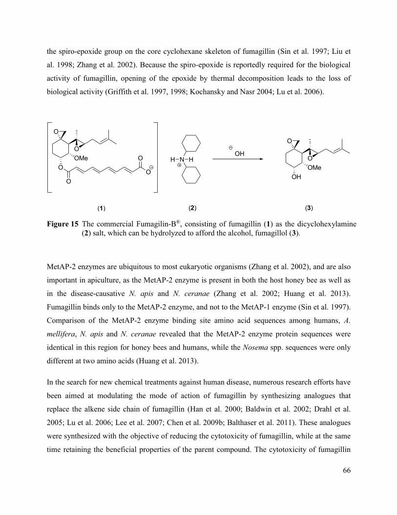
66
the spiro-epoxide group on the core cyclohexane skeleton of fumagillin (Sin et al. 1997; Liu et
al. 1998; Zhang et al. 2002). Because the spiro-epoxide is reportedly required for the biological
activity of fumagillin, opening of the epoxide by thermal decomposition leads to the loss of
biological activity (Griffith et al. 1997, 1998; Kochansky and Nasr 2004; Lu et al. 2006).
O
O
OMe
O
O
O
O
O
O
OMe
OH
(1) (3)
OHN HH
(2)
Figure 15 The commercial Fumagilin-B®, consisting of fumagillin (1) as the dicyclohexylamine
(2) salt, which can be hydrolyzed to afford the alcohol, fumagillol (3).
MetAP-2 enzymes are ubiquitous to most eukaryotic organisms (Zhang et al. 2002), and are also
important in apiculture, as the MetAP-2 enzyme is present in both the host honey bee as well as
in the disease-causative N. apis and N. ceranae (Zhang et al. 2002; Huang et al. 2013).
Fumagillin binds only to the MetAP-2 enzyme, and not to the MetAP-1 enzyme (Sin et al. 1997).
Comparison of the MetAP-2 enzyme binding site amino acid sequences among humans, A.
mellifera, N. apis and N. ceranae revealed that the MetAP-2 enzyme protein sequences were
identical in this region for honey bees and humans, while the Nosema spp. sequences were only
different at two amino acids (Huang et al. 2013).
In the search for new chemical treatments against human disease, numerous research efforts have
been aimed at modulating the mode of action of fumagillin by synthesizing analogues that
replace the alkene side chain of fumagillin (Han et al. 2000; Baldwin et al. 2002; Drahl et al.
2005; Lu et al. 2006; Lee et al. 2007; Chen et al. 2009b; Balthaser et al. 2011). These analogues
were synthesized with the objective of reducing the cytotoxicity of fumagillin, while at the same
time retaining the beneficial properties of the parent compound. The cytotoxicity of fumagillin

67
makes it unsuitable for most human applications. Fumagillin can be hydrolyzed with a suitable
base to remove the alkene side chain (Figure 15), yielding the alcohol, fumagillol (Tarbell et al.
1961; Gochnauer and Furgala 1962; Assil and Sporns 1991). A new chemical moiety, such as a
carboxylic acid, can then be coupled to fumagillol, resulting in a new semisynthetic fumagillin
analogue (Han et al. 2000; Baldwin et al. 2002; Lee et al. 2007). An example of such a
semisynthetic fumagillin analogue is TNP-470, which is a potential new treatment for malaria
(Zhang et al. 2002; Chen et al. 2009b).
I postulated that the known mode of action of fumagillin against the MetAP-2 enzyme in humans
is similar to MetAP-2 enzymes found in the host honey bee as well as in N. apis and N. ceranae.
As such, the core cyclohexane skeleton of fumagillin with the intact spiro-epoxide was used as a
template for the design and synthesis of two semisynthetic analogues of fumagillin, as well as for
the synthesis of several purely synthetic compounds aimed at targeting the MetAP-2 enzyme in
N. ceranae. These, and several other commercially-available compounds that reportedly
exhibited activity against Nosema disease, were evaluated for disease suppression and adult bee
survival using cage trial assays. The effects of DCH, the counter ion of fumagillin in Fumagilin-
B®, were also evaluated.
5.2 Materials and Methods
5.2.1 Reagents and materials
Pure fumagillin isolated from Aspergillus fumigatus Fres. containing no DCH (Cat. # F6771),
dicyclohexylamine nitrite (Cat. # 317837), trimethylsulfoxonium iodide (Cat. # T80500), N,N'-
dicyclohexylcarbodiimide (Cat. # D80002), 4-(dimethylamino) pyridine (Cat. # 107700), acetyl
salicylic acid (Cat. # A5376), piperonylic acid (Cat. # P49805), thymol (Cat. # T0501),
carbendazim (Cat. # 378674), toltrazuril (Cat. # 34000), thiabendazole (Cat. # T8904) and
enilconazole (also known as imazalil or chloramizole, Cat. # Y0000136) were all purchased from
Sigma-Aldrich (St. Louis, MO, USA). Purified fumagillol was produced in-house by hydrolysis
of a quantity of the commercial fumagillin formulation, Fumagilin-B®(Medivet Pharmaceuticals
Ltd., High River, Alberta, Canada; DIN 02231180), followed by purification using column
chromatography (Assil and Sporns 1991). The commercial formulation of fumagillin,

68
Fumagilin-B® (fumagillin as the DCH salt), was used as a positive control in the cage trial
assays. The Dess-Martin reagent used for oxidation reactions was prepared in-house according to
literature procedures (Dess and Martin 1991; Frigerio et al. 1999). Nozevit solution, a natural
plant polyphenol extract, was obtained from Complete Bee (AK, USA).
5.2.2 General synthetic methodology O
O
OMe
O
OO
O
OH
OO
O
+
O
O
OMe
OH
O
O
OMe
OH
+
O
OHO
O O
O
OMe
O
O
O
O
(3) (4) (5)
(3) (6) (7)
DCC
DMAP
DCC
DMAP
Figure 16 Coupling of fumagillol (3) with aspirin (4) or piperonylic acid (6) respectively, under
Steglich reaction conditions using N,N'-dicyclohexylcarbodiimide (DCC) and
dimethylaminopyridine (DMAP), to afford the aspirin analogue (5) and piperonylic
acid analogue (7) of fumagillin.
Fumagillol was prepared by basic hydrolysis according to established methodology (Assil and
Sporns 1991) from the commercial formulation Fumagilin-B® (Figure 15). Oxidation reactions
to transform alcohol starting materials to the corresponding ketones were done using the Dess-
Martin reaction (Dess and Martin 1991; Frigerio et al. 1999). The reactions used to prepare the
semisynthetic and synthetic compounds generally involved the esterification of alcohols with
carboxylic acids (Figure 16), and the epoxidation of ketones (Figure 17). General procedures
for performing these reactions are given in following sections. During the chemical synthesis no

69
attempt was made to synthesize single stereoisomers of compounds, hence presumably racemic
mixtures of analogues were prepared for initial screening. The identity of all the synthesized
compounds was verified by high resolution mass spectrometry on an Agilent 6224 time of flight
(TOF) mass spectrometer, with a summary of the results given in Table 8.
O O
O
O O O
O
OO
O
OH
O
(11)(9) (10)
(12)
(8)
O
O
OO
O
(13) (14)
O
Cyclohexanone
Corey-Chaykovsky
Epoxidation
Figure 17 Purely synthetic compounds prepared by using the Corey-Chaykovsky epoxidation
reaction to afford the cyclohexanone derivative (8), menthol derivative (9) as well as
other synthetic analogues (10, 11, 12, 13, 14).

70
Table 8 Confirmation of the identity of semisynthetic and synthetic compounds that were tested
against N. ceranae by high resolution mass spectrometric analysis.
Compound Found
Formula
Ion Mass
Found
Mass
Calculated
Δ ppm
3 C16H24O3Na [M+Na (-H2O)]+ 287.1631 287.1618 4.65
5 C25H32O7Na [M+Na]+ 467.2033 467.2040 -1.49
7 C24H28O6K [M+K (-H2O)]+ 451.1512 451.1517 -1.23
8 C7H12ONa [M+Na]+ 135.0776 135.0780 -3.49
9 C11H21O [M+H]+ 169.1592 169.1587 2.72
10 C9H13O2 [M+H (-H2O)]+ 153.0913 153.0910 1.96
11 C10H20NO [M+NH4]+ 170.1531 170.1539 -4.72
12 C9H15O3 [M+H]+ 171.1024 171.1016 4.96
13 C11H20O2Na [M+Na]+ 207.1351 207.1356 -2.02
14 C25H33O7 [M+H]+ 445.2234 445.2221 3.00
5.2.3 Ester synthesis
The Steglich esterification (Figure 16) was used to prepare esters from the corresponding
carboxylic acids and alcohols (Neises and Steglich 1978). The general procedure involved
stirring a solution of the carboxylic acid (10 mmol) in anhydrous CH2Cl2 (10 mL). The catalyst,
dimethylaminopyridine (DMAP) was added (5 mmol) followed by drop-wise addition of the
alcohol (11 mmol, 1.1 equivalent). The mixture was then cooled to 0 oC, and then
dicyclohexylcarbodiimide (DCC; 11 mmol, 1.1 equivalent) was slowly added. The mixture was
stirred for 5 min at 0°C, and then for 3 h at room temperature. The precipitated dicyclohexyl urea
(DCU) by-product was then filtered off, and the filtrate washed successively with 0.5 N HCl (2 x
10 mL) and saturated NaHCO3 solution (1 x20 mL). The CH2Cl2 fraction was dried (MgSO4)
and concentrated in vacuo. The residue was then purified by flash chromatography on silica
(230-400 mesh) with a suitable eluent, usually consisting of a mixture of n-hexane and ethyl
acetate.
5.2.4 Epoxide formation from ketones
A modified version of the Corey-Chaykovsky reaction (Corey and Chaykovsky 1965) was used
to prepare epoxides from ketone compounds (Ng 1990). The procedure involved dissolving
trimethylsulfoxonium iodide (10 mmol) into dimethylsulfoxide (15 mL) under a nitrogen

71
atmosphere at room temperature. After stirring for 5 min the ketone (10 mmol) was added,
followed by addition of potassium tert-butoxide (10 mmol), dissolved in dimethylsulfoxide
(10 mL). Stirring was continued overnight at room temperature under nitrogen, and the mixture
was then diluted with water (30 mL) and extracted with CH2Cl2 (3 x 30 mL) and concentrated in
vacuo. The crude epoxide product was then purified by flash chromatography on silica (230-400
mesh) with a suitable eluent system, usually consisting of a mixture of n-hexane and ethyl
acetate. An example of this procedure is shown for the conversion of cyclohexanone to the
corresponding epoxide (Figure 17).
5.2.5 Cage assays – general procedures
Trials were conducted over a four-year period, from 2010 to 2013. To obtain adult honey bees
(A. mellifera L.) for the assays, frames of sealed brood with newly-eclosing bees were obtained
from several colonies at Agriculture and Agri-Food Canada’s Research Farm, in Beaverlodge,
Alberta, Canada (55° 18' N; 119° 17' W). These colonies were repeatedly tested to be free from
both infections of N. apis and N. ceranae, using both light microscopic and molecular methods,
described below. Frames were kept overnight in an incubator (Percival Model 136NLC9,
Percival Scientific Inc., Perry, IA, USA) maintained at hive temperature (33 ± 0.5oC) and a
relative humidity of 70 ± 5%. Adult workers were pooled and mixed from all frames with 100
bees added to each wooden screened cage (8.0 x 9.5 x 12.0 cm I.D.). Bees were then fed 4 mL of
a 60% (w/v) of aqueous sucrose syrup for 24 h, using gravity feeders fashioned from disposable
centrifuge tubes (Cat. #93000-020, VWR International, Radnor, PA, USA).
After the initial 24 h period, each cage was mass inoculated with 5 mL of 60% syrup solution
containing 1×10⁷ freshly-harvested N. ceranae spores, prepared from previously-identified
colonies of honey bees with high levels of N. ceranae infection. Workers from these colonies
were euthanized on dry ice, and then their abdomens were removed and suspended in ultrapure
water (1 mL per bee). After maceration, the crude suspension was filtered through a sieve (~0.8
mm) to remove large body parts, and spores were then counted (described below) to prepare
solutions with the correct inoculation doses. After consumption of the inoculum for 48 h, cages
of bees were fed the test compounds in 60% sucrose syrup, ad libitum for 17 days at prescribed
dosages.
72
Five or six replicate cages of bees were evaluated for each concentration of each compound
tested. The efficacy of test compounds for suppressing N. ceranae spore development was
assessed by determining the average number of spores per bee at 17 days post-inoculation.
Cumulative bee mortality per cage over the duration of the trials was also recorded. For the trials
in which the toxicity of DCH was being assessed (Section 5.2.9), the mortality of bees was
recorded each day, up to 17 days post-inoculation.
5.2.6 Determination of spore levels
To determine mean spore numbers per bee from colonies, 60 adult workers were collected from
peripheral frames of the brood nest; these were used to confirm a source of inoculum for
infections, or a source of Nosema-free bees for cage trials. When conducting the cage trials, 30
surviving workers were analyzed, collected from each cage on day 17 post-inoculation.
Worker abdomens were removed and placed in a stomacher bag containing 70% ethanol (1 mL
per bee), which was then macerated for 1 min at medium speed (Seward Stomacher® 80
Biomaster, Seward Laboratory Systems Inc., Davie, FL, USA). To determine Nosema spore
counts per bee, 6 µL of the macerate was withdrawn using a micropipette, loaded onto a Helber
Z30000 counting chamber (Hawksley, Lancing, UK) and counted according to the generalized
methods of Cantwell (1970), under phase contrast microscopy at 400× magnification. Samples of
the remaining crude macerate were portioned into 1.5 mL microcentrifuge tubes and stored at
-20°C until required for Nosema spp. identification.
5.2.7 Nosema spp. Identification
Identification and confirmation of N. ceranae as the only microsporidian pathogen infecting the
caged bees was conducted, in order to provide evidence that no N. apis infection, or co-infection
of N. ceranae and N. apis had occurred in the caged bees used for testing. The crude frozen
macerate, described in section 5.2.6, was thawed, vortexed, and then 200-400 µL was
centrifuged to remove the ethanol from the sample. DNA extraction was performed using the
DNeasy® Blood & Tissue Kit (Qiagen®, Valencia, CA, USA). The concentration of the extracted
DNA was determined spectrophotometrically (NanoDrop 2000C, Thermo Scientific, West Palm

73
Beach, FL, USA), whereafter 50-100 ng of this DNA extract was amplified using polymerase
chain reactions (PCR).
A multiplex system that co-amplified the 16S rRNA gene of N. apis and N. ceranae (Martín-
Hernández et al. 2007) as well as the honey bee ribosomal protein RpS5 gene (Thompson et al.
2007b) was used within the same reaction. A modified version of the PCR protocol was used,
owing to the fact that early pre-tests indicated that these modifications increased the sensitivity
of simultaneous detection of both N. apis and N. ceranae within any given sample. All PCR
reactions were performed using a Mastercycler® proS thermocycler (Eppendorf, Mississauga,
Canada) and utilizing the Illustra™ PuReTaq Ready-To-Go™ PCR beads (GE Healthcare Life
Sciences, Baie d’Urfe, Quebec, Canada). PCR beads were reconstituted to a 25 µL final volume
by adding sterile H2O, 0.5 µL of 20 mM forward and reverse primers (a final concentration of
0.4 mM) and the DNA (50-100 ng per reaction). To amplify a 218 bp 16S rRNA PCR product
specific for N. ceranae, primers Mitoc-For (5'-CGGCGACGATGTGATATGAAAATATTAA-
3') and Mitoc-Rev (5'-CCCGGTCATTCTCAAACAAAAAACCG-3') were used and to amplify
a 321 bp 16S rRNA PCR product specific for N. apis primers Apis-For (5'-
GGGGGCATGTCTTTGACGTACTATGTA-3') and Apis-Rev (5'-
GGGGGGCGTTTAAAATGTGAAACAACTATG-3) were used, according to Martín-
Hernández et al. (2007). In addition, the honey bee housekeeping gene, RpS5, was also amplified
within the same reaction as a reference, which yielded a PCR product of 115 bp. The primer
pairs used were RpS5-For (5'-AATTATTTGGTCGCTGGAATTG-3'), and RpS5-Rev (5'-
TAACGTCCAGCAGAATGTGGTA-3') respectively (Thompson et al. 2007). The thermocycler
program consisted of an initial DNA denaturation step at 95°C for 5min, followed by 35 cycles
of 94°C for 30sec, 61.8°C for 30sec, and 72°C for 30sec. A final extension of 72°C for 7min was
followed by holding the reactions at 4°C until stopped. All PCR products were visualized on a
2% agarose gel and stained with SYBR® Safe DNA gel stain (Life Technologies, Carlsbad, CA,
USA).

74
5.2.8 Testing of semisynthetic, synthetic and commercially available
compounds
In the 2010 trials, Fumagilin-B® was used to prepare three solutions at concentrations of 4, 40,
and 400 µM fumagillin (and DCH) in 60% sucrose solution. This concentration range was
chosen to encompass the manufacturer’s recommended dose concentration of 41 µM fumagillin
in 60% sucrose solution (105 mg fumagillin DCH salt, corresponding to 5 g of the formulation,
dissolved into 4 L of syrup). This was achieved by first dissolving the appropriate amount of
Fumagilin-B® formulation into 1.0 mL ethanol, before adding it to the 60% sucrose solution, in
order to better solubilize the Fumagilin-B® powder. All test compounds were similarly prepared,
at the same three concentrations of 4, 40 and 400 μM. A negative control consisting of only 60%
sucrose, with 1 mL of added ethanol, was also prepared. The semisynthetic aspirin and piperonyl
analogues of fumagillin (Figure 16, compounds 5 and 7), as well as fumagillol (Figure 15,
compound 3) and the commercially available carbendazim, thiabendazole, thymol (Figure 18,
compounds 15, 17, and 18) were tested. Nozevit, sold as a bee supplement consisting of a
mixture of unspecified polyphenols and other compounds, was prepared as per the
manufacturer’s instructions and tested as well. Several synthetic compounds (Figure 17,
compounds 8, 9, 10 and 11) were also evaluated.
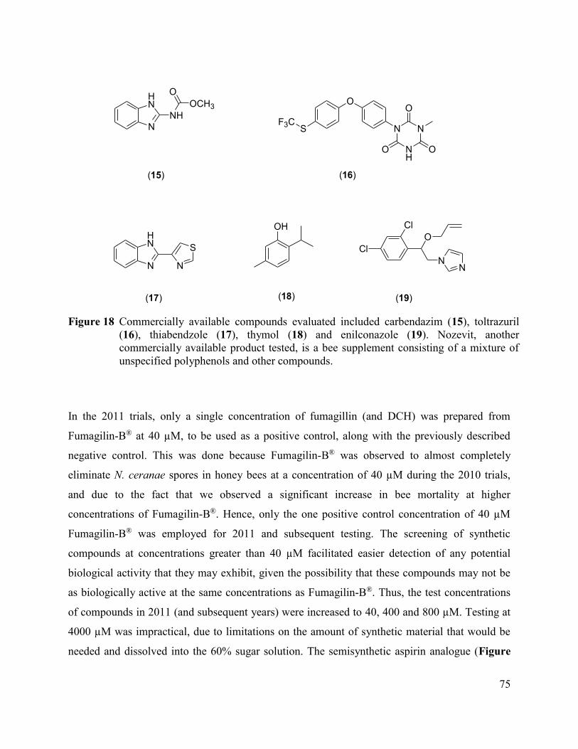
75
N
HN
N
HN
NH
OCH3
O
N
S
OH
O
SF3C
N
NH
N
O
O O
(15) (16)
(17) (18)
Cl
Cl
O
NN
(19)
Figure 18 Commercially available compounds evaluated included carbendazim (15), toltrazuril
(16), thiabendzole (17), thymol (18) and enilconazole (19). Nozevit, another
commercially available product tested, is a bee supplement consisting of a mixture of
unspecified polyphenols and other compounds.
In the 2011 trials, only a single concentration of fumagillin (and DCH) was prepared from
Fumagilin-B® at 40 µM, to be used as a positive control, along with the previously described
negative control. This was done because Fumagilin-B® was observed to almost completely
eliminate N. ceranae spores in honey bees at a concentration of 40 µM during the 2010 trials,
and due to the fact that we observed a significant increase in bee mortality at higher
concentrations of Fumagilin-B®. Hence, only the one positive control concentration of 40 µM
Fumagilin-B® was employed for 2011 and subsequent testing. The screening of synthetic
compounds at concentrations greater than 40 µM facilitated easier detection of any potential
biological activity that they may exhibit, given the possibility that these compounds may not be
as biologically active at the same concentrations as Fumagilin-B®. Thus, the test concentrations
of compounds in 2011 (and subsequent years) were increased to 40, 400 and 800 µM. Testing at
4000 µM was impractical, due to limitations on the amount of synthetic material that would be
needed and dissolved into the 60% sugar solution. The semisynthetic aspirin analogue (Figure

76
16, compound 5), as well as the commercially available carbendazim and toltrazuril (Figure 18,
compound 15 and 16) were tested during the cage trial assays conducted during 2011.
In 2012, testing was conducted using the same 40, 400 and 800 µM concentration regimen as
described above for the 2011 tests. Three synthetic compounds (Figure 17, compounds 12, 13
and 14), as well as the commercially available aspirin (Figure 16 compound 4) and enilconazole
(Figure 18, compound 19), were tested during this year.
5.2.9 Testing of DCH in the commercial Fumagilin-B®
Our observation that Fumagilin-B® appears to lead to an increase in bee mortality at higher
concentrations, along with the fact that the commercial formulations of fumagillin (Fumidil-B®,
Fumagilin-B®) contain the reportedly genotoxic and tumorigenic DCH (Greim et al. 1998),
caused us to suspect that DCH might be a contributing factor to this higher observed bee
mortality (Table 11). This led to an experiment conducted in 2013 that was designed to evaluate
bee mortality associated with the use of purified fumagillin (no DCH), Fumagilin-B® (fumagillin
as the DCH salt) and DCH by itself. Test solutions for each of these compounds or groups of
compounds were prepared at a single concentration of 40 µM in 60% sucrose solutions, along
with a 60% sucrose negative control, as previously described.
5.2.10 Statistical Analysis
For the cage trials conducted to evaluate alternative chemotherapies, the effectiveness of
compounds was determined by using regression models of spore counts that included compound
dose as the explanatory variable. Indicators for each dose were included, compared to 0 µM
negative controls as the referent, because the relationship between dose and spore count was not
linear. Separate negative binomial (NB) or zero-inflated negative binomial (ZINB) regression
models were run for each compound. Models were selected using the following criteria (Dohoo
et al. 2009): 1) Linear regression models for spore count violated residual assumptions and were
not used, despite a log transformation of spore count. 2) NB models were selected as the mean
number of spore counts were not equal to the variance of Poisson models for count data (the
over-dispersion parameter of NB models was significantly different than 0). 3) ZINB models
were selected when the Vuong test was significant (P≤0.05), indicating that there may be a

77
separate process involved for those bees with zero spore counts. 4) For the inflated (logistic)
portion of ZINB models, Akaike’s and Bayesian Information Criteria (AIC and BIC) were used
to determine the best fit for compound dose (dose indicators compared to treated versus non-
treated, lower AIC and BIC representing a better fitting model).
Likelihood ratio tests (LRT) were used to determine if groups of concentration indicators were
significantly associated with changes in spore count (P≤0.05). If the LRT was significant, Wald
tests were used to compare each individual dose to the referent concentration (0 μM). The fit of
NB models was assessed using deviance chi-squared tests (P≤0.05 indicating the model did not
fit the data) and residual analysis. The fit of ZINB models was assessed by residual analysis.
Differences in mortality for compounds significantly related to spore count were evaluated
descriptively by comparing proportions of mortality for bees in all replicates of a given dose
compared to controls. Probit analysis to determine median lethal concentrations (LC50) for each
compound was attempted, using Abbott’s formula for correction for control mortality.
For the cage trial assays used to assess the influence of DCH on the mortality of N. ceranae-
infected bees, data was analyzed using semi-parametric, Cox proportional hazards models with a
complimentary log-log link for survival data (Corrente et al. 2003; Dohoo et al. 2009). The bee
survival for indicator variables for each treatment group was considered against negative controls
using LRT (P≤0.05). To test for proportional hazards across treatment groups, an interaction
between treatment group and survival time was considered. A LRT P≤0.05 indicated non-
proportional hazards, which was solved by including the interaction in the model. Linear
regression was used to assess the effects of these treatment methods on the natural log of spore
counts to normalize the data (zero spore counts were changed to one to allow for the log value to
be zero). The effect of an indicator variable treatment group was tested using the extra sum of
squares F-test (Dohoo et al. 2009). The model was assessed for fit by evaluating the normality
and homoscedasticity of the standardized residuals.
All analysis was conducted in Excel 2010 (Microsoft Corporation, Redmond, WA, USA) and
STATA Intercooled 13.1 (StataCorp LP, College Station, TX, USA).
78
5.3 Results
5.3.1 Potential alternative chemotherapies against N. ceranae
Molecular identification of N. ceranae, using the described PCR techniques confirmed that bees
were only infected with N. ceranae. The results of the NB and ZINB models are shown in Table
9 and Table 10, respectively. Fumagilin-B®, as well as fumagillol (Figure 15, compound 3),
exhibited statistically significant biological activity against N. ceranae under our test conditions
(Table 10). Both semisynthetic analogues of fumagillin, namely the aspirin analogue (Figure 16,
compound 5) and the piperonyl analogue (Figure 16, compound 7) showed statistically
significant biological activity against N. ceranae-infected bees under the test conditions
described.
From Table 9 it can be seen that the purely synthetic compounds (Figure 17, compounds 8, 10,
11 and 14) also exhibited statistically significant biological activity against N. ceranae under the
described test conditions. The commercially available thymol and enilconazole (Figure 18,
compound 18 and 19) were also active against N. ceranae (Table 10). Model assessment for the
NB models indicated that they were a good fit (Deviance chi-squared P>0.05 and normal
residuals), with the exception of one NB model for carbendazim, where the P=0.05; however,
carbendazim was not significantly associated with reduced spore counts. Model assessment for
the ZINB models indicated that they were a good fit using the tools available (Vuong P≤0.05).
Detected outliers were evaluated and kept in all models.
Cumulative bee mortalities by concentrations for those compounds shown to significantly impact
spore count are shown in Table 11. It is interesting to note the higher bee mortality associated
with Fumagilin-B®, when compared to the negative control. The mortality associated with
Fumagilin-B® also increases with an increase in concentration. Further, initial attempts to
conduct probit analysis to determine LC50 values determined that models did not fit the data
using Pearson Chi-squared tests (data not shown). For most compounds, testing was only
conducted at three concentrations, making it difficult to determine an LC50 value, where it is
generally accepted that having two to three concentrations above as well as below the LC50 is
best for this calculation.
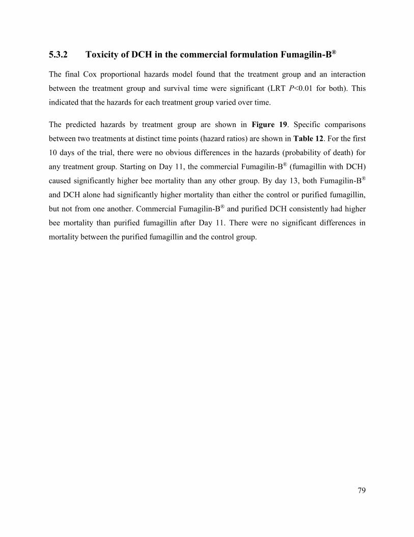
79
5.3.2 Toxicity of DCH in the commercial formulation Fumagilin-B®
The final Cox proportional hazards model found that the treatment group and an interaction
between the treatment group and survival time were significant (LRT P<0.01 for both). This
indicated that the hazards for each treatment group varied over time.
The predicted hazards by treatment group are shown in Figure 19. Specific comparisons
between two treatments at distinct time points (hazard ratios) are shown in Table 12. For the first
10 days of the trial, there were no obvious differences in the hazards (probability of death) for
any treatment group. Starting on Day 11, the commercial Fumagilin-B® (fumagillin with DCH)
caused significantly higher bee mortality than any other group. By day 13, both Fumagilin-B®
and DCH alone had significantly higher mortality than either the control or purified fumagillin,
but not from one another. Commercial Fumagilin-B® and purified DCH consistently had higher
bee mortality than purified fumagillin after Day 11. There were no significant differences in
mortality between the purified fumagillin and the control group.

80
Table 9 Results of the negative binomial models to assess the relationship between test
compound concentration and average N. ceranae spore count at 17 days post-infection
after feeding each compound ad libitum in 60% sugar solution.
Compound n1 Dose
(µM)2 IRR3
LRT P-
value4
Wald
P-value5 95% CI6
Cyclohexanone derivative (8) 28 0 Referent 0.03 -- --
6 4 0.35
0.02 0.14-0.87
6 40 0.41
0.06 0.16-1.03
6 400 0.32
0.02 0.13-0.81
Compound( 10) 28 0 Referent 0.03 -- --
6 4 0.24
0.01 0.08-0.73
6 40 0.21
0.01 0.07-0.63
6 400 0.44
0.15 0.15-1.35
Compound (11) 28 0 Referent <0.01 -- --
6 4 0.55
0.22 0.21-1.43
6 40 0.08
<0.01 0.03-0.21
6 400 0.43
0.09 0.17-1.13
Compound (14) 28 0 Referent <0.01 -- --
5 40 2.56
0.07 0.93-7.04
5 400 3.49
0.02 1.27-9.60
4 800 5.64
<0.01 1.85-17.20
Aspirin (4) 43* -- Dose 0.52 -- --
Compound (9) 46* -- Dose 0.07 -- --
Compound (12) 46* -- Dose 0.44 -- --
Compound (13) 46* -- Dose 0.85 -- --
Carbendazim (15) 63* -- Dose 0.67 -- --
Toltrazuril (16) 47* -- Dose 0.09 -- --
Thiabendazole (17) 46* -- Dose 0.12 -- --
Nozevit 46* -- Dose 0.32 -- -- 1Total number of cages of 100 bees for each dose. Spore counts for each cage represent the average number of
spores per bee. 2The dose of each compound tested compared to negative controls (0 µM). 3Incident rate ratio (ratio of spore counts per bee of the dose in question compared to the referent negative control
dose). IRRs are not shown for compounds where the dose variable was not significantly associated with changes in
spore counts (see 5). 4Likelihood ratio test of whether or not the group of indicators for dose is significant to the model for spore count. 5Wald tests of whether or not IRR’s for each individual dose compared to controls is significantly different than 1.0. 6The 95% confidence interval for the IRR.
*These numbers also include 28 cages of controls

81
Table 10 Results of the zero-inflated negative binomial models to assess the relationship between
test compound concentration and average N. ceranae spore count at 17 days post-
infection after feeding each compound ad libitum in 60% sugar solution.
Compound (model portion)1 n2 Dose
(µM)3 IRR/OR4
LRT/Wald P-
value5
Wald P-
value6 95% CI7
Fumagilin-B® (1) 28 0 Referent <0.01 -- --
NegBin 11 4 0.36
<0.01 0.19-0.68
23 40 0.04
<0.01 0.02-0.07
11 400 1.1e-7
1.00 0-∞
Inflated (treated) 28 No Referent N/A -- --
45 Yes 31.82
<0.01 3.71-273.14
Fumagillol (3) 28 0 Referent <0.01 -- --
NegBin 6 4 0.84
0.62 0.41-1.70
6 40 0.27
<0.01 0.12-0.59
5 400 0.03
<0.01 0.01-0.15
Inflated (dose) 28 0 Referent 0.02 -- --
6 4 0.00
1.00 0-∞
6 40 6.75
0.21 0.35-130.32
5 400 107.77 <0.01 5.58-2100.65
Aspirin Analogue (5) 28 0 Referent <0.01 -- --
NegBin 6 4 0.55
0.11 0.27-1.14
12 40 0.38
<0.01 0.22-0.64
13 400 0.10
<0.01 0.06-0.28
5 800 0.13
<0.01 0.06-0.28
Inflated (treated) 28 No Referent N/A -- --
36 Yes 6.51
0.09 0.75-56.49
Piperonyl Analogue (7) 28 0 Referent <0.01 -- --
NegBin 6 4 0.26
<0.01 0.13-0.52
6 40 0.28
<0.01 0.14-0.56
6 400 0.43
0.03 0.20-0.92
Inflated (treated) 18 No Referent N/A -- --
28 Yes 7.69
0.08 0.79-75.94
Thymol (18) 28 0 Referent <0.01 -- --
NegBin 6 4 0.31
<0.01 0.16-0.62
6 40 0.30
<0.01 0.15-0.59
6 400 0.54
0.08 0.27-1.07
Inflated (treated) 28 No Referent N/A -- --
18 Yes 5.42
0.16 0.52-56.83
Enilconazole (19) 28 0 Referent <0.01 -- --
NegBin 5 40 0.47
0.051 0.22-1.005
5 400 0.16
<0.01 0.07-0.34
3 800 0.12
<0.01 0.04-0.37
Inflated (treated) 28 No Referent N/A -- --
13 Yes 11.25
0.04 1.19-106.70 1Model portion refers to the Negative Binomial (NegBin) portion or the zero-inflated logistic (inflated) portion. 2Total number of cages of 100 bees for each dose. Spore counts for each cage represent the average number of spores per bee. 3The dose of each compound tested compared to negative controls (0 µM). 4Incident rate ratio (ratio of spore counts per bee of the dose in question compared to the referent negative control dose) for the
NegBin portion. Odds ratio for the Inflated portion (either indicators for each dose or for a variable that is treated – yes or no). 5Likelihood ratio test of whether or not the group of indicators for dose is significant to the model for spore count for the NegBin
model. Wald test of the group of indicators for dose for the Inflated model. If treatment – yes or no – was used, refer to the Wald
test in 6. 6Wald tests of whether or not IRR’s or OR’s for each individual dose compared to controls is significantly different than 1.0. 7The 95% confidence interval for the IRR or the OR.

82
Table 11 Cumulative mortalities for bees infected with N. ceranae that were fed varying
concentrations of test compounds ad libitum in 60% sugar solution. Only compounds
that had a statistically significant impact on spore counts are included.
Treatment Dose (µM) dead total mortality Mean Spore
Count
Control 0 1587 2810 56.5% 7,214,286
Fumagilin-B® 4 634 1100 57.6% 4,068,182
40 1634 2300 71.0% 423,913
400 939 1100 85.4% 22,727
Fumagillol (3) 4 226 600 37.7% 2,916,667
40 413 600 68.8% 1,000,000
400 541 600 90.2% 350,000
Aspirin analogue (5) 4 304 600 50.7% 3,548,333
40 267 600 44.5% 2,166,667
400 1084 1900 57.1% 1,315,789
800 441 600 73.5% 750,000
Piperonyl analogue (7) 4 288 600 48.0% 1,625,000
40 276 600 46.0% 1,750,000
400 416 600 69.3% 2,166,667
Compound (8) 4 280 600 46.7% 2,500,000
40 267 600 44.5% 2,958,333
400 332 600 55.3% 2,333,333
Compound (10) 4 302 600 50.3% 1,750,000
40 351 600 58.5% 1,500,000
400 217 600 36.2% 3,208,333
Compound (11) 4 264 600 44.0% 3,958,333
40 332 600 55.3% 583,333
400 320 600 53.3% 3,125,000
Compound (14) 40 182 500 36.4% 18,500,000
400 427 500 85.4% 25,200,000
800 494 500 98.8% 40,700,000
Thymol (18) 4 268 600 44.7% 1,583,333
40 276 600 46.0% 5,375,000
400 308 600 51.3% 2,541,667
Eniconazole (19) 40 355 500 71.0% 4,950,000
400 340 500 68.0% 10,800,000
800 493 500 98.6% 2,416,667
*One cage contained 110, rather than 100 bees
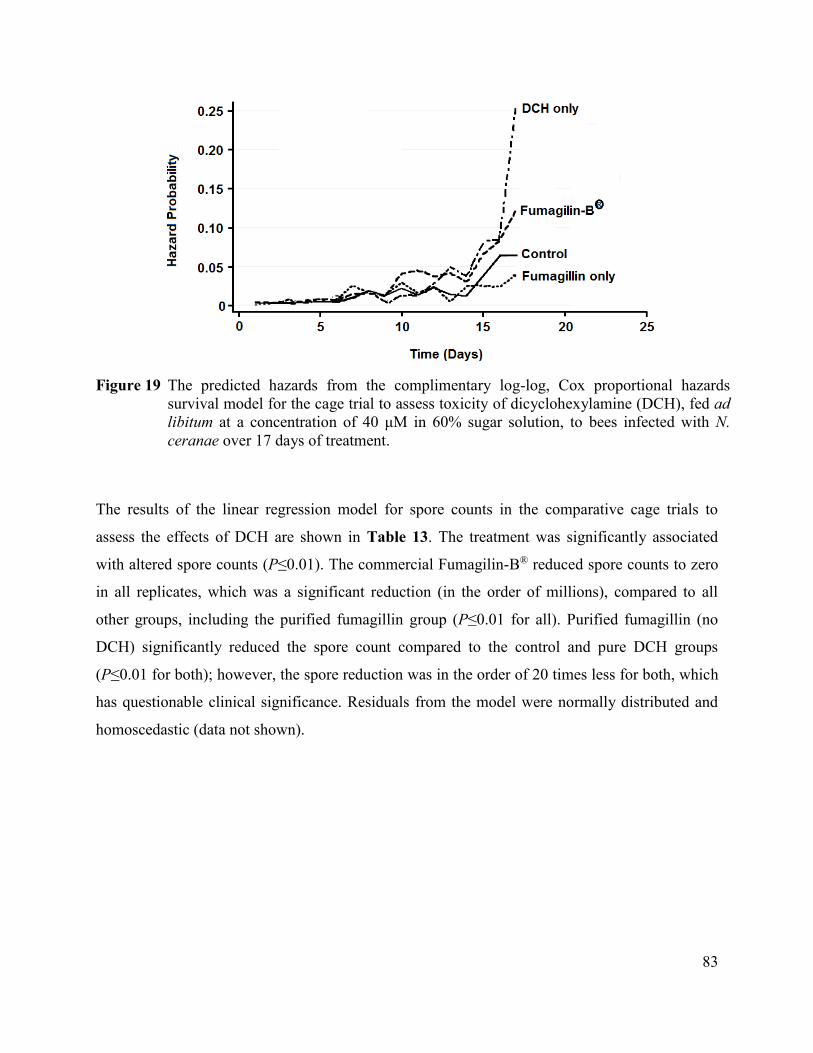
83
Figure 19 The predicted hazards from the complimentary log-log, Cox proportional hazards
survival model for the cage trial to assess toxicity of dicyclohexylamine (DCH), fed ad
libitum at a concentration of 40 μM in 60% sugar solution, to bees infected with N.
ceranae over 17 days of treatment.
The results of the linear regression model for spore counts in the comparative cage trials to
assess the effects of DCH are shown in Table 13. The treatment was significantly associated
with altered spore counts (P≤0.01). The commercial Fumagilin-B® reduced spore counts to zero
in all replicates, which was a significant reduction (in the order of millions), compared to all
other groups, including the purified fumagillin group (P≤0.01 for all). Purified fumagillin (no
DCH) significantly reduced the spore count compared to the control and pure DCH groups
(P≤0.01 for both); however, the spore reduction was in the order of 20 times less for both, which
has questionable clinical significance. Residuals from the model were normally distributed and
homoscedastic (data not shown).

84
Table 12 Results of the complimentary log-log, Cox proportional hazards survival model for the
cage trial to assess toxicity of dicyclohexylamine (DCH), fed ad libitum at a
concentration of 40 μM in 60% sucrose solution, to bees infected with N. ceranae over
17 days of treatment.
Treatment HR1 P-value 95% CI2
DCH only vs. Negative Control
Day 11 0.97 0.96 0.31-3.02
Day 13 3.66 0.01 1.48-9.02
Day 15 2.20 0.01 1.21-4.00
Day 16 1.36 0.24 0.81-2.28
Day 17 4.34 <0.01 2.78-6.77
Fumagilin-B® vs. Negative Control
Day 11 3.44 0.01 1.38-8.57
Day 13 3.06 0.02 1.21-7.76
Day 15 1.77 0.07 0.95-3.32
Day 16 1.34 0.27 0.79-2.27
Day 17 1.94 0.01 1.18-3.20
Fumagillin only vs. Negative Control
Day 11 1.23 0.71 0.41-3.67
Day 13 0.35 0.20 0.07-1.74
Day 15 0.66 0.31 0.30-1.46
Day 16 0.36 0.01 0.17-0.76
Day 17 0.60 0.11 0.31-1.13
DCH only vs. Fumagillin only
Day 11 0.79 0.67 0.27-2.35
Day 13 10.40 <0.01 2.45-44.22
Day 15 3.33 <0.01 1.64-6.76
Day 16 3.82 <0.01 1.82-8.00
Day 17 7.27 <0.01 4.20-12.57
Fumagilin-B® vs. Fumagillin only
Day 11 2.79 0.02 1.18-6.60
Day 13 8.70 <0.01 2.01-37.66
Day 15 2.68 0.01 1.29-5.58
Day 16 3.77 <0.01 1.79-7.93
Day 17 3.25 <0.01 1.80-5.89
Fumagilin-B® vs. DCH only
Day 11 3.53 0.01 1.42-8.79
Day 13 0.84 0.58 0.44-1.58
Day 15 0.80 0.41 0.48-1.35
Day 16 0.99 0.96 0.60-1.62
Day 17 0.45 <0.01 0.31-0.65 1HR – hazard ratio 2CI – confidence interval

85
Table 13 Results of linear regression model for the cage trial to assess the effects of various
treatment preparations with or without dicyclohexylamine (DCH), fed ad libitum at a
concentration of 40 μM in 60% sugar solution, on spore counts in bees infected with N.
ceranae over 17 days of treatment.
Compound
Median
Spores
(106/bee)
SD1
(106)
Coefficient
(spores/bee) P-value 95% CI2
Mean Spore
Count
Negative Control 12.0 3.72 Referent --
12,900,000
DCH only 13.0 4.51 1 0.47 -1 to 2 15,100,000
Fumagilin-B® 0 0 -12.38e+6 <0.01 (-19.64 to -7.91) e+6 0
Fumagillin only 0.75 0.29 -21 <0.01 -33 to -13 650,000
Fumagilin-B® vs. Fumagillin only -- -- -0.59e+6 <0.01 (-0.93 to -0.37) e+6 N/A
Fumagilin-B® vs. DCH only -- -- -14.53e+6 <0.01 (-23.05 to -9.16) e+6 N/A
Fumagillin only vs. DCH only -- -- -25 <0.01 -39 to -16 N/A
1standard deviation. 2confidence interval.
e – exponent (-12.38e+6 = -12,380,000).
5.4 Discussion
All of the semisynthetic and pure synthetic compounds tested were chosen to be easily
preparable from readily available starting materials, which were preferentially selected to be
naturally occurring compounds (piperonylic acid, menthol, aspirin – the acetylated derivative of
salicylic acid, etc.) possessing antifungal properties. All starting materials were also selected to
contain either an alcohol or ketone functional group located on a cyclohexane moiety that
mimics the core structure of fumagillin. The alcohols and ketones could then be easily converted
into epoxides, using the described synthetic methodologies. Both semisynthetic derivatives of
fumagillol (Figure 15, compound 3), namely the aspirin and piperonylic acid analogues (Figure
16, compounds 5 and 7) were observed to exhibit statistically significant, although lower,
activity against N. ceranae-infected bees in cage trial assays compared with Fumagilin-B®
(Table 10). Fumagillol itself also exhibited statistically significant, but lower biological activity
than Fumagilin-B® (Table 10). This is consistent with reports citing lower biological activity of
the alcohol (Gochnauer and Furgala 1962). The diastereomeric UV-decomposition products of
fumagillin (Figure 19, compounds 20) have been reported to retain some of the biological
activity of the parent fumagillin (Kochansky and Nasr 2004). The observed biological activity of
the aspirin analogue (Figure 16, compound 5), which is structurally very similar to the UV-
decomposition products of fumagillin, is consistent with their activity. Aspirin, alone, did not

86
exhibit any statistically significant activity against N. ceranae-infected bees in our study (Table
9), even though it does possess antifungal properties.
Four of the purely synthetic compounds (Figure 17, compounds 8, 10, 11 and 14) were also
observed to exhibit statistically significant, activity against N. ceranae, though inferior to that of
Fumagilin-B® (Tables 9 and 10). One of these compounds was the simple cyclohexanone
derivative (Figure 17, compound 8). This simple spiro-epoxide compound was previously
synthesised and tested for activity against the MetAP-2 enzyme, where it was observed to have
only trace amounts of biological activity (Arico-Muendel et al. 2009).
Of the six commercially available compounds tested (Figure 18, compounds 15, 16, 17, 18 and
19), including Nozevit, only thymol and enilconazole (Figure 18, compound 18 and 19), were
observed to be biologically active against N. ceranae (Table 10). In our experiments thymol
lowered the N. ceranae spore count by an average of 40% over all three test concentrations after
17 d, when compared to the control group. In a previously reported cage trial study, a similar
beneficial effect was observed for thymol, where N. ceranae-infected bees treated with
0.12 mg g-1 thymol in candy (85% icing sugar, 10% honey, 5% water and 3.2 µL g-1 ethanol)
were reported to have only 8.8% of the amount of spores after 25 d, when compared to the
control group (Maistrello et al. 2008). In a subsequent study the same researchers reported that
thymol, when fed to N. ceranae-infected bees in a 0.1 mg g-1 concentration in either a 50% (w/v)
sugar syrup or in a candy form (85% icing sugar, 10% honey, 5% water and 3.2 µL g-1 ethanol),
reduced the N. ceranae spore counts by 50% after 25d when compared to the control group
(Costa et al. 2010). The lifespan of the bees treated with thymol was also increased by three
days, when compared to the control group. Resveratrol was also observed to have a similar effect
to that of thymol in both studies (Maistrello et al. 2008; Costa et al. 2010). Thymol is registered
in Canada under the trade name of ThymovarTM (Reg. #. 29747), and is sold as 15 g thymol
impregnated cellulose wafers for use against Varroa destructor (Anderson and Trueman 2000)
infestations. Thymol is sold worldwide under different trade names and formulations, and is
applied as a fumigant. For example, in the EU and the USA it is sold as a gel (Apiguard®), or in
combination with menthol, camphor and eucalyptol, as an impregnated wafer (Apilife Var®). In
our study, as well as others, thymol was applied orally (Maistrello et al. 2008; Costa et al. 2010).
It would be of interest to learn if treatments of varroa mite infestations with products that contain
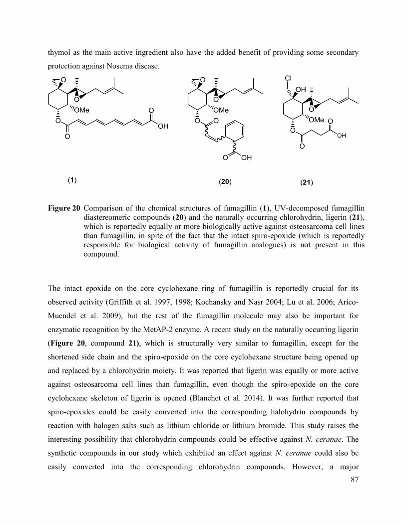
87
thymol as the main active ingredient also have the added benefit of providing some secondary
protection against Nosema disease.
O
O
OMe
O
O
OH
O
O
O
OMe
O
O
OHO
OH
O
OMe
O
OH
O
(1) (21)(20)
O
Cl
Figure 20 Comparison of the chemical structures of fumagillin (1), UV-decomposed fumagillin
diastereomeric compounds (20) and the naturally occurring chlorohydrin, ligerin (21),
which is reportedly equally or more biologically active against osteosarcoma cell lines
than fumagillin, in spite of the fact that the intact spiro-epoxide (which is reportedly
responsible for biological activity of fumagillin analogues) is not present in this
compound.
The intact epoxide on the core cyclohexane ring of fumagillin is reportedly crucial for its
observed activity (Griffith et al. 1997, 1998; Kochansky and Nasr 2004; Lu et al. 2006; Arico-
Muendel et al. 2009), but the rest of the fumagillin molecule may also be important for
enzymatic recognition by the MetAP-2 enzyme. A recent study on the naturally occurring ligerin
(Figure 20, compound 21), which is structurally very similar to fumagillin, except for the
shortened side chain and the spiro-epoxide on the core cyclohexane structure being opened up
and replaced by a chlorohydrin moiety. It was reported that ligerin was equally or more active
against osteosarcoma cell lines than fumagillin, even though the spiro-epoxide on the core
cyclohexane skeleton of ligerin is opened (Blanchet et al. 2014). It was further reported that
spiro-epoxides could be easily converted into the corresponding halohydrin compounds by
reaction with halogen salts such as lithium chloride or lithium bromide. This study raises the
interesting possibility that chlorohydrin compounds could be effective against N. ceranae. The
synthetic compounds in our study which exhibited an effect against N. ceranae could also be
easily converted into the corresponding chlorohydrin compounds. However, a major

88
complication in developing new fumagillin analogues for apicultural usage based on their mode
of action against the MetAP-2 enzyme is still the presumably low selectivity of fumagillin and
other analogues between the host honey bee and the pathogen (N. ceranae) MetAP-2 enzyme
(Huang et al. 2013). Further research into the identification of other enzymes that may be present
in N. ceranae is desirable. This might lead to the discovery of different enzymes that may be
crucial for the proliferation of the Nosema spp., while ideally not affecting the host honey bee,
which could afford different biological targets other than the MetAP-2 enzymes, which could be
pursued in future chemotherapy research.
It is important to note that there are limitations to using spore counts from cage trials to evaluate
the efficacy of alternative compounds against N. ceranae, as well as against other bee diseases.
For example, in cage trial studies bees cannot fly, and therefore do not defecate or conduct
housekeeping operations such as the removal of dead bees from the hive. This could lead to
different results being obtained when applying antibiotics in field trials to whole bee colonies. In
our study, which was designed as a preliminary screen to evaluate alternative compounds for use
against N. ceranae, only those bees that survived to day 17 had their spores counted. It is
possible that bees dying earlier than day 17 could have greater spore counts than that of those
that survived. Further studies that track daily mortality, using potentially effective compounds
are certainly warranted. Testing of these compounds under field conditions would also give a
better indication of their effectiveness.
A recent study reported that very low concentrations of Fumagilin-B® (1000 to 20 000 times
lower than the manufacturer’s recommended dosage) are estimated to be present in beehives at
approximately 2 to 5.5 months post Fumagilin-B® treatment, and that these residual
concentrations of fumagillin negatively impacted the health of the host honey bee, while not
supressing N. ceranae spore formation (Huang et al. 2013). This study also provided evidence
that these low residual concentrations of Fumagilin-B® leads to a hyperproliferation of N.
ceranae spore production. These conditions could be conducive to the development of resistance
by N. ceranae against Fumagilin-B®. Although no resistance to Fumagilin-B® has been reported
in apiculture for either N. apis or N. ceranae, there has been a report of resistance being observed
to the usage of Fumidil-B® for an unidentified Nosema spp. infecting the diamondback moth,
Pluetella xylostella L. (Idris et al. 2000, 2001). Based on this report, it could be speculated that
89
resistance to fumagillin may develop in apiculture in future, owing to the prolonged usage of
fumagillin against Nosema disease, dating back almost to its discovery in the early 1950’s.
Research into the development of new chemical treatments against Nosema disease in apiculture
is therefore of utmost importance, given that Fumagilin-B® is currently the only effective
chemical treatment available.
It is important to remember that Fumagilin-B® is commercially sold as the dicyclohexylamine
(DCH) salt, containing both fumagillin and DCH in a 1:1 stoichiometric ratio (Figure 15,
compound 1 and 2). Using Fumagilin-B® or Fumidil-B® therefore introduces not only one, but
two potentially biologically active compounds to the beehive (van den Heever et al. 2014). From
our study to determine the toxicity of DCH by examining bee mortality, it was observed that the
risk of bee mortality of N. ceranae-infected caged bees treated with DCH only was not
statistically different than commercial Fumagilin-B® for most of the study period, with the
exception of days 11 and 17 (Table 12). Bees treated with DCH or commercial Fumagilin-B®
had significantly higher risk of mortality compared to controls or those treated with purified
fumagillin for most days after day 11, with a few exceptions (Table 13). The observed high bee
mortality associated with the use of the commercial Fumagilin-B® needs to be investigated
further, as Fumagilin-B® contains not only fumagillin, but also DCH. DCH has also been shown
to be significantly more stable in honey under a variety of conditions (van den Heever et al.
2015b), and is reportedly also genotoxic and tumorigenic. In our study the beneficial properties
of purified fumagillin was also observed to be almost twenty times less than that of the
commercial Fumagilin-B®. This is presumably due to the fact that the purified fumagillin is not
chemically stabilized as a salt and therefore prone to decomposition during handling and
application.
5.5 Conclusions
From the results obtained during the cage trial evaluation of alternative chemotherapies against
N. ceranae, four purely synthetic compounds were found to exhibit statistically significant
biological activity against N. ceranae, although none are as effective as Fumagilin-B®.
Fumagillol, as well as the semisynthetic aspirin and piperonyl analogues also exhibited
statistically significant activity against N. ceranae, but were also not as effective as
90
Fumagilin-B®. Despite our knowledge of fumagillin’s activity against the MetAP-2 enzyme, no
commercially viable alternative to fumagillin was discovered in this study - nevertheless, it is
hoped that the information described here will stimulate other research efforts aimed at finding
new alternative chemotherapies against N. ceranae. Amongst the commercially available
compounds evaluated, only thymol and enilconazole were found to have some activity against N.
ceranae. The high bee mortality related to enilconazole usage makes this compound less
attractive for commercial purposes, however thymol is a commercially available product that
might be used to combat Nosema disease.
Evaluation of the toxicity of the DCH, which is present in a 1:1 stoichiometric ratio to fumagillin
in the commercial formulations of fumagillin, showed that DCH appears to have a negative
effect on bee survival. The genotoxic and mutagenic properties of DCH, combined with its
lipophilicity, could lead to the accumulation of DCH in comb wax, where it could potentially
impact the development of the bee larvae and pupa. DCH is also a contaminant of concern in
hive products, with regard to food safety and human health. The frequent detection of DCH
residues in honey by LC-MS/MS, even in the absence of detectable fumagillin residues, was
recently reported (van den Heever et al. 2015a). The risk to the consumer associated with DCH
residues in honey at the levels detected (20-235 ng g-1), should be evaluated. DCH was also
reported to be significantly more stable than fumagillin in honey (van den Heever et al. 2015b).
A different formulation of fumagillin that uses a less toxic counter ion to form the salt should be
further investigated. This would eliminate DCH from the commercial fumagillin formulation,
and would clearly benefit bee health, and improve the quality of honey destined for human
consumption.

91
Chapter 6 General discussion and conclusions
6.1 Significance of this research
Several key observations and findings made during the course of this research enhance the body
of knowledge available to the apicultural industry. The significance of dicyclohexylamine
(DCH), used in the formulation of the salt form of fumagillin in the commercial products
(Fumagilin-B® and Fumidil-B®) was recognized (Chapter 2) and further investigated with the
development of an analytical method to quantitate not only fumagillin and its degradation
products, but also DCH in honey.
A LC-MS/MS method and extraction procedure were developed to quantitate and confirm DCH,
fumagillin, as well as its UV decomposition products in honey (Chapter 3). The sample
extraction procedure used in this method is compatible with current published methods which do
not yet include DCH as an analyte. This should facilitate the adoption of this analytical method.
A stable deuterium labelled analogue of DCH was synthesized to counteract the matrix effects
observed for this analyte during the method development. This internal standard is easily
prepared in a one-pot reaction form commercially available starting materials. When honey
samples were analyzed it was found that DCH was present at significant concentrations in honey,
even when no fumagillin was detected. This indicates that DCH is a much better marker residue
than fumagillin itself, when analyzing honey to determine Fumagilin-B® or Fumidil-B® usage.
The apparent higher stability of DCH in honey, as compared to that of fumagillin, was therefore
further examined.
The relative stability of fumagillin and DCH in honey was investigated under a variety of
conditions (Chapter 4) simulating bulk storage, hive and shelf conditions. It was observed that
DCH was significantly more stable than fumagillin in honey under all conditions tested. This
information could be used with caution by honey producers and regulators trying to estimate the
amount of DCH in honey resulting from fumagillin usage, but further field trial research needs to
be performed in order to verify the results obtained from the simulated stability study. In my
research, the half-life of DCH was observed to be approximately 1-3 years in honey under the
various simulated conditions examined. DCH can therefore be expected to accumulate in
beeswax, and to have a long half-life in this matrix, but this remains to be confirmed. The

92
relative stability of DCH, when compared to that of fumagillin confirms that DCH is indeed a
better marker residue than fumagillin, when examining Fumagilin-B® or Fumidil-B® usage in
apiculture.
The mode of action of fumagillin against the MetAP-2 enzyme in humans is well established
(Ingber et al. 1990). It has also been determined that the MetAp-2 enzyme in humans, honey
bees, N. ceranae and N. apis is almost identical (Huang et al. 2007). I hypothesised that the mode
of action of fumagillin is the same in humans, honey bees, N. ceranae and N. apis, and this
hypothesis was tested in the design, synthesis and evaluation of alternative chemical compounds
other than fumagillin to treat Nosema disease (Chapter 5). Several commercially available, as
well as some novel synthetic and semisynthetic compounds, were evaluated in vivo against N.
ceranae-infected bees in cage trial experiments. The effectiveness of the test compounds were
evaluated using spore count as the measure of efficacy. Four purely synthetic compounds were
found to exhibit statistically significant biological activity against N. ceranae, although none
were as effective as Fumagilin-B®. Fumagillol (the hydrolysis product of fumagillin) as well as
the semisynthetic aspirin and piperonyl fumagillin analogues also exhibited statistically
significant activity against N. ceranae, but again none were as effective as Fumagilin-B® in
suppressing N. ceranae spore production from infected caged bees. Amongst the commercially
available compounds evaluated, only thymol and enilconazole were found to be active against N.
ceranae. The high bee mortality related to enilconazole usage makes this compound less
attractive for commercial purposes, however thymol might be used as an alternative treatment for
Nosema disease. Interestingly, high cumulative bee mortality was observed in the control groups
of N. ceranae-infected bees being treated with Fumagilin-B®.
The observed high bee mortality related to Fumagilin-B® usage was further examined in an
experiment designed to evaluate the effects of fumagillin, DCH and Fumagilin-B® (fumagillin
and DCH) on N. ceranae-infected caged bees (Chapter 5). It was observed that that oral
exposure of N. ceranae-infected caged honey bees to DCH and to Fumagilin-B® leads to a
statistically significant risk of increased bee mortality, when compared to pure fumagillin and the
control group. This indicates that DCH appears to have a negative effect on bee health.

93
6.2 Conclusions and Future Prospects
In this research the prevalence and fate of Fumagilin-B® was examined in honey only. Other hive
products such as wax need to be examined for DCH and fumagillin residues. The current
analytical method sample extraction (Chapter 3) is unsuitable for analyzing wax samples. The
labelled d10-DCH internal standard that I developed for the LC-MS/MS method used to analyze
honey would however be very useful for the analysis of DCH in matrices such as wax or pollen.
Determining the prevalence of DCH in wax would be very useful for apiculturists as well as for
the cosmetic industry applications of beeswax. The prevalence of DCH in pollen and runoff
water next to busy roads and industries should be examined, since this might indicate whether an
environmental source of DCH is indeed of concern. The fact that fumagillin decomposes fairly
rapidly when exposed to light could be considered as a benefit as far as residues in honey is
concerned, but remains a problem for commercial beekeepers when treating large numbers of
colonies, due to the exposure of fumagillin in the prepared sugar solution to light during transit,
or when barrel feeding. The results of this research suggest that in order to ensure the least
amount of chemical residues in honey destined for human consumption, Fumagilin-B® should
only be applied well before the bees start to collect nectar for honey production. A fall
application of Fumagilin-B® should concentrate chemical residues in the brood chambers, and
minimize their presence in the “supers” used for honey production. Fumagillin should also be
more beneficial during the winter in controlling Nosema disease, as the bees cannot leave the
hive easily for cleansing flights, and are closely confined with longer lifespans, which leads to
conditions conducive to the spread of disease. Spring application should likely be avoided.
The observed relative stability of DCH (Chapter 4) in honey indicates that it is likely that DCH
will accumulate in wax, and that it will be present at elevated concentrations. The general
stability of chemical contaminants in wax (Bogdanov 2004) supports this assumption, but it
remains to be investigated. Accumulation of DCH in the comb wax could negatively impact the
development of young bee larvae, which are in close proximity to the wax at a stage of their
lifecycle that make them potentially more susceptible to the influence of chemical contaminants
such as the reportedly genotoxic DCH. The observed increased bee mortality associated with
DCH and Fumagilin-B® in N. ceranae-infected caged bees (Chapter 5) supports this conclusion,
but further research is needed.

94
The development of an alternative salt of fumagillin that does not contain DCH would be
desirable, as this would eliminate DCH residues from the hive and hive products destined for
human consumption. Fumagillin is not chemically synthesized, but is produced through a fungal
fermentation process. The DCH salt of fumagillin is presumably used to crystallize fumagillin
from the fermentation broth, before further formulation occurs. The DCH-fumagillin salt could
potentially be purified by using for example an ion exchange resin column that would “trap” the
DCH, while allowing the fumagillin to exit the column bed along with the eluent. Fumagillin
could then be re-crystallized with another suitable less toxic counter ion. This will add another
purification step to the process, which would result in an increase in the cost of production, but
would ultimately result in improved bee health and food safety.
The potential synergistic effects resulting from the combination DCH and fumagillin with other
known chemical contaminants in the hive needs to be examined. Previous research has shown
that combined exposure to Fumagilin-B® and to tau-fluvalinate leads to an increase in tau-
fluvalinate toxicity to honey bees, resulting in increased mortality (Johnson et al. 2013). Tau-
fluvalinate was reportedly the most abundant pyrethroid chemical found North American
apiaries (Mullin et al. 2010) in wax (98.1% detection at a median concentration of 3595 ng g-1),
pollen (88.3% detection at a median concentration of 40.2 ng g-1) and bees (83.6% detection at a
median concentration of 53 ng g-1). The concentration of Fumagilin-B® used for the synergism
study (designed to evaluate subtherapeutic concentration effects) was however only 0.78 μM
(Johnson et al. 2013), which is 50 times less than the manufacturer’s prescribed minimum
therapeutic concentration of 40 μM for Fumagilin-B®. A significant increase in toxic synergism
with tau-fluvalinate can therefore be expected at the therapeutic concentration of Fumidil-B® (or
Fumagilin-B®). It would be interesting to establish whether it is fumagillin or whether it is DCH
that was responsible for this observed synergistic effect, as the commercially available
Fumidil-B® was used in this study (Johnson et al. 2013). Pyrethroids, including tau-fluvalinate,
are reportedly very stable in wax, where they accumulate, with estimated half-lives of
approximately 5 years (Bogdanov 2004). The synergistic effects of both DCH and fumagillin
with tau-fluvalinate (as well as other frequently found chemical residues in hive products)
warrants further research.

95
Fumagilin-B® (and Fumidil-B®) has been used extensively since almost 1950 (van den Heever et
al. 2014), combined with a recent report that resistance to Fumidil-B® was observed in an
unidentified Nosema sp. infecting the diamondback moth (Idris et al. 2000, 2001), makes finding
alternative chemical treatment against Nosema disease important, should antibiotic resistance
develop to fumagillin. Our observation that orally administered thymol has a statistically
significant effect on N. ceranae (Chapter 5) is promising, and it would be interesting to
determine if fumigation with thymol, which is used to combat mite infections, also acts against
N. ceranae at the same time. Evaluation of the chlorohydrin analogues of the four synthetic
analogues of fumagillin that we tested could also result in increased activity against N. ceranae.
Further research is however needed in order to determine whether there are enzymes other than
MetAP-2 that are crucial for the survival and functioning of N. ceranae. In the best case scenario
these potential enzymes would be unique to N. ceranae (and to N. apis) and would not be present
in the host honey bee. Such an enzyme, if found, would be a much better target for the design of
alternative chemotherapies, since fumagillin (and mimics of fumagillin) target the MetAP-2
enzyme that is also crucial to the survival of the host honey bee. Alternative chemical treatments
that target the MetAP-2 enzyme will most likely also become ineffective if resistance to
fumagillin should develop, since they are designed to mimic the same mode of action as that of
fumagillin.
Cage trial in vivo testing used for this research is however very labor and time intensive, and also
seasonally bound, as newly-eclosed bees free from infection are required. In vitro testing of
alternative treatments would be a much better option, but this is dependent on the availability of
bee cell lines which can be used to propagate the obligatory intracellular N. ceranae
microsporidian fungal pathogen. N. ceranae needs host cells to propagate, because it can only
exist as metabolically inactive spores outside of the host cell (Bigliardi and Sacchi 2001). The
susceptibility of the heterologous cell line IPL-LD-65Y obtained from the ovaries of the gypsy
moth, Lymantria dispar (Goodwin et al. 1978) was however recently used to successfully
propagate both N. apis and N. ceranae in the laboratory in vitro (Gisder et al. 2011). This
development, along with another cell line derived from honey bee (A. mellifera) embryonic
tissues (Goblirsch et al. 2013b) could enable the in vitro screening of potential new chemical

96
treatments against Nosema disease, which would improve the efficiency of screening potential
alternative chemotherapies.
My research should also lead to more informed decisions with regards to setting a MRL for DCH
and fumagillin, since the significance of DCH in Fumagilin-B®, Fumidil-B®, honey and other
hive products has been overlooked since 1950. The harmonization of MRL standards worldwide
would be the ideal situation that we should aspire to, but it is a moving target at best. The lack of
harmonization can impede fair trade, by excluding honey from one country while it is acceptable
in another. A recent example of this is the change in the MRL for tylosin in honey from 10 ng g-1
to a zero tolerance limit in Japan (The Japan Food Chemical Research Foundation 2014), while
there is no requirement for a tylosin MRL in the USA. Disputes can take a long time to resolve
through organizations such as the World Trade Organization (WTO). At best it is probably up to
the producer to be fully aware of the requirements of the market where he or she hopes to sell his
products. A thorough risk assessment of fumagillin and DCH needs to be conducted. The
proposed MRL of 25 ng g-1 for fumagillin by Health Canada (Fishbein 2013) needs to be re-
examined to include a MRL for DCH as well. This research may eventually lead to policy
changes regarding the MRL status of fumagillin and DCH in honey destined for human
consumption, and also perhaps to a change in the formulated product to exclude the use of DCH
as the counter ion of the salt form in the commercial product.

References
Anderson, D. L.; Trueman, J. W. H. Varroa jacobsoni (Acari: Varroidae) is more than one
species. Exp. Appl. Acarol. 2000, 24, 165-189.
Arico-Muendel, C. C.; Benjamin, D. R.; Caiazzo, T. M.; Centrella, P. A.; Contonio, B. D.;
Cook, C. M.; Doyle, E. G.; Hannig, G.; Labenski, M. T.; Searle, L. L.; Lind, K.; Morgan, B.
A.; Olson, B.; Paradise, C. L.; Self, C.; Skinner, S. R.; Sluboski, B.; Svedsen, J. L.;
Thompson, C.D.; Westlin, W.; White, K.F.. Carbamate analogues of fumagillin as potent,
targeted inhibitors of methionine aminopeptidase-2. J. Med. Chem. 2009, 52, 8047-8056.
Assil, H. I.; Sporns, P. ELISA and HPLC methods for analysis of fumagillin and its
decomposition products in honey. J. Agric. Food Chem. 1991, 39, 2206-2213.
Bailey, L. Effect of fumagillin upon Nosema apis (Zander). Nature 1953, 171, 212-213.
Bailey, L.; Ball, B. V. Honey Bee Pathology, 2nd ed.; Academic Press: London, 1991.
Bajić, V.; Stanimirović, Z.; Stevanović, J. Genotoxicity potential of 8-Cl-cyclic adenosine
monophosphate assessed with cytogenic tests in vivo. Arch. Med. Res. 2004, 35, 209-214.
Baldwin, J. E.; Bulger, P. G.; Marquez, R. Fast and efficient synthesis of novel fumagillin
analogues. Tetrahedron 2002, 58, 5441-5452.
Balthaser, B. R.; Maloney, M. C.; Beeler, A. B.; Porco, J. A. J.; Snyder, J. K. Remodelling
of the natural product fumagillol employing a reaction discovery approach. Nature Chem.
2011, 3, 969-973.
Bigliardi, E.; Sacchi, L. Cell biology and invasion of the microsporidia. Microbes Infect.
2001, 3, 373-379.

Blanchet, E.; Vansteelandt, M.; Le Bot, R.; Egrov, M.; Guitton, Y.; Pouchus, Y. F.; Grovel,
O. Synthesis and antiproliferative activity of ligerin and new fumagillin analogs against
osteosarcoma. Eur. J. Med. Chem. 2014, 79, 2444-2250.
Bloch, G.; Francoy, T. M.; Wachtel, I.; Panitz-Cohen, N.; Fuchs, S.; Mazar, A. Industrial
apiculture in the Jordan valley during Biblical times with Anatolian honeybees. PNAS 2010,
107, 11240-11244.
Blood, D.C.; Studdert, V.P.; Gay, C.C. Saunders Comprehensive Medical Dictionary, 3rd
ed.; Elsevier: St. Louis, Missouri, 2007, p 2172.
Bogdanov, S. Beeswax: quality issues today. Bee World 2004, 85, 46-50.
Botías, C.; Anderson, D. L.; Meana, A.; Garrido-Bailón, E.; Martín-Hernández, R.; Higes,
M. Further evidence of an oriental origin for Nosema ceranae (Microsporidia:
Nosematidae). J. Invertebr. Pathol. 2012a, 110, 108-113.
Botías, C.; Martín-Hernández, R.; Garrido-Bailón, E.; González-Porto, A.; Martínez-
Salvador, A.; De La Rúa, P.; Meana, A.; Higes, M. The growing prevalence of Nosema
ceranae in honey bees in Spain, an emerging problem for the last decade. Res. Vet. Sci.
2012b, 93, 150-155.
Brackett, J. M.; Arguello, M. D.; Schaar, J. C. Determination of fumagillin by high
performance liquid chromatography. J. Agric. Food Chem. 1988, 36, 762-764.
Brorström-Lundén, E.; Hansson, K.; Remberger, M.; Kaj, L.; Magnér, J.; Andersson, H.;
Haglund, P.; Andersson, R.; Liljelind, P.; Grabic, R. Screening of benzothiazoles,
benzenediamines, dicyclohexylamine and benzotriazoles. IVL Report B2023:1-53 2011.
Cantwell, G.E. Standard methods for counting nosema spores. Am. Bee J. 1970, 110, 222-
223.

CFIA 2014 http://www.inspection.gc.ca/food/honey/product-inspection/wrls/questions-and-
answers/eng/ (accessed September 10, 2015).
Chaimanee, V.; Warrit, N.; Chantawannakul, P. Infections of Nosema ceranae in four
different honeybee species. J. Invertebr. Pathol. 2010, 105, 207-210.
Chambers, E.; Wagrowski-Diehl, D. M.; Lu, Z.; Mazzeo, J. R. Systematic and
comprehensive strategy for reducing matrix affects in LC/MS/MS analyses. J. Chrom. B
2007, 852, 22-34.
Chen, Y.; Evans, J. D.; Smith, B. I.; Pettis, J. S. Nosema ceranae is a long-present and
wide-spread microsporidian infection of the European honey bee (Apis mellifera) in the
United States. J. Invertebr. Pathol. 2008, 97, 186-188.
Chen, Y.; Evans, J. D.; Zhou, L.; Boncristiani, H.; Kimura, K.; Xiao, T.; Litkowski, A. M.;
Pettis, J. S. Asymmetrical coexistence of Nosema ceranae and Nosema apis in honey bees.
J. Invertebr. Pathol. 2009a, 101, 204-209.
Chen, X.; Xie, S.; Bhat, S.; Kumar, N.; Shapiro, T. A.; Liu, J. O. Fumagillin and fumarranol
interact with P. falciparum methionine aminopeptidase 2 and inhibit malaria parasite
growth in vitro and in vivo. Chem. Biol. 2009b, 16, 193-202.
COM. Committees on Toxicity, Mutagenicity, Carcinogenicity of Chemicals in Food,
Consumer Products and the Environment;, 2009a.
cot.food.gov.uk/sites/default/files/cot/comsection2009.pdf (accessed September 15, 2015)
COM. Committees on Toxicity, Mutagenicity, Carcinogenicity of Chemicals in Food,
Consumer Products and the Environment Joint Annual Report 2009;, 2009b.
cot.food.gov.uk/sites/default/files/cot/cotcomcocreport2009.pdf (accessed September 15,
2015)

COM. Committees on Toxicity, Mutagenicity, Carcinogenicity of Chemicals in Food,
Consumer Products and the Environment;, 2011.
cot.food.gov.uk/sites/default/files/cot/cotcomcocreport2011.pdf (accessed September 15,
2015)
Commission Regulation (EU) No 37/2010. 2009. ec.europa.eu/health/files/eudralex/vol-
5/reg_2010_37/reg_2010_37_en.pdf (accessed December 03, 2014).
Conteas, C. N.; Berlin, O. G. W.; Ash, L. R.; Pruthi, J. S. Therapy for human
gastrointestinal microsporidiosis. Am. J. Trop. Med. Hyg. 2000, 63, 121-127.
Corey, E. J.; Chaykovsky, M. Methylsulfinyl carbanion (CH3-SO-CH2-). Formation and
applications to organic synthesis. J. Am. Chem. Soc. 1965, 87, 1345-1353.
Corrente, J. E.; Chalita, L. V. A. S.; Moreira, J. A. Choosing between Cox proportional
hazards and logistic models for interval-censored data via bootstrap. J. Appl. Stat. 2003, 30,
37-57.
Costa, C.; Lodesani, M.; Maistrello, L. Effect of thymol and resveratrol administered with
candy or syrup on the development of Nosema ceranae and on the longevity of honeybees
(Apis mellifera L.) in laboratory conditions. Apidologie 2010, 41, 141-150.
Cox-Foster, D. L.; Conlan, S.; Holmes, E. C.; Palacios, G.; Evans, J. D.; Moran, N. A.;
Quan, P.-L.; Briese, T.; Hornig, M.; Geiser, D. M.; Martinson, V.; vanEngelsdorp, D.;
Kalkstein, A. L.; Drysdale, A.; Hui, J.; Zhai, J.; Cui, L.; Hutchinson, S. K.; Simons, J.F.;
Egholm, M.; Pettis, J.S.; Lipkin, W.I. A metagenomic survey of microbes in honey bee
colony collapse disorder. Science 2007, 318, 283-287.
Crane, E., Ed. Honey, A comprehensive Survey., 1st ed.; William Heinemann Ltd.: London,
England, 1975.

Crane, E. The Archaelogy of Beekeeping, 1st ed.; Gerald Duckworth & Co. Ltd.: London,
England, 1983.
Currie, R. W.; Pernal, S. F.; Guzmán-Novoa, E. Honey bee colony losses in Canada. J.
Apic. Res. 2010, 49, 104-106.
Darrach, M; Page, S. Statistical overview of the Canadian honey and bee Industry and the
economic contribution of honey bee pollination 2013-2014; AAFC No: 12297E, 2015.
Dess, D. B.; Martin, J. C. A usefull 12-I-5 Triacetoxyperiodinane (the Dess-Martin
periodinane) for the selective oxidation of primary or secondary alcohols and a variety of
related 12-I-5 Species. J. Am. Chem. Soc. 1991, 113, 7277-7287.
Didier, E. S. Effects of albendazole, fumagillin, and TNP-470 on microsporidial replication
in vitro. Antimicrob. Agents Chemother. 1997, 41, 1541-1546.
Didier, P. J.; Phillips, J. N.; Kuebler, D. J.; Nasr, M.; Brindley, P. J.; Stoval, M. E.; Bowers,
L. C.; Didier, E. S. Antimicrosporidial activities of fumagillin, TNP-470, ovalicin, and
ovalicin derivatives in vitro and in vivo. Antimicrob. Agents Chemother. 2006, 50, 2146-
2155.
Didier, E. S.; Weiss, L. M. Overview of microsporidia and microsporidiosis. Protistol.
2008, 5, 243-255.
Dmitrovic, J.; Durden, D. A. Analysis of fumagillin in honey by LC-MS/MS. J. AOAC Int.
2013, 96, 687-695.
Dohoo, I.; Martin, W.; Stryhn, H. Veterinary epidemiologic research; VER Inc.:
Charlottetown, P.E.I., 2009.
Drahl, C.; Cravatt, B. F.; Sorensen, E. J. Protein-reactive natural products. Angew. Chem.
Int. Ed. 2005, 44, 5788-5809.
Eble, T. E.; Hanson, F. R. Fumagillin, an antibiotic from Aspergillus fumigatus H-3.
Antibiot. Chemother. 1951, 1, 54-58.
Eble, T. E.; Garrett, E. R. Studies on the stability of fumagillin II. Photolytic degradation of
crystalline fumagillin. J. Am. Pharm. Assoc. 1954, 43, 536-538.
Ellis, J. D.; Munn, P. A. The worldwide health status of honeybees. Bee World 2005, 86,
88.
El-Matbouli, M.; Hoffmann, R. W. Prevention of experimentally induced whirling disease
in rainbow trout Oncorhynus mykiss by fumagillin. Dis. Aquat. Org. 1991, 10, 109-113.
FAO/WHO. Joint FAO/WHO Technical Workshop on Residues of Veterinary Drugs
without ADI/MRL. ftp.fao.org/docrep/fao/008/y5723e/y5723e00.pdf 2004. (accessed July
14, 2015).
Fishbein, D. Proceedings of the Canadian Assosiation of Professional Apiculturists meeting
held at Edmonton, AB on January 28-29, 2014. CFIA Honey Program 56-58.
http://capabees.org/content/uploads/2012/11/2013-CAPA-AGM-Proceedings.pdf 2013.
(accessed July 31, 2014).
Folkman, J. Tumor angiogenesis. J. Adv. Cancer Res. 1985, 43, 175-204.
Folkman, J.; Shing, Y. Minireview: angiogenesis. J. Biol. Chem. 1992, 267, 10931-10934.
Forsgren, E.; Fries, I. Comparative virulence of Nosema ceranae and Nosema apis in
individual European honey bees. Vet. Parasitol. 2010, 170, 212-217.
Forsgren, E.; Fries, I. Temporal study of Nosema spp. in a cold climate. Environ. Microbiol.
Rep. 2013, 5, 78-82.
Franzen, C. Microsporidia: a review of 150 years of research.. Open Parasitol. J. 2008, 2,
1-34.

Fries, I. Nosema apis – a parasite in the honey bee colony. Bee World 1993, 74, 5-19.
Fries, I.; Fend, F.; Da Silva, A.; Slemenda, S. B.; Pieniazek, N. J. Nosema ceranae n. sp.
(Microspora, Nosematidae), morphological and molecular characterization of a
microsporidian parasite of the Asian honey bee Apis cerana (Hymenoptera: Apidae). Eur. J.
Protistol. 1996, 32, 356-365.
Fries, I. Protozoa. In Honey bee pests, predators, and diseases; Morse, R. A., Ed.; A.I. Root
Company: Medina, Ohio, 1997; p 57−76.
Fries, I. Nosema ceranae in European honey bees (Apis mellifera). J. Invertebr. Pathol.
2010, 103, S73-S79.
Fries, I.; Chauzat, M.-P.; Chen, Y.-P.; Doublet, V.; Genersch, E.; Gisder, S.; Higes, M.;
McMahon, D. P.; Martín-Hernández, R.; Natsopoulou, M.; Paxton, R. J.; Tanner, G.;
Webster, T. C.; Williams, G. R. Standard methods for nosema research. J. Apic. Res. 2013,
51(5): http://dx.doi.org/10.3896/IBRA.1.52.1.14
Frigerio, M.; Santagostino, M.; Sputore, S. A user friendly entry to 2-Iodobenzoic acid
(IBX). J. Org. Chem. 1999, 64, 4537-4538.
Furgala, B.; Boch, R. The effect of Fumidil-B, Nosemack and Humatin on Nosema apis. J.
Apic. Res. 1970, 9, 79-85.
Furgala, B.; Gochnauer, T. A. Chemotherapy of nosema disease. Further studies on the
residual activity of fumagillin in stored sugar syrup and “bee manipulated” sugar syrup. Am.
Bee. J. 1969a, 109, 218-219.
Furgala, B.; Gochnauer, T. A. Chemotherapy of nosema disease. Effect of treatment
method with Fumidil-B. Am. Bee. J. 1969b, 109, 380-381; 392.
Garrett, E. R. Studies on the stability of fumagillin III. Thermal degradation in the presence
and absence of air. J. Am. Pharm. Assoc. 1954, 43, 539-543.

Garrett, E. R.; Eble, T. E. Studies on the stability of fumagillin I. Photolytic degradation in
alcohol solution. J. Am. Pharm. Assoc. 1954, 3, 385-390.
Genersch, E. Honey bee pathology: current threats to honey bees and beekeeping. Appl.
Microbiol. Biotechnol. 2010, 87, 87-97.
Giersch, T.; Berg, T.; Galea, F.; Hornitzky, M. Nosema ceranae infects honey bees (Apis
mellifera) and contaminates honey in Australia. Apidologie 2009, 40, 117-123.
Gisder, S.; Möckel, N.; Linde, A.; Genersch, E. A cell culture model for Nosema ceranae
and Nosema apis allows new insights into the life cycle of these important honey bee-
pathogenic microsporidia. Environ. Microbiol. 2011, 13, 404-413.
Goblirsch, M.; Huang, Z. Y.; Spivak, M. Physiological and behavioral changes in honey
bees (Apis mellifera) induced by Nosema ceranae infection. PLoS ONE 2013a, 8, e58165.
Goblirsch, M.J.; Spivak, M.S.; Kurtti, T.J. A cell line resource derived from honey bee
(Apis Mellifera) embryonic tissues. PLoS ONE 2013b, 8, e69831.
Gochnauer, T. A.; Furgala, B. The nosema inhibitory activity of alcohol I, a component of
fumagillin. J. Insect Pathol. 1962, 1962, 4, 489-1962, 4,491.
Goodwin, R. H.; Tompkins, G. J.; McCawley, P. Gypsy moth cell lines divergent in viral
susceptibility. In Vitro 1978, 14, 485-494.
Greim, H.; Bury, D.; Klimisch, H.-J.; Oeben-Negele, M.; Ziegler-Skylakakis, K. Toxicity
of aliphatic amines: structure-activity relationship. Chemosphere 1998, 36, 271-295.
Griffith, E. C.; Su, Z.; Turk, B. E.; Chen, S.; Chang, Y.-H.; Wu, Z.; Biemann, K.; Liu, J. O.
Methionine aminopeptidase (type 2) is the common target for angiogenesis inhibitors
AGM-1470 and ovalicin. Chem. Biol. 1997, 4, 461-471.

Griffith, E. C.; Su, Z.; Niwayama, S.; Ramsay, C. A.; Chang, Y.-H.; Liu, J. O. Molecular
recognition of angiogenesis inhibitors fumagillin and ovalicin by methionine
aminopeptidase 2. Proc. Natl. Acad. Sci. USA 1998, 95, 15183-15188.
Guyonnet, J.; Richard, M.; Hellings, P. Determination of fumagillin in muscle tissue of
rainbow trout using automated ion-pairing liquid chromatography. J. Chromatogr. B 1995,
666, 354-359.
Han, C. K.; Ahn, S. K.; Choi, N. S.; Hong, R. K.; Moon, S. K.; Chun, H. S.; Lee, S. J.; Kim,
J. W.; Hong, C. I.; Kim, D.; Yoon, J. H.; No, K. T. Design and synthesis of highly potent
fumagillin analogues from homology modeling for a human MetAP-2. Bioorg. Med. Chem.
Lett. 2000, 10, 39-43.
Hanson, F. R.; Eble, T. E. An antiphage agent isolated from Aspergillus sp. J. Bacteriol.
1949, 58, 527-529.
Health Canada, 2015a http://www.hc-sc.gc.ca/dhp-mps/vet/legislation/pol/cfia-acia_amr-
ram_table-eng.php (accessed September 10, 2015).
Health Canada, 2015b www.hc-sc.gc.ca/dho-mps/vet/mrl-lmr/mrl-lmr_versus_new-
nouveau-eng.php (accessed September 10, 2015).
Health Canada, 2015c http://pr-rp.hc-sc.gc.ca/mrl-lrm/index-eng.php (accessed September
10, 2015).
Heil, J.; Reifferscheid, G.; Waldman, P.; Leyhausen, G.; Geurtsen, W. Genotoxicity of
dental materials. Mut. Res. 1996, 368, 181-194.
Higes, M.; Martín, R.; Meana, A. Nosema ceranae, a new microsporidian parasite in
honeybees in Europe. J. Invertebr. Pathol. 2006, 92, 93-95.
Higes, M.; Martín-Hernández, R.; Botías, C.; Garrido-Bailón, R.; Gonzales-Porto, A. V.;
Barrios, L.; Nozal, M. J.; Bernal, J. L.; Jimenez, J. J.; Palencia, P. G.; Meana, A. How
natural infection by Nosema ceranae causes honeybee colony collapse. Environ. Microbiol.
2008, 10, 2659-2669.
Higes, M.; Martín-Hernández, R.; Garcia-Palencia, P.; Martin, P.; Meana, A. Horizontal
transmission of Nosema ceranae (Microsporidia) from worker honeybees to queens (Apis
mellifera). Environ. Microbiol. Rep. 2009a, 1, 495-498.
Higes, M.; Martín-Hernández, R.; Garrido-Bailón, E.; Botías, C.; Meana, A. First detection
of Nosema ceranae (Microsporidia) in African honey bees (Apis mellifera intermissa). J.
Apic. Res. 2009b, 48, 217-219.
Higes, M.; Martín-Hernández, R.; Garrido-Bailón, E.; Gonzalez-Porto, A. V.; Garcia-
Palencia, P.; Meana, A.; del Nozal, M. J.; Mayo, R.; Bernal, J. L. Honeybee colony collapse
due to Nosema ceranae in professional apiaries. Environ. Microbiol. Rep. 2009c, 1, 110-
113.
Higes, M.; Martín-Hernendez, R.; Meana, A. Nosema ceranae in Europe: an emergent type
C nosemosis. Apidologie 2010, 41, 375-392.
Higes, M.; Nozal, M. J.; Alvaro, A.; Barrios, L.; Meana, A.; Martín-Hernández, R.; Bernal,
J. L.; Bernal, J. The stability and effectiveness of fumagillin in controlling Nosema ceranae
(Microsporidia) infection in honey bees (Apis mellifera) under laboratory and field
conditions. Apidologie 2011, 42, 364-377.
Higes, M.; Meana, A.; Bartolomé, C.; Botías, C.; Martín-Hernández, R. Nosema ceranae
(Microsporidia), a controversial 21st century honey bee pathogen. Environ. Microbiol. Rep.
2013, 5, 17-29.
Huang, W.-F.; Jiang, J.-H.; Chen, Y.-W.; Wang, C.-H. A Nosema ceranae isolate from the
honeybee Apis mellifera. Apidologie 2007, 38, 30-37.

Huang, W.-F.; Bocquet, M.; Lee, K.-C.; Sung, I.-H.; Jiang, J.-H.; Chen, Y.-W.; Wang, C.-
H. The comparison of rDNA spacer regions of Nosema ceranae isolates from different
hosts and locations. J. Invertebr. Pathol. 2008, 97, 9-13.
Huang, W.-F.; Solter, L. F.; Yau, P. M.; Imai, B. S. Nosema ceranae escapes fumagillin
control in honey bees. PLoS Path. 2013, 9, e1003185.
Idris, A. B.; Paul, A. Y.; Noraman, M. N.; Zainal Abidin, B. A.-H. Evidence of antibiotic
resistant development on Nosema spp, infecting diamondback moth larvae. Resistant Pest
Management Newsletter 2000, 11, 21-23.
Idris, A. B.; Paul, A. Y.; Zainal Abidin, B. A. H.; Sajap, A. S.; Hussan, A. K. Preliminary
evidence on Nosema spp. infecting diamondback moth larvae on Fumidil-B. Pakistan J.
Biol. Sci. 2001, 4, 329-330.
Ingber, D.; Fujita, T.; Kishimoto, S.; Sudo, K.; Kanamaru, T.; Brem, H.; Folkman, J.
Synthetic analogues of fumagillin that inhibit angiogenesis and suppress tumor growth.
Nature 1990, 348, 555-557.
Johansson, T.S.K.; Johansson, M.P. Lorenzo L. Langstroth and the bee space. Bee Wld.
1967, 48, 133-143.
Johnson, R. M.; Dahlgren, L.; Siegfried, B. D.; Ellis, M. D. Acaricide, fungicide and drug
interactions in honey bees (Apis mellifera). PLoS ONE 2013, 8, e54092.
Kanda, M.; Sasamoto, T.; Takeba, K.; Hayashi, H.; Kusano, T.; Matsushima, Y.; Nakajima,
T.; Kanai, S.; Takano, I. Rapid determination of fumagillin residues in honey by liquid
chromatography-tandem mass spectrometry using the QuEChERS method. J. AOAC Int.
2011, 94, 878-885.
Katznelson, H.; Jamieson, C. A. Control of Nosema disease of honeybees with fumagillin.
Science 1952, 115, 70-71.
Kent, M. L.; Dawe, S. C. Efficacy of Fumagillin DCH against experimentally induced
Loma salmonae (Microsporea) infections in Chinook salmon Oncorhynus tshawytscha. Dis.
Aquat. Org. 1994, 20, 231-233.
Killough, J. T.; Magill, G. B.; Smith, R. C. The treatment of amebiasis with fumagillin.
Science 1952, 115, 71-72.
Kirkby, W. W.; Salmond, G. W. A.; Davies, K. W. The assessment for safety of food
additives. J. Royal Stat. Soc. D (The Statistician) 1972, 21, 47-61.
Klee, J.; Besana, A. M.; Genersch, E.; Gisder, S.; Nanetti, A.; Quyet Tam, D.; Xuan Chinh,
T.; Puerta, F.; Ruz, J. M.; Kryger, P.; Message, D.; Hatjina, F.; Korpela, S.; Fries, I.;
Paxton, R. J. Widespread dispersal of the microsporidian Nosema ceranae, an emergent
pathogen of the western honey bee, Apis mellifera. J. Invertebr. Pathol. 2007, 96, 1-10.
Kochansky, J.; Nasr, M. Laboratory studies on the photostability of fumagillin, the active
ingredient of Fumidil-B. Apidologie 2004, 35, 301-310.
Kulić, M.; Aleksić, N.; Stanimirović, Z.; Ristić, S.; Medenica, S. Examination of genotoxic
effects of fumagillin in vivo. Genetika 2009, 41, 329-338.
Kusaka, M.; Sudo, K.; Fujita, T.; Marui, S.; Itoh, F.; Ingber, D.; Folkman, J. Potent anti-
angiogenic action of AGM-1470: comparison to the fumagillin parent. Biochem. Biophys.
Res. Comm. 1991, 174, 1070-1076.
Lanternier, F.; Boutboul, D.; Menotti, J.; Chandesris, M. O.; Sarfati, C.; Mamzer Bruneel,
M. F.; Calmus, Y.; Mechai, F.; Viard, J. P.; Lecuit, M.; Bougnoux, M. E.; Lortholary, O.
Microsporidiosis in solid organ transplant recipients: two Enterocytozoon bieneusi cases
and review. Transpl. Infect. Dis. 2009, 11, 83-88.
Lee, H. W.; Cho, C. S.; Kang, S. K.; Yoo, Y. S.; Shin, J. S.; Ahn, S. K. Design, synthesis
and antiangiogenic effects of a series of potent novel fumagillin analogues. Chem. Pharm.
Bull. 2007, 55, 1024-1029.
Lijnen, H. R.; Frederix, L.; Van Hoef, B. Fumagillin reduces adipose tissue formation in
murine models of nutritionally induced obesity. Obesity 2010, 18, 2241-2246.
Liu, T. P. Ultrastructural changes in the secretion granules of the hypopharangeal glands of
the honeybee infected by Nosema apis and after treatment with fumagillin. Tissue and Cell
1990, 22, 523-531.
Liu, S.; Widom, J.; Kemp, C. W.; Crews, C. M.; Clardy, J. Structure of human methionine
aminopeptidase-2 complexed with fumagillin. Science 1998, 282, 1324-1327.
Lopez, M. I.; Pettis, J. S.; Barton Smith, I.; Chu, P.-S. Multiclass determination and
confirmation of antibiotic residues in honey using LC-MS/MS. J. Agric. Food Chem. 2008,
56, 1553-1559.
Lu, J.; Chong, C. R.; Hu, X.; Liu, J. O. Fumarranol, a rearranged fumagillin analogue that
inhibits angiogenesis in vivo. J. Med. Chem. 2006, 49, 5645-5648.
Maistrello, L.; Lodesani, M.; Costa, C.; Leonardi, F.; Marani, G.; Caldon, M.; Mutinelli, F.;
Granato, A. Screening of natural compounds for the control of nosema disease in honeybees
(Apis mellifera L.). Apidologie 2008, 39, 436-445.
Martínez, J.; Leal, G.; Conget, P. Nosema ceranae an emergent pathogen of Apis mellifera
in Chile. Parasitol. Res. 2012, 111, 601-607.
Martin, S. J.; Hardy, J.; Villalobos, E.; Martín-Hernández, R.; Nikaido, S.; Higes, M. Do
the honeybee pathogens Nosema ceranae and deformed wing virus act synergistically?
Environ. Microbiol. Rep. 2013, 5, 506-510.

Martín-Hernández, R.; Meana, A.; Prieto, L.; Salvador, A. M.; Garrido-Bailón, E.; Higes,
M. Outcome of colonization of Apis mellifera by Nosema ceranae. Appl. Environ.
Microbiol. 2007, 73, 6331.
Martín-Hernández, R.; Botías, C.; Garrido-Bailón, E.; Martínez-Salvador, A.; Prieto, L.;
Meana, A.; Higes, M. Microsporidia infecting Apis mellifera: coexistence or competition. Is
Nosema ceranae replacing Nosema apis? Environ. Microbiol. 2012, 14, 2127-2138.
Mavric, E.; Wittmann, S.; Barth, G.; Henle, T. Identification and quantitation of
methylglyoxal as the dominant antibacterial constituent of Manuka (Leptospermum
scoparium) honeys from New Zealand. Mol. Nutr. Food Res. 2008, 52, 483-489.
McCowan, M. C.; Callender, M. E.; Lawlis, J. F. Fumagillin (H-3), a new antibiotic with
amebicidal properties. Science 1951, 113, 202-203.
Medici, S. K.; Sarlo, E. G.; Porrini, M. P.; Braunstein, M.; and Eguaras, M. J. Genetic
variation and widespread dispersal of Nosema ceranae in Apis mellifera apiaries from
Argentina. Parasitol. Res. 2012, 110, 859–864.
Molina, J. M.; Goguel, J.; Sarfati, C.; Michiels, J.-F.; Desportes-Livage, I.; Balkan, S.;
Chastang, C.; Cotte, L.; Maslo, C.; Struxiano, A.; Derouin, F.; Decazes, J.-M. Trial of oral
fumagillin for the treatment of intestinal microsporidiosis in patients with HIV infection.
AIDS 2000, 14, 1341-1348.
Molina, J.-M.; Tourneur, M.; Sarfati, C.; Chevret, S.; de Gouvello, A.; Gobert, J.-G.;
Balkan, S.; Derouin, F. Fumagillin treatment of intestinal microsporidiosis. N. Engl. J. Med.
2002, 346, 1963-1969.
Molnar, K. Recent achievements in the chemotherapy of myxosporean infections of fish.
Acta. Vet. Hung. 1994, 41, 51-58.
Mortelmans, K.; Haworth, S.; Lawlor, T.; Speck, W.; Tainer, B.; Zeiger, E. Salmonella
mutagenicity tests: II. Results from the testing of 270 chemicals. Environ. Mut. 1986, 8
(Supl 7), 1-119.
Mullin, C. A.; Frazier, M.; Frazier, J.; Ashcraft, S.; Simonds, R.; vanEngelsdorp, D.; Pettis,
J. S. High levels of miticides and agrochemicals in North American apiaries: Implications
for honey bee health. PLoS ONE 2010, 5, e9754.
Neises, B.; Steglich, W. Simple method for the esterification of carboxylic acids. Angew.
Chem. Int. Edit. Engl. 1978, 17, 522-524.
Ng, J. S. Epoxide formation from aldehydes and ketones - A modified method for preparing
the Corey-Chaykovsky reagents. Synth. Commun. 1990, 20, 1193-1202.
Nozal, M. J.; Bernal, J. L.; Martín, M. T.; Bernal, J.; Alvaro, A.; Martín, R.; Higes, M.
Trace analysis of fumagillin in honey by liquid chromatography-diode array-electrospray
ionisation mass spectrometry. J. Chromatogr. A 2008, 1190, 224-231.
Paxton, R. J.; Klee, J.; Korpela, S.; Fries, I. Nosema ceranae has infected Apis mellifera in
Europe since at least 1998 and may be more virulent than Nosema apis. Apidologie 2007,
38, 558.
Pegg, A. E.; Bitonti, A. J.; McCann, P. P.; Coward, J. K. Inhibition of bacterial
aminopropyltransferases by S-adenosyl-1,8-diamino-3-thiooctane and by
dicyclohexylamine. FEBS Lett. 1983, 155, 192-196.
Pelter, A.; Rosser, R. M.; Mills, S. Reductive aminations of ketones and aldehydes using
borane-pyridine..J Chem. Soc. Perkin Trans. 1 1984, 717-720.
Pettis, J. S.; Lichtenberg, E. M.; Andree, M.; Stitzinger, J.; Rose, R.; vanEngelsdorp, D.
Crop pollination exposes honey bees to pesticides which alters their susceptibility to the gut
pathogen Nosema ceranae. PLoS ONE 2013, 8, e70182.

Pieper, B. Honey-based dressings and wound care. J. Wound Ostomy Continence Nurs.
2009, 36, 60-66.
Plischuck, S.; Martín-Hernández, R.; Prieto, L.; Lucia, M.; Botías, C.; Meana, A.;
Abrahamovich, A. H.; Lange, C.; Higes, M. South American native bumblebees
(Hymenoptera: Apidae) infected by Nosema ceranae (Microsporidia), an emerging
pathogen of honeybees (Apis mellifera). Environ. Microbiol. Rep. 2009, 1, 131-135.
Pliss, G. B. On the carcinogenic activity of dicyclohexylamine and dicyclohexylamine
nitrite. Vopr. Onk. 1958, 4, 659-667, Original reference in Russian – Abstract accessed
only.
Purchase, I. F. H.; Longstaff, E.; Ashby, J.; Styles, J. A.; Anderson, D.; Lefevre, P. A.;
Westwood, F. R. An evaluation of 6 short-term tests for detecting organic chemical
carcinogens. Br. J. Cancer. 1978, 37, 873-903.
Rada, V.; Máchová, M.; J., J.; Marounek, M.; Dušková, D. Microflora in the honeybee
digestive tract: counts, characteristics and sensitivity to veterinary drugs. Apidologie 1997,
28, 357-365.
Reybroeck, W.; Daeseleire, E.; De Brabander, H. F.; Herman, L. Antimicrobials in
beekeeping. Vet. Microb. 2012, 158, 1-11.
Rigos, G. R.; Kotzamanis, I.; Gialamas, I.; Nengas, I.; Alaxis, M. Toxicity and digestability
of fumagillin DCH in gilthead sea bream, Sparus aurata L. J. Fish Dis. 2000, 23, 161-164.
Scroyen, I.; Christiaens, V.; Lijnen, H. R. Effect of fumagillin on adipocyte differentiation
and adipogenesis. Biochim. Biophys. Acta 2010, 1800, 425-429.
Sin, N.; Meng, L.; Wang, M. Q. W.; Wen, J. J.; Bornmann, W. G.; Crews, C. M. The anti-
angiogenic agent fumagillin covalently binds and inhibits the methionine aminopeptidase,
MetAP-2. Proc. Natl. Acad. Sci. USA 1997, 94, 6099-6103.

Sina, M.; Alastair, G.; Farmer, M.; Andersen, R.; Anderson, O.; Barta, J.; Bowser, S.;
Brugerolle, G.; Fensome, R.; Fredericq, S.; James, T.; Karpov, S.; Kugrens, P.; Krug, J.;
Lane, C.; Lewis, L.; Lodge, J.; Lynn, D.; Mann, D.; Maccourt, R.; Mendoza, L.; Moestrup,
O.; Mozley, S; Nerad, T.; Shearer, C.; Smirnov, A.; Spiegel, F.; Taylor, M.. The new higher
level classification of Eukaryotes with emphasis on the taxonomy of Protists. J. Eukaryot.
Microbiol. 2005, 52, 399–451.
Smith, M. L. The honey bee parasite Nosema ceranae: transmissible via food exchange?
PLoS ONE 2012, 7, e43319.
Stanimirović, Z.; Stevanović, J.; Kulić, M.; Stojic, V. Frequency of chromosomal
aberrations in the evaluation of genotoxic potential of dicyclohexylamine (fumagillin) in
vivo. Acta Vet. Beograd. 2006, 56, 353-366.
Stanimirović, Z.; Ivana, P. I.; Kulišić, Z.; Milanka, D. Evaluation of genotoxic effects of
fumagillin by sister chromatide exchange and chromosomal aberration tests in human cell
cultures. Acta Vet. Beograd. 2007a, 57, 257-273.
Stanimirović, Z.; Stevanović, J. B. V.; Radovic, I. Evaluation of genotoxic effects of
fumagillin by cytogenic tests in vivo. Mut. Res. 2007b, 628, 1-10.
Stanimirović, Z.; Aleksić, N.; Kulić, M.; Maletić, M. Fumagillin-induced chromosome
aberrations in mouse bone-marrow cells. Arch. Biol. Sci. Belgrade 2010, 62, 47-55.
Stevanović, J.; Stanimirović, Z.; Ivana, P. I.; Lazarević, M. Monitoring of mitotic index and
frequency of micronuclei in evaluation of genotoxic potential of fumagillin
(dicyclohexylamine) in vivo. Acta Vet. Beograd. 2006, 56, 437-448.
Stevanović, J.; Stanimirović, Z.; Radakovic, M.; Stojic, V. In vitro evaluation of the
clastogenicity of fumagillin. Environ. Mol. Mut. 2008, 49, 594-601.

Stevanović, J.; Stanimirović, Z.; Genersch, E.; Kovacevic, S. R.; Ljubenkovic, J.;
Radakovic, M.; Aleksić, N. Dominance of Nosema ceranae in honey bees in the Balkan
countries in the absence of symptoms of colony collapse disorder. Apidologie 2010, 42, 49-
58.
Stevanović, J.; Simeunović, P.; Gajić, B.; Lakić, N.; Radović, D.; Fries, I.; Stanimirović, Z.
Characteristics of Nosema ceranae infection in Serbian honey bee colonies. Apidologie
2013, 44, 522-536.
Stoltz, D. R.; Khera, K. S.; Bendall, R.; Gunner, S. W. Cytogenetic studies with cyclamate
and related compounds. Science 1970, 167, 1501-1502.
Szabo, T. I.; Heikel, D. T. Effect of fumagillin treatment on Nosema infection, survival and
populations of overwintering honeybee colonies. J. Apic. Res. 1987, 26, 186-190.
Tarbell, D. S.; Carman, R. M.; Chapman, D. D.; Cremer, S. E.; Cross, A. D.; Huffman, K.
R.; Kunstmann, M.; McCorkindale, N. J.; McNally, J. G. J.; Rosowsky, A.; Varino, F. H.
L.; West, R. L. The chemistry of fumagillin. J. Am. Chem. Soc. 1961, 83, 3096-3113.
The Japan Food Chemical Research Foundation. 2014. Revision of MRLs of agricultural
chemicals, feed additives and veterinary drugs in foods (May 19, 2015)
www.ffcr.or.jp/zaidan/ffcrhome.nsf/TrueMainE (accessed September 15, 2015).
Thompson, T. S.; Pernal, S. F.; Noot, D. K.; Melethopoulos, A. P.; van den Heever, J. P.
Degradation of incurred tylosin to desmycosin - Implications for residue analysis of honey.
Anal. Chim. Acta 2007a, 586, 304-311.
Thompson, G. J.; Yockey, H.; Lim, J.; Oldroyd, B. P. Experimental manipulation of ovary
activation and gene expression in honey bee (Apis mellifera) queen and workers: testing
hypothyses of reproductive regulation. J. Exp. Zool. 2007b, 307A, 600-610.
Thompson, T. S.; van den Heever, J. P. Degradation of erythromycin in honey and selection
of suitable marker residues for food safety analysis. Food Chem. 2012, 133, 1510-1520.
Traver, B. E.; and Fell, R. D. Low natural levels of Nosema ceranae in Apis mellifera
queens. J. Invertebr. Pathol. 2012, 110, 408–410.
van den Heever, J. P.; Thompson, T. S.; Curtis, J. M.; Ibrahim, A.; Pernal, S. Fumagillin: an
overview of recent scientific advances and their significance for apiculture. J. Agric. Food
Chem. 2014, 62, 2728-2737.
van den Heever, J. P.; Thompson, T. S.; Curtis, J. M.; Pernal, S. F. Determination of
dicyclohexylamine and fumagillin in honey by LC-MS/MS. Food Anal. Methods 2015a, 8,
767-777.
van den Heever, J. P.; Thompson, T. S.; Curtis, J. M.; Pernal, S. F. Stability of
dicyclohexylamine and fumagillin in honey. Food Chem. 2015b, 179, 152-158.
Van Eeckhaut, A.; Lanckmans, K.; Sarre, S.; Smolders, I.; Michotte, Y. Validation of
bioanalytical LC-MS/MS assays: evaluation of matrix effects. J. Chrom. B 2009, 877,
2198-2207.
vanEngelsdorp, D.; Evans, J. D.; Saegerman, C.; Mullin, C.; Haubruge, E.; Nguyen, B. K.;
Frazier, M.; Frazier, J.; Cox-Foster, D.; Chen, Y.; Underwood, R.; Tarpy, D. R.; Pettis, J. S.
Colony collapse disorder: a descriptive study. PLoS ONE 2009, 4, e6481.
Webster, T. C. Fumagillin affects Nosema apis and honey bees (Hymenoptera: Apidae). J.
Econ. Entomol. 1994, 87, 601-604.
Weinstein Teixeira, E.; Guimarães dos Santos, L.; Sattler, A.; Message, D.; Teles Marques
Florencio Alves, M. L.; Fonseca Martins, M.; Lopes Grassi-Sella, M. Nosema ceranae has
been present in Brazil for more than three decades infecting Africanized honey bees. J.
Invertebr. Pathol. 2013, 114, 250-254.
Weiss, L. M.; Edlind, T. D.; Vossbrinck, C. R.; Hashimoto, T. Microsporidian molecular
phylogeny: the fungal connection. J. Eukaryot. Microbiol. 1999, 46, 17S-18S.
Weiss, L. M.; Zhou, G. C.; Zhang, H. Characterization of recombinant microsporidian
methionine aminopeptidase type 2. J. Eukaryot. Microbiol. 2003, 50 (Suppl.), 597-599.
WHO. World Health Organisation (2010) Codex Alimentarius Commission E: Joint
FAO/WHO Food Standards Programme Codex Committee on Residues of Veterinary
Drugs in Foods, Nineteenth Session. 2010.
http://www.codexalimentarius.org/input/download/report/761/REP11_RVe.pdf (accessed
September 15, 2015)
Williams, G. R.; Sampson, M. A.; Shutler, D.; Rogers, R. E. L. Does fumagillin control the
recently detected invasive parasite Nosema ceranae in western honey bees (Apis mellifera)?
J. Invertebr. Pathol. 2008a, 99, 342-344.
Williams, G. R.; Shafer, A. B. A.; Rogers, R. E. L.; Shutler, D.; Stewart, D. T. First
detection of Nosema ceranae, a microsporidian parasite of European honey bees (Apis
mellifera), in Canada and central USA. J. Invertebr. Pathol. 2008b, 97, 189-192.
Williams, G. R.; Shutler, D.; Little, C. M.; Burgher-MacLellan, K. L.; Rogers, R. E. L. The
microsporidian Nosema ceranae, the antibiotic Fumagilin-B®, and western honey bee (Apis
mellifera) colony strength. Apidologie 2011, 42, 15-22.
Woldegiorgis, A.; Wiklund, P.; Remberger, M.; Kaj, L.; Viktor, T.; Lilja, K.; Brorstrom-
Lunden, E.; Dye, I. C. Results from the Swedish National Screening Programme 2007, Sub
Report 1:Amines IVL Report B1817. 2007, 1-36.
Woyke, J. Increase in life-span, unit honey productivity and honey surplus with fumagillin
treatment of honeybees. J. Apic. Res. 1984, 23, 209-212.

Yamaoka, M.; Yamamoto, T.; Masaki, T.; Ikeyama, S.; Sudo, K.; Fujita, T. Angiogenesis
inhibitor TNP-470 (AGM-1470) potentially inhibits the tumor growth of hormone-
independent human breast and prostate carcinoma cell lines. Cancer Res. 1993a, 53, 5233-
5236.
Yamaoka, M.; Yamamoto, T.; Masaki, T.; Ikeyama, S.; Sudo, K.; Fujita, T. Inhibition of
tumor growth and metastasis of rodent tumors by the angiogenesis inhibitor O-
(chloroacetyl-carbamoyl)fumagillol (TNP-470; AGM-1470). Cancer Res. 1993b, 53, 4262-
4267.
Yanase, T.; Tamura, M.; Fujita, K.; Kodama, S.; Tanaka, K. Inhibitory effect of
angiogenesis inhibitor TNP-470 on tumor growth and metastasis of human cell lines in vitro
and in vivo. Cancer Res. 1993, 53, 2566-2570.
Zander, E. Tierische parasite als krankenheitserreger bei der biene. Munchener
Bienenzeitung 1909, 31, 196.
Zhang, P.; Nicholson, D. E.; Bujnicki, J. M.; Su, X.; Brendle, J. J.; Ferdig, M.; Kyle, D. E.;
Milhous, W. K.; Chiang, P. K. Angiogenesis inhibitors specific for methionine
aminopeptidase 2 as drugs for malaria and leishmaniasis. J. Biomed. Sci. 2002, 9, 34-40.
Zhu, W.; Schmehl, D.R.; Mullin, C.A.; Frazier, J.L. Four common pesticides, their mixtures
and a formulation solvent in the hive environment have high oral toxicity to honey bee
larvae. PLoS ONE 2014, 9 (1), e77547.