functional chitosan nanocarriers for potential applications in gene therapy
TRANSCRIPT

Materials Letters 68 (2012) 261–264
Contents lists available at SciVerse ScienceDirect
Materials Letters
j ourna l homepage: www.e lsev ie r .com/ locate /mat le t
Functional chitosan nanocarriers for potential applications in gene therapy
Amit Jaiswal a, Arun Chattopadhyay a,b,⁎, Siddhartha Sankar Ghosh a,c,⁎⁎a Centre for Nanotechnology, Indian Institute of Technology Guwahati, Guwahati-39, Assam, Indiab Department of Chemistry, Indian Institute of Technology Guwahati, Guwahati-39, Assam, Indiac Department of Biotechnology, Indian Institute of Technology Guwahati, Guwahati-39, Assam, India
⁎ Correspondence to: A. Chattopadhyay, Departmentof Technology Guwahati, Guwahati-39, Assam, India.+91 361 258 2349.⁎⁎ Correspondence to: S.S. Ghosh, Department of BioTechnology Guwahati, Guwahati-39, Assam, India. Tel.:361 258 2249.
E-mail addresses: [email protected] (A. Chattopadhy(S.S. Ghosh).
0167-577X/$ – see front matter © 2011 Elsevier B.V. Aldoi:10.1016/j.matlet.2011.10.082
a b s t r a c t
a r t i c l e i n f oArticle history:Received 27 June 2011Accepted 22 October 2011Available online 29 October 2011
Keywords:FunctionalNanocarriersPolymeric compositesQuantum dotsGene therapy
Functional chitosan nanocarriers for suicide gene therapy have been developed. Folic acid conjugated chito-san (FA-chitosan) was used to synthesize zinc sulphide quantum dots (ZnS QDs), which was further con-verted to chitosan nanocarriers, where the integrated FA acts as targeting, and the embedded QDs asimaging functionalities, respectively. The synthesized nanocarriers were almost spherical with sizes of~75 nm and were nontoxic to the mammalian cell lines. Fluorescence of the QDs was exploited to imagethe cellular uptake of the nanocarriers. Binding of the plasmid DNA, containing the suicide genes encodingfor cytosine deaminase-uracil phosphoribosyltransferase (pCD-UPRT) with the nanocarriers, was investigat-ed by gel retardation assay. DNAse protection assay proved the stability of the nanocarrier–DNA complex. Thefunctional effect of the nanocarriers to sensitize cell death represents a safe non-viral vector system for genetherapy.
© 2011 Elsevier B.V. All rights reserved.
1. Introduction
Gene therapy is a promising tool, involving transfer of a thera-peutic gene into specific cells for the treatment of certain forms ofcancers and viral infections. The challenge remains in the develop-ment of suitable carrier, namely viral and non-viral vectors; the for-mer exhibit higher transfection efficiency and rapid transcription,but with the evident drawbacks of non-specific targeting, elicitingimmune responses and cytotoxicity, however, the latter bear an ad-vantage of superior safety [1].
Chitosan, a biocompatible polysaccharide has emerged as one of thesuitable non-viral vectors for gene therapy [2]. Chitosan nanoparticleshave been reported to deliver DNA, where targeting was achieved eitherby FA conjugation or using cell penetrating peptides [2,3], still there islack of functionalities, to track the delivery process. Though, organicfluorophore has been used to tag polymeric particles, the intrinsic draw-back of photobleaching has limited its use in cell labelling. In this respect,semiconductor quantumdots have emerged as a benign fluorophore dueto their excellent optical properties and resistance to photobleaching [4].Fluorescent chitosan nanoparticles, incorporating Cadmium (Cd) basedquantum dots, have been reported [5,6], but the inherent toxicity of
of Chemistry, Indian InstituteTel.: +91 361 258 2304; fax:
technology, Indian Institute of+91 361 258 2206; fax: +91
ay), [email protected]
l rights reserved.
Cd2+ ions impedes clinical applications. Therefore, there is a requisitefor the functional nanocarriers which will be nontoxic, targeted andtraceable.
Herein, we report a strategy for the facile synthesis of functional chit-osan nanocarriers for suicide genes encoding the proteins CD-UPRT. En-zyme CD-UPRT converts the nontoxic prodrug 5-fluorocytosine (5-FC)to 5-fluorouracil (5-FU) and other toxic metabolites against cancercells [7]. We used FA-chitosan for the synthesis of ZnS QDs, which aresubsequently transformed to nanocarriers by ionic gelation. These nano-carriers depict DNA binding ability and are non-cytotoxic. The preparednanocarriers showed transfecting ability towards HT 29 cells.
2. Experimental section
2.1. Materials
Chitosan (low MW, >75% deacetylated), Folic acid, Pluronic F127,Sodium tripolyphosphate (TPP), 2,3-Bis(2-methoxy-4-nitro-5-sulfo-phenyl)-2H-tetrazolium-5-carboxanilide (XTT) were purchased fromSigma-Aldrich, USA. N-(3-dimethyaminopropyl)-N′-ethyl carbodii-mide hydrochloride (EDC), zinc acetate dihydrate, sodium sulfide, wasobtained from Merck, India. Milli-Q ultra-pure water (>18 MΩ cm,Millipore) was used in all experiments. Instrumentation details aregiven in the supplementary data.
2.2. Synthesis of ZnS QDs stabilized by FA-chitosan
Conjugation of FA to chitosan was carried out following previouslyreported method [8]. The synthesis of FA-chitosan stabilized ZnS QDs
262 A. Jaiswal et al. / Materials Letters 68 (2012) 261–264
was done using an inorganic wet–chemical synthesis method reportedearlier [9] with modifications. Briefly, zinc acetate (2 mM) was addedto 0.05% (w/v) FA-chitosan solution under constant stirring and heatedat 80 °C for 15 min. After cooling down to room temperature, 2 mM offreshly prepared sodium sulfide was added drop-wise in an ice bathwith continuous stirring which resulted in the formation of ZnS QDs.The as prepared colloidal solution of ZnS QDs, was ultrasonicated to re-move the dissolved gases, if any, and preserved at room temperature indark.
2.3. Synthesis of chitosan nanocarriers
Method of ionic gelation was adapted to synthesize the chitosannanocarriers [10]. FA-chitosan stabilised ZnS QDs (5 mL) were takenand to it pluronic F127 (0.05 mg/mL) was added and stirred. Drop-wiseaddition of TPP (0.14 mg/mL), under vigorous stirring at pH 4.5 resultedin the appearance of a faint milky solution indicating the formation ofchitosan nanoparticle. Next, the prepared solution was centrifuged at15,000 rpm and the pellet obtainedwaswashed several times and finallydissolved in water to form a colloidal solution of the nanocarriers.
2.4. Cell viability assay and cellular uptake of the nanocarriers
HT29 (human colon adenocarcinoma) andU87MG (Human glioblas-toma astrocytoma) cell lines were maintained in the DMEM mediumsupplemented with 10% FBS, 50 U/mL penicillin and 50 mg/mL strepto-mycin in a humidified atmosphere containing 5% CO2 at 37 °C. For thecell viability assay, cells were seeded (104cells/well) into a 96-wellmicroplates and grown overnight. After treating with varying concen-trations (0–30 μg/mL) of the nanocarriers for 24 h, XTT based cell prolif-eration assay was carried out according to the manufacturer's protocolto determine the percentage of viable cells. For cellular uptake study,HT29 cells were seeded in 35 mm cell culture plates and grown for48 h. Itwas then incubatedwith the nanocarriers for 24 h and finally ob-served under afluorescencemicroscope (Nikon eclipse Ti) afterwashingwith phosphate buffered saline (PBS; 0.01 M, pH 7.4).
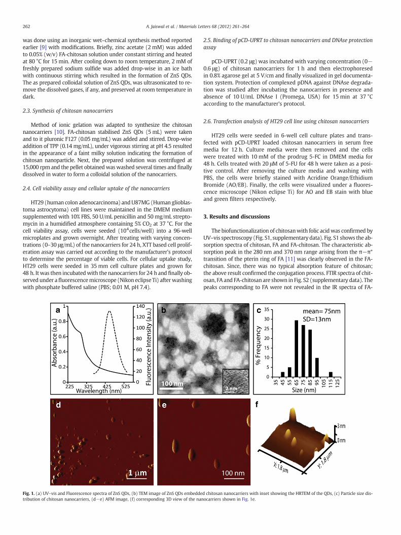
Fig. 1. (a) UV–vis and Fluorescence spectra of ZnS QDs, (b) TEM image of ZnS QDs embeddetribution of chitosan nanocarriers, (d−e) AFM image, (f) corresponding 3D view of the nan
2.5. Binding of pCD-UPRT to chitosan nanocarriers and DNAse protectionassay
pCD-UPRT (0.2 μg) was incubated with varying concentration (0−0.6 μg) of chitosan nanocarriers for 1 h and then electrophoresedin 0.8% agarose gel at 5 V/cm and finally visualized in gel documenta-tion system. Protection of complexed pDNA against DNAse degrada-tion was studied after incubating the nanocarriers in presence andabsence of 10 U/mL DNAse I (Promega, USA) for 15 min at 37 °Caccording to the manufacturer's protocol.
2.6. Transfection analysis of HT29 cell line using chitosan nanocarriers
HT29 cells were seeded in 6-well cell culture plates and trans-fected with pCD-UPRT loaded chitosan nanocarriers in serum freemedia for 12 h. Culture media were then removed and the cellswere treated with 10 mM of the prodrug 5-FC in DMEM media for48 h. Cells treated with 20 μM of 5-FU for 48 h were taken as a posi-tive control. After removing the culture media and washing withPBS, the cells were briefly stained with Acridine Orange/EthidiumBromide (AO/EB). Finally, the cells were visualized under a fluores-cence microscope (Nikon eclipse Ti) for AO and EB stain with blueand green filters respectively.
3. Results and discussions
The biofunctionalization of chitosanwith folic acidwas confirmed byUV–vis spectroscopy (Fig. S1, supplementary data). Fig. S1 shows the ab-sorption spectra of chitosan, FA and FA-chitosan. The characteristic ab-sorption peak in the 280 nm and 370 nm range arising from the π−π*transition of the pterin ring of FA [11] was clearly observed in the FA-chitosan. Since, there was no typical absorption feature of chitosan;the above result confirmed the conjugation process. FTIR spectra of chit-osan, FA and FA-chitosan are shown in Fig. S2 (supplementary data). Thepeaks corresponding to FA were not revealed in the IR spectra of FA-
d chitosan nanocarriers with inset showing the HRTEM of the QDs, (c) Particle size dis-ocarriers shown in Fig. 1e.
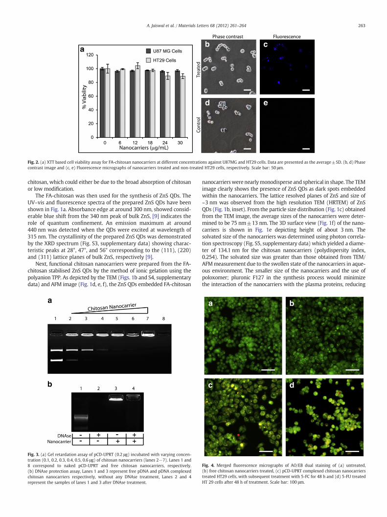
Fig. 2. (a) XTT based cell viability assay for FA-chitosan nanocarriers at different concentrations against U87MG and HT29 cells. Data are presented as the average±SD. (b, d) Phasecontrast image and (c, e) Fluorescence micrographs of nanocarriers treated and non-treated HT29 cells, respectively. Scale bar: 50 μm.
263A. Jaiswal et al. / Materials Letters 68 (2012) 261–264
chitosan, which could either be due to the broad absorption of chitosanor low modification.
The FA-chitosan was then used for the synthesis of ZnS QDs. TheUV–vis and fluorescence spectra of the prepared ZnS QDs have beenshown in Fig. 1a. Absorbance edge at around 300 nm, showed consid-erable blue shift from the 340 nm peak of bulk ZnS, [9] indicates therole of quantum confinement. An emission maximum at around440 nm was detected when the QDs were excited at wavelength of315 nm. The crystallinity of the prepared ZnS QDs was demonstratedby the XRD spectrum (Fig. S3, supplementary data) showing charac-teristic peaks at 28°, 47°, and 56° corresponding to the (111), (220)and (311) lattice planes of bulk ZnS, respectively [9].
Next, functional chitosan nanocarriers were prepared from the FA-chitosan stabilised ZnS QDs by the method of ionic gelation using thepolyanion TPP. As depicted by the TEM (Figs. 1b and S4, supplementarydata) and AFM image (Fig. 1d, e, f), the ZnS QDs embedded FA-chitosan
Fig. 3. (a) Gel retardation assay of pCD-UPRT (0.2 μg) incubated with varying concen-tration (0.1, 0.2, 0.3, 0.4, 0.5, 0.6 μg) of chitosan nanocarriers (lanes 2−7). Lanes 1 and8 correspond to naked pCD-UPRT and free chitosan nanocarriers, respectively.(b) DNAse protection assay, Lanes 1 and 3 represent free pDNA and pDNA complexedchitosan nanocarriers respectively, without any DNAse treatment, Lanes 2 and 4represent the samples of lanes 1 and 3 after DNAse treatment.
nanocarrierswere nearlymonodisperse and spherical in shape. The TEMimage clearly shows the presence of ZnS QDs as dark spots embeddedwithin the nanocarriers. The lattice resolved planes of ZnS and size of~3 nm was observed from the high resolution TEM (HRTEM) of ZnSQDs (Fig. 1b, inset). From the particle size distribution (Fig. 1c) obtainedfrom the TEM image, the average sizes of the nanocarriers were deter-mined to be 75 nm±13 nm. The 3D surface view (Fig. 1f) of the nano-carriers is shown in Fig. 1e depicting height of about 3 nm. Thesolvated size of the nanocarriers was determined using photon correla-tion spectroscopy (Fig. S5, supplementary data) which yielded a diame-ter of 134.1 nm for the chitosan nanocarriers (polydispersity index,0.254). The solvated size was greater than those obtained from TEM/AFMmeasurement due to the swollen state of the nanocarriers in aque-ous environment. The smaller size of the nanocarriers and the use ofpoloxomer; pluronic F127 in the synthesis process would minimizethe interaction of the nanocarriers with the plasma proteins, reducing
Fig. 4. Merged fluorescence micrographs of AO/EB dual staining of (a) untreated,(b) free chitosan nanocarriers treated, (c) pCD-UPRT complexed chitosan nanocarrierstreated HT29 cells, with subsequent treatment with 5-FC for 48 h and (d) 5-FU treatedHT 29 cells after 48 h of treatment. Scale bar: 100 μm.

264 A. Jaiswal et al. / Materials Letters 68 (2012) 261–264
opsonisation, and could thereby increase the circulation time of thenanocarriers [12]. Zeta potential of the nanocarriers at 25 °C in waterwas measured to be +24.65 mV, which rendered stability to the nano-carriers against aggregation.
To evaluate the cytotoxicity of the prepared nanocarriers, we haveperformed the XTT based cell viability assay. Notably, for both the celllines tested, more than 95% of the cells were viable (Fig. 2a) after 24 hof treatment even at the highest concentration of nanocarriers used,thereby confirming its biocompatibility. Next, the cellular uptake ofthe nanocarriers was studied in HT29 cells by using fluorescence mi-croscopy. The representative phase contrast and fluorescence micro-graphs of the cells after 24 h of incubation with the functionalnanocarriers, is shown in Fig. 2b and c respectively. The uptake ofthe nanocarriers was confirmed from the blue fluorescence observed(Fig. 2c) which was ascribed to the quantum dots present within thenanocarriers. Though the fluorescence intensity observed (Fig. 2c)was low, in comparison to the control cells (Fig. 2e) where no fluores-cence was detected indicated that the fluorescence obtained was in-deed due to the cellular internalization of the nanocarriers.
The gel retardation assay (Fig. 3a) visibly showed that the amount ofpDNA in the lanes reduced (lane 2 to lane 7) with increasing concentra-tion of the nanocarriers indicating efficient binding. The bands observedin thewells of lane 2 to lane 7 confirmed the complexation of pDNAwiththe nanocarriers which retarded the movement of the pDNA from thewells. Saturation of the nanocarriers with pDNA was observed (lane 7)for the maximum concentration of nanocarriers (the ratio of nanocar-riers/pDNAwas 3 μg/μg) used. Lane 1 and lane 8 represented the controlsamples containing naked pDNA and the free nanocarriers, respectively.Further, to check the stability of the pDNA during in vitro conditions, wecarried out DNAse protection assay which showed (Fig. 3b) that bindingof pDNAwith the chitosan nanocarriers protected thepDNA fromdegra-dation (well 4) as compared to naked DNA (lane 2), where no bandwasseen indicating complete digestion of pDNA by DNAse. Lane 1 contain-ing only the pDNA and lane 3 containing the pDNA complexed chitosannanocarriers without any DNAse treatment served as the experimentalcontrols. The zeta potential of the chitosan nanocarriers after bindingwith pDNA was measured to be +23.76 mV. Since, the nanocarrierswere already synthesized using the polyanion TPP, the zeta potentialof the nanocarriers after binding with pDNA was not affected much. Inother words, as the concentration of TPP was much higher in compari-son to that of pDNA, even upon complexation the effective concentra-tion of TPP was considerably high. Hence the effective charge (andthus zeta potential) remained unaffected.
The transfecting ability of the chitosan nanocarriers was demon-strated by the induction of cell death in HT29 cell lines after additionof the prodrug, 5-FC, as revealed from AO/EB staining (Fig. S6). Thefluorescence microscopic images obtained with blue and green filterspost AO/EB dual staining were merged and are shown in Fig. 4. Tran-sient expression of the suicide genes to CD-UPRT enzymes convertsthe added prodrug 5-FC to 5-FU and other toxic metabolites, whichleads to cell death [7]. Fig. 4a and b showed that untreated andunloaded chitosan nanocarriers treated cells stained green due tothe uptake of AO dye and both have well organized chromatin
structures indicating normal morphology. On the other hand, incase of pCD-UPRT complexed chitosan nanocarriers treated cells,nuclear uptake of EB was observed as indicated by the orange-redstain of the dye (Fig. 4c) which corroborated compromised cell mem-brane and cell death. Cells treated only with the drug 5-FU showedextensive cell death and served as positive control (Fig. 4d).
4. Conclusion
In summary, we demonstrated a facile method for the preparationof functional chitosan nanocarriers containing folic acid and ZnS QDsas targeting and imaging modalities, respectively. The prepared nano-carriers showed no toxicity against the cell lines tested, enabling it tobe used for gene delivery. The cellular uptake of the nanocarriers wasalso demonstrated by fluorescence microscopy. Gel retardation assayshowed efficient binding of pCD-UPRT with chitosan nanocarriers,and DNAse protection assay proved the stability of the pDNAcomplexed chitosan nanocarriers against degradation. The functionaleffect of the nanocarriers in transfecting HT29 cells can further beexplored as a potential non-viral system for gene therapy.
Acknowledgements
This research was supported by the Department of Biotechnology(Nos. BT/49/NE/TBP/2010, and BT/01/NE/PS/08), the Department ofScience and Technology (SR/S5/NM-108/2006 and 2/2/2005-S.F.). AmitJaiswal is thankful to Shubaash A. R. G. for helpingwith AFM and Depart-ment of Chemical Engineering, IIT Guwahati for Zeta potential measure-ment. We are also thankful to the reviewer whose suggestions havehelped in improving the manuscript. Assistance from CIF, IIT Guwahatiis acknowledged.
Appendix A. Supplementary data
Supplementary data to this article can be found online at doi:10.1016/j.matlet.2011.10.082.
References
[1] Patil SD, Rhodes DG, Burgess DJ. AAPS J 2005;7:E61–77.[2] Mao HQ, Roy K, Troung-Le VL, Janes KA, Lin KY, Wang Y, et al. J Control Release
2001;70:399–421.[3] Mansouria S, Cuieb Y, Winnikb F, Shia Q, Lavignea P, Benderdoura M, et al. Bioma-
terials 2006;27:2060–5.[4] Michalet X, Pinaud FF, Bentolila LA, Tsay JM, Doose S, Li JJ, et al. Science 2005;307:
538–44.[5] Nie Q, Tan WB, Zhang Y. Nanotechnology 2006;17:140–4.[6] Li L, Chen D, Zhang Y, Deng Z, Ren X, Meng X, et al. Nanotechnology 2007;18:1–6.[7] Gopinath P, Ghosh SS. Biotechnol Lett 2008;30:1913–21.[8] Yang SJ, Lin FH, Tsai KC, Wei MF, Tsai HM, Wong JM, et al. Bioconjug Chem
2010;21:679–89.[9] Jaiswal A, Sanpui P, Chattopahyay A, Ghosh SS. Plasmonics 2011;6:125–32.[10] Sanpui P, Chattopahyay A, Ghosh SS. ACS Appl Mater Interfaces 2011;3:218–28.[11] Liu F, Deng D, Chen X, Qian Z, Achilefu S, Gu Y. Mol Imaging Biol 2010;12:
595–607.[12] Alexis F, Pridgen E, Molnar LK, Farokhzad OC. Mol Pharm 2008;5:505–15.