functional implication of b-carotene - plant physiology
TRANSCRIPT

Functional Implication of b-Carotene Hydroxylasesin Soybean Nodulation1[C][W][OA]
Yun-Kyoung Kim2, Sunghan Kim2, Ji-Hyun Um, Kyunga Kim, Sun-Kang Choi, Byung-Hun Um,Suk-Woo Kang, Jee-Woong Kim, Shinichi Takaichi, Seok-Bo Song, Choon-Hwan Lee, Ho-Seung Kim,Ki Woo Kim, Kyoung Hee Nam, Suk-Ha Lee, Yul-Ho Kim, Hyang-Mi Park, Sun-Hwa Ha,Desh Pal S. Verma, and Choong-Ill Cheon*
Department of Biological Science (Y.-K.K., S.K., J.-H.U., K.H.N. C.-I.C.) and Department of Statistics (K.K.), SookmyungWomen’s University, Seoul 140–742, Korea; Gangneung Science Industry Foundation, Gangneung 210-340, Korea(S.-K.C.); Natural Products Research Center, KIST Gangneung Institute, Gangneung 210-340, Korea (B.-H.U.,S.-W.K.); Electron Microscopy Laboratory, Dental Research Institute (J.-W.K.), and School of Plant Science(S.-H.L.), Seoul National University, Seoul 151-742, Korea; Department of Biology, Nippon Medical School,Nakahara, Kawasaki 113-8602, Japan (S.T.); Department of Functional Crops, National Institute of CropScience, Milyang 441-857, Korea (S.-B.S.); Department of Molecular Biology, Pusan National University, Busan609-735, Korea (C.-H.L., H.-S.K.); School of Ecological and Environmental Systems, Kyungpook NationalUniversity, Sangju 702-701, Korea (K.W.K.); National Institute of Crop Science, Suwon 441-857, Korea (Y.-H.K.,H.-M.P.); Graduate School of Biotechnology and Crop Biotech Institute, Kyung Hee University, Yongin 446-701, Korea (S.-H.H.); and Biotechnology Center, Ohio State University, Columbus, Ohio 43210 (D.P.S.V.)
Legume-Rhizobium spp. symbiosis requires signaling between the symbiotic partners and differential expression of plant genesduring nodule development. Previously, we cloned a gene encoding a putative b-carotene hydroxylase (GmBCH1) from soybean(Glycine max) whose expression increased during nodulation with Bradyrhizobium japonicum. In this work, we extended our studyto three GmBCHs to examine their possible role(s) in nodule development, as they were additionally identified as nodule specific,along with the completion of the soybean genome. In situ hybridization revealed the expression of three GmBCHs (GmBCH1,GmBCH2, and GmBCH3) in the infected cells of root nodules, and their enzymatic activities were confirmed by functional assaysin Escherichia coli. Localization of GmBCHs by transfecting Arabidopsis (Arabidopsis thaliana) protoplasts with green fluorescentprotein fusions and by electron microscopic immunogold detection in soybean nodules indicated that GmBCH2 and GmBCH3were present in plastids, while GmBCH1 appeared to be cytosolic. RNA interference of the GmBCHs severely impaired nitrogenfixation as well as nodule development. Surprisingly, we failed to detect zeaxanthin, a product of GmBCH, or any othercarotenoids in nodules. Therefore, we examined the possibility that most of the carotenoids in nodules are converted orcleaved to other compounds. We detected the expression of some carotenoid cleavage dioxygenases (GmCCDs) in wild-typenodules and also a reduced amount of zeaxanthin in GmCCD8-expressing E. coli, suggesting cleavage of the carotenoid. In viewof these findings, we propose that carotenoids such as zeaxanthin synthesized in root nodules are cleaved by GmCCDs, and wediscuss the possible roles of the carotenoid cleavage products in nodulation.
Legume-Rhizobium spp. symbiosis results in theformation of the root nodule, in which rhizobia fixatmospheric nitrogen. Nodule development requires
diverse events, such as Nod factor synthesis in therhizobia, perception of the Nod factor on plant rootsby receptor-like kinases, endocytosis of rhizobia intoplant cells, and so on (Stacey et al., 2006; Oldroydet al., 2011; Singh and Parniske, 2012). Sequentialexpression of numerous plant genes occurs duringnodulation, contributing to different stages includingnitrogen fixation. Arbuscular mycorrhizal (AM) sym-biosis exhibits many similarities to the nodulationprocess (Oldroyd et al., 2009). For example, SymRK,the receptor-like kinase gene, is required for bothrhizobial and AM symbioses (Stracke et al., 2002).Similarly, the signal transduction pathways follow-ing perception are also in part the same, and thegenes common to the two pathways have been re-ferred to as the common symbiosis (SYM) genes(Kistner et al., 2005). These similarities may reflectcommon mechanisms for host plant cells to respondto symbionts, although the commonality is not globallydefined yet.
1 This work was supported by the Basic Science Research Programthrough the National Research Foundation of Korea funded by the Min-istry of Education, Science, and Technology (grant no. 2–1205–0037) andby the Next-Generation BioGreen 21 Program (grant no.PJ009076012013), Rural DevelopmentAdministration, Republic of Korea.
2 These authors contributed equally to the article.* Corresponding author; [email protected] author responsible for distribution of materials integral to the
findings presented in this article in accordance with the policy de-scribed in the Instructions for Authors (www.plantphysiol.org) is:Choong-Ill Cheon ([email protected]).
[C] Some figures in this article are displayed in color online but inblack and white in the print edition.
[W] The online version of this article contains Web-only data.[OA] Open Access articles can be viewed online without a subscrip-
tion.www.plantphysiol.org/cgi/doi/10.1104/pp.113.215020
1420 Plant Physiology�, July 2013, Vol. 162, pp. 1420–1433, www.plantphysiol.org � 2013 American Society of Plant Biologists. All Rights Reserved.
Dow
nloaded from https://academ
ic.oup.com/plphys/article/162/3/1420/6110706 by guest on 22 D
ecember 2021

Plant carotenoids are mostly C40 tetraterpenoidpigments with a series of double bonds (DellaPennaand Pogson, 2006; Lu and Li, 2008). They play essentialroles in photosynthesis. The phytohormone abscisicacid (ABA) is synthesized from xanthophylls, oxy-genated derivatives of carotenoids. The beneficial ef-fects of carotenoids for human disease prevention andhealth promotion are well established and are basedon their antioxidant activities (Kopsell and Kopsell,2006; Rao and Rao, 2007; von Lintig, 2010). Metabolicengineering approaches have produced crop plantswith enhanced carotenoid contents and improvednutritional value (Giuliano et al., 2008). For example,enhancement of b-carotene, provitamin A, by engi-neering the carotenoid biosynthetic pathway resultedin the development of cv Golden rice (Oryza sativa; Yeet al., 2000; Paine et al., 2005; Ha et al., 2010).The initial step of carotenoid biosynthesis is the
production of phytoene by the enzyme phytoenesynthase (Fig. 1; DellaPenna and Pogson, 2006; Cazzonelliand Pogson, 2010). The subsequent activities of desat-urases, isomerase, and cyclase convert phytoene intolycopene and further into b-carotene. Xanthophyllsynthesis begins with the action of b-carotene hy-droxylase (BCH) on b-carotene, producing initiallyb-cryptoxanthin and thereafter zeaxanthin (Kim et al.,2009). Overexpression of BCH has been found to confertolerance to light stress (Davison et al., 2002). The sub-sequent steps catalyzed by zeaxanthin epoxidase (ZEP)and neoxanthin synthase lead to the synthesis of ABA(Takaichi and Mimuro, 1998).
Various carotenoid cleavage dioxygenases (CCDs)catalyze the formation of apocarotenoids with func-tions as hormones, flavors, and pigments (Auldridgeet al., 2006b; Strack and Fester, 2006; Tsuchiya andMcCourt, 2009; Walter et al., 2010). Recently, CCD7and CCD8 were shown to control the synthesis ofstrigolactones, newly discovered hormones that inhibitshoot branching (Gomez-Roldan et al., 2008; Umeharaet al., 2008; Vogel et al., 2010; Ruyter-Spira et al., 2013).In addition, carotenoid cleavage products have beendiscovered in plant roots colonized by AM fungi(Strack and Fester, 2006). During AM symbiosis, rootssynthesize apocarotenoids at the same time as acti-vating plant genes for carotenoid metabolism. Al-though RNA interference (RNAi)-mediated inhibitionof apocarotenoid synthesis suggests that apocar-otenoids are functionally significant (Snowden et al.,2005; Floss et al., 2008), their role in AM symbiosis isunknown.
In a search for genes differentially induced duringsoybean (Glycine max)-Rhizobium spp. symbiosis, sev-eral antioxidant genes, including a gene encoding aputative BCH, were identified. In this report, we de-scribe genes (GmBCHs) encoding a putative BCHwhose expression increased in soybean root nodules.Therefore, the biochemical activities of BCHs wereinvestigated. RNAi inhibition of GmBCH expressioninterfered with nitrogen fixation as well as noduledevelopment. Subsequent analysis of the expressionand biochemical activities of GmCCDs in root nodulesled us to hypothesize that GmCCD8 could be involvedin the synthesis of apocarotenoids from zeaxanthin inthese nodules.
RESULTS
Expression of Genes Encoding Putative BCHs in Soybean
We isolated from root nodules of soybean a com-plementary DNA (cDNA) with strong homology toBCH (GmBCH1), whose expression was higher innodules than in roots (Fig. 2A; Lee et al., 2005). Ina BLAST search with the GmBCH1 sequence againstthe soybean genome (http://www.phytozome.net/soybean.php; Schmutz et al., 2010), we found severalopen reading frames encoding products with aminoacid sequences highly homologous to that of GmBCH1;they were designated GmBCH2 to GmBCH5 (GenBankaccession numbers are as follows: GmBCH1 [AY575953],GmBCH2 [BT093388], GmBCH3 [BT098487], GmBCH4[JF970190], GmBCH5 [JF970191]). The open readingframes of GmBCH1 and GmBCH2 are very similar tofunctionally confirmed BCHs (e.g. they have 74% and75% sequence identity, respectively, to the BCH fromcoffee [Coffea arabica], CaCRTR-B; Simkin et al., 2008;Supplemental Fig. S1A). They have divergentN-terminal regions, like most previously reported BCHs,but carry four His-containing motifs, in which thespacing of the His residues was conserved (e.g.
Figure 1. The biosynthetic pathway of carotenoids in plants. GGPP,Geranylgeranyl diphosphate; PSY, phytoene synthase; PDS, phytoenedesaturase; ZDS, z-carotene desaturase; CRTISO, carotene isomer-ase; LCYB, lycopene b-cyclase; CYP97A3 and CYP97C1, cytochromeP450 enzymes; NSY, neoxanthin synthase; LCYE, lycopene «-cyclase;CRTR-E, «-carotene hydroxylase. Enzymes in red were examined inthis study.
Plant Physiol. Vol. 162, 2013 1421
Carotenoid Metabolism in Soybean Root Nodules
Dow
nloaded from https://academ
ic.oup.com/plphys/article/162/3/1420/6110706 by guest on 22 D
ecember 2021
HXXXXH and HXXHH). The presence of the His resi-dues in these motifs has been confirmed to be essentialfor the enzymatic activity, as mutagenesis abolishingthese His residues resulted in no enzymatic conversionof b-carotene into zeaxanthin (Supplemental Fig. S1A;Bouvier et al., 1998). GmBCH2 appears to possess aplastid transit sequence (see below; Yu et al., 2007), andGmBCH3 has a sequence almost identical to GmBCH1except for its N-terminal 33 amino acids, so it alsocontains the above-mentioned motifs common to BCHs.The 59 untranslated regions of GmBCH1 and GmBCH3differ. Whereas the GmBCH3 locus could be identifiedin the present version of the soybean genome database,the unique 59 region of the GmBCH1 sequence was notdetected in the database. Therefore, we attempted toestablish the presence of GmBCH1 in the soybean ge-nome by cloning its specific 59 DNA region (see “Ma-terials and Methods”). A 432-bp DNA fragment, whichincluded the upstream promoter region of GmBCH1,was cloned (Supplemental Fig. S2A), and this 59 regionas well as the coding region of GmBCH1 were alsodetected by genomic PCR (Supplemental Fig. S2B, lanes3 and 6). GmBCH4 and GmBCH5 also contain themotifs mentioned above and thus are regarded as ad-ditional BCH paralogs (Supplemental Fig. S1A).
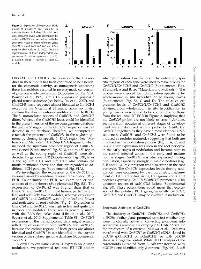
We investigated the expression of the GmBCHs invarious tissues by real-time reverse transcription (RT)-PCR. To optimize the PCR, we examined criticalaspects of the primers (Supplemental Fig. S3). Theexpression of GmBCH2 was higher than that ofGmBCH1 and GmBCH3 in most tissues, particularly inleaf, and relatively low in nodules, whereas expressionof GmBCH1 and GmBCH3 was high in leaf and flowerand noticeable in root nodules (Fig. 2). Expression ofGmBCH4 and GmBCH5 was high in leaf and quite lowin roots and nodules. These results generally matchwith the RNA-Seq Atlas data (Libault et al., 2010;Severin et al., 2010; Supplemental Table S1). GmBCH3expression in the transcriptomic data might actuallyreflect the expression of both GmBCH1 and GmBCH3,because the coding regions of both genes are almostidentical and GmBCH1 is not identified in the currentversion of the soybean genome database (SupplementalTable S1).
In order to examine GmBCH expression duringnodulation, we performed real-time RT-PCR and in
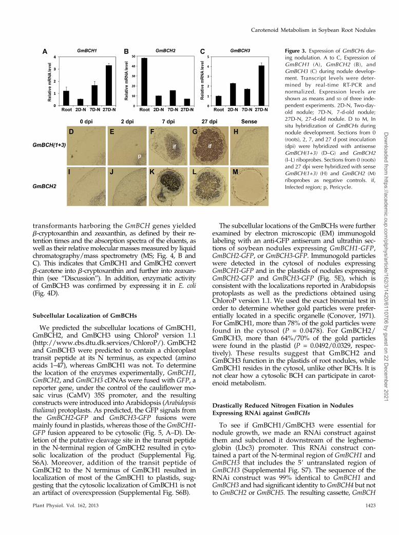
situ hybridization. For the in situ hybridization, spe-cific regions of each gene were used to make probes forGmBCH1/GmBCH3 and GmBCH2 (Supplemental Figs.S1 and S4, A and B; see “Materials and Methods”). Theprobes were checked for hybridization specificity bywhole-mount in situ hybridization to young leaves(Supplemental Fig. S4, C and D). The relative ex-pression levels of GmBCH1/GmBCH3 and GmBCH2obtained from whole-mount in situ hybridization inyoung leaves were found to be comparable to thosefrom the real-time RT-PCR in Figure 2, implying thatthe GmBCH probes are not likely to cross hybridize.Sections from nodules at different stages of develop-ment were hybridized with a probe for GmBCH1/GmBCH3 together, as they have almost identical DNAsequences. GmBCH1 and GmBCH3 were found to beinduced as nodules matured, suggesting that both areinvolved in the nodulation process (Fig. 3, A, C, andD–G). Their expression was seen in the root pericyclein the early stages of nodulation and became high inthe central infected zones in the early and maturenodule stages. GmBCH2 was also expressed duringnodulation, especially strongly in 7-d-old nodules (Fig.3, B and I–L). Its expression was also strong in the rootpericycle. The GmBCH expression levels during nod-ulation were confirmed by the fluorometric measure-ment of GUS activities using transgenic roots andnodules expressing GmBCH2/GmBCH3 promoter (1.6-kbupstream regions of each)-GUS fusions (SupplementalFig. S5). These observations could mean that expres-sion of the putative BCH genes, especially GmBCH1,GmBCH2, and GmBCH3, may be involved in nodulation.
Enzymatic Activities of GmBCHs
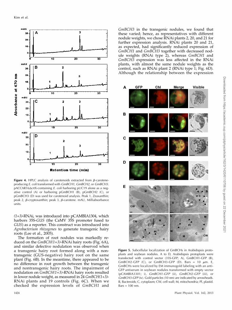
The similarity of GmBCH1, GmBCH2, and GmBCH3to BCHs of other plants prompted us to test whether theywere functionally active in converting b-carotene intozeaxanthin. Escherichia coli carrying pACCAR16DcrtX forthe production of b-carotene (Misawa et al., 1990) wastransformed with GmBCH1 or GmBCH2 cDNA cloned inpUC19 (pGmBCH1 or pGmBCH2) or with pUC19alone as a negative control. While HPLC analysis of thecarotenoids extracted from E. coli transformed withpUC19 alone identified only b-carotene (Fig. 4A), E. coli
Figure 2. Expression of the soybean BCHsGmBCH1, GmBCH2, and GmBCH3 insoybean tissues, including 27-d-old nod-ules. Transcript levels were determined byreal-time RT-PCR and normalized with thegeometric mean of three reference genes(GmELF1b, GmActin2/GmActin7, and Ubiq-uitin; Vandesompele et al., 2002). Data arerepresentative of three independent ex-periments. Error bars represent SD (n = 3).L, Leaf; S, stem; F, flower; R, root; N,nodule.
1422 Plant Physiol. Vol. 162, 2013
Kim et al.
Dow
nloaded from https://academ
ic.oup.com/plphys/article/162/3/1420/6110706 by guest on 22 D
ecember 2021
transformants harboring the GmBCH genes yieldedb-cryptoxanthin and zeaxanthin, as defined by their re-tention times and the absorption spectra of the eluents, aswell as their relative molecular masses measured by liquidchromatography/mass spectrometry (MS; Fig. 4, B andC). This indicates that GmBCH1 and GmBCH2 convertb-carotene into b-cryptoxanthin and further into zeaxan-thin (see “Discussion”). In addition, enzymatic activityof GmBCH3 was confirmed by expressing it in E. coli(Fig. 4D).
Subcellular Localization of GmBCHs
We predicted the subcellular locations of GmBCH1,GmBCH2, and GmBCH3 using ChloroP version 1.1(http://www.cbs.dtu.dk.services/ChloroP/). GmBCH2and GmBCH3 were predicted to contain a chloroplasttransit peptide at its N terminus, as expected (aminoacids 1–47), whereas GmBCH1 was not. To determinethe location of the enzymes experimentally, GmBCH1,GmBCH2, and GmBCH3 cDNAs were fused with GFP, areporter gene, under the control of the cauliflower mo-saic virus (CaMV) 35S promoter, and the resultingconstructs were introduced into Arabidopsis (Arabidopsisthaliana) protoplasts. As predicted, the GFP signals fromthe GmBCH2-GFP and GmBCH3-GFP fusions weremainly found in plastids, whereas those of the GmBCH1-GFP fusion appeared to be cytosolic (Fig. 5, A–D). De-letion of the putative cleavage site in the transit peptidein the N-terminal region of GmBCH2 resulted in cyto-solic localization of the product (Supplemental Fig.S6A). Moreover, addition of the transit peptide ofGmBCH2 to the N terminus of GmBCH1 resulted inlocalization of most of the GmBCH1 to plastids, sug-gesting that the cytosolic localization of GmBCH1 is notan artifact of overexpression (Supplemental Fig. S6B).
The subcellular locations of the GmBCHs were furtherexamined by electron microscopic (EM) immunogoldlabeling with an anti-GFP antiserum and ultrathin sec-tions of soybean nodules expressing GmBCH1-GFP,GmBCH2-GFP, or GmBCH3-GFP. Immunogold particleswere detected in the cytosol of nodules expressingGmBCH1-GFP and in the plastids of nodules expressingGmBCH2-GFP and GmBCH3-GFP (Fig. 5E), which isconsistent with the localizations reported in Arabidopsisprotoplasts as well as the predictions obtained usingChloroP version 1.1. We used the exact binomial test inorder to determine whether gold particles were prefer-entially located in a specific organelle (Conover, 1971).For GmBCH1, more than 78% of the gold particles werefound in the cytosol (P = 0.0478). For GmBCH2/GmBCH3, more than 64%/70% of the gold particleswere found in the plastid (P = 0.0492/0.0329, respec-tively). These results suggest that GmBCH2 andGmBCH3 function in the plastids of root nodules, whileGmBCH1 resides in the cytosol, unlike other BCHs. It isnot clear how a cytosolic BCH can participate in carot-enoid metabolism.
Drastically Reduced Nitrogen Fixation in NodulesExpressing RNAi against GmBCHs
To see if GmBCH1/GmBCH3 were essential fornodule growth, we made an RNAi construct againstthem and subcloned it downstream of the leghemo-globin (Lbc3) promoter. This RNAi construct con-tained a part of the N-terminal region of GmBCH1 andGmBCH3 that includes the 59 untranslated region ofGmBCH3 (Supplemental Fig. S7). The sequence of theRNAi construct was 99% identical to GmBCH1 andGmBCH3 and had significant identity to GmBCH4 but notto GmBCH2 or GmBCH5. The resulting cassette, GmBCH
Figure 3. Expression of GmBCHs dur-ing nodulation. A to C, Expression ofGmBCH1 (A), GmBCH2 (B), andGmBCH3 (C) during nodule develop-ment. Transcript levels were deter-mined by real-time RT-PCR andnormalized. Expression levels areshown as means and SD of three inde-pendent experiments. 2D-N, Two-day-old nodule; 7D-N, 7-d-old nodule;27D-N, 27-d-old nodule. D to M, Insitu hybridization of GmBCHs duringnodule development. Sections from 0(roots), 2, 7, and 27 d post inoculation(dpi) were hybridized with antisenseGmBCH(1+3) (D–G) and GmBCH2(I–L) riboprobes. Sections from 0 (roots)and 27 dpi were hybridized with senseGmBCH(1+3) (H) and GmBCH2 (M)riboprobes as negative controls. if,Infected region; p, Pericycle.
Plant Physiol. Vol. 162, 2013 1423
Carotenoid Metabolism in Soybean Root Nodules
Dow
nloaded from https://academ
ic.oup.com/plphys/article/162/3/1420/6110706 by guest on 22 D
ecember 2021
(1+3)-RNAi, was introduced into pCAMBIA1304, whichharbors 35S-GUS (the CaMV 35S promoter fused toGUS) as a reporter. This construct was introduced intoAgrobacterium rhizogenes to generate transgenic hairyroots (Lee et al., 2005).
The formation of root nodules was markedly re-duced on the GmBCH(1+3)-RNAi hairy roots (Fig. 6A),and similar defective nodulation was observed whena transgenic hairy root formed along with a non-transgenic (GUS-negative) hairy root on the sameplant (Fig. 6B). In the meantime, there appeared to beno difference in root growth between the transgenicand nontransgenic hairy roots. The impairment ofnodulation on GmBCH(1+3)-RNAi hairy roots resultedin lower nodule weight, as measured in 24 GmBCH(1+3)-RNAi plants and 19 controls (Fig. 6C). When wechecked the expression levels of GmBCH1 and
GmBCH3 in the transgenic nodules, we found thatthese varied; hence, as representatives with differentnodule weights, we chose RNAi plants 2, 20, and 21 forfurther expression analysis. RNAi plants 20 and 21,as expected, had significantly reduced expression ofGmBCH1 and GmBCH3 together with decreased nod-ule weights (RNAi type 2), whereas GmBCH1 andGmBCH3 expression was less affected in the RNAiplants, with almost the same nodule weights as thecontrol, such as RNAi plant 2 (RNAi type 1; Fig. 6D).Although the relationship between the expression
Figure 5. Subcellular localization of GmBCHs in Arabidopsis proto-plasts and soybean nodules. A to D, Arabidopsis protoplasts weretransfected with control vector (35S-GFP; A), GmBCH1-GFP (B),GmBCH2-GFP (C), or GmBCH3-GFP (D). Bars = 10 mm. E,GmBCHs were localized by EM immunogold labeling with an anti-GFP antiserum in soybean nodules transformed with empty vector(pCAMBIA3301; i), GmBCH1-GFP (ii), GmBCH2-GFP (iii), orGmBCH3-GFP (iv). Gold particles (10 nm) are indicated by arrowheads.B, Bacteroids; C, cytoplasm; CW, cell wall; M, mitochondria; Pl, plastid.Bars = 100 nm.
Figure 4. HPLC analysis of carotenoids extracted from b-carotene-producing E. coli transformed withGmBCH1, GmBCH2, orGmBCH3.pACCAR16ΔcrtX-containing E. coli harboring pUC19 alone as a neg-ative control (A) or harboring pGmBCH1 (B), pGmBCH2 (C), orpGmBCH3 (D) was used for carotenoid analysis. Peak 1, Zeaxanthin;peak 2, b-cryptoxanthin; peak 3, b-carotene. mAU, Milliabsorbanceunits.
1424 Plant Physiol. Vol. 162, 2013
Kim et al.
Dow
nloaded from https://academ
ic.oup.com/plphys/article/162/3/1420/6110706 by guest on 22 D
ecember 2021

Figure 6. Nodule development on transgenic hairy roots expressing an RNAi construct against both GmBCH1 and GmBCH3.A, Nodules formed on transgenic hairy roots containing pCAMBIA1304 alone (control; 35S-GUS; left plant) or theGmBCH(1+3)-RNAi cloned in pCAMBIA1304 (right plant). After GUS assay, only GUS-positive (transgenic) hairy roots were inoculated withB. japonicum (K599). These experiments were repeated three times, and representative results are shown. In each experiment,five to seven plants were used per construct. B, A GUS-negative (untransformed control) hairy root is shown on the right. C,Total nodule weights (mg) of control (35S-GUS) and GmBCH(1+3)-RNAi plants were measured and are shown as means and SD
of three independent experiments. D, Transcript levels are shown in nodules from a transgenic control plant (35S-GUS) and
Plant Physiol. Vol. 162, 2013 1425
Carotenoid Metabolism in Soybean Root Nodules
Dow
nloaded from https://academ
ic.oup.com/plphys/article/162/3/1420/6110706 by guest on 22 D
ecember 2021

levels of GmBCH(1+3) and nodule weight is onlyshown for the three representative transgenic plants(i.e. 2, 20, and 22) in Figure 6D, we actually examinedthe expression levels and nodule weights in 12 trans-genic plants and four controls.
To establish the statistical significance of the RNAiplant groups (i.e. control versus RNAi type 1 versusRNAi type 2), we conducted an ANOVA with Tukey’smultiple comparison procedure and found significantdifferences in nodule weight between control andRNAi type 2 as well as between RNAi type 1 andRNAi type 2 (Supplemental Fig. S8A); there was also asignificant difference in the expression of GmBCH(1+3)between these pairs (Supplemental Fig. S8B; Maritz,1981). In addition, using all 16 plants making up thethree groups, we examined the correlation betweengene expression level and nodule weights based on theSpearman rank correlation coefficient. This yieldeda strong positive correlation of 0.76 (P = 0.001;Supplemental Fig. S8C). Meanwhile, GmBCH4 ex-pression, which was quite low in root nodules, wassomewhat affected in most of the RNAi plants. Theexpression of GmBCH2 as well as GmBCH5 was notaltered in any of the plants (Fig. 6D). These observa-tions imply that the effect of the RNAi on GmBCHtranscript levels was specific to GmBCH1 andGmBCH3. Expression of GmVDE, a gene contributingpositively to the accumulation of zeaxanthin from theopposite direction of GmBCH in the xanthophyll cycle(Fig. 1; Cazzonelli and Pogson, 2010), was also unaf-fected, indicating that silencing of the GmBCHs in theRNAi nodules was not compensated, at least at thetranscription level, by the induction of GmVDE (Fig.6D). In agreement with the observed retardation ofnodule development, on the other hand, expression ofLbc3, which encodes leghemoglobin, an oxygen carrierfor symbiosis with rhizobia that is one of the hallmarkgenes for the development of nitrogen-fixing nodules(Ott et al., 2005), was decreased in RNAi plants 20 and21 (Fig. 6D). In addition, acetylene reduction assaysshowed that nitrogen-fixing ability was also lowerin the RNAi nodules (Fig. 6F). The RNAi nodules
examined by electron microscopy contained emptyvesicles rather than symbiosomes with rhizobia inabout 60% of the cells examined. In addition, the rhi-zobia were not even enclosed by symbiosome mem-branes in about 5% of the cells of the RNAi nodules withstrongly repressed GmBCH(1+3) (Fig. 6E; SupplementalFig. S9). These data indicate that the expression ofGmBCH1 and/or GmBCH3may be essential for noduledevelopment.
Expression of GmBCH2 was prominent duringnodulation; hence, we also silenced GmBCH2 in nod-ules using the leghemoglobin promoter-driven RNAiapproach. When we compared nine GmBCH2-RNAiplants with eight control plants, we found that thereduction in GmBCH2 expression resulted in decreasednodule weight and nitrogenase activity (Fig. 6, G–K).GmBCH5 expression was also reduced in the GmBCH2-RNAi nodules, probably due to its strong homologywith GmBCH2, while the expression of GmBCH1 andGmBCH3 was not affected and GmBCH4 expressionwas somewhat increased (Fig. 6I). About 65% of theinfected cells examined contained empty vesicles, andmost of them exhibited the presence of bacteroidsoutside symbiosomes (Fig. 6J). It is not clear whetherthe phenotypic difference observed by electronmicroscopy of GmBCH(1+3)-RNAi nodules and GmBCH2-RNAi nodules indicates their different roles in nodu-lation. In addition, the expression of GmBCH4 andGmBCH5 was also observed in nodules, albeit at a lowlevel (Supplemental Fig. S1, B and C). Taken together,these results suggest that the GmBCHs, comprisingGmBCH1 to GmBCH5, may be essential for noduledevelopment.
Decreased Expression of the Putative ZeaxanthinEpoxidase Gene during Nodulation
The impairment of nodulation in the RNAi hairyroots suggested to us that a product of BCH action, orsome other derivatives of the carotenoid metabolicpathway, plays an important role in nodulation. Thisled us to examine the expression of genes encoding
Figure 6. (Continued.)three representative transgenic plants: GmBCH(1+3)-RNAi 2, GmBCH(1+3)-RNAi 20, and GmBCH(1+3)-RNAi 21. RNAi plant2 had almost the same nodule weight as the control, RNAi plant 20 had a reduced nodule weight, and RNAi plant 21 had adrastically reduced nodule weight. Transcript levels of GmBCH(1+3), GmBCH2, GmBCH4, GmBCH5, GmVDE, and Lbc3were determined by real-time RT-PCR in controls (35S-GUS) and three GmBCH(1+3)-RNAi nodules and normalized. Ex-pression levels are shown as means and SD of three independent experiments. E, EM images of 27-d-old control (35S-GUS) andGmBCH(1+3)-RNAi plants. GmBCH(1+3)-RNAi nodules often contained empty vesicles (arrow) and bacteroids outside thesymbiosomes (arrowhead). F, Nitrogenase activities were measured by the acetylene reduction assay, and data are averagedfrom three independent experiments. G, Total nodule weights (mg) of control (35S-GUS) and GmBCH2-RNAi plants weremeasured and are shown as means and SD of three independent experiments. H, Nodules formed on transgenic hairy rootscontaining pCAMBIA1304 alone (control; 35S-GUS; left plant) or the GmBCH2-RNAi cloned in pCAMBIA1304 (right plant).After GUS assay, nodules were formed as in A. I, Transcript levels in nodules from one transgenic control plant (35S-GUS) andtwo differentially repressed transgenic plants (GmBCH2-RNAi 14 and GmBCH2-RNAi 16) are shown. Transcript levels ofGmBCH2, GmBCH(1+3), GmBCH4, GmBCH5, GmVDE, and Lbc3 were determined by real-time RT-PCR and normalized.Expression levels are shown as means and SD of three independent experiments. J, EM images of 27-d-old control (35S-GUS)and GmBCH2-RNAi nodules. GmBCH2-RNAi nodules often contained empty vesicles (arrow) and bacteroids outside thesymbiosomes (arrowhead). K, Nitrogenase activities were measured by the acetylene reduction assay as in F.
1426 Plant Physiol. Vol. 162, 2013
Kim et al.
Dow
nloaded from https://academ
ic.oup.com/plphys/article/162/3/1420/6110706 by guest on 22 D
ecember 2021

two enzymes of the xanthophyll cycle: ZEP and vio-laxanthin deepoxidase (VDE). Since we identified threeGmZEPs (Supplemental Fig. S1D) and two GmVDEs(Supplemental Fig. S1E) in the soybean genome se-quence, we examined their expression. Actually, wecame across a few more DNA sequences homologousto the ZEP and VDE genes in other plants, but theyshowed only partial identity and were not furtherstudied (Supplemental Fig. S1, D and E). The primersused to examine the expression of GmZEPs andGmVDEs were designed using DNA sequences highlyconserved among other plants. The transcript levels ofboth genes were considerably lower in roots andnodules than in the aerial parts of the plant, suchas leaves, stems, and flowers (Fig. 7, A and B;Supplemental Fig. S10). As nodules matured, GmZEPexpression decreased and was almost undetectablein mature 27-d-old nodules (Fig. 7D). Expression ofthe GmVDEs remained relatively constant through-out nodule development (Fig. 7E). In addition, weexamined the expression of two soybean 9-cis-epoxycarotenoid dioxygenase1 (NCED1) orthologs(GmNCED1a and GmNCED1b; Supplemental Fig. S1F)and found that their expression was high in flowersbut quite low in roots and nodules, although expres-sion in 7-d-old nodules was a little higher than in otherstages of nodulation (Fig. 7, C and F). The expressionlevels of GmZEPs, GmVDEs, and GmNCED1s generallymatched with those in the soybean RNA-Seq Atlas,especially expression in the aerial parts of the soybean(Libault et al., 2010; Severin et al., 2010; SupplementalTable S1). These expression data appear to indicatethat GmZEPs, GmVDEs, and GmNCED1s do not play
major roles, if any, in further carotenoid metabolismafter BCH during nodulation, although we cannot ruleout the possibility that their protein levels are higherthan suggested by their transcript levels.
Expression of CCDs in Soybean Root Nodules
We hypothesized that zeaxanthin might be synthe-sized in root nodules by BCHs and converted to othercarotenoids, although not via xanthophyll to ABA, be-cause ZEP expression was very low (Fig. 7D). There-fore, we extracted and quantified carotenoids from rootnodules. Contrary to our expectation, carotenoids in-cluding zeaxanthin were almost undetectable in rootnodules (data not shown). This raised the question ofwhat biochemical reactions occur in root nodules sub-sequent to zeaxanthin production by the BCHs. Sincecarotenoid cleavage products have been found in rootsinfected with mycorrhizal fungi and are regarded asimportant in AM symbiosis (Strack and Fester, 2006),we reasoned that the carotenoids synthesized in rootnodules might be depleted if CCDs were active. Thewhite-colored petals of Chrysanthemum spp. expresshigh levels of CmCCD4a, with the result that no carot-enoid can be detected (Ohmiya et al., 2006), and RNAi-mediated suppression of CmCCD4a expression wasfound to lead to the accumulation of carotenoids andyellow petal color, confirming the relationship betweenthe amount of carotenoid and the expression ofCmCCD4a. Thus, we considered the possibility that theconversion of carotenoids into apocarotenoids by CCDsmight explain our failure to detect carotenoids in rootnodules.
Figure 7. Expression of GmZEPs, GmVDEs, and GmNCED1s in soybean. A to C, Expression of GmZEPs, GmVDEs, andGmNCED1s in different tissues. RNA was extracted from different tissues, including 27-d-old nodules of soybean. D to F,Expression ofGmZEPs,GmVDEs, andGmNCED1s during nodule development. RNAs were extracted from roots and 2-, 7-, and27-d-old nodules, and transcript levels were determined by real-time RT-PCR and normalized. Expression of GmZEP1 andGmZEP2 was examined simultaneously using primers for DNA regions of high identity, while the expression of GmZEP3, agene with low overall homology to other GmZEPs, was measured separately and is shown in Supplemental Figure S10. Ex-pression of GmVDE1 and GmVDE2 was also examined simultaneously using primers for the DNA regions of high identity.Expression of GmNCED1a and GmNCED1b was also examined simultaneously using primers for the DNA regions of highidentity. Data are from three independent experiments. L, Leaf; S, stem; F, flower; R, root; N, nodule; 2D-N, 2-d-old nodule;7D-N, 7-d-old nodule; 27D-N, 27-d-old nodule.
Plant Physiol. Vol. 162, 2013 1427
Carotenoid Metabolism in Soybean Root Nodules
Dow
nloaded from https://academ
ic.oup.com/plphys/article/162/3/1420/6110706 by guest on 22 D
ecember 2021
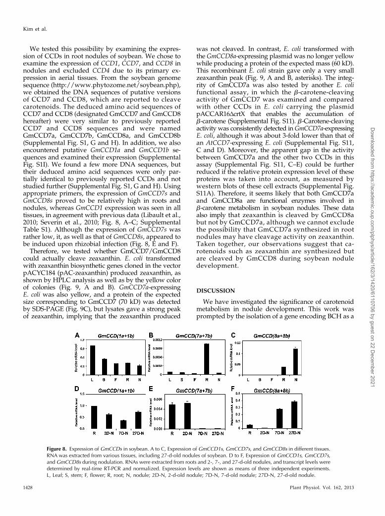
We tested this possibility by examining the expres-sion of CCDs in root nodules of soybean. We chose toexamine the expression of CCD1, CCD7, and CCD8 innodules and excluded CCD4 due to its primary ex-pression in aerial tissues. From the soybean genomesequence (http://www.phytozome.net/soybean.php),we obtained the DNA sequences of putative versionsof CCD7 and CCD8, which are reported to cleavecarotenoids. The deduced amino acid sequences ofCCD7 and CCD8 (designated GmCCD7 and GmCCD8hereafter) were very similar to previously reportedCCD7 and CCD8 sequences and were namedGmCCD7a, GmCCD7b, GmCCD8a, and GmCCD8b(Supplemental Fig. S1, G and H). In addition, we alsoencountered putative GmCCD1a and GmCCD1b se-quences and examined their expression (SupplementalFig. S1I). We found a few more DNA sequences, buttheir deduced amino acid sequences were only par-tially identical to previously reported CCDs and notstudied further (Supplemental Fig. S1, G and H). Usingappropriate primers, the expression of GmCCD7s andGmCCD8s proved to be relatively high in roots andnodules, whereas GmCCD1 expression was seen in alltissues, in agreement with previous data (Libault et al.,2010; Severin et al., 2010; Fig. 8, A–C; SupplementalTable S1). Although the expression of GmCCD7s wasrather low, it, as well as that of GmCCD8s, appeared tobe induced upon rhizobial infection (Fig. 8, E and F).
Therefore, we tested whether GmCCD7/GmCCD8could actually cleave zeaxanthin. E. coli transformedwith zeaxanthin biosynthetic genes cloned in the vectorpACYC184 (pAC-zeaxanthin) produced zeaxanthin, asshown by HPLC analysis as well as by the yellow colorof colonies (Fig. 9, A and B). GmCCD7a-expressingE. coli was also yellow, and a protein of the expectedsize corresponding to GmCCD7 (70 kD) was detectedby SDS-PAGE (Fig. 9C), but lysates gave a strong peakof zeaxanthin, implying that the zeaxanthin produced
was not cleaved. In contrast, E. coli transformed withthe GmCCD8a-expressing plasmid was no longer yellowwhile producing a protein of the expected mass (60 kD).This recombinant E. coli strain gave only a very smallzeaxanthin peak (Fig. 9, A and B, asterisks). The integ-rity of GmCCD7a was also tested by another E. colifunctional assay, in which the b-carotene-cleavingactivity of GmCCD7 was examined and comparedwith other CCDs in E. coli carrying the plasmidpACCAR16ΔcrtX that enables the accumulation ofb-carotene (Supplemental Fig. S11). b-Carotene-cleavingactivity was consistently detected inGmCCD7a-expressingE. coli, although it was about 3-fold lower than that ofan AtCCD7-expressing E. coli (Supplemental Fig. S11,C and D). Moreover, the apparent gap in the activitybetween GmCCD7a and the other two CCDs in thisassay (Supplemental Fig. S11, C–E) could be furtherreduced if the relative protein expression level of theseproteins was taken into account, as measured bywestern blots of these cell extracts (Supplemental Fig.S11A). Therefore, it seems likely that both GmCCD7aand GmCCD8a are functional enzymes involved inb-carotene metabolism in soybean nodules. These dataalso imply that zeaxanthin is cleaved by GmCCD8abut not by GmCCD7a, although we cannot excludethe possibility that GmCCD7a synthesized in rootnodules may have cleavage activity on zeaxanthin.Taken together, our observations suggest that ca-rotenoids such as zeaxanthin are synthesized butare cleaved by GmCCD8 during soybean noduledevelopment.
DISCUSSION
We have investigated the significance of carotenoidmetabolism in nodule development. This work wasprompted by the isolation of a gene encoding BCH as a
Figure 8. Expression of GmCCDs in soybean. A to C, Expression of GmCCD1s, GmCCD7s, and GmCCD8s in different tissues.RNA was extracted from various tissues, including 27-d-old nodules of soybean. D to F, Expression of GmCCD1s, GmCCD7s,and GmCCD8s during nodulation. RNAs were extracted from roots and 2-, 7-, and 27-d-old nodules, and transcript levels weredetermined by real-time RT-PCR and normalized. Expression levels are shown as means of three independent experiments.L, Leaf; S, stem; F, flower; R, root; N, nodule; 2D-N, 2-d-old nodule; 7D-N, 7-d-old nodule; 27D-N, 27-d-old nodule.
1428 Plant Physiol. Vol. 162, 2013
Kim et al.
Dow
nloaded from https://academ
ic.oup.com/plphys/article/162/3/1420/6110706 by guest on 22 D
ecember 2021

gene differentially expressed in root nodules (Lee et al.,2005). GmBCH1, GmBCH2, and GmBCH3 expressionwas found to increase during nodulation and was es-pecially localized to the infected region of nodules(Figs. 2 and 3). In addition, RNAi-mediated repressionof these genes resulted in the retardation of noduledevelopment, including impairment in symbiosomeformation (Fig. 6; Supplemental Fig. S9). In the RNAinodules, nitrogen fixation appeared to be damaged,judging from nitrogenase assays as well as the ex-pression of lbc3, an essential gene for nitrogen fixation.We have thus presented evidence, to our knowledgefor the first time, that carotenoid metabolism by GmBCHsis required for proper nodule development.
We confirmed that the GmBCHs we isolated fromroot nodules encoded active enzymes. cDNAs wereexpressed in E. coli harboring a vector for producingb-carotene (Misawa et al., 1990), and HPLC analyses ofE. coli expressing the GmBCHs revealed the accumu-lation of zeaxanthin and also of large amounts ofb-cryptoxanthin (Fig. 4). Similar results have beenreported from functional assays in E. coli (Yu et al.,2007), while the expression of other BCH orthologsresulted in the synthesis of more zeaxanthin thancryptoxanthin (Sun et al., 1996; Galpaz et al., 2006).The synthesis of b-cryptoxanthin, that is, the asym-metric addition of hydroxyl groups to the b-end groupof b-carotene, could be due to a slightly differentconformation of the BCHs in E. coli or the failure of theBCHs to form a stable dimer (Sun et al., 1996; Yu et al.,2007). In any case, the results confirm that the putativeGmBCHs are, indeed, functional BCHs.
To our surprise, we failed to detect any carotenoidsin root nodules. To account for this, we tested thepossibility that all the carotenoids were cleaved tosynthesize ABA, since ABA is able to coordinate someaspects of nodulation (Ding et al., 2008). However,the expression of putative GmZEPs and putativeGmNCED1s was found to be quite low in nodules (Fig.7, A and C; Supplemental Fig. S10A), and furthermore,GmZEP expression became almost undetectable inmature nodules (Fig. 7D; Supplemental Fig. S10B),while the expression of putative GmVDEs in rootnodules was similar to the level in roots (Fig. 7E). Infact, nodule number is decreased by ABA treatment(Suzuki et al., 2004), and a Lotus japonicus mutant withreduced endogenous ABA exhibited enhanced nodu-lation and nitrogen fixation (Tominaga et al., 2009).Since ABA was suggested to be a negative regulator ofnodulation, it is possible that its concentration is nothigh in effective nodules. However, we cannot ex-clude the possibility that a certain amount of carote-noid is metabolized to ABA during specific stages ofnodulation.
Figure 9. Functional assays of GmCCD7 and GmCCD8 in E. coli. A,Expression of GmCCD7a (middle) or GmCCD8a (right) in E. colistrains that carry pAC-zeaxanthin and accumulate zeaxanthin. Azeaxanthin-accumulating E. coli strain with empty vector alone (left)served as a negative control. B, HPLC analysis of carotenoids extractedfrom zeaxanthin-accumulating E. coli cells expressing GmCCD7a (ii)or GmCCD8a (iii) or with empty vector (i). The zeaxanthin peak isindicated by the asterisks. mAU, Milliabsorbance units. C, Proteinsfrom zeaxanthin-accumulating E. coli cells expressing HA-GmCCD7(lane 2) and HA-GmCCD8 (lane 3) or with empty vector (lane 1) wereisolated (right panel) and immunoblotted with hemagglutinin (HA)antibody (left panel). Proteins of the expected sizes corresponding to
GmCCD7 (70 kD) and GmCCD8 (60 kD) were detected. Immuno-detected bands are indicated by arrowheads. [See online article forcolor version of this figure.]
Plant Physiol. Vol. 162, 2013 1429
Carotenoid Metabolism in Soybean Root Nodules
Dow
nloaded from https://academ
ic.oup.com/plphys/article/162/3/1420/6110706 by guest on 22 D
ecember 2021

Despite its high sequence homology with other plantCCD8s (Supplemental Fig. S1H), GmCCD8a appar-ently exhibited activity on both b-carotene and zea-xanthin in our E. coli functional assay, contradictingthe current view of substrate specificity and the pro-posed role for CCD8 in the sequential cleavage reac-tions of C40 carotenoids (Alder et al., 2008; Walter et al.,2010). On the other hand, this result is consistent withan earlier report that showed direct cleavage activity ofAtCCD8 on a few C40 carotenoids (Auldridge et al.,2006a). In addition, it was reported that CCD8 inter-fered with carotenoid biosynthesis when it was over-expressed in E. coli (Alder et al., 2008). Therefore,our results here, together with the previous data ofAuldridge et al. (2006a), appear to suggest that morein-depth studies on the reaction catalyzed by CCD8,including the nature of the substrate and the cleavageproduct, are needed. Before drawing a conclusion thatGmCCD8a is able to cleave zeaxanthin, it will benecessary to identify the cleavage products generatedin the assay. The data in Figure 9 also show thatGmCCD7a did not alter the HPLC profile ofzeaxanthin-producing E. coli. Since recombinantAtCCD7 has broad substrate specificity and cleavesC40 carotenoids, including b-carotene, into C27 and C13apocarotenoids (Booker et al., 2004; Schwartz et al.,2004) and GmCCD7 cleaved b-carotene, albeit less ef-ficiently than AtCCD7 (Supplemental Fig. S11), wecannot exclude the possibility that the GmCCD7 syn-thesized in root nodules can cleave diverse C40 carot-enoids, including zeaxanthin. Based on these observations,we propose that GmCCD7, and possibly GmCCD8, cleaveC40 carotenoids and that the cleavage products are furthercleaved by GmCCD8 (Alder et al., 2008) inside noduletissue.
The carotenoids synthesized inside the chloroplastsof leaves play essential roles in photosynthesis. On theother hand, those present in flowers, fruits, or rootsare not needed for photosynthesis and accumulate inspecial subcellular compartments such as chromo-plasts and cytoplasmic lipid vesicles. For example, ab-carotene oxygenase in a unicellular green alga(Haematococcus pluvialis) was localized to the lipidvesicles outside plastids (Grünewald et al., 2001). Inour study, both in silico prediction and actual exper-iment showed that GmBCH2 and GmBCH3 werepresent in plastids, whereas GmBCH1 was present,unexpectedly, in the cytosol (Fig. 5). Interestingly, thelocalization of GmBCH1 in the cytosol seems to beclosely associated with a short stretch of N-terminalsequences present only in GmBCH1, in addition tothe putative transit-peptide sequence that is present inall of the plastidial GmBCH isoform sequences. Per-haps adding this sequence makes the transit peptidenonfunctional. Although no corresponding locus toGmBCH1 was found in the soybean genome data-base (http://www.phytozome.net), we have beenable to clone GmBCH1 repeatedly by RT-PCR and alsocould isolate its 0.45-kb upstream sequences from cvWilliams 82, from which the soybean sequence database
was generated, as well as cv Sinpaldal 2, which hasbeen used as the material of this study, by DNAwalking (Supplemental Fig. S2). Therefore, it is likelythat GmBCH1 may be present in an unsequenced gapof the present soybean genome sequence. The sub-strate of GmBCH1 in the cytosol of infected cells, andthe significance of its cytosolic location for the symbi-otic interaction between soybean and Rhizobium spp.,need to be studied in the future.
Carotenoid cleavage products such as b-ionone ordihydroactinidiolide are synthesized in conditions ofstress, being involved in plant protection mechanisms(Bouvier et al., 2005). It is thought that the synthesis ofcarotenoids and their cleavage products promotessymbiosis between plants and the arbuscular mycor-rhiza (Strack and Fester, 2006; Walter et al., 2010).However, since little attention has been paid to thepresence of (apo)carotenoids in root nodules, it re-mains unclear what role the former plays in rootnodule symbiosis, given that our work points to thepresence of apocarotenoids as well as carotenoids inthe nodules. Since nodulation and nitrogen fixationwere severely inhibited in the GmBCH-RNAi rootnodules (Fig. 6) and the expression of GmBCHs andGmCCDs was induced during nodulation, the biosyn-thesis of carotenoids and presumably apocarotenoidsappears to play a significant role in nodulation. Apossible role of carotenoid cleavage products is toprotect the infected cells from oxidative stress. Thesymbiosomes enclosing rhizobia must produce tre-mendous amounts of reactive oxygen species, since anitrogen-fixing infected cell contains about 20,000 rhi-zobia. Alternatively, apocarotenoids may act as sig-naling molecules during the maturation of nodules. Afurther possibility is that C13 and C14 apocarotenoidsare essential for rhizobial symbiosis, as proposed forAM symbiosis (Walter et al., 2010). While there seemsto be no clear indication which apocarotenoid(s) iseffective in AM symbiosis, identification of the apoc-arotenoids present in root nodules may offer a key tounderstanding the establishment and functioning oflegume-Rhizobium spp. symbiosis.
MATERIALS AND METHODS
Plants, Rhizobia, and Growth Conditions
Soybean (Glycine max ‘Sinpaldal 2’) seeds were sterilized and grown indarkness on moist, absorbent paper at 28°C for 3 d. Three-day-old seedlingswere inoculated with rhizobia (Bradyrhizobium japonicum ‘USDA110’), trans-ferred to sterilized vermiculites, and grown at 28°C for 1 month. Tissuesharvested from soybean were frozen immediately in liquid nitrogen andstored at 270°C until used for RNA extraction. For real-time PCR, matureleaves (fully expanded), stems, flowers (including flower buds and matureflowers), roots, and the mature nodule (27 d old) were collected separately.
Gene Isolation and Vector Construction
A partial cDNA clone of GmBCH2 (BE607999) was identified by a BLASTsearch at the National Center for Biotechnology Information EST database.Because this clone did not contain the 59 end of the open reading frame, RACEPCR was performed to recover the missing 59 DNA sequence, and the coding
1430 Plant Physiol. Vol. 162, 2013
Kim et al.
Dow
nloaded from https://academ
ic.oup.com/plphys/article/162/3/1420/6110706 by guest on 22 D
ecember 2021

sequence was extended using the CapFishing kit (Seegene). Full-length first-strand cDNA synthesized with oligo(dT)-ACP was amplified using theprimers listed in Supplemental Table S2. The degenerate primers for GmZEPand the primers for the full-length cDNA are shown in Supplemental Table S2as well. To generate the constructs for bacterial expression, the coding regionsof the GmBCHs were amplified with Pfu DNA polymerase (Corebio;Supplemental Table S2). Full-length cDNAs encoding the putative CCD7 andCCD8 in soybean were obtained from the soybean genome sequence (http://www.phytozome.net/soybean.php), and full-length GmCCD7 and GmCCD8cDNAs were amplified from nodule RNA by RT-PCR (Supplemental TableS2). The resulting amplified products were cloned into pUC19 to makepGmBCH1, pGmBCH2, pGmBCH3, p3HA-GmCCD7, and p3HA-GmCCD8,and their sequences were confirmed by DNA sequencing.
A DNA fragment including the 59 region as well as the coding region ofGmBCH1 was isolated from soybean genomic DNA using the DNA WalkingSpeedUP Premix Kit (Seegene). PCR was performed with an adaptor providedin the kit and the following gene-specific primers: GmBCH1 primer, 59-GAGAGT-GTTTGTGTTCGCCTGCG-39; second nested GmBCH1 primer, 59-AGTAAGGAAT-GTGATGATCCC-39; third nested GmBCH1 primer, 59-CTATCCCCCATGAA-GCGAATGCC-39. The PCR products were cloned into the pGEM-T Easy vector(Promega) and sequenced.
To make GmBCH promoter-GUS fusions, the 59 upstream sequences ofGmBCH2 and GmBCH3 were identified in the soybean genome (http://www.phytozome.net/soybean.php) as shown in Supplemental Figure S5. The 1.5-kb59 upstream regions of GmBCH3 and GmBCH2 were amplified by PCR withPfu DNA polymerase (Corebio) using the primers shown in SupplementalTable S2 and cloned upstream of GUS in pCAMBIA3301. Fluorometric assaysof GUS activity were performed as described by Jefferson et al. (1987) withmodifications.
Real-Time RT-PCR
Total RNA was extracted with the RNeasy Plant Mini Kit (Qiagen), andcDNAs were synthesized with Moloney murine leukemia virus reverse tran-scriptase (Promega) after treatment with DNase I to remove contaminatinggenomic DNA. One microliter of first-strand cDNA was used as a template,and the primers used are listed in Supplemental Table S2. Real-time RT-PCRwas performed with SYBR Green PCR Master Mix (Takara) using a Rotor-Gene 3000 (Corbett Research) and the ABI Prism 5700 sequence detectionsystem. All RT-PCR transcript levels were normalized with the geometricmean of three reference genes (GmELF1b, GmActin2/GmActin7, and Ubiquitin;Vandesompele et al., 2002).
In Situ RNA Hybridization
In situ RNA hybridization was performed according to Oh et al. (2001).Nodules were harvested 2, 7, and 27 d after rhizobial inoculation. Each nodulewas processed by microtechniques, hybridized with digoxigenin-labeled an-tisense and sense RNA probes under standard conditions, and washed withlow-stringency and high-stringency buffers for longer than under standardconditions. Hybridization stringency was established by the washing steps inorder to avoid cross hybridization. The probes for GmBCH(1+3) and GmBCH2were made using the N-terminal regions of the GmBCHs, which have nosignificant sequence similarity to each other (Supplemental Fig. S4); we used a400-bp region for the GmBCH(1+3) probe and a 300-bp region for the GmBCH2probe, as indicated in Supplemental Figure S1A. Whole-mount in situ hy-bridization of soybean leaves was performed with the GmBCH(1+3) andGmBCH2 probes according to Weigel and Glazebrook (2002), and images fromwhole-mount in situ hybridization were quantified with ImageJ (NationalInstitutes of Health) as described by Ubuka and Bentley (2009).
HPLC Analysis of Carotenoids
To measure their activities, pGmBCH1, pGmBCH2, pGmBCH3, and pUC19as a negative control were introduced into Escherichia coli JM109 carryingpACCAR16ΔcrtX, which expresses genes for the production of b-carotene(Misawa et al., 1990). E. coli transformants were grown overnight at 28°C in2 mL of Luria-Bertani liquid medium containing 50 mg mL21 ampicillin and35 mg mL21 chloramphenicol. The overnight cultures were used to inoculate50 mL of Luria-Bertani medium with the same antibiotics. After 3 h, 0.1 mM
isopropylthio-b-galactoside was added, and the E. coli was further incubatedin darkness at 28°C for 72 h. For the expression of GmCCD7 or GmCCD8,
p3HA-GmCCD7 or p3HA-GmCCD8 was introduced into E. coli (BL21) contain-ing a carotenoid-producing construct (pAC-zeaxanthin or pACCAR16ΔcrtX).The transformants were grown as described above and incubated for 24 hafter adding isopropylthio-b-galactoside. Carotenoid cleavage activity wasinferred from the absence of accumulating carotenoids (i.e. the absence ofyellow color).
Cell pellets of E. coli cultures were resuspended in 80% acetone and con-centrated. After redissolving in methanol, 10-mL samples were used for HPLC.Assays were performed in ambient conditions using a Prostar 230 ternarygradient pump, a Prostar 430 autosampler, and a Prostar 335 photodiodearray detector (Varian). Separation was carried out on a 4.6- 3 150-mm ca-rotenoid column (YMC Co.) with a particle size of 3 mm. The mobile phaseconsisted of solvent A (methanol:tert-butyl methyl ether, 10:90, v/v) andsolvent B (water:methanol, 5:95, v/v). A linear gradient was used (10% solventA at 0 min, 65% solvent A at 40 to 45 min, 95% solvent A at 45 to 50 min). Theflow rate was maintained at 1 mL min21, and the chromatographic profile wasrecorded at 470 nm. MS data for the carotenoids were obtained using the1200L liquid chromatography/MS apparatus (Varian). MS conditions were asfollows: Atmospheric Pressure Chemical Ionization positive ion mode; massrange, mass-to-charge ratio of 200 to 800; corona current, 2.0 mA; nebulizinggas pressure (nitrogen), 60 p.s.i.; drying gas (nitrogen) flow rate, 4 L min21;drying gas temperature, 300°C.
Subcellular Localization of GmBCHs Using ArabidopsisProtoplasts and EM Immunogold Labeling
To make GFP fusion constructs, the full-length cDNAs of the GmBCHswereamplified by PCR with Pfu DNA polymerase (Corebio) using primers(Supplemental Table S2) containing XbaI and BamHI sites and fused in frameto GFP. To make a truncated GmBCH2-GFP, a part of GmBCH2 (corre-sponding to amino acids 48–314) was used. To make a fusion of GmBCH1-GFP with the transit peptide of GmBCH2, we used a region of GmBCH2corresponding to amino acids 1 to 47. Protoplasts isolated from Arabidopsis(Arabidopsis thaliana) were transfected by the polyethylene glycol method asdescribed by Yoo et al. (2007). After 16 h of incubation, fluorescence was ex-amined with a fluorescence microscope.
To make the transgenic plants expressing GmBCH1-GFP, GmBCH2-GFP, orGmBCH3-GFP for EM immunogold labeling, fusions of GmBCHs-GFP underthe control of the CaMV 35S promoter were inserted between the HindIII/EcoRI sites of pCAMBIA3301. Transgenic nodules were produced according toLee et al. (2005). Immunoelectron microscopic studies were performedaccording to Lin et al. (2011). Sections of 27-d-old nodule on copper grids werelabeled with anti-GFP rabbit antibody (Abcam) and then with 10-nm gold-conjugated goat anti-rabbit antibody (Abcam). The sections were viewed in aJSM-1200EX II transmission electron microscope (JEOL).
Generation of Transgenic Root Nodules
To make GmBCH(1+3)-RNAi and GmBCH2-RNAi constructs, a 230 bp-fragment targeting both GmBCH1 and GmBCH3 and a 180 bp-fragment ofGmBCH2 were amplified by PCR with Pfu DNA polymerase (Corebio) usingthe primers shown in Supplemental Table S2. The amplified fragments wereinserted into the HindIII/XbaI and XhoI/KpnI sites of pKANNIBAL (Wesleyet al., 2001). The GmBCH(1+3)-RNAi construct was transferred into the binaryplasmid pCAMBIA1304, and the resulting plasmid was introduced intoAgrobacterium rhizogenes (K599) by the freeze-thaw method. Hairy rootsemerging after infection with the agrobacteria were examined for GUS ex-pression in order to identify the transgenic hairy roots, and only one trans-genic hairy root in each plant was spared to be used for nodulation, removingall the others (Lee et al., 2005).
Acetylene Reduction Assay
Ethylenes produced per g (fresh weight) of nodules were determined asdescribed previously (Oh et al., 2001).
Transmission Electron Microscopy
Nodule specimens (approximately 1 3 3 mm2 with 1-mm-thick underlyingtissues) from transgenic roots containing the GmBCH(1+3)-RNAi constructwere excised with a razor blade and processed as reported previously (Kim,
Plant Physiol. Vol. 162, 2013 1431
Carotenoid Metabolism in Soybean Root Nodules
Dow
nloaded from https://academ
ic.oup.com/plphys/article/162/3/1420/6110706 by guest on 22 D
ecember 2021
2008). After metal staining, the sections were examined with a transmissionelectron microscope (JEM-1010; JEOL) operated at an accelerating voltage of80 kV.
Supplemental Data
The following materials are available in the online version of this article.
Supplemental Figure S1. Comparison of the amino acid sequences ofBCHs, ZEPs, VDEs, NCED1s, CCD7s, CCD8s, and CCD1s.
Supplemental Figure S2. The upstream region of GmBCH1 isolated byDNA walking.
Supplemental Figure S3. Assessment of the PCR efficiency of primer setsfor GmBCHs.
Supplemental Figure S4. Assessment of probes for in situ hybridization.
Supplemental Figure S5. Expression of GmBCH2-GUS and GmBCH3-GUSin transgenic soybean nodules measured by fluorometric assay of GUS.
Supplemental Figure S6. Subcellular localization of N-terminally deletedGmBCH2 and a fusion of GmBCH1 with the transit peptide ofGmBCH2.
Supplemental Figure S7. DNA sequences used in preparing for GmBCH(1+3)-RNAi and GmBCH2-RNAi constructs.
Supplemental Figure S8. Statistical analyses of nodule weight and geneexpression in GmBCH(1+3)-RNAi nodules.
Supplemental Figure S9. EM analysis of GmBCH(1+3)-RNAi nodules.
Supplemental Figure S10. Expression of GmZEP3 in soybean.
Supplemental Figure S11. Functional assay of GmCCD7 and GmCCD8 inb-carotene-accumulating E. coli.
Supplemental Table S1. RNA-Seq expression data for soybean genes invarious tissues.
Supplemental Table S2. Primers used for gene cloning and gene expres-sion by real-time RT-PCR.
ACKNOWLEDGMENTS
We thank Drs. Norihiko Misawa and Francis X. Cunningham, Jr., forgenerously providing pACCAR16ΔcrtX and pAC-zeaxanthin, respectively.
Received January 26, 2013; accepted May 14, 2013; published May 22, 2013.
LITERATURE CITED
Alder A, Holdermann I, Beyer P, Al-Babili S (2008) Carotenoid oxygen-ases involved in plant branching catalyse a highly specific conservedapocarotenoid cleavage reaction. Biochem J 416: 289–296
Auldridge ME, Block A, Vogel JT, Dabney-Smith C, Mila I, Bouzayen M,Magallanes-Lundback M, DellaPenna D, McCarty DR, Klee HJ(2006a) Characterization of three members of the Arabidopsis carote-noid cleavage dioxygenase family demonstrates the divergent roles ofthis multifunctional enzyme family. Plant J 45: 982–993
Auldridge ME, McCarty DR, Klee HJ (2006b) Plant carotenoid cleavageoxygenases and their apocarotenoid products. Curr Opin Plant Biol 9:315–321
Booker J, Auldridge M, Wills S, McCarty D, Klee H, Leyser O (2004)MAX3/CCD7 is a carotenoid cleavage dioxygenase required for thesynthesis of a novel plant signaling molecule. Curr Biol 14: 1232–1238
Bouvier F, Isner JC, Dogbo O, Camara B (2005) Oxidative tailoring ofcarotenoids: a prospect towards novel functions in plants. Trends PlantSci 10: 187–194
Bouvier F, Keller Y, d’Harlingue A, Camara B (1998) Xanthophyll bio-synthesis: molecular and functional characterization of carotenoid hy-droxylases from pepper fruits (Capsicum annuum L.). Biochim BiophysActa 1391: 320–328
Cazzonelli CI, Pogson BJ (2010) Source to sink: regulation of carotenoidbiosynthesis in plants. Trends Plant Sci 15: 266–274
Conover WJ (1971) Practical Nonparametric Statistics. John Wiley & Sons,New York, pp 97–104
Davison PA, Hunter CN, Horton P (2002) Overexpression of b-carotene hy-droxylase enhances stress tolerance in Arabidopsis. Nature 418: 203–206
DellaPenna D, Pogson BJ (2006) Vitamin synthesis in plants: tocopherolsand carotenoids. Annu Rev Plant Biol 57: 711–738
Ding Y, Kalo P, Yendrek C, Sun J, Liang Y, Marsh JF, Harris JM, OldroydGE (2008) Abscisic acid coordinates nod factor and cytokinin signalingduring the regulation of nodulation in Medicago truncatula. Plant Cell 20:2681–2695
Floss DS, Schliemann W, Schmidt J, Strack D, Walter MH (2008) RNAinterference-mediated repression of MtCCD1 in mycorrhizal roots ofMedicago truncatula causes accumulation of C27 apocarotenoids, shed-ding light on the functional role of CCD1. Plant Physiol 148: 1267–1282
Galpaz N, Ronen G, Khalfa Z, Zamir D, Hirschberg J (2006) Achromoplast-specific carotenoid biosynthesis pathway is revealed bycloning of the tomato white-flower locus. Plant Cell 18: 1947–1960
Giuliano G, Tavazza R, Diretto G, Beyer P, Taylor MA (2008) Metabolicengineering of carotenoid biosynthesis in plants. Trends Biotechnol 26:139–145
Gomez-Roldan V, Fermas S, Brewer PB, Puech-Pagès V, Dun EA, PillotJP, Letisse F, Matusova R, Danoun S, Portais JC, et al (2008) Strigo-lactone inhibition of shoot branching. Nature 455: 189–194
Grünewald K, Hirschberg J, Hagen C (2001) Ketocarotenoid biosynthesisoutside of plastids in the unicellular green alga Haematococcus pluvialis. JBiol Chem 276: 6023–6029
Ha SH, Liang YS, Jung H, Ahn MJ, Suh SC, Kweon SJ, Kim DH, Kim YM,Kim JK (2010) Application of two bicistronic systems involving 2A andIRES sequences to the biosynthesis of carotenoids in rice endosperm.Plant Biotechnol J 8: 928–938
Jefferson RA, Kavanagh TA, Bevan MW (1987) GUS fusions: beta-glucuronidase as a sensitive and versatile gene fusion marker in higherplants. EMBO J 6: 3901–3907
Kim J, Smith JJ, Tian L, Dellapenna D (2009) The evolution and functionof carotenoid hydroxylases in Arabidopsis. Plant Cell Physiol 50: 463–479
Kim KW (2008) Visualization of micromorphology of leaf epicuticularwaxes of the rubber tree Ficus elastica by electron microscopy. Micron 39:976–984
Kistner C, Winzer T, Pitzschke A, Mulder L, Sato S, Kaneko T, Tabata S,Sandal N, Stougaard J, Webb KJ, et al (2005) Seven Lotus japonicusgenes required for transcriptional reprogramming of the root duringfungal and bacterial symbiosis. Plant Cell 17: 2217–2229
Kopsell DA, Kopsell DE (2006) Accumulation and bioavailability of die-tary carotenoids in vegetable crops. Trends Plant Sci 11: 499–507
Lee MY, Shin KH, Kim YK, Suh JY, Gu YY, Kim MR, Hur YS, Son O, KimJS, Song E, et al (2005) Induction of thioredoxin is required for noduledevelopment to reduce reactive oxygen species levels in soybean roots.Plant Physiol 139: 1881–1889
Libault M, Farmer A, Joshi T, Takahashi K, Langley RJ, Franklin LD, HeJ, Xu D, May G, Stacey G (2010) An integrated transcriptome atlas ofthe crop model Glycine max, and its use in comparative analyses inplants. Plant J 63: 86–99
Lin WL, Dickson DW, Sahara N (2011) Immunoelectron microscopic andbiochemical studies of caspase-cleaved tau in a mouse model of tau-opathy. J Neuropathol Exp Neurol 70: 779–787
Lu S, Li L (2008) Carotenoid metabolism: biosynthesis, regulation, andbeyond. J Integr Plant Biol 50: 778–785
Maritz JS (1981) Distribution-Free Statistical Methods. Chapman & Hall,New York, p 217
Misawa N, Nakagawa M, Kobayashi K, Yamano S, Izawa Y, Nakamura K,Harashima K (1990) Elucidation of the Erwinia uredovora carotenoidbiosynthetic pathway by functional analysis of gene products expressedin Escherichia coli. J Bacteriol 172: 6704–6712
Oh HS, Son O, Chun JY, Stacey G, Lee MS, Min KH, Song ES, Cheon CI(2001) The Bradyrhizobium japonicum hsfA gene exhibits a unique de-velopmental expression pattern in cowpea nodules. Mol Plant MicrobeInteract 14: 1286–1292
Ohmiya A, Kishimoto S, Aida R, Yoshioka S, Sumitomo K (2006) Ca-rotenoid cleavage dioxygenase (CmCCD4a) contributes to white colorformation in Chrysanthemum petals. Plant Physiol 142: 1193–1201
Oldroyd GE, Murray JD, Poole PS, Downie JA (2011) The rules of en-gagement in the legume-rhizobial symbiosis. Annu Rev Genet 45:119–144
1432 Plant Physiol. Vol. 162, 2013
Kim et al.
Dow
nloaded from https://academ
ic.oup.com/plphys/article/162/3/1420/6110706 by guest on 22 D
ecember 2021

Oldroyd GED, Harrison MJ, Paszkowski U (2009) Reprogramming plantcells for endosymbiosis. Science 324: 753–754
Ott T, van Dongen JT, Günther C, Krusell L, Desbrosses G, Vigeolas H,Bock V, Czechowski T, Geigenberger P, Udvardi MK (2005) Symbioticleghemoglobins are crucial for nitrogen fixation in legume root nodulesbut not for general plant growth and development. Curr Biol 15: 531–535
Paine JA, Shipton CA, Chaggar S, Howells RM, Kennedy MJ, Vernon G,Wright SY, Hinchliffe E, Adams JL, Silverstone AL, et al (2005) Im-proving the nutritional value of Golden Rice through increased pro-vitamin A content. Nat Biotechnol 23: 482–487
Rao AV, Rao LG (2007) Carotenoids and human health. Pharmacol Res 55:207–216
Ruyter-Spira C, Al-Babili S, van der Krol S, Bouwmeester H (2013) Thebiology of strigolactones. Trends Plant Sci 18: 72–83
Schmutz J, Cannon S, Schlueter J, Ma J, Mitros T, Nelson W, Hyten D,Song Q, Thelen J, Cheng J, et al (2010) Genome sequence of the pale-opolyploid soybean. Nature 463: 178–183
Schwartz SH, Qin X, Loewen MC (2004) The biochemical characterizationof two carotenoid cleavage enzymes from Arabidopsis indicates that acarotenoid-derived compound inhibits lateral branching. J Biol Chem279: 46940–46945
Severin AJ, Woody JL, Bolon Y-T, Joseph B, Diers BW, Farmer AD,Muehlbauer GJ, Nelson RT, Grant D, Specht JE, et al (2010) RNA-SeqAtlas of Glycine max: a guide to the soybean transcriptome. BMC PlantBiol 10: 160
Simkin AJ, Moreau H, Kuntz M, Pagny G, Lin C, Tanksley S, McCarthy J(2008) An investigation of carotenoid biosynthesis in Coffea canephoraand Coffea arabica. J Plant Physiol 165: 1087–1106
Singh S, Parniske M (2012) Activation of calcium- and calmodulin-dependent protein kinase (CCaMK), the central regulator of plant rootendosymbiosis. Curr Opin Plant Biol 15: 444–453
Snowden KC, Simkin AJ, Janssen BJ, Templeton KR, Loucas HM,Simons JL, Karunairetnam S, Gleave AP, Clark DG, Klee HJ (2005)The Decreased apical dominance1/Petunia hybrida CAROTENOID CLEAV-AGE DIOXYGENASE8 gene affects branch production and plays a rolein leaf senescence, root growth, and flower development. Plant Cell 17:746–759
Stacey G, Libault M, Brechenmacher L, Wan J, May GD (2006) Genetics andfunctional genomics of legume nodulation. Curr Opin Plant Biol 9: 110–121
Strack D, Fester T (2006) Isoprenoid metabolism and plastid reorganizationin arbuscular mycorrhizal roots. New Phytol 172: 22–34
Stracke S, Kistner C, Yoshida S, Mulder L, Sato S, Kaneko T, Tabata S, SandalN, Stougaard J, Szczyglowski K, et al (2002) A plant receptor-like kinaserequired for both bacterial and fungal symbiosis. Nature 417: 959–962
Sun Z, Gantt E, Cunningham FX Jr (1996) Cloning and functional analysisof the b-carotene hydroxylase of Arabidopsis thaliana. J Biol Chem 271:24349–24352
Suzuki A, Akune M, Kogiso M, Imagama Y, Osuki K, Uchiumi T, HigashiS, Han SY, Yoshida S, Asami T, et al (2004) Control of nodule numberby the phytohormone abscisic acid in the roots of two leguminousspecies. Plant Cell Physiol 45: 914–922
Takaichi S, Mimuro M (1998) Distribution and geometric isomerism ofneoxanthin in oxygenic phototrophs: 99-cis, a sole molecular form. PlantCell Physiol 39: 968–977
Tominaga A, Nagata M, Futsuki K, Abe H, Uchiumi T, Abe M, Kucho K,Hashiguchi M, Akashi R, Hirsch AM, et al (2009) Enhanced nodulationand nitrogen fixation in the abscisic acid low-sensitive mutant enhancednitrogen fixation1 of Lotus japonicus. Plant Physiol 151: 1965–1976
Tsuchiya Y, McCourt P (2009) Strigolactones: a new hormone with a past.Curr Opin Plant Biol 12: 556–561
Ubuka T, Bentley GE (2009) Identification, localization, and regulation ofpasserine GnRH-I messenger RNA. J Endocrinol 201: 81–87
Umehara M, Hanada A, Yoshida S, Akiyama K, Arite T, Takeda-KamiyaN, Magome H, Kamiya Y, Shirasu K, Yoneyama K, et al (2008) Inhi-bition of shoot branching by new terpenoid plant hormones. Nature 455:195–200
Vandesompele J, De Preter K, Pattyn F, Poppe B, Van Roy N, De Paepe A,Speleman F (2002) Accurate normalization of real-time quantitative RT-PCR data by geometric averaging of multiple internal control genes.Genome Biol 3: RESEARCH0034
Vogel JT, Walter MH, Giavalisco P, Lytovchenko A, Kohlen W,Charnikhova T, Simkin AJ, Goulet C, Strack D, Bouwmeester HJ, et al(2010) SlCCD7 controls strigolactone biosynthesis, shoot branchingand mycorrhiza-induced apocarotenoid formation in tomato. Plant J 61:300–311
von Lintig J (2010) Colors with functions: elucidating the biochemical andmolecular basis of carotenoid metabolism. Annu Rev Nutr 30: 35–56
Walter MH, Floss DS, Strack D (2010) Apocarotenoids: hormones, my-corrhizal metabolites and aroma volatiles. Planta 232: 1–17
Weigel D, Glazebrook J (2002) Arabidopsis: A Laboratory Manual. ColdSpring Harbor Laboratory Press, Cold Spring Harbor, NY, pp 212–214
Wesley SV, Helliwell CA, Smith NA, Wang MB, Rouse DT, Liu Q,Gooding PS, Singh SP, Abbott D, Stoutjesdijk PA, et al (2001) Con-struct design for efficient, effective and high-throughput gene silencingin plants. Plant J 27: 581–590
Ye X, Al-Babili S, Klöti A, Zhang J, Lucca P, Beyer P, Potrykus I (2000)Engineering the provitamin A (b-carotene) biosynthetic pathway into(carotenoid-free) rice endosperm. Science 287: 303–305
Yoo SD, Cho YH, Sheen J (2007) Arabidopsis mesophyll protoplasts: aversatile cell system for transient gene expression analysis. Nat Protoc 2:1565–1572
Yu B, Lydiate DJ, Schäfer UA, Hannoufa A (2007) Characterization of abeta-carotene hydroxylase of Adonis aestivalis and its expression inArabidopsis thaliana. Planta 226: 181–192
Plant Physiol. Vol. 162, 2013 1433
Carotenoid Metabolism in Soybean Root Nodules
Dow
nloaded from https://academ
ic.oup.com/plphys/article/162/3/1420/6110706 by guest on 22 D
ecember 2021