functions of the mre11-rad50-nbs1 complex in dna double
TRANSCRIPT

Functions of the
MRE11-RAD50-NBS1 complex
in DNA double strand break repair
Inaugural-Disseration
zur
Erlangung des Doktorgrades
Dr. rer. nat.
der Fakultät für Biologie
an der
Universität Duisburg-Essen
Standort Essen
vorgelegt von
Swetlana Konkow
aus Novosibirsk, Russland
Juni, 2012

Die der vorliegenden Arbeit zugrunde liegenden Experimente wurden am Institut für
Medizinische Strahlenbiologie an der Universität Duisburg-Essen, Standort Essen,
durchgeführt.
1. Gutachter: _____________________________
2. Gutachter: _____________________________
3. Gutachter: _____________________________
Vorsitzender des Prüfungsausschusses: _______________________________
Tag der mündlichen Prüfung: _______________________________

“Comparing is the end of happiness and the begin of discontent.”
Søren Aabye Kierkegaard (1813-1855)

Table of contents
iv
Table of contents
Acknowledgements ............................................................................................... viii
List of abbreviations................................................................................................ ix
1 Introduction .................................................................................................. - 16 -
1.1 Preamble ................................................................................................. - 16 -
1.2 Ionizing radiation and induction of DNA damage ..................................... - 17 -
1.2.1 Physics of ionizing radiation .............................................................. - 17 -
1.2.2 DNA damage induction by IR ............................................................ - 20 -
1.2.2.1 Complex lesions induced by IR .................................................. - 21 -
1.3 Cell cycle checkpoints, DNA damage sensing and signaling .................. - 23 -
1.3.1 Mechanisms of DNA damage checkpoint response .......................... - 24 -
1.3.1.1 The ATM-CHK2 signaling pathway ............................................ - 26 -
1.4 Eukaryotic DSB repair and its regulation ................................................. - 30 -
1.4.1 Homologous recombination repair .................................................... - 31 -
1.4.2 Non-homologous end-joining ............................................................ - 35 -
1.4.2.1 D-NHEJ ...................................................................................... - 35 -
1.4.2.2 B-NHEJ ...................................................................................... - 38 -
1.4.3 Regulation of DSB repair pathway choice ......................................... - 41 -
1.5 The MRN complex ................................................................................... - 45 -
1.5.1 Structural and functional characteristics of the MRN complex .......... - 46 -
1.5.1.1 MRE11 ....................................................................................... - 46 -
1.5.1.2 RAD50 ........................................................................................ - 48 -
1.5.1.3 NBS1 .......................................................................................... - 49 -
1.5.1.4 Structural appearance of MRN complex ..................................... - 50 -
1.5.2 The function of MRN complex in DDR .............................................. - 52 -
Table of contents
v
1.5.3 The MRN complex in DSB repair ...................................................... - 55 -
1.6 IR-induced foci formation – protein accumulation at DNA damage sites . - 57 -
2 Hypotheses and specific aims .................................................................... - 60 -
3 Materials and methods ................................................................................ - 62 -
3.1 Materials .................................................................................................. - 62 -
3.2 Methods ................................................................................................... - 69 -
3.2.1 Tissue culture and growth conditions ................................................ - 69 -
3.2.2 Drug treatments ................................................................................ - 70 -
3.2.3 Cell transfection by electroporation ................................................... - 70 -
3.2.4 Cell synchronization .......................................................................... - 71 -
3.2.5 Fluorescence activated cell sorting ................................................... - 71 -
3.2.6 Irradiation .......................................................................................... - 73 -
3.2.6.1 X-ray irradiation .......................................................................... - 73 -
3.2.6.2 Neutron irradiation ...................................................................... - 74 -
3.2.6.3 Heavy ion irradiation ................................................................... - 74 -
3.2.6.4 Multiphoton irradiation ................................................................ - 75 -
3.2.7 Immunofluorescence staining ........................................................... - 76 -
3.2.8 Confocal laser scanning microscopy ................................................. - 77 -
3.2.8.1 Live cell imaging by CLSM imaging systems .............................. - 79 -
3.2.9 Image acquisition and digital image analysis .................................... - 81 -
3.2.10 Biochemical protein fractionation ................................................... - 83 -
3.2.11 Electrophoresis and immunoblotting .............................................. - 83 -
3.2.11.1 Cell lysate preparation and electrophoresis ................................ - 83 -
3.2.11.2 Immunoblotting and western blot detection ................................ - 84 -
3.2.12 Pulsed field gel electrophoresis – PFGE ....................................... - 85 -

Table of contents
vi
4 Results .......................................................................................................... - 88 -
4.1 Analysis of nuclear MRN relocalization dynamics in response to IR ....... - 88 -
4.1.1 MRE11 forms foci after IR ................................................................. - 88 -
4.1.2 MRE11, RAD50 and NBS1 IRIF colocalize within the cell nucleus ... - 90 -
4.1.3 MRE11 interacts with damaged DNA in vivo..................................... - 91 -
4.1.4 IR-induced MRE11 foci have qualitatively distinguishable features .. - 93 -
4.1.5 The bimodal response of MRN IRIF is cell cycle-independent .......... - 95 -
4.1.6 High LET irradiation alters the response of MRE11 IRIF ................ - 100 -
4.1.7 The yields of MRE11 IRIF are cell line specific ............................... - 102 -
4.1.8 Dose-dependent accretion of MRE11 in the cell nucleus after IR ... - 104 -
4.1.9 MRE11 chromatin association does not limit its availability ............ - 106 -
4.1.10 The formation of MRE11 and ATM foci is inter-dependent .......... - 108 -
4.1.11 MRE11 has distinct functions in DDR .......................................... - 110 -
4.2 Investigation of complex functions of MRN in DSB repair ..................... - 114 -
4.2.1 HRR is incomplete in cells without a functional MRN and ATM ...... - 115 -
4.2.2 MRN complex is not required for DSB repair by D-NHEJ ............... - 117 -
4.2.3 DSB repair by B-NHEJ requires MRN ............................................. - 122 -
4.3 Examination of DNA-PK impact in DDR and DSB repair ....................... - 125 -
4.3.1 DNA-PKcs influences the MRE11 IRIF response ........................... - 125 -
4.3.2 DNA-PK has a regulatory function in DSB repair by HRR .............. - 128 -
5 Discussion .................................................................................................. - 131 -
5.1 The formation of MRN foci is IR- and DNA damage-dependent ............ - 131 -
5.2 IR-induced MRN foci have different qualitative features ........................ - 132 -
5.3 The DNA damage-dependent response of MRN is bimodal .................. - 134 -
5.4 The MRN complex has different functions in DDR ................................ - 137 -

Table of contents
vii
5.5 The MRN complex acts as a factor in DSB repair ................................. - 138 -
5.6 The DNA-PK has a regulatory function in DSB repair ........................... - 141 -
6 Summary and prospects ........................................................................... - 144 -
7 Bibliography ............................................................................................... - 146 -
Declaration ........................................................................................................ - 164 -
Curriculum vitae ............................................................................................... - 165 -

Acknowledgements
viii
Acknowledgements
I would like to thank the Helmholtz Centre for Heavy Ion Research, Darmstadt,
Germany (Gesellschaft fuer Schwerionenforschung, GSI) for the stipend that
supported my activities in this project. The support throughout the project of Prof. Dr.
M. Durante, Drs. G. Taucher-Scholz and B. Jakob is greatly appreciated.
My profound appreciation goes to my mentor, Prof. Dr. George Iliakis. I would like to
thank you very much for giving me the opportunity to be a member of your team – the
best team ever, with you as the best boss! You were always there when I needed
support and advice at every level of my thesis or personal life. Special thanks also to
you for the critical reading of my thesis and the useful suggestions in content and
form.
I am very grateful to Drs. C. Staudt and E. Mladenov. Surely, it was not always easy
with me and my many questions. Yet, I greatly appreciate your patience and
understanding along the way. Thank you for teaching me the very many things that
made this work possible – it would certainly not be the same without you.
I am also grateful to my friends and colleagues in the lab. Thank you all for your
support and thanks to you girls for the nice time we always had at work and at our
cooking evenings.
Special thanks go to Profs. M. Stuschke and A. Bockisch for making possible
irradiations with neutrons. I would like to thank G. Huedepohl for his help with all
neutron irradiations.
And last but not least, I would like to thank my family and friends, who supported me
during my thesis. You’re the best… Alia, Jochen, Max, Miguel, Nadja, Patrick, Peter,
Rudolf, Sara, Thomas, Tim…

List of abbreviations
ix
List of abbreviations
< “Less-than” sign
~ “Approximately” sign
°C Degree Celsius
% Percent
2-P Two-photon laser
2-ME 2-Mercaptoethanol
53BP1 P53 Binding Protein 1
aa Amino acid
AK Adenylate kinase
ATLD Ataxia-telangiectasia-like-disorder
ATM Ataxia-telangiectasia-mutated
ATP Adenosine triphosphate
ATR Ataxia-telangiectasia and RAD3 related kinase
ATRIP ATR-interacting protein
AUX Auxiliary
BLM Bloom syndrome protein
B-NHEJ Backup non-homologous end-joining
bp Base pair
BRCA1 Breast cancer susceptibility protein 1
BRCA2 Breast cancer susceptibility protein 2
BRCT BRCA1 C-terminal domain
BSA Bovine serum albumin
CDK1 Cyclin-dependent protein kinase 1
CHK1 Checkpoint kinase 1

List of abbreviations
x
CHK2 Checkpoint kinase 2
CHO Chinese hamster ovary
CLSM Confocal laser scanning microscopy
cm Centimeter
cm2 Square centimeter
CO2 Carbon dioxide
cDNA Complementary DNA
CTIP C-terminal binding protein interacting protein
CSR Class switch recombination
d Day
Da Dalton
DAPI 4',6-diamidino-2-phenylindole
ddH2O Double distilled water
DDR DNA damage response
DEQ Dose equivalent
DI DNA index
DMEM Dulbecco's modified eagle medium
DMSO Dimethyl sulfoxide
DNA Deoxyribonucleic acid
DNA-PKcs DNA-dependent protein kinase, catalytic subunit
D-NHEJ DNA-PK-dependent non-homologous end-joining
ds Double-stranded
DSB DNA double strand break
DTT Dithiothreitol
EDTA Ethylenediaminetetraacetic acid

List of abbreviations
xi
EGTA Ethyleneglycoltetraacetic acid
e.g. Latin abbreviation for “exempli gratia”
et al. Latin abbreviation for “et alii”
etc. Latin abbreviation for “et cetera”
eV Electronvolt
EXO1 Exonuclease 1
FACS Fluorescence activated cell sorting
FBS Fetal bovine serum
FDR Fraction of DNA released
Fe Iron
FHA Forkhead-associated domain
fs Femtosecond
g Gravity
Gd Gallus gallus domesticus
GeV Gigaelectronvolt
GFP Green fluorescent protein
GSI Gesellschaft fuer Schwerionenforschung
Gy Gray
h Hour
HEPES 4-(2-hydroxyethyl)-1-piperazineethanesulfonic acid
HRR Homologous recombination repair
HST Histogram files
i.e. Latin abbreviation for “id est”
Inc. Incorporated
IR Ionizing radiation

List of abbreviations
xii
IRIF Ionizing radiation-induced foci
J Joule
k Kilo
kDa Kilodalton
keV Kiloelectronvolt
kg Kilogram
Ltd. Limited
l Liter
LET Linear energy transfer
LMDS Locally multiply damaged sites
M Molar (mol/l)
mA Milliampere
MDC1 Mediator of DNA damage checkpoint protein 1
MEM Minimum essential medium
MeV Megaelectronvolt
MHz Megahertz
min Minute
MIP Maximum intensity projection
ml Milliliter
mm Millimeter
mM Millimolar
mMAb Mouse monoclonal antibody
Mn Manganese
MP Multiphoton
MR MRE11-RAD50

List of abbreviations
xiii
MRN MRE11-RAD50-NBS1 complex
Mrx Mre11-Rad50-Xrs2 complex
ms Millisecond
mW Milliwatt
µm Micrometer
ng Nanogram
Ni Nickel
NIR Near-infrared
nm Nanometer
NBS Nijmegen breakage syndrome
NBS1 Nibrin
NLS N-lauryl sarcosine
NHEJ Non-homologous end-joining
NTP Nucleotide triphosphate
OH• Hydroxyl radical
p53 Tumor protein 53
PARP1 Poly [ADP-ribose] polymerase 1
PARP2 Poly [ADP-ribose] polymerase 2
PBG PBS, BSA, gelatin
PBS Phosphate-buffered saline
PCR Polymerase chain reaction
PFA Paraformaldehyde
PFGE Pulsed field gel electrophoresis
PI Propidium iodide
PIKK Phosphoinositide-3-kinase-related protein kinase
List of abbreviations
xiv
PI-3K Phosphatidylinositol 3-kinase
PMSF Phenylmethanesulfonylfluoride
PMT Photomultiplier tube
PNKP Polynucleotide kinase 3’-phosphatase
Prof. Professor
RAG1 Recombination activating gene 1
RAG2 Recombination activating gene 2
RBE Relative biological effectiveness
RNA Ribonucleic acid
ROI Region of interest
RPA Replication protein factor A
rPAb Rabbit polyclonal antibody
rpm Rounds per minute
RT Room temperature
RT-PCR Reverse transcription-polymerase chain reaction
s Second
SDS Sodium dodecyl sulfate
SDSA Synthesis-dependent strand annealing
SDS-PAGE Sodium dodecyl sulfate polyacrylamide gel electrophoresis
Ser Serine
SMC Structural maintenance of chromosomes
ss Single-stranded
SSB Single strand break (DNA)
SV40 Simian virus 40
TDP1 Tyrosyl-DNA phosphodiesterase 1
List of abbreviations
xv
TDT Deoxynucleotidyl transferase
tet Tetracycline
Thr Threonine
Ti Titanium
Tris Tris(hydroxymethyl)amino methane
Tyr Tyrosine
UK United Kingdom
USA United States of America
UV Ultraviolet light
V Volt
W Watt
wt Wild-type
w/v Weight per volume
XRCC1/2/3 X-ray repair cross-complementing group 1/2/3
YFP Yellow fluorescent protein
Zn Zinc

Introduction
- 16 -
1 Introduction
1.1 Preamble
In a living organism maintenance of genomic stability and integrity is of extreme
importance. Thus, any chemical change in the cellular deoxyribonucleic acid (DNA)
molecule is considered as damage. Among various forms of DNA damage the DNA
double stand break (DSB) is the most deleterious DNA lesion, since if misrepaired or
left unrepaired it can cause loss or rearrangement of genetic material. This can lead
to permanent cell cycle arrest, apoptosis, mutations, genomic instability and with that
to a variety of diseases ranging from genetic disorders, chronic diseases, cancer and
accelerated ageing (Ward 1988; Olive 1998; Khanna and Jackson 2001; van Gent,
Hoeijmakers et al. 2001; d'Adda di Fagagna, Reaper et al. 2003).
Modifications of DNA can have different sources. They may arise naturally in a
programmed fashion during cellular processes like replication (Arnaudeau, Lundin et
al. 2001), meiotic recombination (Richardson, Horikoshi et al. 2004), V(D)J and class
switch recombination (CSR) (Weaver 1995; Cui and Meek 2007) and DNA repair
(Helleday, Lo et al. 2007). However, DNA changes might also derive from DNA
damage induced by reactive by-products of the normal cellular metabolism, oxidative
and mechanical stress (Kanaar, Hoeijmakers et al. 1998) or by exogenous agents
like ionizing radiation (IR) (Rydberg 2001) or chemical compounds, e.g. bleomycin
(Olive and Banath 1993).
To deal with these assaults, cells have evolved sophisticated mechanisms to
efficiently detect, signal and repair DNA damage and thus to maintain genomic
integrity (Shiloh and Lehmann 2004). Eukaryotic cells have at least two pathways to
repair DSBs: (1) non-homologous end-joining (NHEJ) and (2) homologous
recombination repair (HRR) (Kanaar, Hoeijmakers et al. 1998; Pardo, Gómez-
González et al. 2009). In addition, an extensive signaling network, comprising several
different proteins, recognizes DSBs and coordinates repair pathways with cellular
checkpoint responses, commonly summarized under the term – DNA damage
response (DDR).

Introduction
- 17 -
To provide an introduction into the theoretical background of this thesis, in the
following chapters we outline the basics of radiation physics and give an overview of
the eukaryotic DDR system emphasizing DSB repair and the role of the MRE11-
RAD50-NBS1 (MRN) complex.
1.2 Ionizing radiation and induction of DNA damage
1.2.1 Physics of ionizing radiation
In physics, radiation describes any process in which energy emitted by one body
travels through a medium or through space, ultimately to be absorbed by another
body, leading to excitation or ionization. There are several forms of electromagnetic
radiation that are classified by the frequency of their waves. The photon is the basic
“unit” of all forms of electromagnetic radiation. Electromagnetic spectrum consists of
radio waves, microwaves, infrared radiation, visible light, ultraviolet (UV) radiation, x-
rays and -rays; they all vary in their frequency and wavelength, and hence in the
energy of constituting photons. Photons of high wavelength and low frequency have
low energy contrary to those of low wavelength and high frequency. Some types of
radiation have enough energy to ionize atoms or molecules – about 33eV are thought
to be required to disrupt a chemical bond under the conditions encountered in
biological systems (Hall and Giaccia 2006). Generally, this involves an electron being
“kicked out” of an atom's electron shell, which will give the atom positively charged.
Radiation with sufficient energy to generate this effect is then said to be ionizing
radiation.
It is customary to classify ionizing radiations as either electromagnetic or particulate.
X-rays or -rays are a form of electromagnetic radiation that do not differ in their
nature or basic properties, however particulate radiations include electrons, protons,
-particles, neutrons and heavy charged particles. Moreover, radiation can be
classified as directly or indirectly ionizing. Directly ionizing radiation constitutes
charged particles that have sufficient kinetic energy to disrupt atomic structure of the
absorber, directly producing chemical and biological changes within the absorber.

Introduction
- 18 -
In contrast, electromagnetic radiation is considered indirectly ionizing, as it deposits
the majority of its energy through the production of secondary electrons.
Radiation is measured in units of Gray (Gy) describing the amount of energy
absorbed by a certain mass. The unit of 1Gy is 1J/kg (Hall and Giaccia 2006).
Absorbed energy, deposited by IR, is not distributed at random but tends to localize
along the tracks of directly ionizing particles in a pattern that depends on their type,
energy and speed (Mothersill and Seymour 2006). One can distinguish between
densely and sparsely ionizing radiation depending on the ionization patterns it
generates. This property is described by the parameter linear energy transfer (LET)
that is defined as the energy that an ionizing particle deposits per unit length of track
(keV/µm) as it traverses matter. LET also reflects the pattern of ionizations a type of
radiation generates. Sparsely ionizing radiation is of low LET, whereas highly ionizing
radiation is of high LET.
It is important to note that the biological effects of a type of radiation depend strongly
on its LET, frequently increasing with increasing LET. X-rays and -rays are mostly
low LET radiations, whereas charged particles are generally high LET radiations, e.g.
α-irradiation is a high LET radiation with low penetration depth (Hall and Giaccia
2006). For charged particles, the density of ionizations decreases as the particle
energy increases. Fig. 1 presents track-structure segments of different ions in water.

Introduction
- 19 -
Figure 1: 2D projection of track-structure segments in liquid water for different ions with same velocity (115MeV/nucleon), as calculated with the PARTRAC code (from top to bottom and from left to right: H, He, C and Fe; note the different scale for the proton track) (Ballarini, Alloni et al. 2008).
Notably, each particle has a distinct track structure (i.e. distribution of ionization along
its path) with randomly varying distances between the ionizations that decreases as
the particles lose energy along their paths. Densely ionizing charged particles and
electrons near the ends of their tracks display large increases in the density of
ionizations, and as a result multiple ionizations occur in a rather small volume (Nikjoo
and Goodhead 1991; Nikjoo, Charlton et al. 1994).
In general, at the same absorbed dose, the biological effects of high LET radiations
are stronger compared to those of low LET radiations (Kadhim, Hill et al. 2006). It is
generally assumed that this is because high LET radiation deposits most of its energy
in ways producing highly accumulated damage in the DNA, other cellular structures
and molecules (Goodhead and Nikjoo 1989).
Introduction
- 20 -
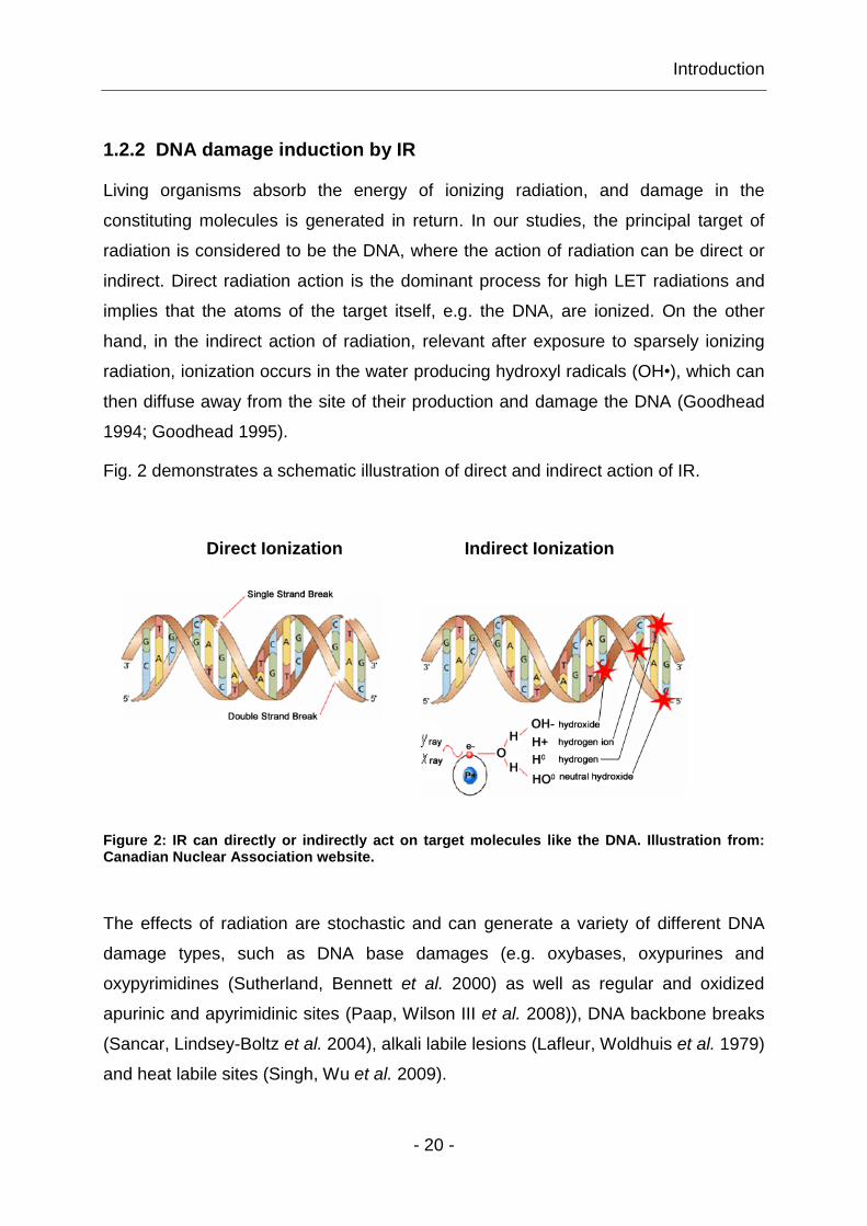
1.2.2 DNA damage induction by IR
Living organisms absorb the energy of ionizing radiation, and damage in the
constituting molecules is generated in return. In our studies, the principal target of
radiation is considered to be the DNA, where the action of radiation can be direct or
indirect. Direct radiation action is the dominant process for high LET radiations and
implies that the atoms of the target itself, e.g. the DNA, are ionized. On the other
hand, in the indirect action of radiation, relevant after exposure to sparsely ionizing
radiation, ionization occurs in the water producing hydroxyl radicals (OH•), which can
then diffuse away from the site of their production and damage the DNA (Goodhead
1994; Goodhead 1995).
Fig. 2 demonstrates a schematic illustration of direct and indirect action of IR.
Direct Ionization Indirect Ionization
Figure 2: IR can directly or indirectly act on target molecules like the DNA. Illustration from: Canadian Nuclear Association website.
The effects of radiation are stochastic and can generate a variety of different DNA
damage types, such as DNA base damages (e.g. oxybases, oxypurines and
oxypyrimidines (Sutherland, Bennett et al. 2000) as well as regular and oxidized
apurinic and apyrimidinic sites (Paap, Wilson III et al. 2008)), DNA backbone breaks
(Sancar, Lindsey-Boltz et al. 2004), alkali labile lesions (Lafleur, Woldhuis et al. 1979)
and heat labile sites (Singh, Wu et al. 2009).
Introduction
- 21 -
Backbone damage includes abasic sites, single strand (ss) and double strand (ds)
DNA breaks. Each Gy of low LET radiation, e.g. x-rays, is estimated to induce around
20-40 prompt DSBs, ~1000 single strand breaks (SSBs) and equal number of base
damages (Ward 1990). SSBs have low biological consequences, since they can be
repaired very fast with the complementary strand as template DNA. However, if
breaks occur opposite one another or are separated by just a few base pairs (bp) on
the opposite DNA strands, a DSB is generated that is a deleterious DNA lesion, since
if left unrepaired or misrepaired it leads to severe genomic instability, cell death,
carcinogenesis or mutations (Jackson 2002).
In conclusion, DNA damage produced by direct or indirect radiation action leads to
DNA change of the character of the molecule, and thus impairs its function as career
of genetic information. As a result, cell death, mutation and/or transformation can
ensue.
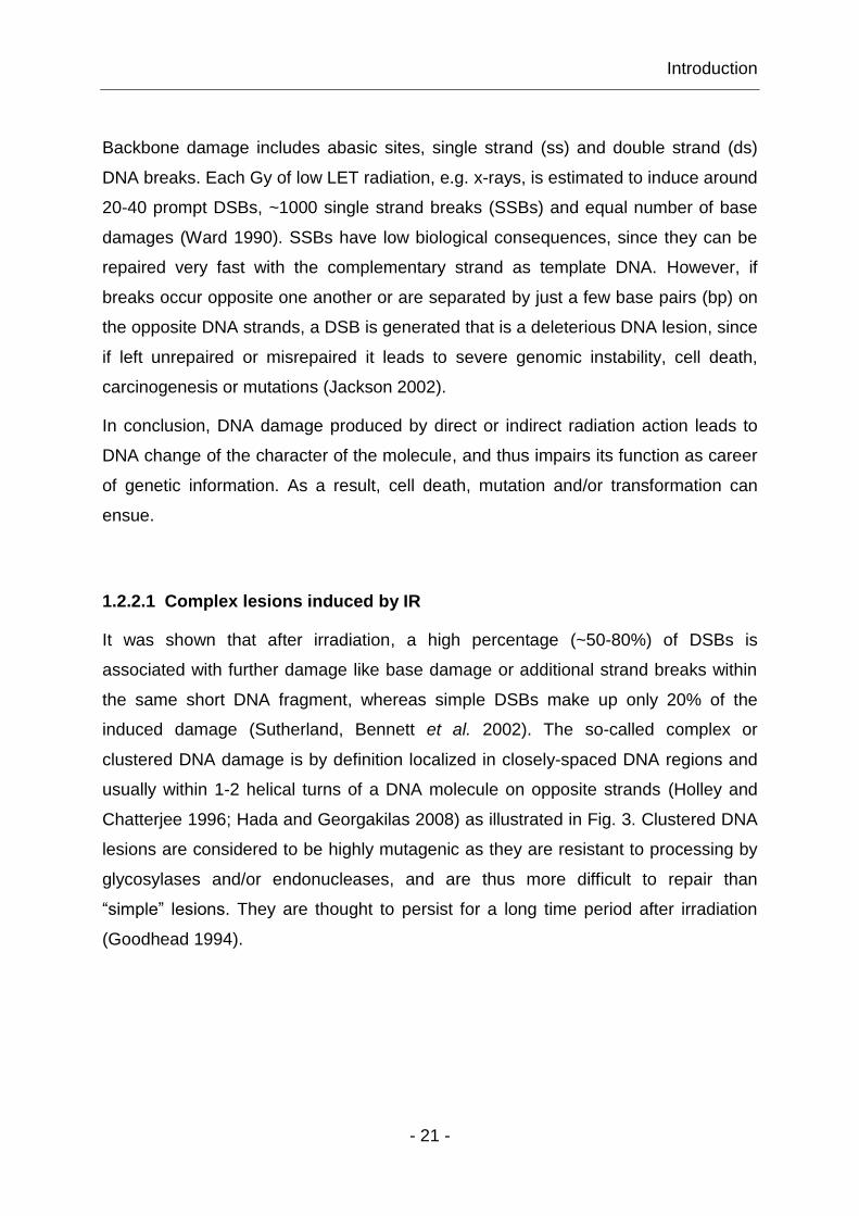
1.2.2.1 Complex lesions induced by IR
It was shown that after irradiation, a high percentage (~50-80%) of DSBs is
associated with further damage like base damage or additional strand breaks within
the same short DNA fragment, whereas simple DSBs make up only 20% of the
induced damage (Sutherland, Bennett et al. 2002). The so-called complex or
clustered DNA damage is by definition localized in closely-spaced DNA regions and
usually within 1-2 helical turns of a DNA molecule on opposite strands (Holley and
Chatterjee 1996; Hada and Georgakilas 2008) as illustrated in Fig. 3. Clustered DNA
lesions are considered to be highly mutagenic as they are resistant to processing by
glycosylases and/or endonucleases, and are thus more difficult to repair than
“simple” lesions. They are thought to persist for a long time period after irradiation
(Goodhead 1994).

Introduction
- 22 -
Figure 3: Clustered DNA damage is present when closely-spaced DNA lesions are generated within one or two helical turns. The schematic diagram illustrates typical examples of strand breakage (solid symbols) and also includes examples of associated base damage (open symbols) (Goodhead 2006).
Clustered DNA damage has also been termed as locally multiply damaged site
(LMDS) (Hall and Giaccia 2006), and are generated more efficiently after exposure to
high LET irradiation. However, LMDS are also generated after exposure to low LET
radiation. Indeed, different theoretical and experimental data suggests that the
induction of clustered DNA lesions, LMDSs, is the result of electrons depositing high
amounts of energy in the form of multiple ionization in a small volume at the end of
their tracks (Hada and Georgakilas 2008). Although clustering of DNA damage is
influenced by the LET of radiation, it is also dependent on chromatin structure in the
sense that after low LET irradiation clustered damage is present in small regions of
the DNA and the nucleosomes, whereas after exposure to high LET irradiation,
clustered damage can spread over large regions of chromatin (Rydberg 1996).

Introduction
- 23 -
1.3 Cell cycle checkpoints, DNA damage sensing and signaling
Eukaryotic cells have evolved a complex cell cycle control system that governs
proper progression through the cell cycle at regulatory transitions – the cell cycle
checkpoints. Stress conditions, inside or outside the cell, activate these checkpoints,
resulting in cell cycle arrest (Khanna and Jackson 2001). Cell cycle checkpoints are
highly conserved in all eukaryotes (Hartwell and Weinert 1989), and include
responses that enforce the right sequence in cell progression through the cell cycle,
respond to and facilitate repair of DNA damage, ensure high fidelity of DNA
replication and assist in proper chromosome segregation at mitosis (Niida and
Nakanishi 2006). Checkpoint deficiency results in genomic instability and is
associated with carcinogenesis (Hartwell and Weinert 1989).
There are three cell cycle checkpoints that control progression throughout the cell
cycle; (1) at late G1, the start checkpoint or the restriction point, where the cell
commits to the cell cycle and chromosome duplication, (2) at G2/M-border, where the
control system checks the completion of DNA replication, and (3) at metaphase to
anaphase transition, where the control system checks attachment of chromosome to
the mitotic spindle, as presented in Fig. 4 (Sherr and Roberts 1995).
Figure 4: Cell cycle phases and checkpoint control system. The division cycle of mammalian cells consists of four distinct phases: M, G1, S and G2. Three cell cycle checkpoints are depicted in this figure; late G1, G2/M-border and M (Alberts, Johnson et al. 2008).
Introduction
- 24 -
Regulation of cell cycle control system is based on a connected series of biochemical
switches, each of which initiates a specific cell cycle event. Central components are
the cyclin-dependent kinases (CDKs). The activity of these kinases rises and falls as
the cell progresses through the cell cycle, leading to cyclical changes in the
phosphorylation of specific intracellular proteins that activate and trigger major cell
cycle events. This is controlled by a complex array of enzymes – the so-called
cyclins, the most important CDK regulators. Cyclins undergo a cycle of synthesis and
degradation in each cell cycle, while the levels of CDKs are constant throughout the
cell cycle. Cyclins bind to CDKs and control their ability to phosphorylate downstream
proteins, i.e. CDKs are dependent on cyclins for their activity, since unless they are
complexed with a cyclin, they have no kinase activity. There are four classes of
cyclins, each defined by the stage of the cell cycle at which they bind CDKs and
function: G1-cyclins (cyclin D), G1/S-cyclins (cyclin E), S-cyclins (cyclin A) and G2/M-
cyclins (cyclin B) (Alberts, Johnson et al. 2008).
In addition to the checkpoints that enforce the correct progression of the cells through
the cell cycle, the cell also has checkpoints activated by DNA damage and these are
described next.
1.3.1 Mechanisms of DNA damage checkpoint response
In order to maintain genomic stability, higher eukaryotic cells have evolved efficient
DNA damage response mechanisms to sense, signal and repair damaged DNA as
improper processing of DSBs can lead to chromosomal instability, resulting in
apoptosis, carcinogenesis and mutations. Briefly, in response to different types of
DNA damage or stalled replication forks, the cell activates its DNA damage
checkpoint response system, which arrests normal cell cycle progression at different
cell cycle phases. This process includes a step-by-step activation of several different
proteins, categorized as sensors, transducers and effectors that control cell cycle
progression and facilitate DNA repair (Jackson 2002; Shiloh and Lehmann 2004;
Pardo, Gómez-González et al. 2009). Fig. 5 depicts the fundamental components of
a signal-transduction pathway initiated by DNA damage that activates cell cycle
checkpoints and regulates apoptosis, transcription or DNA repair.

Introduction
- 25 -
Figure 5: General outline of DDR signal-transduction pathway, consisting of signals, sensors, transducers and effectors. For simplicity, the network of interacting pathways are depicted as a linear pathway with arrowheads representing activating events, whereas inhibitory events are symbolized by perpendicular ends (Zhou and Elledge 2000).
Specifically, upon DNA damage induction, immediate cellular response to a DSB,
which acts as a signal, is the initial DNA damage sensing and detection by sensor
proteins, such as the MRN complex or DNA-PK. Sensor proteins activate a signal
transduction cascade that involves activation of key checkpoint regulators, the DNA-
damage-response transducing kinases – ataxia-telangiectasia mutated (ATM) and
ataxia-telangiectasia and RAD3 related kinase (ATR) (Kastan and Lim 2000; Kastan
2001). Interestingly, for the maintenance of genomic integrity after DNA damage
induction, activation of all three phosphatidylinositol 3-kinases (PI-3K), ATM, ATR
and also DNA-dependent protein kinase, catalytic subunit (DNA-PKcs), is very
important, as all three kinases are significant sensors of genotoxic stress (Yang, Yu
et al. 2003). When activated, these kinases phosphorylate many downstream
mediator, transducer and effector proteins, such as the checkpoint kinase 1 (CHK1)
and the checkpoint kinase 2 (CHK2) (Bakkenist and Kastan 2003). These effector
kinases are responsible for activation of cell cycle checkpoints that can lead to cell
cycle arrest in G1- or G2-phases of the cell cycle facilitating thus DNA repair.
Remarkably, there is some evidence that the DNA repair machinery can distinguish
between different types of damage, which translates it into different modes of
checkpoint activation in G1- and S/G2-phase cells (Barlow, Lisby et al. 2008).

Introduction
- 26 -
The classical view of the DDR system is that the ATM-CHK2 pathway is activated in
response to IR-induced DNA damage, acting at initial stages of DNA damage signal
transduction in mammals (Canman, Lim et al. 1998), while the ATR-CHK1 pathway is
mainly activated by UV light-induced DNA damage and/or ssDNA regions, which may
evolve during processing of chromosomal lesions or result from stalled DNA
replication (Ünsal-Kacmaz, Makhov et al. 2002). However, activation of ATR is not
only restricted to S-phase cells, although the majority of replication protein factor A
(RPA)-coated ssDNA is generally present during DNA replication in S-phase cells
(Fanning, Klimovich et al. 2006). Recent studies suggest that ATR and ATM are not
acting in an independent fashion during the DNA damage checkpoint response, but
rather that they are co-operating to initiate and maintain the DNA damage checkpoint
response (Helt, Cliby et al. 2005). This is also supported by the fact that ATM and
ATR overlap in their substrate specificity indicating the possibility of a crosstalk
between these two pathways (Tibbetts, Cortez et al. 2000; Yajima, Lee et al. 2006).
1.3.1.1 The ATM-CHK2 signaling pathway
One of the first signaling events after exposure of eukaryotic cells to IR is the
activation of the ATM kinase (Canman, Lim et al. 1998). ATM kinase activity is
thought to be required for the activation of the DNA damage checkpoints in G1/S-,
intra-S- and G2/M-phases of the cell cycle (Shiloh and Kastan 2001; Shiloh 2003).
Fig. 6 shows the sequence of coordinated events involved in this activation including
sensor, mediator and transducer proteins. It is generally assumed that MRN is the
primary DNA damage sensor, rapidly accumulating at DSBs (Lavin 2007). MRE11
binds both, ssDNA and dsDNA, in a sequence-independent manner, hence
contributing to ATM kinase activation and the fast recruitment of ATM to damaged
sites (Lee and Paull 2004; You, Bailis et al. 2007; Borde and Cobb 2009) (Fig. 6 C).
As a consequence of MRN protein retention at DNA damaged sites the local
concentration of this complex at the DSB sites increases; this accumulation consists
of a H2AX / mediator of DNA damage checkpoint protein 1 (MDC1)-dependent
fraction on chromatin and a H2AX-independent fraction near the DSB (Bekker-
Jensen, Lukas et al. 2006; Berkovich, Monnat Jr. et al. 2007).
Introduction
- 27 -
Activation of inactive ATM dimers to active ATM monomers is known to involve
intermolecular ATM autophosphorylation events at different Ser-residues including
Ser-1981, Ser-367 and Ser-1893 (O'Neill, Dwyer et al. 2000; Bakkenist and Kastan
2003); these events cause dimer dissociation and initiate ATM monomer formation. It
was shown that ATM autophosphorylation at Ser-1981 is necessary for both, its
monomerization and the binding to regions flanking DSBs (Berkovich, Monnat Jr. et
al. 2007). Activated ATM molecules phosphorylate various downstream ATM
substrates such as NBS1 (Gatei, Young et al. 2000), MRE11 (Dong, Zhong et al.
1999), MDC1 (Goldberg, Stucki et al. 2003), breast cancer susceptibility protein 1
(BRCA1) (Cortez, Wang et al. 1999), CHK2 (Matsuoka, Rotman et al. 2000), tumor
protein 53 (p53) (Banin, Moyal et al. 1998), RPA (Wang, Guan et al. 2001), RAD17
and the H2A histone variant, H2AX at its conserved C-terminus on Ser-139 (Burma,
Chen et al. 2001; Stiff, O'Driscoll et al. 2004), hence initiating a cellular DNA damage
signal.

Introduction
- 28 -
Figure 6: A model of the cellular DSB response cycle. (A) Undamaged section of a chromosome, showing two chromatin loops and an inactive ATM dimer. (B, C) Induction of a DSB, modification of chromatin, ATM activation and recruitment of both ATM and MRN complex to the DSB. The thin black line indicates modified chromatin. (D, E) A wave of H2AX phosphorylation is followed by recruitment and ATM-dependent phosphorylation of mediator proteins, such as MDC1, p53 Binding Protein 1 (53BP1) and BRCA1 to the growing focus. (F) Disassembly of the focus, ATM inactivation and chromatin remodeling. Note that MRN complex is also a component of the growing focus but, for clarity, has been excluded here (van den Bosch, Bree et al. 2003).
Phosphorylation of H2AX (the phosphorylated form of H2AX is termed -H2AX) is
MRN-dependent, since MRE11-depletion abrogates H2AX phosphorylation
(Rogakou, Pilch et al. 1998; Kinner, Wu et al. 2008; Di Virgilio, Ying et al. 2009).
MRN-regulated phosphorylation of H2AX marks DSB sites, and provides at the same
time a phosphorylation-regulated recruitment and retention platform for the -H2AX-
dependent assembly of further mediator proteins (Stucki, Clapperton et al. 2005; Lou,
Minter-Dykhouse et al. 2006). Interestingly, the primary migration of factors to DSBs
has no need of -H2AX (Celeste, Fernandez-Capetillo et al. 2003; Kim, Minter-
Dykhouse et al. 2006).

Introduction
- 29 -
Amplification of the initial DNA damage signal in mammalian cells is facilitated by
molecular recognition modes involving direct binding of the phosphorylated -H2AX
tail to the BRCA1 C-terminal (BRCT)-domain of MDC1. Localization of MDC1 to the
vicinity of a DSB initiates the recruitment of additional, activated ATM molecules that
allow further phosphorylation and spreading of -H2AX on chromatin (Rogakou, Pilch
et al. 1998). This ATM-dependent -H2AX expansion, occurring at megabase regions
surrounding the break (Fig. 6 D), facilitates the recruitment of other scaffolding and
enzymatic repair factors, e.g. 53BP1 and BRCA1, to chromatin regions distal to the
breaks. Such protein accumulation processes at sites of DSBs lead to the formation
of large protein foci (Fig. 6 E), which can be microscopically visualized by
immunofluorescence staining. After completion of repair proteins that have
accumulated at the site dissociate from chromatin and the protein focus disappears
(Fig. 6 F).
In summary, upon DNA damage induction, MRN activates ATM, MDC1 enhances
kinase activity of ATM, which then transduces the genotoxic stress signal by
activating its downstream substrates, particularly the effector kinase, CHK2. This
kinase is essential for the entire DNA damage response as it phosphorylates several
cell-cycle proteins, and thus initiates the activation of cellular DNA damage
checkpoints (Chaturvedi, Eng et al. 1999; Matsuoka, Rotman et al. 2000).
In general, cellular DNA damage, in particular DSBs can be removed by two major
repair pathways. After completion of repair and restoration of DNA integrity, imposed
cell cycle brakes in the form of checkpoints are released and cell cycle progression
resumes (Hartwell and Weinert 1989; Niida and Nakanishi 2006). In case of
irreparable DNA lesions apoptosis is initiated (Rich, Allen et al. 2000). The next
chapters describe in detail DSB repair by different pathways and review the current
status of understanding regarding the regulation of repair pathway choice.

Introduction
- 30 -
1.4 Eukaryotic DSB repair and its regulation
Higher eukaryotic cells remove DNA damage by two main repair pathways – NHEJ
and HRR. The fundamental difference between these two repair pathways is that
HRR requires a homologous template whereas NHEJ does not (Essers, van Steeg et
al. 2000). Consequently, HRR ensures accurate DNA repair by using either an
undamaged sister chromatid or a homologous chromosome as a repair template
(Khanna and Jackson 2001), whereas NHEJ rejoins two DNA ends without any
needs for homology (Karran 2000), thus the term “non-homologous end-joining”
(Weterings and Chen 2008). Repair of DSBs by NHEJ is accompanied with limited or
extensive additions or deletions of nucleotides at the generated junction generated
during the process of producing ligatable ends. The result is an altered sequence of
the repaired DNA molecule due to the fact that NHEJ does not restore sequence
information in the damaged DNA molecule, although it restores its molecular integrity
(Iliakis, Wang et al. 2004; Lieber 2010). Thus, DSB repair by NHEJ is considered as
error-prone.
The relative contribution of the two repair pathways is likely to be determined by the
phase of the cell cycle and the abundance of repetitive DNA, although the
importance and usage of NHEJ varies greatly among species (Karran 2000). As
NHEJ has no need for a DNA template, it can operate throughout the cell cycle,
although it is thought to dominate repair in the G1-phase of the cell cycle, where HRR
cannot operate. In contrast, HRR is restricted to S- and G2-phases of the cell cycle,
where a sister chromatid is available that can be used as a repair template (Krüger,
Rothkamm et al. 2004; Moynahan and Jasin 2010). Fig. 7 presents the function of
the two DSB repair pathways throughout the cell cycle with their dependency on DNA
template.

Introduction
- 31 -
Figure 7: Illustration shows two major DSB repair pathways with its dependency on DNA template and their occurrence in different cell cycle phases (Hall and Giaccia 2006).
In theory, HRR could also occur in diploid G1-phase cells, using existing copy of the
chromosome as a template for repair. However, homologous chromosomes are
usually not directly available due to nuclear chromosome compartmentalization. It is
therefore believed that NHEJ is the prevailing repair pathway during G1/G0- and M-
phases of the cell cycle (Lee, Mitchell et al. 1997), whereas HRR is the main repair
pathway during S- and G2-phases of the cell cycle. In the next section are described
the processes of HRR and NHEJ in detail.
1.4.1 Homologous recombination repair
As mentioned above, DSB repair by HRR utilizes a homologous sequence either on
the same DNA molecule, on a sister chromatid or on a homologous chromosome
(Krüger, Rothkamm et al. 2004), hence the term “homologous recombination repair”.
This template-dependent process is relatively slow but provides the mammalian
genome a high-fidelity mechanism for repairing DNA damages including DNA gaps,
DSBs and DNA interstrand crosslinks in an error-free manner. In addition to these
repair mechanisms, HRR is also implicated in several other biological processes
such as meiotic crossover during allelic rearrangement in gametes, proper
chromosome segregation, mating type switching in yeast as well as epitope
immunoglobulin class switching in many organisms.
Introduction
- 32 -
The basic HRR machinery and its regulation are greatly conserved among
eukaryotes. Interestingly, null mutations in core HRR genes, e.g. RAD51, BRCA1
and breast cancer susceptibility protein 2 (BRCA2) are lethal (Thompson and Schild
2002), and cells with mutated HRR genes present reduced HR levels, resulting in
high levels of chromosomal aberrations and miss-segregation at mitosis (Pierce,
Johnson et al. 1999; Griffin, Simpson et al. 2000), elevated radiosensitivity to killing
and increased tumorigenesis (Pierce, Stark et al. 2001). In yeast and bacteria, HRR
is the primary mechanism of DSB repair (San Filippo, Sung et al. 2008).
In general, HRR involves the following distinct steps: (1) processing of DNA ends, (2)
search for homology, strand invasion and formation of holiday junction, (3) DNA
synthesis, branch migration and final resolution of synapsed DNA molecules (Kinner,
Wu et al. 2008). Fig. 8 shows the key steps of HRR with its main players.

Introduction
- 33 -
Figure 8: Outline of main HRR repair players. Illustration courtesy: Emil Mladenov, Institute of Medical Radiation Biology, Medical School, University of Duisburg-Essen, Germany.

Introduction
- 34 -
Initial DNA damage sensing by MRN and its binding to damaged DNA initiates an
intracellular DNA damage signal leading to the recruitment of different proteins to
damaged DNA ends, e.g. BRCA1, Bloom syndrome helicase (BLM), C-terminal
binding interacting protein (CTIP) and the nucleases – exonuclease 1 (EXO1) and
DNA2 (Stucki, Clapperton et al. 2005; Lou, Minter-Dykhouse et al. 2006). MRN, CTIP
and activated EXO1 and DNA2 nucleases promote resection of DSBs to form
recombinogenic 3’-ssDNA overhangs (Sartori, Lukas et al. 2007; Takeda, Nakamura
et al. 2007; Bolderson, Tomimatsu et al. 2010). The resulting 3’-ssDNA tails are then
coated by RPA, hence protecting the ends from degradation and preventing the
formation of secondary structures (Wold 1997). Interestingly, formation of 3’-ssDNA
tails might also determine the switch from NHEJ to HRR, particularly as NHEJ
preferentially utilizes unprocessed DNA ends for ligation (Zhu, Chung et al. 2008;
Yun and Hiom 2009). Accordingly, ssDNA ends activate the ATR-CHK1 pathway that
might also promote DSB repair by HRR (Sorensen, Hansen et al. 2005).
With the aid of RAD52 epistasis group members like RAD54, RAD51 paralogs
(RAD51B, RAD51C, RAD51D, XRCC2 and XRCC3) and BRCA2 (Symington 2002;
Sy, Huen et al. 2009), the DNA strand exchange protein, RAD51 displaces RPA from
ssDNA tails, consequently generating a RAD51 nucleoprotein filament that can be
composed of thousands RAD51 monomers. This RAD51 nucleoprotein filament is
bound to ssDNA and searches for homology by invading homologous duplex DNA
segments for polymerase-mediated DNA synthesis (West 2003; Mazin, Mazina et al.
2010). The 3’-end from the invading end is used to prime a leading strand DNA
synthesis templated by the donor duplex. The other end of the break interacts with
the displaced strand from the donor duplex, forming a so-called Holiday Junction,
thus priming DNA synthesis from the other end of the break. Resolution of Holiday
Junctions may result in crossover or non-crossover products, although crossing over
is a seldom event during somatic HRR (Richardson, Moynahan et al. 1998; Nagaraju,
Odate et al. 2006). In the process of synthesis-dependent strand annealing (SDSA),
the invading strand, previously being extended by DNA synthesis, is extended by
branch migration and then leaves the template chromatid. This extended strand
anneals to complementary DNA sequences exposed by 5’-3’-resection of the other
site of the break, and the molecule is completed by filling remaining gaps by DNA
Introduction
- 35 -
synthesis by polymerase and/or and LIG1-mediated sealing of the nicks (Mimitou
and Symington 2009). In the case of Holiday Junction resolution, HRR is completed
by separation of the synapsed DNA molecule through resolvases.
1.4.2 Non-homologous end-joining
Cells of higher eukaryotes rejoin DSBs in their DNA predominantly by NHEJ,
comprising four general steps: A set of enzymes (1) recognizes and (2) mediates the
capture of both ends of the broken DNA molecule; (3) formation of a molecular bridge
between the two DNA ends and (4) finally re-ligation of the broken DNA molecule.
In general, NHEJ utilizes proteins like DNA-PKcs, KU, ARTEMIS, LIG4, XRCC4,
XLF/CERNUNNOS, as well as DNA polymerase . Here, this pathway will be termed
as DNA-PK-dependent non-homologous end-joining (D-NHEJ), stressing its
dependence on DNA-PK, where KU and DNA-PKcs act in a coordinated manner to
direct the DNA end-joining process towards the D-NHEJ pathway (Perrault, Wang et
al. 2004; Lieber and Wilson 2010).
Interestingly, D-NHEJ mutants remove a large proportion of DSBs from their genome
using an alternative pathway of end-joining (Wang, Perrault et al. 2003). This repair
pathway may therefore have a backup function becoming active whenever D-NHEJ is
inactivated (Wang, Rosidi et al. 2005). In order to differentiate it from D-NHEJ, and to
indicate its putative backup function, we term it here as backup NHEJ (B-NHEJ)
(Iliakis 2009). Both end-joining pathways are described in detail below.
1.4.2.1 D-NHEJ
As briefly discussed in 1.4, D-NHEJ repair pathway is error-prone but is extremely
efficient in removing DSBs from the genome, with half times of 15-30min. Many
components involved in D-NHEJ are conserved from yeast to humans with the
difference that mammals utilize DNA-PKcs for DSB repair by NHEJ, while a homolog
of this enzyme in yeast as well as in lower eukaryotes has not been found (Critchlow
and Jackson 1998).
Introduction
- 36 -
Moreover, it is proposed, that the high speed of D-NHEJ is an evolutionary
development in higher eukaryotes orchestrated around the newly evolved DNA-PKcs
protein and pre-existing factors. Within a few minutes it achieves restoration of
chromosome integrity through an optimized synapsis mechanism operating by a
sequence of protein-protein interactions in the context of chromatin and nuclear
matrix (Iliakis, Wang et al. 2004). In addition, D-NHEJ is indispensable in processing
DSB intermediates generated during V(D)J recombination (Karran 2000; Weterings
and Chen 2008), where defects in D-NHEJ lead to chromosomal aberrations,
immunodeficiency and sensitivity against IR (Couedel, Mills et al. 2004).
The heterodimeric KU70/80 complex, consisting of 70kDa and 80kDa subunits
(Walker, Corpina et al. 2001), is one of the most abundant cellular proteins with about
300,000 molecules per cell (Lieber, Grawunder et al. 1997). In an early stage of D-
NHEJ-mediated DSB repair, DNA ends are recognized by the KU heterodimer that
captures DNA ends (blunt, 5’- or 3’-overhangs). Through its asymmetric ring
structure, that wraps around the DNA helix (Walker, Corpina et al. 2001), it recruits
DNA-PKcs to DNA ends (Lees-Miller and Meek 2003), thus activating its kinase
activity. DNA-PKcs forms a synaptic complex and brings both DNA ends together.
Moreover, it undergoes conformational changes, and probably dimerizes to generate
a platform for subsequent processing of non-ligatable DNA termini before final re-
ligation (Meek, Gupta et al. 2004). Once the two DNA ends have been captured and
tethered by DNA-PK (complex of KU70/80 and DNA-PKcs), the D-NHEJ repair
process is initiated, thus enabling the phosphorylation of various downstream
substrate proteins, e.g. RPA2, WRN helicase, ARTEMIS, XLF/CERNUNNOS, DNA
LIG4 and XRCC4, as well as autophosphorylation of DNA-PKcs itself (Chen, Trujillo
et al. 2000; Jeggo and Löbrich 2005; Otsuki, Seki et al. 2007; Cruet-Hennequart,
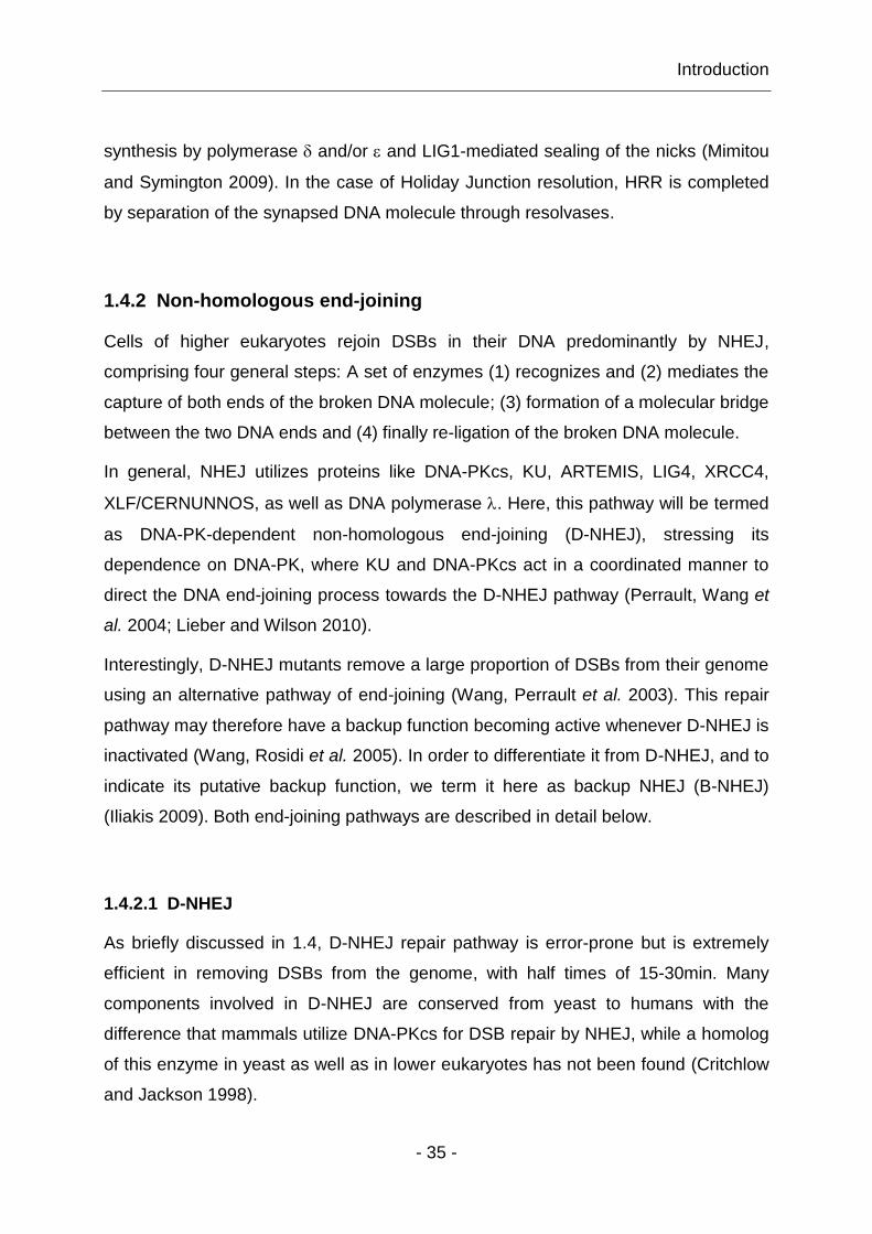
Glynn et al. 2008; Kinner, Wu et al. 2008; Yu, Mahaney et al. 2008). Fig. 9 depicts
DSB repair by D-NHEJ and the main proteins involved.

Introduction
- 37 -
Figure 9: Outline of the main players in the different D-NHEJ steps. D-NHEJ efficiently restores genomic integrity without ensuring sequence restoration. Association of KU to DNA ends facilitates recruitment of DNA-PKcs, which is activated by the DNA ends, thus regulating the efficiency of this repair pathway. DNA-PKcs promotes end-processing by the ARTEMIS nuclease, and subsequent rejoining of broken DNA ends utilizing the LIG4/XRCC4/XLF complex (Mladenov and Iliakis 2011).
Several enzymes have been identified, like tyrosyl-DNA phosphodiesterase (TDP1),
polynucleotide kinase 3'-phosphatase (PNKP), terminal deoxynucleotidyl transferase
(TDT), nucleases like ARTEMIS or polymerases, that are able to either remove or fill-
in ss and non-compatible overhangs. During this step of D-NHEJ process, occasional
loss of nucleotides is possible (Valerie and Povirk 2003). Finally and after release of
DNA-PK from DNA ends, LIG4/XRCC4/XLF complex catalyzes/coordinates the
ligation of processed DNA ends by the help of polymerase or (Mahajan,
McElhinny et al. 2002; Capp, Boudsocq et al. 2006; Wu, Frit et al. 2007).

Introduction
- 38 -
Interestingly, it is believed that dissociation of NHEJ proteins from repaired damage
sites is facilitated by its autophosphorylation; in yeast, it also depends on MRX and
the ATPase functions of RAD50 (Wu, Topper et al. 2008).
1.4.2.2 B-NHEJ
As pointed out above, cells with mutations in components of D-NHEJ, or when D-
NHEJ components are either absent from the vicinity of the break or are genetically
or chemically compromised, are still capable of repairing the majority of IR-induced
DSBs, utilizing an alternative repair pathway, which is, rather surprisingly, not
sensitive to mutations in HRR genes (Feldmann, Schmiemann et al. 2000; Wang,
Zeng et al. 2001; Wang, Zhao-Chong et al. 2001). This alternative pathway is a
distinct form of end-joining, and is likely to be an evolutionarily older pathway with
less optimized synapsis mechanisms, rejoining DNA ends with slower kinetics with
half-times of 2-10h. It is thought that the rapid DNA end-joining of D-NHEJ kinetically
suppresses this slower alternative pathway (DiBiase, Zeng et al. 2000). However,
alternative end-joining pathways (B-NHEJ) are expected to contribute significantly to
genome maintenance and stability, playing an important role in the overall repair of
DSBs.
Interestingly, B-NHEJ has a strong growth-state dependency (Windhofer, Wu et al.
2007). Its activity is markedly reduced in plateau and G1-phase cells, but is
significantly elevated during the G2-phase of the cell cycle, suggesting that
suppression of growth signaling significantly compromises DSB repair by B-NHEJ
(Wu, Wang et al. 2008). Moreover, B-NHEJ is implicated in telomere maintenance
(Rai, Zheng et al. 2010), but it can also robustly substitute for D-NHEJ in CSR in
LIG4-deficient mice (Soulas-Sprauel, Le Guyader et al. 2007; Yan, Boboila et al.
2007), and is likewise found to substitute for V(D)J recombination in D-NHEJ-
deficient cells, where mutations in recombination activating genes (RAG1/RAG2)
lead to proteins able to generate DNA ends but unable to hold them (Lee, Neiditch et
al. 2004; Iliakis, Rosidi et al. 2005; Jones and Simkus 2009).
Introduction
- 39 -
However, the slow kinetics and suboptimal synapsis mechanisms of B-NHEJ allow
more time for exchanges through the joining of incorrect DNA ends, consequently
leading to formation of chromosome aberrations during the repair of IR-induced
DSBs in wild-type and D-NHEJ mutant cells. This error-prone nature of B-NHEJ
(Virsik-Köpp, Rave-Fränk et al. 2003) could also be demonstrated for other
endpoints, as in XRCC4- and LIG4-deficient mice chromosome abnormalities
included translocations at the immunoglobulin heavy locus chain (IgH) that give rise
to lymphoid malignancies (Soulas-Sprauel, Le Guyader et al. 2007; Yan, Boboila et
al. 2007), hence B-NHEJ is placed at the center of carcinogenesis.
Despite the potential consequences of B-NHEJ function, little is known about the
underlying mechanism, its regulation, integration into the cellular DSB processing
apparatus, and its interaction with components of D-NHEJ and HRR. Genetic and
biochemical experiments have shown that proteins like poly [ADP-ribose] polymerase
1 and 2 (PARP1, PARP2), MRN, Werner syndrome (WRN), histone H1, LIG3 and X-
ray repair cross-complementing protein 1 (XRCC1), are implicated in B-NHEJ (Wang,
Zhao-Chong et al. 2001; Haince, McDonald et al. 2008; Rosidi, Wang et al. 2008;
Sallmyr, Tomkinson et al. 2008; Davies and Chen 2010; Della-Maria, Zhou et al.
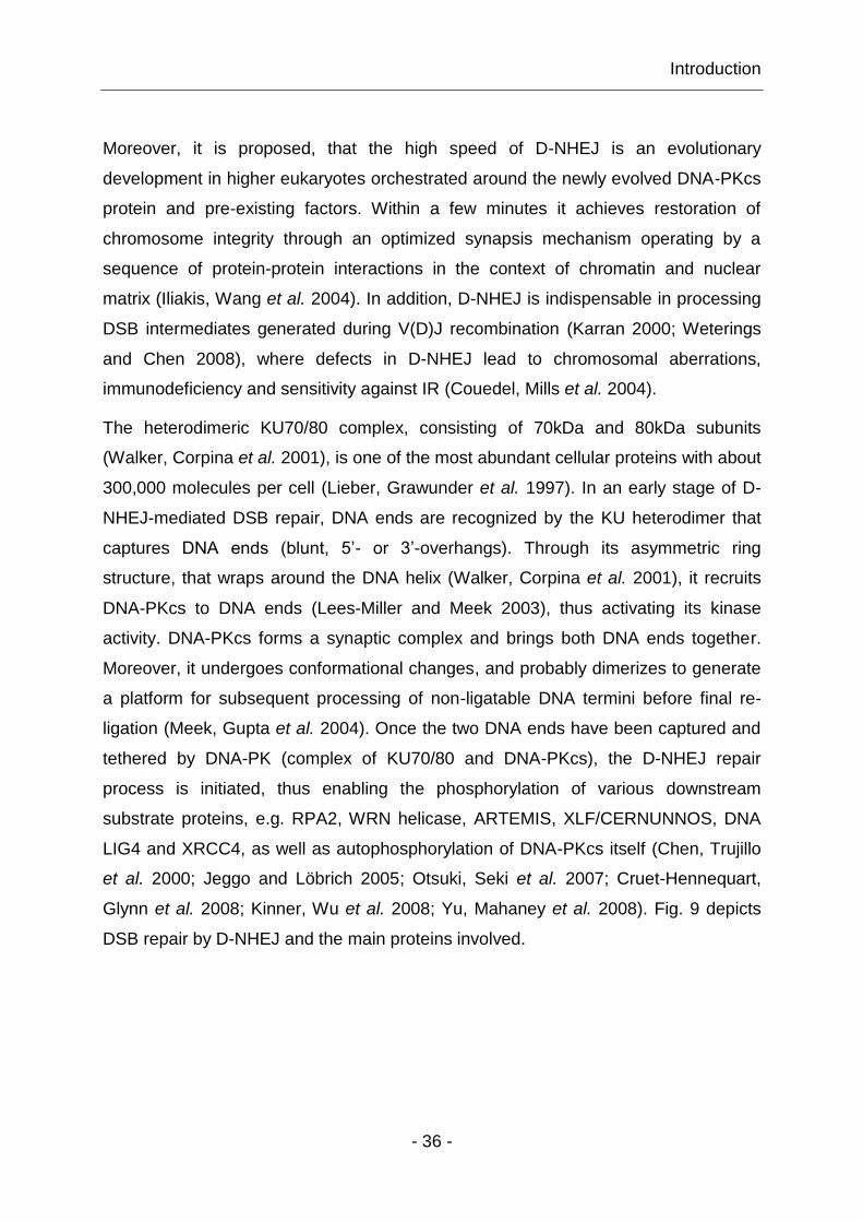
2011). Fig. 10 illustrates the characterized B-NHEJ repair pathway steps and the
proteins involved.

Introduction
- 40 -
Figure 10 Outline of B-NHEJ key steps with its main players (Mladenov and Iliakis 2011).
Upon DNA damage induction, proteins such as MRN, PARP1 and PARP2 are
recruited to DSBs. Prior to ligation, H1 helps to align the DNA ends (Rosidi, Wang et
al. 2008). Remarkably, MRN seems to be essential for end-joining by B-NHEJ, as its
inhibition in D-NHEJ mutants decreases end-joining frequency (Rass, Grabarz et al.
2009); LIG3 functions in a complex with XRCC1 being regulated by PARP1 (Robert,
Dantzer et al. 2009). Interestingly, PARP1 might compete with KU for DNA ends,
where particularly KU’s much higher affinity for DNA ends provides a kinetics basis
for the backup character of B-NHEJ. Another cause for this backup character might
be the fact that other forms of lesions, like SSBs or base damages, also compete for
PARP1, and LIG3 as the PARP1/XRCC1/LIG3 complex is likewise involved in the
repair of SSBs and base damages (Audebert, Salles et al. 2004; Wang, Wu et al.
2006).
Introduction
- 41 -
1.4.3 Regulation of DSB repair pathway choice
Notably, in the field of DNA repair a central and largely unanswered question is how
and when a cell selects which pathway to use for the repair of a certain DSB. This is
because the regulatory mechanisms of DSB repair pathway choice are still unknown.
Thus, it remains open whether the decision for pathway choice is determined by the
nature of the DSB, or whether it is regulated by a global regulatory network
responding to uncharacterized signals and physiological conditions. In the remaining
of this section, several models of DSB repair pathway choice and its coordination are
discussed.
It is widely assumed that in higher eukaryotes NHEJ is the main DSB repair pathway,
and that HRR has only a minimal contributing function in DSB repair, restricted to G2-
phase of the cell cycle (1.4.1, 1.4.2) (Jeggo and Löbrich 2005; Beucher, Birraux et al.
2009). This is supported by the observation that in higher eukaryotes gene targeting
is inefficient, whereas random integration is very efficient (Fattah, Lichter et al. 2008).
In line with this, HRR-deficient mutants have comparable DSB processing activity as
wild-type cells throughout the cell cycle (Wang, Zeng et al. 2001). However, other
results suggest a substantial contribution of HRR to DSB processing, as mutants
defective in HRR components, are radiosensitive, and have highly compromised
HRR of site-specific DSBs (Pierce, Stark et al. 2001). When analyzing the DSB repair
capability of HRR mutants by measuring -H2AX foci decay, a relatively small but
clearly visible defect in removing DSBs is measurable (Rothkamm, Krüger et al.
2003). Thus, it can be assumed that HRR has an important role in DSB repair.
Clearly, further investigations are needed to unravel the contribution of different
repair pathways in DSB repair.
For instance, one theory suggests that D-NHEJ and HRR compete for the
recruitment of DSBs. It was shown that transfected plasmid DNA could be repaired in
mammalian cells by both pathways, and that both D-NHEJ and HRR proteins bind to
broken DNA ends. According to this model, pathway choice reflects the outcome of
this competition and may have a stochastic component. In apparent agreement with
this passive competition model, cells with defects in D-NHEJ have increased HRR
(Allen, Kurimasa et al. 2002). Specifically, inactivation of D-NHEJ by eliminating
DNA-PKcs resulted in elevated HRR (Delacote, Han et al. 2002; Shrivastav, Miller et

Introduction
- 42 -
al. 2009). In contrast to this observation, chemical inhibition or functionally
compromised DNA-PKcs leads to reduced HRR, implicating that non-functional DNA-
PKcs has a dominant negative effect; it abolishes alternative DSB processing by
capturing DNA ends, hence blocking access to other repair factors (Wang, Perrault et
al. 2003; Perrault, Wang et al. 2004; Cui, Yu et al. 2005). In line with this competition
model, two groups reported that Mre11-Rad50-Xrs2 (Mrx) complex (homolog for
MRN in yeast) and Ku compete for DNA end binding (Taylor, Cecillon et al. 2009). In
the absence of Ku, Mre11 binding to DNA ends was increased in G1-phase cells,
leading to a higher number of DNA ends being processed by HRR. Conversely, over-
expression of Ku can inhibit Mre11 binding to DSBs, and with that the initiation of
DNA end resection in G2-phase cells. Similarly, EXO1 can also compete with KU for
DNA end binding and, if present at high concentrations it can process DNA ends for
HRR (Barlow, Lisby et al. 2008). Also, Ku can prevent Exo1 and Sgs1-dependent
resection of DNA ends in the absence of a functional Mrx or Sae2 (Mimitou and
Symington 2010), inhibiting HRR, and promoting DSB repair towards D-NHEJ. In
addition, KU can also inhibit B-NHEJ throughout the genome as well as HR at
telomeres (Fattah, Lee et al. 2010). However, other results indicate that differences in
the DSB binding properties of MRE11 and KU determine different efficiencies of HRR
and NHEJ, at least for the repair of high LET radiation-induced DSBs (Wang, Zhang
et al. 2010).
In opposition to this competition model, other studies revealed no competition
between HRR and NHEJ, when repair of IR-induced DSBs was followed in different
repair mutants. After all, HRR deficient mutants presented no evident defects in
removing DSBs, whereas D-NHEJ mutants showed strong repair defects – although
they rejoined the majority of DSBs with slower kinetics using B-NHEJ (DiBiase, Zeng
et al. 2000). More importantly, it was shown that this rejoining capability is not HRR-
dependent (Wang, Zeng et al. 2001) as one would expect, based on a competition
model. However, in view of the fact that higher eukaryotes predominantly use NHEJ
for DSB repair, a simple competition model appears to be incompatible with all
available facts, and we hypothesize the existence of determinants that mediate
selection while at the same time allowing D-NHEJ to dominate. Of course, all these
Introduction
- 43 -
models of pathway selection will be valid only in S- and G2-phases of the cell cycle,
where HRR can operate.
There is evidence that pathway choice might be controlled by the expression or post-
translational modification of repair proteins such as CTIP, BRCA1 or MRN (Esashi,
Christ et al. 2005; You and Bailis 2010), as well as by the ability of cyclin-dependent
protein kinase 1 (CDK1) activity to promote and regulate DSB end resection (Aylon,
Liefshitz et al. 2004). For instance, it has been reported that loss of MRE11 affects
both D-NHEJ and B-NHEJ, thus decreasing end-joining frequencies in experimental
systems, whereas MRE11 overexpression activates resection of ssDNA leading to
increased DSB repair by B-NHEJ, particularly as MRE11 nuclease activity was found
to be an essential factor favoring B-NHEJ (Xie, Kwok et al. 2009).
Other results report that dissociation of NHEJ proteins from repaired damage sites
depends on Mrx complex and the ATPase activity of Rad50, indicating that Mrx may
be the critical factor in repair pathway switching, at least at DNA ends that failed D-
NHEJ (Wu, Topper et al. 2008). The most obvious mechanism for this dissociation
would be the formation of recombinogenic ss-3’-tails, by nucleases like MRE11,
CTIP, EXO1 and DNA2, presumably because D-NHEJ mostly uses unresected DNA
ends for ligation (Zhu, Chung et al. 2008; Mimitou and Symington 2009; Yun and
Hiom 2009; Cejka, Cannavo et al. 2010; Rupnik, Lowndes et al. 2010). In DNA end
resection, CTIP is directly implicated in the cell cycle specific HRR activities of MRN,
and functional interactions between MRN and CTIP have been observed. Similar
observations were also made in yeast, where the major task of Sae2 (CTIP
homologue in yeast) is to control Mrx’ activity, and thus to regulate the balance
between HRR and NHEJ.
Interestingly, Sae2 was identified as one target of CDK1-dependent control of DNA
end resection (Huertas, Cortes-Ledesma et al. 2008). Recently, it was proposed that
MRN can initiate different forms of DNA damage and checkpoint signaling depending
on the type of DNA ends it is bound to; two-ended DSBs, which generally arise in
chromatin after IR, cause ATM activation, whereas one-ended DSB that arise from
stalled replication forks lead to ATR activation. Through such mechanisms MRN can
control repair pathway choice at a DSB, with ATM and ATR activation resulting in
Introduction
- 44 -
replication fork rescue and leading to DSB repair by HRR (Williams, Lees-Miller et al.
2010).
However, activation of ATR-CHK1 pathway by ssDNA is also implicated in HRR
(Sorensen, Hansen et al. 2005), possibly because CHK1 phosphorylates RAD51 that
in turn attenuates its interaction with BRCA2 (Sorensen, Hansen et al. 2005). Post-
translational modifications of MRN like phosphorylation, methylation and acetylation
are clearly involved in regulating MRN activities (Olson, Nievera et al. 2007; Zhuang,
Jiang et al. 2009; Bennetzen, Larsen et al. 2010; Olsen, Vermeulen et al. 2010),
even though the effects of these post-translational modifications on MRE11, RAD50
and NBS1 activities are not well understood. Thus, it is still unknown, whether
processes like the formation of ss-3’-DNA ends, reflect coordinating functions or are
themselves the result of overarching coordination.

Introduction
- 45 -
1.5 The MRN complex
In the context of DSB repair pathway utilization and coordination, the early function of
MRN complex, immediately upon generation of DSBs, places this complex at the
interface of DSB repair pathway choice, although its precise functional significance in
diverse aspects of the response to DNA damage, DSB repair and pathway choice
remains to be elucidated. The reported MRE11 and KU heterodimer interaction
(Goedecke, Eijpe et al. 1999; Wu, Topper et al. 2008) is compatible with the roles
proposed for these proteins in NHEJ, particularly when considering that they are the
earliest players in the choice of the repair pathway that will remove a DSB. In the
following, we give an overview of MRN, and its biochemical and structural
characteristics, its involvement in DDR signaling and its contribution to different DSB
repair pathways. This information forms the framework for arguments and
interpretations of results obtained in the experiments described in the following
section.
Eukaryotic MRN is composed of three subunits: MRE11, RAD50 and NBS1. MRE11
and RAD50 are highly conserved from archaea to humans, whereas NBS1 (homolog
of Xrs2 in yeast) is a less conserved eukaryotic-specific protein (D'Amours and
Jackson 2002; Williams, Williams et al. 2007). MRN has different enzymatic activities,
and is thus considered as a cornerstone complex, rather than a simple component
within a linear pathway. It is involved in DNA damage sensing (Lee and Paull 2007),
DNA repair (Tauchi, Kobayashi et al. 2002; Buis, Wu et al. 2008), DNA replication
(Tittel-Elmer, Alabert et al. 2009), meiosis (Ajimura, Leem et al. 1993), mitosis
(Mimitou and Symington 2009), telomere maintenance (Dimitrova and de Lange
2009) and apoptosis (Stracker, Morales et al. 2007). MRN is also required for V(D)J
recombination (Saidi, Li et al. 2010), and the programmed DSB induction during CSR
(Dinkelmann, Spehalski et al. 2009).
Cells with deficiencies in MRN components have phenotypes similar to ATM-deficient
cells including impaired IR-induced cell cycle checkpoint activation, ineffective DNA
repair, dramatic reduction of targeted integration of exogenous DNA, gene
conversion, sister chromatid exchanges, impaired ss-annealing and apoptosis.
However, in contrast with ATM loss, which is not embryonically lethal, null mutations

Introduction
- 46 -
in any of these three proteins leads to embryonic lethality (Demuth, Frappart et al.
2004).
Links between defects in the MRN complex and tumorigenesis in humans, and the
recognition that defects in MRN subunits, cause cancer-prone diseases like
Nijmegen breakage syndrome (NBS) or Ataxia Telangiectasia like disorder (ATLD)
(Stewart, Maser et al. 1999; Tauchi, Matsuura et al. 2002; McKinnon 2004; Taylor,
Grom et al. 2004; Uchisaka, Takahashi et al. 2009) underscore the importance of
MRN in cellular functions. To date, mutations in RAD50 gene have been reported in
only a few cases of patients, and have not yet been associated with a defined human
phenotype (Heikkinen, Rapakko et al. 2006; Tommiska, Seal et al. 2006). However,
recent results report a patient with heterozygous mutations in RAD50 gene with low
levels of unstable RAD50 protein. Cells from this patient were characterized with a
RAD50 deficiency resulting in a phenotype that can be classified as NBS (Waltes,
Kalb et al. 2009). All three disorders share similar clinical and cellular phenotypes,
such as immunodeficiency, cerebellar degeneration, defects in DNA damage-induced
checkpoint activation, failure to form DNA damage-induced MRN foci, increased
chromosome instability, radio-resistant DNA synthesis, radiation sensitivity and
premature ageing (Matsuura, Tauchi et al. 1998; Varon, Vissinga et al. 1998; Petrini
2000; Waltes, Kalb et al. 2009).
1.5.1 Structural and functional characteristics of the MRN complex
1.5.1.1 MRE11
MRE11 is composed of 708 amino acids (aa), and is the central 80kDa protein-
nucleic acid and protein-protein interaction core component of the MRN complex
(Hopfner, Karcher et al. 2001; Williams, Williams et al. 2007). MRE11 comprises
several protein domains involved in different functions. The amino-terminal end has
four conserved phosphoesterase motifs and a NBS1-binding site, whereas its C-
terminus includes a RAD50-binding site and two DNA binding domains, enabling
binding of duplexed as well as ssDNA-ends (Borde and Cobb 2009). The protein
domains of MRE11 are presented in Fig. 11.

Introduction
- 47 -
Figure 11: Scheme of MRE11 protein domains. Regions important for the NBS1 and RAD50 interactions are indicated, blue regions show the relative position of four phosphoesterase motifs, whereas the DNA binding domains are shown in orange (Borde and Cobb 2009).
The nuclease domain of MRE11 is responsible for different enzymatic activities
including ssDNA endonuclease activity on 5’-overhangs, 3’-flaps and 3’-branches, as
well as the double-stranded (ds) DNA 3’-5’-exonuclease activity that is manganese-
(Mn2+), ATM- and NBS1-dependent. Furthermore, the protein has DNA annealing, as
well as DNA unwinding activities that are regulated through interactions with RAD50
and NBS1 (Paull and Gellert 1998; Buis, Wu et al. 2008; Jazayeri, Balestrini et al.
2008). Moreover, MRE11 is known to homodimerize via poorly characterized
mechanisms that is required for basic MRN functions, such as MRE11-DNA-binding
in vitro and MRE11 repair in vivo activities (Williams, Moncalian et al. 2008).
Because MRE11’s nuclease activity does not seem to be required for resection of
clean DSBs but rather for processing of DNA ends with covalent adducts, such as
those created by IR (Llorente and Symington 2004), it is likely that MRE11 is
necessary for 5’-3’ resection of DNA ends suitable for HRR (Mimitou and Symington
2009). However, structural and biochemical data suggest that generation of 3’-tails
for HRR in vivo requires additional factors with reverse nuclease directionality
compared to MRE11, such as CTIP or EXO1 (Schaetzlein, Kodandaramireddy et al.
2007). Thus, one possible mechanism for DNA end resection might be a two-step
mechanism, involving a helicase, e.g. BLM, in conjunction with a ss-specific endo- or
exonuclease for resection of DSBs (Mimitou and Symington 2008; Mimitou and
Symington 2009). There is some evidence, that in yeast the first step depends on
Mrx and Sae2, resulting in endonucleolytic removal of about 50-100 nucleotides from
the 5’-end, which gives rise to an intermediate with short 3’-ssDNA tails that is rapidly
processed in a second step by a ss-specific nuclease, like Exo1 or Sgs1 (BLM
orthologue in yeast) (Niu, Raynard et al. 2009). Interestingly, it was shown that the
nuclease acting with Sgs1 is Dna2, a conserved endonuclease implicated in Okazaki

Introduction
- 48 -
fragment and DSB processing in post-replication repair pathways. In yeast Mrx and
Sae2 initiate DSB resection, and either Sgs1 with Dna2 or Exo1 rapidly degrade 5’-
strands to expose long 3’-ssDNA tails. Thus, it appears that Sgs1 and Dna2 function
in a parallel pathway to Exo1 (Zhu, Chung et al. 2008).
1.5.1.2 RAD50
RAD50 is a 150kDa ATP-dependent subunit of MRN belonging to the structural
maintenance of chromosome (SMC) group of proteins. At each end of the protein,
Walker A and B nucleotide triphosphate (NTP)-binding motifs are separated by two
long heptad-repeat regions bearing two MRE11 binding sites and a central
conserved zinc-hook structure (CXXC motif) that are depicted in Fig. 12 (Alani,
Subbiah et al. 1989; Hopfner, Karcher et al. 2000).
Figure 12: The domain structure of RAD50 shows Walker A and B motifs at each end of the protein, MRE11 binding sites, adjacent to ATPase domains are shown in green. Coiled-coil regions meet at the hook region in the center of the protein. This central RAD50 region contains a CXXC motif that reverses directionality of the coiled coil, coordinating Zinc (Zn
2+) to
mediate RAD50 hook-hook assembly (Borde and Cobb 2009).
The CXXC motif consists of 14 non-helical residues (aa 440-453) with two main-
chain hydrogen bonds, and is important for MRN complex assembly as well as
recombinational repair (Hopfner, Craig et al. 2002). The heptad-repeat regions fold
into an extended intramolecular anti-parallel coiled-coil structure that brings N-
terminal Walker A and C-terminal Walker B ATPase motifs in close proximity, stably
associating to form a bipartite ATP-binding cassette (ABC)-ATPase domain (Alani,
Subbiah et al. 1989; Hopfner, Craig et al. 2002; Mimitou and Symington 2009). It
appears that ATPase motifs in RAD50 are essential for all known activities of MRN
including ATM activation by DSBs in vitro (Lee and Paull 2005). Adenylate kinase
(AK) and ATPase activities act competitively, and regulate the conformational

Introduction
- 49 -
(allosteric) changes of RAD50. Both activities are important for various functions of
MRN in vivo, e.g. DNA binding, unwinding and/or DNA tethering (Bhaskara, Dupre et
al. 2007). However, the proposed main task of RAD50 is to bind damaged DNA ends
and to hold them in close proximity.
1.5.1.3 NBS1
NBS1 consists of 754 aa (~90kDa) including two important ATM phosphorylation
sites on Ser-278 and Ser-343, as well as three functional regions. Fig. 13 shows the
main domains of the protein, which include (1) the amino-terminal forkhead-
associated (FHA)-domain, (2) the central BRCT-domain and (3) the C-terminal
MRE11-binding and phosphoinositide-3-kinase-related protein kinase (PIKK)-
interacting domains (Kobayashi, Tauchi et al. 2002; Williams, Williams et al. 2007).
Figure 13: Schematic presentation of NBS1 protein domains. Functional FHA- and two BRCT-domains are in the N-terminal half of the protein, followed by two important central ATM phosphorylation sites, whereas essential regions for MRE11 binding (green) and ATM interaction (purple) are located in the C-terminus of NBS1 (Borde and Cobb 2009).
N-terminal NBS1-FHA-domain mediates phospho-specific protein-protein
interactions, thus being important for effective DNA damage signaling (Lloyd,
Chapman et al. 2009). It recognizes phosphorylated target proteins, e. g. -H2AX and
interacts with them, leading to recruitment of MRN to damaged DNA sites. The BRCT
domain contributes to chromatin association, nuclear foci formation, IR-induced
responses, and is implicated in intra S-phase checkpoint control (Tauchi, Kobayashi
et al. 2002; Lee and Paull 2007). The C-terminal NBS1-MRE11-interacting domain is
essential for viability, as truncation in this region interrupts MRE11-binding to NBS1,
leading to no offspring in mice (Difilippantonio, Celeste et al. 2005). In addition,
association of ATM and NBS1 is involved in chromatin remodeling processes in order
Introduction
- 50 -
to permit access of repair-related factors to DNA organized in highly ordered
eukaryotic chromatin (Iijima, Ohara et al. 2008).
Cells from NBS1 patients with a N-terminal deletion in NBS1 protein, present
abrogated MRE11 phosphorylation, and lack nuclear focal recruitment of MRE11
after irradiation, indicating that both MRE11 phosphorylation as well as MRE11 foci
formation are NBS1-dependent events (Dong, Zhong et al. 1999). However, other
results show that the C-terminal half of NBS1 is sufficient to localize MRE11-RAD50
(MR) complex to the nucleus, although these interactions are not sufficient to
stimulate nuclear focus formation and/or ATM interaction (Lee and Paull 2007). The
catalytic MR complex functions are likely regulated through NBS1 by forming a
complex with them (Paull and Gellert 1999; Lee, Ghirlando et al. 2003). All in all,
NBS1 is an essential eukaryotic protein, as it regulates several different MRN
functions, like nucleotide-dependent DNA binding, ATP-dependent DNA unwinding
and ATM activation. However, the primary functions of NBS1 are to localize the MR
complex to the nucleus and to activate dimeric ATM.
1.5.1.4 Structural appearance of MRN complex
Scanning atomic force microscopy revealed that the core MR complex exists as a
heterohexameric assembly (M2R2N2), whose morphology can be divided into different
molecular regions – a globular head, coiled-coils and hook domains, as presented in
Fig. 14 A (Williams, Lees-Miller et al. 2010).
The M2R2 complex has a bipolar architecture, in which, at the one end, one MRE11
homodimer associates with two RAD50-ATPase domains, leading to the formation of
a globular DNA binding head, and mediating MRN binding to chromatin. While at the
other end, coiled-coil regions of RAD50 form an extended intra-molecular flexible arm
with a CXXC motif at its end (de Jager, van Noort et al. 2001; Hopfner, Karcher et al.
2001).
As mentioned above, the CXXC motif of RAD50 is important for MRN complex
assembly, as MR complex dimerization requires two RAD50 cysteine hooks. These
cysteine hooks create one-half of a composite Zn2+-binding site hence linking two MR
molecules (Hopfner, Craig et al. 2002).
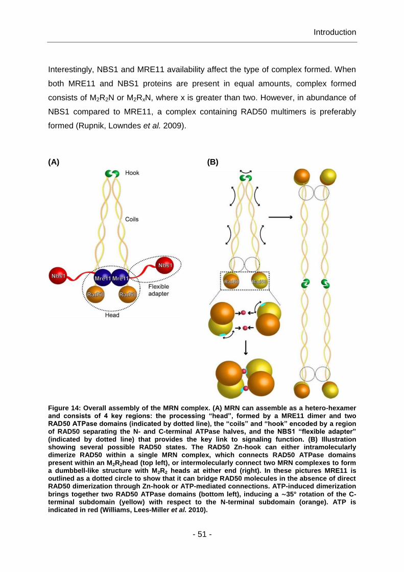
Introduction
- 51 -
Interestingly, NBS1 and MRE11 availability affect the type of complex formed. When
both MRE11 and NBS1 proteins are present in equal amounts, complex formed
consists of M2R2N or M2RxN, where x is greater than two. However, in abundance of
NBS1 compared to MRE11, a complex containing RAD50 multimers is preferably
formed (Rupnik, Lowndes et al. 2009).
(A) (B)
Figure 14: Overall assembly of the MRN complex. (A) MRN can assemble as a hetero-hexamer and consists of 4 key regions: the processing “head”, formed by a MRE11 dimer and two RAD50 ATPase domains (indicated by dotted line), the “coils” and “hook” encoded by a region of RAD50 separating the N- and C-terminal ATPase halves, and the NBS1 “flexible adapter” (indicated by dotted line) that provides the key link to signaling function. (B) Illustration showing several possible RAD50 states. The RAD50 Zn-hook can either intramolecularly dimerize RAD50 within a single MRN complex, which connects RAD50 ATPase domains present within an M2R2head (top left), or intermolecularly connect two MRN complexes to form a dumbbell-like structure with M2R2 heads at either end (right). In these pictures MRE11 is outlined as a dotted circle to show that it can bridge RAD50 molecules in the absence of direct RAD50 dimerization through Zn-hook or ATP-mediated connections. ATP-induced dimerization
brings together two RAD50 ATPase domains (bottom left), inducing a ∼35° rotation of the C-terminal subdomain (yellow) with respect to the N-terminal subdomain (orange). ATP is indicated in red (Williams, Lees-Miller et al. 2010).
Introduction
- 52 -
Moreover, MRN is able to slide along the DNA, which would allow further MRN
molecules to bind to DNA ends (Moreno-Herrero, de Jager et al. 2005) – an ability
that might also be useful for the initial steps of DNA repair. For instance, Fig. 14 B
depicts two microscopically distinct MRN-DNA tethering architectures – a long-range
DNA tethering structure and a short range DNA synapsis. Short-range DNA bridging,
within a single head of a M2R2 complex may promote direct DNA end joining,
whereas long-range DNA tethering plays an important role in recombinational DNA
repair (Hopfner, Craig et al. 2002; Williams, Williams et al. 2007; Williams, Lees-
Miller et al. 2010), as the RAD50 hook structure allows for bridging of dsDNA ends by
facilitating the assembly of octameric long-range MRN-DNA-tethering scaffolds (de
Jager, van Noort et al. 2001), resulting in the formation of heterotetrameric MRE11
complex dimers ((M2R2)2). This is in line with the observation that any mutations in
the RAD50 hook region disrupt scaffolding abilities of the complex leading to an
increased sensitivity to genotoxic stress or radiation sensitivity (Hopfner, Craig et al.
2002).
In summary, the MRN complex contains 5 super classes of DNA repair domains: (1)
DNA damage recognition, (2) protein-protein interaction and (3) nuclease domains as
well as (4) ATP-driven conformational switches and (5) an ATPase motor for opening
DNA at DSBs. Hence, it serves in part as a multipurpose DNA tether that acts to
directly bridge severed DNA ends and chromatin domains, hence preventing DNA
and chromosome separation, respectively.
1.5.2 The function of MRN complex in DDR
In undamaged cells MRE11 is homogenously distributed within the nucleus. Upon
DNA damage induction MRN localizes very rapidly to DSBs, showing association
with damaged DNA within 10min post-irradiation (D'Amours and Jackson 2002).
NBS1 recruits ATM to damage-sites and moreover activates it (Berkovich, Monnat Jr.
et al. 2007). This process is initiated by autophosphorylation events of inactive ATM
dimers on Ser-1981 leading to its monomerization, and hence the activation of ATM
kinase activity. For localizing ATM to damage sites, the ATP-dependent DNA
unwinding ability of MRE11 is of great importance (Paull and Gellert 1999).
Introduction
- 53 -
Once ATM is activated and recruited to DNA damage sites, it phosphorylates further
ATM substrates such as MRE11 and NBS1, thus activating DNA damage signaling,
DNA repair pathways and other cellular responses (Bakkenist and Kastan 2003; Lee
and Paull 2007). However, MRN is not only implicated in ATM activation, but is also a
target for ATM kinase in amplifying cell-cycle checkpoint signals or in downstream
signaling to the DNA-repair machinery (Lavin 2007). Cell-cycle checkpoint signals
can be amplified by NBS1-binding to -H2AX, resulting in a second wave of MRN
recruitment to damage sites (Lee and Paull 2007), which is in line with the results
showing that NBS1 mutants (Ser-343A) present no checkpoint response as they fail
to stimulate CHK2 phosphorylation by ATM in vitro (Difilippantonio, Celeste et al.
2005). In addition, phosphorylation of MRN by ATM triggers MRN’s inactivation and
its disassembly from chromatin, thus down-regulating DNA damage response,
following the recovery of checkpoint response (Di Virgilio, Ying et al. 2009). This
cascade of cellular events in response to DNA damage is illustrated in Fig. 15.

Introduction
- 54 -
Figure 15: A model for early events in the cellular response to DSBs. MRN is essential for initial damage processing, where processed DNA lesions lead to recruitment of MRN and ATM activation. Active ATM phosphorylates its substrates, like NBS1 and MRE11, whereas phosphorylated NBS1 facilitates phosphorylation of certain ATM substrates, and furthermore plays an important role in activating cell cycle checkpoints (Uziel, Lerenthal et al. 2003).
Interestingly, in mammals MRE11 nuclease activity has been implicated in the
activation of ATM through several approaches including effects on the stability of
replication forks, as well as the initiation of cell-cycle checkpoints and repair
processes (Jazayeri, Balestrini et al. 2008; Hashimoto, Chaudhuri et al. 2010).

Introduction
- 55 -
Accordingly, there is some evidence for a putative role of MRE11 nuclease activity in
a continued ATM activation (Jazayeri, Balestrini et al. 2008), suggesting that while
nuclease activity of MRE11 is not required for the initiation of ATM activation, it
contributes to the maintenance of ATM checkpoint signaling (Uziel, Lerenthal et al.
2003; Lee and Paull 2005). Other results show that processing of DSBs into ssDNA
ultimately inhibits ATM, and contributes to the switch from ATM to ATR activation
(Shiotani and Zou 2009). However, MRN may function to regulate ATM activity during
ongoing DSB signaling by controlling the production of suitable DNA substrates at
distinct stages of the DSB response, even though nucleolytic processing by MRE11
is an essential function of fundamental importance in DNA repair, distinct from MRN
control of ATM signaling (Buis, Wu et al. 2008). Taken together, these observations
demonstrate that MRN does not only act as a DNA damage sensor and as an
upstream regulator of ATM activity but that it also plays an essential role downstream
of ATM activation in DNA damage signaling and DSB repair.
1.5.3 The MRN complex in DSB repair
As discussed above, MRN senses DNA breaks, activates cell cycle checkpoints and
has a central role in all DSB repair pathways, like HRR, D-NHEJ and B-NHEJ.
Depending on the particular function of MRN, there are several distinct models for its
role in DNA end tethering and processing during DSB repair.
For instance, HRR of DSBs requires architectural, structural and enzymatic
components. Architectural components are needed to tether DSB ends in close
proximity to prevent inappropriate recombination, and to pair them with homologous
sequences on the sister chromatid, as illustrated in Fig. 16 A. Enzymatic activities are
required to process DNA ends and for repair synthesis (de Jager, Trujillo et al. 2004;
Shin, Chahwan et al. 2004). Thus, the structurally implied primary activity of MRN
seems to be a linker function for homologous stretches of DNA and/or broken ends
(Hopfner, Craig et al. 2002), whereas the resecting ability of MRE11 is required for
the formation of RAD51 nucleoprotein filament during the presynapsis step of
recombination (Hopkins and Paull 2008). In this model, the nuclease activity of MRN
is downstream of DNA break binding and the initiation of ATM activation.

Introduction
- 56 -
The involvement of MRN in HRR is supported by data showing that deletion of
MRE11 or NBS1 in chicken DT40 cells results in reduced HR and sister chromatid
exchange without affecting end-joining frequencies in plasmid based assays (Tauchi,
Kobayashi et al. 2002; Yang, Saidi et al. 2006). In contrast, other results suggest that
MRN deficiency confers a strong defect in CSR, affecting both D-NHEJ and B-NHEJ
(Helmink, Bredemeyer et al. 2009). Furthermore, other results showed that silencing
of MRE11, RAD50 or CTIP decreased NHEJ probably because MRE11 could control
end-joining through both, ATM-dependent and ATM-independent pathways. The
latter set of results suggests that MRN may play an inhibitory role in NHEJ in
mammals (Rass, Grabarz et al. 2009).
(A)
(B)
(C)
Figure 16: Models for the role of MRN in HRR and NHEJ. (A) The nuclease activity of MRN can process dsDNA ends for the subsequent repair steps. The architecture of tail-to-tail linked MRN complexes suggests a mechanism for bridging sister chromatids, and initiating and stabilizing displacement loop formation – a common intermediate in recombination, break-induced replication and telomere maintenance. (B) In NHEJ, head domains of two M2R2 complexes bind separate broken DNA ends, aligning and tethering them after a structural transition. (C) Circularization of a single M2R2 complex could potentially contribute to productive repair of broken DNA ends (Hopfner, Craig et al. 2002).

Introduction
- 57 -
However, the DNA end recognition and processing by MRN suggests a role for MRN
during the early stage of NHEJ (Daley, Palmbos et al. 2005) as MRN can stabilize
distant breaks by collapse of a Zn2+-linked (M2R2)2 octamer (Fig. 16 B). Alternatively,
two broken DNA ends might bind to the head of same MRN complex that has an
internal Zn2+ linkage, as depicted in Fig. 16 C (Hopfner, Craig et al. 2002). There is
also evidence that DSB repair by B-NHEJ is facilitated by MRN, particularly as
MRE11 processes DNA termini (Deriano, Stracker et al. 2009; Lamarche, Orazio et
al. 2010; Taylor, Cecillon et al. 2010), and because rapid accumulation of MRN at
DNA damage sites requires PARP-1 (Haince, McDonald et al. 2008). Nonetheless,
the nuclease activity of MRE11 is not important for accurate D-NHEJ (Moreau,
Ferguson et al. 1999), so that a specific role of MRN in D-NHEJ is still unclear and
hence requires further investigation.
1.6 IR-induced foci formation – protein accumulation at DNA
damage sites
As described above, in response to IR-induced DNA damage several proteins
involved in DDR signaling or DSB repair are recruited. Many of these proteins have
been shown to locally accumulate into large nuclear domains – the so-called ionizing
radiation-induced foci (IRIF) (Rogakou, Boon et al. 1999; Paull, Rogakou et al. 2000;
Fernandez-Capetillo, Celeste et al. 2003). Indirect immunofluorescence or imaging of
live cells expressing fluorescently tagged proteins can be used for the visualization of
such protein accumulations. In order to detect discrete protein foci, hundreds to
thousands of molecules must be present at DNA damage sites (Paull, Rogakou et al.
2000). The examination of IRIF formation and decay of DDR signaling and DSB
repair proteins offers a powerful way to analyze effects of DNA damage in signaling
and repair.
Nowadays many proteins are known to form IRIF, e.g. -H2AX, MRE11, RAD50,
NBS1, RAD51, ATM, RPA, 53BP1 etc. (Maser, Monsen et al. 1997; Chen,
Bhandoola et al. 2000; Anderson, Henderson et al. 2001; Daboussi, Dumay et al.
2002; Balajee and Geard 2004; Young, Jonnalagadda et al. 2005; Whalen, Gurai et
al. 2008).

Introduction
- 58 -
Human MRE11 and RAD50 form IR-induced discrete and subnuclear foci (Maser,
Monsen et al. 1997), surrounding the break within a 10kb distance (Di Virgilio, Ying et
al. 2009), where it exists in two fractions: (1) in a H2AX / MDC1-dependent fraction
on chromatin and (2) in a H2AX-independent fraction at DSBs (Bekker-Jensen,
Lukas et al. 2006; Berkovich, Monnat Jr. et al. 2007). However, the best documented
foci-forming protein is the chromatin bound histone variant, H2AX, although -H2AX
foci formation is a special case as no protein movements are involved in the foci
forming process. In response to IR, H2AX phosphorylation at Ser-139 occurs at
megabase regions surrounding the DSB within seconds after DNA damage induction
(Rogakou, Pilch et al. 1998; Fernandez-Capetillo, Celeste et al. 2003). In the course
of DSB repair, the initial -H2AX foci number decreases, which makes this protein an
indirect marker for studying DSB processing. Thus, scoring of -H2AX foci is now
generally accepted as a method to evaluate DSB repair kinetics at low radiation
doses (up to 4Gy) (Rogakou, Pilch et al. 1998; Sedelnikova, Rogakou et al. 2002;
Sedelnikova, Horikawa et al. 2004; Kinner, Wu et al. 2008). Although, not every -
H2AX focus can be linked to a DSB at all times (Tanaka, Huang et al. 2007).
Moreover, after DNA damage induction -H2AX foci colocalize with many other DNA
damage signaling and repair protein foci such as ATM, MDC1, 53BP1, BRCA1,
RAD51, MRN etc. (Paull, Rogakou et al. 2000). Therefore, colocalization analysis of
IRIF can be of great importance in analyzing the involvement of different proteins in
DSB repair.
However, there are two classes of proteins accumulating to foci after DNA damage
induction distinguishable based on their spatial distributions: those present directly at
the damage sites, coating ssDNA resulting from DSB resection, and those associated
with DSB-flanking chromatin (Bekker-Jensen, Lukas et al. 2006).
Furthermore, DDR proteins assemble in a sequential, coordinated manner at DSBs,
rather than being recruited as a preformed protein complex. The accumulation
kinetics of signaling proteins has been described as a two-stage process in which
initial recruitment occurs independently of H2AX phosphorylation, possibly involving
MRN, followed by sustained DDR factor retention in a -H2AX-dependent manner
(Celeste, Fernandez-Capetillo et al. 2003; Xie, Kwok et al. 2009; Yuan and Chen
2010).

Introduction
- 59 -
It is widely assumed that foci formation by DDR proteins potentiates interactions
between these proteins and damaged DNA, thus mounting rapid and effective
responses to DNA breaks. Recruitment of DDR proteins to DNA damage sites is very
fast but transient, reaching its accumulation maximum within the first 1-2min
(Mortusewicz, Ame et al. 2007). Factors involved in DSB repair by NHEJ are
recruited within seconds upon break formation, normally dissociating within 2h (Mari,
Florea et al. 2006), whereas HRR factors show delayed and persistent recruitment to
DSBs, reflecting the different repair kinetics between these two pathways (Kim,
Krasieva et al. 2005).
Importantly, responses to DSBs are markedly influenced by the cell cycle status.
Thus, foci from DDR factors like -H2AX, MRN, ATM or MDC1 occur regardless of
cell cycle stage. In contrast, proteins like CTIP, RPA, ATR, BRCA1 and RAD51, form
effective foci only in association with ssDNA formation after DNA resection in S/G2-
phase of the cell cycle (Lisby, Barlow et al. 2004; Sartori, Lukas et al. 2007).
Additionally, in most cases the number of focus-positive cells, and of the foci number
per cell increase in a dose-dependent manner (van Veelen, Cervelli et al. 2005). For
some proteins, even the foci size increases over time (van Veelen, Cervelli et al.
2005), reflecting the spread of these proteins from the DSB into adjacent chromatin
(Rogakou, Boon et al. 1999). However, not all DDR or DSB repair factors accumulate
at DNA breaks in such a way that foci can be observed, e.g. IRIF of CHK1 or CHK2
(Smits, Reaper et al. 2006). No foci are also forming by most of the NHEJ repair
proteins. This is probably because NHEJ components do not spread substantially
into adjacent chromatin, which would make them visible as foci. As a result
accumulation is only observed only when high levels of damage are induced (Bekker-
Jensen, Lukas et al. 2006).

Hypotheses and specific aims
- 60 -
2 Hypotheses and specific aims
It is evident from the outline above that the cellular mechanisms of DSB repair
pathway choice are only incompletely understood, despite their importance in our
understanding of the DDR. There are hints that MRN is involved in this function, but
the available information of its mechanistic significance is uncertain. Thus, the first
aim of this study was to contribute new information on the possible role of MRN in the
selection between HRR and D-NHEJ in the repair of IR-induced DSBs. The
overarching hypothesis was that MRN has a key function in bridging NHEJ with the
initiating steps of HRR.
To this end, we further hypothesized that the analysis of MRN foci formation and
decay would provide important insights into its function in DDR, in general, and its
contribution to DSB repair pathway choice, in particular. To begin addressing this
question, we studied the dynamics of MRN foci formation and decay in different cell
types after exposure to IR of different LET. For this purpose, we employed confocal
laser scanning microscopy (CLSM) to ensure the highest possible sensitivity and
resolution of detection. Furthermore, we attempted an analysis throughout the cell
cycle, particularly in G1- and G2-phases, as it is amply documented that the
repertoire of DSB repair pathways varies throughout the cell cycle. Analysis of the
relative contribution of MRN in DSB repair was carried out with double staining for -
H2AX and MRE11.
A question we asked beyond a function of MRN in DSB repair pathway choice was its
actual contribution to the different DSB repair pathways. This is because there are
reports in the literature implicating MRN in all DSB repair pathways. To investigate
the contribution of MRN to DSB repair by HRR, we scored RAD51 IRIF in wild-type
and MRN-deficient cells. To evaluate DSB repair by D-NHEJ we used PFGE. PFGE
was also used to measure DSB repair by B-NHEJ in repair-proficient, MRN-deficient,
as well as in repair-proficient and MRN-deficient cells treated with a specific DNA-
PKcs inhibitor, NU7441.

Hypotheses and specific aims
- 61 -
Lastly, we made an effort to unveil the putative regulatory role of the DNA-PK protein
complex in DSB repair pathway choice. Indeed, there are reports implicating DNA-PK
in the regulatory process of DSB repair pathway choice. Therefore, we decided to
study to what extent DNA-PK deficiency affects HRR. For this purpose, we examined
IR-induced MRE11 and RAD51 foci formation and decay dynamics in different DNA-
PK mutants and compared the results to wild-type cells.
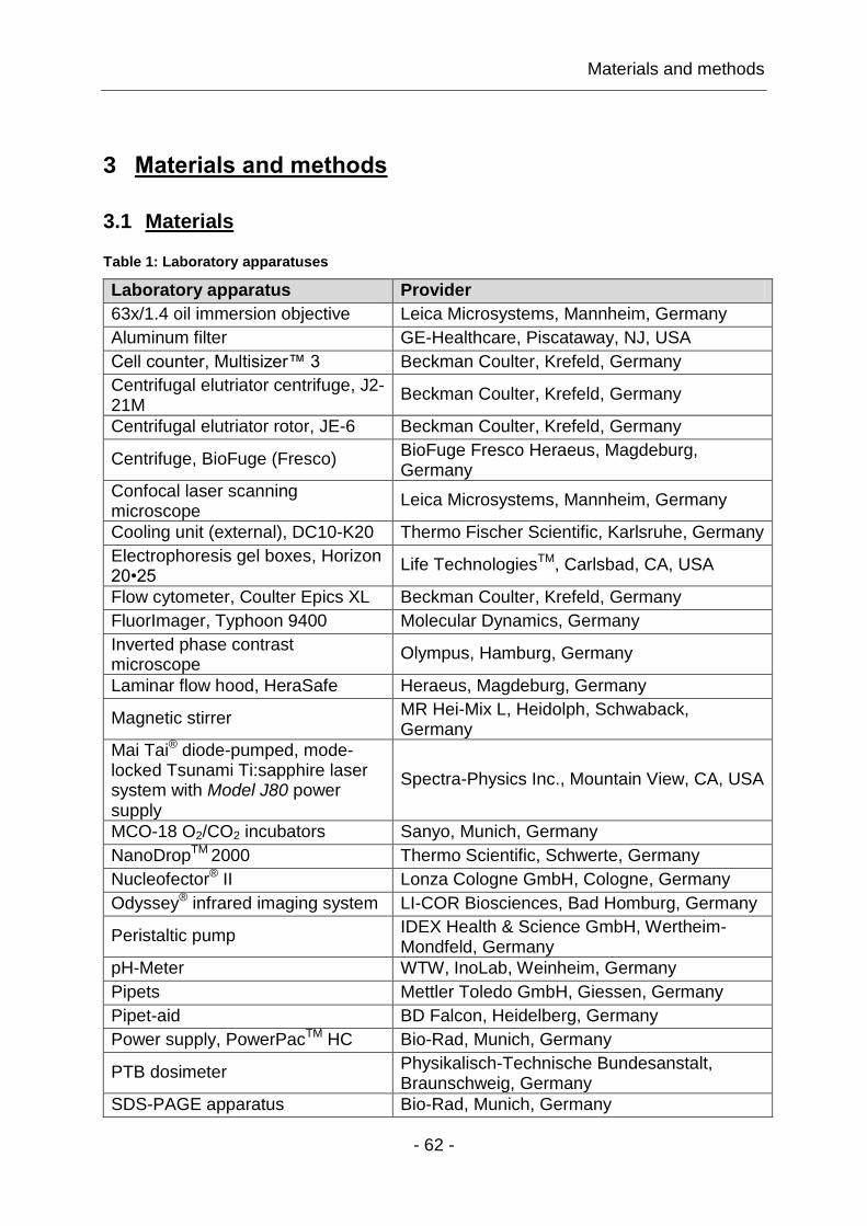
Materials and methods
- 62 -
3 Materials and methods
3.1 Materials
Table 1: Laboratory apparatuses
Laboratory apparatus Provider
63x/1.4 oil immersion objective Leica Microsystems, Mannheim, Germany
Aluminum filter GE-Healthcare, Piscataway, NJ, USA
Cell counter, Multisizer™ 3 Beckman Coulter, Krefeld, Germany
Centrifugal elutriator centrifuge, J2-21M
Beckman Coulter, Krefeld, Germany
Centrifugal elutriator rotor, JE-6 Beckman Coulter, Krefeld, Germany
Centrifuge, BioFuge (Fresco) BioFuge Fresco Heraeus, Magdeburg, Germany
Confocal laser scanning microscope
Leica Microsystems, Mannheim, Germany
Cooling unit (external), DC10-K20 Thermo Fischer Scientific, Karlsruhe, Germany
Electrophoresis gel boxes, Horizon 20•25
Life TechnologiesTM, Carlsbad, CA, USA
Flow cytometer, Coulter Epics XL Beckman Coulter, Krefeld, Germany
FluorImager, Typhoon 9400 Molecular Dynamics, Germany
Inverted phase contrast microscope
Olympus, Hamburg, Germany
Laminar flow hood, HeraSafe Heraeus, Magdeburg, Germany
Magnetic stirrer MR Hei-Mix L, Heidolph, Schwaback, Germany
Mai Tai® diode-pumped, mode-locked Tsunami Ti:sapphire laser system with Model J80 power supply
Spectra-Physics Inc., Mountain View, CA, USA
MCO-18 O2/CO2 incubators Sanyo, Munich, Germany
NanoDropTM 2000 Thermo Scientific, Schwerte, Germany
Nucleofector® II Lonza Cologne GmbH, Cologne, Germany
Odyssey® infrared imaging system LI-COR Biosciences, Bad Homburg, Germany
Peristaltic pump IDEX Health & Science GmbH, Wertheim-Mondfeld, Germany
pH-Meter WTW, InoLab, Weinheim, Germany
Pipets Mettler Toledo GmbH, Giessen, Germany
Pipet-aid BD Falcon, Heidelberg, Germany
Power supply, PowerPacTM HC Bio-Rad, Munich, Germany
PTB dosimeter Physikalisch-Technische Bundesanstalt, Braunschweig, Germany
SDS-PAGE apparatus Bio-Rad, Munich, Germany
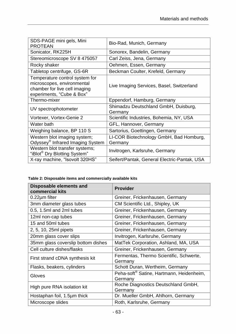
Materials and methods
- 63 -
SDS-PAGE mini gels, Mini PROTEAN
Bio-Rad, Munich, Germany
Sonicator, RK225H Sonorex, Bandelin, Germany
Stereomicroscope SV 8 475057 Carl Zeiss, Jena, Germany
Rocky shaker Oehmen, Essen, Germany
Tabletop centrifuge, GS-6R Beckman Coulter, Krefeld, Germany
Temperature control system for microscopes, environmental chamber for live cell imaging experiments, “Cube & Box”
Live Imaging Services, Basel, Switzerland
Thermo-mixer Eppendorf, Hamburg, Germany
UV spectrophotometer Shimadzu Deutschland GmbH, Duisburg, Germany
Vortexer, Vortex-Genie 2 Scientific Industries, Bohemia, NY, USA
Water bath GFL, Hannover, Germany
Weighing balance, BP 110 S Sartorius, Goettingen, Germany
Western blot imaging system; Odyssey® Infrared Imaging System
LI-COR Biotechnology GmbH, Bad Homburg, Germany
Western blot transfer systems; “iBlot® Dry Blotting System”
Invitrogen, Karlsruhe, Germany
X-ray machine, “Isovolt 320HS” Seifert/Pantak, General Electric-Pantak, USA
Table 2: Disposable items and commercially available kits
Disposable elements and commercial kits
Provider
0.22µm filter Greiner, Frickenhausen, Germany
3mm diameter glass tubes CM Scientific Ltd., Shipley, UK
0.5, 1.5ml and 2ml tubes Greiner, Frickenhausen, Germany
12ml non-cap tubes Greiner, Frickenhausen, Germany
15 and 50ml tubes Greiner, Frickenhausen, Germany
2, 5, 10, 25ml pipets Greiner, Frickenhausen, Germany
20mm glass cover slips Invitrogen, Karlsruhe, Germany
35mm glass coverslip bottom dishes MatTek Corporation, Ashland, MA, USA
Cell culture dishes/flasks Greiner, Frickenhausen, Germany
First strand cDNA synthesis kit Fermentas, Thermo Scientific, Schwerte, Germany
Flasks, beakers, cylinders Schott Duran, Wertheim, Germany
Gloves Peha-soft® Satine, Hartmann, Heidenheim, Germany
High pure RNA isolation kit Roche Diagnostics Deutschland GmbH, Germany
Hostaphan foil, 1.5µm thick Dr. Mueller GmbH, Ahlhorn, Germany
Microscope slides Roth, Karlsruhe, Germany

Materials and methods
- 64 -
Para film Lab Depot, Dawsonville, GA, USA
Pasteur pipettes BD Falcon, Heidelberg, Germany
Pipettes Greiner, Frickenhausen, Germany
Pipet tips Greiner, Frickenhausen, Germany
Real time PCR LightCycler® capillaries
Roche Diagnostics Deutschland GmbH, Germany
Western blot blotting stacks; iBlot® blotting stacks
Invitrogen, Karlsruhe, Germany
UV cuvettes Hellma, Muellheim, Germany
Table 3: Chemicals
Chemical Provider
2-Mercaptoethanol Sigma-Aldrich, Steinheim, Germany
4-(2-hydroxyethyl)-1-piperazineethanesulfonic acid
Roth, Karlsruhe, Germany
4',6-diamidino-2-phenylindole Sigma-Aldrich, Steinheim, Germany
6-(4-hydroxyphenyl)-2-thioxo-2,3-dihydro-4(1H)-pyrimidinone
Santa Cruz Biotechnology, Inc., Heidelberg, Germany
Agarose Bio-Rad, Munich, Germany
Aluminum sulphate Roth, Karlsruhe, Germany
Ammonium persulfate Sigma-Aldrich, Steinheim, Germany
Boric acid Roth, Karlsruhe, Germany
Bovine serum albumin fraction IV Roth, Karlsruhe, Germany
Bromophenol blue Sigma-Aldrich, Steinheim, Germany
Chicken serum Gibco™, Invitrogen, Karlsruhe, Germany
Coomassie brilliant blue R 250 SERVA Electrophoresis GmbH, Heidelberg, Germany
Crystal violet Merck, Darmstadt, Germany
Dimethyl sulfoxide Sigma-Aldrich, Steinheim, Germany
Dithiothreitol Roth, Karlsruhe, Germany
Doxycycline hydrochloride Sigma-Aldrich, Steinheim, Germany
Dulbecco's Modified Eagle Medium Gibco™, Invitrogen, Karlsruhe, Germany
Ethanol Sigma-Aldrich, Steinheim, Germany
Ethidium bromide Roth, Karlsruhe, Germany
Ethylenediaminetetraacetic acid Roth, Karlsruhe, Germany
Ethyleneglycoltetraacetic acid Sigma-Aldrich, Steinheim, Germany
Fetal bovine serum Biochrom, Berlin, Germany; PAA, Coelbe, Germany; Gibco™, Invitrogen, Karlsruhe, Germany
Gelatin Calbiochem, Invitrogen, Karlsruhe, Germany
Glycerol Roth, Karlsruhe, Germany
Glycine Roth, Karlsruhe, Germany
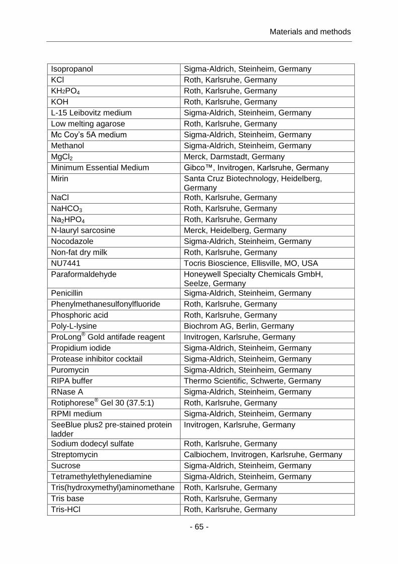
Materials and methods
- 65 -
Isopropanol Sigma-Aldrich, Steinheim, Germany
KCl Roth, Karlsruhe, Germany
KH2PO4 Roth, Karlsruhe, Germany
KOH Roth, Karlsruhe, Germany
L-15 Leibovitz medium Sigma-Aldrich, Steinheim, Germany
Low melting agarose Roth, Karlsruhe, Germany
Mc Coy’s 5A medium Sigma-Aldrich, Steinheim, Germany
Methanol Sigma-Aldrich, Steinheim, Germany
MgCl2 Merck, Darmstadt, Germany
Minimum Essential Medium Gibco™, Invitrogen, Karlsruhe, Germany
Mirin Santa Cruz Biotechnology, Heidelberg, Germany
NaCl Roth, Karlsruhe, Germany
NaHCO3 Roth, Karlsruhe, Germany
Na2HPO4 Roth, Karlsruhe, Germany
N-lauryl sarcosine Merck, Heidelberg, Germany
Nocodazole Sigma-Aldrich, Steinheim, Germany
Non-fat dry milk Roth, Karlsruhe, Germany
NU7441 Tocris Bioscience, Ellisville, MO, USA
Paraformaldehyde Honeywell Specialty Chemicals GmbH, Seelze, Germany
Penicillin Sigma-Aldrich, Steinheim, Germany
Phenylmethanesulfonylfluoride Roth, Karlsruhe, Germany
Phosphoric acid Roth, Karlsruhe, Germany
Poly-L-lysine Biochrom AG, Berlin, Germany
ProLong® Gold antifade reagent Invitrogen, Karlsruhe, Germany
Propidium iodide Sigma-Aldrich, Steinheim, Germany
Protease inhibitor cocktail Sigma-Aldrich, Steinheim, Germany
Puromycin Sigma-Aldrich, Steinheim, Germany
RIPA buffer Thermo Scientific, Schwerte, Germany
RNase A Sigma-Aldrich, Steinheim, Germany
Rotiphorese® Gel 30 (37.5:1) Roth, Karlsruhe, Germany
RPMI medium Sigma-Aldrich, Steinheim, Germany
SeeBlue plus2 pre-stained protein ladder
Invitrogen, Karlsruhe, Germany
Sodium dodecyl sulfate Roth, Karlsruhe, Germany
Streptomycin Calbiochem, Invitrogen, Karlsruhe, Germany
Sucrose Sigma-Aldrich, Steinheim, Germany
Tetramethylethylenediamine Sigma-Aldrich, Steinheim, Germany
Tris(hydroxymethyl)aminomethane Roth, Karlsruhe, Germany
Tris base Roth, Karlsruhe, Germany
Tris-HCl Roth, Karlsruhe, Germany

Materials and methods
- 66 -
Triton X-100 Sigma-Aldrich, Steinheim, Germany
Trypsin Biochrom AG, Berlin, Germany
Tween 20 Roth, Karlsruhe, Germany
Table 4: Antibodies
Antibody Provider
MRE11 (NB 100-142) (rPAb)* Novus Biologicals, Littleton, CO, USA
MRE11 (12D7) (mMAb) Novus Biologicals, Littleton, CO, USA
MRE11 (12D7) (mMAb) GeneTex, Irvine, CA, USA
RAD50 (13B3) (mMAb) GeneTex, Irvine, CA, USA
NBS1 (NB100-143) (rPAb) Novus Biologicals, Littleton, CO, USA
RAD51 (14B4) (mMAb) GeneTex, Irvine, CA, USA
pS1981-ATM (rPAb) Abcam, Cambridge, UK
-H2AX (3F2) (mMAb) Abcam, Cambridge, UK
Cyclin B1 (H433) (rPAb) Santa Cruz Biotechnology, Heidelberg, Germany
GAPDH (mMAb) Millipore, Darmstadt, Germany
LAMIN A/C (636) (mMAb) Santa Cruz Biotechnology, Heidelberg, Germany
α-TUBULIN (AA13) (mMAb) Sigma-Aldrich, Steinheim, Germany
Alexa Fluor 488 (mPAb, rPAb) Invitrogen, Karlsruhe, Germany
Alexa Fluor 568 (mPAb, rPAb) Invitrogen, Karlsruhe, Germany
Alexa Fluor 633 (mPAb, rPAb) Invitrogen, Karlsruhe, Germany
IRDye 680 (mPAb, rPAb) LI-COR Biosciences, Bad Homburg, Germany
IRDye 800 (mPAb, rPAb) LI-COR Biosciences, Bad Homburg, Germany
*In immunofluorescence applications, the antibody MRE11 (NB 100-142) was used in all experiments, as this antibody was the only functional one, among all other tested MRE11 antibodies, in detecting human MRE11 IRIF. None of the antibodies tested worked in other species.

Materials and methods
- 67 -
Table 5: Plasmid
Plasmid Description Provider
pLEGFP-MRE11 Wild-type MRE11 cDNA cloned into a pLEGFP vector
Huichen Wang, Department of Radiation Oncology, Emory University School of Medicine, Atlanta, GA, USA
Table 6: Softwares
Software Provider
Adobe® Creative Suite® 5.5 Adobe Systems Inc., USA
ImageQuant™ 5.0 GE Healthcare Life Sciences, USA
ImarisXT® 6.0 Bitplane Scientific Software,
Switzerland
Microsoft Office 2010® Microsoft, USA
Quantity One® Bio-Rad Laboratories, Inc., USA
SigmaPlot® 11.0 Systat Software, USA
WincycleTM Phoenix Flow Systems, USA

Materials and methods
- 68 -
Table 7: Alphabetical list of cell lines used in experiments
Cell line Description [Reference] Culture medium
A549 Human lung carcinoma, repair-proficient cell line with wild-type p53 expression level but decreased repair of mismatched DNA bases.
Mc Coy’s 5A + 10% FBS
AT5-BIVA Human AT fibroblasts (Luo, Tang
et al. 1996)
MEM + 10% FBS
CHO-10B4 Chinese hamster ovarian fibroblasts, standard repair-proficient cell line (Jeggo and Kemp 1983)
Mc Coy’s 5A + 10% FBS
DT40-DNA-PKcs-/- DT40-MRE11 DT40-MRE11-/-MRE11+
Chicken B cell lymphocytes, KO for DNA-PKcs (Fukushima, Takata et al. 2001) Wild-type cell line for MRE11 and conditional KO for MRE11 (Yamaguchi-Iwai, Sonoda et al. 1999)
RPMI + 10% FBS + 1% chicken serum + 10µM 2-ME
HT1080 HT1080-YFP-MRE11 HT1080-MRE11-YFP
Human osteosarcoma tumor fibroblasts Cell clones stably express YFP-tagged MRE11 proteins with high and low expression rates, respectively (Giesen, Langner et al. 2011).
DMEM + 10% FBS + 0.4µg/ml Puromycin
Human lymphocytes Human lymphocytes from a healthy donor
M059K M059J
Human glioma control cell line for M059J cells Mutant in DNA-PKcs; counterpart of M059K
DMEM + 10% FBS
MEF PK34N (DNA-PKcs+/+) PK33N (DNA-PKcs-/-) KU70-/- KU80-/-
PK80-193A (DNA-PKcs-/-KU80-/-)
Mouse embryonic fibroblasts Wt cell line for DNA-PKcs KO KO for DNA-PKcs (Kurimasa, Ouyang et al. 1999) KO for KU70 KO for KU80 (Nussenzweig, Chen et al. 1996) Double KO for DNA-PKcs and KU80
MEM + 10% FBS DMEM + 10% FBS MEM + 10% FBS
MRC5-SV1 Human repair-proficient fibroblasts MEM + 10% FBS
NBS1-LB Human fibroblasts from NBS patients with defective NBS1 function
MEM + 10% MEM
Materials and methods
- 69 -
3.2 Methods
3.2.1 Tissue culture and growth conditions
Tissue culture was performed in SANYO MCO-18 O2/CO2 incubators at 37°C (in
case of DT40 cells at 39.5°C) in a humidified atmosphere with 5% CO2 and 95% air.
Adherent cells were grown in 100mm cell culture dishes with 15ml of the appropriate
growth medium, supplemented with fetal bovine serum (FBS). Suspension cells were
maintained in 100mm bacteria dishes with 10ml growth medium supplemented with
chicken serum and 10µM 2-ME (Table 7). All the media included 100µg/ml penicillin
as well as 100µg/ml streptomycin.
Adherent cells were grown as a monolayer and were passaged every 3 days while
avoiding confluency levels above 80%. For passage, media was removed and cells
were briefly washed with 10ml cold 1x phosphate-buffered saline (PBS; 137mM
NaCl, 2.7mM KCl, 10mM Na2HPO4, 1.76mM KH2PO4, pH 7.4). Cells were then
covered with 2ml trypsin solution (0.05% trypsin in EDTA) and incubated for 5min at
37°C. Detached cells were resuspended in 10ml cold media. Single cell suspensions
were counted (Multisizer™ 3, Beckman Coulter), and appropriate numbers of cells
were further incubated for experiments or for subculture. HT1080-MRE11-YFP, as
well as HT1080-YFP-MRE11 cells were maintained in selection marker-containing
medium (0.4µg/ml puromycin) in order to ensure stable transgenic cells expressing
MRE11 tagged with YFP protein. Suspension cells were subcultured every 3 days
without exceeding a cell number of 2 million cells per ml of medium. For splitting, the
cell suspension was gently pipetted to break clumps and the cell density was
determined. Calculated cell volume with desired cell number was re-seeded into a
new dish and incubated for growth.
When frozen cells were taken to subculture, they were passaged at least two times
before being used in experiments and were discarded after about 40 passages. In all
experiments exponentially growing cells were used unless otherwise indicated. The
distribution of cells throughout the cell cycle was measured by flow cytometry.
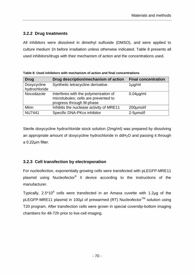
Materials and methods
- 70 -
3.2.2 Drug treatments
All inhibitors were dissolved in dimethyl sulfoxide (DMSO), and were applied to
culture medium 1h before irradiation unless otherwise indicated. Table 8 presents all
used inhibitors/drugs with their mechanism of action and the concentrations used.
Table 8: Used inhibitors with mechanism of action and final concentrations
Drug Drug description/mechanism of action Final concentration
Doxycycline hydrochloride
Synthetic tetracycline derivative 1µg/ml
Nocodazole Interferes with the polymerization of microtubules; cells are prevented to progress through M-phase
0.04µg/ml
Mirin Inhibits the nuclease activity of MRE11 200µmol/l
NU7441 Specific DNA-PKcs inhibitor 2-5µmol/l
Sterile doxycycline hydrochloride stock solution (2mg/ml) was prepared by dissolving
an appropriate amount of doxycycline hydrochloride in ddH2O and passing it through
a 0.22μm filter.
3.2.3 Cell transfection by electroporation
For nucleofection, exponentially growing cells were transfected with pLEGFP-MRE11
plasmid using Nucleofector® II device according to the instructions of the
manufacturer.
Typically, 2.5*106 cells were transfected in an Amaxa cuvette with 1.2μg of the
pLEGFP-MRE11 plasmid in 100µl of prewarmed (RT) NucleofectorTM solution using
T20 program. After transfection cells were grown in special coverslip-bottom imaging
chambers for 48-72h prior to live-cell imaging.

Materials and methods
- 71 -
3.2.4 Cell synchronization
Cell populations enriched in G1- or G2-phases of the cell cycle were obtained by
centrifugal elutriation – a method for isolating cellular subpopulations on the basis of
their sedimentation coefficient. For the elutriation process exponentially growing
single cell suspension of 140-200 millions cells were placed into a specially designed
centrifuge rotor chamber (Beckman JE-6 Elutriation Rotor, Beckman Coulter). They
were subjected to a centrifugal field that was balanced by a counter-flow (25ml/min)
of medium (culture medium with 1% FBS) that was selected to ensure that cells
remained in the chamber and aligned themselves according to size. Cells were then
sequentially elutriated out of the rotor based on their size, with small cells coming out
first. For an exponentially growing culture, cell size approximates the phase of the
cell cycle, with G1-phase cells being small and G2-phase cells being large.
Rotor speeds for cell cycle fractionations of eukaryotic cell populations were usually
in the 1,200-3,000rpm range with 100-200rpm steps.
Cell fractions of 250ml were collected by gradually reducing the rotor speed. Cell
cycle analysis was carried out by flow cytometry. Typically, cell populations with at
least 90% G1-phase cells were obtained in G1-fraction, and 65-75% G2-phase cells
in G2-fraction, respectively.
3.2.5 Fluorescence activated cell sorting
Flow cytometry allows measurement of several different cellular parameters as well
as cell sorting on the basis of different properties by assessing fluorescence intensity.
This technique is also termed as fluorescence activated cell sorting (FACS). For
instance, cell cycle distribution can be evaluated by measuring fluorescence intensity
of propidium iodide (PI) bound to DNA, as PI binds to DNA proportionally to its mass.
Hence, the fluorescence intensity of PI is proportional to the DNA amount present in
a cell. Because after replication the DNA amount is doubled, there are twice as many
signals generated from G2-phase cells as compared to G1-phase cells. In this way,
cells in different cell cycle phases are distinguishable from each other by the DNA
amount detected.

Materials and methods
- 72 -
For measurements, single cell suspensions of about one million cells were fixed
overnight in 70% ethanol at -20°C. Supernatant was removed by centrifugation at
100 x g for 5min, and the pellet was incubated in 500µl of PI stain (40µg/ml PI,
62µg/ml RNaseA; PI stain buffer: 0.1M Tris, 0.1M NaCl, 5mM MgCl2, 0.05% Triton X-
100, RNaseA buffer: 10mM Tris, 100mM EDTA, 50mM NaCl, pH 7.6) for 15min at
37°C. Samples were measured in a flow cytometer (Coulter Epics XL, Beckman
Coulter) according to pre-established protocols (Table 9); 15,000 events were
counted, and gated to obtain standard histograms for each sample. Histogram files
(*.HST) were generated by counting the frequency of cells with the same PI signal
intensity. The fraction/percentage of cells in different cell cycle phases was
automatically calculated using WincycleTM software. For that, HST files were loaded
into WincycleTM, and the parameter “S-phase growing order” was carefully chosen
between 0-2 until the prediction model fitted the histogram shape.
Table 9: Settings for flow cytometer, Coulter Epics XL
Parameter Setting/value
Flow cytometry Coulter Epics XL
Sheath speed middle
Total cells sampled 15,000
Maximal running duration 900s
Working mode carousel
Excitation 488nm
Filter spectrum 655-735nm
FS-PMT 55V
FS-Gain 2
SS-PMT 400V
SS-Gain 1
AUX-PMT 300V
AUX-Gain 2
PI-PMT 520-650V
PI-Gain 2
Discriminator PI>5
Gate single

Materials and methods
- 73 -
3.2.6 Irradiation
For irradiation experiments exponentially growing cells were plated at the appropriate
cell density as required by the experimental protocol, and were grown for two days. In
experiments analyzing IRIF, cells were seeded at 125,000 cells onto sterilized 20mm
glass cover slips in 35mm dishes with 2ml growth medium, two days prior to IR
exposure. In case of using synchronized cells, 500,000 cells were seeded onto poly-
L-lysine coated cover slips, and given 1h time to attach before irradiation. To coat
cover slips with poly-L-lysine, sterilized cover slips were incubated in 2ml poly-L-
lysine solution (1mg/ml) for 15min, followed by a washing step with ddH2O.
3.2.6.1 X-ray irradiation
X-rays were generated by an x-ray machine (“Isovolt 320HS”, Seifert/Pantak,
General Electric-Pantak) with an effective photon energy of approximately 90keV.
The machine was operated at 320kV and 10mA using a 1.65mm aluminum filter (GE-
Healthcare) to absorb soft x-rays. Dosimetry was performed with a PTB dosimeter
(Physikalisch-Technische Bundesanstalt, Braunschweig, Germany) that was used to
calibrate an in-field ionization monitor. Radiation dose was confirmed with Fricke’s
chemical dosimetry. Even irradiation was ensured by rotating the radiation table.
Cells were irradiated with different x-ray doses at a dose rate of 1.3Gy/min.
Immediately after IR exposure cells were returned to the incubator, and collected at
different times according to the experimental procedure. Unirradiated control cells
were treated similarly (sham-irradiated).

Materials and methods
- 74 -
3.2.6.2 Neutron irradiation
Neutrons cannot be accelerated like charged particles as they are uncharged.
Therefore, neutrons have to be produced in nuclear reactions by accelerating light
ions and letting them collide with selected target nuclei, e.g. beryllium nuclei. The ion
accelerator used was a rotatable, variable energy “TCC CV-28 cyclotron” operated by
the Departments of Nuclear Medicine and Radiotherapy, University Hospital Essen,
Germany. This cyclotron provides beams of protons (19MeV), deuterons (13.5MeV)
and α-particles (28MeV). Cells were irradiated with different neutron doses (mean
energy: 5.8MeV) at a dose rate of 0.3Gy/min in a rectangular 25 x 25cm2 irradiation
field with a photon contamination of ~10%.
3.2.6.3 Heavy ion irradiation
Heavy ion irradiation experiments were performed at the irradiation facility of the
Helmholtz Centre for Heavy Ion Research, Darmstadt, Germany (GSI), where ion
beams of all elements up to the heaviest, uranium, can be generated in any state of
electric charge, and accelerated to nearly the speed of light. The GSI has two
different accelerators: (1) UNILAC, a linear 120m long accelerator that accelerates
ions up to 20% of light speed, and (2) the heavy-ion SIS synchrotron, shown in Fig.
17, which is able to accelerate ion beams up to 90% of the speed of light in the
course of several hundred thousand revolutions.

Materials and methods
- 75 -
Figure 17: Outer view of the heavy-ion synchrotron facility (SIS) at the Helmholtz Centre for Heavy Ion Research, Darmstadt, Germany (GSI). Electromagnets, which direct and focus the ion beam, are shown in red and yellow. Copyright: Helmholtz Centre for Heavy Ion Research, Darmstadt, Germany (GSI).
We performed two different experiments at the GSI. Fe and Ni heavy ions,
respectively with an ion fluence of 1.12*106 ions/cm2 per scan were generated by the
SIS synchrotron. These heavy ions had a kinetic energy of 1GeV/nucleon, and a
corresponding LET of 150keV/μm.
3.2.6.4 Multiphoton irradiation
Multiphoton (MP) absorption processes resulting in light-induced subcellular
alterations have been detected in living cells (Calmettes and Berns 1983). The study
of MP absorption can yield new information about the basic interactions within and
between molecules. Due to the fact that spatial resolution of two-photon (2-P)
absorption can produce subcellular destructive events (Berns, Wang et al. 2000), MP
(2-P) laser irradiation of individual live cells can be used for localized intracellular
DNA damage induction. This kind of approach has been already used extensively for
cell division, and gene expression analyses in cells with organelles altered by micro-
irradiation.
Materials and methods
- 76 -
The obvious advantage of near-infrared (NIR) MP laser micro-irradiation consists in
its three-dimensional confinement of the lesions as the laser can specifically target
single cells and subnuclear structures, such as nucleoli, mitochondria, nuclear
membranes, and any other structures that can be labeled and visualized by a
fluorescent tag. However, the specificity with respect to damage type is limited. Thus,
this technique is a powerful tool to study intracellular protein localization processes
as a function of space and time (Meldrum, Botchway et al. 2003), enabling
investigation of DNA repair dynamics, intracellular signaling, and cell-cell
communication processes with a spatio-temporal resolution.
To generate strictly localized and clearly tractable sub-nuclear DNA damage, cells
were brought into focus and positioned in such a way that the pulsed 2-P
femtosecond laser (Mai Tai® Tsunami Ti:sapphire laser, Spectra-Physics) would
center on a cell nucleus. The region to be damaged was always of the same size and
shape (bleaching points). Laser treatment was done at constant maximum laser
output power of 80mW to exclude variations in dose. Time of cell exposure to the
focused laser beam was 850ms (pulse width <100fs, pulse repetition rate ~80MHz,
MP gain 40%, MP offset 65%, Attenuation min). This process was controlled by a
software-connected internal electromechanical shutter.
3.2.7 Immunofluorescence staining
For immunofluorescence staining, cells were briefly washed with cold 1x PBS, fixed
in 2ml 2% paraformaldehyde (PFA) for 15min at room temperature (RT), washed with
1x PBS, permeabilized in 2ml P-solution (100mM Tris, pH 7.4, 50mM EDTA, 0.5%
Triton X-100) for 10min at RT, again washed with 1x PBS and finally blocked in 2ml
PBG blocking solution (0.2% gelatin, 0.5% BSA fraction V in 1x PBS, pH 7.4)
overnight at 4°C or for 1h at RT.
Primary and conjugated secondary antibodies (Table 4) were diluted 1:200 and 1:400
in PBG, respectively; 70μl of the antibody solution were dispensed on parafilm, cover
slips were mounted on top of the antibody solution and incubated at RT for 1.5h with
primary antibodies and for 1h with secondary antibodies.
Materials and methods
- 77 -
After each antibody incubation, cover slips were returned into the dishes and washed
three times with 1x PBS for 5min each. Subsequently, cell nuclei were counterstained
in 2ml 4',6-diamidino-2-phenylindole (DAPI) solution (2µg/ml DAPI, 0.1M Tris, 0.1M
NaCl, 5mM MgCl2, 0.05% Triton X-100), with a final DAPI concentration of 50ng/ml,
for 15min at RT, three times washed with 1x PBS and mounted in 13µl ProLong®
Gold antifade reagent on microscope slides. For the polymerization of ProLong® Gold
antifade, slides were kept in the dark for at least 24h at RT and then stored at 4°C
before analysis.
However, immunofluorescence staining with anti-MRE11 antiserum, after fixing the
cells with PFA, exhibited very high background signals, thus making MRE11 foci
analysis impossible (data not shown). High background signals can be a
consequence of remaining non-bound protein; probably because of high cross-linking
ability of PFA, and the high nuclear abundance of the MRN complex. To reduce
background signal of non-bound MRE11 in the nucleoplasm, cell fixation was
performed using a methanol-acetone fixation protocol. For this purpose, cells were
fixed in 2ml 100% methanol for 20min at -20°C, permeabilized in acetone for 10s at
RT, washed with 1x PBS and blocked in 2ml 10% FBS in PBS solution overnight at
4°C, or alternatively 1h at RT.
3.2.8 Confocal laser scanning microscopy
In contrast to conventional two-dimensional light microscopy, CLSM comprises three
process steps: (1) line-by-line scanning of the specimen with a focused laser beam
(monochromatic light of a discrete wavelength) deflected in X-Y directions by means
of two mirrors mounted on two so-called – galvanometric scanners, (2) pixel-by-pixel
detection of the fluorescence emitted by the scanned specimen details, by means of
a photomultiplier tube (PMT), and (3) digitalization of the optical object information
contained in the electrical signal provided by the PMT (Wilhelm, Gröbler et al. 2009).
The decisive design feature of a CLSM compared to a conventional microscope is
the confocal aperture – the pinhole, a small diaphragm in front of the detector,
arranged in a plane conjugate to the intermediate image plane and thus to the object
plane of the microscope. As a result, the detector (PMT) can only detect light that has

Materials and methods
- 78 -
passed the pinhole. The significant amount of fluorescence emission that occurs at
points above and below the objective focal plane is not confocal with the pinhole, and
is termed as “out-of-focus light rays”; this extraneous light is not detected by the
PMT, and does not contribute to the resulting high-resolution optical image. By
varying the pinhole diameter, the degree of confocality can be adapted to practical
requirements (Claxton, Fellers et al.).
In a CLSM system a laser beam passes through a light source aperture, and is then
focused to a diffraction-limited spot, using a diffraction mirror, which illuminates only a
small focal volume at a time within or on the surface of a specimen. After illuminating
this defined focal plane, secondary fluorescence emitted from illuminated points on
the specimen in the same focal plane, passes back through the dichroic mirror, and is
focused as a confocal point at the detector pinhole aperture. Thus, specimens are
irradiated in a pointwise fashion, where the point illuminated and the point observed
are situated in conjugate planes, i.e. they are focused onto each other. The result is
what is called a confocal beam path. In order to obtain information about the entire
specimen, it is necessary to guide the laser beam in X and Y directions across the
specimens. A schematic diagram of CLSM key components is shown in Fig. 18.
Figure 18: Schematic diagram of principal components and optical pathway in a CLSM system. Illustration from: Leica Microsystems, Mannheim, Germany.
Materials and methods
- 79 -
In addition, to observe a single plane (or slice) of a thick specimen in good contrast,
optical sectioning allows a great number of slices to be cut and recorded at different
planes of the specimen, with specimens being moved along the optical axis (Z) by
controlled increments. The result is a 3-dimensional data set – a "z-stack", which
provides information about the spatial structure of the object (Wilhelm, Gröbler et al.
2009). Moreover, by using more than one fluorescent dye with different spectral
properties, several target molecules can be identified simultaneously (Wilhelm,
Gröbler et al. 2009). Taken together, the CLSM system is used to acquire high-
resolution optical images with depth selectivity, in both, fixed specimens and living
cells.
3.2.8.1 Live cell imaging by CLSM imaging systems
Live cell imaging is the observation of living cells over time using images acquired by
imaging systems such as confocal scanning microscopes. CLSM systems can record
images like a camera, and can therefore be used to record a series of time-resolved
confocal images, known as a time series. Such time-lapse experiments provide an
important tool in studies of cell physiology, cellular developmental and cancer
biology, whenever interest is focused on the visualization and quantification of
cellular dynamic changes. Thus, live-cell imaging provides critical insights into the
fundamental nature of cellular and tissue functions.
A major challenge of live cell imaging is keeping cells alive and functioning as
naturally as possible for the duration of the experiment since fluorescence
illumination, especially in the UV range, is harmful for cells, and causes
photobleaching and phototoxicity. The use of high power lasers as an excitation
source adds to this challenge. Successful experiments must be designed to minimize
such specimen illumination whilst maintaining an appropriate environment for the
cells. Fig. 19 represents a general setup of a live cell imaging apparatus with heating
capabilities, consisting of a microscope inside an environmental chamber. Within this
temperature-controlled system for microscopes, cells can be maintained at an
appropriate temperature.

Materials and methods
- 80 -
Figure 19: Schematic illustration of a live cell imaging microscope inside an environmental chamber. Illustration from: Carl Zeiss Microscopy, Jena, Germany.
For live cell imaging experiments, human fibroblasts (HT1080-MRE11-YFP and
HT1080-YFP-MRE11 cells; detailed described in Table 7) that stably express yellow
fluorescent protein (YFP) tagged to MRE11, were grown in 35mm coverslip bottom
dishes with 2ml growth medium at an initial cell number of 300,000 cells for 24h. In
experiments using cells that transiently express MRE11 tagged to a green
fluorochrome (GFP), CHO-10B4 and MEF-PK34N cells were grown in coverslip
bottom live cell imaging chambers for 48-72h post transfection (transfection
conditions in 3.2.3).
The requirement of keeping cells at 5% CO2 during the investigation span can be
minimized by using 4-(2-hydroxyethyl)-1-piperazineethanesulfonic acid (HEPES)
buffered medium, e.g. L-15 Leibovitz medium, which was formulated for the use in
CO2-free systems requiring sodium bicarbonate. Hence, before starting live cell
imaging experiments, growth medium was exchanged with 1ml of L-15 Leibovitz
medium to ensure proper and stable pH for the duration of experiment. The software
was programmed to start image sequence acquisition just before irradiation for an
initial control image, and immediately after damage induction to take z-stack images
for 1-8h in 5min intervals. Further details concerning image acquisition and image
analysis are described below. Normally, the scanned field included also some
unirradiated cells, to ensure a proper control for protein accumulation at damage
sites.

Materials and methods
- 81 -
3.2.9 Image acquisition and digital image analysis
For image acquisition, fixed, immunofluorescently stained cells were imaged at a
resolution of 1024 x 1024 and a zoom factor of 1.2, resulting in a pixel size of
240.74nm, whereas live cells were imaged in a 512 × 512 pixel format and a zoom
factor of 3. Fluorescence protein signals were visualized by exciting fluorochromes
with an appropriate laser line, and by detecting the spectral range of the fluorescence
emitted with proper PMT gain and offset settings. To minimize photobleaching of
fluorescence protein-signal during data collection, laser power was kept at about 10-
15% of maximum. Scanning parameters, like laser intensity, PMT gain and offset
settings (Table 10, 11) were individually adjusted for different fluorochromes, and
kept constant within an experiment to ensure comparability of data. When using
multiple fluorescent proteins, bidirectional scans were applied in a sequential scan
modus with predefined scanning parameters in order to avoid cross talk between
different channels.
Table 10: Image acquisition settings of the confocal microscope, Leica TCS SP5
Parameter Mode/setting/value
Instrument parameter setting Confocal LMD
Argon laser intensity 30%
Objective HCX PL APO lambda blue 63.0x1.4 OIL UV
Sequential scan Between stacks
Speed 400Hz
Refraction index 1.52
Scan direction Bidirectional
Pinhole 1
Frame average 2
Frame accumulation 2
Line average 2
Line accumulation 2
Zoom 1.2-3
Resolution 512 x 512 or 1024 x 1024, 8bits
Z-step size 0.5μm

Materials and methods
- 82 -
Typically, at least 5 fields were scanned by taking z-stack images with 0.5µm steps.
After each scan, three-dimensional data sets (LIF files), and maximum intensity
projection TIFF images of the field scanned were generated and processed for
further analysis.
Table 11: Scanning parameters for the detection of fluorescent proteins by CLSM
Fluorochrome parameter
DAPI Alexa-488; GFP
YFP Alexa-568 Alexa-633
Excitation
Laser intensity
Detected spectral range PMT gain
PMT offset
405nm
15%
410-480nm
500-750V
-0.3V
488nm
10-15%
495-560nm
550-850V
-0.3V
514nm
10%
520-580nm
650-1200
-0,3
568nm
15%
570-635nm
650-850V
-0.3V
633nm
15%
640-760nm
750-950V
-0.3V
The recorded (time-resolved) z-stack images were analyzed for foci numbers, foci
colocalization, fluorescence total signal intensity, and protein accumulation dynamics
at damaged sites, using the quantitative image processing software – ImarisXT® 6.0
(Bitplane).
LIF files of imaged live cells were processed with the “Easy 3-D” mode, and
subsequently converted into video clip files to visualize protein accumulation, and
dissociation dynamics in a spatio-temporal resolution. In contrast, foci number and
foci colocalization analyses were achieved by using “spots”, “split spots” and
“colocalize spots” functions, respectively. During foci analysis, a gray value threshold
was set to separate signal from background. This threshold value varied from 12 to
20, and was kept constant throughout one experiment in order to be able to compare
the data. Only objects with a minimum diameter of 0.5μm and intensity above the
threshold were counted as foci. For each dose and repair time point, about 200 cells
were analyzed. Finally, mean foci number per nucleus, standard deviation as well as
standard error were calculated using Microsoft Excel 2007® and graphs generated
with SigmaPlot® 11.0.

Materials and methods
- 83 -
3.2.10 Biochemical protein fractionation
For sub-cellular biochemical protein fractionation, 2-3x10⁶ cells were collected with a
cell scraper and washed twice in 1x PBS. Pellets were resuspended and incubated
for 10min on ice in 200µl cytoskeleton buffer (10mM Hepes-KOH (pH 6.8), 100mM
NaCl, 300mM sucrose, 1mM EGTA (pH 8.0), 3mM MgCl2, 0.5% Triton X-100, 0.2mM
PMSF, 1mM DTT), containing 1x protease inhibitor cocktail (complete inhibitors).
Skeletal frameworks were pelleted by centrifugation at low speed (700 x g, 5min,
4°C) and 2/3 of the supernatant was collected. The insoluble pellet was incubated in
200µl buffer A (10mM Tris-HCl (pH 8.0), 10mM NaCl, 250mM sucrose, 1mM EGTA
(pH 8.0), 5mM MgCl2), containing 1x protease inhibitors and 0.2M ammonium
sulphate, in order to extract proteins that are loosely attached to chromatin. After
10min incubation on ice, insoluble chromatin-bound proteins were collected by
centrifugation at 2,000 x g for 5min at 4°C; eluted proteins were added to the
previous supernatant. Total supernatant was clarified by high speed centrifugation
(10,000 x g, 10min, 4°C) and termed as soluble protein fraction. Final insoluble
chromatin-enriched nuclear fraction, containing chromatin-bound proteins was
resuspended in 50µl of 2x Laemmli sample buffer (126mM Tris-HCl (pH 6.8), 0.01M
EDTA, 10% glycerol, 3% SDS, 0.02% bromophenol blue, 1/20 2-mercaptoethanol),
and subjected to five pulses of sonication.
3.2.11 Electrophoresis and immunoblotting
3.2.11.1 Cell lysate preparation and electrophoresis
For cell lysate preparation, 2-3x10⁶ cells were once washed with cold 1x PBS by
centrifugation and subsequently lysed in 200µl cold RIPA buffer (ready-to-use cell
lysis reagent), containing 1x protease inhibitor cocktail. After 30min incubation on ice,
cell lysate was spun down (14,000 x g, 15min, 4°C) to pellet cell debris, and the
supernatant, with mostly all membrane, cytoplasmic, soluble and nucleoplasmic
proteins, was transferred into a new tube. Protein concentration in solution was
determined by colorimetric Bradford assay. RIPA buffer cell lysates or biochemically
fractionated soluble protein reactions were mixed with 50µl 2x Laemmli sample

Materials and methods
- 84 -
buffer, denaturated at 95°C for 5min and spun down for 1min at 13,000 x g before
electrophoresis. Protein separation was performed by electrophoresis on SDS-
polyacrylamide mini gels (“Mini PROTEAN”, Bio-Rad), consisting of a 5% stacking
gel and a resolving gel of varying polyacrylamide concentrations. RIPA buffer cell
lysates and biochemically fractionated soluble protein reactions were loaded with a
total protein amount of 20µg on SDS-10% polyacrylamide gels, whereas only 3-5µl of
chromatin-bound protein reactions were electrophoresed on SDS-12.5%
polyacrylamide gels at a constant voltage of 130V for 1.5h at RT. SDS-PAGE gels
were cast following instructions of the manufacturer.
3.2.11.2 Immunoblotting and western blot detection
For western blot analysis, electrophoretically resolved proteins were transferred onto
a nitrocellulose membrane using the “iBlot® Dry Blotting System” (Invitrogen)
following the manufacturer’s instructions. The self-contained iBlot® device uses
disposable blotting stacks with integrated nitrocellulose membranes that efficiently
and reliably blots proteins from polyacrylamide gels on membranes in less than
10min without the need for additional buffers or an external power supply. Equal
loading and transfer was monitored by immunodetection with antibodies against
appropriate protein fraction loading controls. After transfer, gels were removed from
the blotting stacks and stained in Coomassie Brilliant Blue R 250 solution (0.02%
Coomassie Brilliant Blue R 250, 2% (w/v) phosphoric acid, 5% aluminum sulphate in
10% ethanol) overnight with gentle agitation, whereas membranes were incubated in
blocking buffer (5% non-fat dry milk in PBS-T (0.05% Tween-20 in 1x PBS)) for 1h at
RT or overnight at 4°C. Subsequently, membranes were incubated with the primary
antibody overnight at 4°C, gently washed 3 times for 10min with PBS-T and probed
with the secondary antibody for 1h at RT. Primary antibodies were diluted 1:5000 in
PBS-T containing 2.5% non-fat dry milk, whereas secondary antibodies were diluted
1:10,000 (Table 4 includes all used antibodies). Subsequently, membranes were
gently washed 3 times for 10min with PBS-T and processed for western blot
detection using “Odyssey® Infrared Imaging System” (LI-COR Biosciences) following
instructions of the manufacturer.

Materials and methods
- 85 -
3.2.12 Pulsed field gel electrophoresis – PFGE
Pulsed field gel electrophoresis (PFGE) was introduced by Stamato and Denko in
1990 (Stamato and Denko 1990) and is a specific technique to measure IR-induced
DSBs. This technique takes advantage of the fact that fragmentation of chromosomal
DNA with radiation leads to a linear dose-dependent increase in the fraction of DNA
that enters the gel, whereas intact mammalian chromosomes are unable to enter the
gel. PFGE resolves DNA fragments ranging in size from 0.2-6Mbp, and detects
damage induced by as little as 2Gy of x-rays, whereas gel electrophoresis with a
constant electric field cannot resolve DNA fragments much above 50kbp.
To evaluate induction of DSBs at different radiation doses (i.e. dose response
experiment) asynchronous cells were used. Cells were resuspended in cold HEPES-
buffered (20mM HEPES, 5mM NaHCO3) serum-free media, and mixed with an equal
volume of pre-warmed (50°C) HEPES-buffered serum-free media containing 1% low
melting agarose to reach a concentration of 3x10⁶ cells/ml. The cell-agarose
suspension was then pipetted into 3mm diameter glass tubes and placed into ice to
allow for solidification. Solidified cell-agarose suspension was extruded from glass
tubes and cut into 5mm long cylindrical plugs containing approximately 2x10⁵
cells/plug. Subsequently, agarose plugs were placed in a 60mm dish containing 5ml
cold serum-free medium and exposed to different x-ray doses (3.2.6.1). Plugs were
immediately placed in lysis buffer (10mM Tris-HCl, 50mM NaCl, 100mM EDTA, 2%
N-lauryl sarcosine (NLS), pH 7.6 and freshly added 0.2mg/ml protease), and
incubated first at 4°C for 1h and then at 50°C for 16-18h.
For repair kinetics of IR-induced DSBs, irradiated cells were returned to the incubator
for the indicated repair times. After completion of the repair time interval, cells were
collected by centrifugation at 100 x g for 5min at 4°C and embedded into agarose.
This was followed by an immediate lysis step as described above. For each dose and
repair time point at least four plugs were prepared. Due to technical difficulties in
determining the 0h repair time point, the initial value of repair kinetics was obtained
from dose response curves. After lysis, agarose plugs were washed with washing
buffer (10mM Tris-HCl, 50mM NaCl, 100mM EDTA, pH 7.6) for 1h at 37°C while
gently shaking, and were then treated for 1h at 37°C in the same buffer with
0.1mg/ml freshly added RNase A.

Materials and methods
- 86 -
To determine the signals generated by non-irradiated cells as background, cells from
identically treated non-irradiated cultures were processed at pre-defined times, e.g.
15min and 4h.
Asymmetric field inversion gel electrophoresis was used for quantification of IR-
induced DSBs. This was carried out in 0.5% agarose gels containing
0.5µg/ml·ethidium bromide (stock solution 10mg/ml in ddH2O). Agarose plugs were
loaded into the gel-wells and sealed with 1% agarose. Gel electrophoresis was run in
0.5x TBE buffer (45mM Tris, 4.5mM boric acid, 0.1mM EDTA, pH 8.8) using
conventional gel boxes (Horizon 20•25, Life TechnologiesTM) connected to a
switching apparatus that regulated both, forward and reverse pulse times as well as
voltages, from a standard power supply (PowerPacTM HC, Bio-Rad). PFGE was
performed with 40 alternating cycles – a 900s pulse of 1.25V/cm in (forward) direction
of DNA migration, followed by a 75s pulse of 5V/cm in reverse direction. Before
starting electrophoresis, 0.5x TBE buffer was pre-cooled through an external cooling
unit (DC10-K20, Thermo Fischer Scientific) to 8°C and circulated at constant
temperature throughout the run. After electrophoresis, the gel was scanned with a
FluorImager (Typhoon™ 9410, Molecular Dynamics) using appropriate settings (see
Table 12).
Table 12: Scanning Parameters for FluorImager, Typhoon™ 9410
Mode Setting
Acquisition mode Fluorescence
Laser Green (532)
PMT gain 470V
Sensitivity Normal
Emission filter 610 BP SPYRO RyPy EtBr
Pixel size 200 microns
Focal plane +3mm
To estimate DSBs, the fraction of DNA released (FDR) was calculated using
ImageQuant™ 5.0. The FDR parameter is defined as the fraction of DNA found in the
lane and is calculated by dividing the signal in the lane with the total signal of the
sample; it is equivalent to the fraction of unrepaired DSBs in the sample.
Materials and methods
- 87 -
Measured FDR in non-irradiated cells (background) was subtracted from the FDR of
irradiated cells. When induction of DSBs was measured at different radiation doses,
FDR was plotted against radiation dose to obtain dose response curves. In order to
facilitate the inter comparison of results obtained, and to account for differences in
the dose response curves between different cell lines and experiments, repair
kinetics are not presented as FDR versus time but rather as dose equivalent (DEQ)
versus time. We used dose response curves to estimate DEQ values from each FDR
value. This way of analysis has an advantage as it corrects for non-linear dose
response curves. Repair kinetics curves were fitted using non-linear regression
analysis to calculate repair half times. In general, two components were assumed to
exist in the repair curves and fitting algorithms were selected accordingly. For all
graphs and curve fitting analyses SigmaPlot® 11.0 was used.

Results
- 88 -
4 Results
4.1 Analysis of nuclear MRN relocalization dynamics in response
to IR
As discussed above, the precise functions of the MRN complex, although clearly
critical for the cellular DDR, are only incompletely understood, and are therefore
presently under intensive investigation. We reasoned that analysis of the dynamics of
foci formation and decay for proteins implicated in DDR will yield relevant insights
into their function. Therefore, as a first step, we studied the IRIF dynamics of MRN in
different cell types after exposure to IR of different LET. Experiments were carried out
either by monitoring time-resolved MRE11-GFP/YFP protein accumulation to DNA
damage sites in live cells, or by quantitatively analyzing, using LSCM, IR-induced
MRN foci in cells fixed after irradiation and repair.
4.1.1 MRE11 forms foci after IR
We used indirect immunofluorescence to examine the response of MRE11 to
radiation-induced DNA damage. We wished to examine whether MRE11 foci form in
cells exposed to low LET radiation. For this purpose, several repair-proficient, human
cell lines were used in the exponential phase of growth. These included: A549,
MRC5-SV1, M059K and HT1080 cells (see Table 7 for more information regarding
cell origin and other cell line characteristics). Cells were irradiated with 1Gy x-rays,
and immediately returned to pre-irradiation conditions. Cells were subsequently fixed
and stained with an anti-MRE11 antibody, while DNA was counterstained with DAPI.
LSCM was used to image randomly selected cell nuclei. Representative micrographs
of captured images from unirradiated as well as irradiated cells are presented in
Fig. 20.

Results
- 89 -
Figure 20: MRE11 forms nuclear foci in response to IR. Cells shown were either mock irradiated (upper panel) or irradiated with 1Gy x-rays (lower panel), and fixed 1h post-irradiation. Cells were then processed for immunofluorescence staining with an anti-MRE11 antiserum (green) while the DNA was counterstained with DAPI (blue). Merged maximum intensity projection (MIP) images represent an overlay of the green and blue channels.
Evidently, after IR all tested cell lines showed detectable MRE11 protein
accumulation to discrete nuclear foci, visible in the majority of irradiated cells (up to
95%). In contrast, almost no MRE11 foci could be detected in unirradiated cells. Non-
specific binding of the fluorochrome-labeled secondary antibody (Alexa Fluor 488), or
auto-fluorescence was not observed in cells only stained with the secondary antibody
or with DAPI. Moreover, DAPI counterstaining of irradiated cells indicated that IRIF-
containing nuclei were not grossly aberrant, and did not show signs of apoptosis. In
line with this, flow cytometry analysis of irradiated cells did not detect a significant
population of apoptotic cells (data not shown).
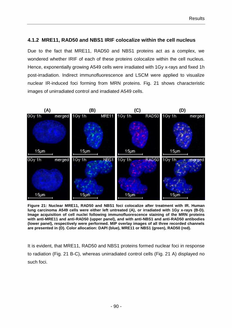
Results
- 90 -
4.1.2 MRE11, RAD50 and NBS1 IRIF colocalize within the cell nucleus
Due to the fact that MRE11, RAD50 and NBS1 proteins act as a complex, we
wondered whether IRIF of each of these proteins colocalize within the cell nucleus.
Hence, exponentially growing A549 cells were irradiated with 1Gy x-rays and fixed 1h
post-irradiation. Indirect immunofluorescence and LSCM were applied to visualize
nuclear IR-induced foci forming from MRN proteins. Fig. 21 shows characteristic
images of unirradiated control and irradiated A549 cells.
Figure 21: Nuclear MRE11, RAD50 and NBS1 foci colocalize after treatment with IR. Human lung carcinoma A549 cells were either left untreated (A), or irradiated with 1Gy x-rays (B-D). Image acquisition of cell nuclei following immunofluorescence staining of the MRN proteins with anti-MRE11 and anti-RAD50 (upper panel), and with anti-NBS1 and anti-RAD50 antibodies (lower panel), respectively were performed. MIP overlay images of all three recorded channels are presented in (D). Color allocation: DAPI (blue), MRE11 or NBS1 (green), RAD50 (red).
It is evident, that MRE11, RAD50 and NBS1 proteins formed nuclear foci in response
to radiation (Fig. 21 B-C), whereas unirradiated control cells (Fig. 21 A) displayed no
such foci.
(A) (B) (C) (D)
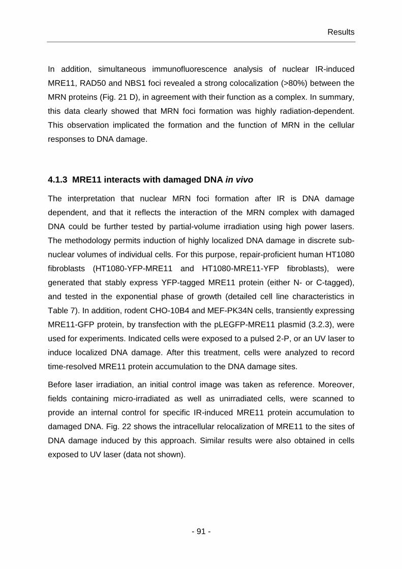
Results
- 91 -
In addition, simultaneous immunofluorescence analysis of nuclear IR-induced
MRE11, RAD50 and NBS1 foci revealed a strong colocalization (>80%) between the
MRN proteins (Fig. 21 D), in agreement with their function as a complex. In summary,
this data clearly showed that MRN foci formation was highly radiation-dependent.
This observation implicated the formation and the function of MRN in the cellular
responses to DNA damage.
4.1.3 MRE11 interacts with damaged DNA in vivo
The interpretation that nuclear MRN foci formation after IR is DNA damage
dependent, and that it reflects the interaction of the MRN complex with damaged
DNA could be further tested by partial-volume irradiation using high power lasers.
The methodology permits induction of highly localized DNA damage in discrete sub-
nuclear volumes of individual cells. For this purpose, repair-proficient human HT1080
fibroblasts (HT1080-YFP-MRE11 and HT1080-MRE11-YFP fibroblasts), were
generated that stably express YFP-tagged MRE11 protein (either N- or C-tagged),
and tested in the exponential phase of growth (detailed cell line characteristics in
Table 7). In addition, rodent CHO-10B4 and MEF-PK34N cells, transiently expressing
MRE11-GFP protein, by transfection with the pLEGFP-MRE11 plasmid (3.2.3), were
used for experiments. Indicated cells were exposed to a pulsed 2-P, or an UV laser to
induce localized DNA damage. After this treatment, cells were analyzed to record
time-resolved MRE11 protein accumulation to the DNA damage sites.
Before laser irradiation, an initial control image was taken as reference. Moreover,
fields containing micro-irradiated as well as unirradiated cells, were scanned to
provide an internal control for specific IR-induced MRE11 protein accumulation to
damaged DNA. Fig. 22 shows the intracellular relocalization of MRE11 to the sites of
DNA damage induced by this approach. Similar results were also obtained in cells
exposed to UV laser (data not shown).

Results
- 92 -
(A) (B) (C)
1
2
3
4
Figure 22: MRE11 rapidly accumulates to sites of localized DNA damage. Control images of unirradiated cells are shown in column (A). Subnuclear areas (B) were exposed to a pulsed 2-P laser, each for 850ms. Z-stack images, taken at the indicated times after laser irradiation, show a prompt MRE11 protein accumulation to the DNA damage sites (red arrows in column C). All pictures shown are MIPs.
It is evident, that upon localized DNA damage all examined cell lines showed a fast
redistribution of intracellular MRE11 to the sites of DNA damage (Fig. 22 C).
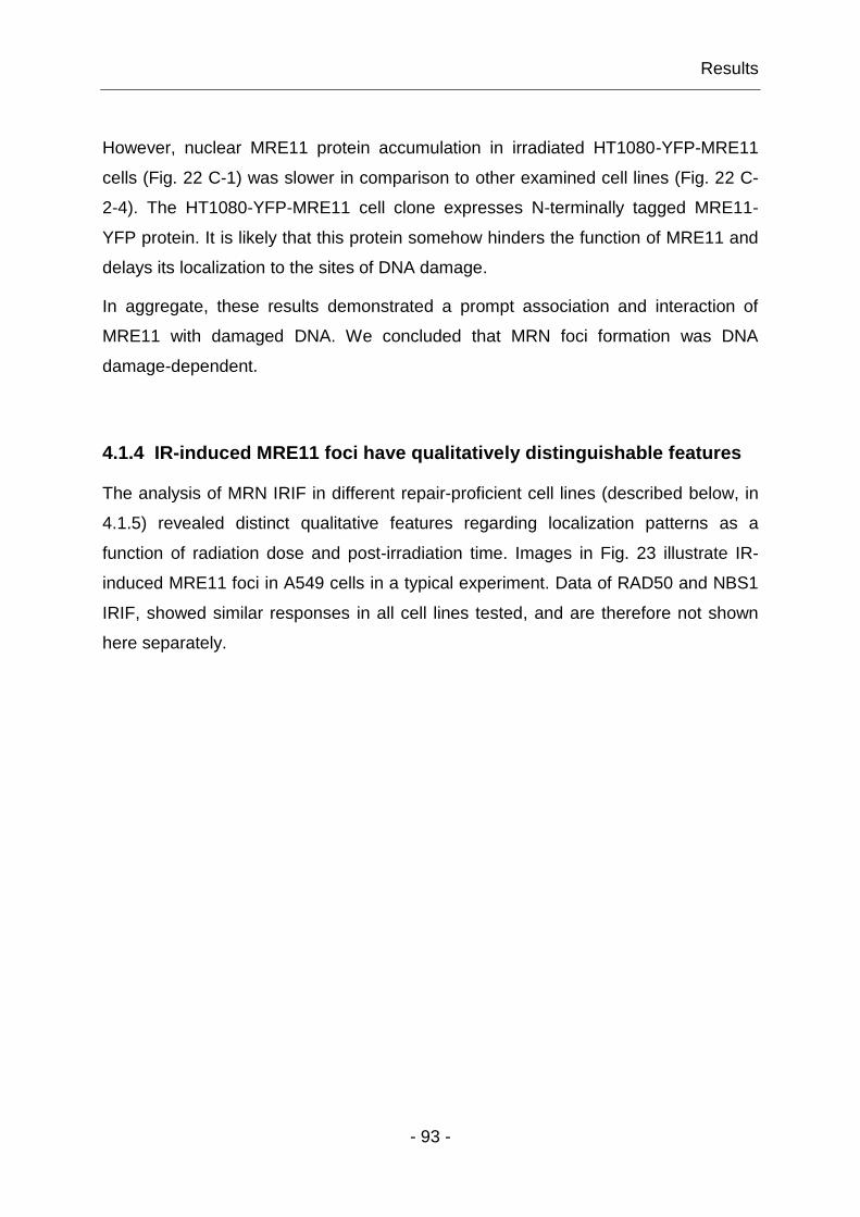
Results
- 93 -
However, nuclear MRE11 protein accumulation in irradiated HT1080-YFP-MRE11
cells (Fig. 22 C-1) was slower in comparison to other examined cell lines (Fig. 22 C-
2-4). The HT1080-YFP-MRE11 cell clone expresses N-terminally tagged MRE11-
YFP protein. It is likely that this protein somehow hinders the function of MRE11 and
delays its localization to the sites of DNA damage.
In aggregate, these results demonstrated a prompt association and interaction of
MRE11 with damaged DNA. We concluded that MRN foci formation was DNA
damage-dependent.
4.1.4 IR-induced MRE11 foci have qualitatively distinguishable features
The analysis of MRN IRIF in different repair-proficient cell lines (described below, in
4.1.5) revealed distinct qualitative features regarding localization patterns as a
function of radiation dose and post-irradiation time. Images in Fig. 23 illustrate IR-
induced MRE11 foci in A549 cells in a typical experiment. Data of RAD50 and NBS1
IRIF, showed similar responses in all cell lines tested, and are therefore not shown
here separately.
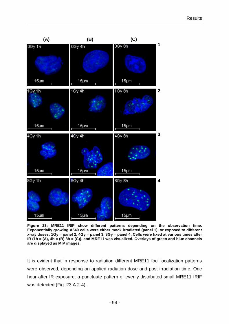
Results
- 94 -
(A) (B) (C)
1
2
3
4
Figure 23: MRE11 IRIF show different patterns depending on the observation time. Exponentially growing A549 cells were either mock irradiated (panel 1), or exposed to different x-ray doses; 1Gy = panel 2, 4Gy = panel 3, 8Gy = panel 4. Cells were fixed at various times after IR (1h = (A), 4h = (B) 8h = (C)), and MRE11 was visualized. Overlays of green and blue channels are displayed as MIP images.
It is evident that in response to radiation different MRE11 foci localization patterns
were observed, depending on applied radiation dose and post-irradiation time. One
hour after IR exposure, a punctuate pattern of evenly distributed small MRE11 IRIF
was detected (Fig. 23 A 2-4).

Results
- 95 -
These small MRE11 foci begun to change at about 4h after irradiation to large,
irregularly shaped and more sparsely distributed foci (Fig. 23 B/C 2-4). Interestingly,
this was only observed after irradiation with a low x-ray dose of 1Gy. After high
radiation doses (4-8Gy) the early response diminished leaving the late and large
MRE11 foci to dominate. Thus, two distinct MRE11 localization patterns could be
distinguished: small MRE11 IRIF mainly detected at early time points; and late
developing larger MRE11 foci that persisted for up to 8h after exposure to IR.
4.1.5 The bimodal response of MRN IRIF is cell cycle-independent
As discussed above, among proteins involved in DDR, MRN takes a unique place
through its apparent intimate involvement in the cellular response to DNA damage
and DSB repair (1.5.2, 1.5.3). IRIF of many DDR factors form, more or less, in all
phases of the cell cycle. An exception to this rule, presents proteins that effectively
form foci only in association with ssDNA formation generated by DNA resection – a
process that appears to reach a maximum in S/G2-phase of the cell cycle. We were
thus wondering whether MRN IRIF formation is cell cycle-dependent. This is
because, MRN is involved, on the one hand in DNA damage sensing, which occurs
irrespective of the cell cycle phase and, on the other hand in DSB repair by HRR,
which operates in the S/G2-phase of the cell cycle.
For this purpose, we examined several repair-proficient cell lines in G1- and G2-
phase of the cell cycle IR-induced MRN foci formation. We started with the human
lung carcinoma A549 cell line, which is deficient in mismatch repair, but express a
wild-type p53. The presence of a functional p53 in A549 cells is advantageous to our
studies, as it leaves intact important DNA damage signaling cascades (Tanaka,
Huang et al. 2007). Thus, enriched G1- and G2-phase A549 cells were irradiated with
0, 1, 4 or 8Gy, and MRE11, RAD50 and NBS1 protein localization was measured by
immunofluorescence on fixed samples using CLSM. For a quantitative analysis we
measured over 200 cell nuclei for MRE11, RAD50 or NBS1 foci at different times
after exposure to different doses of radiation.

Results
- 96 -
The cell cycle specific time course of IR-induced MRN foci dynamics in G1- and G2-
phase A549 cells after different radiation doses are summarized in Fig. 24 A-D.
Comparable results were obtained for RAD50 and NBS1 and were therefore omitted
for clarity.
(A) (B)
Repair Time (h)
0 2 4 6 8
0 2 4 6 8
MR
E1
1 F
oc
i p
er
Nu
cle
us
0
5
10
15
20
0
5
10
15
200Gy
1Gy
4Gy
8Gy
A549, G1-phase
Repair Time (h)
0 2 4 6 8
0 2 4 6 8
MR
E1
1 F
oc
i p
er
Nu
cle
us
0
5
10
15
20
0
5
10
15
200Gy
1Gy
4Gy
8Gy
A549, G2-phase
(C) (D)
Repair Time (h)
0 2 4 6 8
0 2 4 6 8
RA
D5
0 F
oc
i p
er
Nu
cle
us
0
5
10
15
20
0
5
10
15
200Gy
1Gy
4Gy
8Gy
A549, G1-phase
Repair Time (h)
0 2 4 6 8
0 2 4 6 8
NB
S1
Fo
ci p
er
Nu
cle
us
0
5
10
15
20
0
5
10
15
200Gy
1Gy
4Gy
8Gy
A549, G1-phase
Figure 24: The bimodal response of IR-induced MRN foci is cell cycle-independent. Centrifugal elutriation was applied for the enrichment of A549 cells in different cell cycle phases, as described in 3.2.4. Enriched G1- and G2-phase A549 cells were irradiated with 0, 1, 4 or 8Gy x-rays, fixed after the indicated recovery times, and MRN foci numbers quantitatively analyzed, as described above. Mean MRE11 foci numbers per cell nucleus (A, B) were enumerated from four independent experiments, whereas average RAD50 and NBS1 (C, D) foci numbers per nucleus were determined from two different experiments. Error bars represent +/- standard errors.

Results
- 97 -
Quantitative analysis of MRE11, RAD50 and NBS1 IRIF in enriched G1- and G2-
phase A549 cells revealed that irrespectively of the cell cycle phase, comparable
MRN foci formation and decay dynamics with similar MRN foci patterns (4.1.4) were
obtained. The dose response of MRN IRIF was cell cycle-independent and sub-linear
in the range of doses tested (1 to 8Gy). Remarkably, the MRN foci formation kinetics
depended on radiation dose, with a maximum been reached at later times as the
dose of radiation increased. After low radiation doses (1Gy), fast developing MRN
foci maxima were detected at 0.5-1h post-irradiation. However, these fast developing
MRN foci promptly decayed to almost background values within a repair time interval
of ~3h. In contrast to that, high doses of radiation (4-8Gy), led to a slower formation
of MRN IRIF.
After observing this bimodal response of MRN IRIF in A549 cells, we wished to
examine the generality of this phenotype. Therefore, we measured MRE11 IRIF
formation and decay in MRC5-SV1 and M059K cells. Here again, cells were enriched
in different cell cycle phases by centrifugal elutriation, treated and analyzed as
described above. Fig. 25 A-B shows MRE11 foci formation and decay dynamics only
in G1-phase MRS5-SV1 and M059K cells, respectively.

Results
- 98 -
(A) (B)
Repair Time (h)
0 2 4 6 8
0 2 4 6 8
MR
E1
1 F
oc
i p
er
Nu
cle
us
0
5
10
15
20
25
30
0
5
10
15
20
25
300Gy
1Gy
4Gy
8Gy
MRC5-SV1, G1-phase
Repair Time (h)
0 2 4 6 8
0 2 4 6 8
MR
E1
1 F
oc
i p
er
Nu
cle
us
0
5
10
15
20
25
30
0
5
10
15
20
25
300Gy
1Gy
4Gy
8Gy
M059K, G1-phase
Figure 25: All examined human repair-proficient cell types show a comparable bimodal cell cycle-independent response of MRE11 IRIF. Cells were enriched by centrifugal elutriation in different phases of the cell cycle, irradiated with different x-ray doses (0, 1, 4, 8Gy), fixed at specified times after irradiation, and the mean MRE11 IRIF numbers per cell nucleus were evaluated, as described above. Three and two independent experiments were quantitatively analyzed for average MRE11 foci numbers in MRC5-SV1 cells (A) and M059K cells (B), respectively. Error bars represent +/-standard errors.
Obviously, all examined repair-proficient cell lines showed nearly the same
phenotype – a fast response, with MRE11 foci number maxima developing at 30-
60min after exposure to low radiation doses, and a slower MRE11 foci formation after
exposure high doses of radiation (4-8Gy).
In all experiments and with all cell lines tested, the response of MRE11 IRIF showed
a sub-linear dose dependency in the range of doses tested (1 to 8Gy). For a better
visualization of this bimodal but quite general response of MRE11 foci formation as a
function of radiation dose, quantitative data of all investigated cell lines is
summarized in Fig. 26 A-D.
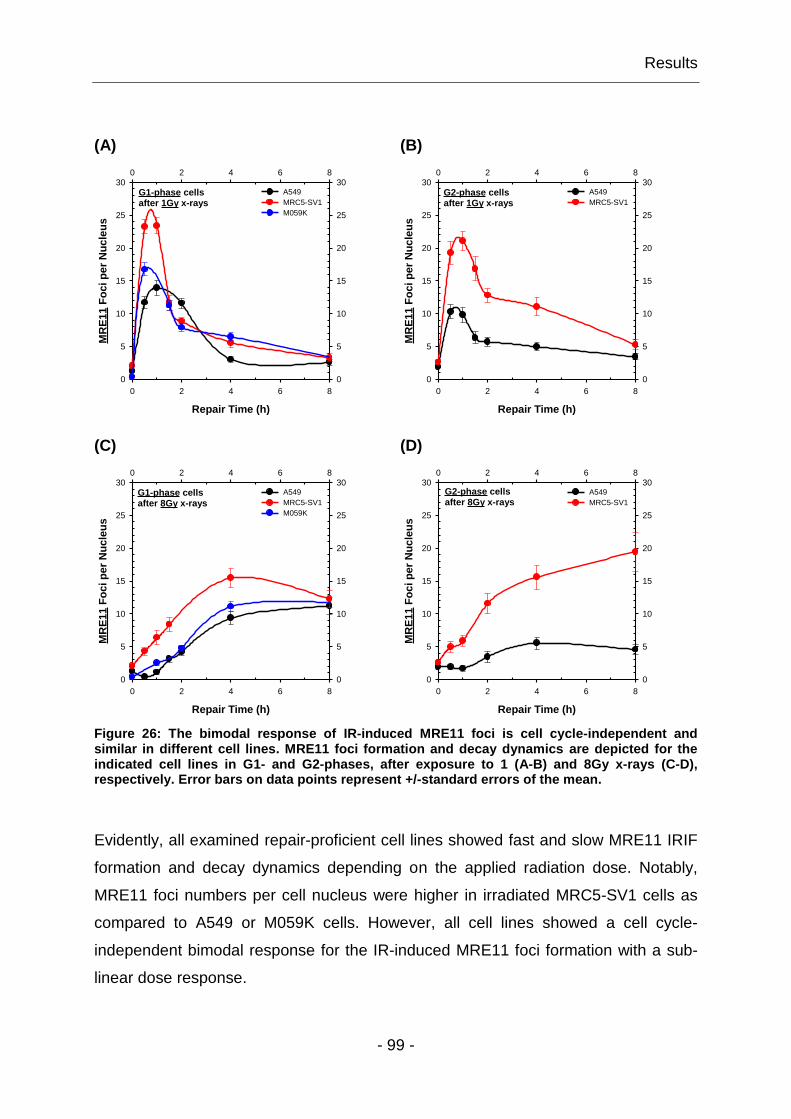
Results
- 99 -
Figure 26: The bimodal response of IR-induced MRE11 foci is cell cycle-independent and similar in different cell lines. MRE11 foci formation and decay dynamics are depicted for the indicated cell lines in G1- and G2-phases, after exposure to 1 (A-B) and 8Gy x-rays (C-D), respectively. Error bars on data points represent +/-standard errors of the mean.
Evidently, all examined repair-proficient cell lines showed fast and slow MRE11 IRIF
formation and decay dynamics depending on the applied radiation dose. Notably,
MRE11 foci numbers per cell nucleus were higher in irradiated MRC5-SV1 cells as
compared to A549 or M059K cells. However, all cell lines showed a cell cycle-
independent bimodal response for the IR-induced MRE11 foci formation with a sub-
linear dose response.
(A) (B)
Repair Time (h)
0 2 4 6 8
0 2 4 6 8
MR
E1
1 F
oc
i p
er
Nu
cle
us
0
5
10
15
20
25
30
0
5
10
15
20
25
30A549
MRC5-SV1
M059K
G1-phase cells after 1Gy x-rays
Repair Time (h)
0 2 4 6 8
0 2 4 6 8
MR
E1
1 F
oc
i p
er
Nu
cle
us
0
5
10
15
20
25
30
0
5
10
15
20
25
30A549
MRC5-SV1
G2-phase cells after 1Gy x-rays
(C) (D)
Repair Time (h)
0 2 4 6 8
0 2 4 6 8
MR
E1
1 F
oc
i p
er
Nu
cle
us
0
5
10
15
20
25
30
0
5
10
15
20
25
30A549
MRC5-SV1
M059K
G1-phase cells after 8Gy x-rays
Repair Time (h)
0 2 4 6 8
0 2 4 6 8
MR
E1
1 F
oc
i p
er
Nu
cle
us
0
5
10
15
20
25
30
0
5
10
15
20
25
30A549
MRC5-SV1
G2-phase cells after 8Gy x-rays
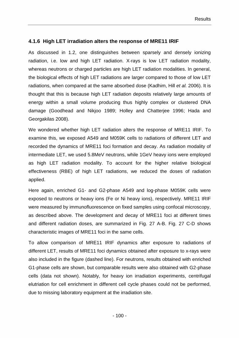
Results
- 100 -
4.1.6 High LET irradiation alters the response of MRE11 IRIF
As discussed in 1.2, one distinguishes between sparsely and densely ionizing
radiation, i.e. low and high LET radiation. X-rays is low LET radiation modality,
whereas neutrons or charged particles are high LET radiation modalities. In general,
the biological effects of high LET radiations are larger compared to those of low LET
radiations, when compared at the same absorbed dose (Kadhim, Hill et al. 2006). It is
thought that this is because high LET radiation deposits relatively large amounts of
energy within a small volume producing thus highly complex or clustered DNA
damage (Goodhead and Nikjoo 1989; Holley and Chatterjee 1996; Hada and
Georgakilas 2008).
We wondered whether high LET radiation alters the response of MRE11 IRIF. To
examine this, we exposed A549 and M059K cells to radiations of different LET and
recorded the dynamics of MRE11 foci formation and decay. As radiation modality of
intermediate LET, we used 5.8MeV neutrons, while 1GeV heavy ions were employed
as high LET radiation modality. To account for the higher relative biological
effectiveness (RBE) of high LET radiations, we reduced the doses of radiation
applied.
Here again, enriched G1- and G2-phase A549 and log-phase M059K cells were
exposed to neutrons or heavy ions (Fe or Ni heavy ions), respectively. MRE11 IRIF
were measured by immunofluorescence on fixed samples using confocal microscopy,
as described above. The development and decay of MRE11 foci at different times
and different radiation doses, are summarized in Fig. 27 A-B. Fig. 27 C-D shows
characteristic images of MRE11 foci in the same cells.
To allow comparison of MRE11 IRIF dynamics after exposure to radiations of
different LET, results of MRE11 foci dynamics obtained after exposure to x-rays were
also included in the figure (dashed line). For neutrons, results obtained with enriched
G1-phase cells are shown, but comparable results were also obtained with G2-phase
cells (data not shown). Notably, for heavy ion irradiation experiments, centrifugal
elutriation for cell enrichment in different cell cycle phases could not be performed,
due to missing laboratory equipment at the irradiation site.
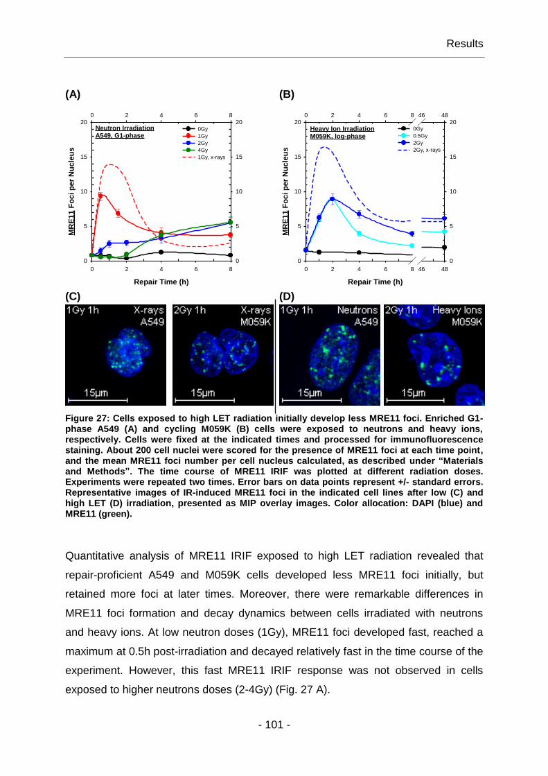
Results
- 101 -
(A) (B)
Repair Time (h)
0 2 4 6 8
0 2 4 6 8
MR
E1
1 F
oc
i p
er
Nu
cle
us
0
5
10
15
20
0
5
10
15
200Gy
1Gy
2Gy
4Gy
1Gy, x-rays
Neutron IrradiationA549, G1-phase
Repair Time (h)
0 2 4 6 8 46 48
0 2 4 6 8 46 48
MR
E1
1 F
oc
i p
er
Nu
cle
us
0
5
10
15
20
0
5
10
15
200Gy
0.5Gy
2Gy
2Gy, x-rays
Heavy Ion IrradiationM059K, log-phase
(C) (D)
Figure 27: Cells exposed to high LET radiation initially develop less MRE11 foci. Enriched G1-phase A549 (A) and cycling M059K (B) cells were exposed to neutrons and heavy ions, respectively. Cells were fixed at the indicated times and processed for immunofluorescence staining. About 200 cell nuclei were scored for the presence of MRE11 foci at each time point, and the mean MRE11 foci number per cell nucleus calculated, as described under “Materials and Methods”. The time course of MRE11 IRIF was plotted at different radiation doses. Experiments were repeated two times. Error bars on data points represent +/- standard errors. Representative images of IR-induced MRE11 foci in the indicated cell lines after low (C) and high LET (D) irradiation, presented as MIP overlay images. Color allocation: DAPI (blue) and MRE11 (green).
Quantitative analysis of MRE11 IRIF exposed to high LET radiation revealed that
repair-proficient A549 and M059K cells developed less MRE11 foci initially, but
retained more foci at later times. Moreover, there were remarkable differences in
MRE11 foci formation and decay dynamics between cells irradiated with neutrons
and heavy ions. At low neutron doses (1Gy), MRE11 foci developed fast, reached a
maximum at 0.5h post-irradiation and decayed relatively fast in the time course of the
experiment. However, this fast MRE11 IRIF response was not observed in cells
exposed to higher neutrons doses (2-4Gy) (Fig. 27 A).

Results
- 102 -
On the other hand, in response to heavy ions, M059K cells showed, independently of
the radiation dose applied, similar MRE11 foci formation dynamics. Hence, a MRE11
foci peak appeared at 2h post-irradiation; however cells exposed to 2Gy heavy ions
showed slower MRE11 foci decay kinetics over time than cells irradiated with heavy
ions at doses of 0.5Gy (Fig. 27 B). In aggregate, these results confirmed the sub-
linear dose-dependency of MRE11 foci formation for radiations of high LET.
Notably, after high LET irradiation the initial MRE11 IRIF response included mostly
large MRE11 foci (Fig. 27 D), which decreased in size over time (data not shown),
suggesting a progress in the repair of the underlying complex lesions. In contrast, low
doses of low LET radiation quickly induced the formation of small MRE11 foci (Fig.
27 C), which developed to larger foci at later times. However, these small MRE11 foci
were only observed after low doses (1Gy); after high doses (4-8Gy) the small MRE11
foci diminished leaving the late, slower developing and larger foci to dominate the
response (see also the discussion in 4.1.4). These observations indicated that the
dynamic assembly of large MRE11 foci may be required for the repair of particularly
complex and difficult to repair lesions associated with high LET irradiation, and when
high doses of low LET radiation are applied.
We concluded that high LET radiation changed the response of MRE11 to DNA
damage, since in general comparatively low numbers of large and longer persisting
foci were observed as compared to low LET irradiation. The significance of this
observation remains to be elucidated.
4.1.7 The yields of MRE11 IRIF are cell line specific
The fact that irradiated MRC5-SV1 cells showed higher MRE11 foci numbers
compared to other cell lines, such as A549 and M059K, led us inquire on the possible
causes of this phenomenon. One likely explanation is differences in the DNA content
among the cell lines tested; higher DNA content is expected to result in a larger
number of foci for the same radiation dose.
Results
- 103 -
To test this hypothesis, quantitative measurements of the nuclear DNA content for
the cell lines employed, were carried out using flow cytometry. Generally, DNA
content measurements are expressed as DNA index (DI) values, representing the
ratio of sample DNA G1 peak channel to reference DNA G1 peak channel. Thus, a DI
of 1.0 is equal to normal diploid DNA content, while DI values greater than 1.0
implicate DNA aneuploidy. We evaluated the nuclear DNA content of exponentially
growing A549, MRC5-SV1 and M059K cells by FACS (detailed information in 3.2.5),
using human lymphocytes as reference. The results of these measurements are
presented as FACS histograms in Fig. 28 A-C.
(A) (B) (C)
Figure 28: Exponentially growing MRC5-SV1 cells have the highest nuclear DNA content among the investigated cell lines. The nuclear DNA content was measured in exponentially growing A549 (A), MRC5-SV1 (B) and M059K (C) cells, using human lymphocytes as reference (i.e. DI of 1.0). To obtain the histograms, about 15,000 events were counted for each sample, and files were generated as described above. DI values were determined as G1 cell DNA content of the test cell line relative to G1 cell DNA content of the lymphocytes used as standard.
Evidently, cycling MRC5-SV1 cells had the highest cellular DNA content with a DI of
2.06, compared to exponentially growing A549 and M059K cells, which had a DI of
1.8. This result suggests that MRE11 IRIF numbers depend on cellular DNA content.
Indeed, higher IR-induced MRE11 foci numbers were detected in cells with higher
nuclear DNA content, e.g. for A549, MRC5-SV1 and M059K cells.
DI (A549) = 1.8 DI (MRC5-SV1) = 2.06 DI (M059K) = 1.8

Results
- 104 -
4.1.8 Dose-dependent accretion of MRE11 in the cell nucleus after IR
It is estimated that each Gy of low LET radiation, e.g. x-rays, will induce ~20-40
DSBs. Higher radiation doses will therefore induce hundreds of DSBs. Our
observation that after exposure to high x-ray doses (4-8Gy) only very small MRE11
foci formed at early post-irradiation times, led us consider whether the bimodal
response (4.1.5) derived from detection limitations. Possibly, in response to high
radiation doses only few MRN molecules bind to damage sites, limiting thus their
detectability. This possibility is supported by the fact that the detection of cytologically
discrete foci by indirect immunofluorescence depends on the number of protein
molecules that accumulate at the site, and is estimated to be of the order of one
thousand (Paull, Rogakou et al. 2000). Sites with fewer protein molecules might thus
escape detection.
To test this possibility, we performed an evaluation of total nuclear MRE11
fluorescence in cells irradiated with different x-rays doses as a function of time. This
kind of analysis provides quantitative information on local protein accretion in imaged
cells, and allows examination whether the degree of local protein accretion is dose-
dependent. Nuclear MRE11 and DAPI fluorescence total signal intensities were
measured in specific regions of interest (ROI), in A549 cell nuclei. Subsequently,
normalized MRE11 fluorescence total signal intensity values of analyzed ROIs were
calculated by the formula below and plotted as a function of time for the different
radiation doses in Fig. 29.
( )
( )
( )

Results
- 105 -
Repair Time (h)
0 2 4 6 8
0 2 4 6 8
MR
E1
1 F
luo
res
ce
nc
e
To
tal S
ign
al In
ten
sit
y (
%)
0
5
10
15
20
25
0
5
10
15
20
250Gy
1Gy
4Gy
8Gy
Figure 29: Accretion of nuclear MRE11 is dose-dependent. Exponentially growing A549 cells were irradiated with various x-ray doses (1-8Gy), fixed at the indicated times, and stained for MRE11 IF analysis as previously described. About 20 nuclei were analyzed for MRE11 and DAPI fluorescence total signal intensities for each dose and repair time. Normalized MRE11 fluorescence intensities were calculated and graphically presented as a function of time.
The results showed that nuclear MRE11 total signal intensities fluctuated with the
applied radiation dose and the time measured. After low radiation doses (1Gy), a fast
developing MRE11 fluorescence signal maximum was observed at 1h post-
irradiation, reaching a higher fluorescence intensity than that measured after
exposure to high radiation doses. However, this signal promptly decreased to almost
background fluorescence values within 4h. On the other hand, no fluorescence
intensity maxima formed in response to higher radiation doses (4-8Gy) within the
observed time intervals. Remarkably, after irradiation with 4Gy higher fluorescence
intensities were detected than after 8Gy.
This data showed that nuclear MRE11 fluorescence signal intensities followed non-
linear dose-yields in the range of doses tested (1-8Gy). Moreover, MRE11 protein
accretion was dependent on dose and showed distinct time dependencies. We
concluded that the observed slow MRN IRIF response after high dose irradiation
treatment was not due to technical limitations; if this were the case, we would have
expected a higher MRE11 protein accretion at high x-ray doses.
Results
- 106 -
4.1.9 MRE11 chromatin association does not limit its availability
The detection of nuclear IR-induced MRN foci suggests that MRE11 interacts with
damaged DNA, which should be associated with active retention on chromatin.
However, after exposure to low x-ray doses (1Gy), only a subset of the total
intracellular MRE11 protein should be associated with damaged DNA, and this
association should increase with dose. We considered that at high radiation doses,
limitations in MRE11 protein availability lead to the accretion characteristics
described above.
To address this question, an analysis of MRE11 protein availability was carried out in
cells exposed to different radiation doses. A549 cells were irradiated with 0, 1, 4 and
8Gy x-rays, harvested at different post-irradiation times, and subsequently subjected
to sub-cellular biochemical protein fractionation (3.2.10). This analysis allows
estimates of the intracellular chromatin-bound and soluble protein levels.
Immunoblots of showing the results of this fractionation are shown in Fig. 30 A-B.

Results
- 107 -
(A)
MRE11
LAMIN A/C
α-TUBULIN
(B)
MRE11
LAMIN A/C
α-TUBULIN
Figure 30: IR-induced MRE11 chromatin association is not limited by its availability. Log-phase A549 cells were either mock irradiated or irradiated as indicated, collected at 1, 4 and 8h post-irradiation and biochemically fractionated into chromatin-bound (A) and soluble protein (B) fractions. Protein fractions were loaded on gels and analyzed by Western blotting, as described under “Materials and Methods”. LAMIN A/C served as control for the chromatin-bound protein fraction, whereas the cytoskeletal microtubule protein, α-TUBULIN, was used as control for the soluble protein fraction. The absence of α-TUBULIN and LAMIN A/C in chromatin-bound and soluble protein fractions, respectively, confirmed the quality of the biochemical protein fractionation.
Western blot analysis of the resulting fractions demonstrated that both the chromatin-
bound and the soluble protein fractions contained MRE11. Surprisingly, IR treatment
induced only a slight increase of MRE11 chromatin association. In addition,
chromatin-bound MRE11 protein maxima were observed at different times after
irradiation. After exposure to higher radiation doses, the maximum in MRE11
chromatin association developed at later times (red circles in Fig. 30 A). However,
the detection of chromatin-bound MRE11 protein in unirradiated cells reflects also a
radiation-independent MRE11 chromatin association. On the other hand, no notable
reduction in MRE11 protein level could be detected in the soluble protein fraction in
response to radiation exposure (Fig. 30 B).

Results
- 108 -
The biochemical fractionation shown here indicated that although there was a dose-
dependent increase in MRE11 chromatin association, enough protein remained
soluble at the highest dose to exclude availability as the cause for the accretion
kinetics observed above.
4.1.10 The formation of MRE11 and ATM foci is inter-dependent
As discussed in 1.5, MRN is considered to be the major sensor of DSBs, initiating
signaling and regulating effector responses to DSBs. A key signaling function of MRN
is the recruitment and activation of ATM, which then initiates a cascade of
phosphorylation events that leads to signal amplification, cell cycle arrest and DNA
repair. It has been reported that MRN and ATM are inter-dependent in the recognition
and signaling of DSBs (Lavin 2007), as MRN mutations abolish ATM activation
(Difilippantonio, Celeste et al. 2005).
We showed above a rapid DNA damage-dependent relocalization of MRE11 to sites
of DNA damage. To examine the dependence of MRE11 IRIF formation on the ATM-
controlled response to DNA damage, we measured the time course of MRE11 foci
formation in SV40 immortalized human A-T fibroblasts, AT5-BIVA cells and
compared the results to those obtained with A549 cells (Luo, Tang et al. 1996).
Furthermore, to investigate the possible interdependence between ATM and MRE11
in response to DNA damage, we also examined the formation of phospho-Ser-1981
(pS1981) ATM IRIF in NBS1-deficient, NBS1-LB cells and used A549 cells as a
control. Exponentially growing cells were exposed to 1Gy x-rays and fixed at different
times post-irradiation. The results obtained are summarized in Fig. 31 A-B.
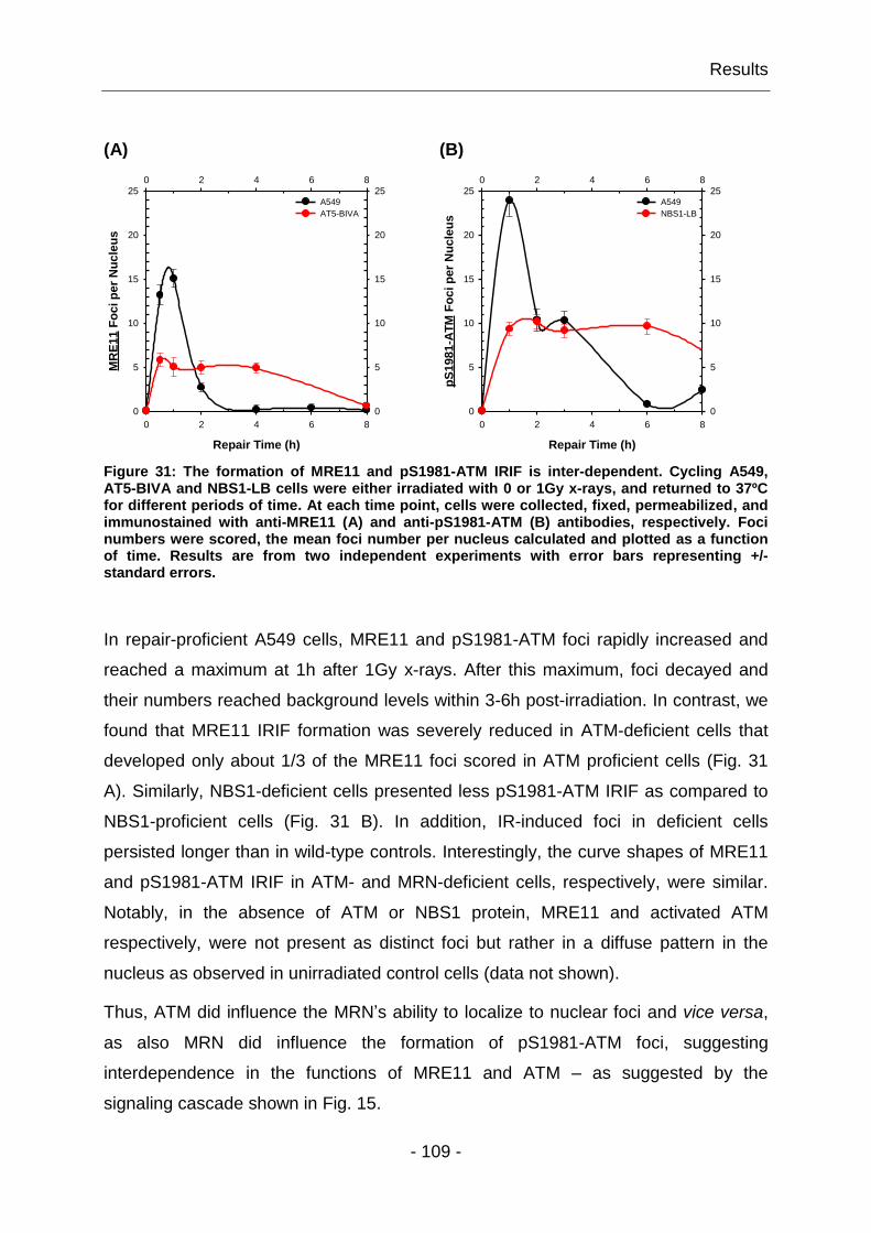
Results
- 109 -
(A) (B)
Repair Time (h)
0 2 4 6 8
0 2 4 6 8
MR
E1
1 F
oc
i p
er
Nu
cle
us
0
5
10
15
20
25
0
5
10
15
20
25A549
AT5-BIVA
Repair Time (h)
0 2 4 6 8
0 2 4 6 8
pS
19
81
-AT
M F
oc
i p
er
Nu
cle
us
0
5
10
15
20
25
0
5
10
15
20
25A549
NBS1-LB
Figure 31: The formation of MRE11 and pS1981-ATM IRIF is inter-dependent. Cycling A549, AT5-BIVA and NBS1-LB cells were either irradiated with 0 or 1Gy x-rays, and returned to 37ºC for different periods of time. At each time point, cells were collected, fixed, permeabilized, and immunostained with anti-MRE11 (A) and anti-pS1981-ATM (B) antibodies, respectively. Foci numbers were scored, the mean foci number per nucleus calculated and plotted as a function of time. Results are from two independent experiments with error bars representing +/- standard errors.
In repair-proficient A549 cells, MRE11 and pS1981-ATM foci rapidly increased and
reached a maximum at 1h after 1Gy x-rays. After this maximum, foci decayed and
their numbers reached background levels within 3-6h post-irradiation. In contrast, we
found that MRE11 IRIF formation was severely reduced in ATM-deficient cells that
developed only about 1/3 of the MRE11 foci scored in ATM proficient cells (Fig. 31
A). Similarly, NBS1-deficient cells presented less pS1981-ATM IRIF as compared to
NBS1-proficient cells (Fig. 31 B). In addition, IR-induced foci in deficient cells
persisted longer than in wild-type controls. Interestingly, the curve shapes of MRE11
and pS1981-ATM IRIF in ATM- and MRN-deficient cells, respectively, were similar.
Notably, in the absence of ATM or NBS1 protein, MRE11 and activated ATM
respectively, were not present as distinct foci but rather in a diffuse pattern in the
nucleus as observed in unirradiated control cells (data not shown).
Thus, ATM did influence the MRN’s ability to localize to nuclear foci and vice versa,
as also MRN did influence the formation of pS1981-ATM foci, suggesting
interdependence in the functions of MRE11 and ATM – as suggested by the
signaling cascade shown in Fig. 15.
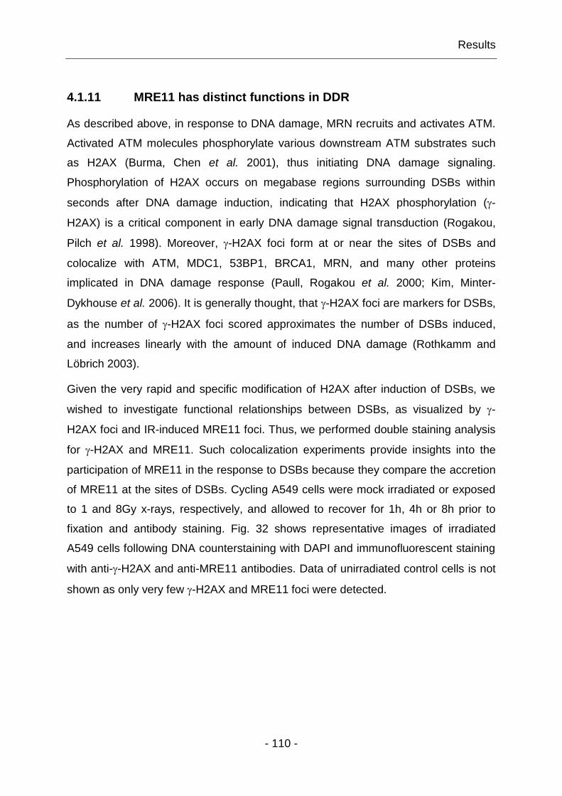
Results
- 110 -
4.1.11 MRE11 has distinct functions in DDR
As described above, in response to DNA damage, MRN recruits and activates ATM.
Activated ATM molecules phosphorylate various downstream ATM substrates such
as H2AX (Burma, Chen et al. 2001), thus initiating DNA damage signaling.
Phosphorylation of H2AX occurs on megabase regions surrounding DSBs within
seconds after DNA damage induction, indicating that H2AX phosphorylation (-
H2AX) is a critical component in early DNA damage signal transduction (Rogakou,
Pilch et al. 1998). Moreover, -H2AX foci form at or near the sites of DSBs and
colocalize with ATM, MDC1, 53BP1, BRCA1, MRN, and many other proteins
implicated in DNA damage response (Paull, Rogakou et al. 2000; Kim, Minter-
Dykhouse et al. 2006). It is generally thought, that -H2AX foci are markers for DSBs,
as the number of -H2AX foci scored approximates the number of DSBs induced,
and increases linearly with the amount of induced DNA damage (Rothkamm and
Löbrich 2003).
Given the very rapid and specific modification of H2AX after induction of DSBs, we
wished to investigate functional relationships between DSBs, as visualized by -
H2AX foci and IR-induced MRE11 foci. Thus, we performed double staining analysis
for -H2AX and MRE11. Such colocalization experiments provide insights into the
participation of MRE11 in the response to DSBs because they compare the accretion
of MRE11 at the sites of DSBs. Cycling A549 cells were mock irradiated or exposed
to 1 and 8Gy x-rays, respectively, and allowed to recover for 1h, 4h or 8h prior to
fixation and antibody staining. Fig. 32 shows representative images of irradiated
A549 cells following DNA counterstaining with DAPI and immunofluorescent staining
with anti--H2AX and anti-MRE11 antibodies. Data of unirradiated control cells is not
shown as only very few -H2AX and MRE11 foci were detected.

Results
- 111 -
(A) (B) (C) (D)
Figure 32: -H2AX and MRE11 foci colocalize at sites of DSBs. Exponentially growing A549 cells were irradiated with 1Gy (A-B) or 8Gy (C-D), and fixed at the indicated times.
Subsequently, cells were stained with anti--H2AX (red; panel 1) and anti-MRE11 antibodies (green; panel 2); cell nuclei were counterstained with DAPI (blue). Image acquisition was performed as described above. MIP overlay images of all three recorded channels are presented in panel 3.
Immunostaining with antibodies directed against -H2AX and MRE11 revealed that
after damage induction both molecules formed nuclear foci. After exposure to IR, -
H2AX appeared in 100% of the cell population and formed distinct foci that
developed to larger foci over time. The number of -H2AX foci increased with higher
radiation doses (Fig. 32, panel 1). In contrast, two major MRE11 IRIF patterns were
detected: At early post-irradiation times mainly small MRE11 foci developed, which
subsequently formed a larger MRE11 focus (Fig. 32, panel 2). However, this was
only observed after low dose radiation (1Gy); after high dose radiation (8Gy) the early
response diminished leaving the late and large MRE11 foci to dominate (4.1.4).
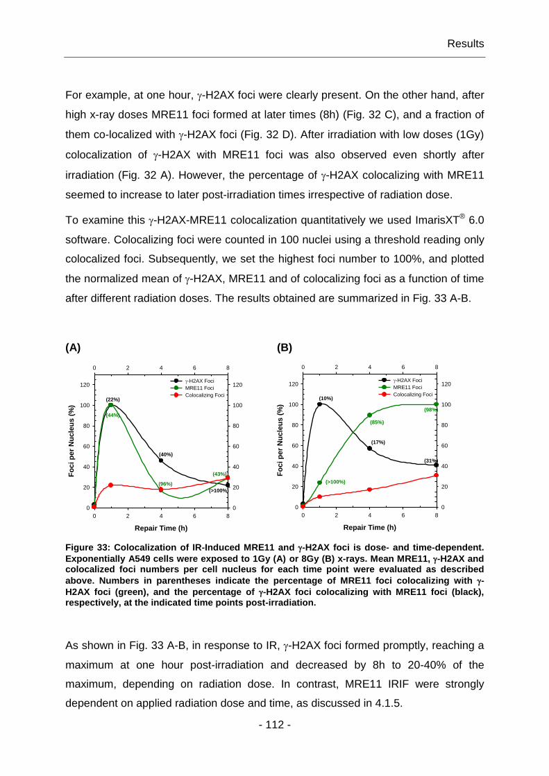
Results
- 112 -
For example, at one hour, -H2AX foci were clearly present. On the other hand, after
high x-ray doses MRE11 foci formed at later times (8h) (Fig. 32 C), and a fraction of
them co-localized with -H2AX foci (Fig. 32 D). After irradiation with low doses (1Gy)
colocalization of -H2AX with MRE11 foci was also observed even shortly after
irradiation (Fig. 32 A). However, the percentage of -H2AX colocalizing with MRE11
seemed to increase to later post-irradiation times irrespective of radiation dose.
To examine this -H2AX-MRE11 colocalization quantitatively we used ImarisXT® 6.0
software. Colocalizing foci were counted in 100 nuclei using a threshold reading only
colocalized foci. Subsequently, we set the highest foci number to 100%, and plotted
the normalized mean of -H2AX, MRE11 and of colocalizing foci as a function of time
after different radiation doses. The results obtained are summarized in Fig. 33 A-B.
(A) (B)
Repair Time (h)
0 2 4 6 8
0 2 4 6 8
Fo
ci p
er
Nu
cle
us (
%)
0
20
40
60
80
100
120
0
20
40
60
80
100
120-H2AX Foci
MRE11 Foci
Colocalizing Foci
(44%)
(96%)
(43%)
(22%)
(40%)
(>100%)
Repair Time (h)
0 2 4 6 8
0 2 4 6 8
Fo
ci p
er
Nu
cle
us (
%)
0
20
40
60
80
100
120
0
20
40
60
80
100
120-H2AX Foci
MRE11 Foci
Colocalizing Foci
(>100%)
(85%)
(98%)
(10%)
(17%)
(31%)
Figure 33: Colocalization of IR-Induced MRE11 and -H2AX foci is dose- and time-dependent.
Exponentially A549 cells were exposed to 1Gy (A) or 8Gy (B) x-rays. Mean MRE11, -H2AX and colocalized foci numbers per cell nucleus for each time point were evaluated as described
above. Numbers in parentheses indicate the percentage of MRE11 foci colocalizing with -
H2AX foci (green), and the percentage of -H2AX foci colocalizing with MRE11 foci (black), respectively, at the indicated time points post-irradiation.
As shown in Fig. 33 A-B, in response to IR, -H2AX foci formed promptly, reaching a
maximum at one hour post-irradiation and decreased by 8h to 20-40% of the
maximum, depending on radiation dose. In contrast, MRE11 IRIF were strongly
dependent on applied radiation dose and time, as discussed in 4.1.5.

Results
- 113 -
Evidently, there was no one-to-one colocalization between -H2AX and MRE11 IRIF.
In cells exposed to 1Gy x-rays, the amount of colocalized foci per nucleus was
relatively constant, varying between 18-29%, whereas after irradiation with 8Gy,
colocalization gradually increased from 10-31%. However, the percentage of -H2AX
IRIF colocalizing with MRE11 foci (black numbers in parentheses, Fig. 33) increased
over time and was highest at later time points, when DSB repair had progressed, as
indicated by the decay of -H2AX foci. On the other hand, the presence of MRE11 to
-H2AX sites (green numbers in parentheses, Fig. 33) was strongly dose- and time-
dependent. In general, DSBs at early recovery times were typically not associated
with MRE11, whereas DSBs, detectable as -H2AX foci at later times, showed with
higher probability the presence of MRE11. One might speculate that these structures
represent MRE11 accumulation at irreparable or slowly repairing DSBs.
Overall, the colocalization analysis presented above revealed that MRE11 clearly
localized to sites of -H2AX, which are considered sites of DSBs. However the
kinetics of localization appears complex and pertains only for a subset of the DSBs.
These results in aggregate suggest functionally distinct modes of MRE11 activity in
DDR that manifest in the complex kinetics and dose response relationships
measured.
Results
- 114 -
4.2 Investigation of complex functions of MRN in DSB repair
The above results implicate different functions of MRN in DDR. Moreover, one could
hypothesize that MRN acts as a repair factor in DSB repair, as MRE11 was
associated with -H2AX sites, particularly at later times post-irradiation. This is in line
with published reports which indicate that MRN is involved in all DSB repair
pathways: HRR, D-NHEJ and B-NHEJ (Hopkins and Paull 2008; Helmink,
Bredemeyer et al. 2009; Rass, Grabarz et al. 2009). However, from our
investigations, information on the contribution of MRN to HRR versus NHEJ could not
be extracted. Thus, its precise role and exact contribution to different DSB repair
pathways is still unknown. This is partly because studies on MRN functions are
complicated by the fact that null mutations in any of its constituent proteins are
embryonically lethal in mice and are lethal as well in cells.
To begin addressing the complex functions of MRN in DSB repair, we investigated
the contribution of MRN to different DSB repair pathways using wild-type cell lines or
cell lines with different MRN deficiencies. DSB repair efficiency by HRR was
evaluated by documenting decay of RAD51 foci formation and decay in fixed cells
after IR exposure, whereas PFGE was used to quantitate radiation-induced DSBs
and thus to evaluate DSB repair by NHEJ.

Results
- 115 -
4.2.1 HRR is incomplete in cells without a functional MRN and ATM
There is ample evidence that MRN is involved in DSB repair by HRR. This repair
pathway requires architectural, structural and enzymatic components, which can be
partly provided by MRN. The DNA-tethering architectures of MRN implicate it in a
linker function for homologous stretches of DNA to tether the ends of the DSB in
close proximity, and to pair the damaged molecule with the homologous section of
the sister chromatid. Furthermore, the end-resecting activity of MRE11 is implicated
in DNA end processing that is essential for the formation of RAD51 nucleoprotein
filament during the presynapsis step of recombination (see 1.4.1). It also needs to be
pointed out that mutation in key proteins that recognize and signal DSBs, such as
MRN or ATM, results in genetic disorders, which generate hyper sensitivity to IR,
genome instability, cancer and/or neurodegeneration. However, frequently, these
mutation do not dramatically reduce the capacity of cells to repair DSBs overall
(Girard, Foray et al. 2000; Riballo, Kühne et al. 2004).
To examine the contribution of MRN to DSB repair by HRR, and moreover to
investigate the inter-dependent function of MRN and ATM in HRR, we carried out
experiments, in which RAD51 foci formation and decay were quantitatively analyzed
in wild-type cells, NBS1-deficient and ATM-deficient cells. The formation of a RAD51
focus is generally taken as evidence that a DSB is engaged by HRR (van Veelen,
Cervelli et al. 2005). Thus, it is thought that the number of RAD51 foci detected
reflects the number of DSBs that are repaired by HRR at a given radiation dose and
repair time point. Exponentially growing cells were irradiated, and RAD51 foci were
detected and scored in G2-phase cyclin B1 positive cells at different times after
exposure to a range of radiation doses. The development and decay of RAD51 foci in
repair-proficient, NBS1- and ATM-deficient cells could be established at three
representative doses, as summarized in Fig. 34 A-C. Control experiments with HRR-
deficient cells revealed no RAD51 foci formation after irradiation (data not shown).
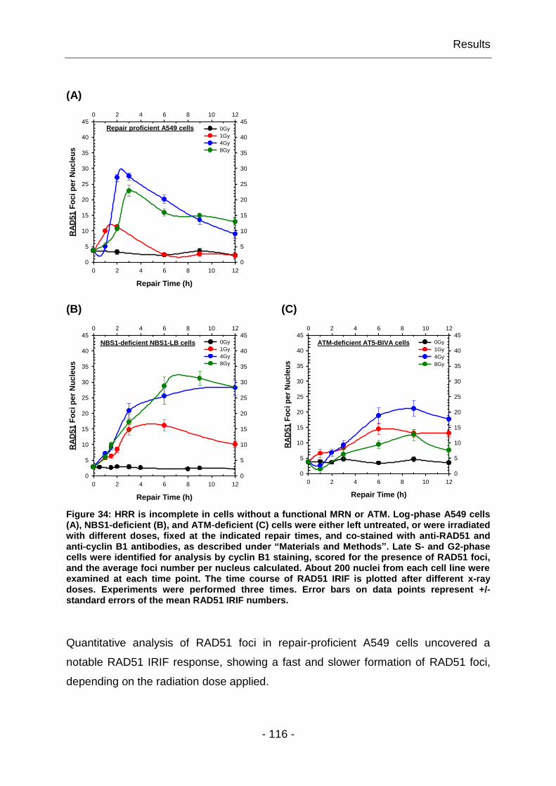
Results
- 116 -
(A)
Repair Time (h)
0 2 4 6 8 10 12
0 2 4 6 8 10 12
RA
D5
1 F
oc
i p
er
Nu
cle
us
0
5
10
15
20
25
30
35
40
45
0
5
10
15
20
25
30
35
40
450Gy
1Gy
4Gy
8Gy
Repair proficient A549 cells
(B) (C)
Repair Time (h)
0 2 4 6 8 10 12
0 2 4 6 8 10 12
RA
D5
1 F
oc
i p
er
Nu
cle
us
0
5
10
15
20
25
30
35
40
45
0
5
10
15
20
25
30
35
40
450Gy
1Gy
4Gy
8Gy
NBS1-deficient NBS1-LB cells
Repair Time (h)
0 2 4 6 8 10 12
0 2 4 6 8 10 12
RA
D5
1 F
oc
i p
er
Nu
cle
us
0
5
10
15
20
25
30
35
40
45
0
5
10
15
20
25
30
35
40
450Gy
1Gy
4Gy
8Gy
ATM-deficient AT5-BIVA cells
Figure 34: HRR is incomplete in cells without a functional MRN or ATM. Log-phase A549 cells (A), NBS1-deficient (B), and ATM-deficient (C) cells were either left untreated, or were irradiated with different doses, fixed at the indicated repair times, and co-stained with anti-RAD51 and anti-cyclin B1 antibodies, as described under “Materials and Methods”. Late S- and G2-phase cells were identified for analysis by cyclin B1 staining, scored for the presence of RAD51 foci, and the average foci number per nucleus calculated. About 200 nuclei from each cell line were examined at each time point. The time course of RAD51 IRIF is plotted after different x-ray doses. Experiments were performed three times. Error bars on data points represent +/-standard errors of the mean RAD51 IRIF numbers.
Quantitative analysis of RAD51 foci in repair-proficient A549 cells uncovered a
notable RAD51 IRIF response, showing a fast and slower formation of RAD51 foci,
depending on the radiation dose applied.

Results
- 117 -
At low doses (1Gy), RAD51 foci development reached a maximum at 2h, while at
intermediate (4Gy) and high (8Gy) doses, RAD51 foci developed slower and the
maximum was reached 2-3h after irradiation. In addition, the dose response for
RAD51 foci formation was not linear in the range of doses tested (1 to 8Gy). In
contrast to that, RAD51 foci formation was delayed but not completely blocked, and
no pronounced foci maxima were developed in cells with defects in NBS1 and ATM,
respectively. Accordingly, the dose response for RAD51 foci formation was sub-linear
for the tested x-ray doses of 1 to 8Gy. However, in NBS1- and ATM-deficient cells
RAD51 foci persisted for much longer than in wild-type controls.
Taken together, these results demonstrated a dose-dependent RAD51 foci formation
– with fast and slow developing RAD51 foci maxima in repair-proficient cells, while in
MRN- or ATM-deficient cells, RAD51 foci formation dynamics were slower and
showed a much slower decay. This indicated that in cells without a functional MRN
complex or a functional ATM, DNA end resection was initiated, as visualized by
RAD51 foci, but the DSB repair could not be efficiently completed. This observation
demonstrated that HRR was indeed severely impaired in NBS1- and ATM-deficient
cells. We could thus conclude that MRN and ATM had a strong impact on HRR.
4.2.2 MRN complex is not required for DSB repair by D-NHEJ
There are reports suggesting that MRN contributes to DSB repair by D-NHEJ.
However, the evidence is contradictory with some reports showing that MRN
deficiency confers a strong defect in DSB repair by D-NHEJ (Rass, Grabarz et al.
2009), while others conclude that MRE11 is not important for accurate D-NHEJ
(Moreau, Ferguson et al. 1999) (1.5.3). To investigate the functional significance of
MRN in DSB repair by D-NHEJ, we measured DSB repair capability by PFGE in cells
with different known MRN deficiencies, and compared the results to those of repair-
proficient cells.
Results
- 118 -
Since depletion of MRE11 is lethal, we used conditional MRE11-null mutants (DT40-
MRE11-/-MRE11+ cells). This mutant was generated in the lab of Prof. Shunichi
Takeda through targeted integration of MRE11 deletion constructs, which disrupt the
reading frame of MRE11 sequence from aa 98 to 385. For viability, MRE11-/- clones
carry a chicken MRE11-transgene (complementary DNA (cDNA) of a complete open
reading frame of GdMRE11) that is under the control of a tet-repressible promoter.
Notably, this transgene has a 10- to 20-fold higher MRE11 expression level than that
of endogenous MRE11. Upon addition of tetracycline, which suppresses the
expression of MRE11-transgene, the level of MRE11 expression is reduced by 10-
fold 12h later, and is undetectable at 24h after the addition of tetracycline. Reverse
transcription-polymerase chain reaction (RT-PCR) revealed that the transcript level of
GdMRE11 is reduced by two orders of magnitude three days later (Yamaguchi-Iwai,
Sonoda et al. 1999). In our experiments we used doxycycline hydrochloride, a
synthetic tetracycline derivative, which is more stable than tetracycline, and thus
does not need to be replaced in the culture medium during the time course of the
experiment.
At the beginning we tested the proliferative properties of wild-type DT40-MRE11 and
DT40-MRE11-/-MRE11+ cells in the absence of doxycycline by monitoring cell
number and carrying out cell-cycle analysis. The growth curve of DT40-MRE11-/-
MRE11+ cells was indistinguishable from that of the wild-type DT40-MRE11 cells,
which divided approximately every 8h. FACS analysis revealed that the cell-cycle
distribution was essentially the same in both cell lines (data not shown). To suppress
the expression of chicken MRE11-transgene, exponentially growing DT40-MRE11-/-
MRE11+ cells were cultivated in media containing doxycycline (1µg/ml). As a function
of time thereafter, we examined cell growth and viability. DT40-MRE11-/-MRE11- cells
were capable of proliferating for several rounds and started to die at day 5. During
the time course of this measurement MRE11 protein level was followed. We
observed that MRE11 significantly decreased at 24h, and was undetectable at 48h
after addition of doxycycline hydrochloride (Fig. 35).

Results
- 119 -
Doxycycline (-) (+) MRE11
GAPDH
Figure 35: MRE11 is undetectable in DT40-MRE11-/-
MRE11- cells. Whole cell lysates were
prepared 2.5d after the addition of doxycycline hydrochloride from cycling conditional MRE11-null mutants. Untreated DT40-MRE11
-/-MRE11
+cells were used as control for MRE11
expression. Following SDS-PAGE, western blots were simultaneously probed with anti-MRE11 and anti-GAPDH antibodies, which served as loading control.
Western blot analysis revealed that at 2.5d after the addition of doxycycline
hydrochloride no MRE11 could be detected in DT40-MRE11-/-MRE11- cells, whereas
untreated cells expressed robust levels of MRE11. On the basis of this result,
subsequent experiments with DT40-MRE11-/-MRE11- cells were performed at 2.5d
after addition of doxycycline.
To begin addressing the contribution of MRN to DSB repair by D-NHEJ, we
measured DSB repair capacity by PFGE in repair-proficient DT40-MRE11-/-MRE11+
and A549 cells as well as in two MRN mutants, the MRE11-deficient DT40-MRE11-/-
MRE11- cells and human NBS-defective, NBS1-LB cells. Fig. 36 A-B shows the
results obtained, with typical gels and cell cycle distribution, from wild-type cells that
were compared to those of MRN-deficient cells.

Results
- 120 -
(A) (B)
DT40-MRE11-/-MRE11+ 0 5 10 15 20Gy 0 0.25 0.5 1 2 4h
A549 0 10 20 30 40Gy 0 0.25 0.5 1 2 4 8h
DT40-MRE11-/-MRE11-
0 5 10 15 20Gy 0 0.25 0.5 1 2 4h
NBS1-LB 0 10 20 30 40Gy 0 0.25 0.5 1 2 4 8h
Time (h)
0 1 2 3 4
0 1 2 3 4
DE
Q (
Gy)
0
5
10
15
20
0
5
10
15
20
DT40-MRE11-/-
MRE11+
DT40-MRE11-/-
MRE11-
Time (h)
0 2 4 6 8
0 2 4 6 8
DE
Q (
Gy)
0
5
10
15
20
0
5
10
15
20A549
NBS1-LB
DT40-MRE11-/-
MRE11+ DT40-MRE11
-/- MRE11
- 0Gy, 0h 20Gy, 4h 0Gy, 0h 20Gy, 4h
A549 NBS1-LB
Gy, 0h 20Gy, 8h 0Gy, 0h 20Gy, 8h
Figure 36: MRN has no effect on DSB repair by D-NHEJ. Upper two panels show typical gels obtained with repair-proficient and MRN-deficient cells exposed to x-rays. To measure the dose response, cells were irradiated with different IR doses, whereas repair kinetics of the same cells was followed after exposure to 20Gy x-rays. The amount of DSBs present in the cells is reflected by the value of FDR. In the calculation, the value of FDR measured in non-irradiated cells was subtracted from all data points with irradiated samples. To account for non-linear dose-response curves as well as for differences among cell lines and experiments, the DEQ was calculated and plotted as a function of time. Plotted are the mean and standard errors calculated from four determinations in three independent experiments. Results obtained were fitted to the sum of two exponentials. Bottom panel shows DNA histograms for the different cells before and after exposure to IR. Evidently, radiation exposure led to the activation of cell cycle-dependent checkpoints that caused an increase in the fraction of cells in G2-phase of the cell cycle several hours after irradiation.

Results
- 121 -
The top panel shows characteristic gels of dose response and repair kinetics
experiments obtained with repair-proficient, DT40-MRE11-/-MRE11+ and A549 cells.
The lower gel panel shows typical gels of MRN-deficient cells, such as
DT40-MRE11-/-MRE11- and NBS1-LB cells. The released DNA from the well into the
lane, as a function of dose, demonstrates the increase in the induction of DSBs,
whereas the gradual reduction in the amount of DNA released into the lane, as a
function of time after exposure to IR, is a measure of the ability of cells to repair
DSBs. Non-irradiated control cells were used to determine the background FDR at
0Gy (data not shown) that was subsequently subtracted from the FDR values of
irradiated cells.
The determination of the dose response is very important as it allows the
recalculation of the repair kinetics from the initially measured FDR versus time plots
to DEQ versus time plots. The plotting of DEQ, instead of FDR as a function of time,
has the advantage that it eliminates differences deriving from fluctuations in the
migrating characteristics of the DNA. An additional advantage is that it corrects for
non-linear dose response curves that can significantly skew the repair kinetics data.
Accordingly, we plotted calculated DEQ values as a function of time, where the initial
values for DEQ at t=0h were derived from the dose response curves, since these
measurements are carried out under conditions that ensure nearly complete absence
of DSB repair.
DSB repair efficiency by D-NHEJ was followed in indicated cells after exposure to x-
rays. The analysis of the repair kinetics obtained with repair-proficient and MRN-
deficient cells clearly showed efficient rejoining of DSBs in all used cell lines, with
most of the DSBs rejoined at 0.25h in DT40 cells (Fig. 36 A) and at 2h in
A549/NBS1-LB cells (Fig. 36 B), respectively. This is indicated by the observed rapid
reduction in DEQ as a function of the repair time. Moreover, the overall rates of DSB-
rejoining in repair-proficient DT40-MRE11-/-MRE11+ and A549 cells were found to be
very similar to those of MRN-deficient DT40-MRE11-/-MRE11- and NBS1-LB cells.
Taken together, the above results demonstrated that MRN did not contribute
detectably to DSB repair by D-NHEJ, at least as measured by PFGE.
Results
- 122 -
4.2.3 DSB repair by B-NHEJ requires MRN
As outlined in the introduction (1.5.3), there is evidence that MRN is involved in DSB
repair by B-NHEJ – possibly because MRE11 processes DNA termini that facilitates
B-NHEJ. It was also reported that MRN deficiency is associated with strong defects
in the repair of RAG-mediated DSBs generated during V(D)J recombination, when
repaired either by D-NHEJ and B-NHEJ (Helmink, Bredemeyer et al. 2009).
However, there are no results showing clear contribution of MRN to DSB repair by B-
NHEJ. This void prompted us to investigate the possible role of MRN in B-NHEJ.
Here again, we used PFGE to measure DSB repair by B-NHEJ in repair-proficient
and MRN-deficient cells, treated with a specific DNA-PKcs inhibitor (NU7441) as a
tool for inhibiting D-NHEJ. One hour after addition of NU7441, cells were irradiated
and DSB repair kinetics were obtained with wild-type A549 cells as well as with the
conditional MRE11-null DT40 and the NBS1-LB mutants. Control experiments with
DT40-DNA-PKcs-/- cells revealed best results, using NU7441 at a concentration of
5µM; at higher inhibitor concentrations repair was inhibited suggesting off-target
effects (data not shown). Therefore, subsequent experiments with NU7441 were
carried out at a concentration of 5µM, given 1h prior to irradiation.
A summary of the results obtained with repair-proficient cells and MRN mutants
regarding rejoining of DSBs by B-NHEJ is given in Fig. 37 A-B. To allow direct
comparison of the repair capability by D-NHEJ and B-NHEJ, results of untreated cells
were also included (dashed line). Finally, the figure includes flow cytometry data of
the cell-cycle distribution of the initial cell populations, and those exposed to radiation
at 4 and 8h post-irradiation.
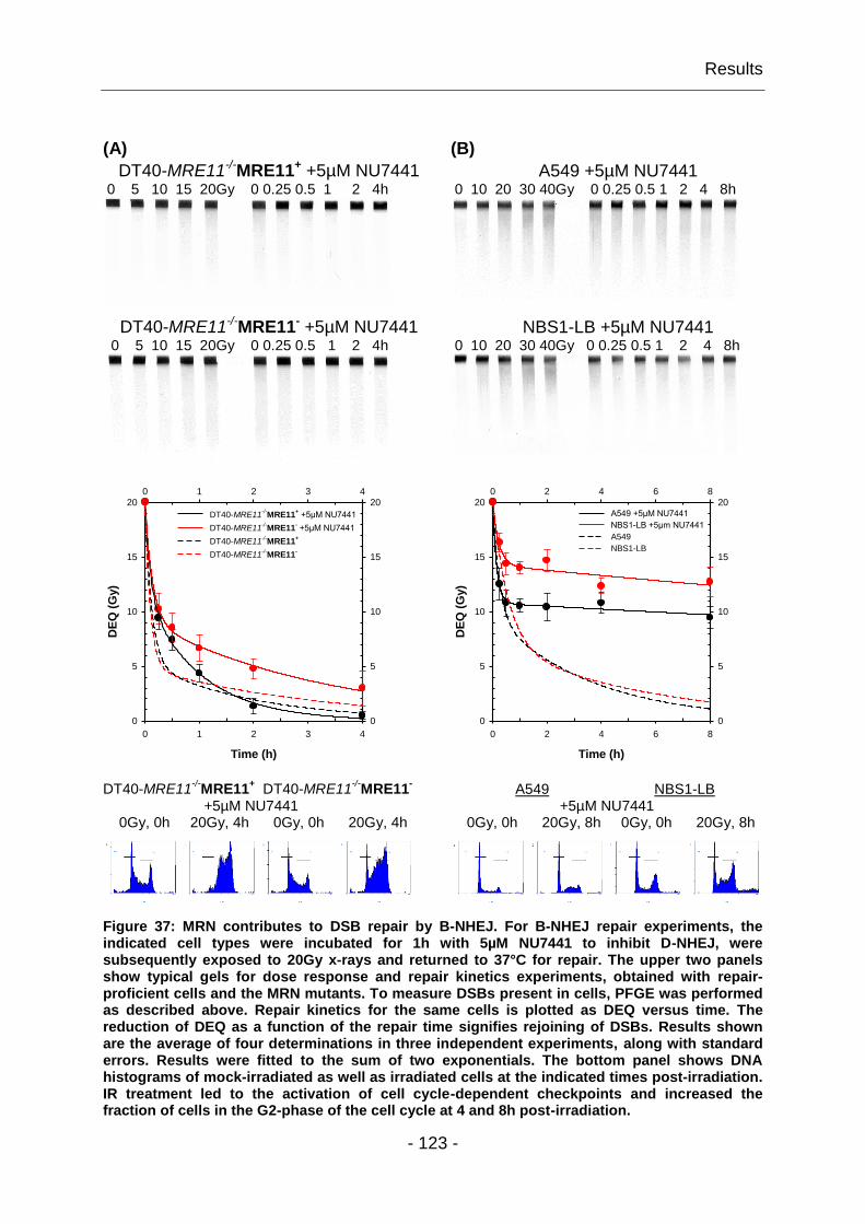
Results
- 123 -
(A) (B)
DT40-MRE11-/-MRE11+ +5µM NU7441 0 5 10 15 20Gy 0 0.25 0.5 1 2 4h
A549 +5µM NU7441 0 10 20 30 40Gy 0 0.25 0.5 1 2 4 8h
DT40-MRE11-/-MRE11- +5µM NU7441 0 5 10 15 20Gy 0 0.25 0.5 1 2 4h
NBS1-LB +5µM NU7441 0 10 20 30 40Gy 0 0.25 0.5 1 2 4 8h
Time (h)
0 1 2 3 4
0 1 2 3 4
DE
Q (
Gy)
0
5
10
15
20
0
5
10
15
20
DT40-MRE11-/-
MRE11+ +5µM NU7441
DT40-MRE11-/-
MRE11- +5µM NU7441
DT40-MRE11-/-
MRE11+
DT40-MRE11-/-
MRE11-
Time (h)
0 2 4 6 8
0 2 4 6 8
DE
Q (
Gy)
0
5
10
15
20
0
5
10
15
20A549 +5µM NU7441
NBS1-LB +5µm NU7441
A549
NBS1-LB
DT40-MRE11-/-
MRE11+ DT40-MRE11
-/-MRE11
-
+5µM NU7441 0Gy, 0h 20Gy, 4h 0Gy, 0h 20Gy, 4h
A549 NBS1-LB +5µM NU7441 0Gy, 0h 20Gy, 8h 0Gy, 0h 20Gy, 8h
Figure 37: MRN contributes to DSB repair by B-NHEJ. For B-NHEJ repair experiments, the indicated cell types were incubated for 1h with 5µM NU7441 to inhibit D-NHEJ, were subsequently exposed to 20Gy x-rays and returned to 37°C for repair. The upper two panels show typical gels for dose response and repair kinetics experiments, obtained with repair-proficient cells and the MRN mutants. To measure DSBs present in cells, PFGE was performed as described above. Repair kinetics for the same cells is plotted as DEQ versus time. The reduction of DEQ as a function of the repair time signifies rejoining of DSBs. Results shown are the average of four determinations in three independent experiments, along with standard errors. Results were fitted to the sum of two exponentials. The bottom panel shows DNA histograms of mock-irradiated as well as irradiated cells at the indicated times post-irradiation. IR treatment led to the activation of cell cycle-dependent checkpoints and increased the fraction of cells in the G2-phase of the cell cycle at 4 and 8h post-irradiation.
Results
- 124 -
Repair was evident in all cell types tested. However, when D-NHEJ was inhibited, to
allow B-NHEJ to dominate, the DSB rejoining kinetics were slowed down in all cells
as compared to their untreated counterparts (dashed line). This decrease in DSB
rejoining efficiency is especially visible in human A549 and NBS1-LB cells (Fig. 37
B), where nearly 50% and 65% of the initial IR-induced DSBs were still present 8h
after irradiation. In contrast, a significant rejoining of DSBs was observed in treated
A549 cells at early times. Interestingly, chicken DT40 cells showed superior ability to
repair DSBs, even when D-NHEJ was inhibited. In these cells, the observed DSB
rejoining kinetics was very efficient although slightly slower than those obtained with
untreated cells. Remarkably, no significant difference in the rejoining of DSBs was
detectable between untreated and D-NHEJ-inhibited DT40-MRE11-/-MRE11+ cells at
later post-irradiation times. These results demonstrated the strong potential of DT40-
MRE11-/-MRE11+ cells to remove DSBs from their genome despite inhibited DNA-
PKcs, as nearly 95% of the initial DSB load was rejoined by 2h after IR exposure.
Repair of DSBs in the presence of NU7441 that inhibits DNA-PKcs is thought to be
carried out by alternative pathways of end joining.
Following NU7441 treatment, MRN mutants showed a significant reduction in DSB
rejoining as compared to MRN proficient cells. Interestingly, the kinetics of both
MRN-deficient mutants indicated faster rejoining at early times, which later slowed
down. In addition, D-NHEJ-inhibited DT40-MRE11-/-MRE11- cells were more efficient
in rejoining IR-induced DSBs, than NBS1-deficient cells; they removed the majority of
DSBs (>85%) by 4h, whereas nearly 65% of the initial DSBs remained unrepaired 8h
post-irradiation in NBS1-LB cells. Overall, these results unveiled a substantial role of
MRN in DSB repair by B-NHEJ.
Results
- 125 -
4.3 Examination of DNA-PK impact in DDR and DSB repair
As discussed above (1.4), DSBs can be repaired by various pathways including
NHEJ and HRR. It is widely assumed that in higher eukaryotes NHEJ is the main
DSB repair pathway and that HRR has only a small contributing function to DSB
repair, restricted to the G2-phase of the cell cycle (Jeggo and Löbrich 2005; Beucher,
Birraux et al. 2009). However, the cellular mechanisms that regulate DSB repair
pathway choice are not well understood. Some studies suggest that these two
mechanistically distinct repair pathways compete for DNA ends, whereas others
report that DDR signaling and DSB repair pathways are actively controlled, and thus
act in a well-coordinated manner to detect and process IR induced DNA damage
(Allen, Kurimasa et al. 2002; Cui, Yu et al. 2005). In apparent agreement with the
competition model, cells with defects in D-NHEJ showed increased HRR (Allen,
Kurimasa et al. 2002). In particular, inactivation of D-NHEJ by eliminating DNA-PKcs
resulted in elevated HRR (Delacote, Han et al. 2002; Shrivastav, Miller et al. 2009).
To determine the impact of DNA-PK in DDR and DSB repair by HRR, we examined
IR-induced MRE11 and RAD51 foci in different DNA-PK mutants after different
radiation doses and at different repair times.
4.3.1 DNA-PKcs influences the MRE11 IRIF response
To begin addressing the significance of DNA-PKcs in DDR, we examined MRE11 foci
formation and decay dynamics in M059J cells. These cells are known to be
completely lack DNA-PKcs (Lees-Miller, Godbout et al. 1995), and to also have low
levels of ATM (Gately, Hittle et al. 1998) as a result of a frame-shift mutation. Thus,
the expression level of DNA-PKcs in M059J cells is about 200 times lower than in
M059K cells and DSBs cannot be repaired using the D-NHEJ repair pathway with
fast half-time kinetics as an essential protein for D-NHEJ is missing. We therefore
speculated that M059J cells would present an altered DNA damage response as
measured by MRE11 IRIF.
Results
- 126 -
To test this, enriched G1- and G2-phase, as well as log-phase M059J cells were
irradiated with different radiation doses using radiation modalities of different LET.
The time course of MRE11 IRIF response in M059J cells was examined and
compared to that of M059K cells. To allow better comparison of MRE11 foci
formation and decay dynamics in M059J and M059K cells, results of MRE11 IRIF
response with M059K cells (dashed line), were also included in the graphs.
Comparable results were obtained for both cell cycle phases after x-ray and neutron
irradiation, respectively. For heavy ion irradiation experiments, centrifugal elutriation
could not be performed, due to missing laboratory equipment at the irradiation facility.
The results summarized in Fig. 38 A-C show MRE11 foci formation and decay
dynamics in enriched G2-phase M059J cells after x-rays or neutrons and in log-
phase cells after heavy ion irradiation.
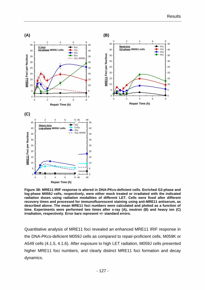
Results
- 127 -
(A) (B)
Repair Time (h)
0 2 4 6 8
0 2 4 6 8
MR
E1
1 F
oc
i p
er
Nu
cle
us
0
5
10
15
20
25
30
35
40
45
0
5
10
15
20
25
30
35
40
450Gy
1Gy
4Gy
8Gy
1Gy, M059K
X-raysG2-phase M059J cells
Repair Time (h)
0 2 4 6 8
0 2 4 6 8
MR
E1
1 F
oc
i p
er
Nu
cle
us
0
5
10
15
20
25
30
35
40
45
0
5
10
15
20
25
30
35
40
450Gy
1Gy
2Gy
4Gy
NeutronsG2-phase M059J cells
(C)
Repair Time (h)
0 2 4 6 8 46 48
0 2 4 6 8 46 48
MR
E1
1 F
oc
i p
er
Nu
cle
us
0
5
10
15
20
25
30
35
40
45
0
5
10
15
20
25
30
35
40
450Gy
0.5Gy
2Gy
2Gy, M059K
Heavy IonsLog-phase M059J cells
Figure 38: MRE11 IRIF response is altered in DNA-PKcs-deficient cells. Enriched G2-phase and log-phase M059J cells, respectively, were either mock treated or irradiated with the indicated radiation doses using radiation modalities of different LET. Cells were fixed after different recovery times and processed for immunofluorescent staining using anti-MRE11 antiserum, as described above. The mean MRE11 foci numbers were calculated and plotted as a function of time. Experiments were performed two times after x-ray (A), neutron (B) and heavy ion (C) irradiation, respectively. Error bars represent +/- standard errors.
Quantitative analysis of MRE11 foci revealed an enhanced MRE11 IRIF response in
the DNA-PKcs-deficient M059J cells as compared to repair-proficient cells, M059K or
A549 cells (4.1.5, 4.1.6). After exposure to high LET radiation, M059J cells presented
higher MRE11 foci numbers, and clearly distinct MRE11 foci formation and decay
dynamics.
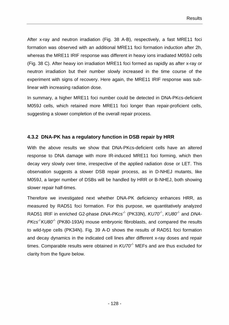
Results
- 128 -
After x-ray and neutron irradiation (Fig. 38 A-B), respectively, a fast MRE11 foci
formation was observed with an additional MRE11 foci formation induction after 2h,
whereas the MRE11 IRIF response was different in heavy ions irradiated M059J cells
(Fig. 38 C). After heavy ion irradiation MRE11 foci formed as rapidly as after x-ray or
neutron irradiation but their number slowly increased in the time course of the
experiment with signs of recovery. Here again, the MRE11 IRIF response was sub-
linear with increasing radiation dose.
In summary, a higher MRE11 foci number could be detected in DNA-PKcs-deficient
M059J cells, which retained more MRE11 foci longer than repair-proficient cells,
suggesting a slower completion of the overall repair process.
4.3.2 DNA-PK has a regulatory function in DSB repair by HRR
With the above results we show that DNA-PKcs-deficient cells have an altered
response to DNA damage with more IR-induced MRE11 foci forming, which then
decay very slowly over time, irrespective of the applied radiation dose or LET. This
observation suggests a slower DSB repair process, as in D-NHEJ mutants, like
M059J, a larger number of DSBs will be handled by HRR or B-NHEJ, both showing
slower repair half-times.
Therefore we investigated next whether DNA-PK deficiency enhances HRR, as
measured by RAD51 foci formation. For this purpose, we quantitatively analyzed
RAD51 IRIF in enriched G2-phase DNA-PKcs-/- (PK33N), KU70-/-, KU80-/- and DNA-
PKcs-/-KU80-/- (PK80-193A) mouse embryonic fibroblasts, and compared the results
to wild-type cells (PK34N). Fig. 39 A-D shows the results of RAD51 foci formation
and decay dynamics in the indicated cell lines after different x-ray doses and repair
times. Comparable results were obtained in KU70-/- MEFs and are thus excluded for
clarity from the figure below.
Results
- 129 -
(A) (B)
Repair Time (h)
0 2 4 6 8 10 12
0 2 4 6 8 10 12
RA
D5
1 F
oc
i p
er
Nu
cle
us
0
5
10
15
20
25
30
35
40
45
50
55
60
65
70
75
0
5
10
15
20
25
30
35
40
45
50
55
60
65
70
750Gy
1Gy
4Gy
8Gy
Wild-type cells
Repair Time (h)
0 2 4 6 8 10 12
0 2 4 6 8 10 12
RA
D5
1 F
oc
i p
er
Nu
cle
us
0
5
10
15
20
25
30
35
40
45
50
55
60
65
70
75
0
5
10
15
20
25
30
35
40
45
50
55
60
65
70
750Gy
1Gy
4Gy
8Gy
DNA-PKcs-/-
cells
(C) (D)
Repair Time (h)
0 2 4 6 8 10 12
0 2 4 6 8 10 12
RA
D5
1 F
oc
i p
er
Nu
cle
us
0
5
10
15
20
25
30
35
40
45
50
55
60
65
70
75
0
5
10
15
20
25
30
35
40
45
50
55
60
65
70
750Gy
1Gy
4Gy
8Gy
KU80-/-
cells
Repair Time (h)
0 2 4 6 8 10 12
0 2 4 6 8 10 12
RA
D5
1 F
oc
i p
er
Nu
cle
us
0
5
10
15
20
25
30
35
40
45
50
55
60
65
70
75
0
5
10
15
20
25
30
35
40
45
50
55
60
65
70
750Gy
1Gy
4Gy
8Gy
DNA-PKcs-/-
KU80-/-
cells
Figure 39: KU heterodimer strongly affects the initiation of HRR. The indicated cell lines were enriched in G2-phase of the cell cycle by centrifugal elutriation, and were irradiated with 0, 1, 4 and 8Gy x-rays. After different repair times cells were fixed, and stained with anti-RAD51 antibody. RAD51 foci were scored as described under “Materials and Methods”. The time course of mean RAD51 IRIF per cell nucleus is plotted after different radiation doses in wild-type (A), DNA-PKcs
-/- (B), KU80
-/- (C) and DNA-PKcs
-/KU80
-/- (D) mouse embryonic fibroblasts.
Results are from three independent experiments. Error bars represent +/- standard errors.
Evidently, in response to IR similar RAD51 foci formation and decay dynamics were
obtained in wild-type (PK34N) and DNA-PKcs-deficient (PK33N) cells (Fig. 39 A-B).
In these cells, IR-induced RAD51 foci reached a maximum within 1-2h post-
irradiation, and decayed to almost background levels during the time course of the
experiment.
Results
- 130 -
In contrast to that, KU80-deficient cells presented a different RAD51 IRIF response
(Fig. 39 C), with three times higher RAD51 foci numbers per cell nucleus as
compared to the other used cell lines. Remarkably, IR-induced RAD51 foci in these
cells only moderately decayed over time following 1Gy of x-rays. On the other hand,
less RAD51 IRIF developed in DNA-PKcs-/-KU80-/- (PK80-193A) cells (Fig. 39 D),
which rapidly reached a plateau and did not decay during the time interval of
analysis. This plateau was not caused by scoring limitations because it was reached
at ~20 foci per cell, which could be readily counted. The dose response for RAD51
foci formation was not linear in the range of doses tested (1 to 8Gy) in any of the cell
lines examined.
Thus, following IR treatment significantly higher RAD51 foci numbers were obtained
in KU-deficient cells, indicating a higher initiation of HRR in those cells. Moreover, KU
and DNA-PK deficiency, resulted in incomplete HRR as almost no decay of RAD51
foci was observed in KU-deficient cells and the double KO for KU and DNA-PKcs.
Hence, this observation suggests a regulatory function for DNA-PK in DSB repair by
HRR. However, it raises questions regarding its mechanistic basis and warrants
further investigations.
Discussion
- 131 -
5 Discussion
5.1 The formation of MRN foci is IR- and DNA damage-dependent
We were able to show that MRE11 accumulates to discrete nuclear foci following
exposure to IR. This was confirmed for several different human cell lines, such as
lung carcinoma A549 cells, MRC5-SV1 fibroblasts, osteosarcoma tumor HT1080
fibroblasts and glioma M059K and M059J cells (4.1.1). All examined cell lines
presented a detectable MRE11 protein accumulation to discrete nuclear foci only
after radiation exposure, since almost no MRE11 foci could be detected in
unirradiated cells. Moreover, simultaneous immunofluorescence analysis of nuclear
IR-induced MRE11, RAD50 and NBS1 foci revealed a strong colocalization of MRN
IRIF (4.1.2). These results allowed two important conclusions – (1) the formation of
MRN foci was radiation-dependent, and (2) foci forming generally comprised all three
component of the MRN complex, i.e. MRE11-RAD50-NBS1, indicating functional
interaction between the MRN components and damaged DNA upon irradiation. In
addition, by inducing localized DNA damage using high power lasers in discrete
subnuclear volumes in live cells, we could demonstrate that MRE11 rapidly
associates with damaged DNA in vivo (4.1.3).
These results are in line with several other studies, which showed that in response to
treatment with DSB-inducing agents, human MRE11, RAD50 and NBS1 proteins
accumulated to discrete small nuclear foci. It has been demonstrated that these
proteins colocalized at DNA damage sites, confirming that MRE11-RAD50-NBS1
proteins act as a complex (Maser, Monsen et al. 1997; Carney, Maser et al. 1998;
Mirzoeva and Petrini 2001; Lee, Fernandez-Capetillo et al. 2005; Robison, Dixon et
al. 2007; Dellaire, Kepkay et al. 2009; Takahashi, Mori et al. 2010). Thus, the Kenshi
Komatsu group provided in vivo direct evidence that MRE11, RAD50 and NBS1
proteins localized to IR-induced nuclear foci (Tauchi, Kobayashi et al. 2001).
Furthermore, the Wyman laboratory demonstrated in vitro by scanning force
microscopy-based volume analysis that MRE11, RAD50 and NBS1 formed stable
complexes (van der Linden, Sanchez et al. 2009).

Discussion
- 132 -
We confirmed that the formation of MRN IRIF was dependent on NBS1 as NBS1-
deficient cells, which have no functional NBS1 protein as a result of a 5bp deletion
within the NBS1 gene, showed no MRE11 foci formation (data not shown). It is
known, that upon DNA damage induction, NBS1 recruits the MR complex from the
cytoplasm to the nucleus, and then to sites of DSBs (Carney, Maser et al. 1998),
particularly through the interaction between its C-terminal MRE11-binding domain,
and the N-terminal FHA- and BRCT-domains of MRE11 (Desai-Mehta, Cerosaletti et
al. 2001; Tauchi, Kobayashi et al. 2001; Zhao, Renthal et al. 2002). In addition, it was
reported that mutations of conserved residues in FHA- and BRCT-domains of NBS1
disrupted nuclear MRN focus formation (Cerosaletti and Concannon 2004; Hari,
Spycher et al. 2010). We were thus not surprised that NBS1-deficient cells did not
form any MRE11 foci, implicating that MRE11 had cytoplasmic localization in NBS1-
deficient cells.
In addition, we reported here that DNA damage-dependent nuclear MRE11 protein
accumulation in HT1080-YFP-MRE11 cells that express N-terminally tagged MRE11-
YFP proteins was slower than in those cells with a C-terminal YFP-tag. Due to the
fact that the relocation of MRE11 is dependent on the interaction between the N-
terminal NBS1-binding domain of MRE11 and NBS1, we are speculating that the N-
terminal protein tag could hinder NBS1 from effectively binding to MRE11 affecting
thus its nuclear localization to damaged DNA sites. However, to confirm this
hypothesis further investigations are required.
5.2 IR-induced MRN foci have different qualitative features
We observed that the formation of nuclear MRN foci was strictly dependent upon the
prior induction of DSBs. Moreover, the analysis of MRE11 foci formation in all
examined cell types after exposure to either low or high LET radiation showed
different MRE11 foci patterns at different times post-irradiation (4.1.4). In general,
small foci were observed after short recovery times and low doses (1-2Gy) of low
LET radiation, whereas after high radiation doses (4-8Gy) or high LET radiation the
early response diminished leaving large foci to dominate the late response.
Discussion
- 133 -
As discussed above, MRN is a keystone complex, rather than a single component
within a linear pathway, as it has different enzymatic activities important for DNA
damage sensing, signaling and DSB repair. We inquired whether IR-induced MRN
foci of different size might reflect different functions of MRN in DSB repair. The
dynamic assembly of large foci may be required for the repair of particularly complex
and difficult to repair lesions. Such lesions are induced with higher frequency after
exposure to high LET radiation, which also explains the dominance of large foci
under these conditions. Small foci on the other hand, may be associated with
signaling emanating from a larger fraction of DSBs.
The Petrini group was the first to report distinct localization patterns for nuclear
MRE11 that were depended on the radiation dose applied and recovery times
(Maser, Monsen et al. 1997). Furthermore, the Bonner laboratory hypothesized that
the IRIF occur at sites where the repair machinery has difficulty repairing the breaks
as a result of multiple ionization events along a radiation track (Paull, Rogakou et al.
2000). In line with this, Mirzoeva and Petrini (Mirzoeva and Petrini 2001) suggested
that the later type of IR-induced MRE11 foci might represent MRE11’s accumulation
at irreparable or slowly repairing lesions that corresponds to downstream roles of
MRN in DSB repair. Further studies reported that in response to DSBs MRN formed
two types of nuclear foci: the first MRE11 foci type developed rapidly, was ATM-
independent, and probably reflected the sensor mode of MRN action, while the
second type of IR-induced MRE11 foci was ATM-dependent and prolonged
(Mirzoeva and Petrini 2001; Mirzoeva and Petrini 2003). Accordingly, Petrini and
Stracker (Petrini and Stracker 2003) proposed that following the initial action of MRN
as a sensor, the subsequent phosphorylation of NBS1 turns it into a mediator in
further stages of DDR. In addition, it was shown that high LET ions produced clear
and large MRE11 foci (Karlsson and Stenerlöw 2004), and that the radiation dose
and the time post-irradiation strongly influenced the number and the size of individual
foci (van Veelen, Cervelli et al. 2005). Similar results were obtained for IR-induced-
H2AX, ATM and RAD51 foci respectively, demonstrating that in response to high LET
irradiation, the size and frequency of IRIF varied as a function of radiation quality,
dose and recovery time (Costes, Boissiere et al. 2006).
Discussion
- 134 -
5.3 The DNA damage-dependent response of MRN is bimodal
We showed in several repair-proficient cell lines, using radiation modalities of
different LET, a cell cycle-independent bimodal MRN IRIF response with a sub-linear
dose-dependency (4.1.5, 4.1.6). In general, after low radiation doses, fast developing
MRN foci were detected, whereas high radiation doses caused a slower formation of
MRN foci. Similar kinetics was also observed for nuclear chromatin accretion of
MRE11 after exposure to IR. Notably, we were able to demonstrate that the bimodal
response of IR-induced MRN foci was neither due to technical limitations in detection
nor due to intracellular MRE11 protein depletion (4.1.8, 4.1.9).
We were thus wondering whether the bimodal response of MRN to DNA damage
reflects its different functions in DSB repair, as discussed above for the different IR-
dependent MRN foci patterns. This seems possible, since there is strong evidence
that MRN is a primary DNA damage sensor that activates ATM, and hence initiates
the intracellular DNA damage signaling cascade (Nelms, Maser et al. 1998; Carson,
Schwartz et al. 2003; Uziel, Lerenthal et al. 2003; Bekker-Jensen, Lukas et al. 2006),
but that it is also essential for all major DSB repair pathways (Tauchi, Kobayashi et
al. 2002; Dinkelmann, Spehalski et al. 2009; Xie, Kwok et al. 2009; Taylor, Cecillon et
al. 2010) despite the fact that they operate with different half times (Iliakis, Wang et
al. 2004). Accordingly, the fast formation of small MRN foci and the increase of
MRE11 protein accretion could be associated with MRN’s activity as a DNA damage
sensor, upstream of ATM, whereas after exposure to high radiation doses the slow
formation of big IR-induced MRN foci and the slow accumulation of MRE11 protein at
DNA damage sites, might reflect MRN’s involvement in DSB repair pathways. Why
so many MRN molecules are necessary at the site of one or a few DSBs is not
known and remains to be resolved.
Our results are in agreement with previous studies, where in response to high x-ray
doses (12Gy) only a slow appearance of IR-induced RAD50 foci was observed,
associated with gradually increasing total fluorescence intensity of the individual foci
(Paull, Rogakou et al. 2000; Gerashchenko and Dynlacht 2009). Eva Lee’s group
demonstrated a cell cycle-independent formation of nuclear DNA damage-dependent
NBS1 foci (Yuan, Chang et al. 2003), and the Kanaar laboratory showed that
radiation dose influenced the number of foci per nucleus and that the post-irradiation

Discussion
- 135 -
time influenced the size of the foci (van Veelen, Cervelli et al. 2005). Interestingly,
association of MRN with chromatin in vivo was shown by Zhao et al. (Zhao, Renthal
et al. 2002). These authors claimed that MRN chromatin association was not
exclusively dependent on IR, but increased also when cells entered S-phase. In
support of this notion, we were also able to show a radiation-independent association
of MRE11 with chromatin. However, we observed chromatin-bound MRE11 protein
maxima at different recovery times after exposure to different radiation doses,
indicating a radiation dose-dependent association of MRE11 with damaged DNA
(Fig. 30 B). Moreover, the association of MRE11 with chromatin mirrored the kinetics
of nuclear MRE11 protein abundance as well as the bimodal MRN IRIF response,
following IR exposure. These data in aggregate demonstrated an interaction of MRN
with damaged DNA in a time- and dose-dependent manner.
Quantitative analysis of MRN IRIF revealed that in response to high LET radiation,
repair-proficient cells presented with less foci initially, retained however more foci at
later times. As discussed above, these persisting foci may represent particularly
complex and difficult to repair lesions associated with the increase in LET of
radiation. The lower MRN foci numbers, following high LET irradiation, could be
explained by the energy deposition pattern of high LET radiation. It is well
established that high LET radiation deposits a large amount of energy in a much
more localized area and creates thus a more localized and clustered DNA damage,
including complex DSBs with multiple lesions within a helical turn. This is in contrast
to low LET irradiation, where a more even distribution of energy will induce more
frequently a uniform distribution of DNA damage that is separated by rather large
distances (Goodhead 1994; Terato and Ide 2004; Okayasu, Okada et al. 2006). As a
result of these characteristics of high LET radiation, the distribution pattern of
clustered and complex DNA damage several DSBs can be generated in close
proximity that will be detected as a single focus. This will affect both focus
appearance, as well as the kinetics of its development and decay.

Discussion
- 136 -
Our observations are in agreement with previous reports, where it was shown that
after high LET irradiation, less IRIF formed than after x-rays, reflecting a clustering of
multiple foci along the high LET ion track (Karlsson and Stenerlöw 2004; Costes,
Boissiere et al. 2006). Moreover, it was shown that MRN remained associated to the
damaged site until the bulk of DSB repair was completed (Nelms, Maser et al. 1998).
Other groups have shown a slower DSB rejoining in response to high LET radiation
(Stenerlöw, Höglund et al. 2000), presumably correlating with difficulties in the repair
of the induced DSBs (Karlsson and Stenerlöw 2004).
However, the slower decay of MRN foci after high LET irradiation might also be
because high LET radiation seems only to inhibit the KU-dependent DSB repair
pathway (D-NHEJ), but not HRR or B-NHEJ. This hypothesis was postulated by the
Ya Wang laboratory, demonstrating that high LET IR might induce small (<40bp)
DSB fragments, which prevent KU from binding efficiently to the DNA ends, resulting
in a delayed KU-dependent repair (Wang, Wang et al. 2008).
We concluded that persisting MRN foci represented DNA damage sites, which were
difficult to repair and moreover suggested that with increasing lesion complexity,
associated with an increase in LET, more DSBs were repaired slowly and recruited
HRR for their removal from the genome.
In summary, while our results confirmed published data, they also extended the
available information in several important ways, including highly sensitive detection
methodology. Thus, previous studies used very high and very few radiation doses
with only very few repair time points (Maser, Monsen et al. 1997). Therefore,
important facts concerning the DNA damage-dependent response of MRN were
missed. This study is the first to present full MRN IRIF formation and decay dynamics
in several human cell lines at different LETs. The bimodal cell cycle-independent
response of MRN foci formation and decay was shown here for the first time.
Discussion
- 137 -
5.4 The MRN complex has different functions in DDR
The investigation of functional relationships between -H2AX and MRE11 IRIF
revealed that following IR exposure, MRE11 co-localized with -H2AX to nuclear foci
in a strongly dose- and time-dependent manner (4.1.11). There was no one-to-one
colocalization between -H2AX and MRE11, indicating temporally and possibly also
functionally distinct modes of operation of MRE11 in DDR. Generally, the early
function of MRE11 was not overly DNA damage signaling-dependent, as only a small
proportion of MRE11 foci colocalized with -H2AX foci (Fig. 33 A). On the other hand,
-H2AX foci that were still present at later recovery times, when the vast majority of
DSB repair was completed, showed a high colocalization with IR-induced MRE11
foci, implicating MRE11’s involvement in DSB repair (Fig. 33 B). Hence, we
speculated that these structures represent MRE11’s accumulation at irreparable or
slowly repairing lesions.
This is in keeping with several other studies, where a colocalization of -H2AX and
MRN IRIF was shown (Paull, Rogakou et al. 2000; Mirzoeva and Petrini 2003;
Karlsson and Stenerlöw 2004; Bekker-Jensen, Lukas et al. 2006; Hari, Spycher et al.
2010; Nakamura, Rao et al. 2010). Moreover, it was reported that the intensity of
individual foci gradually increased over time, as did the extent of colocalization within
individual cells (Paull, Rogakou et al. 2000). However, in previous sections (5.2, 5.3)
we have postulated that the fast response of small MRN IRIF might reflect MRN’s
involvement in DNA damage sensing and signaling, whereas the slow response of
big IR-induced MRN foci could reflect its contribution in the course of DSB repair.
Indeed, there are several studies indicating that MRN acts as a primary DNA damage
sensor, but is also involved in DNA damage signaling and DSB repair (Maser,
Monsen et al. 1997; Nelms, Maser et al. 1998; Tauchi, Kobayashi et al. 2002;
Carson, Schwartz et al. 2003; Uziel, Lerenthal et al. 2003; Taylor, Cecillon et al.
2010). Nevertheless, if one MRN focus represented a DNA damage sensing event, a
larger MRN--H2AX colocalization would be expected at early time points post-
irradiation.

Discussion
- 138 -
Taken together, while we could not exclude that the small, rapidly induced MRN foci
did not reflect MRN’s participation in DNA damage sensing and signaling, it seemed
more reasonable to postulate that they reflect the involvement of MRN to DSB repair
– presumably by HRR and/or B-NHEJ that operate with slower kinetics than NHEJ
(Iliakis, Wang et al. 2004). It was hypothesized by several groups that the IRIF occur
at sites where the repair machinery has difficulty repairing the breaks that are present
(Paull, Rogakou et al. 2000; Petrini and Stracker 2003; Karlsson and Stenerlöw
2004). However, it remains to be solved, why so many proteins molecules are
necessary at one DSB site and how MRN contributes to DDR.
5.5 The MRN complex acts as a factor in DSB repair
Our investigations confirmed that MRN acts as a repair factor in DSB repair.
Moreover, we unveiled the contribution of MRN to different DSB repair pathways
(4.2). Specifically, we could demonstrate a clear inter-dependent function of MRN
and ATM in DSB repair by HRR. Interestingly, in MRN- and ATM-deficient cells
respectively, DNA end resection was initiated, as visualized by RAD51 foci, but the
DSB repair by HRR was incomplete as RAD51 foci did not decayed over time (4.2.1).
Thus, HRR was impaired in cells without a functional MRN or ATM. We could thus
conclude that MRN and ATM play an important role in HRR.
On the other hand, we could not detect a contribution of MRN to DSB repair by D-
NHEJ, as measured by PFGE (4.2.2). But we were able to demonstrate that MRN
has a role in DSB repair by B-NHEJ (4.2.3). These results allowed the important
conclusion that MRN was not involved in all DSB repair pathways – it functioned
specifically in HRR and B-NHEJ.
The above results are consistent with findings of other groups. It is well documented
that the functions of MRN and ATM are intertwined (Maser, Monsen et al. 1997;
Gatei, Young et al. 2000; Uziel, Lerenthal et al. 2003; Lee and Paull 2004;
Difilippantonio, Celeste et al. 2005; Jazayeri, Balestrini et al. 2008; Di Virgilio, Ying et
al. 2009). Moreover, the clinical presentation of human AT, ATLD and NBS patients
is similar (Carney, Maser et al. 1998; Stewart, Maser et al. 1999; Shiloh and Kastan
2001).

Discussion
- 139 -
It was hence not surprising that the MRE11 IRIF formation was severely reduced in
ATM-deficient cells, which developed only 1/3 of the MRE11 foci scored in ATM-
proficient cells and vice versa (4.1.10). There are some studies showing that in
response to DSBs, MRN forms two types of nuclear foci: the first MRE11 foci type is
rapid and ATM-independent, and probably reflects its sensor mode of action, while
the second type of IR-induced MRE11 foci is ATM-dependent and prolonged
(Mirzoeva and Petrini 2001; Mirzoeva and Petrini 2003). We might thus speculate
that MRN IRIF-types reflected the independent function between MRN and ATM in
focus formation.
Regarding MRN’s function in recombinational repair, there are many studies showing
that MRN is involved in DSB repair by HRR. This repair pathway requires
architectural, structural and enzymatic components that can be provided by MRN.
The DNA-tethering architectures of MRN implicate it as linker for homologous
stretches of DNA and for tethering DNA ends in close proximity to facilitate repair
using the sister chromatid. On the other hand, the end-resecting ability of MRE11 is
implicated in DNA end processing that is essential for the formation of RAD51
nucleoprotein filament during the presynapsis step of recombination (Paull and
Gellert 1998; Yamaguchi-Iwai, Sonoda et al. 1999; Tauchi, Kobayashi et al. 2002; de
Jager, Trujillo et al. 2004; Yang, Saidi et al. 2006; Hopkins and Paull 2008; Budd and
Campbell 2009; Cejka, Cannavo et al. 2010; Nimonkar, Genschel et al. 2011; Liao,
Guay et al. 2012). Other groups showed that ATM defects impaired HR-mediated
DSB repair (Golding, Rosenberg et al. 2004; Beucher, Birraux et al. 2009),
particularly as ATM-/- cells showed altered kinetics of IR-induced RAD51 foci
formation (Haaf, Golub et al. 1995; Maser, Monsen et al. 1997; Chen, Yuan et al.
1999; Morrison, Sonoda et al. 2000; Yuan, Chang et al. 2003). This is in agreement
with our results, where we could clearly demonstrate that MRN and ATM had a
strong impact on DSB repair by HRR, as documented by RAD51 IRIF. IR-induced
RAD51 foci formed more rapidly in wild-type cells than in MRN- and ATM-deficient
cells, although many more RAD51 foci accumulated in deficient cells (4.2.1),
probably reflecting increased numbers of unrepaired breaks. We interpreted these
findings to mean that the initial stages of recombination repair, mediated principally
by RAD51 were disrupted by MRN and ATM deficiency.
Discussion
- 140 -
Overall, these observations confirmed that MRN and ATM are major players in HRR
and function in an inter-dependent manner. Notably, this study is the first to show
detailed MRN and ATM foci formation and decay dynamics.
Whereas, it is well established that MRN participates in DSB repair by HRR, it is less
clear how these proteins are involved in D-NHEJ or B-NHEJ. It is anticipated that
MRN does contribute to both, D-NHEJ and B-NHEJ. However, published data is
contradictory. Several studies claim that MRE11 is not important for accurate D-
NHEJ (Moreau, Ferguson et al. 1999; Yamaguchi-Iwai, Sonoda et al. 1999; Di Virgilio
and Gautier 2005; Taylor, Cecillon et al. 2010), and that it does not affect end-joining
frequencies in plasmid based assays (Tauchi, Kobayashi et al. 2002; Yang, Saidi et
al. 2006). In support of this notion, one group found the overall rates of DSB-rejoining
in NBS cells, as measured by PFGE, to be very similar to those of wild-type cells
(Kraakman-van der Zwet, Overkamp et al. 1999). On the other hand, other groups
reported that MRN deficiency confers a strong defect in DSB repair by D-NHEJ,
because MRE11 could control end-joining through both, ATM-dependent and ATM-
independent pathways (Rass, Grabarz et al. 2009). The notion of MRN’s involvement
in D-NHEJ is supported by its ability to stabilize distant breaks (Fig. 16 A-B) (Hopfner,
Craig et al. 2002), and by its DNA end recognition potential that implies a role in the
early stage of NHEJ (Daley, Palmbos et al. 2005).
In addition, there is also strong evidence that MRN is involved in B-NHEJ (Xie, Kwok
et al. 2009; Davies and Chen 2010; Della-Maria, Zhou et al. 2011). A likely function in
this pathway is the processing by MRE11 of the DNA termini that seem to facilitate
repair of DSBs by B-NHEJ (Deriano, Stracker et al. 2009; Rass, Grabarz et al. 2009;
Lamarche, Orazio et al. 2010; Rahal, Henricksen et al. 2010; Taylor, Cecillon et al.
2010). In line with this postulate, several groups demonstrated that MRN deficiency
was associated with strong defects in end-joining pathways involved in isotype class
switching, and in the repair of chromosomal RAG-mediated DSBs generated during
V(D)J recombination, affecting both B-NHEJ and D-NHEJ (Dinkelmann, Spehalski et
al. 2009; Helmink, Bredemeyer et al. 2009; Saidi, Li et al. 2010). Overall, the precise
functional significance of MRN in D-NHEJ and B-NHEJ requires further
investigations.
Discussion
- 141 -
However, in this study we provided further insights into how MRN contributed to
different DSB repair pathways. We demonstrated that MRN deficiency had a
dramatic impact on HRR. However, we also presented strong evidence that MRN
was not required for D-NHEJ, although it played a substantial role in B-NHEJ. We
concluded that MRN acted as a repair factor only in DSB repair pathways, where
DNA end processing is an essential or a likely step, like HRR and B-NHEJ.
5.6 The DNA-PK has a regulatory function in DSB repair
As discussed above (1.4.3), the regulation of DSB repair pathway choice is still
unknown. Thus, it is not clear how and when a cell selects which pathway to use for
the repair of a given DSB, and whether the decision is affected by the nature of the
DSB, or by a global regulatory network involving key regulatory proteins. Within this
study we attempted to uncover such regulatory proteins by examining the putative
role of DNA-PK in DSB repair pathway choice. This protein complex is one of the
central components of D-NHEJ, and has been implicated in DDR signaling as a
member of the PI3KK family. Moreover, DNA-PK is considered as one of the most
important players in the maintenance of genomic stability (Chen, Trujillo et al. 2000;
Meek, Gupta et al. 2004).
To investigate the role of DNA-PK in DSB repair, we used different DNA-PK-deficient
cell lines and showed that DNA-PKcs contributes to DDR, as measured by MRE11
IRIF (4.3.1). Human DNA-PKcs-deficient M059J cells presented an altered response
to DNA damage, irrespective of radiation dose or LET. Remarkably, these cells
retained more MRE11 foci at later time points than their repair-proficient counterpart,
M059K cells. This observation suggested a slower completion of the overall repair,
probably because in D-NHEJ mutants a larger number of DSBs is handled by HRR or
B-NHEJ, which operate with slower half-times (Wang, Zeng et al. 2001). This is in
agreement with previous studies showing that MRE11 IRIF response was increased
in D-NHEJ mutants (Maser, Monsen et al. 1997), and that DNA-PKcs-deficient cells
were not capable of repairing DSBs with fast kinetics (Stiff, O'Driscoll et al. 2004).
Moreover, Karlsson and Stenerloew (Karlsson and Stenerlöw 2004) reported that
after high LET radiation exposure a high level of remaining foci correlated with the
Discussion
- 142 -
lack of apparent DSB rejoining in M059J cells. Thus, functional DNA-PKcs seems
also to be critical for DSB rejoining of breaks produced by radiation of high LET.
Furthermore, we detected a higher initiation of HRR in KU-deficient cells – although
DSB repair appeared incomplete since IR-induced RAD51 foci did not decay over
time (4.3.2). Increased HRR in cells with D-NHEJ defects was previously shown by
several other groups, and can be explained by a passive competition model between
D-NHEJ and HRR repair proteins (Pierce, Hu et al. 2001; Allen, Kurimasa et al. 2002;
Delacote, Han et al. 2002; Barlow, Lisby et al. 2008; Taylor, Cecillon et al. 2010).
However, the fact that higher eukaryotes predominantly use NHEJ for DSB repair
makes a simple competition model difficult to accommodate as key determinant of
DSB repair pathway choice.
Several lines of evidence suggest that DNA-PKcs is an active regulator of DSB
repair. First there was the puzzling finding that inactivation of NHEJ by elimination of
DNA-PKcs indeed increased HRR, but chemical inhibition of DNA-PKcs had the
opposite effect (Perrault, Wang et al. 2004). These results suggested a model in
which chemically inhibited DNA-PKcs fails to dissociate from DNA ends, and thereby
blocks access to other NHEJ and HRR repair factors. The idea that DNA-PKcs
actively regulates DSB repair pathway choice gained additional support when the
Meek laboratory identified DNA-PKcs splice variants that lack the kinase domain and
which surprisingly had dominant negative effects on DSB repair by HRR (Convery,
Shin et al. 2005). Thus, the group of Jack Nickoloff reported more persistent IR-
induced RAD51 foci formation in DNA-PKcs null cells, and proposed an active
regulation of DSB repair by DNA-PKcs, despite an existing passive shunt to HRR
(Shrivastav, Miller et al. 2009). These results strongly support the notion that DNA-
PKcs is a key regulator of DSB repair pathway choice in higher eukaryotes and
moreover indicate that DNA-PKcs is not only involved in DSB repair by D-NHEJ but
also seems to have an effect on HRR. The latter function may contribute to DSB
repair pathway choice, perhaps through a complex regulatory network that may
involve crosstalk with ATM, and the regulation of other proteins involved in HRR,
which are phosphorylated by DNA-PKcs and/or ATM.
Discussion
- 143 -
However, based on the competition model between D-NHEJ and HRR, we were
stunned about the DSB repair phenotype detected in DNA-PKcs-/- (PK33N) and DNA-
PKcs-/-KU80-/- double mutant mouse embryonic fibroblasts (Fig. 39 B, D). Those cells
presented a RAD51 IRIF response comparable to that of wild-type cells (Fig. 39 A).
We were thus wondering, whether the RAD51 IRIF response in DNA-PKcs-/- and
DNA-PKcs-/-KU80-/- cells was the expression of a regulatory network involving DNA-
PKcs, KU and MRN proteins. It is well established that KU is the first factor of D-
NHEJ to interact with DSB ends, and which may help to bring and hold the DNA ends
together (Feldmann, Schmiemann et al. 2000; Walker, Corpina et al. 2001), and for
recruiting DNA-PKcs to the DNA ends (Lees-Miller and Meek 2003). As discussed
above, DNA-PKcs seems to regulate HRR. Additionally, KU and MRN are known to
interact with each other in somatic and yeast cells, respectively (Goedecke, Eijpe et
al. 1999; Wu, Topper et al. 2008). Thus, we hypothesized that the response of IR-
induced RAD51 foci in DNA-PKcs-/- cells (Fig. 39 B) was mediated by the KU
heterodimer that was binding to DNA ends, hence preventing DNA end processing by
MRN; a function required for efficient HRR. The response observed in the double
DNA-PKcs and KU mutant might be due to the fact that the active HRR regulator –
DNA-PKcs – acts in a KU-dependent manner. Hence, even though KU is not there to
block the DNA ends for processing by MRN, DNA-PKcs is missing to regulate repair
by HRR.

Summary and prospects
- 144 -
6 Summary and prospects
In summary, our results confirmed that following IR, MRN forms, in fixed and live
cells, nuclear foci, which comprised the entire MRE11-RAD50-NBS1 protein
complex, and which localized at the sites of DSBs. However, the results obtained
also provided new insights. To date, we are the first to show a bimodal cell cycle-
independent MRN IRIF response with different MRE11 foci patterns, using different
radiation modalities. We documented small MRE11 IRIF mainly forming at early time
points and which later developed to larger MRE11 foci. Notably, after high LET
irradiation the initial MRE11 IRIF response mostly included large MRE11 foci.
These observations indicated that the dynamic assembly of large MRE11 foci may be
required for the repair of particularly complex and difficult to repair DSBs induced by
high LET irradiation or by high doses of low LET radiation. We concluded that
complex DSBs induced by high LET radiation changed the accretion characteristics
of MRE11. Moreover, we confirmed an inter-dependent function of MRE11 and ATM
to IR, as ATM influenced MRN’s ability to localize to nuclear foci and vice versa.
However, the colocalization analysis of -H2AX and MRE11 IRIF suggested
functionally distinct modes of MRE11’s activity in DDR, as MRE11 clearly localized to
sites of -H2AX but could be found only at a subset of DSBs.
In addition, our results unveiled the contribution of MRN to different DSB repair
pathways, and confirmed that MRN acts as a repair factor in several of them.
Specifically, we showed that MRN and ATM deficiency had a dramatic impact on
HRR. Thus, in cells with defective MRN or ATM, DNA end resection was initiated but
repair remained incomplete. On the other hand, we could not detect a contribution of
MRE11 to DSB repair by D-NHEJ. However, we were able to show that MRN plays a
major role in DSB repair by B-NHEJ. These results in aggregate provided strong
evidence that MRN is not involved in all DSB repair pathways, as it is occasionally
anticipated in published reports. The MRN complex seems to specifically function in
HRR and B-NHEJ, where DNA end processing is an essential or a likely step.

Summary and prospects
- 145 -
In addition, we demonstrated a clear effect of DNA-PKcs in DDR, as higher and
longer persisting MRE11 foci numbers were detected in DNA-PKcs-deficient cells. In
addition, we showed that DNA-PK not only plays a role in D-NHEJ but also has a
function in HRR. Specifically, we detected a higher initiation of HRR in KU-deficient
cells and, more interestingly, incomplete HRR in DNA-PK-deficient cells.
Still many questions remain unanswered. It remains to be elucidated what exactly a
small and a large MRN focus represents. Does a small and rapidly forming MRN
focus represent MRN’s participation in DNA damages sensing and signaling, or do all
MRN IRIF reflect its involvement in DSB repair, independently of their size. It remains
also unknown why so many protein molecules are necessary at one DSB site. How
MRN contributes to DDR and why MRN IRIF do form in a bimodal and cell cycle-
independent mode certainly requires further investigation.
Although we were able to unveil MRN’s contribution to different DSB repair pathways,
its precise role and exact mechanistic contribution to HRR and B-NHEJ remains
unknown. Moreover, it remains to be solved how the different DSB repair pathways
are controlled. Is it just a simple competition between the different DSB repair
pathways or are there regulatory proteins, like MRN or DNA-PK, which actively
regulate this choice. Is DNA end processing the key step in DSB pathway choice?
Addressing these questions is certain to be the focus of numerous future studies.
Bibliography
- 146 -
7 Bibliography
Ajimura, M., S.-H. Leem, et al. (1993). "Identification of new genes required for meiotic recombination in Saccharomyces cerevisiae." Genetics 133: 51-66.
Alani, E., S. Subbiah, et al. (1989). "The yeast RAD50 gene encodes a predicted 153-kD protein containing a purine nucleotide-binding domain and two large heptad-repeat regions." Genetics 122: 47-57.
Alberts, B., A. Johnson, et al. (2008). Molecular Biology of The Cell. New York, Garland Science, Taylor & Francis Group.
Allen, C., A. Kurimasa, et al. (2002). "DNA-dependent protein kinase suppresses double-strand break-induced and spontaneous homologous recombination." Proceedings of the National Academy of Sciences of the United States of America 99: 3758-3763.
Anderson, L., C. Henderson, et al. (2001). "Phosphorylation and Rapid Relocalization of 53BP1 to Nuclear Foci upon DNA Damage." Molecular and Cellular Biology 21(5): 1719-1508.
Arnaudeau, C., C. Lundin, et al. (2001). "DNA double-strand breaks associated with replication forks are predominantly repaired by homologous recombination involving an exchange mechanism in mammalian cells." Journal of Molecular Biology 307(5): 1235-1245.
Audebert, M., B. Salles, et al. (2004). "Involvement of Poly(ADP-ribose) Polymerase-1 and XRCC1/DNA Ligase III in an Alternative Route for DNA Double-strand Breaks Rejoining." Journal of Biological Chemistry 279: 55117-55126.
Aylon, Y., B. Liefshitz, et al. (2004). "The CDK regulates repair of double-strand breaks by homologous recombination during the cell cycle." EMBO Journal 23(24): 4868-4875.
Bakkenist, C. J. and M. B. Kastan (2003). "DNA damage activates ATM through intermolecular autophosphorylation and dimer dissociation." Nature 421: 499-506.
Balajee, A. S. and C. R. Geard (2004). "Replication protein A and -H2AX foci assembly is triggered by cellular response to DNA double-strand breaks." Experimental Cell Research 300: 320-334.
Ballarini, F., D. Alloni, et al. (2008). "Heavy-ion effects: from track structure to DNA and chromosome damage." New Journal of Physics 10: doi:10.1088/1367-2630/1010/1087/075008.
Banin, S., L. Moyal, et al. (1998). "Enhanced phosphorylation of p53 by ATM in response to DNA damage." Science 281: 1674-1677.
Barlow, J. H., M. Lisby, et al. (2008). "Differential Regulation of the Cellular Response to DNA Double-Strand Breaks in G1." Molecular Cell 30(1): 73-85.
Bekker-Jensen, S., C. Lukas, et al. (2006). "Spatial organization of the mammalian genome surveillance machinery in response to DNA strand breaks." Journal of Cell Biology 173(2): 195-206.
Bennetzen, M. V., D. H. Larsen, et al. (2010). "Site-specific Phosphorylation Dynamics of the Nuclear Proteome during the DNA Damage Response." Molecular & Cellular Proteomics 9: 1314-1323.
Berkovich, E., R. J. Monnat Jr., et al. (2007). "Roles of ATM and NBS1 in chromatin structure modulation and DNA double-strand break repair." Nature Cell Biology 9(6): 683-690.
Bibliography
- 147 -
Berns, M. W., Z. Wang, et al. (2000). "Gene inactivation by multiphoton-targeted photochemistry." Proceedings of the National Academy of Sciences of the United States of America 97(17): 9504-9507.
Beucher, A., J. Birraux, et al. (2009). "ATM and Artemis promote homologous recombination of radiation-induced DNA double-strand breaks in G2." EMBO Journal 28in press: 3413-3427.
Bhaskara, V., A. Dupre, et al. (2007). "Rad50 Adenylate Kinase Activity Regulates DNA Tethering by Mre11/Rad50 Complexes." Molecular Cell 25: 647-661.
Bolderson, E., N. Tomimatsu, et al. (2010). "Phosphorylation of Exo1 modulates homologous recombination repair of DNA double-strand breaks." Nucleic Acids Research 38(6): 1821-1831.
Borde, V. and J. Cobb (2009). "Double functions for the Mre11 complex during DNA double-strand break repair and replication." International Journal of Biochemistry & Cell Biology 41(6): 1249-1253.
Budd, M. E. and J. L. Campbell (2009). "Interplay of Mre11 Nuclease with Dna2 plus Sgs1 in Rad51-Dependent Recombinational Repair." PLoS ONE 4(1): e4267.
Buis, J., Y. Wu, et al. (2008). "Mre11 Nuclease Activity Has Essential Roles in DNA Repair and Genomic Stability Distinct from ATM Activation." Cell 135(1): 85-96.
Burma, S., B. P. Chen, et al. (2001). "ATM phosphorylates histone H2AX in response to DNA double-strand breaks." Journal of Biological Chemistry 276: 42462-42467.
Calmettes, P. P. and M. W. Berns (1983). "Laser-induced multiphoton processes in living cells." Proceedings of the National Academy of Sciences of the United States of America 80: 7197-7199.
Canman, C. E., D.-S. Lim, et al. (1998). "Activation of the ATM kinase by ionizing radiation and phosphorylation of p53." Science 281: 1677-1679.
Capp, J.-P., F. Boudsocq, et al. (2006). "The DNA polymerase {lambda} is required for the repair of non-compatible DNA double strand breaks by NHEJ in mammalian cells." Nucleic Acids Research 34(10): 2998-3007.
Carney, J. P., R. S. Maser, et al. (1998). "The hMre 11/hRad50 protein complex and Nijmegen breakage syndrome: Linkage of double-strand break repair." Cell 93: 477-486.
Carson, C. T., R. A. Schwartz, et al. (2003). "The Mre11 complex is required for ATM activation and the G2/M checkpoint." EMBO Journal 22: 6610-6620.
Cejka, P., E. Cannavo, et al. (2010). "DNA end resection by Dna2-Sgs1-RPA and its stimulation by Top3-Rmi1 and Mre11-Rad50-Xrs2." Nature 467(7311): 112-116.
Celeste, A., O. Fernandez-Capetillo, et al. (2003). "Histone H2AX phosphorylation is dispensable for the initial recognition of DNA breaks." Nature Cell Biology 5: 675-679.
Cerosaletti, K. and P. Concannon (2004). "Independent Roles for Nibrin and Mre11-Rad50 in the Activation and Function of Atm." Journal of Biological Chemistry 279: 38813-38819.
Chaturvedi, P., W. K. Eng, et al. (1999). "Mammalian Chk2 is a downstream effector of the ATM-dependent DNA damage checkpoint pathway." Oncogene 18: 4047-4054.
Chen, G., S.-S. F. Yuan, et al. (1999). "Radiation-induced assembly of Rad51 and Rad52 recombination complex requires ATM and c-Abl." Journal of Biological Chemistry 274: 12748-12752.

Bibliography
- 148 -
Chen, H. T., A. Bhandoola, et al. (2000). "Response to RAG-Mediated V(D)J Cleavage by
NBS1 and -H2AX." Science 290: 1962-1964.
Chen, L., K. Trujillo, et al. (2000). "Interactions of the DNA Ligase IV-XRCC4 Complex with DNA Ends and the DNA-dependent Protein Kinase." Journal of Biological Chemistry 275(34): 26196-26205.
Claxton, N. S., T. J. Fellers, et al.
Convery, E., E. K. Shin, et al. (2005). "Inhibition of homologous recombination by variants of the catalytic subunit of the DNA-dependent protein kinase (DNA-PKcs)." Proceedings of the National Academy of Sciences of the United States of America 102(5): 1345-1350.
Cortez, D., Y. Wang, et al. (1999). "Requirement of ATM-Dependent Phosphorylation of Brca1 in the DNA Damage Response to Double-Strand Breaks." Science 286: 1162-1166.
Costes, S. V., A. Boissiere, et al. (2006). "Imaging Features that Discriminate between Foci Induced by High- and Low-LET Radiation in Human Fibroblasts." Radiation Research 165: 505-515.
Couedel, C., K. D. Mills, et al. (2004). "Collaboration of homologous recombination and nonhomologous end-joining factors for the survival and integrity of mice and cells." Genes & Development 18: 1293-1304.
Critchlow, S. E. and S. P. Jackson (1998). "DNA end-joining: from yeast to man." Trends in Biochemical Sciences 23: 394-398.
Cruet-Hennequart, S., M. T. Glynn, et al. (2008). "Enhanced DNA-PK-mediated RPA2 hyperphosphorylation in DNA polymerase [eta]-deficient human cells treated with cisplatin and oxaliplatin." DNA Repair 7(4): 582-596.
Cui, X. and K. Meek (2007). "Linking double-stranded DNA breaks to the recombination activating gene complex directs repair to the nonhomologous end-joining pathway." Proceedings of the National Academy of Sciences of the United States of America 104(43): 17046-17051.
Cui, X., Y. Yu, et al. (2005). "Autophosphorylation of DNA-Dependent Protein Kinase Regulates DNA End Processing and May Also Alter Double-Strand Break Repair Pathway Choice." Molecular and Cellular Biology 25(24): 10842-10852.
d'Adda di Fagagna, F., P. M. Reaper, et al. (2003). "A DNA damage checkpoint response in telomere-initiated senescence." Nature 426: 194-198.
D'Amours, D. and S. P. Jackson (2002). "The Mre11 Complex: At the Crossroads of DNA Repair and Checkpoint Signalling." Nature Reviews 3: 317-327.
Daboussi, F., A. Dumay, et al. (2002). "DNA double-strand break repair signalling: The case of RAD51 post-translational regulation." Cellular Signalling 14: 969-975.
Daley, J. M., P. L. Palmbos, et al. (2005). "Nonhomologous End Joining in Yeast." Annual Review of Genetics 39: 431-451.
Davies, A. J. and D. J. Chen (2010). "A role for ATM kinase activity and Mre11 in microhomology-mediated end-joining." Cell Cycle 9(16): 3147-3148.
de Jager, M., K. M. Trujillo, et al. (2004). "Differential Arrangements of Conserved Building Blocks among Homologs of the Rad50/Mre11 DNA Repair Protein Complex." Journal of Molecular Biology 339: 937-949.
Bibliography
- 149 -
de Jager, M., J. van Noort, et al. (2001). "Human Rad50/Mre11 Is a Flexible Complex that Can Tether DNA Ends." Molecular Cell 8: 1129-1135.
Delacote, F., M. Han, et al. (2002). "An xrcc4 defect or Wortmannin stimulates homologous recombination specifically induced by double-strand breaks in mammalian cells." Nucleic Acids Research 30: 3454-3463.
Della-Maria, J., Y. Zhou, et al. (2011). "Human Mre11/Human Rad50/Nbs1 and DNA Ligase IIIα/XRCC1 Protein Complexes Act Together in an Alternative Nonhomologous End Joining Pathway." Journal of Biological Chemistry 286(39): 33845-33853.
Dellaire, G., R. Kepkay, et al. (2009). "High resolution imaging of changes in the structure
and spatial organization of chromatin, -H2A.X and the MRN complex within etoposide-induced DNA repair foci." Cell Cycle 8(22): 3750-3769.
Demuth, I., P.-O. Frappart, et al. (2004). "An inducible null mutant murine model of Nijmegen breakage syndrome proves the essential function of NBS1 in chromosomal stability and cell viability." Human Molecular Genetics 13: 2385-2397.
Deriano, L., T. H. Stracker, et al. (2009). "Roles for NBS1 in Alternative Nonhomologous End-Joining of V(D)J Recombination Intermediates." Molecular Cell 34(1): 13-25.
Desai-Mehta, A., K. M. Cerosaletti, et al. (2001). "Distinct Functional Domains of Nibrin Mediate Mre11 Binding, Focus Formation, and Nuclear Localization." Molecular and Cellular Biology 21(6): 2184-2025.
Di Virgilio, M. and J. Gautier (2005). "Repair of double-strand breaks by nonhomologous end joining in the absence of Mre11." Journal of Cell Biology 171(5): 765-771.
Di Virgilio, M., C. Y. Ying, et al. (2009). "PIKK-dependent phosphorylation of Mre11 induces MRN complex inactivation by disassembly from chromatin." DNA Repair 8(11): 1311-1320.
DiBiase, S. J., Z.-C. Zeng, et al. (2000). "DNA-dependent protein kinase stimulates an independently active, nonhomologous, end-joining apparatus." Cancer Research 60: 1245-1253.
Difilippantonio, S., A. Celeste, et al. (2005). "Role of Nbs1 in the activation of the Atm kinase revealed in humanized mouse models." Nature Cell Biology 7(7): 675-685.
Dimitrova, N. and T. de Lange (2009). "Cell Cycle-Dependent Role of MRN at Dysfunctional Telomeres: ATM Signaling-Dependent Induction of Nonhomologous End Joining (NHEJ) in G1 and Resection-Mediated Inhibition of NHEJ in G2." Molecular and Cellular Biology 29(20): 5552-5563.
Dinkelmann, M., E. Spehalski, et al. (2009). "Multiple functions of MRN in end-joining pathways during isotype class switching." Nature Structural & Molecular Biology 16(8): 808-813.
Dong, Z., Q. Zhong, et al. (1999). "The Nijmegen breakage syndrome protein is essential for Mre11 phosphorylation upon DNA damage." Journal of Biological Chemistry 274: 19513-19516.
Esashi, F., N. Christ, et al. (2005). "CDK-dependent phosphorylation of BRCA2 as a regulatory mechanism for recombinational repair." Nature 434: 598-604.
Essers, J., H. van Steeg, et al. (2000). "Homologous and non-homologous recombination differentially affect DNA damage repair in mice." EMBO Journal 19: 1703-1710.
Fanning, E., V. Klimovich, et al. (2006). "A dynamic model for replication protein A (RPA) function in DNA processing pathways." Nucleic Acids Research 34(15): 4126-4137.
Bibliography
- 150 -
Fattah, F., E. H. Lee, et al. (2010). "Ku Regulates the Non-Homologous End Joining Pathway Choice of DNA Double-Strand Break Repair in Human Somatic Cells." PLoS Genetics 6(2): e1000855.
Fattah, F. J., N. F. Lichter, et al. (2008). "Ku70, an essential gene, modulates the frequency of rAAV-mediated gene targeting in human somatic cells." Proceedings of the National Academy of Sciences of the United States of America 105(25): 8703-8708.
Feldmann, E., V. Schmiemann, et al. (2000). "DNA double-strand break repair in cell-free extracts from Ku80-deficient cells: implications for Ku serving as an alignment factor in non-homologous DNA end joining." Nucleic Acids Research 28: 2585-2596.
Fernandez-Capetillo, O., A. Celeste, et al. (2003). "Focusing on foci: H2AX and the recruitment of DNA-damage response factors." Cell Cycle 2(5): 426-427.
Fukushima, T., M. Takata, et al. (2001). "Genetic Analysis of the DNA-dependent Protein Kinase Reveals an Inhibitory Role of Ku in Late S-G2 Phase DNA Double-strand Break Repair." Journal of Biological Chemistry 276: 44413-44418.
Gatei, M., D. Young, et al. (2000). "ATM-dependent phosphorylation of nibrin in response to radiation exposure." Nature Genetics 25: 115-119.
Gately, D. P., J. C. Hittle, et al. (1998). "Characterization of ATM expression, localization, and associated DNA-dependent protein kinase activity." Molecular Biology of the Cell 9: 2361-2374.
Gerashchenko, B. I. and J. R. Dynlacht (2009). "A tool for enhancement and scoring of DNA repair foci." Cytometry Part A 75A(3): 245-252.
Giesen, U., F. Langner, et al. (2011). "Online imaging of initial DNA damages at the PTB microbeam." Radiation Protection Dosimetry 143(2-4): 349-352.
Girard, P.-M., N. Foray, et al. (2000). "Radiosensitivity in Nijmegen breakage syndrome cells is attributable to a repair defect and not cell cycle checkpoint defects." Cancer Research 60: 4881-4888.
Goedecke, W., M. Eijpe, et al. (1999). "Mre11 and Ku70 interact in somatic cells, but are differentially expressed in early meiosis." Nature Genetics 23: 194-198.
Goldberg, M., M. Stucki, et al. (2003). "MDC1 is required for the intra-S-phase DNA damage checkpoint." Nature 421: 952-956.
Golding, S. E., E. Rosenberg, et al. (2004). "Double Strand Break Repair by Homologous Recombination Is Regulated by Cell Cycle-independent Signaling via ATM in Human Glioma Cells." Journal of Biological Chemistry 279: 15402-15410.
Goodhead, D. T. (1994). "Initial events in the cellular effects of ionizing radiations: clustered damage in DNA." International Journal of Radiation Biology 65(1): 7-17.
Goodhead, D. T. (1995). "Molecular and cell models of biological effects of heavy ion radiation." Radiation and Environmental Biophysics 34: 67-72.
Goodhead, D. T. (2006). "Energy deposition stochastics and track structure: what about the target?" Radiation Protection Dosimetry 122(1-4): 3-15.
Goodhead, D. T. and H. Nikjoo (1989). "Track structure analysis of ultrasoft X-rays compared to high- and low-LET radiations." International Journal of Radiation Biology 55(4): 513-529.
Griffin, C. S., P. J. Simpson, et al. (2000). "Mammalian recombination-repair genes XRCC2 and XRCC3 promote correct chromosome segregation." Nature Cell Biology 2: 757-761.
Bibliography
- 151 -
Haaf, T., E. I. Golub, et al. (1995). "Nuclear foci of mammalian Rad51 recombination protein in somatic cells after DNA damage and its localization in synaptonemal complexes." Proceedings of the National Academy of Sciences of the United States of America 92: 2298-2302.
Hada, M. and A. G. Georgakilas (2008). "Formation of Clustered DNA Damage after High-LET Irradiation: A Review." Journal of Radiation Research 49(3): 203-210.
Haince, J.-F., D. McDonald, et al. (2008). "PARP1-dependent Kinetics of Recruitment of MRE11 and NBS1 Proteins to Multiple DNA Damage Sites." Journal of Biological Chemistry 283(2): 1197-1208.
Hall, E. J. and A. J. Giaccia (2006). Radiobiology for the Radiologist. Philadelphia, Baltimore, New York, London, Buenos Aires, Hong Kong, Sydney, Tokyo, Lippincott Williams & Wilkins.
Hari, F. J., C. Spycher, et al. (2010). "A divalent FHA/BRCT-binding mechanism couples the MRE11-RAD50-NBS1 complex to damaged chromatin." EMBO Reports 11(5): 387-392.
Hartwell, L. H. and T. A. Weinert (1989). "Checkpoints: Controls that ensure the order of cell cycle events." Science 246: 629-634.
Hashimoto, Y., A. R. Chaudhuri, et al. (2010). "Rad51 protects nascent DNA from Mre11-dependent degradation and promotes continuous DNA synthesis." Nature Structural & Molecular Biology 17(11): 1305-1311.
Heikkinen, K., K. Rapakko, et al. (2006). "RAD50 and NBS1 are breast cancer susceptibility genes associated with genomic instability." Carcinogenesis 27(8): 1593-1599.
Helleday, T., J. Lo, et al. (2007). "DNA double-strand break repair: From mechanistic understanding to cancer treatment." DNA Repair 6(7): 923-935.
Helmink, B. A., A. L. Bredemeyer, et al. (2009). "MRN complex function in the repair of chromosomal Rag-mediated DNA double-strand breaks." Journal of Experimental Medicine 206(3): 669-679.
Helt, C. E., W. A. Cliby, et al. (2005). "Ataxia Telangiectasia Mutated (ATM) and ATM and Rad3-related Protein Exhibit Selective Target Specificities in Response to Different Forms of DNA Damage." Journal of Biological Chemistry 280(2): 1186-1192.
Holley, W. R. and A. Chatterjee (1996). "Clusters of DNA damage induced by ionizing radiation: Formation of short DNA fragments. 1. Theoretical modeling." Radiation Research 145: 188-199.
Hopfner, K.-P., L. Craig, et al. (2002). "The Rad50 zinc-hook is a structure joining Mre11 complexes in DNA recombination and repair." Nature 418: 562-566.
Hopfner, K.-P., A. Karcher, et al. (2001). "Structural biochemistry and interaction architecture of the dna double-strand break repair mre11 nuclease and rad50-atpase." Cell 105(4): 473-485.
Hopfner, K.-P., A. Karcher, et al. (2000). "Structural biology of Rad50 ATPase: ATP-driven conformational control in DNA double-strand break repair and the ABC-ATPase superfamily." Cell 101: 789-800.
Hopkins, B. B. and T. T. Paull (2008). "The P. furiosus Mre11/Rad50 Complex Promotes 5' Strand Resection at a DNA Double-Strand Break." Cell 135(2): 250-260.
Huertas, P., F. Cortes-Ledesma, et al. (2008). "CDK targets Sae2 to control DNA-end resection and homologous recombination." Nature 455: 689-692.

Bibliography
- 152 -
Iijima, K., M. Ohara, et al. (2008). "Dancing on Damaged Chromatin: Functions of ATM and the RAD50/MRE11/NBS1 Complex in Cellular Responses to DNA Damage." Journal of Radiation Research 49: 451-464.
Iliakis, G. (2009). "Backup pathways of NHEJ in cells of higher eukaryotes: Cell cycle dependence." Radiotherapy and Oncology 92: 310-315.
Iliakis, G., B. Rosidi, et al. (2005). Plasmid-Based Assays for DNA End-Joining In Vitro. Methods in Molecular Biology: DNA Repair Protocols: Mammalian Systems. D. S. Henderson. Totowa, N.M., Humana Press Inc.: 123-131.
Iliakis, G., H. Wang, et al. (2004). "Mechanisms of DNA double strand break repair and chromosome aberration formation." Cytogenetic and Genome Research 104: 14-20.
Jackson, S. P. (2002). "Sensing and repairing DNA double-strand breaks." Carcinogenesis 23: 687-696.
Jazayeri, A., A. Balestrini, et al. (2008). "Mre11-Rad50-Nbs1-dependent processing of DNA breaks generates oligonucleotides that stimulate ATM activity." EMBO Journal 27(14): 1953-1962.
Jeggo, P. A. and L. M. Kemp (1983). "X-ray-sensitive mutants of Chinese hamster ovary cell line. Isolation and cross-sensitivity to other DNA-damaging agents." Mutation Research 112: 313-327.
Jeggo, P. A. and M. Löbrich (2005). "Artemis Links ATM to Double Strand Break Rejoining." Cell Cycle 4: e-42-e-44.
Jones, J. and C. Simkus (2009). "The roles of the RAG1 and RAG2 “non-core” regions in V(D)J recombination and lymphocyte development." Archivum Immunologiae et Therapiae Experimentalis 57(2): 105-116.
Kadhim, M. A., M. A. Hill, et al. (2006). "Genomic instability and the role of radiation quality." Radiation Protection Dosimetry 122(1-4): 221-227.
Kanaar, R., J. H. J. Hoeijmakers, et al. (1998). "Molecular mechanisms of DNA double-strand break repair." Trends in Cell Biology 8: 483-489.
Karlsson, K. H. and B. Stenerlöw (2004). "Focus Formation of DNA Repair Proteins in Normal and Repair-Deficient Cells Irradiated with High-LET Ions." Radiation Research 161: 517-527.
Karran, P. (2000). "DNA double strand break repair in mammalian cells." Current Opinion in Genetics & Development 10: 144-150.
Kastan, M. B. (2001). "Checking two steps." Nature 410: 766-767.
Kastan, M. B. and D.-S. Lim (2000). "The many substrates and functions of ATM." Nature Reviews. Molecular Cell Biology 1: 179-186.
Khanna, K. K. and S. P. Jackson (2001). "DNA double-strand breaks: signaling, repair and the cancer connection." Nature Genetics 27: 247-254.
Kim, J.-E., K. Minter-Dykhouse, et al. (2006). "Signaling Networks Controlled by the MRN Complex and MDC1 During Early DNA Damage Responses." Molecular Carcinogenesis 45: 403-408.
Kim, J.-S., T. B. Krasieva, et al. (2005). "Independent and sequential recruitment of NHEJ and HR factors to DNA damage sites in mammalian cells." Journal of Cell Biology 170(3): 341-347.
Bibliography
- 153 -
Kinner, A., W. Wu, et al. (2008). "-H2AX in recognition and signaling of DNA double-strand breaks in the context of chromatin." Nucleic Acids Research 36(17): 5678-5694.
Kobayashi, J., H. Tauchi, et al. (2002). "NBS1 localizes to -H2AX foci through interaction with the FHA/BRCT domain." Current Biology 12: 1846-1851.
Kraakman-van der Zwet, M., W. J. I. Overkamp, et al. (1999). "Immortalization and characterization of Nijmegen Breakage Syndrome fibroblasts." Mutation Research 434: 17-27.
Krüger, I., K. Rothkamm, et al. (2004). "Enhanced fidelity for rejoining radiation-induced DNA double-strand breaks in the G2 phase of Chinese hamster ovary cells." Nucleic Acids Research 32(9): 2677-2684.
Kurimasa, A., H. Ouyang, et al. (1999). "Catalytic subunit of DNA-dependent protein kinase: Impact on lymphocyte development and tumorigenesis." Proceedings of the National Academy of Sciences of the United States of America 96: 1403-1408.
Lafleur, M. V. M., J. Woldhuis, et al. (1979). "Alkali-labile sites and post-irradiation effects in gamma-irradiated biologically active double-stranded DNA in aqueous solution." International Journal of Radiation Biology 36(3): 241-247.
Lamarche, B. J., N. I. Orazio, et al. (2010). "The MRN complex in double-strand break repair and telomere maintenance." FEBS Letters 584(17): 3682-3695.
Lavin, M. F. (2007). "ATM and the Mre11 complex combine to recognize and signal DNA double-strand breaks." Oncogene 26(56): 7749-7758.
Lee, A. C., O. Fernandez-Capetillo, et al. (2005). "Specific Association of Mouse MDC1/NFBD1 with NBS1 at Sites of DNA-Damage." Cell Cycle 4(1): 177-182.
Lee, G. S., M. B. Neiditch, et al. (2004). "RAG Proteins Shepherd Double-Strand Breaks to a Specific Pathway, Suppressing Error-Prone Repair, but RAG Nicking Initiates Homologous Recombination." Cell 117: 171-184.
Lee, J.-H., R. Ghirlando, et al. (2003). "Regulation of Mre11/Rad50 by Nbs1." Journal of Biological Chemistry 278(46): 45171-45181.
Lee, J.-H. and T. T. Paull (2004). "Direct Activation of the ATM Protein Kinase by the Mre11/Rad50/Nbs1 Complex." Science 304: 93-100.
Lee, J.-H. and T. T. Paull (2005). "ATM Activation by DNA Double-Strand Breaks Through the Mre11-Rad50-Nbs1 Complex." Science 308(5721): 551-554.
Lee, J. H. and T. T. Paull (2007). "Activation and regulation of ATM kinase activity in response to DNA double-strand breaks." Oncogene 26(56): 7741-7748.
Lee, S. E., R. A. Mitchell, et al. (1997). "Evidence for DNA-PK-dependent and -independent DNA double-strand break repair pathways in mammalian cells as a function of the cell cycle." Molecular and Cellular Biology 17: 1425-1433.
Lees-Miller, S. P., R. Godbout, et al. (1995). "Absence of p350 subunit of DNA-activated protein kinase from a radiosensitive human cell line." Science 267: 1183-1185.
Lees-Miller, S. P. and K. Meek (2003). "Repair of DNA double strand breaks by non-homologous end joining." Biochimie 85: 1161-1173.
Liao, S., C. Guay, et al. (2012). "Analysis of MRE11's function in the 5′→3′ processing of DNA double-strand breaks." Nucleic Acids Research in press.

Bibliography
- 154 -
Lieber, M. R. (2010). "The Mechanism of Double-Strand DNA Break Repair by the Nonhomologous DNA End-Joining Pathway." Annual Review of Biochemistry 79: 1.1-1.31.
Lieber, M. R., U. Grawunder, et al. (1997). "Tying loose ends: roles of Ku and DNA-dependent protein kinase in the repair of double-strand breaks." Current Opinion in Genetics & Development 7: 99-104.
Lieber, M. R. and T. E. Wilson (2010). "SnapShot: Nonhomologous DNA End Joining (NHEJ)." Cell 142(3): 496-496.e491.
Lisby, M., J. H. Barlow, et al. (2004). "Choreography of the DNA Damage Response: Spatiotemporal Relationships among Checkpoint and Repair Proteins." Cell 118: 699-713.
Llorente, B. and L. S. Symington (2004). "The Mre11 Nuclease Is Not Required for 5' to 3' Resection at Multiple HO-Induced Double-Strand Breaks." Molecular and Cellular Biology 24(21): 9682-9694.
Lloyd, J., J. R. Chapman, et al. (2009). "A Supramodular FHA/BRCT-Repeat Architecture Mediates Nbs1 Adaptor Function in Response to DNA Damage." Cell 139(1): 100-111.
Lou, Z., K. Minter-Dykhouse, et al. (2006). "MDC1 maintains genomic stability by participating in the amplification of ATM-dependent DNA damage signals." Molecular Cell 21(2): 187-200.
Luo, C.-M., W. Tang, et al. (1996). "High frequency and error-prone DNA recombination in ataxia telangiectasia cell lines." Journal of Biological Chemistry 271: 4497-4503.
Mahajan, K. N., S. A. N. McElhinny, et al. (2002). "Association of DNA Polymerase µ (pol µ) with Ku and Ligase IV: Role for pol µ in End-Joining Double-Strand Break Repair." Molecular and Cellular Biology 22: 5194-5202.
Mari, P.-O., B. I. Florea, et al. (2006). "Dynamic assembly of end-joining complexes requires interaction between Ku70/80 and XRCC4." Proceedings of the National Academy of Sciences of the United States of America 103(49): 18597-18602.
Maser, R. S., K. J. Monsen, et al. (1997). "hMre11 and hRad50 nuclear foci are induced during the normal cellular response to DNA double-strand breaks." Molecular and Cellular Biology 1997: 6087-6096.
Matsuoka, S., G. Rotman, et al. (2000). "Ataxia telangiectasia-mutated phosphorylates chk2 in vivo and in vitro." Proceedings of the National Academy of Sciences of the United States of America 97: 10389-10394.
Matsuura, S., H. Tauchi, et al. (1998). "Positional cloning of the gene for Nijmegen breakage syndrome." Nature Genetics 19: 179-181.
Mazin, A. V., O. M. Mazina, et al. (2010). "Rad54, the motor of homologous recombination." DNA Repair 9(3): 286-302.
McKinnon, P. J. (2004). "ATM and ataxia telangiectasia." EMBO Reports 5: 772-776.
Meek, K., S. Gupta, et al. (2004). "The DNA-dependent protein kinase: the director at the end." Immunological Reviews 200(1): 132-141.
Meldrum, R. A., S. W. Botchway, et al. (2003). "Nanoscale spatial induction of ultraviolet photoproducts in cellular DNA by three-photon near-infrared absorption." EMBO Reports 4(12): 1144-1149.

Bibliography
- 155 -
Mimitou, E. P. and L. S. Symington (2008). "Sae2, Exo1 and Sgs1 collaborate in DNA double-strand break processing." Nature 455(7214): 770-774.
Mimitou, E. P. and L. S. Symington (2009). "DNA end resection: Many nucleases make light work." DNA Repair 8(9): 983-995.
Mimitou, E. P. and L. S. Symington (2009). "Nucleases and helicases take center stage in homologous recombination." Trends in Biochemical Sciences 34(5): 264-272.
Mimitou, E. P. and L. S. Symington (2010). "Ku prevents Exo1 and Sgs1-dependent resection of DNA ends in the absence of a functional MRX complex or Sae2." EMBO Journal 29(19): 3358-3369.
Mirzoeva, O. K. and J. H. J. Petrini (2001). "DNA damage-dependent nuclear dynamics of the Mre11 complex." Molecular and Cellular Biology 21(1): 281-288.
Mirzoeva, O. K. and J. H. J. Petrini (2003). "DNA Replication-Dependent Nuclear Dynamics of the Mre11 Complex." Molecular and Cancer Research 1: 207-218.
Mladenov, E. and G. Iliakis (2011). "Induction and Repair of DNA Double Strand Breaks: The Increasing Spectrum of Non-homologous End Joining Pathways." Mutation Research/Fundamental and Molecular Mechanisms of Mutagenesis 711: 61-72.
Moreau, S., J. R. Ferguson, et al. (1999). "The nuclease activity of Mre11 is required for meiosis but not for mating type switching, end joining, or telomere maintenance." Molecular and Cellular Biology 19: 556-566.
Moreno-Herrero, F., M. de Jager, et al. (2005). "Mesoscale conformational changes in the DNA-repair complex Rad50/Mre11/Nbs1 upon binding DNA." Nature 437: 440-443.
Morrison, C., E. Sonoda, et al. (2000). "The controlling role of ATM in homologous recombinational repair of DNA damage." EMBO Journal 19: 463-471.
Mortusewicz, O., J.-C. Ame, et al. (2007). "Feedback-regulated poly(ADP-ribosyl)ation by PARP-1 is required for rapid response to DNA damage in living cells." Nucleic Acids Research 35(22): 7665-7675.
Mothersill, C. and C. Seymour (2006). "Radiation-Induced Bystander Effects: Evidence for an Adaptive Response to Low Dose Exposures?" Dose-Response 4(4): 283-290.
Moynahan, M. E. and M. Jasin (2010). "Mitotic homologous recombination maintains genomic stability and suppresses tumorigenesis." Nature Reviews. Molecular Cell Biology 11(3): 196-207.
Nagaraju, G., S. Odate, et al. (2006). "Differential Regulation of Short- and Long-Tract Gene Conversion between Sister Chromatids by Rad51C." Molecular and Cellular Biology 26(21): 8075-8086.
Nakamura, A. J., V. A. Rao, et al. (2010). "The complexity of phosphorylated H2AX foci formation and DNA repair assembly at DNA double-strand breaks." Cell Cycle 9(2): 389-397.
Nelms, B. E., R. S. Maser, et al. (1998). "In situ visualization of DNA double strand break repair in human fibroblasts." Science 280: 590-593.
Niida, H. and M. Nakanishi (2006). "DNA damage checkpoints in mammals." Mutagenesis 21(1): 3-9.
Nikjoo, H., D. E. Charlton, et al. (1994). "Monte Carlo Track Structure Studies of Energy Deposition and Calculation of Initial DSB and RBE." Advances in Space Research 14(10): 10161-10180.

Bibliography
- 156 -
Nikjoo, H. and D. T. Goodhead (1991). "Track structure analysis illustrating the prominent role of low-energy electrons in radiobiological effects of low-LET radiations." Physics in Medicine and Biology 36(2): 229-238.
Nimonkar, A. V., J. Genschel, et al. (2011). "BLM-DNA2-RPA-MRN and EXO1-BLM-RPA-MRN constitute two DNA end resection machineries for human DNA break repair." Genes & Development 25(4): 350-362.
Niu, H., S. Raynard, et al. (2009). "Multiplicity of DNA end resection machineries in chromosome break repair." Genes & Development 23(13): 1481-1486.
Nussenzweig, A., C. Chen, et al. (1996). "Requirement for Ku80 in growth and immunoglobulin V(D)J recombination." Nature 382: 551-555.
O'Neill, T., A. J. Dwyer, et al. (2000). "Utilization of Oriented Peptide Libraries to Identify Substrate Motifs Selected by ATM." Journal of Biological Chemistry 275(30): 22719-22727.
Okayasu, R., M. Okada, et al. (2006). "Repair of DNA Damage Induced by Accelerated Heavy Ions in Mammalian Cells Proficient and Deficient in the Non-homologous End-Joining Pathway." Radiation Research 165: 59-67.
Olive, P. L. (1998). "The Role of DNA Single- and Double-Strand Breaks in Cell Killing by Ionizing Radiation." Radiation Research 150 (Suppl.): S42-S51.
Olive, P. L. and J. P. Banath (1993). "Detection of DNA double-strand breaks through the cell cycle after exposure to X-rays, bleomycin, etoposide and 125IDUrd." International Journal of Radiation Biology 64: 349-358.
Olsen, J. V., M. Vermeulen, et al. (2010). "Quantitative Phosphoproteomics Reveals Widespread Full Phosphorylation Site Occupancy During Mitosis." Science Signaling 3(104): ra3.
Olson, E., C. J. Nievera, et al. (2007). "The Mre11 Complex Mediates the S-Phase Checkpoint through an Interaction with Replication Protein A." Molecular and Cellular Biology 27(17): 6053-6067.
Otsuki, M., M. Seki, et al. (2007). "WRN counteracts the NHEJ pathway upon camptothecin exposure." Biochemical and Biophysical Research Communications 355(2): 477-482.
Paap, B., D. M. Wilson III, et al. (2008). "Human abasic endonuclease action on multilesion abasic clusters: implications for radaition-induced biological damage." Nucleic Acids Research 36(8): 2717-2727.
Pardo, B., B. Gómez-González, et al. (2009). "DNA double-strand break repair: how to fix a broken relationship." Cellular and Molecular Life Sciences 66(6): 1039-1056.
Paull, T. T. and M. Gellert (1998). "The 3' to 5' exonuclease activity of Mre11 facilitates repair of DNA double-strand breaks." Molecular Cell 1: 969-979.
Paull, T. T. and M. Gellert (1999). "Nbs1 potentiates ATP-driven DNA unwinding and endonuclease cleavage by the Mre11/Rad50 complex." Genes & Development 13: 1276-1288.
Paull, T. T., E. P. Rogakou, et al. (2000). "A critical role for histone H2AX in recruitment of repair factors to nuclear foci after DNA damage." Current Biology 10: 886-895.
Perrault, R., H. Wang, et al. (2004). "Backup Pathways of NHEJ Are Suppressed by DNA-PK." Journal of Cellular Biochemistry 92: 781-794.
Petrini, J. H. (2000). "The Mre11 complex and ATM: collaborating to navigate S phase." Current Opinion in Cell Biology 12: 293-296.

Bibliography
- 157 -
Petrini, J. H. J. and T. H. Stracker (2003). "The cellular response to DNA double-strand breaks: defining the sensors and mediators." Trends in Cell Biology 13: 458-462.
Pierce, A. J., P. Hu, et al. (2001). "Ku DNA end-binding protein modulates homologous repair of double-strand breaks in mammalian cells." Genes & Development 15: 3237-3242.
Pierce, A. J., R. D. Johnson, et al. (1999). "XRCC3 promotes homology-directed repair of DNA damage in mammalian cells." Genes & Development 13: 2633-2638.
Pierce, A. J., J. M. Stark, et al. (2001). "Double-strand breaks and tumorigenesis." Trends in Cell Biology 11: S52-S59.
Rahal, E. A., L. A. Henricksen, et al. (2010). "ATM regulates Mre11-dependent DNA end-degradation and microhomology-mediated end joining." Cell Cycle 9(14): 2866-2877.
Rai, R., H. Zheng, et al. (2010). "The function of classical and alternative non-homologous end-joining pathways in the fusion of dysfunctional telomeres." EMBO Journal 29(15): 2598-2610.
Rass, E., A. Grabarz, et al. (2009). "Role of Mre11 in chromosomal nonhomologous end joining in mammalian cells." Nature Structural & Molecular Biology 16(8): 819-825.
Riballo, E., M. Kühne, et al. (2004). "A pathway of double-strand break rejoining dependent
upon ATM, artemis, and proteins locating to -H2AX foci." Molecular Cell 16: 715-724.
Rich, T., R. L. Allen, et al. (2000). "Defying death after DNA damage." Nature 407: 777-783.
Richardson, C., N. Horikoshi, et al. (2004). "The role of the DNA double-strand break response network in meiosis." DNA Repair 3: 1149-1164.
Richardson, C., M. E. Moynahan, et al. (1998). "Double-strand break repair by interchromosomal recombination: suppression of chromosomal translocations." Genes & Development 12: 3831-3842.
Robert, I., F. Dantzer, et al. (2009). "Parp1 facilitates alternative NHEJ, whereas Parp2 suppresses IgH/c-myc translocations during immunoglobulin class switch recombination." Journal of Experimental Medicine 206(5): 1047-1056.
Robison, J. G., K. Dixon, et al. (2007). "Cell Cycle-and Proteasome-Dependent Formation of Etoposide-Induced Replication Protein A (RPA) or Mre11/Rad50/Nbs1 (MRN) Complex Repair Foci." Cell Cycle 6(19): 2399-2407.
Rogakou, E. P., C. Boon, et al. (1999). "Megabase chromatin domains involved in DNA double-strand breaks in vivo." Journal of Cell Biology 146: 905-915.
Rogakou, E. P., D. R. Pilch, et al. (1998). "DNA double-stranded breaks induce histone H2AX phosphorylation on serine 139." Journal of Biological Chemistry 273: 5858-5868.
Rosidi, B., M. Wang, et al. (2008). "Histone H1 functions as a stimulatory factor in backup pathways of NHEJ." Nucleic Acids Research 36(5): 1610-1623.
Rothkamm, K., I. Krüger, et al. (2003). "Pathways of DNA Double-Strand Break Repair during the Mammalian Cell Cycle." Molecular and Cellular Biology 23: 5706-5715.
Rothkamm, K. and M. Löbrich (2003). "Evidence of a lack of DNA double-strand break repair in human cells exposed to very low X-ray doses." Proceedings of the National Academy of Sciences of the United States of America 100: 5057-5062.
Rupnik, A., N. Lowndes, et al. (2009). "MRN and the race to the break." Chromosoma in press: doi:10.1007/s00412-00009-00242-00414.

Bibliography
- 158 -
Rupnik, A., N. Lowndes, et al. (2010). "MRN and the race to the break." Chromosoma 119(2): 115-135.
Rydberg, B. (1996). "Clusters of DNA damage induced by ionizing radiation: Formation of short DNA fragments. 11. Experimental detection." Radiation Research 145: 200-209.
Rydberg, B. (2001). "Radiation-induced DNA Damage and Chromatin Structure." Acta Oncologica 40: 682-685.
Saidi, A., T. Li, et al. (2010). "Dual Functions of Nbs1 in the Repair of DNA Breaks and Proliferation Ensure Proper V(D)J Recombination and T-Cell Development." Molecular and Cellular Biology 30(23): 5572-5581.
Sallmyr, A., A. E. Tomkinson, et al. (2008). "Up-regulation of WRN and DNA ligase III in chronic myeloid leukemia: consequences for the repair of DNA double-strand breaks." Blood 112(4): 1413-1423.
San Filippo, J., P. Sung, et al. (2008). "Mechanism of Eukaryotic Homologous Recombination." Annual Review of Biochemistry 77: 229-257.
Sancar, A., L. A. Lindsey-Boltz, et al. (2004). "Molecular Mechanisms of Mammalian DNA Repair and the DNA Damage Checkpoints." Annual Review of Biochemistry 73: 39-85.
Sartori, A. A., C. Lukas, et al. (2007). "Human CtIP promotes DNA end resection." Nature 450: 509-514.
Schaetzlein, S., N. R. Kodandaramireddy, et al. (2007). "Exonuclease-1 Deletion Impairs DNA Damage Signaling and Prolongs Lifespan of Telomere-Dysfunctional Mice." Cell 130(5): 863-877.
Sedelnikova, O., I. Horikawa, et al. (2004). "Senescing human cells and ageing mice accumulate DNA lesions with unrepairable double-strand breaks." Nature Cell Biology 6(2): 168-170.
Sedelnikova, O. A., E. P. Rogakou, et al. (2002). "Quantitative detection of 125IdU-induced
DNA double-strand breaks with -H2AX antibody." Radiation Research 158: 486-492.
Sherr, C. J. and J. M. Roberts (1995). "Inhibitors of mammalian G1 cyclin-dependent kinases. (Review)." Genes & Development 9: 1149-1163.
Shiloh, Y. (2003). "ATM and related protein kinases: Safeguarding genome integrity." Nature Reviews. Cancer 3: 155-168.
Shiloh, Y. and M. B. Kastan (2001). ATM: Genome stability, neuronal development, and cancer cross paths. Advances in Cancer Research, Academic Press. Volume 83: 209-254.
Shiloh, Y. and A. R. Lehmann (2004). "Maintaining integrity." Nature Cell Biology 6: 923-928.
Shin, D. S., C. Chahwan, et al. (2004). "Structure and function of the double-strand break repair machinery." DNA Repair 3: 863-873.
Shiotani, B. and L. Zou (2009). "Single-Stranded DNA Orchestrates an ATM-to-ATR Switch at DNA Breaks." Molecular Cell 33(5): 547-558.
Shrivastav, M., C. A. Miller, et al. (2009). "DNA-PKcs and ATM co-regulate DNA double-strand break repair." DNA Repair 8(8): 920-929.
Singh, S. K., W. Wu, et al. (2009). "Extensive Repair of DNA Double-Strand Breaks in Cells Deficient in the DNA-PK Dependent Pathway of NHEJ after Exclusion of Heat-Labile Sites." Radiation Research 172: 152-164.

Bibliography
- 159 -
Smits, V. A. J., P. M. Reaper, et al. (2006). "Rapid PIKK-Dependent Release of Chk1 from Chromatin Promotes the DNA-Damage Checkpoint Response." Current Biology 16(2): 150-159.
Sorensen, C. S., L. T. Hansen, et al. (2005). "The cell-cycle checkpoint kinase Chk1 is required for mammalian homologous recombination repair." Nature Cell Biology 7: 195-201.
Soulas-Sprauel, P., G. Le Guyader, et al. (2007). "Role for DNA repair factor XRCC4 in immunoglobulin class switch recombination." Journal of Experimental Medicine 204(7): 1717-1727.
Stamato, T. D. and N. Denko (1990). "Asymmetric field inversion gel electrophoresis: A new method for detecting DNA double-strand breaks in mammalian cells." Radiation Research 121: 196-205.
Stenerlöw, B., E. Höglund, et al. (2000). "Rejoining of DNA fragments produced by radiations of different linear energy transfer." International Journal of Radiation Biology 76(4): 549-557.
Stewart, G. S., R. S. Maser, et al. (1999). "The DNA double-strand break repair gene hMRE11 is mutated in individuals with an ataxia-telangiectasia-like disorder." Cell 99(6): 577-587.
Stiff, T., M. O'Driscoll, et al. (2004). "ATM and DNA-PK function redundantly to phosphorylate H2AX after exposure to ionizing radiation." Cancer Research 64: 2390-2396.
Stracker, T. H., M. Morales, et al. (2007). "The carboxy terminus of NBS1 is requireds for induction of apoptosis by the MRE11 complex." Nature 447: 218-221.
Stucki, M., J. A. Clapperton, et al. (2005). "MDC1 directly binds phosphorylated histone H2AX to regulate cellular responses to DNA double-strand breaks." Cell 123: 1213-1226.
Sutherland, B. M., P. V. Bennett, et al. (2000). "Clustered DNA damages induced in isolated DNA and in human cells by low doses of ionizing radiation." Proceedings of the National Academy of Sciences of the United States of America 97: 103-108.
Sutherland, R. M., P. V. Bennett, et al. (2002). "Clustered DNA Damages Induced by X Rays in Human Cells." Radiation Research 157: 611-616.
Sy, S. M. H., M. S. Y. Huen, et al. (2009). "PALB2 is an integral component of the BRCA complex required for homologous recombination repair." Proceedings of the National Academy of Sciences of the United States of America 106: 7155-7160.
Symington, L. S. (2002). "Role of Rad52 epistasis group genes in homologous recombination and double-strand break repair." Microbiology and Molecular Biology Reviews:MMBR 66: 630-670.
Takahashi, A., E. Mori, et al. (2010). "The Foci of DNA Double Strand Break-recognition
Proteins Localize with H2AX after Heat Treatment." Journal of Radiation Research 51: 91-95.
Takeda, S., K. Nakamura, et al. (2007). "Ctp1/CtIP and the MRN Complex Collaborate in the Initial Steps of Homologous Recombination." Molecular Cell 28(3): 351-352.
Tanaka, T., X. Huang, et al. (2007). "ATM activation accompanies histone H2AX phosphorylation in A549 cells upon exposure to tobacco smoke." BMC Cell Biology 8: 26.

Bibliography
- 160 -
Tauchi, H., J. Kobayashi, et al. (2001). "The Forkhead-associated Domain of NBS1 Is Essential for Nuclear Foci Formation after Irradiation but Not Essential for hRAD50.hMRE11.NBS1 Complex DNA Repair Activity." Journal of Biological Chemistry 276(1): 12-15.
Tauchi, H., J. Kobayashi, et al. (2002). "Nbs1 is essential for DNA repair by homologous recombination in higher vertebrate cells." Nature 420: 93-98.
Tauchi, H., S. Matsuura, et al. (2002). "Nijmegen breakage syndrome gene, NBS1, and molecular links to factors for genome stability." Oncogene 21: 8967-8980.
Taylor, A. M., A. Grom, et al. (2004). "Ataxia-telangiectasia-like disorder (ATLD) - its clinical presentation and molecular basis." DNA Repair 3: 1219-1225.
Taylor, E. M., S. M. Cecillon, et al. (2009). "The Mre11/Rad50/Nbs1 complex functions in resection-based DNA end joining in Xenopus laevis." Nucleic Acids Research in press: gkp905.
Taylor, E. M., S. M. Cecillon, et al. (2010). "The Mre11/Rad50/Nbs1 complex functions in resection-based DNA end joining in Xenopus laevis." Nucleic Acids Research 38(2): 441-454.
Terato, H. and H. Ide (2004). "Clustered DNA damage induced by heavy ion particles." Biological Sciences in Space 18(4): 206-215.
Thompson, L. H. and D. Schild (2002). "Recombinational DNA repair and human disease." Mutation Research 509: 49-78.
Tibbetts, R. S., D. Cortez, et al. (2000). "Functional interactions between BRCA1 and the checkpoint kinase ATR during genotoxic stress." Genes & Development 14(23): 2989-3002.
Tittel-Elmer, M., C. Alabert, et al. (2009). "The MRX complex stabilizes the replisome independently of the S phase checkpoint during replication stress." EMBO Journal 28: 1142-1156.
Tommiska, J., S. Seal, et al. (2006). "Evaluation of RAD50 in familial breast cancer predisposition." International Journal of Cancer 118(11): 2911-2916.
Uchisaka, N., N. Takahashi, et al. (2009). "Two Brothers with Ataxia-Telangiectasia-like Disorder with Lung Adenocarcinoma." Journal of Pediatrics 155: 435-438.
Ünsal-Kacmaz, K., A. M. Makhov, et al. (2002). "Preferential binding of ATR protein to UV-damaged DNA." Proceedings of the National Academy of Sciences of the United States of America 99: 6673-6678.
Uziel, T., Y. Lerenthal, et al. (2003). "Requirement of the MRN complex for ATM activation by DNA damage." EMBO Journal 22: 5612-5621.
Valerie, K. and L. F. Povirk (2003). "Regulation and mechanisms of mammalian double-strand break repair." Oncogene 22: 5792-5812.
van den Bosch, M., R. T. Bree, et al. (2003). "The MRN complex: coordinating and mediating the response to broken chromosomes." EMBO Reports 4(9): 844-849.
van der Linden, E., H. Sanchez, et al. (2009). "RAD50 and NBS1 form a stable complex functional in DNA binding and tethering." Nucleic Acids Research 37(5): 1580-1588.
van Gent, D. C., J. H. J. Hoeijmakers, et al. (2001). "Chromosomal stability and the DNA double-stranded break connection." Nature Reviews. Genetics 2: 196-206.
van Veelen, L. R., T. Cervelli, et al. (2005). "Analysis of ionizing radiation-induced foci of DNA damage repair proteins." Mutation Research 574: 22-33.

Bibliography
- 161 -
Varon, R., C. Vissinga, et al. (1998). "Nibrin, a novel DNA double-strand break repair protein, is mutated in Nijmegen breakage syndrome." Cell 93: 467-476.
Virsik-Köpp, P., M. Rave-Fränk, et al. (2003). "Role of DNA-PK in the process of aberration formation as studied in irradiated human glioblastoma cell lines M059K and M059J." International Journal of Radiation Biology 79: 61-68.
Walker, J. R., R. A. Corpina, et al. (2001). "Structure of the Ku heterodimer bound to DNA and its implications for double-strand break repair." Nature 412: 607-614.
Waltes, R., R. Kalb, et al. (2009). "Human RAD50 Deficiency in a Nijmegen Breakage Syndrome-like Disorder." American Journal of Human Genetics 84: 605-616.
Wang, H., J. Guan, et al. (2001). "Replication Protein A2 Phosphorylation after DNA Damage by the Coordinated Action of Ataxia Telangiectasia-Mutated and DNA-dependent Protein Kinase." Cancer Research 61: 8554-8563.
Wang, H., A. R. Perrault, et al. (2003). "Biochemical evidence for Ku-independent backup pathways of NHEJ." Nucleic Acids Research 31: 5377-5388.
Wang, H., B. Rosidi, et al. (2005). "DNA Ligase III as a Candidate Component of Backup Pathways of Nonhomologous End Joining." Cancer Research 65(10): 4020-4030.
Wang, H., X. Wang, et al. (2008). "The Ku-dependent non-homologous end-joining but not other repair pathway is inhibited by high linear energy transfer ionizing radiation." DNA Repair 7(5): 725-733.
Wang, H., Z.-C. Zeng, et al. (2001). "Efficient rejoining of radiation-induced DNA double-strand breaks in vertebrate cells deficient in genes of the RAD52 epistasis group." Oncogene 20: 2212-2224.
Wang, H., X. Zhang, et al. (2010). "Characteristics of DNA-binding proteins determine the biological sensitivity to high-linear energy transfer radiation." Nucleic Acids Research 38(10): 3245-3251.
Wang, H., Z. Zhao-Chong, et al. (2001). "Genetic evidence for the involvement of DNA ligase IV in the DNA-PK-dependent pathway of non-homologous end joining in mammalian cells." Nucleic Acids Research 29: 1653-1660.
Wang, M., W. Wu, et al. (2006). "PARP-1 and Ku compete for repair of DNA double strand breaks by distinct NHEJ pathways." Nucleic Acids Research 34(21): 6170-6182.
Ward, J. F. (1988). "DNA damage produced by ionizing radiation in mammalian cells: Identities, mechanisms of formation, and reparability." Progress in Nucleic Acid Research 35: 95-125.
Ward, J. F. (1990). "The yield of DNA double-strand breaks produced intracellularly by ionizing radiation: a review." International Journal of Radiation Biology 57: 1141-1150.
Weaver, D. T. (1995). "V(D)J recombination and double-strand break repair." Advances in Immunology 58: 29-85.
West, S. C. (2003). "Molecular views of recombination proteins and their control." Nature Reviews. Molecular Cell Biology 4: 1-11.
Weterings, E. and D. J. Chen (2008). "The endless tale of non-homologous end-joining." Cell Research 18(1): 114-124.
Whalen, M. K., S. K. Gurai, et al. (2008). "Specific ATM-Mediated Phosphorylation Dependent on Radiation Quality." Radiation Research 170: 353-364.
Wilhelm, S., B. Gröbler, et al. (2009). Confocal Laser Scanning Microscopy. Microscopy from Carl Zeiss.

Bibliography
- 162 -
Williams, G. J., S. P. Lees-Miller, et al. (2010). "Mre11-Rad50-Nbs1 conformations and the control of sensing, signaling, and effector responses at DNA double-strand breaks." DNA Repair 9(12): 1299-1306.
Williams, R. S., G. Moncalian, et al. (2008). "Mre11 Dimers Coordinate DNA End Bridging and Nuclease Processing in Double-Strand-Break Repair." Cell 135(1): 97-109.
Williams, R. S., J. S. Williams, et al. (2007). "Mre11-Rad50-Nbs1 is a keystone complex connecting DNA repair machinery, double-strand break signaling, and the chromatin template." Biochemistry and Cell Biology 85: 509-520.
Windhofer, F., W. Wu, et al. (2007). "Marked dependence on growth state of backup pathways of NHEJ." International Journal of Radiation Oncology Biology Physics 68(5): 1462-1470.
Wold, M. S. (1997). "Replication Protein A: A heterotrimeric, single-stranded DNA-binding protein required for eukaryotic DNA metabolism." Annual Review of Biochemistry 66: 61-92.
Wu, D., L. M. Topper, et al. (2008). "Recruitment and Dissociation of Nonhomologous End Joining Proteins at a DNA Double-Strand Break in Saccharomyces cerevisiae." Genetics 178(3): 1237-1249.
Wu, P.-Y., P. Frit, et al. (2007). "Interplay between Cernunnos-XLF and Nonhomologous End-joining Proteins at DNA Ends in the Cell." Journal of Biological Chemistry 282(44): 31937-31943.
Wu, W., M. Wang, et al. (2008). "Repair of radiation induced DNA double strand breaks by backup NHEJ is enhanced in G2." DNA Repair 7(2): 329-338.
Xie, A., A. Kwok, et al. (2009). "Role of mammalian Mre11 in classical and alternative nonhomologous end joining." Nature Structural & Molecular Biology 16(8): 814-818.
Yajima, H., K.-J. Lee, et al. (2006). "ATR-Dependent Phosphorylation of DNA-Dependent Protein Kinase Catalytic Subunit in Response to UV-Induced Replication Stress." Molecular and Cellular Biology 26(20): 7520-7528.
Yamaguchi-Iwai, Y., E. Sonoda, et al. (1999). "Mre11 is essential for the maintenance of chromosomal DNA in vertebrate cells." EMBO Journal 18(23): 6619-6629.
Yan, C. T., C. Boboila, et al. (2007). "IgH class switching and translocations use a robust non-classical end-joining pathway." Nature 449: 478-482.
Yang, J., Y. Yu, et al. (2003). "ATM, ATR and DNA-PK: initiators of the cellular genotoxic stress responses." Carcinogenesis 24: 1571-1580.
Yang, Y.-G., A. Saidi, et al. (2006). "Conditional deletion of Nbs1 in murine cells reveals its role in branching repair pathways of DNA double-strand breaks." EMBO Journal 25: 5527-5538.
You, Z. and J. M. Bailis (2010). "DNA damage and decisions: CtIP coordinates DNA repair and cell cycle checkpoints." Trends in Cell Biology 20(7): 402-409.
You, Z., J. M. Bailis, et al. (2007). "Rapid activation of ATM on DNA flanking double-strand breaks." Nature Cell Biology 9(11): 1311-1318.
Young, D. B., J. Jonnalagadda, et al. (2005). "Identification of Domains of Ataxia-telangiectasia Mutated Required for Nuclear Localization and Chromatin Association." Journal of Biological Chemistry 280(30): 27587-27594.
Bibliography
- 163 -
Yu, Y., B. L. Mahaney, et al. (2008). "DNA-PK and ATM phosphorylation sites in XLF/Cernunnos are not required for repair of DNA double strand breaks." DNA Repair 7(10): 1680-1692.
Yuan, J. and J. Chen (2010). "MRE11-RAD50-NBS1 Complex Dictates DNA Repair Independent of H2AX." Journal of Biological Chemistry 285(2): 1097-1104.
Yuan, S.-S. F., H.-L. Chang, et al. (2003). "Ionizing radiation-induced Rad51 nuclear focus formation is cell cycle-regulated and defective in both ATM-/- and c-Abl-/- cells." Mutation Research 525: 85-92.
Yun, M. H. and K. Hiom (2009). "CtIP-BRCA1 modulates the choice of DNA double-strand-break repair pathway throughout the cell cycle." Nature 459(7245): 460-463.
Zhao, S., W. Renthal, et al. (2002). "Functional analysis of FHA and BRCT domains of NBS1 in chromatin association and DNA damage responses." Nucleic Acids Research 30(22): 4815-4822.
Zhou, B. B. and S. J. Elledge (2000). "The DNA damage response: putting checkpoints in perspective." Nature 408: 433-439.
Zhu, Z., W.-H. Chung, et al. (2008). "Sgs1 Helicase and Two Nucleases Dna2 and Exo1 Resect DNA Double-Strand Break Ends." Cell 134(6): 981-994.
Zhuang, J., G. Jiang, et al. (2009). "Exonuclease Function of Human Mre11 Promotes Deletional Nonhomologous End Joining." Journal of Biological Chemistry 284(44): 30565-30573.
Declaration
- 164 -
Declaration
Erklärung:
Hiermit erkläre ich, gem. § 6 Abs. (2) f) der Promotionsordnung der Fakultäten für
Biologie, Chemie und Mathematik zur Erlangung des Dr. rer. nat., dass ich das
Arbeitsgebiet, dem das Thema „Functions of the MRE11-RAD50-NBS1 complex in
DNA double strand break repair“ zuzuordnen ist, in Forschung und Lehre vertrete
und den Antrag von Frau Swetlana Konkow befürworte und die Betreuung, auch im
Falle eines Weggangs, wenn nicht wichtige Gründe dem entgegenstehen,
weiterführen werde.
Essen, _________________ ________________________________________
Unterschrift eines Mitglieds der Universität Duisburg-Essen
Erklärung:
Hiermit erkläre ich, gem. § 7 Abs. (2) c) + e) der Promotionsordnung der Fakultäten
für Biologie, Chemie und Mathematik zur Erlangung des Dr. rer. nat., dass ich die
vorliegende Dissertation selbständig verfasst und mich keiner anderen, als der
angegebenen Hilfsmittel, bedient habe.
Essen, _________________ ________________________________________
Unterschrift der Doktorandin
Erklärung:
Hiermit erkläre ich, gem. § 7 Abs. (2) d) + f) der Promotionsordnung der Fakultäten
für Biologie, Chemie und Mathematik zur Erlangung des Dr. rer. nat., dass ich keine
anderen Promotionen bzw. Promotionsversuche in der Vergangenheit durchgeführt
habe und dass diese Arbeit von keiner anderen Fakultät/Fachbereich abgelehnt
worden ist.
Essen, _________________ ________________________________________
Unterschrift der Doktorandin

- 165 -
The biography is not included in the online version for reasons of data protection

- 166 -
The biography is not included in the online version for reasons of data protection

- 167 -
The biography is not included in the online version for reasons of data protection

- 168 -
The biography is not included in the online version for reasons of data protection