fundamental the nervous system an vous ti - lake · pdf file ·...
TRANSCRIPT
Fundamental the Nervous System an vous Ti
Student Objectives
When you have completed the exercises in this chapter, you will have accomplished the following objectives:
Functions and Divisions of the Nervous System
1. List the basic functions of the nervous system.
2. Explain the structural and functional divisions of the nervous system.
Histology of Nervous Tissue
3. List the types of neuroglia and cite their functions.
4. Define neuron, describe its important structural components, and relate each to a functional role.
5. Differentiate between a nerve and a tract, and between a nucleus and a ganglion.
6. Explain the importance of the myelin sheath and describe how it is formed in the central and peripheral nervous systems.
7. Classify neurons structurally and functionally.
Membrane Potentials
8. Define resting membrane potential and describe its electrochemical basis.
9. Compare and contrast graded potentials and action potentials.
10. Explain how action potentials are generated and propagated along neurons.
11. Define absolute and relative refractory periods.
12. Define saltatory conduction and contrast it to conduction along unmyelinated fibers.
The Synapse and Neurotransmitters and Their Receptors
13. Define synapse. Distinguish between electrical and chemical synapses by structure and by the way they transmit information.
14. Distinguish between excitatory and inhibitory postsynaptic potentials.
15. Describe how synaptic events are integrated and modified.
16. Define neurotransmitter and name several classes of neurotransmitters.
Basic Concepts of Neural Integration
17. Describe common patterns of neuronal organization and processing.
18. Distinguish between serial and parallel processing.
Developmental Aspects of Neurons
19. Describe how neurons develop and form synapses.
247
Chapter 11 Fundamentals of the Nervous System and Nervous Tissue 251
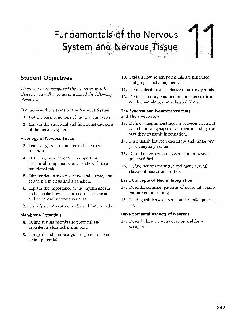
their cytopla mic ex ck CNS.
9. Tn bousendsof~th~e~~~~~~~
2. Relative to neuron anatomy, match the anatomical terms in Column B with the appropriate descriptions of function in Column A. Place the correct answers in the answer blanks.
Column A Column B
1. Releases neurotransmitters A. Axon
2. Conducts local currents toward B. Axon terminal the soma
C. Cell body 3. Increases the speed of impulse
transmission D. Dendrite
4. Location of the nucleus E. Myelin sheath
5. Conducts impulses away from the F. Nissl bodies cell body
6. Most are located and protected within the CNS
7. Short, tapering, diffusely branched extension from the cell body
8. The process called a nerve fiber
9. Formed by Schwann cells in the PNS
10. Clustered ribosomes and rough ER
11. Patchy disappearance in multiple sclerosis
12. Site of biosynthetic activities
3. Circle the term that does not belong in each of the following groupings.
1. Nucleus Soma Centrioles Nucleolus
2. Mitochondria Rough ER Ribosomes Nissl bodies
3. Melanin Glycogen Lipofuscin Pigment
4. Dendritic spine Input Output Receptive
5. Axon terminal Synaptic knob Bouton Axon collateral
252 Study Guide for Human Anatomy & Physiology
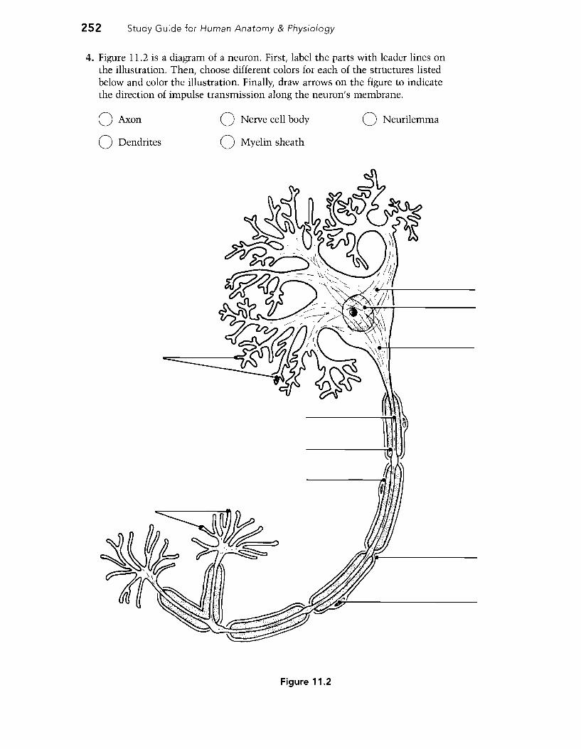
4. Figure 11.2 is a diagram of a neuron. First, label the parts with leader lines on the illustration. Then, choose different colors for each of the structures listed below and color the illustration. Finally, draw arrows on the figure to indicate the direction of impulse transmission along the neuron's membrane.
o Axon o Nerve cell body o Neurilemma
o Dendrites o Myelin sheath
Figure 11.2

262 Study Guide for Human Anatomy & Physiology
5. In Figure 11.6, identify by coloring the following structures, which are typically part of a chemical synapse. Also, bracket the synaptic cleft, and identify the arrows showing (1) the direction of the presynaptic impulse and (2) the direction of net neurotransmitter movements.
0 Axon terminal 0 Postsynaptic membrane 0 Presynaptic membrane
0 Mitochondria 0 Na+ ions 0 Ca2+ ions
0 K+ ions 0 Chemically gated channels 0 Synaptic vesicles
0 Postsynaptic neurotransmitter receptors 0 Neurotransmitter molecules
Figure 11.6
6. Using the key choices, select the phase of action potential generation described in the following statements. Write the correct key letter in the answer blanks.
Key Choices
A. Depolarizing phase C. Repolarization, J, in Na+ permeability E. Resting state
B. Hyperpolarization D. Repolarization, i in K+ permeability
1. Voltage change caused by sodium influx opens more Na+ channels
2. All voltage-gated Na+ and K+ channels are closed
3. Na+ entry declines, voltage-gated K+ channels open
4. Produces the undershoot

Chapter 11 Fundamentals of the Nervous System and Nervous Tissue 263
5. Only leakage channels are open
6. Na+ inactivation gates closing
7. Na+ voltage-sensitive activation gates open
8. AP spike reverses direction
9. Na+ channels resetting to resting state position; K+ entry continues
7. On Figure 11.7, several types of chemical synapses are illustrated. Identify each type, using the key choices. Color the diagram as you wish.
Key Choices
A. Axoaxonic B. Axodendritic C. Axosomatic D. Dendrodendritic
Figure 11.7
B~sic Con i
n
One anticipated response
-------------------
264 Study Guide for Human Anatomy & Physiology
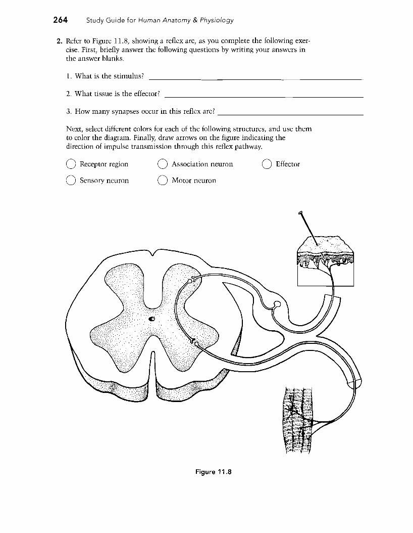
2. Refer to Figure 11.8, showing a reflex arc, as you complete the following exercise. First, briefly answer the following questions by writing your answers in the answer blanks.
1. What is the stimulus?
2. What tissue is the effector?
3. How many synapses occur in this reflex arc?
Next, select different colors for each of the following structures, and use them to color the diagram. Finally, draw arrows on the figure indicating the direction of impulse transmission through this reflex pathway.
o Receptor region o Association neuron o Effector
o Sensory neuron o Motor neuron
Figure 11.8

The Central Nervous System
Student Objectives
When you have completed the exercises in this chapter, you will have accomplished the folluwing ubjectives:
The Brain
1. Describe the process of brain development.
2. Name the major regions of the adult brain.
3. Name and locate the ventricles of the brain.
4. List the major lobes, fissures, and functional areas of the cerebral cortex.
5. Explain lateralization of hemisphere function.
6. Differentiate between commissures, association fibers, and projection fibers.
7. Describe the general function of the basal nuclei (basal ganglia).
8. Describe the location of the diencephalon, and name its subdivisions and functions.
9. Identify the three major regions of the brain stem, and note the functions of each area.
10. Describe the structure and function of the cerebellum.
11. Locate the limbic system and the reticular formation, and explain the role of each functional system.
Higher Mental Functions
12. Define EEG and distinguish between alpha, beta, theta, and delta brain waves.
13. Describe consciousness clinically.
14. Compare and contrast the events and importance of slow-wave and REM sleep, and indicate how their patterns change through life.
15. Compare and contrast the stages and categories of memory.
16. Describe the relative roles of the major brain structures believed to be involved in declarative and procedural memories.
Protection of the Brain
17. Describe how meninges, cerebrospinal fluid, and the blood-brain barrier protect the CNS.
18. Describe the formation of cerebrospinal fluid, and follow its circulatory pathway.
19. Indicate the cause (if known) and major signs and symptoms of cerebrovascular accidents, Alzheimer's disease, Parkinson's disease, and Huntington'S disease.
The Spinal Cord
20. Describe the embryonic development of the spinal cord.
21. Describe the gross and microscopic structure of the spinal cord.
22. List the major spinal cord tracts, and classify each as a motor or sensory tract.
23. Distinguish between flaccid and spastic paralysis, and between paralysis and paresthesia.
Diagnostic Procedures for Assessing CNS Dysfunction
24. List and explain several techniques used to diagnose brain disorders.
Developmental Aspects of the Central Nervous System
25. Indicate several maternal factors that can impair development of the nervous system in an embryo.
26. Explain the effects of aging on the brain.
273
276 Study Guide for Human Anatomy & Physiology
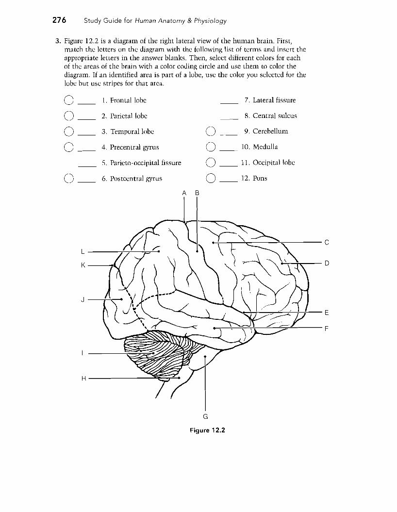
3. Figure 12.2 is a diagram of the right lateral view of the human brain. First, match the letters on the diagram with the following list of terms and insert the appropriate letters in the answer blanks. Then, select different colors for each of the areas of the brain with a color coding circle and use them to color the diagram. If an identified area is part of a lobe, use the color you selected for the lobe but use stripes for that area.
o 1. Frontal lobe 7. Lateral fissure
o 2. Parietal lobe 8. Central sulcus
3. Temporal lobe 9. Cerebellumo o 4. Precentral gyrus 10. Medulla o o 5. Parieto-occipital fissure o 11. Occipital lobe
6. Postcentral gyrus 12. Ponso o B
-+--..,....-~:----....;:.,.,.....----- C L ----.,:--,,.:..----'0:==-.....
K -----.
J
H-----~~
G
Figure 12.2

Chapter 12 The Central Nervous System 277
4. Figure 12.3 illustrates a lisee-through" brain showing the positioning of the ventricles and connecting canals or apertures. Correctly identify all structures having leader lines by using the key choices provided below. One of the lateral ventricles has already been identified. Color the spaces filled with cerebrospinal fluid blue.
Key Choices
A. Anterior horn D. Fourth ventricle G. Lateral aperture
B. Central canal E. Inferior horn H. Third ventricle
C. Cerebral aqueduct F. Interventricular foramen
_--Lateral ventricle
Figure 12.3

278 Study Guide for Human Anatomy & Physiology
5. Figure 12.4 shows a left lateral view of the brain with some of its functional areas indicated by numbers. These areas are listed below. Identify each cortical area by its corresponding number on the diagram. Color the diagram as you wish.
Primary motor cortex Primary somatosensory cortex
Premotor cortex Somatosensory association cortex
Visual cortex Auditory cortex
Prefrontal cortex Broca's area
Frontal eye field Wernicke's area
Posterior association area
Central sulcus
I
Figure 12.4

Chapter 12 The Central Nervous System 281
9. Figure 12.5 is a diagram of the sagittal view of the human brain. First, match the letters on the diagram with the following list of terms and insert the appropriate letters in the answer blanks. Then, color the brain stem areas blue and the areas where cerebrospinal fluid is found yellow.
1. Cerebellum 10. Hypothalamus
2. Cerebral aqueduct 11. Medulla oblongata
3. Cerebral hemisphere 12. Optic chiasma
4. Cerebral peduncle 13. Pineal body
5. Choroid plexus 14. Pituitary gland
6. Corpora quadrigemina 15. Pons
7. Corpus callosum 16. Thalamus (interthalamic adhesion)
8. Fornix 17. Third ventricle
9. Fourth ventricle
ABC D
Figure 12.5 L K J H

Chapter 12 The Central Nervous System 291
Protection of the Brain
1. Figure 12.9 shows a frontal view of the meninges of the brain at the level of the superior sagittal (dural) sinus. First, label arachnoid villi and falx cerebri on the figure. Then, select different colors for each of the following structures and use them to color the diagram.
o Dura mater o Pia mater
o Arachnoid o Subarachnoid space
Scalp -+------..
Bone of skull
Superior sagittal
sinus
Gray matter of cerebral
cortex
Figure 12.9
2. Referring again to the structures in Figure 12.9, identify the meningeal [or associated) structures described here. Write the correct terms in the answer blanks.
1. Innermost covering of the brain; delicate and vascular
2. Structures that return cerebrospinal fluid to the venous blood in the dural sinuses
3. Its outer layer forms the periosteum of the skull
4. Contains cerebrospinal fluid
5. Location of major arteries and veins
6. Contains venous blood
7. Attaches to crista galli of the ethmoid bone

294 Study Guide for Human Anatomy & Physiology
2. Figure 12.10 is a cross-sectional view of the spinal cord. First identify the areas listed in the key choices by inserting the correct letters next to the appropriate leader lines on parts A and B of the figure. Then, color the bones of the vertebral column in part B gold.
Key Choices
A. Central canal E. Dorsal root I. Ventral horn
B. Columns of white matter F. Dorsal root ganglion J. Ventral root
C. Conus medullaris G. Filum terminale
D. Dorsal horn H. Spinal nerve
On part A, color the butterfly-shaped gray matter gray, and color the spinal nerves and roots yellow. Finally, select different colors to identify the following structures and use them to color the figure.
o Pia mater o Dura mater o Arachnoid mater
--.,..::;~,....-----~ Spinal
cord
A
Figure 12.10 B