fundamentals of digital mammography
TRANSCRIPT

IntroductionScreen-film image receptors have been the standard detector
used in conventional mammography. New developments indetector technology and computers are altering the landscape ofmammography imaging. Full Field Digital Mammography(FFDM) offers the promise of revolutionizing the practice ofmammography through its superior dose and contrast perform-ance. Advanced applications made possible through digitalimaging, such as automated computer-aided diagnosis, dual-energy and 3D tomosynthesis are expected to further improvediagnostic sensitivity and specificity.
This primer describes the technical basis for current andfuture advances in mammographic detector technology. Theseinclude all of the following:• Lower dose• Improved image quality• Computer-aided diagnosis• Softcopy review and digital archiving• Tomosynthesis and other three-dimensional visualization tech-
niques• Reduction in breast compression pressure
New flat-panel X-ray detectors offer extremely high quantumefficiency and high resolution. These will translate into lowerdose and improved image quality mammograms. Digital detectorsfor mammography can be categorized as indirect or direct conversion detectors. This primer describes these technologiesand how direct conversion detectors, in particular selenium-based detectors, provide the best performance for full field digitalmammography systems.
Indirect conversion digital detectors utilize a method ofimaging X-rays, similar to screen-film, wherein a scintillatorabsorbs the X-rays and generates a light scintillation. The lightscintillation is then detected by an array of photodetectors.However, indirect conversion systems suffer from resolutiondegradation caused by light spread in the scintillator, and frompoor quantum efficiency caused by the use of thin scintillators.
Direct conversion digital detectors utilize a direct-conversionmethod of imaging, wherein the X-rays are absorbed and theelectrical signals are created in one step. Systems using amor-phous selenium represent the most advanced direct conversiontechnology for digital mammography. Selenium is an ideal
Fundamentals of Digital Mammography:Physics, Technology and Practical ConsiderationsAndrew P. Smith, Ph.D.
material for a mammography detector, because it has high X-rayabsorption efficiency (approaching 100 percent at mammo-graphic X-ray energies), extremely high intrinsic resolution, lownoise and a well-established manufacturing process.
The pixel size requirement for FFDM is also addressed inthis article. Light blurring in indirect conversion systems interfereswith the effective design and performance of systems with veryfine resolution. Direct conversion systems, in contradistinction,can have their resolution characteristics optimized independentlyfrom their quantum detection efficiency and can thus bedesigned with an optimum pixel size.
The field of view for FFDM systems is very important. Inorder to be able to image most of the adult female population,the imaging field of view must be similar to the size of the largestscreen-film cassette commonly used in screen film imaging, 24 x30 cm. Detectors that are the size of the smaller cassette, 18 x 24cm, will require imaging and tiling of many images to cover thebreast field of view for larger breasts. For the U.S. demographics,up to 30 percent of all women require the larger field of view.
Detector performance is commonly quantified by two metrics:Modulation Transfer Function (MTF) and Detective QuantumEfficiency (DQE). MTF is a measure of resolution and DQE isa measure of dose efficiency.The MTF for screen-film is superiorto indirect conversion detectors, while the MTF for direct conversion detectors are superior to both. The DQE of indirectconversion detectors is superior to screen-film, however theDQE of selenium direct conversion detectors is seen to be superioragain to both screen-film and indirect conversion detectors.With superior DQE at all spatial frequencies, selenium detectorsoffer the potential for both improved image quality and lowerpatient dose.
Digital images offer a variety of new and improved applica-tions. Contrast enhancement of the digital image and the widedynamic range of digital detectors will improve visibility ofmammographic features. The digital image will provide imagearchiving and retrieval advantages over film, and will facilitatethe use of computer-aided diagnosis.
Systems with high quantum efficiency, especially atincreased X-ray energies, offer the possibility of decreased breastcompression. As is commonly known, painful breast compressionis an important factor in undercompliance of women to presentfor routine breast screening.

Future applications such as stereo mammography, breasttomosynthesis, contrast enhanced digital mammography andother imaging modalities, are under investigation. Suchadvances in technology will provide improved diagnostic infor-mation and reduced image confusion from overlapping structures.These three dimensional imaging tasks will benefit from thehigh quantum efficiency that selenium detectors offer. The digitaldetectors will also be able to be used for full-field and high resolution stereotactic breast biopsies and diagnostic imagingtasks. It is essential, however, that the full-field digital systemsperform well for its primary task, i.e. breast screening.
In conclusion, digital detectors offer many advantages comparedto screen-film imaging for mammography. It is hoped that thepotentials for improved image quality, lower dose and advancedimaging applications will result in improved diagnostic accuracy.
Screen-Film MammographyAs shown in Figure 1, conventional film systems use inten-
sifying screens to capture X-rays and reduce radiation dose. X-rays that pass through the tissue are collected by phosphorscreens. These screens are often constructed of rare earth phos-phors such as gadolinium oxysulfide (Gd2O2S) that output lightupon absorption of X-rays.When an X-ray is absorbed, theresultant light scintillation creates a number of light photonsthat spread and illuminate the film in a distribution cloud. Filmin close proximity to the screen captures the light photons, andthe image is obtained by exposing the film.
An important parameter to understand is the thickness ofthe intensifying screen. Thicker screens capture more X-rays andare therefore more dose efficient and higher speed. However,thicker screens also create more light scatter and blurring of theimage. Therefore, it is impossible to offer a screen-film systemsimultaneously offering the highest possible resolution and lowestpossible radiation dose. This trade-off between radiation doseand image quality must be optimized for the specific clinicalapplication.
Figure 2 shows the performance trade off of sensitivity versusresolution inherent in the design of screen-film systems. As thescreen is made thicker, the cloud of light on the film will increasein size, on average. This reduces the resolution of the system,however the system’s sensitivity increases because the thickerphosphor increases the probability that the incoming X-ray willbe absorbed. Thus screen-film systems have a performance tradeoff between speed and resolution.
Because X-rays are absorbed with a decaying exponentialspatial distribution, in a screen-film mammography system thefilm is placed at the entrance surface of the scintillating screen.
While a screen-film system offers several advantages, thereare significant disadvantages and limitations of this system. Filmdoes not have a linear sensitivity to photonflux, and there is anarrow range over which it can detect small differences in contrast.In particular, tissue areas of high and low density are often sub-optimally imaged. Frequently, the entire image is poorly exposedbecause of film’s stringent requirements for proper exposure,resulting in repeated imaging.
Another major problem is film granularity, affecting detectivequantum efficiency at high optical densities and visibility ofmicrocalcifications.
Film also requires processing time and storage space. Chemicalprocessing of the film adds time to the exam, and the resultingX-ray films require a large amount of storage space in patienthealth records. Film also must be physically transported to thephysician for viewing.
Digital Mammography DetectorsDigital technology offers the potential for several advances
in mammography detectors. Because images are captured as adigital signal, electronic transfer and storage of images is possible,eliminating physical storage and distribution required by film.Digital systems offer a large dynamic range of operation,improving visualization of all areas of the breast and increasingexposure latitude. Also, the digital format allows grayscale
Line spread function
Scintillating screen,Gd2O2S
X-ray film
Light scintillation
X-ray
~84 µm
Figure 1.Screen-film systems use a scintillating screen to absorb the X-rayand generate light photons, which are captured on the film.
Line spread functions
Screen/film:low speed, high resolution
Screen/film:high speed, low resolution
X-ray X-ray
Figure 2.As the thickness of the screen is increased, its speed increases atthe expense of poorer spatial resolution.

adjustment to optimize contrast for every imaging task. Soft-copy reading, computer-aided diagnosis and three-dimensionalimaging offer additional and potentially important opportunitiesfor improvement in mammographic systems.
Digital Detector TechnologyThere are two methods of image capture used in digital
mammography that represent different generations of technology:indirect conversion and direct conversion.
Indirect-Conversion Digital DetectorsThe earliest digital mammography systems available in the
U.S. used indirect conversion detectors. Such detectors use atwo-step process for X-ray detection, as seen in Figure 3. Thefirst step requires a scintillator layer such as cesium iodide dopedwith thallium [CsI(Tl)] to capture the X-ray energy and convertit to light. An array of thin-film diodes converts light photons toelectronic signals that are captured using thin-film transistors.Some systems, like Charge-Coupled Devices (CCDs), use alter-native light collection and readout methods. In both systems,the light sensitive imaging function of film has been replaced bydigital light imaging. It is in this sense that these can be seen asan evolution of screen film imaging.
Similarly to screen-film, light scatter compromises imagequality, and there is a performance tradeoff between spatial res-olution and radiation sensitivity, as shown in Figure 4. As thescintillator is made thicker, light spread increases resulting indecreased resolution. Because of its columnar structure, CsI(Tl)does not create as much light scatter as other screens. However,compromise between resolution and sensitivity still exists.
The placement of the scintillator is more problematic inindirect conversion digital detectors than with screen-film systems.As with film screens, more X-rays are absorbed near the entranceof the scintillation layer than the exit. While film is placed nearthe entrance side of the scintillator, a photodiode/transistor arrayis not transparent to X-rays and the array must be placed on the
exit surface of the scintillator. This causes degradation in spatialresolution compared to screen-film.
Typical thicknesses of CsI(Tl) used in mammographydetectors range from 150 to 250 microns, and these indirectconversion digital detectors exhibit light spreading similar toscreen-film systems. Examples of indirect conversion detectorsare the Fischer Imaging CsI/CCD-based detector and the GECsI/TFT detector.
Direct-Conversion Digital DetectorsDirect-conversion digital detectors represent a technological
advance, eliminating problems associated with light scatterinherent in indirect conversion systems. In these systems, illus-trated in Figure 5, a photoconductor absorbs the X-rays anddirectly generates the signal (direct conversion). Under the influenceof an external electric field, holes (or electrons, depending uponthe polarity of the applied field) drift towards a pixel electrodeand are collected on a pixel capacitor. Because the electrons andholes travel along the direction of the electric field lines, theymove without lateral charge spreading. This results in an excep-tionally narrow point spread response, of about 1 micron.
The superior photoconductor for use in direct conversionsystems is amorphous selenium (a-Se). Selenium has a long com-mercial history in xerography, and its manufacturing processesare well known and optimized. The image quality of xeromam-mography systems was widely acknowledged, but it sufferedfrom reliability problems as a result of mechanical wear of theplates during toner deposition and crystallization of the seleniumduring the high-temperature erasure cycles. By depositing seleniumon a flat-panel imaging receptor, these problems have been eliminated.
In direct-conversion detectors, the response function maintainsits sharpness even as the thickness of the photoconductor isincreased, so there is no trade off between radiation stoppingpower and spatial resolution. This is shown in Figure 6. In practice,the photoconductor is made sufficiently thick in order to stop
Line spread function
Pixel array
Columnar CsI (T1)150-250 µm
Light scintillation
X-ray
150-250 µm
Figure 3.Indirect conversion detectors utilize a scintillating layer to absorb the X-ray andgenerate light photons, which are detected by a photodiode array.
X-ray X-ray
Line spread functions
Indirect conversion:low speed, high resolution
Indirect conversion:high speed, low resolution
Pixel array
Figure 4.Indirect conversion detectors generate poorer resolution imagesas the scintillator thickness is increased.

the majority of the incident X-rays, and this can be done withoutadversely affecting the spatial resolution, an important considerationin mammography with its dual needs of high resolution and lowradiation exposure.
Using amorphous selenium as the photoconductor, a thicknessof 250 microns is adequate to stop more than 95 percent of theX-rays in the mammographic energy range. This is seen in Fig-ure 7. Standard screens for use in film mammography only haveabout 50 to 70 percent quantum efficiency, and the scintillatorCsI(Tl) used in indirect-conversion digital detectors exhibitsabout 50 to 80 percent quantum efficiency. Systems using amorphous-selenium can achieve almost complete quantumefficiency.
Pixel Design for Digital DetectorsDigital detectors require an array of pixels that collect
electronic signals. The signals on these pixels are transferred to acomputer during a readout sequence. This is known as directreadout, a function of all digital systems, and should not be confused with direct conversion digital detection.
Thin film transistor (TFT) arrays are the active electronicreadout mechanism commonly used in both direct- and indirect-digital radiography systems. The arrays are typicallydeposited onto a glass substrate in multiple layers, beginningwith readout electronics at the lowest level, followed by chargecollector arrays at higher levels. The composition of the top layerdepends upon the type of detector. If the system uses indirectconversion detection, both X-ray elements and light-sensitiveelements are deposited on the top layer. Direct conversion detectorsdo not require conversion of X-rays to light, so light sensitive elements are not necessary for these systems.
Semiconductor arrays for direct conversion detectors aremuch easier to fabricate than arrays for indirect conversiondetectors because selenium-based detectors do not require a photodiode structure on top of the TFT. As a result, these detectorscan utilize the same manufacturing processes as large area TFT-LCD arrays, commonly used in computer displays and do notrequire construction of dedicated manufacturing facilities.
Charge-coupled devices (CCDs) are an alternative to TFTarrays in indirect conversion systems. Basic CCD-based systemsconsist of a series of metal oxide semiconductor capacitors thatare fabricated very close together on the semiconductor surface.These systems use fiber optics to capture images from light emittedfrom scintillators or intensifying screens, but suffer from lightloss and added complexity due to the fiber optics. The only commercially available CCD-based system uses a slot scanningtechnology,whereby a small detector is scanned across the breastto build up the image over several seconds. This is in distinctionto the other FFDM systems that acquire the image in a singlesnapshot.
Line spread function
X-ray
Electron/hole pairs
Photoconductoramorphous selenium
Pixel array
250 µm
Figure 5.Direct-conversion detectors use a photoconductor to absorb the X-ray anddirectly generate the signal.
X-ray X-ray
Line spread functions
Direct conversion:high speed & resolution
Direct conversion:high speed maintainedeven at higher speed
Figure 6.There is no tradeoff between spatial resolution and sensitivity fordirect-conversion systems.
X-ray energy (KeV)
atte
nuat
ion
mu/
rho
(cm
**2/
g)
1010
100
1000
15 20 25 30 35
Figure 7.K-edge for Selenium is just below the diagnostic range for mammography,shown in the area between the two yellow lines.

Pixel Size RequirementsMammographic imaging requires the detection and classifi-
cation of extremely small objects. In particular, microcalcifica-tions can be as small as 100 to 200 microns. Any useful FFDMsystem must be able to image these smallest microcalcificationsof interest.
Digital detectors are comprised of arrays of pixels. Thesmallest feature that can be resolved in any digital imaging system is a function of the pixel size – the smaller the pixel thehigher the resolution. Figure 8 shows the intrinsic resolution fordirect and indirect detectors.
In indirect-conversion digital detectors, as the pixel size isdecreased, a limit is reached beyond which further reductions inpixel size does not improve resolution. This resolution limit is afunction of the light scattering in the scintillator. This limit isabout 100 microns in practical indirect conversion scintillators,which is the upper limit for application to digital mammography.CCD based indirect conversion detectors typically have muchsmaller pixels, however the light spread from the scintillator lim-its the resolution so the result is an image with more pixels butnot superior resolution.
In a direct-conversion digital detector, on the other hand,spatial resolution is only limited by the size of the pixel. The sizeof the pixel in these detectors can be made arbitrarily small(within limits) to make the resolution performance extend tovery high spatial frequencies. The ultimate limit to very smallpixels is the reduced X-ray flux impinging upon the detector.
The pixel size for FFDM systems range from about 50 to100 microns. As explained earlier, the indirect-conversion detectorsall have resolution characteristics that are scintillator limited, sothe raw pixel size does not accurately reflect the actual resolutioncharacteristics of the image. The pixel size for the Hologic’s sele-nium detector is 70 microns, and because of the design of thisdetector, this represents its true resolution characteristic.
Pixel size design represents a compromise between spatial
resolution and system cost. As the pixel size of any digital imagingsystem is made smaller, the amount of data contained in theimage rapidly increases, which increases system costs in terms ofdata storage, network bandwidth and display capabilities. Fabri-cation of large-area detectors with very small pixels is an expensive,low-yield process, and integration of such detectors with analogand digital readout electronics and system interconnects pushesthe state of the art in microfabrication on several fronts.
Field of View RequirementsMQSA requirements for mammography facilities require
the use of at least two screen-film cassette sizes: 18 x 24 cm and24 x 30 cm. The rationale for this requirement is the large distribution in compressed breast sizes. A small cassette cannotaccommodate large breasts in the field of view, and two overlappingexposures will be required to ensure adequate coverage of all thebreast tissue. This is costly, time consuming, and subjects thewomen to additional radiation dose. On the other hand, the useof a large cassette on smaller women is wasteful of film and processing materials.
Digital mammography presents additional field-of-viewissues. At the present time, flat-panel X-ray detectors are consid-erably more expensive than screen-film cassettes, precluding theuse of more than one receptor size on a given machine. Irrespectiveof cost, digital detectors are heavy and fragile and do not lendthemselves to rapid changeover as do film cassettes. Therefore,digital mammography equipment manufacturers must decideon one optimum detector size, one that is large enough toaccommodate most women without requiring double exposures.
A digital detector of size 18 x 24 cm (the smaller film cassettesize) is inadequate to image approximately 20 to 30 percent ofU.S. women. Because of regional differences, some areas of thecountry might have a larger percentage of women that cannot bescreened using the smaller film cassette.
If the breast is too large to image on the detector in oneexposure, multiple exposures that ‘tile’ the breast are required.This solution has disadvantages such as requiring multiple additional compressions, additional setup and imaging times,breast regions that suffer from repeated radiation exposure, andthe difficulty of radiologist’s review of multiple images.
Use of a large detector for digital mammography is animportant design issue. Issues using a large digital detector toimage a small breast does not present the same problems as film.The large image can be cropped to reduce data transfer, displayand storage space requirements, and there are no additionalmaterial costs. The one technical challenge in the use of a largedetector for small breasts is in positioning, however the use ofsmaller compression paddles in conjunction with the largedetector solves this problem.
The larger detector can be used to advantage when imagingsmaller breasts. The imaging can be performed in a magnificationmode, creating the effect of smaller pixels and reducing scatteredradiation at the same time. If a system is designed so that breasts
Line spread functions
Indirect ConversionScreen/Film Direct Conversion
Columnar CsI (T1)150-250 µm
MinR - 2000Gd2O2S amorphous-Selenium
Figure 8.Line spread functions for screen-film and digital detectors. Screen-film andindirect-conversion detectors have broad presampled line spread functions;direct-conversion detectors have narrower presampled line spread functions.

that are adequately served by the smaller film cassette size of 18x 24 cm are imaged in magnification mode to project onto adetector of size 24 x 30 cm, an image of effective pixel size of 56microns will be achieved for systems with inherent 70 mm pixelsizes. Such a system could offer 56 micron imaging in a screeningenvironment for approximately 80 percent of U.S. women. Thisis another possibility offered by use of a large digital detector.
Characterizing Detector PerformanceThere are parameters that characterize the overall image
quality obtained on mammography and other radiography systems.The most important are:• Spatial resolution• Contrast resolution• Signal-to-noise ratio• Dose efficiency
Dynamic RangeScreen-film has a limited dynamic range, which prevents
visualization with equal clarity of all breast tissue regions fromthe chest wall to the skin line. Figure 9 shows the need forimproved dynamic range in breast imaging. Digital detectorsoffer greatly improved performance. It has been shown that foran ideal detector with no inherent detector noise, 3100 gray lev-els are discernable in a typical mammographic image. Thus a system that provides at least 12 bits of dynamic range will notdegrade the underlying information. Digital mammograms will
exhibit uniform quality over a range of exposure conditions andbreast sizes.
X-ray Quantum Detection EfficiencyX-ray quantum detection efficiency is a measure of the per-
centage of X-rays that hit the detector that are absorbed. Systemswith higher quantum efficiency can produce higher qualityimages, at lower dose. As mentioned earlier, selenium systemsstop more than 95 percent of the X-rays in the mammographicenergy range. Standard screens for use in film mammographyonly have about 50 to 70 percent quantum efficiency, andCsI(Tl) used in indirect conversion detectors exhibit about 50 to80 percent quantum efficiency.
Modulation Transfer Function (MTF)Modulation Transfer Function (MTF) and Detective
Quantum Efficiency (DQE) provide quantitative measurementof imaging performance. MTF measures spatial resolution, whileDQE is a measure of signal-to-noise ratio, contrast resolution anddose efficiency. An imaging system is best characterized by exam-ining corresponding MTF and DQE curves; however, it cannotbe adequately described by one number at a single spatial frequency. These measurements are used to determine how wella system captures information over a range of spatial frequencies.
Modulation Transfer Function (MTF) is a measure of signaltransfer over a range of spatial frequencies and quantifies spatialresolution. Spatial resolution is often measured using a line pairphantom, illustrated in Figure 10. The ultimate resolution limitof any system is determined by its pixel size. For example, a system with a 100-micron pixel cannot adequately resolve spatialfrequencies above 5 line pairs/mm (lp/mm). Indirect-conversionmethods can scatter light over several pixels, further limiting theeffective resolution of the system, more so than indicated bypixel size alone.
Direct conversion systems do not suffer from this limitation.
Commercially AvailableDigital Mammography Detectors
The following is a list of full-field-of-view digital mammography detectors commercially available in the U.S.
Indirect–conversion detectors• GE
Scintillator CsI(Tl)Pixel size TFT 100 micronsField of view 18 x 23 cm
• Fischer ImagingScintillator CsI(Tl)Pixel size CCD 24/48 micronsField of view 22 x 30 cm (scanning)
Direct–conversion detectors• Hologic/Lorad
Photoconductor amorphous seleniumPixel size TFT 70 micronsField of view 24 x 29 cm
Relative Exposure
fatty tissue
markersdense tissue
skin marginair
4.00
Optic
al D
ensi
ty
3.50
3.00
2.50
2.00
1.50
1.00
0.50
1 10 100 1000 10,000
Figure 9.Characteristic curve of mammographic film illustrating how the display contrast(slope of the curve) is suboptimal in lucent and dense regions of the breast1.
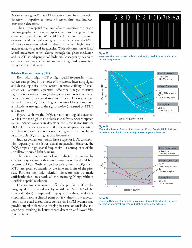
As shown in Figure 11, the MTF of a selenium direct conversiondetector2 is superior to those of screen-film3 and indirect-conversion detectors4.
The intrinsic spatial resolution of selenium direct-conversionmammography detectors is superior to those using indirect-conversion scintillators. While MTFs for indirect conversiondetectors fall dramatically at higher spatial frequencies, the MTFof direct-conversion selenium detectors remain high over agreater range of spatial frequencies. With selenium, there is nolateral movement of the charge through the photoconductor,and its MTF is independent of thickness. Consequently, seleniumdetectors are very efficient in capturing and converting X-rays to electrical signals.
Detective Quantum Efficiency (DQE)Even with a high MTF at high spatial frequencies, small
objects can get lost in the noise of the system. Increasing signaland decreasing noise in the system increases visibility of smallstructures. Detective Quantum Efficiency (DQE) measures signal-to-noise transfer through the system as a function of spatialfrequency, and it is a good measure of dose efficiency. Several factors influence DQE, including the amount of X-ray absorption,amplitude or strength of the signal profile (measured by MTF)and noise.
Figure 12 shows the DQE for film and digital detectors.While film has a high MTF at high spatial frequencies comparedto the indirect conversion detectors, the same is not true forDQE. This is one reason why the potential spatial resolutionwith film is not realized in practice. Film granularity noise limitsits achievable DQE at high spatial frequencies.
Indirect conversion systems have a superior DQE to screen-film, especially at the lower spatial frequencies. However, theDQE drops at high spatial frequencies—a consequence of thescintillator-induced light blurring.
The direct conversion selenium digital mammographydetector outperforms both indirect conversion digital and filmin terms of DQE. With no signal spreading, and the DQE (andMTF) are governed mainly by the inherent limits of the pixelsize. Furthermore, only selenium detectors can be made sufficiently thick to absorb all the incoming X-rays without sacrificing spatial resolution.
Direct-conversion systems offer the possibility of similarimage quality at lower doses (by as little as 1/2 to 1/3 of thescreen-film dose) or improved image quality at the same dose asscreen-film. From a clinical point of view, there is the expecta-tion that at equal doses, direct conversion FFDM systems mayprovide superior diagnostic imaging in terms of sensitivity andspecificity, resulting in better cancer detection and lower falsepositive rates.
Figure 10.X-ray resolution test pattern to determine imaging resolution performance, inunits of line pairs/mm.
Direct Conversiona-Selenium TFT 70 µm pixels
CsI (T1) TFT 1000 µm pixelsCsI (T1) CCD 40 µm pixels
Gd2O2S OrthoM / MinR
Indirect Conversion
Screen/film
Spatial Frequency (lp/mm)
80%
100%
60%
40%
Pres
ampl
ed M
TF20%
0%
0 2 4 6 8 10
Figure 11.Modulation Transfer Function for screen-film (Kodak: OrthoM/MinR), indirectconversion and direct conversion digital mammography detectors.
Frequency (lp/mm)
Direct Conversiona-Selenium TFT 70 µm pixels
CsI (T1) TFT 1000 µm pixelsCsI (T1) CCD 40 µm pixels
Gd2O2S OrthoM / MinR
Indirect Conversion
Screen/film
70%
60%
50%
40%
DQE
30%
20%
10%
0%0 2 4 6 8 10
Figure 12.Detective Quantum Efficiency for screen-film (Kodak: OrthoM/MinR), indirect-conversion and direct-conversion digital mammography detectors.

ACR Phantom Imaging PerformanceFigure 13 shows the acrylic phantom used for ACR accred-
itation and MQSA inspections. The phantom is approximatelyequivalent in X-ray absorption to a 4.2-cm thick compressedbreast consisting of 50 percent glandular and 50 percent adiposetissue. The phantom includes appropriate details that rangefrom visible to invisible on a standard mammographic filmimage. The phantom has fibers with diameters of 1.56, 1.12,0.89, 0.75, 0.54 and 0.40 mm; specks with diameters of 0.54,0.40, 0.32, 0.24 and 0.16 mm; and masses with decreasingdiameters and thicknesses of 2.00, 1.00, 0.75, 0.50 and 0.25 mm.
The visibility of phantom details has been evaluated forscreen-film and for first and second-generation detectors.5 Thephantom was imaged at 28 kVp at a dose of ~3 mGy. (A highdose was used to emphasize the performance differences.)Results are summarized in Table 1. These results are consistentwith predicted performance based on the DQE plots: second-generation systems outperform both screen-film and indirectconversion.
System Design ConsiderationsThere are a variety of system-level design considerations
with full field digital mammography equipment.
X-ray SourceThe spectrum of X-ray energies used in screen-film
mammography has been highly optimized. Digital mammo-graphic detectors offer improved performance characteristics,particularly dynamic range, however the issue of the optimumenergy is still in the investigational phase.6 It is possible thathigher X-ray energies may permit lower dose or higher imagequality with digital mammography, particularly for patients withdense breasts. Detectors that have high intrinsic quantum efficiency allow the use of higher energies.
Selenium has superior stopping power compared to filmscreens and CsI(Tl) material, as seen in Figure 14. At typicalenergies used in mammography (mean energy ~ 20 KeV), selenium’s quantum efficiency is optimal. Selenium maintains itshigh quantum efficiency at higher energies as well. Systems thatreduce breast compression will utilize higher X-ray energies thancurrently used, and the pronounced high-energy performance ofselenium will become even more important.
Scatter Rejection MethodsLarge area flat-panel detectors are subject to the same dele-
terious effects of radiation scatter as conventional screen film systems. The preferred method to reduce scatter is to employ theuse of radiation anti-scatter grids interposed between the patientand the detector.
Anti-scatter GridsThere are two common methods of grid design, shown in
Figure 15. Standard linear grids are constructed of long thin slatsof radio-opaque materials (called septa or laminae) separated by
Figure 13.ACR Mammography Accreditation Phantom, and imaging details.
X-Ray Energy (KeV)
Csl(T1)Gd2O2Sa-Selenium
100%
80%
60%
40%
20%
0%
15% 20% 25% 30%
Abso
rptio
n Pe
rcen
tage
Figure 14.Percent absorption of incident X-rays for materials used in screen-film: Gd2O2S34 mg/cm2, indirect conversion CsI(Tl) : 73 mg/cm2 and second generationcselenium: 250 microns thick.
Table 1.Performance of systems in imaging the ACR Mammography Accreditation Phantom
Detector Technology Fibers visible Specks visible Masses visible
Kodak MinR 2000 Screen film 4 4 3
CCD Indirect conversion 5.5 4 5
a Selenium Direct conversion 6 5 5
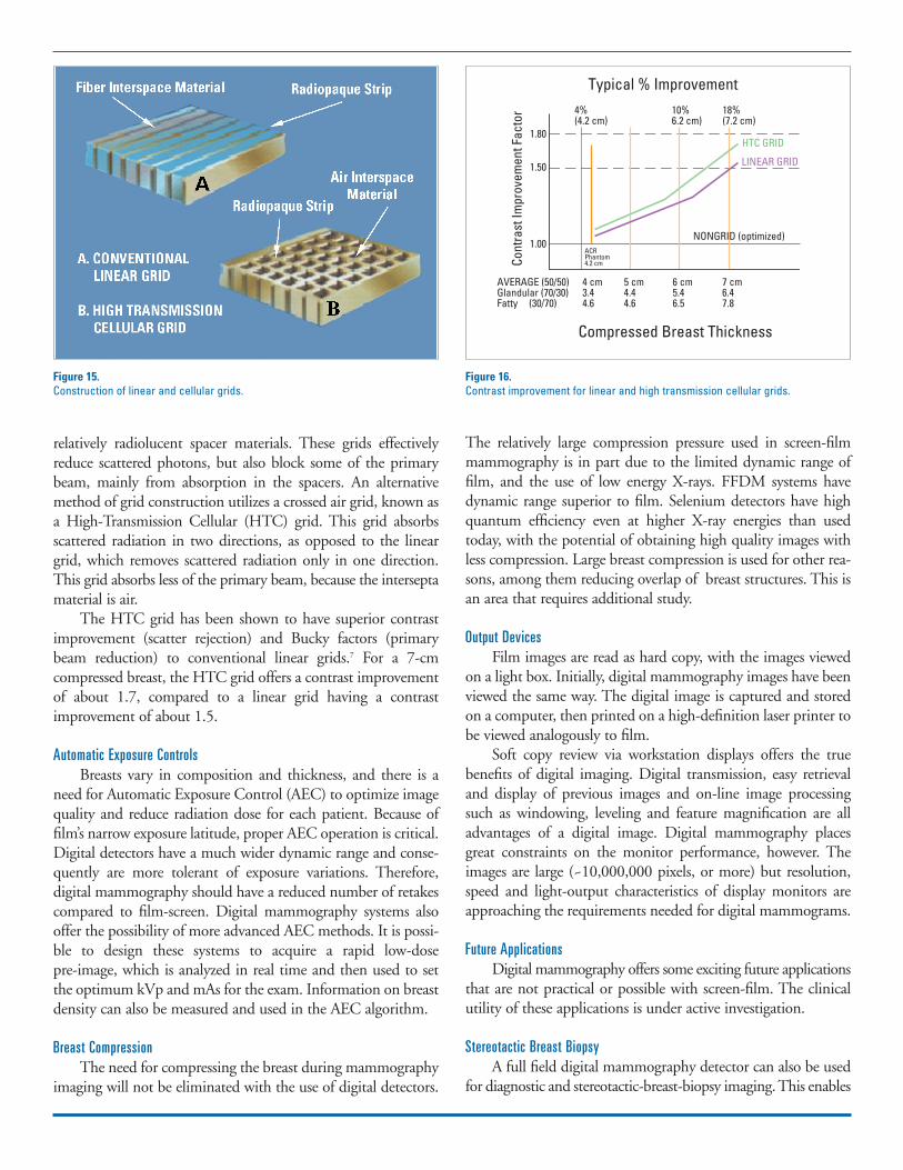
relatively radiolucent spacer materials. These grids effectivelyreduce scattered photons, but also block some of the primarybeam, mainly from absorption in the spacers. An alternativemethod of grid construction utilizes a crossed air grid, known asa High-Transmission Cellular (HTC) grid. This grid absorbsscattered radiation in two directions, as opposed to the lineargrid, which removes scattered radiation only in one direction.This grid absorbs less of the primary beam, because the interseptamaterial is air.
The HTC grid has been shown to have superior contrastimprovement (scatter rejection) and Bucky factors (primarybeam reduction) to conventional linear grids.7 For a 7-cm compressed breast, the HTC grid offers a contrast improvementof about 1.7, compared to a linear grid having a contrastimprovement of about 1.5.
Automatic Exposure ControlsBreasts vary in composition and thickness, and there is a
need for Automatic Exposure Control (AEC) to optimize imagequality and reduce radiation dose for each patient. Because offilm’s narrow exposure latitude, proper AEC operation is critical.Digital detectors have a much wider dynamic range and conse-quently are more tolerant of exposure variations. Therefore, digital mammography should have a reduced number of retakescompared to film-screen. Digital mammography systems alsooffer the possibility of more advanced AEC methods. It is possi-ble to design these systems to acquire a rapid low-dose pre-image, which is analyzed in real time and then used to setthe optimum kVp and mAs for the exam. Information on breastdensity can also be measured and used in the AEC algorithm.
Breast CompressionThe need for compressing the breast during mammography
imaging will not be eliminated with the use of digital detectors.
The relatively large compression pressure used in screen-filmmammography is in part due to the limited dynamic range offilm, and the use of low energy X-rays. FFDM systems havedynamic range superior to film. Selenium detectors have highquantum efficiency even at higher X-ray energies than usedtoday, with the potential of obtaining high quality images withless compression. Large breast compression is used for other rea-sons, among them reducing overlap of breast structures. This isan area that requires additional study.
Output DevicesFilm images are read as hard copy, with the images viewed
on a light box. Initially, digital mammography images have beenviewed the same way. The digital image is captured and storedon a computer, then printed on a high-definition laser printer tobe viewed analogously to film.
Soft copy review via workstation displays offers the truebenefits of digital imaging. Digital transmission, easy retrievaland display of previous images and on-line image processingsuch as windowing, leveling and feature magnification are alladvantages of a digital image. Digital mammography placesgreat constraints on the monitor performance, however. Theimages are large (~10,000,000 pixels, or more) but resolution,speed and light-output characteristics of display monitors areapproaching the requirements needed for digital mammograms.
Future ApplicationsDigital mammography offers some exciting future applications
that are not practical or possible with screen-film. The clinicalutility of these applications is under active investigation.
Stereotactic Breast BiopsyA full field digital mammography detector can also be used
for diagnostic and stereotactic-breast-biopsy imaging. This enables
Figure 15.Construction of linear and cellular grids.
Compressed Breast Thickness
Typical % Improvement
AVERAGE (50/50)Glandular (70/30)Fatty (30/70)
Cont
rast
Impr
ovem
ent F
acto
r
4 cm3.44.6
4%(4.2 cm)
1.80
1.50
1.00ACRPhantom4.2 cm
10%6.2 cm)
18%(7.2 cm)
HTC GRID
LINEAR GRID
NONGRID (optimized)
5 cm4.44.6
6 cm5.46.5
7 cm6.47.8
Figure 16.Contrast improvement for linear and high transmission cellular grids.

a multi-purpose system ideal for both screening and follow-up imaging tasks.
Stereo MammographyAcquisition of binary images, separated by a few degrees,
allows the display of mammograms in a stereo fashion, to facilitatethe visualization of the three-dimensional characteristics ofmammographic features. Computer display of stereo imagesrequires either glasses synchronized to display frame updates, orpolarizing glasses. Investigations of this technique8 show promisein improving three-dimensional depth discrimination in mammograms.
Tomosynthesis and Three-Dimensional ImagingThese applications utilize acquisition of multiple images
from many angles, typically 9-15 about a +- 15 degree range,with the data processed to generate tomographic slices. Theseimages will exhibit reduced confusion from overlapping structures. Preliminary studies9 show that these acquisitions canbe performed with doses similar to screening mammography,and that the images offer diagnostic information not availablewith 2D imaging. High quantum efficiency will be importantfor these applications, to reduce the total dose for the multipleimages required.
Dual-Energy ImagingIn dual-energy imaging, two images are made of the breast
at differing X-ray energies. Because of the differing X-ray attenuation characteristics of glandular tissue, adipose tissue andmicrocalcifications, processing of the dual images can enhancethe visibility of certain structures. The clinical utility of this hasbeen suggested10, but further research is needed. Dual energyimaging can also be used to quantify fibroglandular breast tissuedensity.
Computer-Aided DiagnosisThe use of computer programs to perform preliminary cancer
diagnoses is an area of active research.11 The digital image isexamined by software, and suspicious areas are highlighted forfurther scrutiny by the radiologist. The challenge for these systems is to find the proper balance between sensitivity andspecificity. Increasing sensitivity can result in too many false pos-itives marked on an image, and the radiologist may find thismore of an annoyance than help. Conversely, if not enough truepositives are marked, the system will be offering little help to theradiologist. The low cancer rate per image in a screening envi-ronment makes this task difficult. However, it is expected thateventually these types of systems will become routine, since it isvery easy to perform the CAD procedure on the digitallyacquired image.
Digital mammography CAD systems offer significantadvantages compared to film-based CAD systems. Digitizing ofthe film images is time consuming and yields a less than optimal
digital image.With a true digital image, the increased dynamicrange and access to multiple views and previous studies is expectedto improve the accuracy of CAD systems.
ConclusionsFull field digital mammography offers the potential for
significant advances in breast cancer diagnosis, including lowerradiation dose, reduced breast compression pressure andimproved cancer detection rates. FFDM has advanced rapidly inthe past few years, and there are now several systems commer-cially available for sale in the U.S. These systems offer improve-ments over screen-film imaging, especially in regards to imagingdynamic range and digital storage and display. The differentmanufacturer’s products can be differentiated by the imagereceptor’s detection technology, its resolution dose and scatterrejection performance, and in the detector’s field of view.
Andrew Smith, Ph.D., is principal scientist at Hologic, Inc., in Bed-ford, Mass., where he is involved in research and development indigital radiographic systems. His previous employment was withDigital Scintigraphics Inc., a nuclear medicine company he co-founded. Prior to that, he attended the Massachusetts Institute ofTechnology, where he received a bachelor’s degree and doctoral degreein physics.

Glossarya-Se . . . . . . . . . . . . . . . . .Amorphous selenium. The most
common photoconductor materialused in direct conversion detectors.
a-Si . . . . . . . . . . . . . . . . .Amorphous silicon. The materialused to manufacture TFT arraysused in both indirect and directFFDM image receptors.
CsI(Tl) . . . . . . . . . . . . . .Cesium-iodide with thallium dop-ing. A common scintillator used inindirect conversion detectors.
Direct Conversion . . . . .A method of detecting X-rays utilizinga material that directly absorbs an X-ray and generates an electrical signal.
DQE . . . . . . . . . . . . . . . .Detective Quantum Efficiency. Mea-sure of the square of the output signal/noise ratio to the input signal/noiseratio, as a function of spatial frequency.
FFDM . . . . . . . . . . . . . .Full Field Digital Mammography.Digital systems offering field of viewlarge enough to image the entirebreast.
Gd2O2S . . . . . . . . . . . . . .Gadolinium oxysulfide. A commonphosphor used in film screens andsome indirect conversion detectors.
HTC Grid . . . . . . . . . . .High Transmission Cellular grid.Anti-scatter grid design, using a cel-lular grid construction with air as theinter-septa material.
Indirect Conversion . . . .A two step method of detecting X-rays. First, a scintillator is used togenerate light photons upon absorptionof X-rays, then a light sensitive elementis used to convert the light photonsinto an electrical signal.
MQSA . . . . . . . . . . . . . .Mammography Quality StandardsAct, that defines minimum qualitystandards for mammography equip-ment, facilities and operators.
MTF . . . . . . . . . . . . . . . .Modulation Transfer Function. Measure of the system response, as afunction of spatial frequency.
Scintillator . . . . . . . . . . . .A material that gives off light photonsupon absorption of an X-ray. Usedin screen-film and indirect conversiondigital imaging.
References1 Adapted from Yaffe M, Digital Mammography, RSNA Cate-gorical Course in Breast Imaging 1999; pp 229-238.
2 Gingold E, Lee D, Proc. SPIE Vol. 3977, p. 185-193, MedicalImaging 2000: Physics of Medical Imaging, James T. Dobbins;John M. Boone; Eds.
3 Bunch P, Proc. SPIE Vol. 2708, p. 241-271, Medical Imaging1996: Physics of Medical Imaging, Richard L. Van Metter;Jacob Beutel; Eds.
4 GE Senographe 2000D FDA PMA submission P990066(1999); Vedantham S, Karellas A, et al., Med Phys. 2000Mar;27(3):558-67; Cheung L, Bird R, et al, p. 11-18 in Digi-tal MammographyNijmegen, Kluwer Academic Publishers,1998.
5 Private communication, Zhenxue Jing, Lorad Corp. CCD datafrom the Lorad LDBI system.
6 Fahrig R. Rowlands J. Yaffe M, X-ray imaging with amorphousselenium: optimal spectra for digital mammography, MedPhys. 1996 April 23(4):557-67.
7 de Almeida A,Rezentes P, Barnes G, Mammography grid per-formance. Radiology 1999; 210:227-232.
8 Chan H, Goodsitt M. in Digital Mammography, MedicalPhysics Publishing, 2000 (in press).
9 Niklason L, Christian B, et al, p. 51-56 in Digital Mammoraphy Nijmegen, Kluwer Academic Publishers, 1998.
10 See pg. 390 in Kopans D, Breast Imaging 2nd ed., Lippincott-Raven, 1998.
11 See many papers in Digital Mammography, Medical PhysicsPublishing, 2000 (in press), and also Digital MammographyNijmegen, Kluwer Academic Publishers, 1998. Fundamentalsof Digital Mammography: Physics, Technology and PracticalConsiderations