g l e c e l bi single-cell biology · single cell biol, an oen ae onal ole 7 ie 3 1000173 iss:...
TRANSCRIPT

Volume 7 • Issue 3 • 1000173Single Cell Biol, an open access journalISSN: 2168-9431
Open AccessResearch Article
Single-Cell BiologySi
ngle Cell Biology
ISSN: 2168-9431
Li et al., Single Cell Biol 2018, 7:3DOI: 10.4172/2168-9431.1000173
Identification of Heat-Induced Proteomes in Tomato Microspores Using LCM- Proteomics Analysis Li H1, Zhu Y1, Rangu M1, Wu X1, Bhatti S1, Zhou S1*, Yang Y2, Fish T2 and Thannhauser TW2* 1Department of Agricultural and Environmental Sciences, College of Agriculture, Tennessee State University, Nashville, TN, USA2R.W. Holley Center for Agriculture and Health, USDA-ARS, Cornell University, Ithaca, NY, USA
AbstractPollen development is highly susceptible to heat stress (HS) and the production of inviable pollen causes
reduction in seed- and fruit-set in plants. This study was carried out to identify HS-induced pollen proteins and the associated biological processes in tomato (Solanum lycopersicum). Tomato ‘Micro-Tom’ plants were incubated under 32°C//22°C (day/night, 12/12 h) for two weeks for heat treatment, and the non-treated control plants were incubated for the same time period at 25°C /22°C. Flower buds of 5 mm in length were confirmed to contain the heat sensitive uninucleate microspores. Pollen cells were harvested using laser capture microdissection (LCM) and protein was extracted using a one-step method under high pressure and vacuum. Approximately 60,000 LCM-harvested microspore cells yielded about 18-20 μg proteins. The tandem mass tags (TMT) proteomics analysis identified a total of 6018 proteins, 4784 proteins were quantified, 37 proteins were identified as HS up-regulated significantly changed proteins (SCPs), and 83 proteins as HS down (dn)-regulated SCPs. Further analysis using the plant MetGenMap system showed that the HS up-regulated SCPs were enriched in the heat acclimation, pollen wall formation, protein folding/refolding gene ontology (GO) biological processes, and the HS dn-regulated SCPs were placed in the carbohydrate catabolism and de-novo protein biosynthesis GO terms. Biological processes such as mitosis, resistance to oxidative stresses, and carbohydrate and lipid metabolic processes contain both the HS up-, and dn-regulated SCPs. These results indicate that the LCM-TMT proteomics workflow is highly efficient in the identification of HS-induced pollen proteomes. These HS induced SCPs will be used for exploring heat tolerance of tomato pollens. The proteomics data are available via ProteomeXchange with identifier PXD010218.
*Corresponding author: Zhou S, Department of Agricultural and Environmental Sciences, College of Agriculture, Tennessee State University, 3500 John Merritt Blvd, Nashville, TN 37209, USA, Tel: +1-615-963- 2465; E-mail: [email protected]
Thannhauser TW, R.W. Holley Center for Agriculture and Health, USDA-ARS, Cornell University, Ithaca, NY 14853, USA, Tel: +1-607-255-8808; E-mail: [email protected]
Received July 20, 2018; Accepted July 31, 2018; Published August 03, 2018
Citation: Li H, Zhu Y, Rangu M, Wu X, Bhatti S, et al. (2018) Identification of Heat-Induced Proteomes in Tomato Microspores Using LCM- Proteomics Analysis. Single Cell Biol 7: 173. doi:10.4172/2168-9431.1000173
Copyright: © 2018 Li H, et al. This is an open-access article distributed under the terms of the Creative Commons Attribution License, which permits unrestricted use, distribution, and reproduction in any medium, provided the original author and source are credited.
Keywords: LCM-TMT-proteomics; Microspores; Tomato; Pollen; Viability; Heat stress; Protein functional classification
IntroductionTomato (Solanum lycopersicum L.) is a major crop worldwide. In the US, tomato is the Nation’s fourth most popular fresh-market vegetable behind potatoes, lettuce, and onions in terms of consumption, according to the data from the United States Department of Agriculture, Economic Research Service [1]. For tomato to properly set fruits it requires 21°C to 22°C at night and 24°C to 25°C during the day [2,3]. Tomato pollen production and viability is highly vulnerable to higher temperature (32/26˚C day and night) [4]. Hot summers can result in up to 70% losses in tomato yield due to failure in the production of viable pollens, and several earlier studies have indicated that the reduced carbohydrate contents in the developing anthers are in part responsible for this biological phenomenon [5-7].
In angiosperms, mature pollens are developed through a series of successive phases from microsporocytes [8]. During the microsporogenesis stage, the diploid pollen mother cells (PMC) undergo meiosis to give rise to four haploid microspores (tetrads). As pollen enters the microgametogenesis stage, these microspore cells are released from the tetrad, and the free microspores enlarge and undergo polarization and asymmetric mitosis to form desiccated pollen grains [9]. Quantitative proteomics analysis in tomato and Arabidopsis thaliana showed each of these distinct pollen development stages (pollen mother cell, tetrad, microspore, polarized microspore, and mature pollens) expresses the cell-specific proteomes, and each stage shows a specific reprogramming of the proteome [10-12].
The sensitivity of pollen to heat stress (HS) varies over the course of pollen development, and the most heat sensitive timing occurs from meiotic process of microsporocytes, at the young microspore stage (uninucleate stage of microspore) to during late pollen development
(pollen mitosis) [10,13]. Heat stress induces reorganization of the transcriptome, proteome and metabolome [14]. A recent study indicated that the response of pollen to elevated temperature was mainly regulated at the proteome level [15]. Therefore profiling quantitative proteomic changes in each type of these heat sensitive pollen cells will lead to a systematic understanding of pollen developmental processes under the HS conditions.
Cell-specific quantitative proteomics analysis requires the preparation of a homogenous cell population. In one of the widely used methods, anthers are normally removed from the flower buds and soaked in a buffer to release pollen cells, and high purity pollen samples are obtained by differential centrifugation [10,15]. The fluorescence-activated cell sorting (FACS) method is also used to separate pollen cells into subpopulations and for the removal of non-pollen debris tissues [16]. These pollen harvest methods have the possibility to induce physical or chemical stresses during handling of flower buds, centrifugation, and the FACS process, which may reduce the power of the quantitative proteomics in the identification of HS related proteins.
To avoid these unintended effects during sample preparation, we have

Citation: Li H, Zhu Y, Rangu M, Wu X, Bhatti S, et al. (2018) Identification of Heat-Induced Proteomes in Tomato Microspores Using LCM- Proteomics Analysis. Single Cell Biol 7: 173. doi:10.4172/2168-9431.1000173
Page 2 of 9
Volume 7 • Issue 3 • 1000173Single Cell Biol, an open access journalISSN: 2168-9431
developed a laser capture microdissection (LCM)-proteomics analysis workflow. Flower buds were fixed immediately after harvest to stop any further biological activities. A single-step protein extraction method was developed for the LCM pollen tissue samples, which significantly improved protein extraction efficiency by eliminating tissue grinding and protein purification steps. Here, we report a tandem mass tag (TMT)-quantitative proteomics analysis of the LCM- harvested pollen samples from heat treated tomatoes. The roles of the heat-induced significantly changed proteins (SCPs) in heat tolerance/acclimations of developing pollen were discussed.
Materials and MethodsTomato heat treatments
Tomato ‘Micro-Tom’ plants were grown in a greenhouse (25/22°C) with no supplemental light till flower buds emerged (approximately 40-45 days). Plants were transferred into a preheated growth chamber and exposed to 32°C /22°C (day/night, 12/12 h) for two weeks. Untreated plants were kept in a growth chamber for the same time period at 25°C /22°C. Tomato flowers under the heat treatment condition produced a smaller number of total and viable pollens, which confirmed the presence of HS (unpublished data, Zhu and Zhou, 2018, Tennessee State University). Flower buds were harvested from the heat treated or at the same time from control plants. According to Bokszczanin and Fragkostefanakis [17] tomato flower buds of 4–6 mm in length should contain pollen at the meiotic stage/microspore mother cell stage [17]. In this study, we only selected flower buds of 5 mm in length.
Preparation of pollen samples and laser capture microdissection (LCM)
The LCM sample preparation followed a method described before with minor modification [18]. Tomato flower buds were submerged in a fixative solution containing 75% (v/v) ethanol and 25% (v/v) acetic acid, at a 1:10 volume ratio of tissue to fixative on ice. Fixative was infiltrated into the tissue under vacuum for 15 min on ice and then replaced with fresh solution, before incubating at 4°C overnight. Tissues were transferred into a phosphate-buffered saline buffer containing 10% sucrose and the Halt protease inhibitor cocktail (Thermo Fisher, NY) and infiltrated for 2 × 10 min. Finally, tissues were infiltrated twice in the same buffer except increasing sucrose to 20% (w/v). Flower buds were imbedded in optimal cutting temperature (O.C.T.) compound (TissuePlus; FisherScientific, NY) in 10 mm × 10 mm × 5 mm Tissue-Teck Cryomold (Sakura, USA) and frozen into blocks under liquid nitrogen. Tissues were cut into 20 um thick sections at -20°C using a cryostat (Leica CM1950; Leica, Germany). These frozen slides were washed in 75% ethanol for 10 min and then dipped in absolute ethanol. Microspores inside the pollen sacs were picked using the PALM MicroBeam laser microdissection system (ZEISS, Germany). For each replicate sample, approximately 60,000 cells were collected.
Protein extraction and tandem mass tags (TMT) labeling
Proteins were extracted in a Pressure Cycling Technology (PCT) buffer comprising of 20 mM HEPES, pH 8.0, 4 M urea, 2% SDS, 2 mM EDTA on a Barocycler (2320 EXT; Pressure Biosciences Inc, NY). The LCM capture cap was washed in 45 μl PCT buffer. Solutions containing the LCM cells were collected by centrifugation and transferred to a 50 μl PCT microtube. Protein extraction was performed by running 60 cycles under 45 kPsi pressure at 25°C. After completion of the cycles, the protein extract solution was transferred to a 1.5 ml Eppendorf tube and centrifuged at 13,000 rpm for 10 min at 4°C. Supernatant containing proteins was transferred to a clean tube. Protein concentration was
measured using Qubit Protein Assay Kit (Fisher Scientific), on a Qubit 3.0 Fluorometer (Life Technologies Corporation, NY). Proteins (18 μg each sample) were reduced with tris (2-carboxyethyl) phosphine (TCEP) and cysteines were blocked with methyl methanethiosulfonate (MMTS). After removal of sodium dodecyl sulfate (SDS) and urea using S-TRAP Micro-Kit (PROTIFI, NY, USA ), on-column trypsin digestion was carried out using the sequencing grade modified trypsin (Promega, WI) at 35°C for 16 h. Tryptic peptides were eluted by centrifugation (8,000 rpm for 0.5 min) in 40 μL 50 mM triethylamonium bicarbonate (TEAB) buffer, 40 μL 0.2% formic acid, and finally 40 μL 50% acetonitrile and 0.2% formic acid. Elutes were combined and dried down under vacuum. The tryptic peptides were reconstituted in 50 μl of 50 mM TEAB and labeled with TMT tags (126, 127, 128 for the three heat-treated replicates, and 129,130, 131 for the three non-treated control replicates), using the TMT six-plex label reagent set (Thermo Fisher). After pooling of the labeled peptides, unbound tags, SDS, and salts were removed using the Oasis MCX 1cc 30 mg Extraction Cartridges 9 (Waters, MA). Peptides were eluted twice in 75% ACN/ 10% NH4OH and dried-down under vacuum.
High pH reverse phase (hpRP) fractionation and nano liquid chromatography and mass spectrometry analysis (LC-MS/MS)
The hpRP chromatography was carried out using a Dionex UltiMate 3000 HPLC system with the built-in micro fraction collection option in its autosampler and UV detection (Sunnyvale, CA). Specifically, the TMT 6-plex tagged tryptic peptides were reconstituted in buffer A (20 mM ammonium formate, pH 9.5 in water), and loaded onto an XTerra MS C18 column (3.5 μm, 2.1× 150 mm) from Waters (Milford, MA) with 20 mM ammonium formate (NH4FA), pH 9.5 as buffer A and 80% acetonitrile/20% 20 mM NH4FA as buffer B. The LC was performed using a gradient from 10-45% of buffer B in 30 minutes at a flow rate 200 μL/min. Forty-eight fractions were collected at 1 minute intervals and pooled into a total of 6 fractions based on the UV absorbance at 214 nm and with multiple fraction concatenation strategy. All of the fractions were dried and reconstituted in 40 μL of 2% ACN/0.5% FA for nano LC-MS/MS analysis. Nano LC-MS/MS analysis was carried out using an Orbitrap Fusion (Thermo Scientific, CA) mass spectrometer equipped with nano ion source using high energy collision dissociation (HCD). The Orbitrap is coupled with the UltiMate3000 RSLCnano (Dionex, Sunnyvale, CA). Each reconstituted fraction (8 μL) was injected onto a PepMap C-18 RP nano trap column (3 μm, 75 μm × 20 mm, Dionex) with nanoViper Fittings at 20 μL/min flow rate for on-line desalting and then separated on a PepMap C-18 RP nano column (3 μm, 75 μm × 15 cm), and eluted in a 120 min gradient of 5% to 38% ACN in 0.1% FA at 300 nL/min, followed by a 7-min ramping to 95% ACN-0.1% FA and a 7-min hold at 95% ACN-0.1% FA. The column was re-equilibrated with 2% ACN-0.1% FA for 20 min prior to the next run. The Orbitrap Fusion was operated in positive ion mode with nano spray voltage set at 1.6 kV and source temperature at 275°C. External calibration for FT, IT and quadrupole mass analyzers was performed. An internal calibration was performed using the background polysiloxane ion signal at m/z 445.120025. The instrument was operated in data-dependent acquisition (DDA) mode using the FT mass analyzer to select precursor ions followed by “Top 3 second” data-dependent HCD-MS/MS scans for precursor ions with 2-7 charges/ion above a threshold ion count of 10,000 with normalized collision energy of 37.5%. MS survey scans were carried out at a resolving power of 120,000 (fwhm at m/z 200), for the mass range of m/z 400-1600 with AGC =3e5 and Max IT = 50 ms. MS/MS scans were carried out at 50,000 resolution with AGC=1e5, Max IT = 120ms and with Q isolation window (m/z) at 1.6 for the mass range m/z 105-2000. Dynamic exclusion parameters were set at 1

Citation: Li H, Zhu Y, Rangu M, Wu X, Bhatti S, et al. (2018) Identification of Heat-Induced Proteomes in Tomato Microspores Using LCM- Proteomics Analysis. Single Cell Biol 7: 173. doi:10.4172/2168-9431.1000173
Page 3 of 9
Volume 7 • Issue 3 • 1000173Single Cell Biol, an open access journalISSN: 2168-9431
Figure 1: Microscopic analysis of pollen developmental stages of LCM harvested tissues. Pollen nuclei were stained with DAPI and visualized under Olympus BX-50 upright microscope using oil immersion 40X objective lenses. Images of the DAPI stained nuclei (blue) were overlapped with background (red) tissues using Metamorph acquisition program ( Right). Microspores were harvested with LCM (Left)”.
within 50s exclusion duration with ±10 ppm exclusion mass width. All data were acquired under Xcalibur 3.0 operation software and Orbitrap Fusion Tune 2.0 (Thermo-Fisher Scientific).
Data processing protocol
All MS and MS/MS raw spectra from each set of TMT 6-plex experiments were processed and database searched using Sequest HT software within Proteome Discoverer 2.2 (PD 2.2; Thermo Scientific) against tomato protein database version ITAG3.20. The default search settings used for 6-plex TMT quantitative processing and protein identification in PD 2.2 searching software were: two mis-cleavages for full trypsin with fixed carbamidomethyl of cysteine, fixed 6-plex TMT modifications on lysine and N-terminal amines and variable modifications of methionine oxidation and deamidation on asparagine and glutamine residues. The peptide mass tolerance and fragment mass tolerance values were 10 ppm and 50 mDa, respectively.
Identified peptides were filtered for a maximum 0.05% FDR using the Percolator algorithm in PD 2.2. Peptide confidence was set to high. The TMT 6-plex quantification method within PD 2.2 was used to calculate the reporter ratios. Only peptide spectra containing all reporter ions were designated as “quantifiable spectra” and used for peptide/protein quantitation. The mass spectrometry proteomics data have been deposited to the ProteomeXchange Consortium via the PRIDE partner repository with the dataset identifier PXD010218, under project title “Identification of heat-induced proteomes in tomato microspores using laser capture microdissection proteomics analysis” ( http://www.ebi.ac.uk/pride).
Identification of significantly changed proteins (SCPs) and protein functional analysis
In the quantitative proteins analysis, only proteins quantified with two or more peptides were included. These proteins were first fitted to a principal component analysis (PCA) to determine if there was a
systemic change from heat-treated to non-treated groups. Then the abundance ratios (treated/control) of these proteins were converted to log2 fold values which were fitted to a normal distribution [19]. The threshold criteria for Significantly Changed Proteins (SCPs) were set as: abundance ratio value (treated/control) higher than 2.0 standard deviation (± 2 SD) which was derived from the normal distribution test; proteins quantified with two or more unique peptides; and the Abundance Ratio P-Value (Treatment/Control) ≤0.5 in the PD 2.2 report. Gene Ontology (GO) functional classification of these SCPs was identified using the Plant MetGenMAP system [20]. Additional literature searches were conducted to verify and supplement this database-generated information.
Microscopic analysis of pollen developmental stages
Frozen tissue slides (before and after LCM) were stained with 4′,6-diamidino-2-phenylindole (DAPI) and observed under an Olympus Widefield Imaging System (Metamorph) using 10x and 40x objective magnification and UV excitation filters to confirm the developmental stage of pollen cells.
Statistical analysis
The SAS version 9.0 software (SAS Inc., Cary, NC, USA) was used to perform the analysis of PCA and normal distribution test of quantitative proteomics data.
Results The microscopic analysis showed that the LCM-harvested microspores from heat-treated and non-heat-treated conditions were all at the uninucleate stage (Figure 1). These results confirmed that these LCM-harvested microspores from both treatment conditions were at the same heat sensitive developmental stage. Using LCM, the microspore cells inside the pollen sacks were harvested (Figure 1). Approximately 60,000 pollen cells were collected in each replicate sample which
Citation: Li H, Zhu Y, Rangu M, Wu X, Bhatti S, et al. (2018) Identification of Heat-Induced Proteomes in Tomato Microspores Using LCM- Proteomics Analysis. Single Cell Biol 7: 173. doi:10.4172/2168-9431.1000173
Page 4 of 9
Volume 7 • Issue 3 • 1000173Single Cell Biol, an open access journalISSN: 2168-9431
yielded about 18-20 μg proteins. The proteomics analysis identified a total of 6018 proteins and 4784 proteins were quantified. In the PCA analysis of proteins that were quantified with two and more unique peptides, the three heat treated biological replicate samples were clustered in a separate component from the three non-treated control replicate samples (Supplemental Figure S1) This PCA analysis indicated a systemic change in the microspore proteomes from heat treated to non-heat treated conditions. The log2 fold values (treated/control) of proteins in the normal distribution test produced a standard deviation at 0.26. The SCPs were selected for proteins with log 2 fold > 0.52, or <-0.52, which equals to >1.43, or <0.70 of the Abundance Ratio (Treatment/Control) in the PD 2.2. From these quantified proteins, 37 proteins were identified as HS-up-regulated SCPs, and 83 HS-dn-regulated SCPs (Table 1).
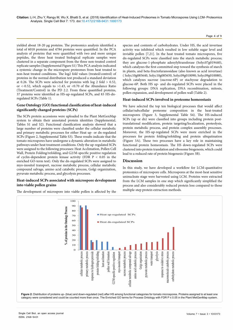
Gene Ontology (GO) functional classification of heat-induced significantly changed proteins (SCPs) The SCPs protein accessions were uploaded to the Plant MetGenMap system to obtain their annotated protein identities (SupplementaL Tables S1 and S2). Functional classification analysis showed that a large number of proteins were classified under the cellular metabolic and primary metabolic processes for either Heat up- or dn-regulated SCPs (Figure 2, Supplemental Table S3). These results indicate that the tomato microspores have undergone a dynamic alteration in metabolic pathways under heat treatment conditions. Only the up-regulated SCPs were assigned to the following processes: Heat Acclimation, Pollen Cell Wall, Protein Folding/refolding, and G2/M-specific positive regulation of cyclin-dependent protein kinase activity (FDR P < 0.05 in the enriched GO-term test). Only the dn-regulated SCPs were assigned to myo-inositol transport, sucrose metabolic process, cellular metabolic compound salvage, amino acid catabolic process, Golgi organization, pyruvate metabolic process, and glycolysis processes.
Heat-induced SCPs associated with microspore development into viable pollen grains
The development of microspore into viable pollen is affected by the
species and contents of carbohydrates. Under HS, the acid invertase activity was inhibited which resulted in low soluble sugar level and inviable pollen [7,21]. In the heat treated tomato microspores, five dn-regulated SCPs were classified into the starch metabolic process; they are glucose-1-phosphate adenylyltransferase (Solyc07g019440), which catalyzes the first committed step toward the synthesis of starch in plants, and beta-fructofuranosidase (also known as acid invertase) ( Solyc10g085640, Solyc10g085650, Solyc09g010090; Solyc09g010080), which catalyzes sucrose (sucrose-6P) or stachyose degradation to glucose-6P. Both HS up- and dn-regulated SCPs were placed in the following groups: DNA replication, DNA recombination, mitosis, pollen expansion, and development of pollen wall (Table 2).
Heat-induced SCPs involved in proteome homeostasis
We have selected the top ten biological processes that would affect cellular/subcellular proteome composition in the heat-treated microspores (Figure 3, Supplemental Table S4). The HS-induced SCPs (up or dn) were classified into groups including protein post-translational modification, protein targeting/localization, proteolysis, protein metabolic process, and protein complex assembly processes. Moreover, the HS-up-regulated SCPs were more enriched in the processes for protein folding/refolding and protein ubiqutination (Figure 3A). These two processes have a key role in maintaining functional protein homeostasis. The HS down-regulated SCPs were clustered into protein translation and ribosome biogenesis, which could lead to a reduced rate of protein biogenesis (Figure 3B).
Discussion In this study, we have developed a workflow for LCM-quantitative proteomics of microspore cells. Microspores at the most-heat sensitive uninucleate stage were harvested using LCM. Proteins were extracted from the LCM samples in one step which significantly simplified the process and also considerably reduced protein loss compared to those multiple-step protein extraction methods.
Figure 2: Distribution of proteins up- (blue) and down-regulated (red) after HS among functional categories for tomato microspores. Proteins assigned to at least one category were considered and could be counted more than once. The Enriched GO terms for Process Ontology with FDR P ≤ 0.05 in the Plant MetGenMap system.
0
10
20
30
40
50
60
70
80
90
100
cellul
ar me
taboli
c proc
esspri
mary
metab
olic p
rocess
respo
nse to
hydro
gen pe
roxide
protei
n fold
ing/re
foldin
ghea
t accli
matio
npo
llen w
all fo
rmati
onG2
/M-sp
ecific
positi
ve reg
ulatio
nmy
o-ino
sitol tr
anspo
rtsuc
rose m
etabo
lic pr
ocess
cellul
ar me
taboli
c com
poun
d salv
ageam
ino ac
id cat
abolic
proce
ssGo
lgi or
ganiza
tion
pyruv
ate m
etabo
lic pr
ocess
water
transp
ortgly
colysi
sres
ponse
to ox
idativ
e stre
ssma
cromo
lecule
catab
olic p
rocess
carbo
hydra
te meta
bolic
proce
ss
% of
protei
ns in
the O
ntolog
y Proc
ess
Heat up-regulated SCPs
Heat dn-regulated SCPs

Citation: Li H, Zhu Y, Rangu M, Wu X, Bhatti S, et al. (2018) Identification of Heat-Induced Proteomes in Tomato Microspores Using LCM- Proteomics Analysis. Single Cell Biol 7: 173. doi:10.4172/2168-9431.1000173
Page 5 of 9
Volume 7 • Issue 3 • 1000173Single Cell Biol, an open access journalISSN: 2168-9431
No. of proteins identfieda No. of proteins quantifiedb No. of HS-up-regulated SCPsC No. of HS-dn-regulated SCPsd
6018 4784 37 83aProteins identified using nano liquid chromatography and mass spectrometry analysis (LC-MS/MS). bProteins quantified using tandem mass tags (TMT) reporter ion intensity of the labled tryptic peptides. c, dProteins classified as Significantly Changed Proteins (SCPs) which have met the following criteria: ratio value (treated/control) higher than 2.0 standard deviation (± 2 SD) derived from the normal distribution test, and proteins quantified with two or more unique peptides having a P ≤0.05 of ratio values (treated/control) using three replicates from each of the two conditions.
Table 1: Profile of proteins identified in heat-treated tomato microscopres.
Studies have shown that pollen and the surrounding anther tissues respond to heat stress at the transcriptome, proteome, and metabolome levels, similar to other plant cell types [13]. This proteomics study indicated that protein translation was reduced in tomato microspores under heat treatment conditions. Global down-regulation of de-novo protein biosynthesis was suggested to be an effective way to reduce protein flux into the folding machinery in the endoplasmic reticulum (ER) lumen as an effective mean to reduce ER stress [22,23]. The
reduction of protein biosynthesis may have a similar role in the heat treated tomato microspores.
At the same time, selective mRNA translation becomes pivotal to maintain proper cell growth and protein homeostasis. In the tomato microspores, ten of the 35 HS-up-regulated SCPs are in the heat acclimation process. These tomato proteins include Hsp20, (Solyc07g045610, Solyc06g076540, Solyc06g076520, Solyc09g059210,
Figure 3: The enriched ontology processes affecting protein homeostasis in tomato microspores. Only the top 10 groups were selected. (3A) Heat-up-regulated SCPs, (3B) Heat-dn-regulated SCPs.
post-translational
protein modification
12%
proteasomal protein
catabolic process
7%
protein complex assembly
8%
protein folding/refoldi
ng9%protein
localization15%
protein metabolic process
23%
protein modification
by small protein
conjugation9%
protein ubiquitination
9%proteolysis
8%
post-translational
protein modification
13%protein amino
acid phosphorylati
on8%
protein complex assembly
6%protein
localization17%
protein metabolic process
31%
proteolysis10%
ribosome biogenesis
8%
translation7%
3A
3B

Citation: Li H, Zhu Y, Rangu M, Wu X, Bhatti S, et al. (2018) Identification of Heat-Induced Proteomes in Tomato Microspores Using LCM- Proteomics Analysis. Single Cell Biol 7: 173. doi:10.4172/2168-9431.1000173
Page 6 of 9
Volume 7 • Issue 3 • 1000173Single Cell Biol, an open access journalISSN: 2168-9431
Ontology Processa Protein Accessionb Ratioc P-valued Protein Descriptione
DNA replication
Solyc06g062690 1.4 0 Nucleosome assembly protein 1-like protein 4
Solyc03g044350 1.8 0 Chaperone dnaJ 3
Solyc07g064970 1.5 0.01 Microtubule-associated protein MAP65-1a
Solyc11g065190 1.5 0.02 Ubiquitin-conjugating enzyme E2 H10
Solyc05g050700 1.5 0.01 LRR receptor-like serine/threonine-protein kinase, RLP
Solyc03g112710 1.6 0.03 SKP1-like 1Solyc10g055610 1.5 0.04 SKP1-like 1
Regulation of DNA metabolic process
Solyc01g087560 0.7 0.03 24-sterol C-methyltransferase
Solyc04g071690 0.6 0.02 Ribonuclease P protein subunit p25
Solyc06g073530 0.7 0.03 Argonaute 4-like protein
Solyc03g097450 0.6 0.03 SWI/SNF complex subunit SMARCC1
Solyc07g049500 0.7 0.03 Argonaute 4-like protein
Chromosome organization
Solyc08g006790 0.6 0.04 Early nodulin-55-1Solyc03g119880 0.7 0.02 Early nodulin-55-1
Solyc03g097450 0.6 0.03 SWI/SNF complex subunit SMARCC1
Solyc07g049500 0.7 0.03 Argonaute 4-like protein
Solyc11g062220 0.7 0.04 Novel protein containing a PHD-finger domain
Solyc05g013740 0.7 0.01 Coilin
Solyc03g095720 0.7 0.01 Transcription elongation factor, TFIIS
Solyc12g010000 0.4 0.03 Early nodulin-55-1
Solyc10g005280 0.6 0.02 Pentatricopeptide repeat-containing protein
Solyc06g073530 0.7 0.03 Argonaute 4-like protein
G2/M transition of mitotic cell cycle
Solyc03g117630 1.6 0.01 Heat shock protein 70
Solyc04g011440 1.4 0.01 Heat shock protein 70
Anaphase
Solyc11g065190 1.5 0.02 Ubiquitin-conjugating enzyme E2 H10
Solyc10g055610 1.5 0.04 SKP1-like 1
Solyc07g064970 1.5 0.01 Microtubule-associated protein MAP65-1a
Solyc05g050700 1.5 0.01 LRR receptor-like serine/threonine-protein kinase, RLP
Interphase of mitotic cell cycle
Solyc10g055610 1.5 0.04 SKP1-like 1Solyc04g011440 1.4 0.01 Heat shock protein 70
Solyc06g062690 1.4 0 Nucleosome assembly protein 1-like protein 4
Solyc03g117630 1.6 0.01 Heat shock protein 70
M phase
Solyc11g065190 1.6 0.02 Ubiquitin-conjugating enzyme E2 H10
Solyc05g050700 1.5 0.01 LRR receptor-like serine/threonine-protein kinase, RLP
Solyc03g112710 1.6 0.03 E3 ubiquitin ligase, SCF complex, Skp subunit
Solyc07g064970 1.5 0.01 Microtubule-associated protein MAP65-1a
Solyc10g055610 1.6 0.04 E3 ubiquitin ligase, SCF complex, Skp subunit
Solyc03g117630 1.6 0.01 Heat shock protein 70Solyc04g011440 1.4 0.01 Heat shock protein 70Solyc04g015270 1.5 0.02 Glycosyltransferase

Citation: Li H, Zhu Y, Rangu M, Wu X, Bhatti S, et al. (2018) Identification of Heat-Induced Proteomes in Tomato Microspores Using LCM- Proteomics Analysis. Single Cell Biol 7: 173. doi:10.4172/2168-9431.1000173
Page 7 of 9
Volume 7 • Issue 3 • 1000173Single Cell Biol, an open access journalISSN: 2168-9431
Mitosis
Solyc11g065190 1.6 0.02 Ubiquitin-conjugating enzyme E2 H10
Solyc07g064970 1.5 0.01 Microtubule-associated protein MAP65-1a
Solyc10g055610 1.6 0.04 E3 ubiquitin ligase, SCF complex, Skp subunit
Solyc03g112710 1.6 0.03 E3 ubiquitin ligase, SCF complex, Skp subunit
Solyc04g051350 0.7 0.01 Ribonucleoside-diphosphate reductase
Solyc11g062220 0.7 0.04 Novel protein containing a PHD-finger domain
Solyc02g068480 0.7 0 Ubiquitin carboxyl-terminal hydrolase
Spindle organization
Solyc07g064970, 1.5 0.01 Microtubule-associated protein MAP65-1a
Solyc08g006790 0.6 0.01 Early nodulin-55-1Solyc07g049500 0.7 0.03 Argonaute 4-like proteinSolyc06g073530 0.7 0.03 Argonaute 4-like protein
Pollen development
Solyc11g065190 1.5 0.02 conjugating enzyme E2 H10
Solyc01g090600 1.6 0.02 Chalcone synthase 3 protein (pollen wall formation)
Solyc07g065770 1.6 0.03 ABC transporter G family member 26
Solyc06g082500 1.5 0 Solute carrier family 35 member E3
Solyc04g008780 1.6 0.02 Dihydroflavonol 4-reductase (wall formation)
Solyc01g010900 1.8 0.01 Cytochrome P450 (pollen wall formation)
Solyc03g051960 1.5 0 Fatty acyl coA reductase/Male sterility
Solyc05g050700 1.5 0.01 LRR receptor-like serine/threonine-protein kinase, RLP
Pollen wall formation
Solyc03g051960 1.5 0 Fatty acyl coA reductase/Male sterility
Solyc01g010900 1.8 0.01 Cytochrome P450Solyc04g008780 1.6 0.02 Dihydroflavonol 4-reductase
Solyc07g065770 1.5 0.01 LRR receptor-like serine/threonine-protein kinase, RLP
Solyc06g082500 1.5 0 Solute carrier family 35 member E3
Solyc01g090600 1.6 0.02 Chalcone synthase 3 protein aEnriched GO of Ontology Process classified in the Plant MetGen Map system.bUnigenes accession number in annotated tomato genome databases in Plant MetGen Map system.cRatio of protein abundance from heat treated to non-treated tissues estimated based on reporter ion intensity values in the Proteome Discoverer 2.2 (PD 2.2, Thermo) report. dP value of the protein ratio between heat treated and non-treated groups (using three biological replicates each) in the Proteome Discoverer 2.2 (PD 2.2, Thermo) report. eAnnotated protein in the Plant MetGenMap system.
Table 2: Heat induced significantly changed proteins associated with pollen development.
1.8- 2.2-fold, p<0.05), DnaJ (Solyc03g044350), Hsp70 (Solyc03g117630, Solyc04g011440, 1.4-1.6-fold, p<0.05), Hsp90 (Solyc06g036290, 1.5-fold, p=0.01), and ClpB chaperone (Solyc03g115230, 1.4-fold, p=0.0001). These tomato HSPs are comprised of the cognate HSPs including HSP 70 (Hsc 70) [24,25] and the heat-inducible ClpB chaperone class [26,27]. These HSPs serve as chaperones for protein folding and refolding and thus have a critical role in maintaining proteome homeostasis throughout microspore and pollen development [13]. Therefore, these tomato HSPs, in particular the heat-inducible ClpB may have a key role for microspores to acquire heat tolerance and develop into viable pollen.
Heat stress affects composition in the primary and secondary metabolites (carbohydrates, lipids, proline, polyamines, glutathione,
and flavonoids) [14,28]. A decrease in pollen viability is often associated with an alteration of metabolite contents such as soluble sugars and starch [5,29]. Sucrose produced in photosynthetic tissues is transported to anther, where it is converted to glucose and fructose under the action of acid invertase. Heat stress was shown to inhibit the acid invertase activity, which is in part responsible for decreased levels of soluble sugars in tomato anthers [7]. Tomato RNAi-lines with silencing of the Lin5 gene, encoding for a homologous enzyme, showed pollen malformation and abortion [30]. In this study, we have identified several acid invertase (β-D-fructofuranosidase) isoenzymes among the HS-dn-regulated SCPs (for instance, Solyc10g085640, Solyc09g010080, 0.36-fold, p<0.01). We have also identified a fatty acyl-CoA reductase (FAR), which was annotated as a homolog to Male Sterility 2 (MS2). The FAR affects sporopollenin composition and reduced FAR activity leads

Citation: Li H, Zhu Y, Rangu M, Wu X, Bhatti S, et al. (2018) Identification of Heat-Induced Proteomes in Tomato Microspores Using LCM- Proteomics Analysis. Single Cell Biol 7: 173. doi:10.4172/2168-9431.1000173
Page 8 of 9
Volume 7 • Issue 3 • 1000173Single Cell Biol, an open access journalISSN: 2168-9431
to infertile pollen [31]. The identification of these SCPs supports the role of carbohydrate and lipid metabolites in pollen thermo-tolerance.
Heat also affects metabolism of secondary metabolites in many plant organs including pollens. A recent study using LC-MS platform showed that a short heat stress at 38°C leads to alteration of the secondary compound in tomato pollen, such as accumulation of flavonoids in the microspore [28]. Chalcone synthase catalyzes the initial step of flavonoid biosynthesis through the phenylpropanoid pathway. A lack of chalcone synthase activity leads to disruption of flavonoid synthesis and loss of pollen fertility [32]. The chalcone synthase 3 protein was induced in the heat treated tomato microspore (Solyc01g090600, 1.6-fold, p=0.02). The dihydroflavonol 4-reductase (1.6-fold, p=0.02) and fatty acyl CoA reductase (1.5-fold, p=0.02) also affect pollen wall formation [31,33]. These HS-upregulated SCPs may have a critical role in determining the fate of microspores under the stress conditions.
The uninucleate stage microspores undergo mitosis to develop into binucleate or trinucleate pollen grains. Several heat induced tomato SCPs are involved in repair of DNA damages (which occurs frequently under heat and other stresses), DNA replication, the mitotic cycles, and microspore cell wall loosening to allow pollen expansion after microspore mitosis [34]. These processes contained both HS up-regulated or dn-regulated SCPs. Future work will focus on functional analysis of these SCPs in thermo-tolerance of the developing pollen.
ConclusionThis study has developed a LCM quantitative proteomics workflow which was used to identify cell-specific pollen proteomes associated with heat stress. Using the LCM, six replicate samples each containing approximately 60,000 tomato microspore cells were harvested. A one-step protein extraction procedure was developed where the LCM–harvested pollens were directly subjected to the high pressure/vacuum extraction cycles. By eliminating tissue grinding and multiple steps of protein purification/precipitation, this protein extraction procedure has proven very effective in working with small amount of tissue such as in the case of LCM collected cells. The TMT-proteomics analysis of the LCM collected microspore proteins has led to the quantification of over 4000 proteins, and the identification of HS-induced SCPs that are involved in heat acclimation, proteome homeostasis, pollen coat formation, mitosis and other biological processes affecting pollen development under HS conditions.
Ethics Approval and Consent to Participate
Not applicable.
Competing Interests
The authors declare that they have no competing interests.
Funding
This project was supported by the Agriculture and Food Research Exploratory Award accession number 1009172, from the USDA National Institute of Food, and Agriculture, the USDA Evans-Allen Research Funds and ARS CRIS Projects 1907-21000-036/037-00D. ARS disclaimer: “Mention of trade names or commercial products in this publication is solely for the purpose of providing specific information and does not imply recommendation or endorsement by the U.S. Department of Agriculture.”
Author’s Contributions
Tomato plant treatments, LCM, protein preparation, TMT labeling, anther, and pollen microscopy, were performed by YZ, HL, XBW, MR, and SZ. TF assisted in the protein extraction, protein quantifications and labeling experiments as well as being responsible for various analytical experiments to guarantee the quality of the analysis. YY carried out all of the MS analysis, evaluated the data, and compiled the experimental results. SZ, SB, and HL prepared the manuscript. TWT and SZ
developed the experimental design. Furthermore, TWT provided critical oversight for all the MS analysis, and contributed significantly to the drafting and revising of the manuscript. All authors read and approved the final manuscript.
Acknowledgements
The authors wish to thank Johanna M. Dela Cruz, and Carol J. Bayles of the Imaging Facility, Sheng Zhang of the Proteomics and Mass Spectrometry Facility, of the Cornell University Institute of Biotechnology for expert technical assistance and helpful discussions.
References
1. United States Department of Agriculture (2016) Economic Research Service Tomato.
2. Geisenberg C, Stewart K (1986) Field crop management the tomato crop. The Tomato Crop (A scientific basis for improvement). Springer Dordrecht pp: 511-557.
3. Peet MM, Bartholomew M (1996) Effect of night temperature on pollen characteristics, growth, and fruit set in tomato (Lycopersicon esculentum Mill). J Amer Soc Hort Sci 121: 514-519.
4. Sakata T, Takahashi H, Nishiyama I, Higashitani A (2000) Effects of high temperature on the development of pollen mother cells and microspores in barley Hordeum vulgare. L J Plant Res 113: 395–402.
5. Pressman E, Peet MM, Pharr DM (2002) The effect of heat stress on tomato pollen characteristics is associated with changes in carbohydrate concentration in the developing anthers. Ann Bot 90: 631–636.
6. Sato S, Peet MM, Thomas JF (2002) Determining critical pre- and post-anthesis periods and physiological processes in Lycopersicon esculentum mill exposed to moderately elevated temperatures. J Exp Bot 53: 1187–1195.
7. Sato S, Kamiyama M, Iwata T, Makita N, Furukawa H, et al. (2006) Moderate increase of mean daily temperature adversely affects fruit set of Lycopersicon esculentum by disrupting specific physiological processes in male reproductive development. Ann Bot 97:731–738.
8. Echlin P, Godwin H (1968) The ultrastructure and ontogeny of pollen in Helleborus foetidus L II Pollen grain development through the callose special wall stage. J Cell Sci 3: 175–186.
9. Chaturvedi P, Ghatak A, Weckwerth W (2016) Pollen proteomics: from stress physiology to developmental priming. Plant Reprod. 29: 119–132.
10. Chaturvedi P, Ischebeck T, Egelhofer V, Lichtscheidl I, Weckwerth W, et al. (2013) Cell-specific analysis of the tomato pollen proteome from pollen mother cell to mature pollen provides evidence for developmental priming. J Proteome Res 12: 4892–4903.
11. Chaturvedi P, Doerfler H, Jegadeesan S, Ghatak A, Castillejo MA, et al. (2015) Heat-treatment-responsive proteins in different developmental stages of tomato pollen detected by targeted mass accuracy precursor alignment (tMAPA). J Proteome Res 14: 4463–4471.
12. Ischebeck T, Valledor L, Lyon D, Gingl S, Nagler M, et al. (2014) Comprehensive cell-specific protein analysis in early and late pollen development from diploid microsporocytes to pollen tube growth. Mol Cell Proteomics 13: 295–310.
13. Rieu I, Twell D, Firon N (2017) Pollen development at high temperature: from acclimation to collapse. Plant Physiol 173: 1967–1976.
14. Paupière MJ, van Heusden AW, Bovy AG (2014) The metabolic basis of pollen thermo-tolerance: Perspectives for breeding. Metabolites 4: 889–920.
15. Keller M, Consortium S, Simm S (2018) The coupling of transcriptome and proteome adaptation during development and heat stress response of tomato pollen. BMC Genomics pp: 1–20.
16. Santos MR, Bispo C, Becker JD (2017) Isolation of Arabidopsis pollen, sperm cells, and vegetative nuclei by fluorescence-activated cell sorting (FACS). In: Plant Germline Development: Methods and Protocols. Springer New York, USA pp: 193–210.
17. Bokszczanin KL, Fragkostefanakis S (2013) Solanaceae Pollen Thermotolerance Initial Training Network (SPOT-ITN) Consortium, Perspectives on deciphering mechanisms underlying plant heat stress response and thermotolerance. Front Plant Sci 4: 1–20.
18. Zhu Y, Li H, Bhatti S, Zhou S, Yong Y (2016) Development of a laser capture microscope-based single-cell-type proteomics tool for studying proteomes of individual cell layers of plant roots. Hort Res 3: 16026.
Citation: Li H, Zhu Y, Rangu M, Wu X, Bhatti S, et al. (2018) Identification of Heat-Induced Proteomes in Tomato Microspores Using LCM- Proteomics Analysis. Single Cell Biol 7: 173. doi:10.4172/2168-9431.1000173
Page 9 of 9
Volume 7 • Issue 3 • 1000173Single Cell Biol, an open access journalISSN: 2168-9431
19. Zhou S, Okekeogbu I, Sangireddy S, Ye Z, Li H, et al. (2016) Proteome modification in tomato plants upon long-term aluminum treatment. J Proteome Res 15: 1670–1684.
20. Joung JG, Corbett AM, Fellman SM, Tieman DM, Klee HJ, et al. (2009) Plant metgenmap: an integrative analysis system for plant systems biology. Plant Physiol 151: 1758–1768.
21. Li Z, Palmer WM, Martin AP, Wang R, Rainsford F, et al. (2012) High invertase activity in tomato reproductive organs correlates with enhanced sucrose import into, and heat tolerance of, young fruit. J Exp Bot 63: 1155–1166.
22. Reid DW, Chen Q, Tay AS, Shenolikar S, Nicchitta CV, et al. (2014) The unfolded protein response triggers selective mRNA release from the endoplasmic reticulum. Cell 158: 1362–1374.
23. Ron D (2002) Translational control in the endoplasmic reticulum stress response. J Clin Invest 110: 1383–1388.
24. Craig EA (1985) The heat shock response. Critical Reviews in Biochemistry 18: 239-280.
25. Fragkostefanakis S, Mesihovic A, Simm S, Paupière M J, Hu Y, et al. (2016) HsfA2 controls the activity of developmentally and stress-regulated heat stress protection mechanisms in tomato male reproductive tissues. Plant Physiol 170: 2461–2477.
26. Duran EC, Weaver CL, Lucius AL (2017) Comparative analysis of the structure and function of AAA+ motors ClpA, ClpB, and Hsp104: Common threads and disparate functions. Front Mol Biosci 4: 54.
27. Lee S, Sowa ME, Watanabe YH, Sigler PB, Chiu W, et al. (2003) The structure
of ClpB: A molecular chaperone that rescues proteins from an aggregated state. Cell 115: 229–240.
28. Paupière MJ, Müller F, Li H, Rieu I, Tikunov YM, et al. (2017) Untargeted metabolomic analysis of tomato pollen development and heat stress response. Plant Reprod 30: 81–94.
29. Firon N, Shaked R, Peet MM, Pharr DM, Zamski E, et al. (2006) Pollen grains of heat tolerant tomato cultivars retain higher carbohydrate concentration under heat stress conditions. Scientia Horti 109: 212–217.
30. Zanor MI, Osorio S, Nunes-Nesi A, Carrari F, Lohse M, et al. (2009) RNA interference of LIN5 in tomato confirms its role in controlling Brix content, uncovers the influence of sugars on the levels of fruit hormones, and demonstrates the importance of sucrose cleavage for normal fruit development and fertility. Plant Physiol 150: 1204–1218.
31. de Azevedo Souza C, Kim SS, Koch S, Kienow L, Schneider K, et al. (2009) A novel fatty acyl-CoA synthetase is required for pollen development and sporopollenin biosynthesis in Arabidopsis. Plant Cell Online 21: 507–525.
32. Mo Y, Nagel C, Taylor LP (1992) Biochemical complementation of chalcone synthase mutants defines a role for flavonols in functional pollen. Proc Natl Acad Sci USA 89: 7213–7217.
33. Tang LK, Chu H, Yip WK, Yeung EC, Lo C (2009) An anther-specific dihydroflavonol 4-reductase-like gene (DRL1) is essential for male fertility in Arabidopsis. New Phytologist 181: 576–587.
34. Hrubá P, Honys D, Twell D, Čapková V, Tupý J, et al. (2005) Expression of β-galactosidase and β-xylosidase genes during microspore and pollen development. Planta 220: 931–940.