gain of function of mutant p53: the mutant p53/nf-y ... · gain of function of mutant p53: the...
TRANSCRIPT

A R T I C L E
Gain of function of mutant p53: The mutant p53/NF-Y proteincomplex reveals an aberrant transcriptional mechanism of cellcycle regulation
Silvia Di Agostino,1 Sabrina Strano,1 Velia Emiliozzi,1 Valentina Zerbini,3 Marcella Mottolese,3 Ada Sacchi,1
Giovanni Blandino,1,2,* and Giulia Piaggio1,2
1 Experimental Oncology Department, Istituto Regina Elena, Via delle Messi D’Oro 156, 00158 Rome, Italy2 Rome Oncogenomic Center (ROC), Istituto Regina Elena, Via delle Messi D’Oro 156, 00158 Rome, Italy3 Pathological Anatomy Department, Istituto Regina Elena, Via Elio Chianesi 53, 00128 Rome, Italy*Correspondence: [email protected]
Summary
This article investigates the mechanistic aspects of mutant p53 ‘‘gain of function’’ in response to DNA damage. We show thatmutant forms of p53 protein interact with NF-Y. The expression of cyclin A, cyclin B1, cdk1, and cdc25C, as well as the cdk1-associated kinase activities, is upregulated after DNA damage, provoking a mutant p53/NF-Y-dependent increase in DNAsynthesis. Mutant p53 binds NF-Y target promoters and, upon DNA damage, recruits p300, leading to histone acetylation.The recruitment of mutant p53 to the CCAAT sites is severely impaired upon abrogation of NF-YA expression. EndogenousNF-Y, mutant p53, and p300 proteins form a triple complex upon DNA damage. We demonstrate that aberrant transcriptionalregulation underlies the ability of mutant p53 proteins to act as oncogenic factors.
Introduction
The human p53 gene is mutated in about 50% of human cancers(Beroud and Soussi, 1998; Hainaut and Hollstein, 2000). The p53protein contains three functional domains: an N-terminal trans-activation domain (TAD), a central DNA binding core domain(DBD), and a C-terminal oligomerization domain (OLD) (Levine,1997). The integrity of these domains is required for p53 activi-ties such as cell cycle arrest, apoptosis, senescence, and differ-entiation (Harris and Levine, 2005). Unlike other tumor suppres-sor genes, whose inactivation occurs mainly by deletions, p53 isa frequent target of missense mutations that mainly reside in theregion coding for the DNA binding domain. The resulting pro-teins, whose half-life is greatly prolonged, are full-length witha single amino acid change and are unable to recognize wild-type p53 (wtp53) consensus DNA binding sites (Prives andHall, 1999). Most of the p53 mutations can be ascribed to twomain classes: DNA contact-defective and conformational mu-tants (Levine, 1997; Prives and Hall, 1999). The biological signif-icance of p53 mutations ranges from the mere loss of function togain of function. Many in vitro and in vivo studies have clearlyshown that some p53 mutants can acquire new functions,thereby contributing actively to the maintenance, the spreading,
CANCER CELL 10, 191–202, SEPTEMBER 2006 ª2006 ELSEVIER INC. D
and the increased resistance to conventional anticancer treat-ments of mutant p53 tumors (Dittmer et al., 1993; Blandinoet al., 1999; Sigal and Rotter, 2000). At the molecular level,gain-of-function mutant p53 proteins can exert their activities ei-ther through the binding, the sequestration, and the inactivationof tumor suppressor proteins or through the transcriptionalmodulation of target genes (Di Como et al., 2002; Strano et al.,2000, 2002; Gaiddon et al., 2001).
wtp53 has been shown to interact with a number of transcrip-tion factors (E2F1, SP1, NF-Y, YY1, TBP, TAFs), giving rise tomacromolecular complexes that modulate the transcription ofgenes whose promoters lack p53 binding sites. These pro-moters are often inhibited by wtp53 in specific phases of thecell cycle after DNA damage (Lu and Levine, 1995; Farmeret al., 1996; Manni et al., 2001; St Clair et al., 2004; Imbrianoet al., 2005).
NF-Y is a heterotrimeric transcription factor with a high bind-ing affinity for the CCAAT consensus motif that is present in 30%of eukaryotic promoters, and it consists of three subunits, NF-YA, -B, and -C, all required for CCAAT binding (Mantovani,1998). NF-Y modulates, at least in part, the activity of the CCAATbox-containing promoters of the E2F1, cyclin A, cyclin B1, cyclinB2, cdk1, cdc25C, chk2, topo IIa, cdc25C, and MDR-1 genes
S I G N I F I C A N C E
Approximately half of all human tumors express mutant p53 proteins. Our study demonstrates the oncogenic cooperation of two majorregulators of the cell cycle, mutant p53 and NF-Y, in proliferating cells and upon DNA damage. This cooperation allows cells to overridecellular failsafe programs, thus permitting tumor progression. We provide evidence supporting the hypothesis that mutant p53, throughits ability to interact with a variety of transcription factors, takes part in the control of key regulatory activities during the cell cycle. Theidentification of specific molecular mechanisms through which mutant p53 exerts its ‘‘gain-of-function’’ activities provides the oppor-tunity to target transcriptionally competent mutant p53 complexes in cancer cells.
OI 10.1016/j.ccr.2006.08.013 191

A R T I C L E
Figure 1. The protein complex mutant p53/NF-Y upregulates cell cycle NF-Y target genes in response to DNA damage
A: Cellular extracts (1 mg) from SKBR3, HT29, and SW480 cells, treated or not with 0.5 mg/ml ADR for 48 hr, were immunoprecipitated with ap53 or sheep serum ascontrol. Twenty micrograms of SKBR3, 40 mg of HT29, and 40 mg of SW480 cellular extracts and half of the related immunoprecipitates were subject to Westernblotting using ap53, NF-YA, and NF-YB antibodies.B: List of some cell cycle genes regulated by NF-Y.C: MCF10A, SKBR3, HT29, and SW480 cells were incubated with 0.5 mg/ml of ADR for 24 and 48 hr. Eight micrograms per sample (MCF10A) and 40 mg/sample(SKBR3, HT29, and SW480) of protein extracts were subjected to Western blotting using ap53, cyclin A, cyclin B1, cdk1, cdc25C, and tubulin antibodies.D: SKBR3, MDA-MB468, and T47D breast cancer cell lines were incubated with 0.5 mg/ml of ADR for 1, 6, 18, and 24 hr. Cellular extracts (40 mg/sample) weresubject to Western blotting using the same antibodies described in C.E: The same experiment described in C and D was performed on H1299 and SAOS p53 null cell lines.
during the different phases of the cell cycle and in response toDNA damage (Huet et al., 1996; Van Ginkel et al., 1997; Bolo-gnese et al., 1999; Farina et al., 1999; Yun et al., 1999; Adachiet al., 2000; Hu et al., 2000). Interestingly, the wtp53-dependenttranscriptional inhibition of these genes upon DNA damage re-quires the integrity of the CCAAT boxes and a functional NF-Ycomplex (Manni et al., 2001; St. Clair et al.,2004; Imbrianoet al., 2005).
Here, we show that diverse mutant p53 proteins interactin vivo with NF-Y. The mutant p53/NF-Y protein complex(mutp53/NF-Y) regulates, at the transcriptional level, NF-Y tar-get genes involved in cell cycle control. Indeed, the expressionof the cyclin A, cyclin B1, cyclin B2, cdk1, and cdc25c genes isupregulated in response to DNA damage in cells harboring en-dogenous mutant p53 proteins. This upregulation requires thepresence within the target promoters of an intact CCAAT box.The functional consequence of this upregulation is increasedDNA synthesis, which is strictly dependent on the existence ofthe protein complex mutp53/NF-Y. We further show the exis-tence of a transcriptional competent complex includingmutp53, NF-Y, and p300 on the promoters of NF-Y cell cycle tar-get genes. The recruitment of p300 is severely impaired in cellswhose endogenous expression of mutant p53 was specificallyknocked down. Of note, the recruitment of mutant p53 to the
192
CCAAT sites of NF-Y target promoters is severely impairedupon abrogation of NF-YA expression. Our observations reveala crosstalk between mutant forms of p53 protein and NF-Y thattakes part in the aberrant regulation of the cell cycle in responseto DNA-damaging agents.
Results
Mutant p53 proteins interact in vivo with NF-YTo investigate whether mutant forms of p53 protein and NF-Y in-teract in vivo, we performed coimmunoprecipitation experi-ments, employing cell extracts derived from tumor cells(SKBR3, HT29, and SW480) harboring endogenous mutantp53His175, p53His273, and p53His273/Ser309 proteins, re-spectively. As shown in Figure 1A (lanes 5 and 6), we foundthe presence of protein complexes between mutant p53 andNF-YA and NF-YB. Immunostaining analysis revealed that mu-tant p53 and NF-YA localize in the nucleus of SKBR3 cells.RNAi knockdown of mutant p53His175 did not affect the local-ization of NF-YA (Figure S1A in the Supplemental Data availablewith this article online). Adriamycin (ADR) treatment does not im-pact on the endogenous levels of mutant p53 and NF-Y, nor onthe formation of the complex (Figure 1A, lanes 1–6). Reciprocalprotein complexes involving mutant p53, NF-YA, and NF-YB
CANCER CELL SEPTEMBER 2006

A R T I C L E
Figure 2. Expression of mutant p53 is necessary for transcriptional activation of NF-Y target genes
A: SKBR3, SW480, shp53-SKBR3, and shp53-SW480 cells were treated with 0.5 mg/ml of ADR for 48 hr. Forty micrograms per sample were subjected to Westernblot analysis using ap53, cyclin A, cyclin B1, cdk1, cdc25C, and tubulin antibodies.B: SKBR3 cells transiently transfected with siLacZ or sip53 oligonucleotides (see Experimental Procedures) were treated and assessed as in A (left panel). SKBR3transfected with shscramblep53 vector or pSuper-altered control p53 was analyzed as described above (right panel).C: SKBR3 and shp53-SKBR3 were treated as in A. Cell extracts (1 mg) were immunoprecipitated with mouse preimmune serum or 50 ml of a specific monoclonalaNF-YA serum. Western blotting was performed with 20 mg/sample of total extracts, and 500 mg of the related immunoprecipitates were probed with sheepap53 serum and rabbit aNF-YA antibody.D: Cell extracts (1 mg) of ADR-treated MCF10A, SKBR3, and shp53-SKBR3 cells were immunoprecipitated with polyclonal acyclin A or cyclin B1 antibodies andpreimmune sera as control. Left panel shows 20 mg of cellular extracts subjected to Western blotting with acdk1 antibody. Immunoprecipitated kinase activitywas assessed using 1 mg of H1 histone and 10 mM 32P-g-ATP as substrates. One of three separate experiments is shown (right panel).E: RT-PCR amplification of cyclin A, cyclin B1, cdk1, cdc25C, and aldolase on total cellular RNA extracted from the cell lines indicated in the figure.
were found in cells whose exogenous expression of mutantp53His175 (H-175#41) or wtp53 (H-wtp53#23) was under thecontrol of ponasterone A (Figure S1B) (Strano et al., 2000). Noprotein complexes were detected in cell lysates immunoprecip-itated with preimmune serum (Figure 1A, lanes 3 and 4;Figure S1B, lanes 8–10). Taken together, these results demon-strate the existence of complexes containing mutant p53 andNF-Y proteins under physiological conditions. Since NF-YBand NF-YC heterodimerization is a prerequisite for NF-YA asso-ciation, our results further indicate that both mutant and wtp53proteins associate with a transcriptional competent NF-Y tri-meric complex.
Mutant p53 proteins upregulate NF-Y targetgenes after DNA damageA number of genes controlling different phases of the cell cycleare regulated at the transcriptional level by NF-Y (Figure 1B)(Mantovani, 1998; Elkon et al., 2003). It has been previously re-ported that the expression of some cell cycle-related NF-Y tar-get genes is inhibited by wtp53 in response to DNA damage(Manni et al., 2001; Imbriano et al., 2005). To verify whetherthe mutp53/NF-Y complex modulates the expression of such
CANCER CELL SEPTEMBER 2006
NF-Y target genes, we assessed the protein levels of cyclin A,cyclin B1, cdc25, and cdk1 in ADR-treated SKBR3, HT29,SW480, MDA-MB468, and T47D mutant p53 expressing cells(Figures 1C and 1D). We found that, irrespective of the type ofp53 mutation, cyclin A, cyclin B1, cdc25, and cdk1 protein levelswere upregulated in response to ADR treatment (Figures 1C and1D). This upregulation starts between 1 and 6 hr after the treat-ment (Figure 1D). Conversely, the expression of these geneswas downregulated in ADR-treated human primary breast epi-thelial cells (MCF10A) carrying endogenous wtp53 protein(Figure 1C). These findings agree with those showing thatADR-induced p53 accumulation in MCF10A cells is critical forthe cellular response to DNA damage (Kohn et al., 2002). Ofnote, we did not find any modulation of cyclin A, cyclin B1,cdc25, and cdk1 protein levels in p53 null cells, such as H1299and Saos-2 cells, in response to DNA damage (Figure 1E). Tofurther confirm the role of mutant p53 in the upregulation ofthe previously analyzed NF-Y target genes, we assessed theirexpression in SKBR3 and SW480 cells whose mutant p53 ex-pression had been knocked down (Figures 2A and 2B). Knock-down was achieved by stable transfection of a shp53 expressionplasmid (Figure 2A), or by p53-siRNA (small interfering RNA)
193
A R T I C L E
oligonucleotide transduction (Figure 2B). As shown in Figures 2Aand 2B, reduction of mutant p53 severely impaired the upregu-lation of cyclin A. cyclin B1, cdk1, and cdc25C in response toADR treatment. This was not observed in cells transducedwith control oligo (LacZ) or with plasmids expressing a scram-bled p53 shRNA or a triple point mutated p53 shRNA (Figure 2B;Zalcenstein et al., 2006). Altogether, our findings indicate thatthe status of p53 plays a role in the modulation of cell cycle-related NF-Y target genes in response to DNA damage.
Next, we investigated whether the increased protein levels ofcyclin B1, cyclin A, and cdk1 resulted in the activation of the cor-responding protein kinases. Indeed, the upregulation of cyclinB1 and cdk1 was accompanied by an upward mobility shift,which suggests phosphorylation and activation of these proteins(Figure 1C). As shown in Figure 2D (lanes 3–10, upper panel), ki-nase assays performed on the indicated immunoprecipitates re-vealed that both cyclin A/cdk1 and cyclin B1/cdk1 kinase activ-ities were upregulated after DNA damage in SKBR3 cells,indicating that the upward mobility shift of cdk1 is due to an ac-tivating phosphorylation. An intact mutp53/NF-Y protein com-plex (Figure 2C) is necessary for the kinase activity associatedwith cyclin B1 and cyclin A complexes, as shown by the lack ofsuch activity in cells whose mutant p53 expression had beenknocked down (Figure 2D, lane 3–10, lower panel). No increasein the activity of cyclin A/cdk1 or cyclin B1/cdk1 kinases was ob-served in MCF10A cells (Figure 2D, lanes 3–10, middle panel).
To study whether the regulation of NF-Y target genes occursat the transcriptional level, we assessed the behavior of theirmRNAs in response to ADR treatment. We found that cyclin A,cyclin B2, cdc2, and cdc25C mRNAs are already upregulatedbetween 1 and 6 hr after the addition of ADR (Figure 2D). Knock-down of mutant p53 expression severely impairs this upregula-tion (Figure 2C).
Altogether, these data indicate that mutant p53 is necessaryfor the upregulation of cell cycle-related NF-Y target gene ex-pression. Notably, the activity of important cyclin/cdk com-plexes involved in control of the G2/M phase of the cell cyclein response to DNA damage is strictly related to the presenceof mutant p53.
Transcriptional activation of NF-Y target promotersby mutant p53 proteins requires CCAAT box integrityTo further dissect the transcriptional control of mutant p53 onthe NF-Y target promoters, we performed transactivation as-says. First, we found that a ponasterone-inducible mutantp53His175 (H-175#41) (Strano et al., 2000) transactivates theexogenously expressed luciferase gene driven by a 1.1 kb frag-ment of the murine cyclin B2 promoter (pCCAAT-B2LUC), whichcontains three CCAAT boxes (Figure 3A). This activity was notrevealed in control cells treated with ponasterone A (Figure3A). Second, H1299 cells were transiently cotransfected withexpression plasmids encoding either mutant p53His175 orwtp53 together with the cyclin B2 promoter reporter employedin Figure 3A. We found that the activity of the cyclin B2 promoterwas upregulated in the presence of mutant p53His175 (Figure3B). In agreement with our previous findings, the activity of thecyclin B2 promoter was downregulated by wtp53 (Manni et al.,2001; Imbriano et al., 2005). These activities were lost in the con-text of a cyclin B2 promoter carrying three mutated CCAATboxes (pmutCCAAT-B2LUC) (Figure 3B). Similar results were
194
obtained using the cyclin A and cdc25C promoters (Figures3C and 3D).
To further evaluate the role of NF-Y in the mutant p53His175-dependent upregulation of NF-Y target gene expression, weused a dominant-negative NF-Y mutant (YA13 m29). This mu-tant bears a triple amino acid substitution in the NF-YA DNAbinding domain, which allows trimer formation but preventsthe binding to CCAAT boxes (Mantovani, 1998). As shown inFigure 3E, the exogenous expression of YA13m29 protein im-paired the opposing transcriptional effects of mutp53 His175and wtp53 on cyclin B2 promoter activity.
To investigate whether endogenous mutant p53His175 pro-tein regulates NF-Y target promoters in response to ADR treat-ment, we analyzed mutant p53 transcriptional activity in SKBR3cells harboring an exogenous cyclin B2 promoter (Figure 3F).We found that cyclin B2 promoter activity was enhanced bythe addition of ADR (Figure 3F). This activity requires the pres-ence of an active mutant p53 and the integrity of the CCAATboxes of cyclin B2 promoter (Figure 3F). Indeed, knockdownof mutant p53 expression abolished the transcriptional activa-tion of the cyclin B2 promoter upon ADR treatment (Figure 3F).In contrast, cyclin B2 promoter activity was decreased inwtp53-carrying MCF10A cells (Figure 3G).
To verify whether the transcriptional effects exerted by mutp53His175 on NF-Y target promoters were also recapitulated byother types of human tumor-derived p53 mutants, we performeda transactivation assay employing mutant p53His273 andp53Gly281 proteins (Figure 3H). To this end, H1299 cells werecotransfected with expression plasmids encoding the abovep53 mutants in combination with luciferase reporter plasmidsdriven by the cyclin B2 or cdc25c promoters. We found thatactivity of both promoters was enhanced by the concomitantexpression of mutant p53His273 and p53 Gly281 (Figure 3H).
To find out whether the transactivation domain of mutantp53His175 takes part in the transcriptional activation of NF-Ytarget promoters, we performed transactivation assays employ-ing the mutant p53His175/22,23, harboring point mutations intwo critical residues within the N-terminal transcriptional activa-tion domain. We found that this mutant bound to NF-YA as effi-ciently as mutant p53His175 (Figure S2A). Coexpression of thismutant with NF-YA resulted in the activation of the cyclin B2 pro-moter to an extent very similar to that seen with mutantp53His175/NF-Y protein complex (Figure S2B). Unlike themutp53/NF-Y complex, the wtp53/NF-Y protein complex re-quired an intact transactivation domain to induce transcriptionalrepression of NF-Y target genes (Figure S2B).
Altogether, these results demonstrate that the mutp53/NF-Yprotein complex is capable of transcriptionally regulating NF-Ytarget promoters in response to ADR. This activity requires thepresence of a transcriptionally competent NF-Y complex andthe integrity of the CCAAT boxes of the target promoters.
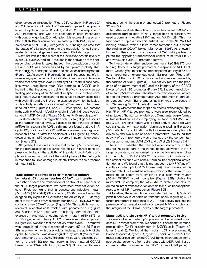
Mutant p53 protein binds NF-Y target promoters in vivoTo assess whether mutant p53 protein can be recruited in vivoonto NF-Y target promoters, we carried out chromatin immuno-precipitation (ChIP) experiments in SKBR3 cells (Figure 4A,lanes 2 and 3). We found that mutant p53 is predominantlybound to cyclin A, cyclin B1, cyclin B2, cdk1, and cdc25C pro-moter sequences containing CCAAT boxes in chromatin immu-noprecipitates derived from cells treated with ADR. A similar oc-cupancy pattern was evident for NF-Y (Figure 4A, left panel). In
CANCER CELL SEPTEMBER 2006

A R T I C L E
Figure 3. The integrity of CCAAT boxes is necessary for mutant p53 transcriptional activation of NF-Y target promoters
A: H-pIND#1 and H-175#41 cells were transfected with pCCAAT-B2-LUC (3 mg). p53 expression was induced by addition of ponasterone A (2.5 mM) for 24 hr.B: H1299 cells were cotransfected with pCCAAT-B2LUC (3 mg) or pmutCCAAT-B2LUC (3 mg) and with pcDNA3-wtp53 (wtp53; 5 mg) or pcDNA3-p53His175(p53175; 5 mg) vectors. pcDNA vector (5 mg) was used as basal control of CMV promoter activity.C and D: The same experiments described in B were performed on H1299 cotransfected with pCCAAT-A2LUC and pCCAAT-cdc25CLUC promoters.E: H1299 cells were transiently cotransfected as in A plus vectors encoding NF-YA (5 mg) or the dominant-negative m29NF-YA (5 mg).F and G: SKBR3, shp53-SKBR3, and MCF10A cells were transiently transfected with pCCAAT-B2LUC (3 mg) or pmutCCAAT-B2LUC (3 mg) vectors. Sixteen hoursafter transfection, cells were treated with 0.5 mg/ml of ADR for 48 hr.H: H1299 cells were cotransfected with pCCAAT-B2LUC (3 mg) or pCCAAT-cdc25CLUC (3 mg) vectors and with pcDNA3-wtp53 (5 mg), pcDNA3-p53His273(p53273; 5 mg), or pcDNA3-p53His281 (p53281; 5 mg) vectors. Luciferase activity of all the above shown experiments was normalized for b-gal activity (cellswere cotransfected with pCDNA3-b-gal vector) and for protein amount. The shown data represent the mean 6 SD of triplicate determinations from threeseparate experiments.
contrast, when using primers corresponding to sequences ofthe cyclin B1 gene that do not contain CCAAT boxes, we didnot find any specific in vivo recruitment (Figure S3A).
It has been previously showed that p300 binds and activatesNF-Y target promoters. As the presence of this HAT on NF-Y tar-get promoters correlates with the presence of highly acetylatedhistones (Caretti et al., 2003; Salsi et al., 2003), we askedwhether the recruitment of p300 affects the acetylation/methyl-ation status of neighboring histones (Figure 4A, lanes 6–10). In-terestingly, p300 was selectively recruited to the indicated pro-moter regions only after ADR treatment (Figure 4A, lane 6). Inagreement with the p300 recruitment, we found that ADR treat-ment induces a striking increase in the global acetylation of pro-moter bound histone 4 and lysine 9 of histone 3, albeit to differentextents (Figure 4A, lanes 7 and 9). Unlike what we observed withthe cyclin A, cyclin B1, and cyclin B2 promoters, methylation oflysine 20 of histone 4 at the cdk1 and cdc25C promoters wasclearly reduced in response to ADR treatment (Figure 4A, lane 8).
To assess the role of mutant p53 in the recruitment of p300,we analyzed the previously described pattern of in vivooccupancy in SKBR3 cells whose endogenous mutant p53had been specifically knocked down (Figure 4B). As shown inFigure 4B (lanes 2–4), while the presence of NF-Y on the
CANCER CELL SEPTEMBER 2006
analyzed promoters did not vary, p300 was not recruited ontothe cyclin A, cyclin B1, cyclin B2, cdk1, and cdc25c promotersin response to ADR treatment. The lack of p300 recruitment re-sulted in the marked reduction of acetylation accompanied bymethylation of neighboring histones in the indicated promoterregions in ADR-treated cells (Figure 4B, lanes 5–9).
Next, we aimed to analyze the kinetics of in vivo occupancy ofmutant p53, NF-Y, and p300 on NF-Y target promoters. To thisend, crosslinked chromatin derived from SKBR3 cells harvestedat different time points after ADR treatment was immunoprecip-itated with antibodies specific for the indicated proteins(Figure 4C). We observed that NF-Y is constantly and ADR-inde-pendently present on the cyclin B2 promoter (Figure 4C, leftpanel). The amount of mutant p53 bound to the cyclin B2 pro-moter is maximal between 1 hr and 6 hr after ADR addition,and p300 is recruited already at 1 hr and peaks at 18 hr. Asshown in Figure 4C, the kinetics of p300 recruitments is compa-rable with that of histone 4 acetylation. Conversely, HDCA1 re-cruitment and H3 methylation on the cyclin B2 promoter, whichare clearly evident at 0 hr and 1 hr, drop down markedly at 6 hrand 18 hr after ADR treatment. Similar findings were obtained inthe analysis of the in vivo occupancy of cdk1 promoter regions(Figure 4C, right panel). We and others have previously shown
195
A R T I C L E
Figure 4. Mutant p53 binds NF-Y target promoters in vivo
A and B: ChIP was performed on SKBR3 and shp53-SKBR3 cells before and after ADR treatment (0.5 mg/ml for 48 hr) with the indicated antibodies. No antibodywas used as a control (No Ab). PCR analysis was performed on the endogenous promoters of Cyclin A2, Cyclin B1, Cyclin B2, Cdk1, and Cdc25C genes.C and D: ChIP was performed on SKBR3 and MCF10A cells treated for 1, 6, and 18 hr with 0.5 mg/ml ADR. PCR analysis was performed on Cyclin B2 and cdk1promoters.
that wtp53 represses NF-Y target promoters in response to DNAdamage (Manni et al., 2001; Imbriano et al., 2005). Therefore, weanalyzed the previously described pattern of in vivo occupancyof the cyclin B2 promoter in normal wtp53-positive breast epi-thelial cells (MCF10A), treated with ADR (Figure 4D). We ob-served that, while the amount of wtp53 bound to the cyclin B2promoter increases with time, that of NF-Y was slightly aug-mented (Figure 4D). The recruitment of p300 and the relatedH4 acetylation markedly decreased from 1 hr to 6 hr and 18 hrafter the addition of ADR (Figure 4C). Of note, the in vivo occu-pancy of HDAC1 and lysine 9 methylation of H3 on the cyclin B2promoter became clearly detectable only in the presence ofADR (Figure 4D).
Altogether, these data demonstrate that mutant p53 binds invivo to NF-Y target promoters. They also indicate that the bind-ing of mutant p53 to those promoters is increased in response toADR treatment and is necessary for the selective recruitment ofp300. The switch between the recruitment of HADC1, whichpromotes histones deacetylation, and that of p300, which re-sults in an increased H4 and H3 acetylation, provides a molecu-lar explanation for the transcriptional activation of NF-Y targetgenes in response to DNA damage.
p300 binds to the mutant p53/NF-Y protein complexThe transcriptional crosstalk between mutant p53, NF-Y, andp300 highlights the possibility that a triple complex might be
196
present in tumor cells. To investigate this issue, we transientlyoverexpressed NF-YA and p300 in H1299 cells expressingponasterone-inducible mutant p53His175. We found that recip-rocal protein complexes involving NF-YA, mutp53, and p300were present in ponasterone-treated cells (Figure 5A, upperpanels). Furthermore, mutant p53 and NF-Y are necessaryfor the binding of p300 (Figure 5A, lanes 1 and 4). Of note,the presence of p300 in the protein complex resulted in theacetylation of both NF-YA and mutant p53His175 (Figure 5B,lanes 2 and 4). To evaluate in a more physiological contextthe existence of mutant p53/NF-Y/p300 protein complexes,we performed coprecipitation experiments in SKBR3 cells. In-terestingly, this complex was found predominantly in cellstreated with ADR (Figure 5C, lanes 5 and 6). We also foundthat the presence of a protein complex involving mutant p53and HDAC1, unlike that containing p300, can be detectedpredominantly in the absence of ADR (Figure 5C, lanes 3–6).Conversely, wtp53 interacted preferentially with HDAC1 andnot p300 in ADR-treated MCF10 cells (Figure 5D). To evaluatethe contribution of p300 to the mutp53-induced transcriptionalcontrol of NF-Y target promoters, we assessed the ability of ap300 acetylase-defective mutant to cooperate with mutant p53in transactivating the cyclin B2 promoter (pCCAAT-B2LUC)(Figure 5E). We found that transient coexpression of thep300LYRR mutant strongly impaired the transcriptional activa-tion of the cyclin B2 promoter by the mutp53/NF-Y protein
CANCER CELL SEPTEMBER 2006

A R T I C L E
Figure 5. p300 binds to the protein complex mutant p53/NF-Y
A: H-175#41 mutp53His175 ponasterone-inducible cells were transfected with vectors for NF-YA and p300 proteins. Immunoprecipitations of related cellextracts were performed with aNF-YA and ap53 antibodies and preimmune serum. Cell extracts (60 mg) and immunoprecipitated samples (500 mg) weresubjected to Western blotting with the indicated antibodies.B: Cell extracts (80 mg) and immunoprecipitations (500 mg) as in A were subjected to Western blotting performed with aAcetyl-Lys, p300, p53, and NF-YAantibodies.C: SKBR3 nuclear extracts treated or not for 24 hr with 0.5 mg/ml ADR were immunoprecipitated (500 mg) with polyclonal ap53 antibody or its preimmune sheepserum as control. Nuclear extracts (30 mg) and 250 mg of immunoprecipitated samples were probed with ap300, HDAC1, p53, and NF-YA antibodies.D: Total cell extracts derived from MCF10A cells were processed and analyzed as in C.E: H1299 cells were cotransfected with pCCAAT-B2LUC (3 mg) and pcDNA3-p53His175 (m175; 5 mg), or pcDNA3-p300 (p300; 5 mg), or pcDNA3-p300LYRR(p300LYRR; 5 mg) vectors or CMV vector as control. All data represent the mean 6 SD of triplicate determinations from three separate experiments. Luciferaseactivity was normalized to b-gal activity in all experiments and to protein amount.
complex, when compared to that promoted by wild-type p300(Figure 5E).
These results indicate that the physical binding to and theacetylase activity of p300 are critical for the transcriptional activ-ity of the mutp53/NF-Y protein complex.
NF-YA is necessary for the recruitment of mutant p53to the CCAAT sitesIn an attempt to provide molecular insights into the involvementof the mutp53/NF-Y protein complex in the regulation of cell cy-cle genes, we analyzed the in vivo recruitment of NF-Y, mutantp53, p300, and HDAC1 onto the cyclin B2 promoter. We foundthat, while NF-Y and mutant p53 are present on that promoter in-dependently of DNA damage, p300 was specifically recruitedupon ADR treatment. Notably, HDAC1 recruitment was inverselycorrelated to that of p300, occurring only in the absence of ADR(Figure 6A, lower panels). By stable transfection of pmutCCAAT-B2LUC promoter, we assessed within the same cell populationthe requirement for CCAAT integrity in order to assemble a tran-scriptional active complex containing NF-Y, mutant p53, p300,or HDAC1 onto the cyclin B2 promoter (Figure 6A, upper andmiddle panels). Indeed, we found that none of the above-men-tioned proteins was recruited to the exogenously expressedpmutCCAAT-B2LUC promoter in the presence or absence of
CANCER CELL SEPTEMBER 2006
ADR. Interestingly, the presence of both mutant p53 and NF-Yon regulatory regions containing intact CCAAT boxes seemsto drive the selective recruitment of an acetylase (p300) or a de-acetylase (HDAC1), whose activities determine the activation orrepression of target genes.
To directly assess the role of NF-YA in the recruitment ofmutant p53 to the CCAAT sites, we performed ChIP experi-ments in SKBR3 cells, whose endogenous NF-YA expressionwas silenced through specific siRNA oligos (Figure 6B). Asshown in Figure 6C, the binding of mutant p53 to the CCAATsites is largely dependent on the presence of NF-YA protein.These findings pair with those of a gel shift analysis showingthat both the DNA binding domain of wtp53 and the core domainof mutant p53His175 (Figure S4A) do not bind to CCAAT sites(Figure S4B).
The protein complex mutant p53/NF-Y playsa role in ADR-induced S phase accumulationIt has been previously reported that conformational and DNAcontact-defective p53 mutants can increase cellular resistanceto chemotherapy or contribute to genome instability (Gualbertoet al., 1998; Li et al., 1998; Blandino et al., 1999; Murphy et al.,2000; El-Hizawi et al., 2002). To explore the molecular basis ofthese effects, we evaluated the response of SKBR3 and
197

A R T I C L E
shp53-SKBR3 cells to different amounts of ADR. As shown inTable 1, the number of dead cells did not vary significantly inSKBR3 cells, even at the higher ADR concentration used.Conversely, shp53-SKBR3 cells showed pronounced cell death(Table 1). These results indicate that mutant p53 contributes tochemoresistance of SKBR3 cells to ADR.
To investigate the role of the mutp53/NF-Y complex in cell cy-cle progression after DNA damage, we focused on SKBR3,whose expression of NF-YA (siNF-YA-SKBR3) and mutant p53(shp53-SKBR3) was independently knocked down (Figures7A–7C). By using nonapoptotic amounts of ADR, we foundthat siGFP-SKBR3 cells accumulate in S phase, as measuredby bromodeoxyuridine (BrdU) incorporation in response toADR treatment (Figure 7A). siNF-YA-SKBR3 cells showed a re-duced BrdU incorporation compared to that of siGFP-SKBR3cells (Figure 7A). Of note, the ADR-induced accumulation in Sphase siNF-YA-SKBR3 cells was abolished (Figure 7A). In
Figure 6. NF-YA is necessary for mutant p53 binding to CCAAT sites
A: Chromatin immunoprecipitation assays were performed on pmutCCAAT-B2LUC-SKBR3 cells before and after 24 hr of 0.5 mg/ml ADR treatment, withaNF-YB, p53, p300, and HDAC1 antibodies. No antibody was used as a con-trol (No Ab). PCR analysis was performed on the same immunoprecipitatedDNA samples using specific primers amplifying endogenous human CyclinB2 promoter, and the luciferase fragment of exogenous mutated CyclinB2 promoter.B: cDNA derived from siGFP-SKBR3 and siNF-YA-SKBR3 cells were subjectedto PCR to analyze NF-YA expression. Amplification of aldolase was used ascontrol.C: Chromatin immunoprecipitation experiments were performed on siGFP-SKBR3 and siNF-YA-SKBR3 cells. Cyclin B2 and cdk1 promoters were ampli-fied by PCR (28 cycles).
198
agreement with these findings, we found that the basal level ofcyclin B1, cdk1, and cdc25C expression was reduced, andADR-induced upregulation of these genes was severely im-paired in siNF-YA-SKBR3 cells (Figure 7B).
Further support for the role of the mutp53/NF-Y protein com-plex in the S phase accumulation in response to DNA damagewas provided by the observations that shp53-SKBR3 andshp53-SW480 cells, unlike their respective control cells, didnot show any increase in DNA synthesis after ADR treatment(Figure 7C). This is coupled with the impairment of NF-Y targetgene upregulation in cells whose mutant p53 expression hadbeen specifically interfered with (Figures 2A and 2B).
Altogether, these results demonstrate that the protein com-plex mutp53/NF-Y plays an important role in cell cycle modifica-tions due to nonapoptotic amounts of DNA damage.
Discussion
In this report, we provide insights into the molecular mecha-nisms that underlie aberrant cell cycle responses of gain-of-function mutant p53-carrying tumor cells to anticancer drugtreatment. We demonstrate that human tumor-derived p53 mu-tants interact physically with the transcription factor NF-Y. Thenet biological output of such protein complexes is the abilityto increase DNA synthesis in response to DNA damage.
Here, we show that mutp53/NF-Y complexes lead to an aber-rant upregulation of the expression of NF-Y cell cycle targetgenes after DNA damage. Notably, this leads to an aberrant ac-tivation of cyclin/cdk1 kinase complexes. We also demonstratethat upregulation of the expression of NF-Y target genes afterDNA damage occurs, at least in part, at the transcriptional level.By analyzing cells whose endogenous expression of both mu-tant p53 and NF-Y was selectively inhibited, we verified thatthe upregulation of NF-Y target promoters was severely im-paired. The functional dissection of the mutp53/NF-Y proteincomplex has allowed us to provide further molecular insightsinto the gain of function of mutant p53. Our findings show thatthe intact transactivation domain of mutant p53 does not playa major role in the transcriptional activation of NF-Y target pro-moters (Figure S2B). Furthermore, we found that mutant p53 isrecruited in vivo onto a region of these promoters that is alsobound by NF-Y. Those promoter regions also recruit p300,whose acetylase activity might represent the key event thatturns on the transcriptional activity of the mutp53/NF-Y proteincomplex. This is further supported by the results showing thatthe recruitment of p300 leads to a global increase of histoneacetylation and a decrease of histone methylation, which havebeen shown to play a critical role in the control of gene transcrip-tion (Kalkhoven, 2004). Acetylated forms of mutant p53 andNF-YA are present in the resulting complex, suggesting that
Table 1. Analysis of nuclei by trypan blue exclusion
Dead cells (% + SD) ADR 0.5 mg/ml ADR 1 mg/ml ADR 2.5 mg/ml
0 hr 24 hr 48 hr 24 hr 48 hr 24 hr 48 hr
SKBR3 14.7 6 1.3 15.5 6 2.1 18.5 6 1.2 16.3 6 2.3 20.2 6 2.4 16.6 6 4.1 16.1 6 2.6shp53-SKBR3 20.1 6 2.4 19.4 6 1.8 24.5 6 3.1 22.9 6 2.5 28.4 6 1.7 43.6 6 3.1 40.3 6 5.6
SKBR3 and shp53-SKBR3 cells were treated for 24 hr and 48 hr with adriamycin (ADR) at a final concentration of 0.5, 1, and 2.5 mg/ml, and death was evaluatedcounting alive cells with trypan blue. Approximately 300 cells were counted from representative microscope fields of each sample. Slides from the threeindependent experiments were analyzed.
CANCER CELL SEPTEMBER 2006

A R T I C L E
Figure 7. The protein complex mutant p53/NF-Y plays a role in ADR-induced S phase accumulation
A: SKBR3, siGFP-SKBR3, and siNF-YA-SKBR3 cells were incubated or not for 24 hr with 0.5 mg/ml ADR. Knockdown expression of NF-YA is shown in the right panel.Cells were then incubated with BrdU for 1 hr, and its incorporation was detected with mouse anti-BrdU antibody (Roche) and cyanin 2-conjugated IgGs(Calbiochem) as secondary antibody. BrdU incorporation is presented as the percentage of positive nuclei to total cell number. The data represent themean 6 SD of triplicate counts. shscramble-SKBR3, shp53-SKBR3, shscramble-SW480, and shp53-SW480 cells were incubated or not for 48 hr in the presenceof 0.5 mg/ml ADR. Cells were then incubated with BrdU for 1 hr, and its incorporation was detected with mouse BrdU antibody (Roche) and cyanin 2-conju-gated IgGs (Calbiochem) as secondary antibody. BrdU incorporation is presented as the percentage of positive nuclei to total cell number. The datarepresent the mean 6 SD of triplicate counts.B: SKBR3 cells were transfected with siGFP and siNF-YA oligonucleotide cells and incubated or not for 24 hr in the presence of 0.5 mg/ml ADR. Western blotanalysis was performed with 50 mg of whole extracts, and aNF-YA, p53, cyclin B1, cdk1, cdc25C, and tubulin antibodies were used.C: shscramble-SKBR3, shp53-SKBR3, shscramble-SW480, and shp53-SW480 cells were incubated or not with 0.5 mg/ml ADR and then processed and evaluatedas in A.D: Model proposing the molecular mechanism underlying the transcriptional control of cell cycle-related genes by mut-p53/NF-Y or wtp53/NF-Y proteincomplexes.
p300 not only leads to local histone acetylation, but also mightregulate their acetylation status. Of note, the presence of mutantp53 on those promoter regions is critical for the recruitment ofp300 (Figure 4B). Altogether, our findings allow us to proposea model (Figure 7D) in which NF-Y brings p53 onto CCAATboxes containing promoters in response to DNA damage. Thestatus of p53 might dictate the identity of the additional mem-bers of the transcriptional competent complex. Indeed, in thepresence of mutant p53 the acetylase p300 is preferentially re-cruited, while wtp53 containing complexes binds to HDACs.The fine balance of this selective recruitment, whose spatialand temporal coordinates need to be further investigated, mighthave a great impact on the cell cycle events and consequentlyon the response of a tumor cell to conventional anticancer treat-ment. Interestingly, immunostaining experiments conducted ina panel of 39 rectal cancer samples of resected patients pre-treated with adjuvant chemotherapy reveal that the expressionof cyclin A and cdk1 is strongly increased in those sampleswhose p53 staining is highly positive, indicating overexpressedmutant p53 (data not shown).
CANCER CELL SEPTEMBER 2006
Recent studies have indicated that NF-Y could serve asa common transcription factor for an increasing number of cellcycle control genes (Elkon et al., 2003). This suggests that othergenes involved in cell cycle progression and known to be targetsof NF-Y could also be transcriptional targets of gain-of-functionmutant p53 proteins through a similar molecular mechanism.Further support to this hypothesis might be provided by micro-array experiments that have identified 91 genes upregulated bymutant p53His175 (G. Fontemaggi and G.B., unpublished data).Interestingly, an in silico analysis reveals that about 68% ofthese genes (62 genes) contain CCAAT boxes within their regu-latory regions (S.D.A. and F. Goeman, unpublished data). Arather speculative hypothesis might suggest that the repertoireof potential target genes of gain-of-function mutant p53 proteinsis as broad as the spectrum of their diverse biological activitiesdescribed in many in vitro and in vivo studies (Sigal and Rotter,2000).
The search for molecular mechanisms underlying gain offunction of mutant p53 has highlighted two potential scenarios.Mutant p53 can function as a transcription factor, whose
199

A R T I C L E
specific DNA binding consensus is still unknown, and conse-quently modulate sets of genes that mediate its protumorigenicactivities (Dittmer et al., 1993; Blandino et al., 1999). Since mu-tant p53 proteins are quite abundant in tumor cells, it is reason-able to speculate that they can physically interact with manyother proteins, including some with antitumoral functions. In-deed, mutant p53 interacts with p73 and p63 and strongly im-pairs their transcriptional activities and their antitumoral effects(Strano et al., 2000, 2002).
Here, we provide evidence that a combination of protein-pro-tein interactions and transcriptional activation of target genessustains gain-of-function activity of human tumor-derived p53mutants.
The gain-of-function activity exerted by the protein complexmutp53/NF-Y seems independent of the particular type of p53mutations. Two of the p53 mutants studied here (p53His175and p53His273) are among the first three most frequent p53 mu-tations found in human cancers. They are also prototypes of thetwo main classes of p53 mutants: conformational and DNA con-tact-defective. Thus, mutant p53, irrespective of the type of mis-sense mutation, might become associated with transcriptionalcompetent complexes, including other transcription factorssuch as NF-Y, acetylases, and/or deacetylases, in order to elicitits ‘‘gain-of-function’’ activity.
Altogether, our data indicate that the deregulated growth con-trol of tumor cells might depend on the excessive expression ofcell cycle genes, which are repressed by wtp53 and become ab-errantly activated in the presence of mutant p53, thereby allow-ing cells to escape from cell proliferation control and fall into ma-lignant transformation.
Experimental procedures
Cell culture and treatments
H1299, H-pIND#1, H-wtp53#23, and H-75#41 cells were cultured and in-
duced as previously reported (Strano et al., 2000; Fontemaggi et al., 2002).
SKBR3, shscramblep53-SKBR3, shp53-SKBR3, MDA-MB468, and T47D
breast cancer cell lines; SW480, shp53-SW480, and HT29 colon cancer
cell lines; and the SAOS p53 null osteosarcoma cell line all were cultured in
DMEM with 10% FCS. MCF10A primary breast epithelial cells (gift from
O. Segatto) were cultured in Ham’s F-12 medium with 5% horse serum,
0.5 mg/ml hidrocortisone, 10 mg/ml insulin, and 20 ng/ml EGF. The cells
were treated with 0.5 mg/ml of ADR for 1, 6, 18, and 24 hr.
Plasmids and transfections
The following plasmids were used in transfection experiments: pcDNA3-
wtp53, pcDNA3-p53His175, pcDNA3-p53His273, pcDNA3-p53Gly281
(Strano et al., 2000), pCCAAT-B2LUC, pmutCCAAT-B2LUC (Bolognese
et al., 1999), pCCAAT-A2LUC (Schulze et al., 1995), pCCAAT-cdc25CLUC
(Manni et al., 2001), D4YA13 vector (NF-YA) and D4YA13m29 dominant-neg-
ative vector (m29) (Mantovani, 1998), pcDNA3-p300, pcDNA3-p300LYRR
(gift from M. Levrero; Thompson et al., 2004), pcDNA-p53wt22,23, pcDNA-
p53His17522,23, and pBABE-PURO. H1299 cells were transiently transfected
as previously shown (Strano et al., 2000). SKBR3, shp53-SKBR3, SW480,
shp53-SW480, and MCF10A cells were transfected with Lipofectamine
2000 following the manufacturer’s instructions (Invitrogen).
SKBR3 cells transfected with pmutCCAAT-B2LUC and pBABE-PURO
(10:1 ratio) were selected in 2 mg/ml puromycin (Sigma) 48 hr after transfec-
tion and then used for ChIP experiments.
RNA interference
pSuper vectors encoding shRNA specific for p53 and targeting p53 in the aa
259–264 sequence (gift from R. Agami), the corresponding scramblep53, and
control shRNA derived from pSuper-p53 by insertion of three base-paired
mismatches in the p53-specific sequence (altered p53Super control; gift
200
from M. Oren) were constructed according to Brummelkamp et al. (2002)
and Zalcenstein et al. (2006).
siRNA oligonucleotides targeting p53 in the aa 245–251 sequence (gift
from M. Oren) were used for transient transfection.
Oligonucleotides for siNF-YA were synthesized by MWG-Biotech. The tar-
geted sequence was 50-TGGGACATTGATGATCACA-30. This sequence
spans from nucleotide 1291 in human NF-YA transcript variant 1 (mRNA)
and from nucleotide 1204 in human NF-YA transcript variant 2 (mRNA). The
sequence of siGFP employed as nonsilencing control was 50-GGCTACGT
CCAGGAGCGCACC-30. The targeting sequence for lacZ-siRNA was 50-
GTGACCAGCGAATACCTGT-30, which is directed to the 1915–1933 region
of the bacterial galactosidase gene. Transfections of siRNAs and shRNAs
were performed using Lipofectamine 2000 reagents (Invitrogen).
Cell extracts and Western blotting
Cell extracts were prepared as previously described (Imbriano et al., 2005).
Solubilized proteins (20–50 mg) were resolved on 10% or 12% SDS-poly-
acrylamide gel electrophoresis. Western blotting was performed using the
following primary antibodies: mouse monoclonal ap53 (DO1); rabbit poly-
clonal aNF-YA and YB (Rockland); rabbit polyclonal acyclin A, acyclin B1,
acdk1, acdc25C, and ap300 (Santa Cruz); mouse monoclonal atubulin (Cal-
biochem); and mouse monoclonal aacetyl-Lys (Upstate Cell). Secondary an-
tibody used were goat anti-mouse and goat anti-rabbit, conjugated to horse-
radish peroxidase (Amersham). Immunostained bands were detected by the
chemiluminescent method (Pierce).
Immunoprecipitations
The following antibodies were used for immunoprecipitations: 3 ml of sheep
serum ap53 Ab7 antibody (Oncogene Science); 50 ml of mouse monoclonal
aNF-YA antibody (gift of R. Mantovani); 1 mg rabbit acyclin A or B1 (Santa
Cruz) antibodies; and sheep serum, rabbit serum, and mouse serum as con-
trol. Precleared extracts were incubated with protein A/G-Sepharose beads
(Pierce) in lysis buffer containing 0.05% BSA and antibodies, under constant
shaking at 4�C for 2 hr. After incubation, Sepharose bead bound immuno-
complexes were rinsed with lysis buffer and eluted in 50 ml of SDS-sample
buffer for Western blotting or washed twice with the appropriate kinase buffer
for immunokinase assays.
Immunokinase assays
Immunoprecipitated cell extracts were rinsed with kinase buffer (50 mM
HEPES [pH 7.5], 5 mM b-glicerophosphate, 5 mM MnCl2, 5 mM NaF, 0.1
mM sodium orthovanadate, 1 mM DTT, 10 mg/ml leupeptin, and 10 mg/ml
aprotinin). Kinase reactions were carried out in 50 ml for 20 min at 37�C in ki-
nase buffer supplemented with 10 mM 32P-g-ATP (0.2 mCi/ml), 10 mM ATP, 1 mg
cAMP-dependent protein kinase inhibitor, and 1 mg full-length H1 histone
(SIGMA, Type III-S, H-5505). Reactions stopped in SDS-sample buffer were
separated on SDS-PAGE, and the dried gel was exposed to autoradiography.
BrdU incorporation assay
BrdU incorporation was visualized by immunostaining. SKBR3 cells, shp53-
SKBR3 and shp53-SW480 cells, shscramblep53-SKBR3 and shscram-
blep53-SW480 cells, and siGFP-SKBR3 and siNF-YA-SKBR3 cells were
treated with 0.5 mg/ml ADR for 24 hr or 48 hr. Twenty-three hours or 47 hr
after stimulation with ADR, 20 mM BrdU (Roche) was added to the cultures
for an additional incubation of 1 hr. Mouse monoclonal aBrdU antibody
(Roche) diluted 1:20 in 0.1% BSA/PBS was used. BrdU incorporation is
presented as the percentage of positive nuclei to total cell number of three
independent experiments.
Transactivation assay
H1299, SKBR3, shp53-SKBR3, and MCF10A cells (1.5 3 105) were tran-
siently transfected with expression plasmids, reporter constructs, and 0.5
mg of CMV b-galactosidase plasmid as an internal control for transfection ef-
ficiency. In the case of SKBR3, shp53-SKBR3, and MCF10A cells, precipi-
tates were removed and cells were treated with 0.5 mg/ml ADR for 48 hr.
LUC activity was assayed on whole-cell extract, as described (Manni et al.,
2001). The luciferase values were normalized to b-galactosidase activity
and protein contents.
CANCER CELL SEPTEMBER 2006

A R T I C L E
RNA extraction and RT-PCR
Total RNA was extracted using the Trizol Reagent (Gibco BRL) and following
the manufacturer’s instructions. The first strand cDNA was synthesized ac-
cording to the manufacturer’s instructions (M-MLV RT kit; Invitrogen). PCR
was performed with HOT-MASTER Taq (Eppendorf) using 2 ml of cDNA reac-
tion, and the conditions were as follows: 94�C, 4 min; 28 cycles of 94�C, 30 s;
58�C, 40 s; 72�C, 40 s; and 72�C, 7 min. PCR products were run on a 2%
agarose gel and visualized with ethidium bromide. The sequences of oligonu-
cleotide primers were as follows: hCycA, 50-AGCAGCCTGCAAACTGCAA
AGTTG-30 (forward), 50-TGGTGGGTTGAGGAGAGAAACAC-30 (reverse);
hCycB2, 50-GGCTGGTACAAGTCCACTCC-30 (forward), 50-GAAGCCAAGA
GCAGAGCAGT-30 (reverse); hCdk1, 50-CCTTGCCAGAGCTTTTGGAATACC-
30 (forward), 50-GACATGGGATGCTAGGCTTCCTGG-30 (reverse); hCdc25C,
50-GTATCTGGGAGGACACATCCAGGG-30 (forward), 50-CAAGTTGGTAGC
CTGTTGGTTTG-30 (reverse). The housekeeping aldolase A mRNA, used as
an external standard, was amplified from the same cDNA reaction mixture
using the following specific primers: 50-CGCAGAAGGGGTCCTGGTGA-30
(forward), 50-CAGCTCCTTCTTCTGCTCCGGGGT-30 (reverse).
ChIP assay
ChIP assay was performed as described (Gurtner et al., 2003). The chromatin
solution was incubated overnight at 4�C with mild shaking with the following
antibodies: 3 ml of sheep serum ap53 Ab7 (Calbiochem); 5 ml of rabbit poly-
clonal aNF-YB (gift of R. Mantovani); 1 mg of rabbit polyclonal ap300 (Santa
Cruz); 6 ml of rabbit serum aH4Ac, aH4MetK20, aH3AcK9, and aH3MetK9
(Upstate); and 5 ml of rabbit polyclonal aHDAC1 (Sigma). Before use, protein
G was blocked with 1 mg/ml sheared herring sperm DNA and 1 mg/ml BSA for 3
hr at 4�C and then incubated with chromatin and antibodies for 2 hr at 4�C.
PCR was performed with HOT-MASTER Taq (Eppendorf) using 2 ml of
immunoprecipitated DNA, and the conditions were as follows: 94�C, 4 min;
28 cycles of 94�C, 30 s; 58�C, 40 s; 72�C, 40 s; and 72�C, 7 min. PCR products
were run on a 2% agarose gel and visualized with ethidium bromide. The
primer sequences of the human promoters used in the PCR reactions are
as follows: luciferase, 50-TTGCTCTCCAGCGGTTCCAT-30 (forward), 50-CAG
CCACTCCGGTCTGCGACA-30 (reverse); hCycA2, 50-GAGTCAGCCTTCGGA
CAGCC-30 (forward), 50-CCAGAGATGCAGCGAGCAGC-30 (reverse); hCycB1,
50-GGCTTCCTCTTCACCAGGCA-30 (forward), 50-CGCGATCGCCCTGGA
AAC-30 (reverse); hCycB2, 50-CAGAGGCGTCCTACGTCTGC-30 (forward),
50-TGCGCACGGGTCGCTGTTCT-30 (reverse); hCdc25C, 50-GAATGGACA
TCACTAGTAAGGCGCG-30 (forward), 50-GCAGGCGTTGACCATTCAAACCT
TC-30 (reverse); hCdk1, 50-GAACTGTGCCAATGCTGGGA (forward), 50-GCA
GTTTCAAACTCACCGCG-30 (reverse).
Supplemental data
The Supplemental Data include Supplemental Experimental Procedures and
four supplemental figures and can be found with this article online at http://
www.cancercell.org/cgi/content/full/10/3/191/DC1/.
Acknowledgments
We thank Moshe Oren, Reuven Agami, M. Levrero, Oreste Segatto, and Cin-
zia Rinaldo for cells and plasmids. We thank Marco Crescenzi, Roberto Man-
tovani, and Moshe Oren for helpful comments on the manuscript. S.D.A. is
the recipient of a FIRC fellowship. S.S. is supported by AIRC and Ministero
della Salute. G.B. is supported by the EU and Ministero della Salute. G.P.
and G.B. are supported by AIRC, MIUR-FIRB, and Italia-USA.
Received: December 5, 2005Revised: July 11, 2006Accepted: August 14, 2006Published: September 11, 2006
References
Adachi, N., Nomoto, M., Kohno, K., and Koyama, H. (2000). Cell-cycle regu-lation of the DNA topoisomerase IIa promoter is mediated by proximalCCAAT boxes: Possible involvement of acetylation. Gene 245, 49–57.
CANCER CELL SEPTEMBER 2006
Beroud, C., and Soussi, T. (1998). p53 mutation: Software and database.Nucleic Acids Res. 26, 200–204.
Blandino, G., Levine, A.J., and Oren, M. (1999). Mutant p53 gain of function:Differential effects p53 mutants on resistance of cultured cells to chemother-apy. Oncogene 18, 477–485.
Bolognese, F., Wasner, M., Dohna, C.L., Gurtner, A., Ronchi, A., Muller, H.,Manni, I., Mossner, J., Piaggio, G., Mantovani, R., et al. (1999). The cyclinB2 promoter depends on NF-Y, a trimer whose CCAAT-binding activity iscell-cycle regulated. Oncogene 18, 1845–1853.
Brummelkamp, T.R., Bernards, R., and Agami, R. (2002). A system for stableexpression of short interfering RNAs in mammalian cells. Science 296, 550–553.
Caretti, G., Salsi, V., Vecchi, C., Imbriano, C., and Mantovani, R. (2003). Dy-namic recruitment of NF-Y and histone acetyltransferases on cell-cycle pro-moters. J. Biol. Chem. 278, 30435–30440.
Di Como, C.J., Urist, M.J., Babayan, I., Drobnjak, M., Hedvat, C.V., Teruya-Feldstein, J., Pohar, K., Hoos, A., and Cordon-Cardo, C. (2002). p63 expres-sion profiles in human normal and tumor tissues. Clin. Cancer Res. 8, 494–501.
Dittmer, D., Pati, S., Zambetti, G., Chu, S., Teresky, A.K., Moore, M., Finlay,C., and Levine, A.J. (1993). Gain of function mutations in p53. Nat. Genet. 4,42–46.
El-Hizawi, S., Lagowski, J.P., Kulesz-Martin, M., and Albor, A. (2002). Induc-tion of gene amplification as a gain-of-function phenotype of mutant p53 pro-teins. Cancer Res. 62, 3264–3270.
Elkon, R., Linhart, C., Sharan, R., Shamir, R., and Shiloh, Y. (2003). Genome-wide in silico identification of transcriptional regulators controlling the cellcycle in human cells. Genome Res. 13, 773–780.
Farina, A., Manni, I., Fontemaggi, G., Tiainen, M., Cenciarelli, C., Bellorini, M.,Mantovani, R., and Piaggio, G. (1999). Down-regulation of cyclin B1 genetranscription in terminally differentiated skeletal muscle cells is associatedwith loss of functional CCAAT-binding NF-Y complex. Oncogene 18, 2818–2827.
Farmer, G., Friedlander, P., Colgan, J., Manley, J.L., and Prives, C. (1996).Transcriptional repression by p53 involves molecular interactions distinctfrom those with the TATA box binding protein. Nucleic Acids Res. 24,4281–4288.
Fontemaggi, G., Kela, I., Amariglio, N., Rechavi, G., Krishnamurthy, J.,Strano, S., Sacchi, A., Givol, D., and Blandino, G. (2002). Identification of di-rect p73 target genes combining DNA microarray and chromatin immunopre-cipitation analyses. J. Biol. Chem. 277, 43359–43368.
Gaiddon, C., Lokshin, M., Ahn, J., Zhang, T., and Prives, C. (2001). A subsetof tumor-derived mutant forms of p53 down-regulate p63 and p73 througha direct interaction with the p53 core domain. Mol. Cell. Biol. 21, 1874–1887.
Gualberto, A.S., Aldape, K., Kozakiewicz, K., and Tlsty, T. (1998). An onco-genic form of p53 confers a dominant, gain-of-function phenotype that dis-rupts spindle checkpoint control. Proc. Natl. Acad. Sci. USA 95, 5166–5171.
Gurtner, A., Manni, I., Fuschi, P., Mantovani, R., Guadagni, F., Sacchi, A., andPiaggio, G. (2003). Requirement for down-regulation of the CCAAT-bindingactivity of the NF-Y transcription factor during skeletal muscle differentiation.Mol. Biol. Cell 14, 2706–2715.
Hainaut, P., and Hollstein, M. (2000). p53 and human cancer: The first tenthousand mutations. Adv. Cancer Res. 77, 81–137.
Harris, S.L., and Levine, A.J. (2005). The p53 pathway: Positive and negativefeedback loops. Oncogene 24, 2899–2908.
Hu, Z., Jin, S., and Scotto, K.W. (2000). Transcriptional activation of theMDR1 gene by UV irradiation. Role of NF-Y and Sp1. J. Biol. Chem. 275,2979–2985.
Huet, X., Rech, J., Plet, A., Vie, A., and Blanchard, J.M. (1996). CyclinA ex-pression is under negative transcriptional control during the cell cycle. Mol.Cell. Biol. 16, 3789–3798.
Imbriano, C., Gurtner, A., Cocchiarella, F., Di Agostino, S., Basile, V., Gos-tissa, M., Dobbelstein, M., Del Sal, G., Piaggio, G., and Mantovani, R.
201
A R T I C L E
(2005). Direct p53 transcriptional repression: In vivo analysis of CCAAT-con-taining G2/M promoters. Mol. Cell. Biol. 25, 3737–3751.
Kalkhoven, E. (2004). CBP and p300: HATs for different occasions. Biochem.Pharmacol. 68, 1145–1155.
Kohn, E.A., Ruth, N.D., Brown, M.K., Livingstone, M., and Eastman, A.(2002). Abrogation of the S phase DNA damage checkpoint results in S phaseprogression or premature mitosis depending on the concentration of 7-hy-droxystaurosporine and the kinetics of Cdc25C activation. J. Biol. Chem.277, 26553–26564.
Levine, A.J. (1997). p53, the cellular gatekeeper for growth and division. Cell88, 323–331.
Li, R., Sutphin, P.D., Schwartz, D., Matas, D., Almog, N., Wolkowicz, R.,Goldfinger, N., Pei, H., Prokocimer, M., and Rotter, V. (1998). Mutant p53protein expression interferes with p53-independent apoptotic pathways. On-cogene 16, 3269–3277.
Lu, H., and Levine, A.J. (1995). Human TAFII31 protein is a transcriptional co-activator of the p53 protein. Proc. Natl. Acad. Sci. USA 92, 51–54.
Manni, I., Mazzaro, G., Gurtner, A., Mantovani, R., Haugwitz, U., Krause, K.,Engeland, K., Sacchi, A., Soddu, S., and Piaggio, G. (2001). NF-Y mediatesthe transcriptional inhibition of the cyclin B1, Cyclin B2 and cdc25 promotersupon induced G2 arrest. J. Biol. Chem. 276, 5570–5576.
Mantovani, R. (1998). A survey of 178 NF-Y binding CCAAT boxes. NucleicAcids Res. 26, 1135–1143.
Murphy, K.L., Dennis, A.P., and Rosen, J.M. (2000). A gain of function p53mutant promotes both genomic instability and cell survival in a novel p53-null mammary epithelial cell model. FASEB J. 14, 2291–2302.
Prives, C., and Hall, P.A. (1999). The p53 pathway. J. Pathol. 187, 112–126.
Salsi, S., Caretti, G., Wasner, M., Reinhard, W., Haugwitz, U., Engeland, K.,and Mantovani, R. (2003). Interactions between p300 and multiple NF-Y tri-mers govern cyclin B2 promoter function. J. Biol. Chem. 278, 6642–6650.
202
Schulze, A., Zerfass, K., Spitkovsky, D., Middendorp, S., Berges, J., Helin, K.,Jansen-Durr, P., and Henglein, B. (1995). Cell cycle regulation of the cyclin Agene promoter is mediated by a variant E2F site. Proc. Natl. Acad. Sci. USA92, 11264–11268.
Sigal, A., and Rotter, V. (2000). Oncogenic mutations of the p53 tumorsuppressor: The demons of the guardian of the genome. Cancer Res. 60,6788–6793.
St Clair, S., Giono, L., Varmeh-Ziaie, S., Resnick-Silverman, L., Liu, W., Padi,A., Dastidar, A., DaCosta, J., Mattia, M., and Manfredi, J. (2004). DNAdamage-induced downregulation of Cdc25C is mediated by p53 via twoindependent mechanisms: One involves direct binding to the cdc25C pro-moter. Mol. Cell 16, 725–736.
Strano, S., Munarriz, E., Rossi, M., Cristofanelli, B., Shaul, Y., Castagnoli, L.,Levine, A.J., Sacchi, A., Cesareni, G., Oren, M., et al. (2000). Physical andfunctional interaction between p53 mutants and different isoforms of p73.J. Biol. Chem. 275, 29503–29512.
Strano, S., Fontemaggi, G., Costanzo, A., Rizzo, M.G., Monti, O., Baccarini,A., Del Sal, G., Levrero, M., Sacchi, A., Oren, M., et al. (2002). Physical inter-action with tumor-derived p53 mutants inhibits p63 activities. J. Biol. Chem.277, 18817–18826.
Thompson, P.R., Wang, D., Wang, L., Fulco, M., Pediconi, N., Zhang, D., An,W., Ge, Q., Roeder, R.G., Wong, J., et al. (2004). Regulation of the p300 HATdomain via a novel activation loop. Nat. Struct. Mol. Biol. 11, 308–315.
Van Ginkel, P.R., Hsiao, K.M., Schjerven, H., and Farnham, P.J. (1997). E2F-mediated growth regulation requires transcription factor cooperation. J. Biol.Chem. 272, 18367–18374.
Yun, J., Chae, H.D., Choy, H.E., Chung, J., Yoo, H.S., Han, M.H., and Shin,D.Y. (1999). p53 negatively regulates cdc2 transcription via the CCAAT-bind-ing NF-Y transcription factor. J. Biol. Chem. 274, 29677–29682.
Zalcenstein, A., Weisz, L., Stambolsky, P., Bar, J., Rotter, V., and Oren, M.(2006). Repression of the MSP/MST-1 gene contributes to the antiapoptoticgain of function of mutant p53. Oncogene 25, 359–369.
CANCER CELL SEPTEMBER 2006