gaining a practical understanding of apoptosis, necrosis ... many roads to cell death...
TRANSCRIPT

Change the size of any window by dragging the lower right corner. Use controls in top right corner to close or maximize each window.
What each widget does:
Facebook login
if you need help
shows speaker bios
download slides and more info
LinkedIn login
shows slide window
opens the Ask a Question box
Twitter login (#ScienceWebinar)
search Wikipedia
shows the audio media player
The Many Roads to Cell DeathGaining a Practical Understanding of Apoptosis, Necrosis, and Autophagy
Instructions for Viewers
Webinar Series

Brought to you by the Science/AAAS Custom Publishing Office
Sponsored by:
The Many Roads to Cell DeathGaining a Practical Understanding of Apoptosis, Necrosis, and AutophagyJune 4, 2014
Participating Experts
John Abrams, Ph.D.University of Texas Southwestern Medical CenterDallas, Texas
William Telford, Ph.D.National Institutes of HealthBethesda, MD
Webinar Series

Cell Death - a holistic viewCell Death - a holistic view
John Abrams, Ph.D.
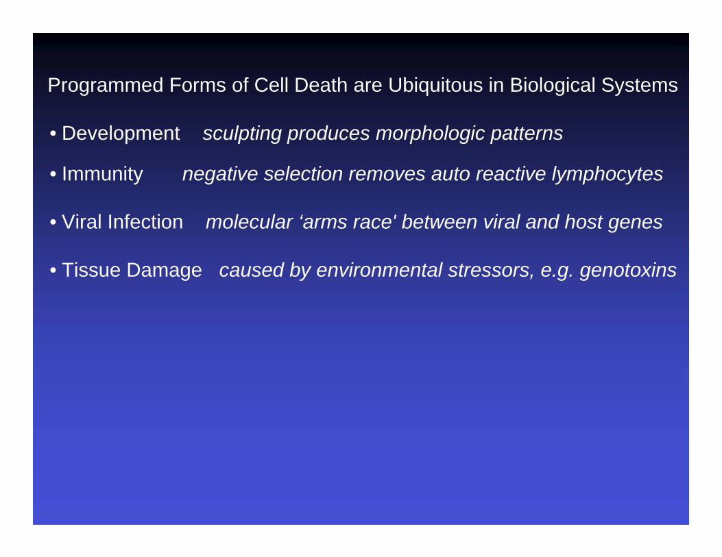
Programmed Forms of Cell Death are Ubiquitous in Biological Systems
• Development sculpting produces morphologic patterns
• Immunity negative selection removes auto reactive lymphocytes
• Viral Infection molecular ‘arms race' between viral and host genes
• Tissue Damage caused by environmental stressors, e.g. genotoxins
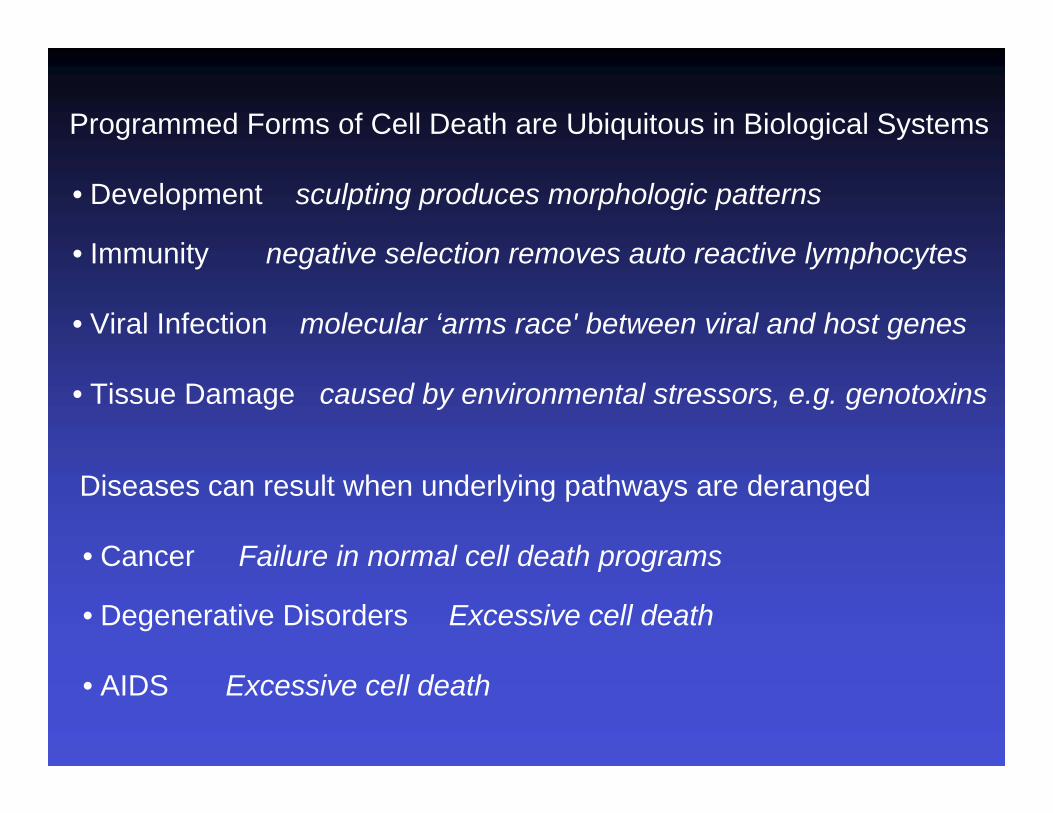
Diseases can result when underlying pathways are deranged
• Cancer Failure in normal cell death programs
• Degenerative Disorders Excessive cell death
• AIDS Excessive cell death

Classification SystemsMolecular definitions of cell death subroutines: recommendations of the Nomenclature Committee on Cell Death 2012 (Cell Death and Differentiation (2012) 19, 107–120)
Appearance Ultrastructural changes within the dying cell and nearby cellsType I associated with heterophagyType II associated with autophagyType III no associated digestion
Programmed vs. incidentalprogrammed = “Naturally occurring cell death”
= predictable in development= dedicated gene-directed pathways= active, cellular process= adaptive, not passive cytotoxicity= not caused by damage, injury or insult

Active Forms of Cell Death
apoptosis, programmed forms of necrosis
specified by natural or non-natural inducers
suicide vs. murder
underlying genetics and biochemistry
dedicated death pathways vs. sabatoge or signaling run ‘amok’
Classification SystemsMolecular definitions of cell death subroutines: recommendations of the Nomenclature Committee on Cell Death 2012 (Cell Death and Differentiation (2012) 19, 107–120)

the ultimate irreversible reaction
Convicted
Condemned
Execution
Disposal
pardon
stay
reversible
no return

9

Real Time Imaging in Culture

11
Live imaging with Stains, Vital Dyes Acridine Orange

12
Live imaging with Stains, Vital Dyes Acridine Orange
13
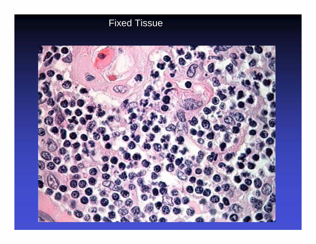
Fixed Tissue

14
Fixed Tissue

Apoptotic Cells are rapidly engulfed by phagocytes

16
• Active site cys, cleave asp
• initiating ‘platforms’ launch amplifying cascade
• Substrates can be activated or activated
• Function in PCD, immunity
• Viral genomes encode inhibitors
Caspases are activated and functional during apoptosis
prodomainlargesubunit
smallsubunit
asp asp
Dormant Proenzyme
Processing and Maturation
Active Tetramer

Peptide Substrates and Antibodies Detect Caspase Activity
In culture, bulk asays

Peptide Substrates and Antibodies Detect Caspase Activity
In culture, bulk asays
Functional studies - Inhibitors, RNAi, mutants

Methods of detection rely on characteristic features
Feature Apoptosis Necrosis
Cell size Shrunken Swollen
Cell fragmentation Yes No
Specific DNA cleavage Yes No
Engulfment Yes No
Inflammatory* No Yes
Caspases Required Yes No
Mitochondria Altered Swollen
Phospatidylserineexposed Yes No

Feature Mode
Cell size imaging
Cell fragmentation imaging
DNA cleavage TUNEL
Engulfment markers, sensors
Inflammatory* markers, sensors
Caspases antibodies, substrates
Phospatidylserine annexin V
Mitochondria imaging, markers, biosensors
Methods of detection rely on characteristic features


TUNEL - terminal deoxytransferase dUTP nick end labeling
Enzyme adds labelled nucleotides to ‘free ends‘ that occur at sites of cleaved DNA

TUNEL - terminal deoxytransferase dUTP nick end labeling
Enzyme adds labelled nucleotides to ‘free ends‘ that occur at sites of cleaved DNA

Defining the mode of cell death
Multiple criterion typically needed
Descriptive criteria vs. Functional criteria
Functional studies define events necessary for killing

Multiple criterion typically needed
Descriptive criteria vs. Functional criteria
Functional studies define events necessary for killing
Defining the mode of cell death

Cell death genes prevent death when eliminated cCell death defectivemacrophages normal engulfment normal
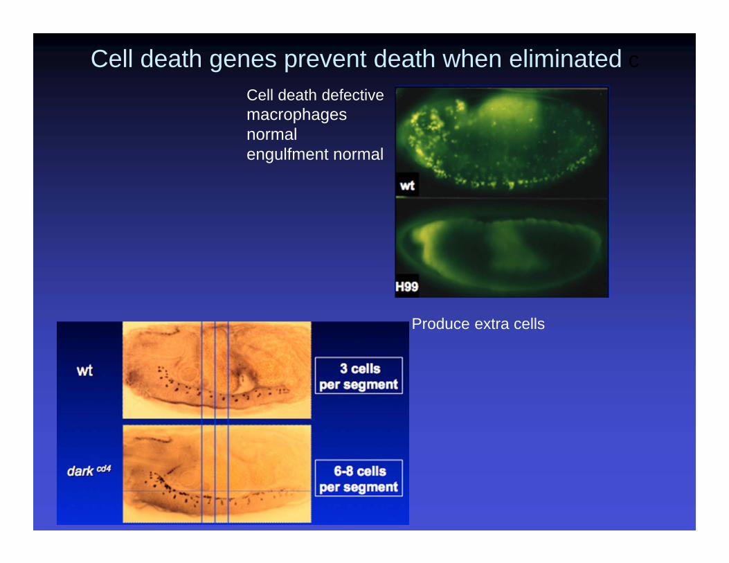
Produce extra cells
Cell death defectivemacrophages normal engulfment normal
Cell death genes prevent death when eliminated c

genetic mosaic tissue cell death defective genes
Cell death defective phenotypes observed in real time through in vivo imagingTransgenic labels: Green fluorescent protein (GFP); Red fluorescent protein RFP

Douglas Green & Beth LevineCell, Volume 157, Issue 1, 2014, 65 - 75
IAPsIAPs
Processed Caspases are physically inhibited by IAPs
smacsmac
IAP antagonists are also pivotal
Pivotal apoptosis regulators impact the extrinsic and/or intrinsic pathway. e.g. Bcl2 family members, caspases, death receptors

Genome scale screens for cell death genes
‘Hits’ reverse killing by smac mimetic
plate Z score
Chew et al (2009) Nature

Genome scale screens for cell death genes
Time lapse
‘Hits’ reverse killing by smac mimetic
plate Z score
Chew et al (2009) Nature

• Anoikis - apoptotic response seen when adherent cells lose matrix interactions. believed to be major tumor suppressive mechanism
• Pyroptosis - associated with inflammation, involves Casapse 1 (Accounts for loss of T cells associated with AIDS)
• Necroptosis - TNFR1 signaling through RIP1 that occurs when Caspase 8 is inhibited• Partial Demolition - Enucleation of Red Blood Cells, Formation of Lens fibers, Skin cell
Keratinization. Often Caspase dependent.• Secondary necrosis - necrosis occuring after full apoptotic program. Seen in culture
and/or when engulfment is compromised• Programmed forms of necrosis -
– Parthanatos - excessive poly(ADP-ribose) polymerase activity depletes ATP and NAD+
– Ferroptosis - dependent upon intracellular iron• Autophagic cell death - induced cytoplasmic vacuolization
– Autophagy is typically a survival adaptation – Death by autophagy vs. death with autophagy? Is autophagy the lethal event?
The Lexicon of Cell Death includes:

Douglas Green & Beth LevineCell, Volume 157, Issue 1, 2014, 65 - 75
The Autophagy Regulatory Network Engages Cell Death Regulators

Many thanks to
Nichole LinkAnwesha GoshSu Kit ChewAlex RodriguezMark CarlsonBeth LevineDoug Green

Brought to you by the Science/AAAS Custom Publishing Office
Sponsored by:
The Many Roads to Cell DeathGaining a Practical Understanding of Apoptosis, Necrosis, and AutophagyJune 4, 2014
Participating Experts
John Abrams, Ph.D.University of Texas Southwestern Medical CenterDallas, Texas
William Telford, Ph.D.National Institutes of HealthBethesda, MD
Webinar Series
Bill Telford, Ph.D.
NCI Flow Cytometry Core LaboratoryNational Cancer Institute
National Institutes of Health
Detecting Apoptosis by Flow Cytometry
Where are we after 25 years?

Apoptosis was first identified as a distinct morphological phenomenon in the 1960s (and probably earlier), and was well-accepted as an important regulatory process by the 1970s…
necrosis
cell shrinkagecytoskeletal collapseincreased cell permability
apoptosis
chromatin condensationDNA fragmentation
transglutaminase crosslinkingcell “blebbing”
mitochondria still intact
recognition andphagocytosis/clearance
From Kerr, J.F.R., J Pathology 105, 13-20, 1971 (!)

Institute of TheoreticalAnd ExperimentalBiophysics
Institute of Cell Biophysics
Pushchino, Serphkov region,Russia

gammairradiatedrat thymus,spleen and BM
Hoechst 33258

gammairradiatedrat thymus,spleen and BM
Hoechst 33258
Radiobiologiia. 1986 Nov-Dec;26(6):728-32. Russian.

Flow cytometry assays for apoptosis are now almost 25 years old…
From Telford et al., Applied Fluorescence Technology 4, 12-17 (1992)
the“sub-G0/G1”peak
changesin lightscatter
The earliest flow cytometry assays for apoptosis analyzed changes in forward and side scatter, and DNA fragmentation / loss following ethanol treatment. Unlike earlier assays, flow cytometry analyzed apoptosis in individual cells.
Apoptosismeasurementin individual cells (not lysates)
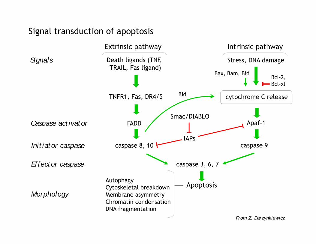
Signal transduction of apoptosis
Signals Death ligands (TNF, TRAIL, Fas ligand)
Stress, DNA damage
TNFR1, Fas, DR4/5 cytochrome C release
Caspase activator
Initiator caspase
Effector caspase
FADD Apaf-1
caspase 8, 10 caspase 9
caspase 3, 6, 7
ApoptosisAutophagyCytoskeletal breakdownMembrane asymmetryChromatin condensationDNA fragmentation
From Z. Darzynkiewicz
Smac/DIABLO
IAPs
Bax, Bam, BidBcl-2, Bcl-xl
Extrinsic pathway Intrinsic pathway
Morphology
Bid

Types of assays…
“early” apoptotic events
“late”apoptoticevents
characteristic flow cytometry assay
Cell volume fluctuationsCytochrome C releaseChanges in cell membrane potentialMitochondrial potential changesSignaling events (bcl-2, Bax, etc.)Initiator (proximal) caspase
activation (1,9,10,8)Effector (distal) caspase
activation (3,6,7)
Organelle changesPS membrane “flipping”Transglutaminase crosslinkingChanges in chromatin organizationDNA strand breaksMembrane “blebbing”Global chromatin damageLoss of membrane permeability
Minor changes in scatterCytochrome C release assayMembrane potential probesMitochondrial potential probesBax translocationFluorgenic caspase substrates
PhiPhiLuxFLICACellEvent Green
Immunolabeling of active caspasesOrganelle-specific probesAnnexin V, structure-specific
plasma membrane probesImmunolabeling of histones and
histone associated proteinsTUNEL assaysMajor changes in scatterLoss of DNA dye binding
Flow cytometry assays now target almost every stage of apoptosis, from the earliestmitochondrial changes to caspase activation, membrane changes and DNA damage.

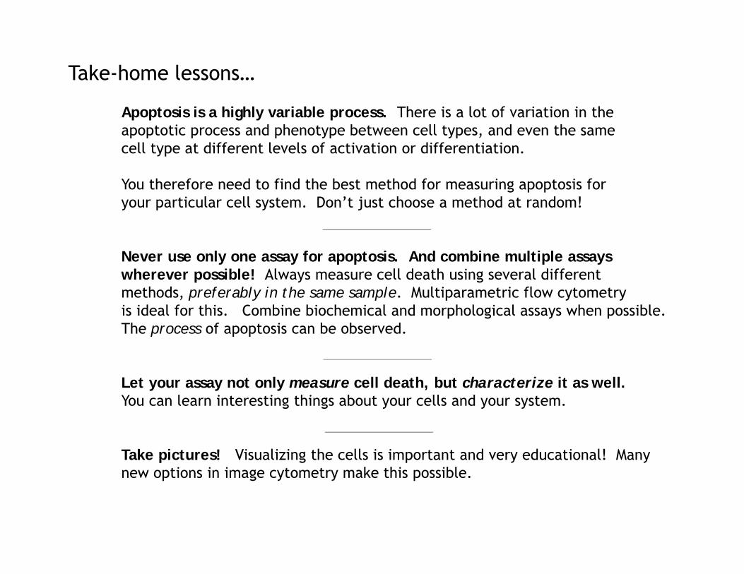
Take-home lessons…
Apoptosis is a highly variable process. There is a lot of variation in theapoptotic process and phenotype between cell types, and even the samecell type at different levels of activation or differentiation.
You therefore need to find the best method for measuring apoptosis foryour particular cell system. Don’t just choose a method at random!
Never use only one assay for apoptosis. And combine multiple assayswherever possible! Always measure cell death using several different methods, preferably in the same sample. Multiparametric flow cytometryis ideal for this. Combine biochemical and morphological assays when possible.The process of apoptosis can be observed.
Let your assay not only measure cell death, but characterize it as well.You can learn interesting things about your cells and your system.
Take pictures! Visualizing the cells is important and very educational! Manynew options in image cytometry make this possible.

Annexin V and a DNA binding dye is an excellent example of combining twoassays into a multiparametric method.
actinomycin D 4 hours
EL4 cells
At least two stages of apoptoticdeath are beingmeasured here.FITC annexin V
16%
7%
75.1%
prop
idiu
m io
dide
PS “flipping” occursprior to 7-AADpermeability
“viable” cells
annexin V+ PI-“early apoptotic”
annexin V+ PI+(late apoptotic/necrotic)
Combining multiple apoptosis assays
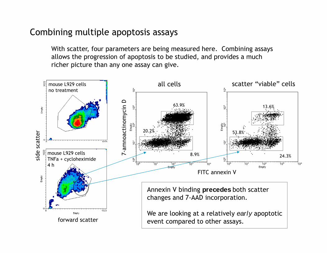
mouse L929 cellsTNFa + cycloheximide4 h
mouse L929 cellsno treatment
all cells scatter “viable” cells
Annexin V binding precedes both scatterchanges and 7-AAD incorporation.
We are looking at a relatively early apoptoticevent compared to other assays.
13.6%
24.3%
53.8%
63.9%
8.9%
20.2%
FITC annexin V
7-am
noac
tino
myc
in D
forward scatter
side
sca
tter
Combining multiple apoptosis assays
With scatter, four parameters are being measured here. Combining assaysallows the progression of apoptosis to be studied, and provides a much richer picture than any one assay can give.
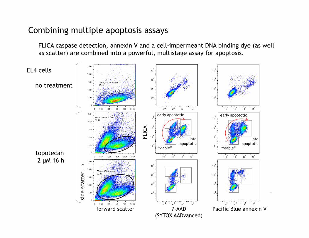
Combining multiple apoptosis assays
no treatment
topotecan2 μM 16 h
side
sca
tter
forward scatter
FLIC
A
7-AAD(SYTOX AADvanced)
Pacific Blue annexin V
EL4 cells
“viable”
early apoptotic
lateapoptotic
“viable”
early apoptotic
lateapoptotic
DC1125
FLICA caspase detection, annexin V and a cell-impermeant DNA binding dye (as well as scatter) are combined into a powerful, multistage assay for apoptosis.
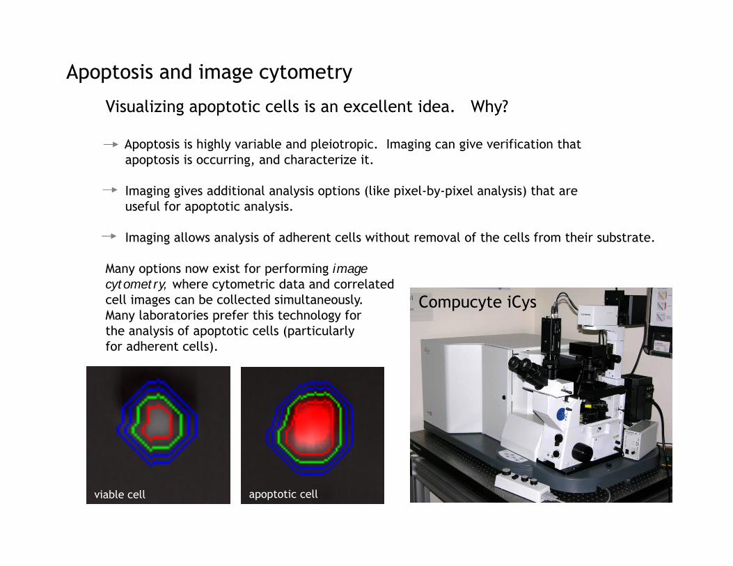
Apoptosis and image cytometry
Visualizing apoptotic cells is an excellent idea. Why?
Apoptosis is highly variable and pleiotropic. Imaging can give verification that apoptosis is occurring, and characterize it.
Imaging gives additional analysis options (like pixel-by-pixel analysis) that are useful for apoptotic analysis.
Imaging allows analysis of adherent cells without removal of the cells from their substrate.
Many options now exist for performing image cytometry, where cytometric data and correlated cell images can be collected simultaneously. Many laboratories prefer this technology for the analysis of apoptotic cells (particularly for adherent cells).
viable cell apoptotic cell
Compucyte iCys
Apoptosis and image cytometry
fluorescein PPL caspase 3
fluorescein PPL caspase 37-
AAD
7-AA
DAl
exa
Fluo
r 64
7an
nexi
n V
Alex
a Fl
uor
647
anne
xin
V
7-AAD
7-AAD
no treatment
camptothecin 6 h
no treatment
camptothecin 6 h
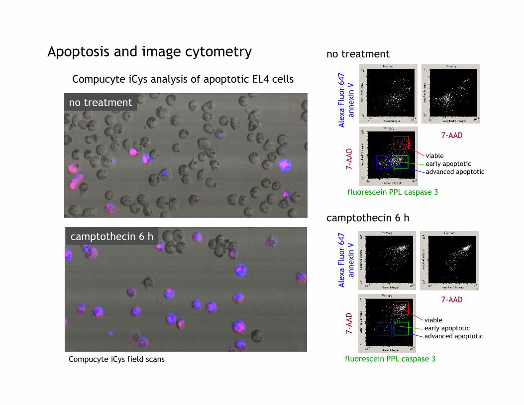
Compucyte iCys analysis of apoptotic EL4 cells
viableearly apoptoticadvanced apoptotic
viableearly apoptoticadvanced apoptotic
Compucyte iCys field scans

fluorescein PPL caspase 3
Alex
a Fl
uor
647
anne
xin
VApoptotic cell analysis with laser scanning cytometry
Direct correlation between thecytometric data and the imagery(relocation analysis).
Morphological analysis usinglight scatter or absorption.

Discrimination of adherent apoptotic cells by image cytometry
Hoechst 33258PhiPhiLux caspase 3
Alexa Fluor 647 annexin VUMR-106 cells
PPL caspase 3Max Pixel
AF64
7 an
nexi
nV
Max Pixel IntegralPPL caspase 3
Image cytometry can analyze site-specific fluorescence from images, improving sensitivity. Trypsin or accutase detachment, which can “muddy” apoptotic labels, is not necessary.
Apoptotic cells “round up”and can be lost from thegrowth substrate.
Max Pixel versus total Integral
caspase 3
AF647 AnnV+ caspase 3
Compucyte iCys field scan

Analyzing apoptosis on the Amnis ImageStream
Brightfield Phi Phi Lux Annexin V PB Draq5 Composite
Composite
BrightfieldA stream-based scanning cytometry system like the Amnis ImageStream orFlowSight similarly isan another excellent way to combine cytometric and morphological analysis.
Again, direct correlation between cytometry and imagery.
Data from Brian Hall and Tad George, Amnis, EMD Millipore
Daudi cells induced with camptothecin

untreated camptothecin 6 h
fluo
resc
ein
PPL
casp
ase
3
Images of live cells Images of PhiPhiLux positives
3% 70%
Brightfield Phi Phi Lux Annexin V Draq5 Composite
Brightfield Phi Phi Lux Annexin V Draq5 Composite
fluo
resc
ein
PPL
casp
ase
3
Analyzing apoptosis on the Amnis ImageStream
Data from Brian Hall and Tad George, Amnis, EMD Millipore

Analyzing apoptosis on the Amnis ImageStream
camptothecin 6 h 77%
Paci
fic
Blue
ann
exin
V
Ann
exin
V I
nten
sity
PPL caspase 3
10%
Brightfield Phi Phi Lux Annexin V Draq5 Composite
Brightfield Phi Phi Lux Annexin V Draq5 Composite Brightfield Phi Phi Lux Annexin V Draq5 Composite
Brightfield Phi Phi Lux Annexin V Draq5 Composite
Viable and very early apoptotic Early apoptotic
Intermediateapoptotic
Late apoptotic
Data from Brian Hall and Tad George, Amnis, EMD Millipore

Autophagy
induced
control
U2OS cells expressing GFP-LC3EMD Millipore
signal
mTor
mTor block
defunct mitochondria
LC3
autophagosomeformation
lysosome
autophagosome-lysosome dockingand fusion
autophagosomebreakdown
plasma membrane
lysozymeinhibitor

During induction, a lysozyme inhibitoris added to block destruction ofautophagosomes by lysosomes.
If autophagy occurs, GFP-LC3 willaccumulate in autophagosomes whenthe inhibitor is present.
The cells are then permeablized.
In the absence of autophagy, theGFP-LC3 will be in the cytoplasm andwill be released into the media.
With autophagy, the GFP-LC3 willbe trapped in the autophagosomesand will not be released into themedia.
Autophagosome associated GFP-LC3can be detected in the intact cellsby flow cytometry.
Autophagy detected by GFP-LC3 translocation
Translocation ofGFP-LC3 to
autophagosomes
permeablization
Most GFP-LC3lost
AutophagosomeassociatedGFP-CL3retained
Assay courtesy EMD Millipore
lysozymeinhibitor

Autophagy detected by GFP-LC3 translocation
During induction, a lysozyme inhibitoris added to block destruction ofautophagosomes by lysosomes.
If autophagy occurs, GFP-LC3 willaccumulate in autophagosomes whenthe inhibitor is present.
The cells are then permeablized.
In the absence of autophagy, theGFP-LC3 will be in the cytoplasm andwill be released into the media.
With autophagy, the GFP-LC3 willbe trapped in the autophagosomesand will not be released into themedia.
Autophagosome-associated GFP-LC3can be detected in the intact cellsby flow cytometry. GFP-LC3
control
induced
permeablized cells intact cells
GFP-LC3 trapped in autophagosomesby inhibitor
Total GFP-LC3
GFP-LC3 trapped in autophagosomesby inhibitor
Total GFP-LC3
level of autophagy
DC950
Assay courtesy EMD Millipore
Take-home lessons…
Apoptosis is a highly variable process. There is a lot of variation in theapoptotic process and phenotype between cell types, and even the samecell type at different levels of activation or differentiation.
You therefore need to find the best method for measuring apoptosis foryour particular cell system. Don’t just choose a method at random!
Never use only one assay for apoptosis. And combine multiple assayswherever possible! Always measure cell death using several different methods, preferably in the same sample. Multiparametric flow cytometryis ideal for this. Combine biochemical and morphological assays when possible.The process of apoptosis can be observed.
Let your assay not only measure cell death, but characterize it as well.You can learn interesting things about your cells and your system.
Take pictures! Visualizing the cells is important and very educational! Manynew options in image cytometry make this possible.

Veena KapoorNga Tu VoongElena I. Kovalenko
NCI ETIB Flow Lab
Acknowledgements
Karen TamulBrian HallThad George
EMD Millipore
Jolene BradfordGayle BullerSuzanne BuckJeff CroissantMike Olszowy
Molecular Probes, Thermo Fisher

To submit yourquestions, type them into the text box and
click
Participating Experts
John Abrams, Ph.D.University of Texas Southwestern Medical CenterDallas, Texas
William Telford, Ph.D.National Institutes of HealthBethesda, MD Sponsored by:
Brought to you by the Science/AAAS Custom Publishing Office
The Many Roads to Cell DeathGaining a Practical Understanding of Apoptosis, Necrosis, and AutophagyJune 4, 2014
Webinar Series

The Many Roads to Cell Death:
Simplified Solutions for Cell Death and Cell Health Analysis with the Muse® Cell Analyzer
Kamala Tyagarajan, Ph.D., EMD Millipore
June 4th, 2014

Study of Cell Death Pathways is Enabled by a Variety of Technologies
• Dissecting and understanding different cell death pathways requires information from multiple steps in the pathway.
• A range of technologies can provide information on different cell death pathways:• Flow cytometry
• Imaging based cytometry
• Technologies for adherent cell imaging
• Time courses and dose responses can provide deeper insights into mechanisms at play and provide a more comprehensive picture.

There are Common, Inherent Drawbacks with Current Methods for Cell Health Analysis
Home-brew reagents and commercial kits
Microscopy
Western Blot, ELISA, Antibodies
Flow Cytometry
Dedicated Instrumentation for Cell Health Applications
Limited Access to Instrumentation
Greater Expertise and Planning Needed
Expense
Lack of Reproducibility
Accuracy of Results

Muse™ Cell Analyzer: Simple, Affordable, Miniature Flow Cytometry
• Closed Platform (instrument and 23 assays) allowing instant assessments of Cell Health, Apoptosis, Cell Signaling, Immunology, Cell Cycle
• Instrument utilizes novel miniaturized flow cytometric technology
• Novel, intuitive software and touchscreeninterface = novel, simplified user experience
• Simple, effortless operation and sample preparation = accessibility to flow novices
• Innovative “personal” cell analysis• Accessible, simplistic approach to
complex questions

Muse ® Provides Multiple Solutions for Evaluation of Cell Death Mechanisms
Late
ROS/NOS Stress
Mitochondrial Protein Release
PhosphatidylserineTranslocation to OuterMembrane
Metabolic Activity/Cell Vitality
Caspase Activity
DNA Condensation
Plasma MembraneDisintegration
Mitochondrial MembranePotential
DNA Fragmentation
MitoPotential
Annexin V & Dead Cell Dye
EarlyC
E
L
L
F
U
N
C
T
I
O
N
Cell Death/Stress Indicators Muse Assays
Multi-Caspase
Count & Viability Assay (2)
LC3 (Autophagy)
Caspase 3,7
Other Indicators
Cell Cycle Cell Proliferation
Oxidative Stress Nitric Oxide Stress

Example: Muse ® MitoPotential Assay
• A simple, no-wash assay that provides percentage and concentration of cells demonstrating mitochondrial membrane depolarization and cell death.
• Assay is based on detection of mitochondrial potential changes using a cationic lipophilic dye.
• Depolarized cells are detected by a decrease in fluorescence.
• 7AAD enters cells that are compromised in dead or late apoptotic cells
Add Reagent Add Cells
Run on MuseTM
Cell AnalyzerAdd 7-AAD
Incubate for20 minutes at
37°C
Incubate for5 minutes at
Room Temperature

Example of Time Course Studies in Cell Death Analysis
Time course of Jurkat cells treated with Staurosporine (top) and Gambogic Acid (bottom) and analyzed with the Muse®
MitoPotential and Muse® Annexin V & Dead Cell Assay

Muse® Assay Offerings Span Multiple Areas
Muse® Count & Viability Kit
Muse® Count & Viability Kit
Muse® Cell Cycle Kit
Muse® Annexin V & Dead Cell Kit
Muse® Caspase 3,7 Kit
Muse® MultiCaspase Kit
Muse® MitoPotential Kit
Muse® Nitric Oxide Kit
Muse® Oxidative Stress Kit
Muse® Ki67 Proliferation Kit
Muse® LC3 Autophagy (Ab Based)
Muse® LC3 Autophagy (Reporter Cell Line)
Cell Health
Muse ® H2A.X Activation Dual Detection Kit
Muse ® MAPK Activation Dual Detection Kit
Muse ® EGFR-RTK Activation Dual Detection Kit
Muse ® PI3 Activation Dual Detection Kit
Muse ® Bcl-2 Activation Dual Detection Kit
Muse ® Multi-Color DNA Damage Kit
Muse ® PI3K/MAPK Activation Dual Detection Kit
Cell Signaling
Muse ® Human CD4 T Cell Kit
Muse ® Human CD8 T Cell Kit
Muse ® Human B Cell Kit
Muse ® Human CD25 Lymphocyte Kit
Muse ® Human CD69 Lymphocyte Kit
Immunology

The Muse Cell Analyzer: Simplified Platform for Rapid Cell Health Analysis
• Small footprint platform based on miniaturized, microcapillary flow cytometry
• Optimized kits for convenient analysis of multiple cell death mechanisms
• Easy-to-follow guided software on touchscreen
• Extremely Affordable ($14,750)
+
Fast & Easy Sample Prep
Load and Run on Muse® Instrument
Quickly Analyze Results!

www.millipore.com/muse

Brought to you by the Science/AAAS Custom Publishing Office
Sponsored by:
The Many Roads to Cell DeathGaining a Practical Understanding of Apoptosis, Necrosis, and AutophagyJune 4, 2014
Participating Experts
John Abrams, Ph.D.University of Texas Southwestern Medical CenterDallas, Texas
William Telford, Ph.D.National Institutes of HealthBethesda, MD
Webinar Series

For related information on this webinar topic, go to:www.millipore.com/muse
The Many Roads to Cell DeathGaining a Practical Understanding of Apoptosis, Necrosis, and AutophagyJune 4, 2014
Look out for more webinars in the series at:
webinar.sciencemag.org
To provide feedback on this webinar, please e‐mailyour comments to [email protected]
Brought to you by the Science/AAAS Custom Publishing Office
Sponsored by:
Webinar Series