gait and balance changes with investigational peripheral
TRANSCRIPT
University of Kentucky University of Kentucky
UKnowledge UKnowledge
Physical Therapy Faculty Publications Physical Therapy
4-15-2021
Gait and Balance Changes with Investigational Peripheral Nerve Gait and Balance Changes with Investigational Peripheral Nerve
Cell Therapy during Deep Brain Stimulation in People with Cell Therapy during Deep Brain Stimulation in People with
Parkinson’s Disease Parkinson’s Disease
Geetanjali Gera University of Kentucky, [email protected]
Zain Guduru University of Kentucky, [email protected]
Tritia R. Yamasaki University of Kentucky, [email protected]
Julie A. Gurwell University of Kentucky, [email protected]
Monica Chau University of Kentucky, [email protected]
See next page for additional authors
Follow this and additional works at: https://uknowledge.uky.edu/rehabsci_facpub
Part of the Neurology Commons, Neurosciences Commons, Neurosurgery Commons, and the
Rehabilitation and Therapy Commons
Right click to open a feedback form in a new tab to let us know how this document benefits you. Right click to open a feedback form in a new tab to let us know how this document benefits you.
Repository Citation Repository Citation Gera, Geetanjali; Guduru, Zain; Yamasaki, Tritia R.; Gurwell, Julie A.; Chau, Monica; Krotinger, Anna; Schmitt, Frederick A.; Slevin, John T.; Gerhardt, Greg A.; van Horne, Craig G.; and Quintero, Jorge E., "Gait and Balance Changes with Investigational Peripheral Nerve Cell Therapy during Deep Brain Stimulation in People with Parkinson’s Disease" (2021). Physical Therapy Faculty Publications. 122. https://uknowledge.uky.edu/rehabsci_facpub/122
This Article is brought to you for free and open access by the Physical Therapy at UKnowledge. It has been accepted for inclusion in Physical Therapy Faculty Publications by an authorized administrator of UKnowledge. For more information, please contact [email protected].

Gait and Balance Changes with Investigational Peripheral Nerve Cell Therapy Gait and Balance Changes with Investigational Peripheral Nerve Cell Therapy during Deep Brain Stimulation in People with Parkinson’s Disease during Deep Brain Stimulation in People with Parkinson’s Disease
Digital Object Identifier (DOI) https://doi.org/10.3390/brainsci11040500
Notes/Citation Information Notes/Citation Information Published in Brain Sciences, v. 11, issue 4, 500.
© 2021 by the authors. Licensee MDPI, Basel, Switzerland.
This article is an open access article distributed under the terms and conditions of the Creative Commons Attribution (CC BY) license (https://creativecommons.org/licenses/by/4.0/).
Authors Authors Geetanjali Gera, Zain Guduru, Tritia R. Yamasaki, Julie A. Gurwell, Monica Chau, Anna Krotinger, Frederick A. Schmitt, John T. Slevin, Greg A. Gerhardt, Craig G. van Horne, and Jorge E. Quintero
This article is available at UKnowledge: https://uknowledge.uky.edu/rehabsci_facpub/122
brainsciences
Communication
Gait and Balance Changes with Investigational PeripheralNerve Cell Therapy during Deep Brain Stimulation in Peoplewith Parkinson’s Disease
Geetanjali Gera 1,2,*, Zain Guduru 2,3, Tritia Yamasaki 2,3,4,5 , Julie A. Gurwell 2,3, Monica J. Chau 2,6,Anna Krotinger 7 , Frederick A. Schmitt 2,3 , John T. Slevin 2,3,5, Greg A. Gerhardt 2,3,4,6, Craig van Horne 2,4,6
and Jorge E. Quintero 2,4,6
�����������������
Citation: Gera, G.; Guduru, Z.;
Yamasaki, T.; Gurwell, J.A.; Chau,
M.J.; Krotinger, A.; Schmitt, F.A.;
Slevin, J.T.; Gerhardt, G.A.; van
Horne, C.; et al. Gait and Balance
Changes with Investigational
Peripheral Nerve Cell Therapy
during Deep Brain Stimulation in
People with Parkinson’s Disease.
Brain Sci. 2021, 11, 500. https://
doi.org/10.3390/brainsci11040500
Academic Editor: Toshikazu Ikuta
Received: 1 February 2021
Accepted: 10 April 2021
Published: 15 April 2021
Publisher’s Note: MDPI stays neutral
with regard to jurisdictional claims in
published maps and institutional affil-
iations.
Copyright: © 2021 by the authors.
Licensee MDPI, Basel, Switzerland.
This article is an open access article
distributed under the terms and
conditions of the Creative Commons
Attribution (CC BY) license (https://
creativecommons.org/licenses/by/
4.0/).
1 Department of Physical Therapy, College of Health Sciences, University of Kentucky,204L 900 South Limestone Street, Lexington, KY 40536, USA
2 Brain Restoration Center, University of Kentucky, Lexington, KY 40536, USA; [email protected] (Z.G.);[email protected] (T.Y.); [email protected] (J.A.G.); [email protected] (M.J.C.);[email protected] (F.A.S.); [email protected] (J.T.S.); [email protected] (G.A.G.);[email protected] (C.v.H.); [email protected] (J.E.Q.)
3 Neurology, University of Kentucky, Lexington, KY 40536, USA4 Neuroscience, University of Kentucky, Lexington, KY 40536, USA5 Veterans Affairs Medical Center, Lexington, KY 40502, USA6 Neurosurgery, University of Kentucky, Lexington, KY 40536, USA7 Department of Neuroscience, Wesleyan University, Middletown, CT 06459, USA; [email protected]* Correspondence: [email protected]; Tel.: +1-859-218-0547
Abstract: Background: The efficacy of deep brain stimulation (DBS) and dopaminergic therapyis known to decrease over time. Hence, a new investigational approach combines implantingautologous injury-activated peripheral nerve grafts (APNG) at the time of bilateral DBS surgery tothe globus pallidus interna. Objectives: In a study where APNG was unilaterally implanted into thesubstantia nigra, we explored the effects on clinical gait and balance assessments over two years in14 individuals with Parkinson’s disease. Methods: Computerized gait and balance evaluations wereperformed without medication, and stimulation was in the off state for at least 12 h to best assessthe role of APNG implantation alone. We hypothesized that APNG might improve gait and balancedeficits associated with PD. Results: While people with a degenerative movement disorder typicallyworsen with time, none of the gait parameters significantly changed across visits in this 24 monthstudy. The postural stability item in the UPDRS did not worsen from baseline to the 24-month follow-up. However, we measured gait and balance improvements in the two most affected individuals,who had moderate PD. In these two individuals, we observed an increase in gait velocity and steplength that persisted over 6 and 24 months. Conclusions: Participants did not show worsening ofgait and balance performance in the off therapy state two years after surgery, while the two mostseverely affected participants showed improved performance. Further studies may better addressthe long-term maintanenace of these results.
Keywords: deep brain stimulation; gait; balance; cell therapy; Parkinson’s disease
1. Introduction
Parkinson’s disease (PD) is a neurodegenerative disease characterized in part by aprogressive loss of dopamine due to neurodegeneration in the substantia nigra [1]. Thisprogressive loss of dopamine, with up to 10% of the remaining dopaminergic cells dyingeach year, is a major cause of disability in those with PD, particularly related to gait andbalance disturbances [2,3]. Gait and balance disturbances are one of the most importantcauses of diminished quality of life, morbidity, and mortality in people with PD [4]. Re-duced gait speed and step length and impaired rhythmicity are all components of gait that
Brain Sci. 2021, 11, 500. https://doi.org/10.3390/brainsci11040500 https://www.mdpi.com/journal/brainsci
Brain Sci. 2021, 11, 500 2 of 9
worsen as the disease progresses [5]. Balance performance in people with PD is measuredby postural control, rigidity, the center of foot pressure (CoP) displacement, velocity, andfrequency [6]. Both gait and balance disturbances lead to increased falls, difficulty nav-igating a home or community environment, and increased overall morbidity [4]. Thus,interventions targeting the causes of disability in those with PD can reduce mortality andimprove independence and quality of life in this population [4]. While there is no cure forPD, levodopa is the gold standard medication for managing PD symptoms but does notalter the pathophysiological trajectory [4].
Deep brain stimulation (DBS) is a therapy for PD in which electrodes are surgicallyimplanted in the subthalamic nucleus (STN) or globus pallidus interna (GPi), areas, whichare responsible for motor control. However, the efficacy of the combination of DBS anddopaminergic therapy is known to decrease over time. Some studies suggest this may bebecause traditional therapies are not targeting the specific cause of the disease. Thus, thepatients still experience disease progression over time [7].
The decreased efficacy of DBS and dopaminergic therapies over time has broughtforth a new, experimental combined approach of implanting autologous injury-activatedperipheral nerve grafts (APNG) at the time of DBS surgery. We have preliminarily foundthat this approach can be safe and feasible [8,9]. In our approach, the participant’s ownperipheral (sural) nerve tissue is implanted into the substantia nigra as investigationalcell therapy. Our strategy is to support the existing neurons in the substantia nigra andprevent degeneration. We implant sural nerve tissue that had been transected to convertthe associated Schwann cells into a reparative phenotype [10–13]. After peripheral nervetransection or injury, Schwann cells undergo a unique cellular reprogramming that transdif-ferentiates the cell from a myelinating phenotype into a reparative phenotype [10,11,13,14].The cells then upregulate and release a whole host of neurotrophic and cell survival factors,including glial-derived neurotrophic factor (GDNF) [15], nerve growth factor (NGF) [16,17],brain-derived neurotrophic factor (BDNF) [18], and nuclear factor erythroid 2-related factor(Nrf2) in animal models. Our RNA Seq analyses support that the human sural nerve tissuesthat we implanted show similarities to the repair phenotype in animal models [19]. Ourstrategy is to use APNG to implant a living repair system with growth factors to reducecell death of dopamine-producing neurons and aid in the long-term maintenance of motorfunction, including gait and balance.
Here we report the effects of APNG on various balance and gait measures, includingstep length, velocity, and cadence over two years. As part of a post hoc analysis, wehypothesized that APNG might improve gait and balance deficits associated with PD. IfAPNG were subsequently shown to be efficacious, it could alter the management of PD,allowing those with this disease to retain their mobility for a better quality of life.
2. Materials and Methods
Study design. This report presents a subpopulation of participants who completedgait and balance assessments from a 24-month, investigator-initiated, open-label, single-center, Phase I trial designed to assess the safety and feasibility of the direct delivery ofperipheral nerve tissue APNG into the substantia nigra (NCT02369003). Gait and balancewere exploratory outcomes of the trial and are reported in detail here separately from therest of the study thus adverse events are not reported. However, as reported in our earlierwork, the most common adverse events related to the study intervention were paresthesia,numbness, and pain of the ankle and foot immediately following the sural nerve biopsy [9].Eighteen participants were implanted with a unilateral graft; however, 14 participantswere included in these analyses (Figure 1). Participants were evaluated at baseline, beforesurgery, and every 6 months for 24 months. (N = 14; 61.9 ± 8.3 y; 10 M/4 F; OFF medicationUPDRS III = 37.6 ± 11.8) (Figure 1).

Brain Sci. 2021, 11, 500 3 of 9
Figure 1. Participants inclusion flowchart.
Participant selection. As previously described [8,9], patients, who were good can-didates for DBS for PD were offered the opportunity to enroll if they met the inclu-sion/exclusion criteria. The eligibility criteria used for the screening phase are presented inFigure 1. The University of Kentucky institutional review board (IRB) approved the study,and all participants provided informed consent.
2.1. Surgical implantation
In the current trial, APNG was implanted after the bilateral DBS electrodes had beenplaced in the GPi (STN for one participant), tested, secured to the skull, and connectedto the lead extensions. The specific details for graft injury conditioning, graft harvestand implantation have been described in detail previously [8,9]. Grafts were implantedunilaterally into the substantia nigra contralateral to the most affected side based onthe lateralized Unified Parkinson’s Disease Rating Scale (UPDRS) part III scores. AfterDBS electrode and tissue implantation procedures, a postoperative MRI was used to aidin confirming electrode placement and APNG implant. Participants underwent routinepostoperative visits, programming visits, and postoperative clinical study visits.
2.2. Clinical Testing
Participants stopped all the dopaminergic medications, and DBS stimulation wasturned off at least 12 h before UPDRS III testing and gait/balance testing. UPDRS PartIII is the motor examination component of the assessment that includes an evaluation ofgait. UPDRS item 30 (postural stability) was used to access balance. The UPDRS item 30(postural stability) scale ranges from 0 to 4, with 0 representing normal/no problems and4 representing a severe loss of balance. Hoehn and Yahr’s (HY) score was also assessed,based on the severity of the disease before undergoing the procedure and every 6 monthsafter the procedure for 2 years. The HY scale is used to describe PD progression andincludes stages 1 through 5, with 1 representing minimal, unilateral symptoms and 5representing confinement to a bed or wheelchair unless aided.
2.3. Gait Data Collection
Each participant performed three walking trials at their self-selected speed across the24 foot GAITrite® walkway (CIR Systems, Franklin, NJ, USA). We present the data forthe participants in the OFF state (off levodopa at baseline for at least 12 h) at the time ofenrollment and off levodopa/off DBS at least 12 h at 6 months and 24 months post-surgery.OFF state (OFF levodopa and OFF DBS) gait data were tested to determine the change ingait performance, as part of the evaluation of the underlying disease progression, in theabsence of a standard of care therapy. Gait data were analyzed using the GAITrite software.The data collected were uploaded into an excel file for data summaries.
Brain Sci. 2021, 11, 500 4 of 9
2.4. Outcome Variables
The gait variables measured and analyzed using the GAITrite software includedspatial measures (step length and velocity) and a temporal measure (cadence). Cadence isdefined as the total number of steps taken in a minute. Step length is the distance betweenthe point of initial contact of one foot and the point of initial contact of the opposite foot.We used a within-subjects repeated measures ANOVA design to investigate if the gaitmetrics changed across visits (SPSS 26). We set the threshold for a significant difference top < 0.05. When there was a significant within-subjects effect, we examined the pair-wisecomparisons to determine the source of significance (no adjustments were performed forthe pair-wise comparison). The clinical scales evaluated were UPDRS III total (overallscore), UPDRS item 29 (gait), UPDRS item 30 (postural stability) and Hoehn and Yahr.Analysis was performed on 14 individuals, as shown in Figure 1.
3. Results3.1. Changes in Clinical Scales 6 and 24 Months after APNG Implantation
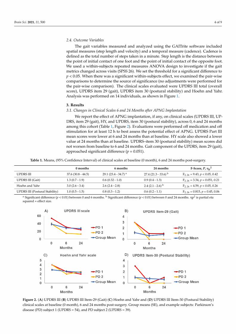
We report the effect of APNG implantation, if any, on clinical scales (UPDRS III, UP-DRS, item 29 (gait), HY, and UPDRS, item 30 (postural stability), across 0, 6 and 24 monthsamong this cohort (Table 1, Figure 2). Evaluations were performed off medication and offstimulation for at least 12 h to best assess the potential effect of APNG. UPDRS Part IIImean scores were lower at 6 and 24 months than at baseline. HY scale also showed a lowervalue at 24 months than at baseline. UPDRS–item 30 (postural stability) mean scores didnot worsen from baseline to 6 and 24 months. Gait component of the UPDRS, item 29 (gait),approached significant difference (p = 0.051).
Table 1. Means, (95% Confidence Interval) of clinical scales at baseline (0 month), 6 and 24 months post-surgery.
0 months 6 months 24 months F-Score, P, ηp2
UPDRS III 37.6 (30.8−44.5) 29.1 (23.4−34.7) a 27.4 (21.3−33.6) b F2, 26 = 9.43, p < 0.05, 0.42
UPDRS III (Gait) 1.3 (0.7−1.9) 0.6 (0.32−1.0) 0.9 (0.4−1.3) F2, 26 = 3.34, p = 0.051, 0.21
Hoehn and Yahr 3.0 (2.6−3.4) 2.6 (2.4−2.8) 2.4 (2.1−2.6) b F2, 26 = 4.59, p < 0.05, 0.26
UPDRS III (Postural Stability) 1.0 (0.5−1.5) 0.8 (0.3−1.2) 0.6 (0.2−1.1) F2, 26 = 0.815, p = 0.45, 0.06a Significant difference (p < 0.01) between 0 and 6 months. b Significant difference (p < 0.01) between 0 and 24 months. ηp2 is partial etasquared = effect size.
Figure 2. (A) UPDRS III (B) UPDRS III Item-29 (Gait) (C) Hoehn and Yahr and (D) UPDRS III Item-30 (Postural Stability)clinical scales at baseline (0 month), 6 and 24 months post-surgery. Group means (SE), and example subjects: Parkinson’sdisease (PD) subject 1 (UPDRS = 54), and PD subject 2 (UPDRS = 39).

Brain Sci. 2021, 11, 500 5 of 9
3.2. Gait Measures for the Cohort
None of the changes in gaitmeasureswere statistically significant across visits. Thus,overall, there was no visit effect of the APNG for gait measures from zero to 6 monthsor 24 months, suggesting no statistically significant decrement or improvement in gaitparameters longitudinally in this cohort (Table 2, Figure 3). However, gait velocity (p = 0.07)and step length for the more affected side (p = 0.08) approached significance.
Table 2. Means, (95% Confidence Interval) of gait parameters at baseline (0 month), 6 and 24 months post-surgery.
0 months 6 months 24 months F-Score, P, ηp2
Cadence 97.71 (90.84−104.59) 101.19 (94.82−107.55) 94.04 (85.22−102.86) F2, 26= 2.05, p = 0.15, ηp2= 0.14
Velocity 82.58 (66.91−98.25) 99.14 (84.32−113.96) 89.72 (71.65−107.79) F2, 26= 2.92, p = 0.07, ηp2 = 0.18
Step Length(less affected side) 52.55 (43.20−61.90) 59.03 (52.11−65.95) 56.55 (48.54−64.55) F2, 26= 2.54, p = 0.1, ηp2 = 0.16
Step Length(more affected side) 51.43 (41.97−60.89) 58.19 (50.72−65.66) 55.86 (46.80−64.91) F2, 26= 2.83, p = 0.08, ηp2 = 0.18
ηp2 is partial eta squared = effect size.
Figure 3. (A) Cadence (B) Velocity (C) Step length (less) and (D) Step length (more) affected sides at baseline(0 month),6 and 24 months post-surgery. Group means (SE), and example subjects: Parkinson’s disease (PD) subject 1 (UPDRS = 54),and PD subject 2 (UPDRS = 39).
3.3. Gait Measures for Subjects with Deficits in Gait and Overall Disease Severity
The cohort in our study was heterogeneous for disease severity and gait/balancedeficits. Moreover, our participants were less severely affected compared to participantstested in previous studies. In our cohort, there were two participants, who had UPDRSscores > 37, classified as moderate disease severity and a gait deficit > 2, also considereda moderate deficit. Unlike the overall group, these two participants showed a reductionin total UPDRS III, UPDRS item 29, HY scores, and UPDRS item 30 (Figure 2). Thesetwo participants also showed an increase in gait velocity and step length at 6 months and24 months (Figure 3).
4. Discussion
We explored the effects of unilateral substantia nigra APNG on gait and balancedeficits. We focused solely on the role of APNG and not on the contribution of DBS or

Brain Sci. 2021, 11, 500 6 of 9
medication by specifically examining participants at times when they were off medicationand off stimulation. While we cannot rule out that stimulation and/or medication mayhave had prolonged effects beyond the washout period, the length of the washout periodfor assessments was consistent before and after surgery. We quantified the effects of APNGon gait and balance for up to 2 years. Gait and balance components did not deteriorate inour cohort over 6 and 24 months postoperatively in DBS OFF and medication OFF state. AsPD is a progressive neurodegenerative disease where with time, symptoms would worsen,this suggests a possible ameliorative effect. The results reported here bolster our approachproviding this investigational cell therapy while still being able to deliver the standard ofcare for symptomatic relief using DBS.
DBS studies have revealed improvements in gait measures with an increase in steplength, gait velocity, and reduced double stance time [4]. Temporal measures of gait, suchas cadence, may not change or decrease with DBS [6,20–31]. Studies that measured changesin gait parameters before and after DBS electrode implants examined participants, whowere more affected than in this report, with an average UPDRS of 50 and average HY of 3.7in the OFF-medication state at baseline [20,32]. Our participants’ PD severity was rated lesssymptomatic than these studies (Table 1). Gait deficits were also more prominent in thesepublished studies, with a gait velocity of approximately 0.74 m/s at baseline compared toour cohort with gait velocity at 0.83 m/s. Thus, individuals included in our study wereless severely affected and may have had a smaller range in which to display improvements;however, when observing the results through the lens of disease progression, notably, wedid not find worsening in gait measures in the OFF state two years after baseline. We willcontinue to follow participants yearly and re-evaluate their gait performance. By evaluatingover a longer time frame, APNG intervention could be shown to slow the progression ofthe disease even in the less severely affected individuals. With regard to time frame, wenote that Defebreve et al. assessed individuals at three months post-surgery, whereas oursextended to 24 months [32].
We further investigated two individuals in our cohort who had moderate PD. Inboth of these individuals that were most affected by PD, we observed an increase in gaitvelocity as well as an increase in step length that persisted over 6 and 24 months. Therewas a reduction in the gait component of the UPDRS score as well as the total UPDRS-IIIscore at both 6 and 24 months. The improved score continued over 6 and 24 monthscompared to baseline for the postural stability component of the UPDRS score. Changesin OFF state scores were not accounted for by interval medication changes for theseparticipants. In line with the usual therapeutic response to DBS, medications were reducedin both participants. Participant 1 remained on stable doses of rasagiline and had reduceddoses of both ropinirole (60% reduction–24 h washout) and levodopa (75% reduction) bythe 24 month period. Participant 2 was on a combination of levodopa, rasagiline androtigotine patch at baseline and by 24 months was on levodopa only (<50% of his baselinedose). Neither participant was on extended-release formulations of levodopa medication.Additionally, testing occurred after a washout period (24 h–ropinirole; 48 h–rotigotinepatch) in accordance with the half-life of the medication.
Importantly, Allert et al. reported a 35% increase in the gait velocity in 3 months in theoff state [20]. In our defined OFF state (OFF stimulation, OFF levodopa), Participant 1 (PD1)experienced a 70% increase in gait velocity at 6-month and a 64% increase at 24-monthfrom baseline. Participant 2 (PD2) experienced a 32% increase in gait velocity at 6-monthand a 45% increase at 24-month from baseline. In the absence of DBS stimulation andmedication treatment, these individuals walked faster with longer step lengths than theirbaseline gait measures. Although at this stage, this interpretation remains speculative, andfurther examination of the effects on gait performance will need to be studied, including inparticipants receiving bilateral APNG to the substantia nigra.
Our observation of changes in more severely affected participantscouldemerge duringsubsequent measures in the less severely affected individuals as their disease progresses.
Brain Sci. 2021, 11, 500 7 of 9
Future studies must also target more severely affected individuals to determine if ourfinding in the two moderately affected participants is more than anecdotal.
Limitations
The small sample size and limited heterogeneity may limit projecting the findingsto the general population of patients with PD. In addition, because of the early stage ofthis trial, there was neither a DBS-only control group nor a medication-only control groupincluded in this small exploratory study. The absence of a comparison group limited ourability to compare natural declines in gait and balance performance over time amongindividuals and limited our ability to draw generalizable conclusions.
5. Conclusions
Here we show that participants with PD, who received a unilateral APNG to thesubstantia nigra, did not show worsening of gait and balance performance two years aftersurgery. Further studies may better address the efficacy and stability of these results.
Author Contributions: Conceptualization, C.v.H., J.E.Q., G.A.G., J.T.S.; methodology, G.A.G., J.E.Q.,C.v.H., F.A.S., J.A.G.; software, Not Applicable.; validation, J.E.Q., A.K.; formal analysis, G.G.;investigation, G.G., J.T.S., Z.G., T.Y., J.E.Q., J.A.G.; resources, C.v.H.; data curation, G.G., J.E.Q., A.K.;writing—original draft preparation, G.G.; writing—review and editing, G.G., J.E.Q., F.A.S., J.T.S.,M.J.C., T.Y., Z.G.; visualization, G.G.; supervision, C.v.H.; project administration, J.E.Q.; fundingacquisition, C.v.H. All authors have read and agreed to the published version of the manuscript.
Funding: Funding provided by gifts to the Brain Restoration Center, Ann Hanley Parkinson’sResearch Fund, the UK College of Medicine BRAIN Alliance, Tom Dupree for Parkinson’s DiseaseResearch, Pro’s Players For Parkinson’s, the Werner Schmitt endowment for Neurobehavior andAging, startup funds to CvH, and the National Center for Advancing Translational Sciences, throughgrant UL1TR001998.
Institutional Review Board Statement: The study was conducted according to the guidelines ofthe Declaration of Helsinki and approved by the Institutional Review Board of the University ofKentucky (protocol number #44749).
Informed Consent Statement: Informed consent was obtained from all participants involved in the study.
Data Availability Statement: The data are not publicly available because of privacy concerns.
Acknowledgments: We thank Kellie Rickard, Chloe Crumpton, Lainey Borgsmiller for their contri-bution to data analysis; Morgan Yazell, Stephanie Morris, and Renee Wagner with study coordination;and Christopher Samaan, Sarah Schreiber and Taylor Whetsell with manuscript preparation.
Conflicts of Interest: The authors declare no conflict of interest.
Ethical Compliance Statement: The University of Kentucky Institutional Review Board (IRB) ap-proved the study, and all participants provided informed consent. We confirm that we have read theJournal’s position on issues involved in ethical publication and affirm that this work is consistentwith those guidelines.
References1. Delamarre, A.; Meissner, W.G. Epidemiology, environmental risk factors and genetics of Parkinson’s disease. Presse Med. 2017, 46,
175–181. [CrossRef] [PubMed]2. Georgem, J.L.; Mok, S.; Moses, D.; Wilkins, S.; Bush, A.I.; Cherny, R.A.; Finkelstein, D.I. Targeting the progression of Parkinson’s
disease. Curr. Neuropharmacol. 2009, 7, 9–36. [CrossRef] [PubMed]3. Morrish, P.K.; Rakshi, J.S.; Bailey, D.L.; Sawle, G.V.; Brooks, D.J. Measuring the rate of progression and estimating the preclinical
period of Parkinson’s disease with [18F]dopa PET. J. Neurol. Neurosurg. Psychiatry 1998, 64, 314–319. [CrossRef] [PubMed]4. George, R.J.S.; Nutt, J.G.; Burchiel, K.J.; Horak, F.B. A meta-regression of the long-term effects of deep brain stimulation on
balance and gait in PD. Neurology 2010, 75, 1292–1299. [CrossRef]5. Mirelman, A.; Bonato, P.; Camicioli, R.; Ellis, T.D.; Giladi, N.; Hamilton, J.L.; Hass, C.J.; Hausdorff, J.M.; Pelosin, E.; Almeida, Q.J.
Gait impairments in Parkinson’s disease. Lancet Neurol. 2019, 18, 697–708. [CrossRef]6. Collomb-Clerc, A.; Welter, M.L. Effects of deep brain stimulation on balance and gait in patients with Parkinson’s disease: A
systematic neurophysiological review. Neurophysiol. Clin. 2015, 45, 371–388. [CrossRef]
Brain Sci. 2021, 11, 500 8 of 9
7. Krack, P.; Batir, A.; Van Blercom, N.; Chabardes, S.; Fraix, V.; Ardouin, C.; Koudsie, A.; Limousin, P.D.; Benazzouz, A.; LeBas, J.F.;et al. Five-year follow-up of bilateral stimulation of the subthalamic nucleus in advanced Parkinson’s disease. N. Engl. J. Med.2003, 349, 1925–1934. [CrossRef]
8. van Horne, C.G.; Quintero, J.E.; Gurwell, J.A.; Wagner, R.P.; Slevin, J.T.; Gerhardt, G.A. Implantation of autologous peripheralnerve grafts into the substantia nigra of subjects with idiopathic Parkinson’s disease treated with bilateral STN DBS: A report ofsafety and feasibility. J. Neurosurg. 2017, 126, 1140–1147. [CrossRef]
9. van Horne, C.G.; Quintero, J.E.; Slevin, J.T.; Anderson-Mooney, A.; Gurwell, J.A.; Welleford, A.S.; Lamm, J.R.; Wagner, R.P.;Gerhardt, G.A. Peripheral nerve grafts implanted into the substantia nigra in patients with Parkinson’s disease during deepbrain stimulation surgery: 1-year follow-up study of safety, feasibility, and clinical outcome. J. Neurosurg. 2018, 129, 1550–1561.[CrossRef]
10. Jessen, K.R.; Arthur-Farraj, P. Repair Schwann cell update: Adaptive reprogramming, EMT, and stemness in regenerating nerves.Glia 2019, 67, 421–437. [CrossRef]
11. Jessen, K.R.; Mirsky, R. The repair Schwann cell and its function in regenerating nerves. J. Physiol. 2016, 594, 3521–3531. [CrossRef]12. Jessen, K.R.; Mirsky, R. The Success and Failure of the Schwann Cell Response to Nerve Injury. Front Cell Neurosci. 2019, 13, 33.
[CrossRef] [PubMed]13. Jessen, K.R.; Mirsky, R.; Lloyd, A.C. Schwann Cells: Development and Role in Nerve Repair. Cold Spring Harb. Perspect. Biol. 2015,
7, a020487. [CrossRef] [PubMed]14. Fontana, X.; Hristova, M.; Da Costa, C.; Patodia, S.; Thei, L.; Makwana, M.; Spencer-Dene, B.; Latouche, M.; Mirsky, R.; Jessen,
K.R.; et al. c-Jun in Schwann cells promotes axonal regeneration and motoneuron survival via paracrine signaling. J. Cell Biol.2012, 198, 127–141. [CrossRef] [PubMed]
15. Naveilhan, P.; ElShamy, W.M.; Ernfors, P. Differential regulation of mRNAs for GDNF and its receptors Ret and GDNFR alphaafter sciatic nerve lesion in the mouse. Eur. J. Neurosci. 1997, 9, 1450–1460. [CrossRef]
16. Heumann, R.; Korsching, S.; Bandtlow, C.; Thoenen, H. Changes of nerve growth factor synthesis in nonneuronal cells in responseto sciatic nerve transection. J. Cell Biol. 1987, 104, 1623–1631. [CrossRef]
17. Brushart, T.M.; Aspalter, M.; Griffin, J.W.; Redett, R.; Hameed, H.; Zhou, C.; Wright, M.; Vyas, A.; Höke, A. Schwann cellphenotype is regulated by axon modality and central–peripheral location, and persists in vitro. Exp. Neurol. 2013, 247, 272–281.[CrossRef]
18. Meyer, M.; Matsuoka, I.; Wetmore, C.; Olson, L.; Thoenen, H. Enhanced synthesis of brain-derived neurotrophic factor in thelesioned peripheral nerve: Different mechanisms are responsible for the regulation of BDNF and NGF mRNA. J. Cell Biol. 1992,119, 45–54. [CrossRef]
19. Welleford, A.S.; Quintero, J.E.; El Seblani, N.; Blalock, E.; Gunewardena, S.; Shapiro, S.M.; Riordan, S.M.; Huettl, P.; Guduru, Z.;Stanford, J.A.; et al. RNA Sequencing of Human Peripheral Nerve in Response to Injury: Distinctive Analysis of the Nerve RepairPathways. Cell Transplant. 2020, 29, 963689720926157. [CrossRef] [PubMed]
20. Allert, N.; Volkmann, J.; Dotse, S.; Hefter, H.; Sturm, V.; Freund, H.J. Effects of bilateral pallidal or subthalamic stimulation on gaitin advanced Parkinson’s disease. Mov. Disord. 2001, 16, 1076–1085. [CrossRef]
21. Faist, M.; Xie, J.; Kurz, D.; Berger, W.; Maurer, C.; Pollak, P.; Lücking, C.H. Effect of bilateral subthalamic nucleus stimulation ongait in Parkinson’s disease. Brain 2001, 124, 1590–1600. [CrossRef]
22. Ferrarin, M.; Lopiano, L.; Rizzone, M.; Lanotte, M.; Bergamasco, B.; Recalcati, M.; Pedotti, A. Quantitative analysis of gait inParkinson’s disease: A pilot study on the effects of bilateral sub-thalamic stimulation. Gait Posture 2002, 16, 135–148. [CrossRef]
23. Ferrarin, M.; Rizzone, M.; Bergamasco, B.; Lanotte, M.; Recalcati, M.; Pedotti, A.; Lopiano, L. Effects of bilateral subthalamicstimulation on gait kinematics and kinetics in Parkinson’s disease. Exp. Brain Res. 2004, 160, 517–527. [CrossRef]
24. Liu, W.; McIntire, K.; Kim, S.H.; Zhang, J.; Dascalos, S.; Lyons, K.E.; Pahwa, R. Quantitative assessments of the effect of bilateralsubthalamic stimulation on multiple aspects of sensorimotor function for patients with Parkinson’s disease. Parkinsonism. Relat.Disord. 2005, 11, 503–508. [CrossRef] [PubMed]
25. Stolze, H.; Klebe, S.; Poepping, M.; Lorenz, D.; Herzog, J.; Hamel, W.; Schrader, B.; Raethjen, J.; Wenzelburger, R.; Mehdorn, H.M.;et al. Effects of bilateral subthalamic nucleus stimulation on parkinsonian gait. Neurology 2001, 57, 144–146. [CrossRef] [PubMed]
26. Xie, J.; Krack, P.; Benabid, A.-L.; Pollak, P. Effect of bilateral subthalamic nucleus stimulation on parkinsonian gait. J. Neurol. 2001,248, 1068–1072. [CrossRef] [PubMed]
27. Hausdorff, J.M.; Gruendlinger, L.; Scollins, L.; O’Herron, S.; Tarsy, D. Deep brain stimulation effects on gait variability inParkinson’s disease. Mov. Disord. 2009, 24, 1688–1692. [CrossRef]
28. Johnsen, E.L.; Mogensen, P.H.; Sunde, N.A.; Østergaard, K. Improved asymmetry of gait in Parkinson’s disease with DBS: Gaitand postural instability in Parkinson’s disease treated with bilateral deep brain stimulation in the subthalamic nucleus. Mov.Disord. 2009, 24, 590–597. [CrossRef] [PubMed]
29. Johnsen, E.L.; Sunde, N.; Mogensen, P.H.; Østergaard, K. MRI verified STN stimulation site-gait improvement and clinicaloutcome. Eur. J. Neurol. 2010, 17, 746–753. [CrossRef] [PubMed]
30. Lubik, S.; Fogel, W.; Tronnier, V.; Krause, M.; König, J.; Jost, W.H. Gait analysis in patients with advanced Parkinson disease:Different or additive effects on gait induced by levodopa and chronic STN stimulation. J. Neural Transm. 2005, 113, 163–173.[CrossRef]

Brain Sci. 2021, 11, 500 9 of 9
31. Vallabhajosula, S.; Haq, I.U.; Hwynn, N.; Oyama, G.; Okun, M.; Tillman, M.D.; Hass, C.J. Low-frequency versus high-frequencysubthalamic nucleus deep brain stimulation on postural control and gait in Parkinson’s disease: A quantitative study. BrainStimul. 2015, 8, 64–75. [CrossRef] [PubMed]
32. Defebvre, L.J.; Krystkowiak, P.; Blatt, J.L.; Duhamel, A.; Bourriez, J.L.; Périna, M.; Blond, S.; Guieu, J.D.; Destée, A. Influence ofpallidal stimulation and levodopa on gait and preparatory postural adjustments in Parkinson’s disease. Mov. Disord. 2002, 17,76–83. [CrossRef] [PubMed]