galactodendritic phthalocyanine targets carbohydrate
TRANSCRIPT

Galactodendritic Phthalocyanine Targets Carbohydrate-Binding Proteins Enhancing Photodynamic TherapyPatrıcia M. R. Pereira1,2, Sandrina Silva1, Jose A. S. Cavaleiro1, Carlos A. F. Ribeiro2, Joao P. C. Tome1,3*,
Rosa Fernandes2,4,5*
1 QOPNA and Department of Chemistry, University of Aveiro, Aveiro, Portugal, 2 Laboratory of Pharmacology and Experimental Therapeutics, Institute for Biomedical
Imaging and Life Sciences (IBILI), Faculty of Medicine, University of Coimbra, Azinhaga de Santa Comba, Coimbra, Portugal, 3 Department of Organic Chemistry, Ghent
University, Gent, Belgium, 4 Center of Investigation in Environment, Genetics and Oncobiology, Coimbra, Portugal, 5 Center of Ophthalmology and Vision Sciences, IBILI,
Faculty of Medicine, University of Coimbra, Coimbra, Portugal
Abstract
Photosensitizers (PSs) are of crucial importance in the effectiveness of photodynamic therapy (PDT) for cancer. Due to theirhigh reactive oxygen species production and strong absorption in the wavelength range between 650 and 850 nm, wheretissue light penetration is rather high, phthalocyanines (Pcs) have been studied as PSs of excellence. In this work, we reportthe evaluation of a phthalocyanine surrounded by a carbohydrate shell of sixteen galactose units distributed in a dendriticmanner (PcGal16) as a new and efficient third generation PSs for PDT against two bladder cancer cell lines, HT-1376 and UM-UC-3. Here, we define the role of galacto-dendritic units in promoting the uptake of a Pc through interaction with GLUT1and galectin-1. The photoactivation of PcGal16 induces cell death by generating oxidative stress. Although PDT with PcGal16
induces an increase on the activity of antioxidant enzymes immediately after PDT, bladder cancer cells are unable to recoverfrom the PDT-induced damage effects for at least 72 h after treatment. PcGal16 co-localization with galectin-1 and GLUT1and/or generation of oxidative stress after PcGal16 photoactivation induces changes in the levels of these proteins.Knockdown of galectin-1 and GLUT1, via small interfering RNA (siRNA), in bladder cancer cells decreases intracellular uptakeand phototoxicity of PcGal16. The results reported herein show PcGal16 as a promising therapeutic agent for the treatmentof bladder cancer, which is the fifth most common type of cancer with the highest rate of recurrence of any cancer.
Citation: Pereira PMR, Silva S, Cavaleiro JAS, Ribeiro CAF, Tome JPC, et al. (2014) Galactodendritic Phthalocyanine Targets Carbohydrate-Binding ProteinsEnhancing Photodynamic Therapy. PLoS ONE 9(4): e95529. doi:10.1371/journal.pone.0095529
Editor: Michael Hamblin, MGH, MMS, United States of America
Received December 17, 2013; Accepted March 27, 2014; Published April 24, 2014
Copyright: � 2014 Pereira et al. This is an open-access article distributed under the terms of the Creative Commons Attribution License, which permitsunrestricted use, distribution, and reproduction in any medium, provided the original author and source are credited.
Funding: The synthesis of the photosensitizer was supported by the grant PTDC/CTM/101538/2008 (FCT- Fundacao para a Ciencia e a Tecnologia, Portugal).Thanks are due to FCT and Fundo Europeu de Desenvolvimento Regional (FEDER) for funding IBILI (Pest-C/SAU/UI3282/2011 and Pest-C/SAU/UI3282/2013) andQOPNA (PEst-C/QUI/UI0062/2013) research units. Thanks are due to ACIMAGO (ref. 12/12). The funders had no role in study design, data collection and analysis,decision to publish, or preparation of the manuscript.
Competing Interests: The authors have declared that no competing interests exist.
* E-mail: [email protected] (JT); [email protected] (RF)
Introduction
Conventional photodynamic therapy (PDT) combines a non-
toxic photosensitizer (PS), light irradiation at a specific wavelength
and tissue molecular oxygen to produce cytotoxic reactive oxygen
species (ROS) [1,2]. The molecular mechanisms underlying PDT
are not clearly understood. However, it has been described that
the generation of ROS will trigger signalling pathways that
ultimately destroy the targeted tissue. Cell death in PDT may
occur by apoptotic and by non-apoptotic mechanisms (e.g.
necrosis), or even by a combination of the two mechanisms [2].
Additionally, studies suggest that cell death pathway induced after
PDT depends on the PS and its intracellular localization, the PDT
dose and the cell metabolic potential (e.g. its intrinsic antioxidant
capacity) [2]. To enhance the specific deliver/target of PSs in
cancer cells, third generation PSs have been synthesized, by
conjugating them with biochemical motifs [3–5]. Among new
third generation PSs, the advances in the past years concerning
glycobiology have spurred the development of carbohydrate-based
molecules for cancer treatment by PDT [3,4,6–14].
Carbohydrates have a strong potential as PS-delivery systems,
because they are biocompatible molecules with a rapid cellular
uptake and specific recognition by lectin proteins, which play an
important role in several biochemical signalling pathways impli-
cated in cancer metastasis, cell growth and inflammation [15,16].
The exact interaction mechanism of PS-carbohydrate conjugates
with cancer cells is still unknown. However, it is expected that the
specific (non-covalent) binding of carbohydrates with lectins [16],
promotes the accumulation of the glyco-conjugate inside cells by
the endocytic pathway. In addition, the expression of certain
carbohydrate-binding lectins (e.g. galectins) is higher in cancer cells
than in non-tumoral cells [17].
Among carbohydrates, the biocompatibility of galactose mole-
cules and their specific recognition by galectins overexpressed in
cancer cells (e.g. galectin-1 and galectin-3 [18]) have led to the
development of galacto-conjugated PSs. Besides galectins, galac-
tose carbohydrates can bind to GLUT1 (a well-known glucose
transporter [19–21]). The steriospecificity of GLUT1 (recognizing
both D-glucose and D-galactose) has been reported [19–21].
Galactose is a C4 epimer of glucose that can bind the glucose-
binding site of GLUT1. There is strong evidence in literature that
conjugation of carbohydrates (monosaccharides such as glucose
and galactose, disaccharides such as lactose) with porphyrinoids
[6,8,9,22–30] can improve the accumulation of PSs in cancer cells
PLOS ONE | www.plosone.org 1 April 2014 | Volume 9 | Issue 4 | e95529

and, consequently, their photoactivity. Furthermore, it has been
reported a marked contrast in terms of adsorption on the cells
between galactose and glucose conjugated PSs. The former
presented a selective uptake by rat hepatoma RLC-116 cells [29].
Recently, the emerging role of dendrimers (with well-defined
nano-scaled structures) in biological systems has highlighted their
potential benefits for the preparation of new anticancer drugs [31–
33]. Regarding dendritic units of specific carbohydrates, it is well-
known their multivalent interactions with lectins, promoting a
synergistic increase in binding affinity [31]. The photodynamic
efficiency of porphyrins conjugated with glycodendrimers has been
reported in the literature [12,34–37]. However, the in vitro PDT
studies with the corresponding phthalocyanines (Pcs) are scarce.
Recently, we have reported the synthesis of a new Pc decorated
with sixteen molecules of galactose (in a dendritic manner,
PcGal16, Figure S1) [34]. PcGal16 demonstrated strong absor-
bance in the red spectral region (600–800 nm), fluorescence
emission bands at 734 and 805 nm, solubility in a phosphate
buffered saline (PBS) solution and interaction with human serum
albumin [34]. Additionally, PcGal16 demonstrated photostability
and ability to generate ROS after photoactivation. The present
study was undertaken to validate the in vitro photodynamic efficacy
of this PcGal16 from the standpoint of its uptake by bladder cancer
cells (HT-1376 and UM-UC-3, derived from transitional cell
carcinoma) to interaction with carbohydrate-binding proteins;
induction of phototoxicity, ROS production and activity of
antioxidant enzymes after PDT. Our findings show that PcGal16
has a strong photodynamic efficiency in an in vitro system of
bladder cancer.
Materials and Methods
Synthesis of galacto-dendrimer phthalocyanine (PcGal16)PcGal16 was synthesized as previously described [34]. Zinc
1,2,3,4,8,9,10,11,15,16,17,18,22,23,24,25-hexadeca-fluoro-phtha-
locyaninato zinc(II) (PcF16) was obtained from Sigma. Stock
solutions of PSs were prepared at a concentration of 2 mM in
dimethyl sulfoxide (DMSO; Sigma-Aldrich, St Louis, MO, USA).
Working solutions of PcGal16 0.5–9 mM were freshly prepared in
sterile phosphate-buffered saline (PBS) keeping the concentration
of DMSO always lower than 0.45% (v/v).
Cells culture and treatmentsHuman bladder cancer cell lines UM-UC-3 and HT-1376
derived from high-grade transitional cell carcinoma (from the
American Type Culture Collection, ATCC, Manassas, VA, USA)
were cultured in Eagle’s Minimum Essential Medium (EMEM;
ATCC) supplemented with 10% (v/v) of fetal bovine serum (Life
Technologies, Carlsbad, CA, USA), 100 U/mL penicillin,
100 mg/mL streptomycin and 0.25 mg/mL amphotericin B
(Sigma).
UM-UC-3 and HT-1376 cells were seeded at a density of
36104 and 46105 cells/well in 96- and 6-well culture plates
(Orange Scientific, Braine-l’Alleud, Belgium), respectively. Twen-
ty-four hours after plating, cells were incubated with the desired
concentrations of PSs in the dark for the indicated period of time.
Photodynamic irradiation was carried out in fresh culture
medium, devoid of PS, covering UM-UC-3 and HT-1376 cell
monolayers and exposing them to red light (620–750 nm)
delivered by an illumination system (LC-122 LumaCare, London).
The light was delivered for 10 min or 40 min at a fluence rate of
2.5 mW/cm2 or 10 mW/cm2, as measured with an energy meter
(Coherent FieldMaxII-Top) combined with a Coherent Power-
Sens PS19Q energy sensor [34]. Sham-irradiated cells, used as
controls, consisted in cells kept in the dark for the same durations
and under the same environmental conditions as the irradiated
cells. In all treatments, triplicate wells were established under each
experimental condition, and each experiment was repeated at least
three times.
Cellular uptake of PcGal16
After incubation with PcGal16 in the dark, UM-UC-3 and HT-
1376 cells were immediately washed with PBS buffer and lysed in
1% m/v sodium dodecyl sulfate (SDS; Sigma) in PBS buffer at
pH 7.0. PcGal16 intracellular concentration was determined by
spectrofluorimetry using an IVIS Lumina XR equipment (Caliper
Life Sciences, Hopkinton MA) with excitation and emission
wavelengths set at 675 nm and Cy 5.5 (695–770 nm), respectively,
and the results were normalized for protein concentration
(determined by bicinchoninic acid reagent; Pierce, Rockford, IL,
USA).
For microscopic evaluation, UM-UC-3 and HT-1376 bladder
cancer cells were grown for 24 h on glass coverslips coated with
poly-L-lysine (Sigma). The cells were incubated with 5 mM
PcGal16 for 2 h, at 37uC. After incubation, cells were fixed with
4% paraformaldehyde (PFA; Merck, Darmstadt, Germany) for
10 min at room temperature. The samples were then rinsed in
PBS, and mounted in VectaSHIELD mounting medium contain-
ing 49,6-diamidino-2-phenylindole (DAPI; Vector Laboratories,
CA, Burlingame) for visualization under a confocal microscope
(LSM 510, Carl Zeiss, Gottingen, Germany). For detection of
PcGal16, the specimen was excited at 633 nm and its emitted light
was collected between 653–750 nm. For DAPI detection, speci-
men was excited at 405 nm and its emitted light was collected
between 430–500 nm.
Cell metabolic activity and membrane integrityTrypan Blue dye exclusion. Cell membrane integrity after
PcGal16 incubation in the dark, irradiation, or both was
determined by the trypan blue dye (Biowhittaker, Walkersville,
MD, USA) exclusion test 24, 48 and 72 h after each treatment.
Cells with intact membrane were counted on a Neubauer chamber
after trypsinization and the cell viability of treated cells was
normalized to that of the untreated cells.
MTT assay. Cell metabolic activity after PcGal16 incubation
in the dark, irradiation, or both was determined 24, 48 and 72 h
after treatments by measuring the ability of bladder cancer cells to
reduce 3-[4,5-dimethylthiazol-2-yl]-2,5-diphenyl-tetrazolium bro-
mide (MTT, Sigma), to a colored formazan using a microplate
reader (Synergy HT, Biotek, Winooski, VT, USA). The data were
expressed in percentage of control (i.e. optical density of formazan
from cells not exposed to PcGal16).
IC50 values (i.e. concentration of PcGal16 required to reduce cell
viability by 50% as compared to the control cells) were calculated
using non-linear regression analysis to fit dose-response curves in
GraphPad Prism 5.0 software (La Jolla, CA, USA).
Detection of intracellular Reactive Oxygen Species (ROS)generation
Immediately after irradiation or sham-irradiation, cancer cells
were washed twice with PBS and incubated with either 2 or 5 mM
29,79-dichlorodihydrofluorescein diacetate (H2DCFDA; Invitrogen
Life Technologies, Carlsbad, CA, USA) for an additional 1 h
period, at 37uC, protected from light. After incubation, cells were
washed with PBS and lysed in 1% (m/v) SDS solution in PBS
(pH 7.0). DCF fluorescence was determined using a microtiter
plate reader (Synergy HT) with the excitation and emission filters
Phototoxic Pc Targets Tumor-Specific Proteins
PLOS ONE | www.plosone.org 2 April 2014 | Volume 9 | Issue 4 | e95529

set at 485/20 nm and 528/20 nm, respectively. Protein concen-
tration was determined using the Pierce BCA Protein Assay Kit.
The ROS levels were also qualitatively evaluated by fluores-
cence microscopy. After PDT treatments, UM-UC-3 and HT-
1376 human bladder cancer cells grown on coverslips were
incubated with 5 mM of H2DCFDA in PBS buffer (in dark
conditions). After washing steps and fixation in 4% (m/v) PFA,
coverslips were mounted using VectaSHIELD mounting medium
and the slides were visualized under a confocal microscope (LSM
710, Carl Zeiss).
Redox quenching studiesImmediately after PcGal16 uptake, photodynamic treatment was
performed with cell monolayers covered with culture medium
containing 50 nM of redox quenchers sodium azide, L-histidine
and L-cysteine obtained from Sigma. The effect of quenchers on
cell viability was evaluated 24 h after PDT by the MTT viability
assay.
TUNEL assayCell death was detected by terminal deoxynucleotidyltransfer-
asedUTP nick end-labeling (TUNEL) assay, using the DeadEnd
Fluorometric TUNEL System (Promega, Madison, WI, USA),
according to the manufacturer’s instructions. Briefly, 24 and 72 h
after PDT treatment, bladder cancer cells were fixed in 4% (m/v)
PFA and permeabilized with 0.2% v/v Triton X-100 in PBS
solution. Cells were stained with TdT reaction cocktail for 60 min
at 37uC. The nuclei were stained with DAPI and the cells were
analyzed under a fluorescence microscope (Leica DFC350 FX,
Leica Microsystems, Bannockburn, IL, USA). Tunel-positive
DAPI-stained cells were counted in 10 randomly selected fields
from three independent experiments. Percentage of dead cells was
expressed as ratio of TUNEL-positive cell numbers to DAPI-
stained cell numbers.
Antioxidant enzyme activitiesCell homogenates were obtained immediately after PDT and
centrifuged at 10,000 g for 10 min at 4uC. The supernatants were
used for measurements of glutathione peroxidase (GPox), gluta-
thione reductase (GR), glutathione S-transferase (GST), superox-
ide dismutase (SOD) and catalase (CAT) activities in 96-well plates
using a Biotek Synergy HT spectrophotometer (Biotek). The
activity was expressed as nmol of substrate oxidized per minute per
mg of protein (mU/mg).
GPox activity was determined at 30uC, measuring the NADPH
(Merck) oxidation at 340 nm. Supernatants were mixed with
1 mM of glutathione-reduced form (GSH; Sigma), 0.5 U/mL GR
(Sigma), 0.18 mM NaDPH, 1 mM EDTA (Sigma) and 0.7 mM
tert-butyl hydroperoxide (t-BOOH; Sigma) in 50 mM imidazole
(Sigma) at pH 7.4. The activity was calculated using the NADPH
extinction coefficient of 0.62 m2/mmoL.
GR activity in cell supernatants was determined at 30uC by
measuring the rate of NADPH oxidation at 340 nm in the
presence of 3 mM glutathione-oxidised form (Sigma), 0.12 mM
NADPH, and 2.5 mM EDTA, in 50 mM Hepes (pH 7.4). The
activity was calculated using the NADPH extinction coefficient of
0.62 m2/mmoL.
GST activity was determined at 30uC by monitoring the
formation of GSH conjugate with 1-chloro-2,4-dinitrobenzene
(CDNB; Sigma) at 340 nm in the presence of 1 mM GSH and
1 mM CDNB in 50 mM Hepes (pH 7.4). The activity was
calculated using the conjugate extinction coefficient of 0.96 m2/
mmoL.
SOD activity was determined at 25uC measuring the cyto-
chrome c (Merck) reduction at 550 nm. The supernatants were
mixed with 40 mM cytochrome c solution (0.05 M potassium
phosphate, 0.5 mM EDTA, pH 7.8) containing 80 mM xanthine
(Merck). To initiate the reaction, 2 U/mL xanthine oxidase
(Merck) was added. The increase in cytochrome c absorbance at
550 nm was recorded. SOD activity was calculated considering
that one unit of SOD activity represents the inhibition of 50% in
the rate of increase in absorbance at 550 nm when compared with
control (sample without SOD under the conditions of the assay).
CAT activity was determined at 25uC by monitoring the rate of
hydrogen peroxide (0.04% w/w) decomposition in 0.05 M
potassium phosphate, pH 7.0. One unit of catalase activity was
defined by the enzyme quantity that produced an absorbance
reduction of 0.43 per minute at 240 nm in this system.
Transfection assaysGalectin-1 or GLUT1 was depleted in human bladder cancer
cells using a pool of three target-specific 20–25 nt siRNA (Santa
Cruz Biotechnology, Inc., Santa Cruz, CA, USA). UM-UC-3 and
HT-1376 bladder cancer cells were transfected in 6- or 96-well
culture plates, at 60–80% confluence, with galectin-1 and
GLUT1, respectively. Cells were also transfected with a scrambled
siRNA in parallel as controls.
For each transfection, cells were treated for 5 h with 2.4 mM of
siRNA in transfection medium (Santa Cruz) containing 0.5 mL/
cm2 of transfection reagent (Santa Cruz). After incubation,
complete media was added and the cells were incubated for 24
or 48 h. Galectin-1 or GLUT1 downregulation was evaluated
24 h or 48 h post-transfection by Western blotting. The uptake
and PDT experiments were performed 24 h or 48 h post-
transfection with GLUT1 hsiRNA or galectin-1 hsiRNA, respec-
tively.
Western blotAfter PDT treatment, UM-UC-3 and HT 1376 cells were
washed twice with ice-cold PBS and harvested in RIPA buffer
(150 mM NaCl, 50 mM Tris-HCl, pH 7.5, 5 mM ethylene glycol
tetraacetic acid (EGTA), 1% Triton X-100, 0.5% sodium
deoxycholate (DOC), 0.1% SDS, 2 mM phenylmethanesulfonyl
(PMSF), 2 mM iodoacetamide (IAD),) and 16 protease inhibitor
cocktail (Roche, Indianapolis, IN, USA)). After centrifugation at
16,000 g for 10 min at 4uC, supernatants were used for protein
quantification using the Pierce BCA Protein Assay Kit, followed
by denaturation of the sample with Laemmli buffer. For the
Western Blotting analysis, 60 mg proteins were loaded per lane on
sodium dodecyl sulphate-polyacrilamide gels (SDS-PAGE). Fol-
lowing electrophoresis and transfer to PVDF membranes (Bio-
Rad, Hercules, CA, USA), the blots were incubated in 5% (m/v)
nonfat milk in TBS-T (20 mM Tris, 150 mM NaCl, Tween 0.2%,
pH 7.6) and probed with rabbit anti-galectin-1 1:1,000 (Abcam,
Cambridge, UK), rabbit anti-GLUT1 1:1,000 (Chemicon, Boston,
MA, USA) and mouse anti b-actin 1:20,000 (Sigma) antibodies.
After washing, the membranes were probed with secondary anti-
rabbit or anti-mouse IgG-HRP-linked antibodies (1:10,000; Bio-
Rad). Immunoreactive bands were detected by enhanced
chemiluminiscence (ECL) substrate using an imaging system
(VersaDoc 4000 MP, Bio-Rad) followed by densitometric analysis.
ImmunofluorescenceUM-UC-3 and HT-1376 human bladder cancer cells were
grown on coverslips as previously described [20,21]. After
treatment, cells were washed with PBS and fixed in 4% PFA.
Cells were then permeabilized with 1% Triton X-100 in PBS
Phototoxic Pc Targets Tumor-Specific Proteins
PLOS ONE | www.plosone.org 3 April 2014 | Volume 9 | Issue 4 | e95529
(pH 7.4) and blocked with 5% bovine serum albumin in PBS
buffer, before incubation with primary antibodies rabbit anti-
galectin-1 1:100 (Abcam) and rabbit anti-GLUT1 1:250 (Chemi-
con). The cells were then rinsed with PBS buffer and incubated
with DAPI and secondary fluorescent antibodies. After washing,
samples were imaged using a confocal microscope (LSM 710, Carl
Zeiss).
Statistical analysisThe results are presented as mean 6 standard deviation (S.D.)
with n indicating the number of experiments. Statistical signifi-
cance among two conditions was assessed using the nonparametric
Mann-Whitney test. Statistical significance among three condi-
tions was assessed by the nonparametric Kruskal-Wallis test.
Statistical significance among several conditions was assessed with
the Friedman test. P-value was considered at the 5% level of
significance to deduce inference of the significance of the data. All
graphs and statistics were prepared using the GraphPad Prism 5.0
software.
Results
PcGal16 accumulates in cancer cells and is non-toxic indarkness
To study the cellular uptake of PcGal16, HT-1376 and UM-
UC-3 bladder cancer cells have been incubated with increasing
concentrations (0.5, 2.5, 5 and 9 mM) of PcGal16 in PBS for up to
4 h. PcGal16 intracellular accumulation was determined by
quantitative spectrofluorimetry and fluorescence microscopy. As
shown in Figure 1A, the uptake of PcGal16 was both concentra-
tion- and time- dependent, reaching a plateau in less than 2 h.
Addition of 5 mM PcGal16 to HT-1376 and UM-UC-3 cells
resulted in an intracellular concentration of 35316125.9 and
29736119.1 nmol PcGal16 per mg of protein, respectively, after
2 h of incubation (Figure 1A). This spectrofluorimetric data was
confirmed by confocal microscopy showing that cells treated with
PcGal16 exhibit strong fluorescence, with occasional bright spots in
the perinuclear region (Figure 1B). PcF16, the non-conjugated Pc
(Figure S1), was used as control. No significant intracellular
accumulation was observed when the cells were incubated with
0.5–9 mM PcF16 (data not shown), showing that the uptake of the
PcGal16 by cancer cells is enhanced relatively to unconjugated
PcF16. After confirmation of PcGal16 uptake by bladder cancer
cells, its cytotoxic effect in darkness was assessed by the MTT
colorimetric assay (Figure S2). No dark toxicity was observed in
untreated cells (up to 4 h) in the presence of 0.45% or less DMSO
in the incubation medium. Moreover, PcGal16 showed no
significant cytotoxicity at concentrations up to 9 mM up to 72 h
after treatment (Figure S2).
PcGal16 induces cytotoxicity after photodynamicactivation
To test the effect of light irradiation (red light at 620–750 nm
delivered at 2.5 mW/cm2 for 40 min, i.e. 6 J/cm2) after PcGal16
uptake on cell viability, MTT was performed 24 h after treatment
(Figure 2). No cytotoxicity was observed in the untreated sham-
irradiated cells (Figure 2A) or untreated irradiated cells in the
presence of 0.45% (v/v) or less DMSO in PBS (data not shown).
However, when HT-1376 and UM-UC-3 cells were incubated
with PcGal16 and then irradiated, there was an increased
phototoxicity in a concentration- and uptake time-dependent
manner (Figure 2A). Data showed that PcGal16 exerted a higher
phototoxicity on UM-UC-3 cells compared to HT-1376 cells
(Figure 2A). Moreover, the percentage of cell death in treated cells
compared to untreated cells was significantly influenced by the
dose of light (Figure 2B). The phototoxicity was higher in cells
irradiated at 6 J/cm2 than in cells irradiated at 1.5 J/cm2 (cells
irradiated with light at 2.5 mW/cm2 for 40 min or 10 min,
respectively). On the other hand, irradiation of cells with light at
10 mW/cm2 for 10 min (i.e. 6 J/cm2) resulted in induction of cell
death in untreated control cells. In subsequent experiments, we
then performed cells irradiation with light at 2.5 mW/cm2 for
40 min. Based on the uptake results (Figure 1A) and MTT data
before (Figure S2) and after PcGal16 photoactivation (Figures 2A
and 2B), we estimate the lowest concentration of PcGal16 and the
lowest dose of light necessary to achieve high phototoxicity for
both bladder cancer cell lines. When cells were incubated with
5 mM PcGal16 for 2 h and then irradiated with light at 6 J/cm2
(cells irradiated for 40 min with light at 2.5 mW/cm2), we
observed a significant increase in phototoxicity of HT-1376 and
UM-UC-3 cells. The cells were also incubated with 5 mM of PcF16
during 2 h and then irradiated. As shown in Figures S2 and 2, the
phototoxicity was higher for PcGal16 than for non-conjugated
PcF16. Based on the critical role of ROS in causing cell death after
PDT and considering the different PDT-induced phototoxicity
observed in UM-UC-3 and HT-1376 cells, the intracellular
production of ROS was evaluated immediately after PDT in the
cells previously incubated with 5 mM PcGal16 for 2 h. The
application of PcGal16 in combination with PDT led to a high
significant augmentation of ROS in both bladder cancer cell lines
compared with the control (Figures 2C and 2D). The ROS levels
(DCF fluorescence fold increase per mg of protein) in HT-1376
and UM-UC-3 cells were 50.52612.77 and 74.88611.49,
respectively, when 5 mM H2DCFDA was used for ROS detection
(Figure 2D).
To assess the contribution of ROS in PcGal16-mediated cell
death, quenchers of ROS (histidine, sodium azide [38] and
cysteine [39]) were added at non-toxic concentrations to the
incubation medium when the cells were irradiated. Cell viability
evaluated 24 h after treatment was dependent on the used
scavenger and cell type (Figure 2E). For the cell line UM-UC-3,
all quenchers at the employed concentration partially decrease the
PcGal16–PDT-induced phototoxicity. For the cell line HT-1376,
none of the quenchers used in these experiments were able to
reduce the phototoxicity induced by photoactivated PcGal16.
To assess whether PDT has a long-term phototoxic effect, we
evaluated cell viability for up to 72 h after PDT treatment. In both
cell lines, the results obtained with the MTT colorimetric assay
(cell metabolic activity) were correlated with the loss of cell
membrane integrity (trypan blue staining) (Figures 3A and 3B).
Overall, UM-UC-3 and HT-1376 bladder cancer cells were
unable to recover from the PDT-induced damage effects 48 or
72 h after treatment, for PcGal16 concentrations above 5 mM.
TUNEL data revealed that there is an induction of cell death in a
time-dependent manner in the cells irradiated after incubation
with PcGal16 (Figure 3C). Twenty-four hours after PDT with
PcGal16, the percentage of TUNEL positive cells in UM-UC-3 cell
line was 1.8 higher than that of the HT-1376 cells, but after 72 h
there was almost the same percentage of TUNEL-positive cells in
both cell lines. The concentrations of PcGal16 necessary to inhibit
the metabolic activity of UM-UC-3 and HT-1376 bladder cancer
cells in 50% can be estimated from Figure 3A. These values,
named as ‘‘photocytotoxic concentrations’’ (IC50) are reported in
Table 1. Data show that 24 h after PDT, IC50 value is lower for
UM-UC-3 when compared with HT-1376 cells and similar for
these cell lines 72 h after PDT.
Phototoxic Pc Targets Tumor-Specific Proteins
PLOS ONE | www.plosone.org 4 April 2014 | Volume 9 | Issue 4 | e95529

PcGal16 induces antioxidant enzyme response afterphotodynamic therapy
Considering the different levels of ROS produced in the two
bladder cancer cell lines after PDT with PcGal16, we investigated
(immediately after PDT) the involvement of specific antioxidant
enzymes [40] in the detoxification of ROS and/or resulting toxic
products. For that, the activities of the three major antioxidant
enzymes, SOD, CAT, and GPox were determined by spectros-
copy [41]. SOD catalyses the dismutation of superoxide radical
anions into hydrogen peroxide and molecular oxygen. Hydrogen
peroxide is then removed by CAT when it is present at high
concentrations or by GPox when present at low concentrations.
Knowing about the indirect antioxidant function [40] of GR in the
replenishment of gluthathione levels in reduced form (GSH) and of
GST in the elimination of reactive compounds through their
conjugation with GSH, their activities were also determined.
In UM-UC-3 control cells, the activities of GR, SOD and CAT
were 1.5-fold, 1.9-fold and 1.5-fold higher, respectively, than in
HT-1376 control cells (Table 2). There was no significant
difference in the activities of GST and GPox between the control
cells of the two cell lines. After PDT with PcGal16, there was a 1.3-
fold, 3.1-fold and 1.5-fold increase in the activities of GR, SOD
and CAT in UM-UC-3 cells. In HT-1376 cells, there was a 2.2-
fold, 4.6-fold and 4.8-fold increase in GR, SOD and CAT
activities and a 2-fold decrease in the activity of GST after PDT
with PcGal16. Treatment of HT-1376 resulted in a 2.3-fold
increased of CAT activity as compared to UM-UC-3-treated cells.
The ability of HT-1376 cells to produce an antioxidant adaptive
response, activating the antioxidant enzymes GR, SOD and CAT
can explain the higher resistance observed 24 h after PDT with
PcGal16 as compared with UM-UC-3 cells.
Knockdown of galectin-1 and GLUT1 decreases theuptake and phototoxicity of PcGal16
We investigated whether the presence of the dendritic galactose
units around the core of Pc molecule could facilitate the
Figure 1. PcGal16 accumulates in UM-UC-3 and HT-1376 human bladder cancer cells. Intracellular uptake of PcGal16 by HT-1376 and UM-UC-3 bladder cancer cells (panel A). The concentration of PcGal16 was determined by fluorescence spectroscopy and the results were normalized toprotein quantity. Data are the mean 6 S.D. of at least three independent experiments performed in triplicates. Representative fluorescence images(panel B) of bladder cancer cells incubated with PcGal16 (red) in darkness and cell nucleus stained with DAPI (blue). Scale bars 20 mm.doi:10.1371/journal.pone.0095529.g001
Phototoxic Pc Targets Tumor-Specific Proteins
PLOS ONE | www.plosone.org 5 April 2014 | Volume 9 | Issue 4 | e95529

Figure 2. PcGal16 generates ROS and produces toxicity after PDT. Photocytotoxic effects after PcGal16-PDT in HT-1376 and UM-UC-3 cellsevaluated 24 h after PDT using the MTT assay (panel A). The percentage of toxicity was calculated relatively to control cells (cells incubated with PBSand irradiated). Data are the mean 6 S.D. of at least three independent experiments performed in triplicates. *(p,0.05), **(p,0.001), ***(p,0.0001)significantly different from control cells. Irradiation dose-dependent cell death in response to PDT with PcGal16 (panel B). Cytotoxicity was assessed24 h after treatment using the MTT assay. The percentage of cytotoxicity was calculated relatively to control cells (untreated cells). Data are the meanvalue 6 S.D. of at least three independent experiments performed in triplicates. *(p,0.05), **(p,0.001), ***(p,0.0001) significantly different fromcontrol cells. Representative fluorescence images (panel C) and quantification (panel D) of DCF fluorescence increase (as a measure of ROSproduction) in HT-1376 and UM-UC-3 cells, after PDT with PcGal16. Scale bars 20 mm. Data are the mean 6 S.D. of at least three independentexperiments performed in triplicates. *(p,0.05), ***(p,0.0001) significantly different from control cells Photocytotoxicity after PDT with PcGal16 in
Phototoxic Pc Targets Tumor-Specific Proteins
PLOS ONE | www.plosone.org 6 April 2014 | Volume 9 | Issue 4 | e95529

the presence of 50 nM of ROS quenchers (sodium azide, histidine and cysteine) in HT-1376 and UM-UC-3 cells (panel E). Cytotoxicity was assessed24 h after treatment using the MTT assay. The percentage of cytotoxicity was calculated relatively to control cells (untreated cells). Data are the meanvalue 6 S.D. of at least three independent experiments performed in triplicates. ***(p,0.0001) significantly different from MTT reduction (%) afterPcGal16-PDT.doi:10.1371/journal.pone.0095529.g002
Figure 3. PDT with PcGal16 has a long-term phototoxicity effect. Cytotoxicity was assessed 24, 48, and 72 h after PcGal16-PDT using the MTT(panel A) and trypan blue staining (panel B) assays. The percentage of cytotoxicity was calculated relatively to control cells (cells incubated with PBSin darkness and then irradiated) at the respective uptake time. Data are the mean value 6 S.D. of at least three independent experiments performedin triplicates. *(p,0.05), **(p,0.001), ***(p,0.0001) significantly different from MTT reduction (%) or excluded trypan blue (%) at 24 h after PDT forthe respective concentration. Representative fluorescence images revealing cell death in HT-1376 and UM-UC-3 cells after PDT with PcGal16 by TUNELstaining 24 and 72 h after treatment (panel C). DAPI was used for nuclei staining (blue) and TUNEL staining was used to visualize dead cells (green).Scale bars 20 mm. Quantification of TUNEL-positive cells 24 and 72 h after PDT with PcGal16. *(p,0.05) significantly. different from control cells. $$(p,
0.001) significantly different from TUNEL-positive cells 24 h after PDT. #(p,0.05) significantly different from TUNEL-positive UM-UC-3 cells at therespective time after PDT.doi:10.1371/journal.pone.0095529.g003
Phototoxic Pc Targets Tumor-Specific Proteins
PLOS ONE | www.plosone.org 7 April 2014 | Volume 9 | Issue 4 | e95529
interaction of this PS with specific domains in the plasma
membrane of cancer cells. We hypothesized that domains
enriched in carbohydrate-binding proteins [42] could facilitate
the interaction with PcGal16, enhancing somehow its cellular
uptake, and therefore its photodynamic potential.
Galectin [18] and glucose [19] proteins are expressed in high
levels in cancer cells and both have affinity for galactose molecules.
Therefore, we have evaluated the protein levels of galectin-1 and
GLUT 1 in UM-UC-3 and HT-1376 cells, by Western Blotting
and immunofluorescence (Figures 4 and 5).
The galectin-1 protein levels were higher in UM-UC-3 than in
HT-1376 control cells (Figure 4A). To determine whether
galectin-1 plays a role in the uptake of PcGal16 by cancer cells,
siRNA was used to knockdown galectin-1 within UM-UC-3
bladder cancer cells. The treatment of UM-UC-3 cells with a pool
of three target-specific siRNAs maximally suppressed galectin-1 by
<50% at 24 h and 48 h post-transfection (Figure 4B), without
affecting the expression of the housekeeping protein b-actin. The
transfected cells were then treated with PcGal16 48 h post-
transfection. As shown in Figures 4C and 4D, transfected cells
displayed a markedly decreased uptake and phototoxicity of
PcGal16. The GLUT1 protein levels were higher in HT-1376 than
in UM-UC-3 control cells (Figure 5A). Therefore, HT-1376
bladder cancer cells were also treated with a pool of three target-
specific GLUT1 siRNAs. Application of GLUT1 siRNA sup-
pressed GLUT1 by <50% and <90% at 24 h and 48 h post-
transfection, respectively (Figure 5B). Treatment of HT-1376 cells
with PcGal16 twenty-four hours post-transfection, resulted in a
substantial decrease in the uptake and phototoxicity (Figures 5C
and D).
PcGal16 decreases the galectin-1 and GLUT1 proteinlevels
To further explore the role of galectin-1 and GLUT1 in the
photodynamic effect induced by PcGal16, we determined the levels
of these proteins before and after PDT. Both incubation of cancer
cells with PcGal16 (i.e. incubation of cancer cells with PcGal16 in
darkness) and PDT with PcGal16 induced a decrease in galectin-1
as observed by Western Blotting and immunofluorescence
(Figures 4E, 4F and 4G). The decrease observed in galectin-1
was higher in UM-UC-3 cells as compared to HT-1376 cells and it
was more evident after PDT. Using confocal fluorescence
microscopy, we observed co-localization of PcGal16 with galec-
tin-1 inside bladder cancer cells (Figure 4G).
Similar to what was observed for galectin-1, there was also a
decrease in GLUT1 (Figures 5E, 5F and 5G) both after PcGal16
uptake and after PDT treatment in HT-1376 cancer cells.
Furthermore, in these cancer cells it was higher after PDT than
after PcGal16 uptake in darkness. In UM-UC-3 cells, PcGal16 was
not able to reduce GLUT1 protein levels (Figure 5F). In both
bladder cancer cell lines there was co-localization of PcGal16 with
GLUT1 (Figure 5G). Overall, these findings clearly indicate show
the critical involvement of the carbohydrate-binding proteins in
the potential of PcGal16 as a therapeutic agent.
Discussion
Third-generation PSs such as Pc coupled to carbohydrates are
interesting for PDT, because they can be recognized by
glycoprotein-based membrane proteins that are overexpressed in
tumors [6]. Besides the enhancement of cellular recognition, the
presence of dendritic galactose molecules improves Pc solubility
and biocompatibility [34]. We have recently reported the synthesis
of a new Pc with dendrimers of galactose sugar (PcGal16) that has
valuable spectroscopic and photochemical properties [34]. In this
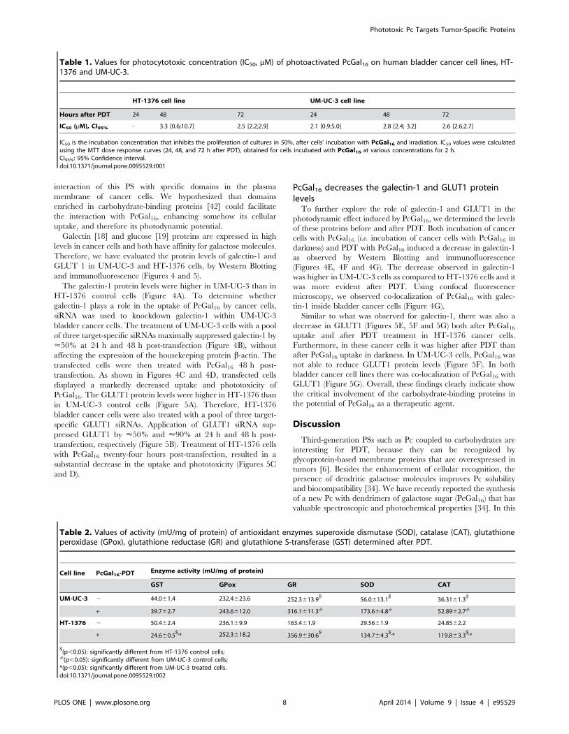
Table 1. Values for photocytotoxic concentration (IC50, mM) of photoactivated PcGal16 on human bladder cancer cell lines, HT-1376 and UM-UC-3.
HT-1376 cell line UM-UC-3 cell line
Hours after PDT 24 48 72 24 48 72
IC50 (mM), CI95% - 3.3 [0.6;10.7] 2.5 [2.2;2.9] 2.1 [0.9;5.0] 2.8 [2.4; 3.2] 2.6 [2.6;2.7]
IC50 is the incubation concentration that inhibits the proliferation of cultures in 50%, after cells’ incubation with PcGal16 and irradiation. IC50 values were calculatedusing the MTT dose response curves (24, 48, and 72 h after PDT), obtained for cells incubated with PcGal16 at various concentrations for 2 h.CI95%: 95% Confidence interval.doi:10.1371/journal.pone.0095529.t001
Table 2. Values of activity (mU/mg of protein) of antioxidant enzymes superoxide dismutase (SOD), catalase (CAT), glutathioneperoxidase (GPox), glutathione reductase (GR) and glutathione S-transferase (GST) determined after PDT.
Cell line PcGal16-PDT Enzyme activity (mU/mg of protein)
GST GPox GR SOD CAT
UM-UC-3 2 44.061.4 232.4623.6 252.3613.9$
56.0613.1$
36.3161.3$
+ 39.762.7 243.6612.0 316.1611.3# 173.664.8# 52.8962.7#
HT-1376 2 50.462.4 236.169.9 163.461.9 29.5661.9 24.8562.2
+ 24.660.5$,* 252.3618.2 356.9630.6
$134.764.3
$,* 119.863.3$,*
$(p,0.05): significantly different from HT-1376 control cells;
#(p,0.05): significantly different from UM-UC-3 control cells;*(p,0.05): significantly different from UM-UC-3 treated cells.doi:10.1371/journal.pone.0095529.t002
Phototoxic Pc Targets Tumor-Specific Proteins
PLOS ONE | www.plosone.org 8 April 2014 | Volume 9 | Issue 4 | e95529

study, we showed that PcGal16 is a nontoxic compound per se, and
has high photocytotoxic efficiency in two bladder cancer cell lines,
which is paralleled with its high ability to produce ROS and to
induce oxidative stress (Figure 6).
The high intracellular uptake of the glycoconjugated PS,
PcGal16, can be explained by the presence of carbohydrate
cellular transporters or receptors present at the cell surface.
Although the PcGal16 uptake was quite similar in the two bladder
cancer cell lines, the expression of carbohydrate-binding proteins
GLUT1 and galectin-1 is different amongst them. Besides its role
in the import and export of glucose [19], the isoform of glucose
transporter GLUT1 also transports D-galactose [19] having lower
Figure 4. Knockdown of galectin-1 decreases de uptake and phototoxicity of PcGal16. Western blotting analysis and quantification ofgalectin-1 protein levels in HT-1376 and UM-UC-3 cells (panel A), in HT-1376 cells (panel E) or UM-UC-3 cells (panel F) after uptake with PcGal16 indarkness and after PDT. b-actin was blotted as loading control. Quantitative analysis of galectin-1 (normalized to b -actin) expressed as a ratio of thelevels found in HT-1376 cells. *(p,0.05) significantly different from HT-1376 cells. Quantitative analysis of galectin-1 (normalized to b-actin) expressedas a ratio of the levels found in untreated HT-1376 or UM-UC-3 cells (panel E, F). Data represents mean 6 S.D. of five independent experiments. *(p,0.05) significantly different from untreated HT-1376 or UM-UC-3 cells. Knockdown of galectin-1 in UM-UC-3 bladder cancer cells as determined byWestern blotting 24 and 48 h post-transfection (panel B). Quantitative analysis of galectin-1 (normalized to b-actin) expressed as a ratio of the levelsfound in non-transfected control cells. Data represents mean 6 S.D. of five independent experiments. *(p,0.05), $(p,0.05) significantly differentfrom non-transfected control cells or cells treated with scrambled siRNA, respectively. Intracellular uptake of PcGal16 by UM-UC-3 bladder cancer cellstransfected with galectin-1 siRNA (panel C). The cells were incubated with PcGal16 48 h post-transfection with galectin-1 siRNA. Data are the mean 6S.D. of at least three independent experiments performed in triplicates. *(p,0.05) significantly different from non-transfected control cells.Photocytotoxic effects after PcGal16-PDT in UM-UC-3 cells transfected with galectin-1 siRNA (panel D). Phototoxicity was evaluated 72 h after PDT.Data are the mean 6 S.D. of at least three independent experiments performed in triplicates. *(p,0.05), ***(p,0.0001) significantly different fromcontrol cells. $(p,0.05), significantly different from PDT with PcGal16 in non-transfected cells. Representative fluorescence images (panel G) ofgalectin-1 protein (green) in cancer cells before and after incubation with PcGal16 (red), with DAPI staining the nucleus (blue). Scale bars 20 mm.doi:10.1371/journal.pone.0095529.g004
Phototoxic Pc Targets Tumor-Specific Proteins
PLOS ONE | www.plosone.org 9 April 2014 | Volume 9 | Issue 4 | e95529

affinity for it than for D-glucose. Studies have been suggested that
the hydroxyl groups in C1, C3 and C4 positions of D-galactose are
hydrogen bond acceptors for GLUT1 sugar uptake site [43]. Like
other galectins, galectin-1 has a carbohydrate-recognition domain
(CRD) able to recognize and bind b-galactose [17]. Our assays
demonstrated that galectin-1 and GLUT1 are both expressed by
UM-UC-3 and HT-1376 cells. However, HT-1376 cells present
higher GLUT1 levels compared with UM-UC-3 cells, and the
contrary was observed for galectin-1. Although a similar PcGal16
uptake was observed in the two bladder cancer cell lines, both
GLUT1 and galectin-1 may contribute for its specificity modu-
lating the intracellular uptake. Knockdown of galectin-1 and
GLUT1 in UM-UC-3 and HT-1376 cells, respectively, was
associated with a marked decrease of PcGal16 uptake and
phototoxicity. Together, these data demonstrated that galectin-1
Figure 5. Knockdown of GLUT1 decreases de uptake and phototoxicity of PcGal16. Western blotting analysis and quantification of GLUT1protein levels in HT-1376 and UM-UC-3 cells (panel A), in HT-1376 cells (panel E) or UM-UC-3 cells (panel F) after uptake with PcGal16 in darkness andafter PDT. b-actin was blotted as loading control. Quantitative analysis of GLUT1 (normalized to b-actin) expressed as a ratio of the levels found in HT-1376 cells (panel A). *(p,0.05) significantly different from HT-1376 cells. Quantitative analysis of GLUT1 (normalized to b-actin) expressed as a ratio ofthe levels found in untreated HT-1376 or UM-UC-3 cells (panel E, F). Data represents mean 6 S.D. of five independent experiments. *(p,0.05)significantly different from untreated HT-1376 cells. Knockdown of GLUT1 in HT-1376 bladder cancer cells as determined by Western blotting 24 and48 h post-transfection (panel B). Quantitative analysis of GLUT1 (normalized to b-actin) expressed as a ratio of the levels found in non-transfectedcontrol cells. Data represents mean 6 S.D. of five independent experiments. *(p,0.05), ***(p,0.0001) significantly different from non-transfectedcontrol cells. $(p,0.05), $$$(p,0.0001) significantly different from cells treated with scrambled siRNA. Intracellular uptake of PcGal16 by HT-1376bladder cancer cells transfected with GLUT1 siRNA (panel C). The cells were incubated with PcGal16 24 h post-transfection. Data are the mean 6 S.D.of at least three independent experiments performed in triplicates. *(p,0.05) significantly different from non-transfected control cells. Photocytotoxiceffects after PcGal16-PDT in UM-UC-3 cells transfected with galectin-1 siRNA (panel D). Phototoxicity was evaluated 72 h after PDT. Data are the mean6 S.D. of at least three independent experiments performed in triplicates. *(p,0.05), ***(p,0.0001) significantly different from control cells. *(p,
0.05), significantly different from PDT with PcGal16 in non-transfected cells. Representative fluorescence images (panel G) of GLUT1 protein (green) inHT-1376 and UM-UC-3 cells before and after incubation with PcGal16 (red), with DAPI staining the nucleus (blue). Scale bars 20 mm.doi:10.1371/journal.pone.0095529.g005
Phototoxic Pc Targets Tumor-Specific Proteins
PLOS ONE | www.plosone.org 10 April 2014 | Volume 9 | Issue 4 | e95529
and GLUT1 contribute for the efficacy of PDT mediated by
PcGal16.
Interestingly, although the similar uptake of PcGal16 by UM-
UC-3 and HT-1376 cells, the phototoxicity induced 24 h after
PDT was higher in UM-UC-3 cells than in HT-1376 cells. Such
lack of association between uptake and phototoxicity has been
described [44,45]. We investigated whether the higher phototox-
icity observed in UM-UC-3 cells was due to higher production of
ROS and/or higher oxidative damage compared with that in HT-
1376 cells. As expected, the ability of PcGal16 to produce ROS
was higher in UM-UC-3 than in HT-1376 cells.
In PDT, it has been described that ROS can be generated by
two photochemical reactions [46,47]. In type-II photochemical
reactions, the excited PS in its triplet state can transfer its energy to
molecular oxygen leading to the formation of singlet oxygen.
Type-I photochemical reactions happen when an excited PS reacts
with a biological substrate forming radicals and radical ions.
Treatment with ROS quenchers demonstrated that in UM-UC-3
cells, singlet oxygen should have a high effect since cell death was
highly reduced with quenchers of singlet oxygen (sodium azide and
histidine). Further studies are needed to gain insight into the
contribution of specific ROS in PcGal16-mediated cell death after
PDT.
Interestingly, we observed that PDT with PcGal16 has a long-
term phototoxic effect in both cancer cell lines. Cytotoxicity assays
(MTT, trypan blue and TUNEL assays) performed 72 h after
PDT demonstrated that UM-UC-3 cells were not able to recover.
Moreover, in HT-1376 cells there was a marked induction of cell
death occurring from 24 to 72 h after PDT with PcGal16. The
three distinct cytotoxic methods used in the present work are
widely applied in the study of cell death: MTT (indicator of
metabolic activity), trypan blue staining (indicator of membrane
integrity loss occurring in necrosis or in late stages of apoptosis)
and TUNEL assay (indicator of DNA fragmentation, a key factor
of apoptosis). Cell death in PDT may occur by apoptosis or
necrosis, or even by a combination of the two mechanisms [2]. A
more specific and comprehensive study is needed to understand
the specific cell death pathways induced after PDT with PcGal16 in
the bladder cancer cells used in this study. The different cell death
obtained 24 h after PDT in UM-UC-3 and HT-1376 cells can be
partially explained by the different amount of ROS present in both
cells lines after irradiation. In addition, the resistance exhibited by
HT-1376 cells could be due to the presence of efficient protective
mechanisms, at least in the first stages after photodynamic
treatment. Cytoprotective mechanisms initiated by cancer cells
after PDT are well-known [47]. The increase of antioxidant
molecules (e.g. gluthathione, vitamin C and vitamin E) [48] and the
induction of genes encoding proteins involved in apoptosis or in
the repair of lesions [49] are two of the well-known cytoprotective
mechanisms induced after PDT. Another one is based on the
equilibrium between photo-oxidative impairment of cells by ROS
versus elimination of ROS by the activity of cellular antioxidant
enzymes. Recent studies have shown that PDT caused increased-
antioxidant enzymes activity and expression [50]. Thus, PDT
efficacy can be influenced by the antioxidant response of the
enzymes SOD, the GSH system and CAT.
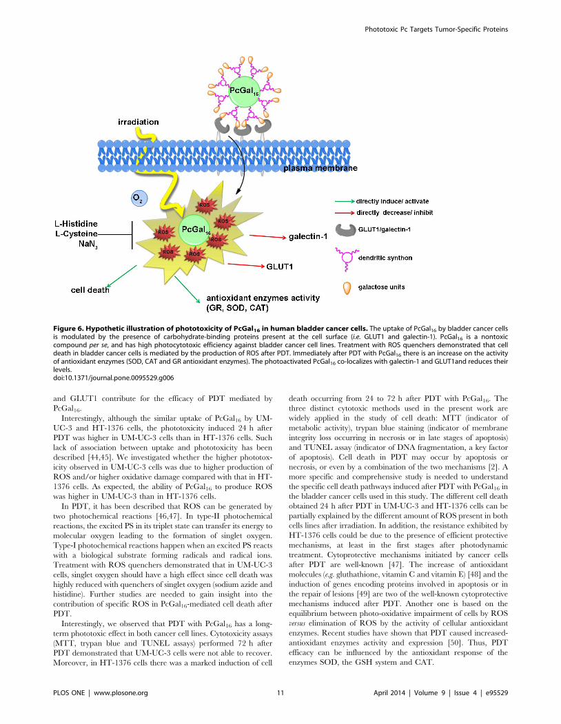
Figure 6. Hypothetic illustration of phototoxicity of PcGal16 in human bladder cancer cells. The uptake of PcGal16 by bladder cancer cellsis modulated by the presence of carbohydrate-binding proteins present at the cell surface (i.e. GLUT1 and galectin-1). PcGal16 is a nontoxiccompound per se, and has high photocytotoxic efficiency against bladder cancer cell lines. Treatment with ROS quenchers demonstrated that celldeath in bladder cancer cells is mediated by the production of ROS after PDT. Immediately after PDT with PcGal16 there is an increase on the activityof antioxidant enzymes (SOD, CAT and GR antioxidant enzymes). The photoactivated PcGal16 co-localizes with galectin-1 and GLUT1and reduces theirlevels.doi:10.1371/journal.pone.0095529.g006
Phototoxic Pc Targets Tumor-Specific Proteins
PLOS ONE | www.plosone.org 11 April 2014 | Volume 9 | Issue 4 | e95529

Our data demonstrated that after PDT with PcGal16 there was
an increase in the activity of SOD, CAT and GR antioxidant
enzymes in both cell lines, being higher in HT-1376 than in UM-
UC-3 cells. This higher antioxidant defense of HT-1376 cells
against ROS can explain the results obtained 24 h after treatment.
However, it is hypothesized that this was not maintained for 72 h
after PDT since for this time point there was a massive cell death.
This not only suggests that in this cell line there is a temporal
relationship between ROS levels and cell death, but shows that
antioxidant enzymes activity is of greater importance in protecting
HT-1376 cells for at least 24 h after PDT with PcGal16. Regarding
the activity of antioxidant enzymes, in HT-1376 cells it was also
observed a decrease in the activity of GST, which is an enzyme
implicated in cells defense against oxidation products. This
enzyme has been described as protecting cells from DNA
desintegration and drug toxicity [51]. GST isoforms are overex-
pressed in multidrug resistant tumors having an important role in
tumors drug resistance by direct detoxification or inhibition of the
MAP kinase pathway [51]. Thus, the higher cell death observed in
HT-1376 cells 72 h after treatment can be also related with the
activity of GST. A decrease in the activity of GST can be
associated with DNA fragmentation and cell death 72 h after
treatment.
Understanding the role of galactose moieties in the recognition
of the PS by cancer cells may allow the investigation and
development of more focused therapeutic strategies. Thus, we
investigated whether PcGal16 could be directly recognized by
specific carbohydrate-binding proteins present at the plasma
membrane. Consistently, the photoactivated PcGal16 was shown
to co-localize and reduce the levels of the plasma membrane
proteins galectin-1 and GLUT1. Moreover, the immunofluores-
cence and Western Blotting studies demonstrated that, although its
non-dark toxicity, PcGal16 decreases the levels of galectin-1 and
GLUT1 proteins. A plausible explanation for the decreased levels
of the galactose binding proteins, galectin-1 and GLUT1, after
incubation with PcGal16 can be the masking of the epitope, which
can block antibody-epitope binding due to changes in protein
conformation or, eventually, endocytosis of these proteins and
subsequent degradation. Thus, the changes observed in the levels
of galectin-1 and GLUT1 could be induced directly by the binding
of PcGal16 to the carbohydrate-binding proteins and/or indirectly
by the generation of ROS after PDT with PcGal16.
Although significant progress has been made in research related
with the role of galectins in cancer, the information underlying the
molecular mechanisms that control the expression of these
proteins in tumour cells is scarce. The interaction of PSs with
galectins (namely galectin-1 and galectin-3) has been studied by
spectroscopic studies [52] and molecular modeling analysis [6,27];
however, they have not been validated by in vitro studies. As far as
we know, there are no in vitro reports indicating whether PSs can
modulate the expression of carbohydrate-binding proteins such as
galectin-1 and GLUT1. Knowing that galectin-1 expression is
correlated with cell metastatic potential [18,53] and contributes to
tumor progression and resistance after conventional cancer
therapeutic modalities [18], the ability of PcGal16 to reduce the
levels of galectin-1 after its uptake and/or photoactivation
prompted us to envisage PcGal16 as a potential candidate for
cancer treatment.
Knowing that the overexpression of GLUTs is involved in
tumor glycolysis - one of the biochemical ‘‘hallmarks’’ of cancer -
the efficiency of PcGal16 as an efficient anti-cancer PS is also
evidenced by its ability to reduce GLUT1. GLUT1 is an attractive
target to consider in the development of new PSs because it is
lower expressed in normal-epithelial tissues or benign epithelial
cell tumors when compared with human cancer cells [54]. The
function of GLUT1 in the tumorogenesis process has been
demonstrated by in vitro and in vivo studies, where the overexpres-
sion of GLUT1 antisense resulted in the inhibition of HL60
leukaemia cells proliferation and MKN-45 derived xenografs,
respectively [55,56]. Based on the results of the current study, we
envisage PcGal16 as a promising therapeutic agent for the
treatment of bladder cancer. Further studies are warranted to
investigate the selectivity and photototoxicity of this PS in an in vivo
model of bladder cancer, to contribute to a possible impact on
clinical practice.
Supporting Information
Figure S1 Chemical structures of free phthalocyanine PcF16 and
galacto-dendrimer phthalocyanine PcGal16.
(TIF)
Figure S2 PcGal16 is non-toxic in darkness, PcF16 is non-toxic in
darkness and after PDT. Non-dark toxicity of various concentra-
tions of PcGal16 in HT-1376 and UM-UC-3 cells (panel A). Non-
dark toxicity was assessed using the MTT colorimetric assay 24,
48, and 72 h after treat HT-1376 and UM-UC-3 cells (panel B).
Toxicity of PcF16 at 5 mM in darkness and after PDT (panel C) in
HT-1376 and UM-UC-3 cells. The toxicity was assessed using the
MTT colorimetric assay 24 h after treat HT-1376 and UM-UC-3
cells. Data are the mean 6 S.D. of at least three independent
experiments performed in triplicates.
(TIF)
Acknowledgments
We grateful appreciate the help of Dr. Celia Gomes (IBILI-FMUC,
Portugal) with the spectrofluorometric measurements in IVIS Lumina XR
equipment.
We thank Dr. Filomena Botelho (IBILI-FMUC, Portugal) for the
generous supply of HT-1376 human bladder cancer cells.
P. Pereira and S. Silva acknowledge fellowships from FCT (SFRH/BD/
85941/2012 and SFRH/BPD/64812/2009, respectively).
We appreciate the help of Dr. Celia Gomes (IBILI-FMUC, Portugal)
with the spectrofluorometric measurements in IVIS Lumina XR
equipment.
Author Contributions
Conceived and designed the experiments: PP RF JT. Performed the
experiments: PP SS. Analyzed the data: PP RF JT. Contributed reagents/
materials/analysis tools: PP SS. Wrote the paper: PP RF JT CFR JC.
References
1. Allison RR, Sibata CH (2010) Oncologic photodynamic therapy photosensitiz-
ers: a clinical review. Photodiagnosis Photodyn Ther 7:61–75.
2. Almeida RD, Manadas BJ, Carvalho AP, Duarte CB (2004) Intracellular
signaling mechanisms in photodynamic therapy. Biochim Biophys Acta
1704:59–86.
3. Soares ARM, Tome JPC, Neves MGPMS, Tome AC, Cavaleiro JAS, et al.
(2009) Synthesis of water-soluble phthalocyanines bearing four or eight D-
galactose units, Carbohyd Res 334:507–510.
4. Silva JN, Silva AMG, Tome JPC, Ribeiro AO, Domingues MRM, et al. (2008)
Photophysical properties of a photocytotoxic fluorinated chlorinconjugated to
four b-cyclodextrins. Photochem Photobiol Sci 7:834–843.
5. Pereira PMR, Carvalho JJ, Silva S, Cavaleiro JAS, Schneider RJ, et al. (2014)
Porphyrin conjugated with serum albumins and monoclonal antibodies boosts
efficiency in targeted destruction of human bladder cancer cells, Org Biomol
Chem 12:1804–1811.
Phototoxic Pc Targets Tumor-Specific Proteins
PLOS ONE | www.plosone.org 12 April 2014 | Volume 9 | Issue 4 | e95529
6. Zheng G, Graham A, Shibata M, Missert JR, Oseroff AR, et al. (2001) Synthesis
of beta-galactose-conjugated chlorins derived by enyne metathesis as galectin-specific photosensitizers for photodynamic therapy. J Org Chem 66:8709–8716.
7. Soares ARM, Neves MGPMS, Tome AC, Iglesias-de la Cruz MC, Zamarron A,
et al. (2012) Glycophthalocyanines as Photosensitizers for Triggering Mitotic:Catastrophe and Apoptosis On Cancer Cells. Chem Res Toxicol 25:940–951.
8. Daly R, Vaz G, Davies AM, Senge MO, Scanlan EM (2012) Synthesis andbiological evaluation of a library of glycoporphyrin compounds. Chemistry
18:14671–14679.
9. Vedachalam S, Choi BH, Pasunooti KK, Ching KM, Lee K, et al. (2011)Glycosylated porphyrin derivatives and their photodynamic activity in cancer
cells. MedChemComm 2:371–377.10. Ernst B, Magnani JL (2009) From carbohydrate leads to glycomimetic drugs.
Nat Rev Drug Discov 8:661–677.11. Lourenco LMO, Tome JPC, Domingues MRM, Domingues P, Costa PJ, et al.
(2009) Synthesis and differentiation of alpha- and beta-glycoporphyrin
stereoisomers by electrospray tandem mass spectrometry. Rapid CommunMass Spectrom 23:3478–3483.
12. Figueira F, Pereira PMR, Silva S, Cavaleiro JAS, Tome JPC (2014) Porphyrinsand Phthalocyanines decorated with dendrimers: Synthesis and biomedical
applications. Curr Org Synth In press.
13. Gary-Bobo M, Vaillant O, Maynadier M, Basile I, Gallud A, et al. (2013)Targeting multiplicity: the key factor for anti-cancer nanoparticles. Curr Med
Chem 20:1946–1955.14. Lourenco LMO, Neves MGPMS, Cavaleiro JAS, Tome JPC (2014) Synthetic
approaches to glycophthalocyanines. Tetrahedron In press.15. David A (2010) Carbohydrate-based Biomedical Copolymers for Targeted
Delivery of Anticancer Drugs. Isr J Chem 50:204–219.
16. Sharon N, Lis H (1989) Lectins as cell recognition molecules. Science 246:227–234.
17. Liu FT, Rabinovich GA (2005) Galectins as modulators of tumour progression.Nat Rev Cancer 5:29–41.
18. Cindolo L, Benvenuto G, Salvatore P, Pero R, Salvatore G, et al. (1999)
Galectin-1 and galectin-3 expression in human bladder transitional-cellcarcinomas. Int J Cancer 84:39–43.
19. Carruthers A, DeZutter J, Ganguly A, Devaskar SU (2009) Will the originalglucose transporter isoform please stand up! Am J Physiol Endocrinol Metab
297:E836–E848.20. Fernandes R, Carvalho AL, Kumagai A, Seica R, Hosoya K, et al. (2004)
Downregulation of retinal GLUT1 in diabetes by ubiquitinylation. Mol Vis
10:618–628.21. Fernandes R, Hosoya K, Pereira P (2011) Reactive oxygen species downregulate
glucose transport system in retinal endothelial cells. Am J Physiol-Cell Ph 300:C927–C936.
22. Silva AMG, Tome AC, Neves MGPMS, Cavaleiro JAS, Perrone D, et al. (2005)
Porphyrins in 1,3-dipolar cycloadditions with sugar azomethine ylides. Synthesisof pyrrolidinoporphyrin glycoconjugates. Synlett 0857–0859.
23. Choi CF, Huang JD, Lo PC, Fong WP, Ng DKP (2008) Glycosylated zinc(II)phthalocyanines as efficient photosensitisers for photodynamic therapy.
Synthesis, photophysical properties and in vitro photodynamic activity. OrgBiomol Chem 6:2173–2181.
24. Iqbal Z, Hanack M, Ziegler T (2009) Synthesis of an octasubstituted galactose
zinc(II) phthalocyanine. Tetrahedron Lett 50:873–875.25. Lee PPS, Lo PC, Chan EYM, Fong WP, Ko WH, et al. (2005) Synthesis and in
vitro photodynamic activity of novel galactose-containing phthalocyanines.Tetrahedron Lett 46:1551–1554.
26. Park YK, Bold B, Cui BC, Bai JQ, Lee WK, et al. (2008) Binding affinities of
carbohydrate-conjugated chlorins for galectin-3. B Korean Chem Soc 29:130–134.
27. Pandey SK, Zheng X, Morgan J, Missert JR, Liu TH, et al. (2007)Purpurinimide carbohydrate conjugates: effect of the position of the carbohy-
drate moiety in photosensitizing efficacy. Mol Pharm 4:448–464.
28. Singh S, Aggarwal A, Thompson S, Tome JPC, Zhu X, et al. (2010) Synthesisand photophysical properties of thioglycosylated- chlorins, isobacteriochlorins
and bacteriochlorins for bioimaging and diagnostics. Bioconjugate Chem21:2136–2146.
29. Fujimoto K, Miyata T, Aoyama Y (2000) Saccharide-directed cell recognitionand molecular delivery using macrocyclic saccharide clusters: Masking of
hydrophobicity to enhance the saccharide specificity. J Am Chem Soc
122:3558–3559.30. Ribeiro AO, Tome JPC, Neves MGPMS, Tome AC, Cavaleiro JAS, et al.
(2006) [1,2,3,4-tetrakis(alpha/beta-D-galactopyranos-6-yl)-phthalocyaninato]-zinc(II): a water-soluble phthalocyanine. Tetrahedron Lett 47:9177–9180.
31. Gillies ER, Frechet JMJ (2005) Dendrimers and dendritic polymers in drug
delivery. Drug Discov Today 10:35–43.
32. Wolinsky JB, Grinstaff MW (2008) Therapeutic and diagnostic applications of
dendrimers for cancer treatment. Adv Drug Deliv Rev 60:1037–1055.
33. Klajnert B, Rozanek M, Bryszewska M (2012) Dendrimers in photodynamic
therapy. Curr Med Chem 19:4903–4912.
34. Silva S, Pereira PMR, Silva P, Paz FA, Faustino MA, et al. (2012) Porphyrin and
phthalocyanine glycodendritic conjugates: synthesis, photophysical and photo-
chemical properties. Chem Commun (Camb) 48:3608–3610.
35. Ballardini R, Colonna B, Gandolfi MT, Kalovidouris SA, Orzel L, et al. (2003)
Porphyrin-containing glycodendrimers. Eur J Org Chem 2:288–294.
36. Wang ZJ, Chauvin B, Maillard P, Hammerer F, Carez D, et al. (2012)
Glycodendrimeric phenylporphyrins as new candidates for retinoblastoma PDT:
Blood carriers and photodynamic activity in cells. J Photochem Photobiol B
115:16–24.
37. Ballut S, Makky A, Loock B, Michel JP, Maillard P, et al. (2009) New strategy for
targeting of photosensitizers. Synthesis of glycodendrimeric phenylporphyrins,
incorporation into a liposome membrane and interaction with a specific lectin.
Chem Comm 2:224–226.
38. Bancirova M (2011) Sodium azide as a specific quencher of singlet oxygen
during chemiluminescent detection by luminol and Cypridina luciferin
analogues. Luminescence 26:685–688.
39. Aruoma OI, Halliwell B, Hoey BM, Butler J (1989) The antioxidant action of N-
acetylcysteine: its reaction with hydrogen peroxide, hydroxyl radical, superoxide,
and hypochlorous acid. Free Radic Biol Med 6:593–597.
40. Sies H (1993) Strategies of antioxidant defense. Eur J Biochem 215:213–219.
41. Weydert CJ, Cullen JJ (2010) Measurement of superoxide dismutase, catalase
and glutathione peroxidase in cultured cells and tissue. Nat Protoc 5:51–66.
42. Lotan R, Raz A (1988) Lectins in cancer cells. Ann N Y Acad Sci 551:385–398.
43. Barnett JEG, Holman GD, Chalkley RA, Munday KA (1975) Evidence for two
asymmetric conformational states in the human erythrocyte sugar-transport
system. Biochem J 145:417–429.
44. Hirohara S, Obata M, Ogata S, Ohtsuki C, Higashida S, et al. (2005) Cellular
uptake and photocytotoxicity of glycoconjugated chlorins in HeLa cells.
J Photoch Photobio B 78:7–15.
45. Laville I, Pigaglio S, Blais JC, Doz F, Loock B, et al. (2006) Photodynamic
efficiency of diethylene glycol-linked glycoconjugated porphyrins in human
retinoblastoma cells. J Med Chem 49:2558–2567.
46. Plaetzer K, Krammer B, Berlanda J, Berr F, Kiesslich T (2009) Photophysics
and photochemistry of photodynamic therapy: fundamental aspects. Laser Med
Sci 24:259–268.
47. Buytaert E, Dewaele M, Agostinis P (2007) Molecular effectors of multiple cell
death pathways initiated by photodynamic therapy. Biochimica et biophysica
acta 1776:86–107.
48. Sattler UGA, Mueller-Klieser W (2009) The anti-oxidant capacity of tumour
glycolysis. International journal of radiation biology 85:963–971.
49. Oleinick NL, Morris RL, Belichenko I (2002) The role of apoptosis in response
to photodynamic therapy: what, where, why, and how. Photochemical &
photobiological sciences : Official journal of the European Photochemistry
Association and the European Society for Photobiology 1:1–21.
50. Saczko J, Kulbacka J, Chwilkowska A, Pola A, Lugowski M, et al. (2007)
Cytosolic superoxide dismutase activity after photodynamic therapy, intracellu-
lar distribution of Photofrin II and hypericin, and P-glycoprotein localization in
human colon adenocarcinoma. Folia Histochem Cyto 45:93–98.
51. Townsend DM, Tew KD (2003) The role of glutathione-S-transferase in anti-
cancer drug resistance. Oncogene 22:7369–7375.
52. Bogoeva VP, Varriale A, John CM, D’Auria S (2010) Human galectin-3
interacts with two anticancer drugs. Proteomics 10:1946–1953.
53. Chiariotti L, Berlingieri MT, De Rosa P, Battaglia C, Berger N, et al. (1992)
Increased expression of the negative growth factor, galactoside-binding protein,
gene in transformed thyroid cells and in human thyroid carcinomas. Oncogene
7:2507–2511.
54. Younes M, Lechago LV, Somoano JR, Mosharaf M, Lechago J (1996) Wide
expression of the human erythrocyte glucose transporter Glut1 in human
cancers. Cancer Res 56:1164–1167.
55. Chan JYW, Kong SK, Choy YM, Lee CY, Fung KP (1999) Inhibition of glucose
transporter gene expression by antisense nucleic acids in HL-60 leukemia cells.
Life Sci 65:63–70.
56. Noguchi Y, Saito A, Miyagi Y, Yamanaka S, Marat D, et al. (2000) Suppression
of facilitative glucose transporter 1 mRNA can suppress tumor growth. Cancer
Letters 154:175–182.
Phototoxic Pc Targets Tumor-Specific Proteins
PLOS ONE | www.plosone.org 13 April 2014 | Volume 9 | Issue 4 | e95529

Galactodendritic Phthalocyanine Targets Carbohydrate-Binding Proteins Enhancing Photodynamic TherapyPatrıcia M. R. Pereira1,2, Sandrina Silva1, Jose A. S. Cavaleiro1, Carlos A. F. Ribeiro2, Joao P. C. Tome1,3*,
Rosa Fernandes2,4,5*
1 QOPNA and Department of Chemistry, University of Aveiro, Aveiro, Portugal, 2 Laboratory of Pharmacology and Experimental Therapeutics, Institute for Biomedical
Imaging and Life Sciences (IBILI), Faculty of Medicine, University of Coimbra, Azinhaga de Santa Comba, Coimbra, Portugal, 3 Department of Organic Chemistry, Ghent
University, Gent, Belgium, 4 Center of Investigation in Environment, Genetics and Oncobiology, Coimbra, Portugal, 5 Center of Ophthalmology and Vision Sciences, IBILI,
Faculty of Medicine, University of Coimbra, Coimbra, Portugal
Abstract
Photosensitizers (PSs) are of crucial importance in the effectiveness of photodynamic therapy (PDT) for cancer. Due to theirhigh reactive oxygen species production and strong absorption in the wavelength range between 650 and 850 nm, wheretissue light penetration is rather high, phthalocyanines (Pcs) have been studied as PSs of excellence. In this work, we reportthe evaluation of a phthalocyanine surrounded by a carbohydrate shell of sixteen galactose units distributed in a dendriticmanner (PcGal16) as a new and efficient third generation PSs for PDT against two bladder cancer cell lines, HT-1376 and UM-UC-3. Here, we define the role of galacto-dendritic units in promoting the uptake of a Pc through interaction with GLUT1and galectin-1. The photoactivation of PcGal16 induces cell death by generating oxidative stress. Although PDT with PcGal16
induces an increase on the activity of antioxidant enzymes immediately after PDT, bladder cancer cells are unable to recoverfrom the PDT-induced damage effects for at least 72 h after treatment. PcGal16 co-localization with galectin-1 and GLUT1and/or generation of oxidative stress after PcGal16 photoactivation induces changes in the levels of these proteins.Knockdown of galectin-1 and GLUT1, via small interfering RNA (siRNA), in bladder cancer cells decreases intracellular uptakeand phototoxicity of PcGal16. The results reported herein show PcGal16 as a promising therapeutic agent for the treatmentof bladder cancer, which is the fifth most common type of cancer with the highest rate of recurrence of any cancer.
Citation: Pereira PMR, Silva S, Cavaleiro JAS, Ribeiro CAF, Tome JPC, et al. (2014) Galactodendritic Phthalocyanine Targets Carbohydrate-Binding ProteinsEnhancing Photodynamic Therapy. PLoS ONE 9(4): e95529. doi:10.1371/journal.pone.0095529
Editor: Michael Hamblin, MGH, MMS, United States of America
Received December 17, 2013; Accepted March 27, 2014; Published April 24, 2014
Copyright: � 2014 Pereira et al. This is an open-access article distributed under the terms of the Creative Commons Attribution License, which permitsunrestricted use, distribution, and reproduction in any medium, provided the original author and source are credited.
Funding: The synthesis of the photosensitizer was supported by the grant PTDC/CTM/101538/2008 (FCT- Fundacao para a Ciencia e a Tecnologia, Portugal).Thanks are due to FCT and Fundo Europeu de Desenvolvimento Regional (FEDER) for funding IBILI (Pest-C/SAU/UI3282/2011 and Pest-C/SAU/UI3282/2013) andQOPNA (PEst-C/QUI/UI0062/2013) research units. Thanks are due to ACIMAGO (ref. 12/12). The funders had no role in study design, data collection and analysis,decision to publish, or preparation of the manuscript.
Competing Interests: The authors have declared that no competing interests exist.
* E-mail: [email protected] (JT); [email protected] (RF)
Introduction
Conventional photodynamic therapy (PDT) combines a non-
toxic photosensitizer (PS), light irradiation at a specific wavelength
and tissue molecular oxygen to produce cytotoxic reactive oxygen
species (ROS) [1,2]. The molecular mechanisms underlying PDT
are not clearly understood. However, it has been described that
the generation of ROS will trigger signalling pathways that
ultimately destroy the targeted tissue. Cell death in PDT may
occur by apoptotic and by non-apoptotic mechanisms (e.g.
necrosis), or even by a combination of the two mechanisms [2].
Additionally, studies suggest that cell death pathway induced after
PDT depends on the PS and its intracellular localization, the PDT
dose and the cell metabolic potential (e.g. its intrinsic antioxidant
capacity) [2]. To enhance the specific deliver/target of PSs in
cancer cells, third generation PSs have been synthesized, by
conjugating them with biochemical motifs [3–5]. Among new
third generation PSs, the advances in the past years concerning
glycobiology have spurred the development of carbohydrate-based
molecules for cancer treatment by PDT [3,4,6–14].
Carbohydrates have a strong potential as PS-delivery systems,
because they are biocompatible molecules with a rapid cellular
uptake and specific recognition by lectin proteins, which play an
important role in several biochemical signalling pathways impli-
cated in cancer metastasis, cell growth and inflammation [15,16].
The exact interaction mechanism of PS-carbohydrate conjugates
with cancer cells is still unknown. However, it is expected that the
specific (non-covalent) binding of carbohydrates with lectins [16],
promotes the accumulation of the glyco-conjugate inside cells by
the endocytic pathway. In addition, the expression of certain
carbohydrate-binding lectins (e.g. galectins) is higher in cancer cells
than in non-tumoral cells [17].
Among carbohydrates, the biocompatibility of galactose mole-
cules and their specific recognition by galectins overexpressed in
cancer cells (e.g. galectin-1 and galectin-3 [18]) have led to the
development of galacto-conjugated PSs. Besides galectins, galac-
tose carbohydrates can bind to GLUT1 (a well-known glucose
transporter [19–21]). The steriospecificity of GLUT1 (recognizing
both D-glucose and D-galactose) has been reported [19–21].
Galactose is a C4 epimer of glucose that can bind the glucose-
binding site of GLUT1. There is strong evidence in literature that
conjugation of carbohydrates (monosaccharides such as glucose
and galactose, disaccharides such as lactose) with porphyrinoids
[6,8,9,22–30] can improve the accumulation of PSs in cancer cells
PLOS ONE | www.plosone.org 1 April 2014 | Volume 9 | Issue 4 | e95529
and, consequently, their photoactivity. Furthermore, it has been
reported a marked contrast in terms of adsorption on the cells
between galactose and glucose conjugated PSs. The former
presented a selective uptake by rat hepatoma RLC-116 cells [29].
Recently, the emerging role of dendrimers (with well-defined
nano-scaled structures) in biological systems has highlighted their
potential benefits for the preparation of new anticancer drugs [31–
33]. Regarding dendritic units of specific carbohydrates, it is well-
known their multivalent interactions with lectins, promoting a
synergistic increase in binding affinity [31]. The photodynamic
efficiency of porphyrins conjugated with glycodendrimers has been
reported in the literature [12,34–37]. However, the in vitro PDT
studies with the corresponding phthalocyanines (Pcs) are scarce.
Recently, we have reported the synthesis of a new Pc decorated
with sixteen molecules of galactose (in a dendritic manner,
PcGal16, Figure S1) [34]. PcGal16 demonstrated strong absor-
bance in the red spectral region (600–800 nm), fluorescence
emission bands at 734 and 805 nm, solubility in a phosphate
buffered saline (PBS) solution and interaction with human serum
albumin [34]. Additionally, PcGal16 demonstrated photostability
and ability to generate ROS after photoactivation. The present
study was undertaken to validate the in vitro photodynamic efficacy
of this PcGal16 from the standpoint of its uptake by bladder cancer
cells (HT-1376 and UM-UC-3, derived from transitional cell
carcinoma) to interaction with carbohydrate-binding proteins;
induction of phototoxicity, ROS production and activity of
antioxidant enzymes after PDT. Our findings show that PcGal16
has a strong photodynamic efficiency in an in vitro system of
bladder cancer.
Materials and Methods
Synthesis of galacto-dendrimer phthalocyanine (PcGal16)PcGal16 was synthesized as previously described [34]. Zinc
1,2,3,4,8,9,10,11,15,16,17,18,22,23,24,25-hexadeca-fluoro-phtha-
locyaninato zinc(II) (PcF16) was obtained from Sigma. Stock
solutions of PSs were prepared at a concentration of 2 mM in
dimethyl sulfoxide (DMSO; Sigma-Aldrich, St Louis, MO, USA).
Working solutions of PcGal16 0.5–9 mM were freshly prepared in
sterile phosphate-buffered saline (PBS) keeping the concentration
of DMSO always lower than 0.45% (v/v).
Cells culture and treatmentsHuman bladder cancer cell lines UM-UC-3 and HT-1376
derived from high-grade transitional cell carcinoma (from the
American Type Culture Collection, ATCC, Manassas, VA, USA)
were cultured in Eagle’s Minimum Essential Medium (EMEM;
ATCC) supplemented with 10% (v/v) of fetal bovine serum (Life
Technologies, Carlsbad, CA, USA), 100 U/mL penicillin,
100 mg/mL streptomycin and 0.25 mg/mL amphotericin B
(Sigma).
UM-UC-3 and HT-1376 cells were seeded at a density of
36104 and 46105 cells/well in 96- and 6-well culture plates
(Orange Scientific, Braine-l’Alleud, Belgium), respectively. Twen-
ty-four hours after plating, cells were incubated with the desired
concentrations of PSs in the dark for the indicated period of time.
Photodynamic irradiation was carried out in fresh culture
medium, devoid of PS, covering UM-UC-3 and HT-1376 cell
monolayers and exposing them to red light (620–750 nm)
delivered by an illumination system (LC-122 LumaCare, London).
The light was delivered for 10 min or 40 min at a fluence rate of
2.5 mW/cm2 or 10 mW/cm2, as measured with an energy meter
(Coherent FieldMaxII-Top) combined with a Coherent Power-
Sens PS19Q energy sensor [34]. Sham-irradiated cells, used as
controls, consisted in cells kept in the dark for the same durations
and under the same environmental conditions as the irradiated
cells. In all treatments, triplicate wells were established under each
experimental condition, and each experiment was repeated at least
three times.
Cellular uptake of PcGal16
After incubation with PcGal16 in the dark, UM-UC-3 and HT-
1376 cells were immediately washed with PBS buffer and lysed in
1% m/v sodium dodecyl sulfate (SDS; Sigma) in PBS buffer at
pH 7.0. PcGal16 intracellular concentration was determined by
spectrofluorimetry using an IVIS Lumina XR equipment (Caliper
Life Sciences, Hopkinton MA) with excitation and emission
wavelengths set at 675 nm and Cy 5.5 (695–770 nm), respectively,
and the results were normalized for protein concentration
(determined by bicinchoninic acid reagent; Pierce, Rockford, IL,
USA).
For microscopic evaluation, UM-UC-3 and HT-1376 bladder
cancer cells were grown for 24 h on glass coverslips coated with
poly-L-lysine (Sigma). The cells were incubated with 5 mM
PcGal16 for 2 h, at 37uC. After incubation, cells were fixed with
4% paraformaldehyde (PFA; Merck, Darmstadt, Germany) for
10 min at room temperature. The samples were then rinsed in
PBS, and mounted in VectaSHIELD mounting medium contain-
ing 49,6-diamidino-2-phenylindole (DAPI; Vector Laboratories,
CA, Burlingame) for visualization under a confocal microscope
(LSM 510, Carl Zeiss, Gottingen, Germany). For detection of
PcGal16, the specimen was excited at 633 nm and its emitted light
was collected between 653–750 nm. For DAPI detection, speci-
men was excited at 405 nm and its emitted light was collected
between 430–500 nm.
Cell metabolic activity and membrane integrityTrypan Blue dye exclusion. Cell membrane integrity after
PcGal16 incubation in the dark, irradiation, or both was
determined by the trypan blue dye (Biowhittaker, Walkersville,
MD, USA) exclusion test 24, 48 and 72 h after each treatment.
Cells with intact membrane were counted on a Neubauer chamber
after trypsinization and the cell viability of treated cells was
normalized to that of the untreated cells.
MTT assay. Cell metabolic activity after PcGal16 incubation
in the dark, irradiation, or both was determined 24, 48 and 72 h
after treatments by measuring the ability of bladder cancer cells to
reduce 3-[4,5-dimethylthiazol-2-yl]-2,5-diphenyl-tetrazolium bro-
mide (MTT, Sigma), to a colored formazan using a microplate
reader (Synergy HT, Biotek, Winooski, VT, USA). The data were
expressed in percentage of control (i.e. optical density of formazan
from cells not exposed to PcGal16).
IC50 values (i.e. concentration of PcGal16 required to reduce cell
viability by 50% as compared to the control cells) were calculated
using non-linear regression analysis to fit dose-response curves in
GraphPad Prism 5.0 software (La Jolla, CA, USA).
Detection of intracellular Reactive Oxygen Species (ROS)generation
Immediately after irradiation or sham-irradiation, cancer cells
were washed twice with PBS and incubated with either 2 or 5 mM
29,79-dichlorodihydrofluorescein diacetate (H2DCFDA; Invitrogen
Life Technologies, Carlsbad, CA, USA) for an additional 1 h
period, at 37uC, protected from light. After incubation, cells were
washed with PBS and lysed in 1% (m/v) SDS solution in PBS
(pH 7.0). DCF fluorescence was determined using a microtiter
plate reader (Synergy HT) with the excitation and emission filters
Phototoxic Pc Targets Tumor-Specific Proteins
PLOS ONE | www.plosone.org 2 April 2014 | Volume 9 | Issue 4 | e95529
set at 485/20 nm and 528/20 nm, respectively. Protein concen-
tration was determined using the Pierce BCA Protein Assay Kit.
The ROS levels were also qualitatively evaluated by fluores-
cence microscopy. After PDT treatments, UM-UC-3 and HT-
1376 human bladder cancer cells grown on coverslips were
incubated with 5 mM of H2DCFDA in PBS buffer (in dark
conditions). After washing steps and fixation in 4% (m/v) PFA,
coverslips were mounted using VectaSHIELD mounting medium
and the slides were visualized under a confocal microscope (LSM
710, Carl Zeiss).
Redox quenching studiesImmediately after PcGal16 uptake, photodynamic treatment was
performed with cell monolayers covered with culture medium
containing 50 nM of redox quenchers sodium azide, L-histidine
and L-cysteine obtained from Sigma. The effect of quenchers on
cell viability was evaluated 24 h after PDT by the MTT viability
assay.
TUNEL assayCell death was detected by terminal deoxynucleotidyltransfer-
asedUTP nick end-labeling (TUNEL) assay, using the DeadEnd
Fluorometric TUNEL System (Promega, Madison, WI, USA),
according to the manufacturer’s instructions. Briefly, 24 and 72 h
after PDT treatment, bladder cancer cells were fixed in 4% (m/v)
PFA and permeabilized with 0.2% v/v Triton X-100 in PBS
solution. Cells were stained with TdT reaction cocktail for 60 min
at 37uC. The nuclei were stained with DAPI and the cells were
analyzed under a fluorescence microscope (Leica DFC350 FX,
Leica Microsystems, Bannockburn, IL, USA). Tunel-positive
DAPI-stained cells were counted in 10 randomly selected fields
from three independent experiments. Percentage of dead cells was
expressed as ratio of TUNEL-positive cell numbers to DAPI-
stained cell numbers.
Antioxidant enzyme activitiesCell homogenates were obtained immediately after PDT and
centrifuged at 10,000 g for 10 min at 4uC. The supernatants were
used for measurements of glutathione peroxidase (GPox), gluta-
thione reductase (GR), glutathione S-transferase (GST), superox-
ide dismutase (SOD) and catalase (CAT) activities in 96-well plates
using a Biotek Synergy HT spectrophotometer (Biotek). The
activity was expressed as nmol of substrate oxidized per minute per
mg of protein (mU/mg).
GPox activity was determined at 30uC, measuring the NADPH
(Merck) oxidation at 340 nm. Supernatants were mixed with
1 mM of glutathione-reduced form (GSH; Sigma), 0.5 U/mL GR
(Sigma), 0.18 mM NaDPH, 1 mM EDTA (Sigma) and 0.7 mM
tert-butyl hydroperoxide (t-BOOH; Sigma) in 50 mM imidazole
(Sigma) at pH 7.4. The activity was calculated using the NADPH
extinction coefficient of 0.62 m2/mmoL.
GR activity in cell supernatants was determined at 30uC by
measuring the rate of NADPH oxidation at 340 nm in the
presence of 3 mM glutathione-oxidised form (Sigma), 0.12 mM
NADPH, and 2.5 mM EDTA, in 50 mM Hepes (pH 7.4). The
activity was calculated using the NADPH extinction coefficient of
0.62 m2/mmoL.
GST activity was determined at 30uC by monitoring the
formation of GSH conjugate with 1-chloro-2,4-dinitrobenzene
(CDNB; Sigma) at 340 nm in the presence of 1 mM GSH and
1 mM CDNB in 50 mM Hepes (pH 7.4). The activity was
calculated using the conjugate extinction coefficient of 0.96 m2/
mmoL.
SOD activity was determined at 25uC measuring the cyto-
chrome c (Merck) reduction at 550 nm. The supernatants were
mixed with 40 mM cytochrome c solution (0.05 M potassium
phosphate, 0.5 mM EDTA, pH 7.8) containing 80 mM xanthine
(Merck). To initiate the reaction, 2 U/mL xanthine oxidase
(Merck) was added. The increase in cytochrome c absorbance at
550 nm was recorded. SOD activity was calculated considering
that one unit of SOD activity represents the inhibition of 50% in
the rate of increase in absorbance at 550 nm when compared with
control (sample without SOD under the conditions of the assay).
CAT activity was determined at 25uC by monitoring the rate of
hydrogen peroxide (0.04% w/w) decomposition in 0.05 M
potassium phosphate, pH 7.0. One unit of catalase activity was
defined by the enzyme quantity that produced an absorbance
reduction of 0.43 per minute at 240 nm in this system.
Transfection assaysGalectin-1 or GLUT1 was depleted in human bladder cancer
cells using a pool of three target-specific 20–25 nt siRNA (Santa
Cruz Biotechnology, Inc., Santa Cruz, CA, USA). UM-UC-3 and
HT-1376 bladder cancer cells were transfected in 6- or 96-well
culture plates, at 60–80% confluence, with galectin-1 and
GLUT1, respectively. Cells were also transfected with a scrambled
siRNA in parallel as controls.
For each transfection, cells were treated for 5 h with 2.4 mM of
siRNA in transfection medium (Santa Cruz) containing 0.5 mL/
cm2 of transfection reagent (Santa Cruz). After incubation,
complete media was added and the cells were incubated for 24
or 48 h. Galectin-1 or GLUT1 downregulation was evaluated
24 h or 48 h post-transfection by Western blotting. The uptake
and PDT experiments were performed 24 h or 48 h post-
transfection with GLUT1 hsiRNA or galectin-1 hsiRNA, respec-
tively.
Western blotAfter PDT treatment, UM-UC-3 and HT 1376 cells were
washed twice with ice-cold PBS and harvested in RIPA buffer
(150 mM NaCl, 50 mM Tris-HCl, pH 7.5, 5 mM ethylene glycol
tetraacetic acid (EGTA), 1% Triton X-100, 0.5% sodium
deoxycholate (DOC), 0.1% SDS, 2 mM phenylmethanesulfonyl
(PMSF), 2 mM iodoacetamide (IAD),) and 16 protease inhibitor
cocktail (Roche, Indianapolis, IN, USA)). After centrifugation at
16,000 g for 10 min at 4uC, supernatants were used for protein
quantification using the Pierce BCA Protein Assay Kit, followed
by denaturation of the sample with Laemmli buffer. For the
Western Blotting analysis, 60 mg proteins were loaded per lane on
sodium dodecyl sulphate-polyacrilamide gels (SDS-PAGE). Fol-
lowing electrophoresis and transfer to PVDF membranes (Bio-
Rad, Hercules, CA, USA), the blots were incubated in 5% (m/v)
nonfat milk in TBS-T (20 mM Tris, 150 mM NaCl, Tween 0.2%,
pH 7.6) and probed with rabbit anti-galectin-1 1:1,000 (Abcam,
Cambridge, UK), rabbit anti-GLUT1 1:1,000 (Chemicon, Boston,
MA, USA) and mouse anti b-actin 1:20,000 (Sigma) antibodies.
After washing, the membranes were probed with secondary anti-
rabbit or anti-mouse IgG-HRP-linked antibodies (1:10,000; Bio-
Rad). Immunoreactive bands were detected by enhanced
chemiluminiscence (ECL) substrate using an imaging system
(VersaDoc 4000 MP, Bio-Rad) followed by densitometric analysis.
ImmunofluorescenceUM-UC-3 and HT-1376 human bladder cancer cells were
grown on coverslips as previously described [20,21]. After
treatment, cells were washed with PBS and fixed in 4% PFA.
Cells were then permeabilized with 1% Triton X-100 in PBS
Phototoxic Pc Targets Tumor-Specific Proteins
PLOS ONE | www.plosone.org 3 April 2014 | Volume 9 | Issue 4 | e95529

(pH 7.4) and blocked with 5% bovine serum albumin in PBS
buffer, before incubation with primary antibodies rabbit anti-
galectin-1 1:100 (Abcam) and rabbit anti-GLUT1 1:250 (Chemi-
con). The cells were then rinsed with PBS buffer and incubated
with DAPI and secondary fluorescent antibodies. After washing,
samples were imaged using a confocal microscope (LSM 710, Carl
Zeiss).
Statistical analysisThe results are presented as mean 6 standard deviation (S.D.)
with n indicating the number of experiments. Statistical signifi-
cance among two conditions was assessed using the nonparametric
Mann-Whitney test. Statistical significance among three condi-
tions was assessed by the nonparametric Kruskal-Wallis test.
Statistical significance among several conditions was assessed with
the Friedman test. P-value was considered at the 5% level of
significance to deduce inference of the significance of the data. All
graphs and statistics were prepared using the GraphPad Prism 5.0
software.
Results
PcGal16 accumulates in cancer cells and is non-toxic indarkness
To study the cellular uptake of PcGal16, HT-1376 and UM-
UC-3 bladder cancer cells have been incubated with increasing
concentrations (0.5, 2.5, 5 and 9 mM) of PcGal16 in PBS for up to
4 h. PcGal16 intracellular accumulation was determined by
quantitative spectrofluorimetry and fluorescence microscopy. As
shown in Figure 1A, the uptake of PcGal16 was both concentra-
tion- and time- dependent, reaching a plateau in less than 2 h.
Addition of 5 mM PcGal16 to HT-1376 and UM-UC-3 cells
resulted in an intracellular concentration of 35316125.9 and
29736119.1 nmol PcGal16 per mg of protein, respectively, after
2 h of incubation (Figure 1A). This spectrofluorimetric data was
confirmed by confocal microscopy showing that cells treated with
PcGal16 exhibit strong fluorescence, with occasional bright spots in
the perinuclear region (Figure 1B). PcF16, the non-conjugated Pc
(Figure S1), was used as control. No significant intracellular
accumulation was observed when the cells were incubated with
0.5–9 mM PcF16 (data not shown), showing that the uptake of the
PcGal16 by cancer cells is enhanced relatively to unconjugated
PcF16. After confirmation of PcGal16 uptake by bladder cancer
cells, its cytotoxic effect in darkness was assessed by the MTT
colorimetric assay (Figure S2). No dark toxicity was observed in
untreated cells (up to 4 h) in the presence of 0.45% or less DMSO
in the incubation medium. Moreover, PcGal16 showed no
significant cytotoxicity at concentrations up to 9 mM up to 72 h
after treatment (Figure S2).
PcGal16 induces cytotoxicity after photodynamicactivation
To test the effect of light irradiation (red light at 620–750 nm
delivered at 2.5 mW/cm2 for 40 min, i.e. 6 J/cm2) after PcGal16
uptake on cell viability, MTT was performed 24 h after treatment
(Figure 2). No cytotoxicity was observed in the untreated sham-
irradiated cells (Figure 2A) or untreated irradiated cells in the
presence of 0.45% (v/v) or less DMSO in PBS (data not shown).
However, when HT-1376 and UM-UC-3 cells were incubated
with PcGal16 and then irradiated, there was an increased
phototoxicity in a concentration- and uptake time-dependent
manner (Figure 2A). Data showed that PcGal16 exerted a higher
phototoxicity on UM-UC-3 cells compared to HT-1376 cells
(Figure 2A). Moreover, the percentage of cell death in treated cells
compared to untreated cells was significantly influenced by the
dose of light (Figure 2B). The phototoxicity was higher in cells
irradiated at 6 J/cm2 than in cells irradiated at 1.5 J/cm2 (cells
irradiated with light at 2.5 mW/cm2 for 40 min or 10 min,
respectively). On the other hand, irradiation of cells with light at
10 mW/cm2 for 10 min (i.e. 6 J/cm2) resulted in induction of cell
death in untreated control cells. In subsequent experiments, we
then performed cells irradiation with light at 2.5 mW/cm2 for
40 min. Based on the uptake results (Figure 1A) and MTT data
before (Figure S2) and after PcGal16 photoactivation (Figures 2A
and 2B), we estimate the lowest concentration of PcGal16 and the
lowest dose of light necessary to achieve high phototoxicity for
both bladder cancer cell lines. When cells were incubated with
5 mM PcGal16 for 2 h and then irradiated with light at 6 J/cm2
(cells irradiated for 40 min with light at 2.5 mW/cm2), we
observed a significant increase in phototoxicity of HT-1376 and
UM-UC-3 cells. The cells were also incubated with 5 mM of PcF16
during 2 h and then irradiated. As shown in Figures S2 and 2, the
phototoxicity was higher for PcGal16 than for non-conjugated
PcF16. Based on the critical role of ROS in causing cell death after
PDT and considering the different PDT-induced phototoxicity
observed in UM-UC-3 and HT-1376 cells, the intracellular
production of ROS was evaluated immediately after PDT in the
cells previously incubated with 5 mM PcGal16 for 2 h. The
application of PcGal16 in combination with PDT led to a high
significant augmentation of ROS in both bladder cancer cell lines
compared with the control (Figures 2C and 2D). The ROS levels
(DCF fluorescence fold increase per mg of protein) in HT-1376
and UM-UC-3 cells were 50.52612.77 and 74.88611.49,
respectively, when 5 mM H2DCFDA was used for ROS detection
(Figure 2D).
To assess the contribution of ROS in PcGal16-mediated cell
death, quenchers of ROS (histidine, sodium azide [38] and
cysteine [39]) were added at non-toxic concentrations to the
incubation medium when the cells were irradiated. Cell viability
evaluated 24 h after treatment was dependent on the used
scavenger and cell type (Figure 2E). For the cell line UM-UC-3,
all quenchers at the employed concentration partially decrease the
PcGal16–PDT-induced phototoxicity. For the cell line HT-1376,
none of the quenchers used in these experiments were able to
reduce the phototoxicity induced by photoactivated PcGal16.
To assess whether PDT has a long-term phototoxic effect, we
evaluated cell viability for up to 72 h after PDT treatment. In both
cell lines, the results obtained with the MTT colorimetric assay
(cell metabolic activity) were correlated with the loss of cell
membrane integrity (trypan blue staining) (Figures 3A and 3B).
Overall, UM-UC-3 and HT-1376 bladder cancer cells were
unable to recover from the PDT-induced damage effects 48 or
72 h after treatment, for PcGal16 concentrations above 5 mM.
TUNEL data revealed that there is an induction of cell death in a
time-dependent manner in the cells irradiated after incubation
with PcGal16 (Figure 3C). Twenty-four hours after PDT with
PcGal16, the percentage of TUNEL positive cells in UM-UC-3 cell
line was 1.8 higher than that of the HT-1376 cells, but after 72 h
there was almost the same percentage of TUNEL-positive cells in
both cell lines. The concentrations of PcGal16 necessary to inhibit
the metabolic activity of UM-UC-3 and HT-1376 bladder cancer
cells in 50% can be estimated from Figure 3A. These values,
named as ‘‘photocytotoxic concentrations’’ (IC50) are reported in
Table 1. Data show that 24 h after PDT, IC50 value is lower for
UM-UC-3 when compared with HT-1376 cells and similar for
these cell lines 72 h after PDT.
Phototoxic Pc Targets Tumor-Specific Proteins
PLOS ONE | www.plosone.org 4 April 2014 | Volume 9 | Issue 4 | e95529
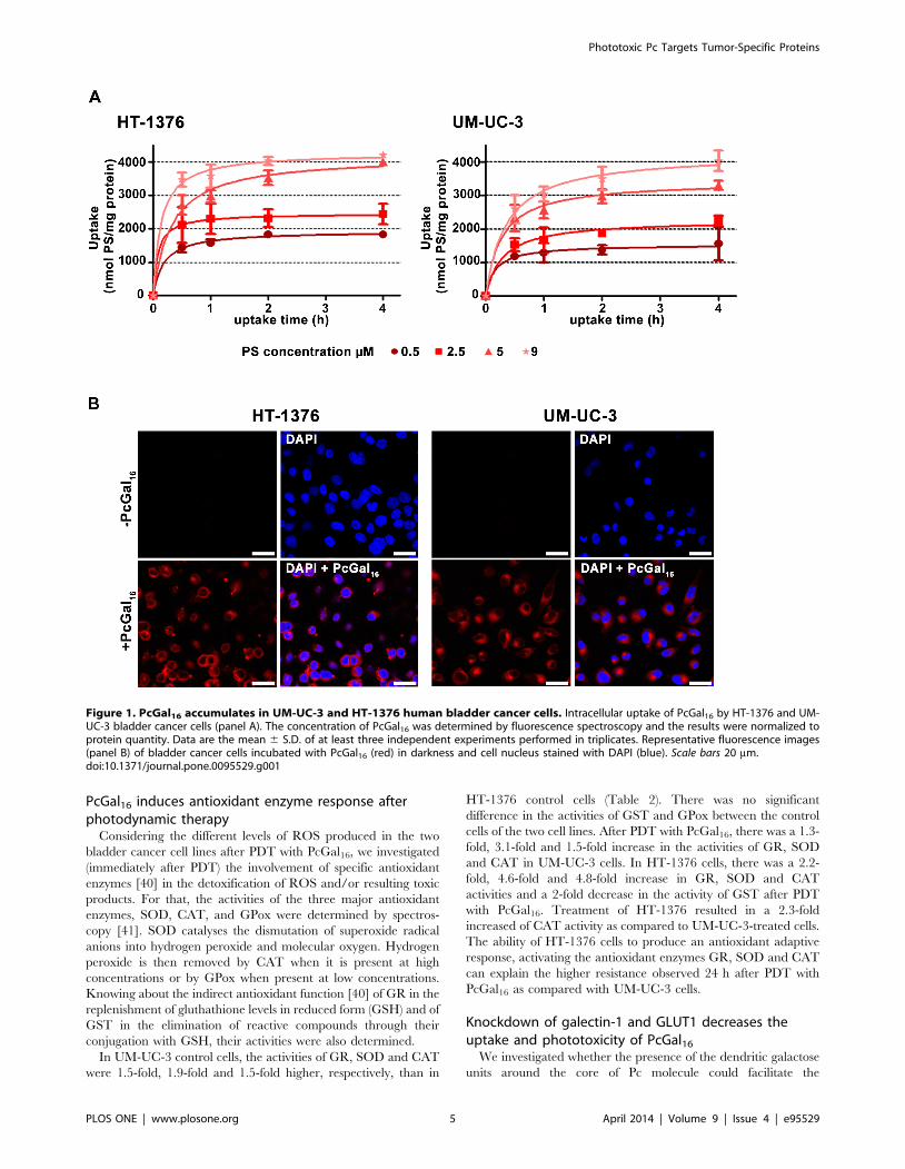
PcGal16 induces antioxidant enzyme response afterphotodynamic therapy
Considering the different levels of ROS produced in the two
bladder cancer cell lines after PDT with PcGal16, we investigated
(immediately after PDT) the involvement of specific antioxidant
enzymes [40] in the detoxification of ROS and/or resulting toxic
products. For that, the activities of the three major antioxidant
enzymes, SOD, CAT, and GPox were determined by spectros-
copy [41]. SOD catalyses the dismutation of superoxide radical
anions into hydrogen peroxide and molecular oxygen. Hydrogen
peroxide is then removed by CAT when it is present at high
concentrations or by GPox when present at low concentrations.
Knowing about the indirect antioxidant function [40] of GR in the
replenishment of gluthathione levels in reduced form (GSH) and of
GST in the elimination of reactive compounds through their
conjugation with GSH, their activities were also determined.
In UM-UC-3 control cells, the activities of GR, SOD and CAT
were 1.5-fold, 1.9-fold and 1.5-fold higher, respectively, than in
HT-1376 control cells (Table 2). There was no significant
difference in the activities of GST and GPox between the control
cells of the two cell lines. After PDT with PcGal16, there was a 1.3-
fold, 3.1-fold and 1.5-fold increase in the activities of GR, SOD
and CAT in UM-UC-3 cells. In HT-1376 cells, there was a 2.2-
fold, 4.6-fold and 4.8-fold increase in GR, SOD and CAT
activities and a 2-fold decrease in the activity of GST after PDT
with PcGal16. Treatment of HT-1376 resulted in a 2.3-fold
increased of CAT activity as compared to UM-UC-3-treated cells.
The ability of HT-1376 cells to produce an antioxidant adaptive
response, activating the antioxidant enzymes GR, SOD and CAT
can explain the higher resistance observed 24 h after PDT with
PcGal16 as compared with UM-UC-3 cells.
Knockdown of galectin-1 and GLUT1 decreases theuptake and phototoxicity of PcGal16
We investigated whether the presence of the dendritic galactose
units around the core of Pc molecule could facilitate the
Figure 1. PcGal16 accumulates in UM-UC-3 and HT-1376 human bladder cancer cells. Intracellular uptake of PcGal16 by HT-1376 and UM-UC-3 bladder cancer cells (panel A). The concentration of PcGal16 was determined by fluorescence spectroscopy and the results were normalized toprotein quantity. Data are the mean 6 S.D. of at least three independent experiments performed in triplicates. Representative fluorescence images(panel B) of bladder cancer cells incubated with PcGal16 (red) in darkness and cell nucleus stained with DAPI (blue). Scale bars 20 mm.doi:10.1371/journal.pone.0095529.g001
Phototoxic Pc Targets Tumor-Specific Proteins
PLOS ONE | www.plosone.org 5 April 2014 | Volume 9 | Issue 4 | e95529
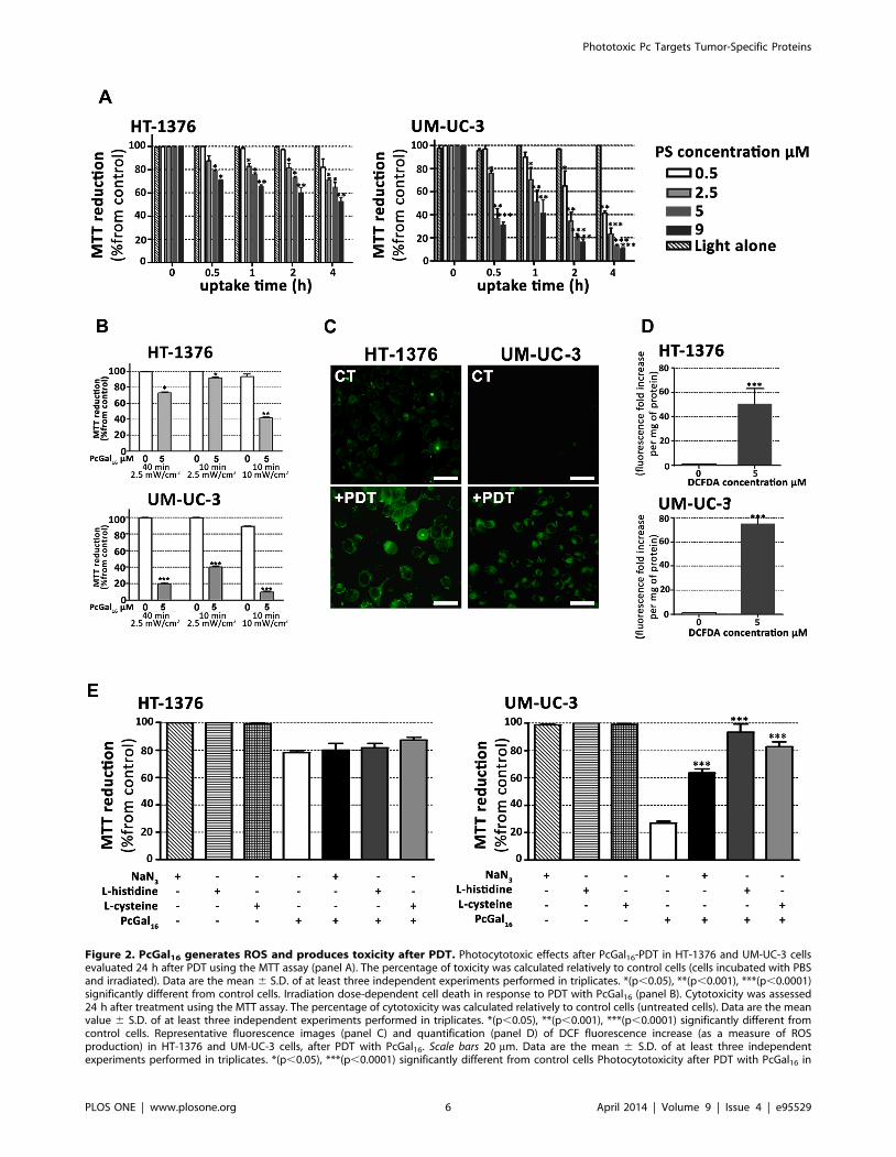
Figure 2. PcGal16 generates ROS and produces toxicity after PDT. Photocytotoxic effects after PcGal16-PDT in HT-1376 and UM-UC-3 cellsevaluated 24 h after PDT using the MTT assay (panel A). The percentage of toxicity was calculated relatively to control cells (cells incubated with PBSand irradiated). Data are the mean 6 S.D. of at least three independent experiments performed in triplicates. *(p,0.05), **(p,0.001), ***(p,0.0001)significantly different from control cells. Irradiation dose-dependent cell death in response to PDT with PcGal16 (panel B). Cytotoxicity was assessed24 h after treatment using the MTT assay. The percentage of cytotoxicity was calculated relatively to control cells (untreated cells). Data are the meanvalue 6 S.D. of at least three independent experiments performed in triplicates. *(p,0.05), **(p,0.001), ***(p,0.0001) significantly different fromcontrol cells. Representative fluorescence images (panel C) and quantification (panel D) of DCF fluorescence increase (as a measure of ROSproduction) in HT-1376 and UM-UC-3 cells, after PDT with PcGal16. Scale bars 20 mm. Data are the mean 6 S.D. of at least three independentexperiments performed in triplicates. *(p,0.05), ***(p,0.0001) significantly different from control cells Photocytotoxicity after PDT with PcGal16 in
Phototoxic Pc Targets Tumor-Specific Proteins
PLOS ONE | www.plosone.org 6 April 2014 | Volume 9 | Issue 4 | e95529
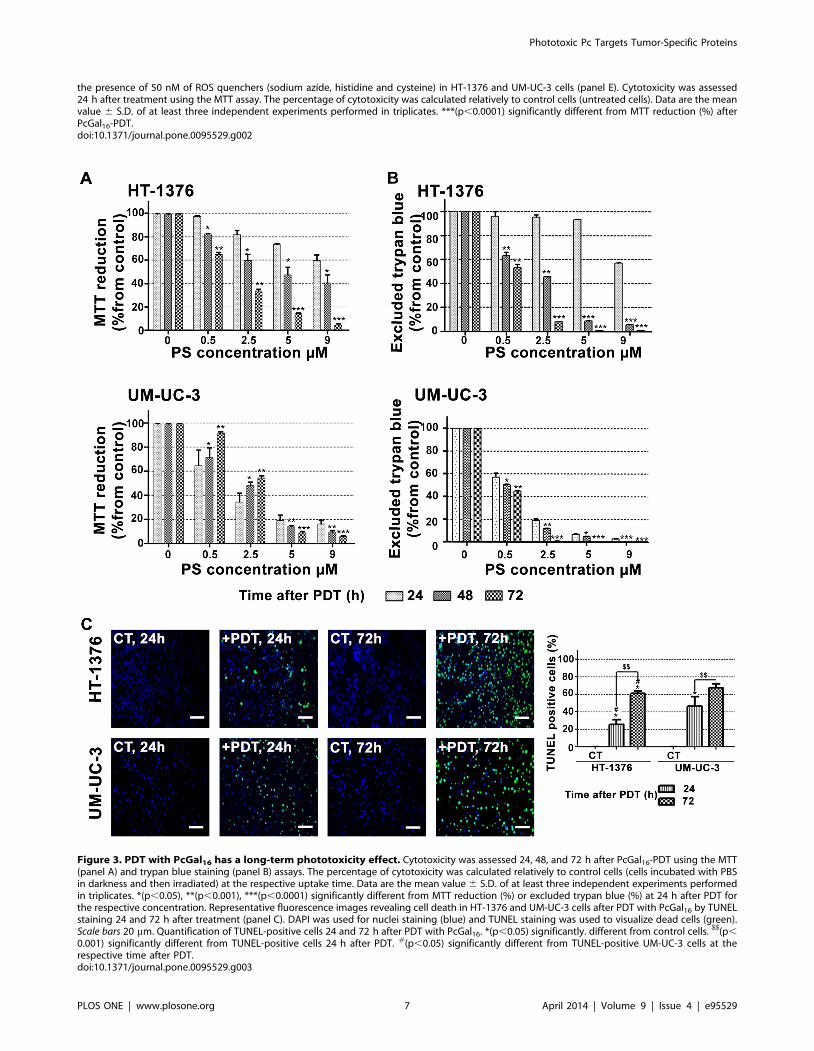
the presence of 50 nM of ROS quenchers (sodium azide, histidine and cysteine) in HT-1376 and UM-UC-3 cells (panel E). Cytotoxicity was assessed24 h after treatment using the MTT assay. The percentage of cytotoxicity was calculated relatively to control cells (untreated cells). Data are the meanvalue 6 S.D. of at least three independent experiments performed in triplicates. ***(p,0.0001) significantly different from MTT reduction (%) afterPcGal16-PDT.doi:10.1371/journal.pone.0095529.g002
Figure 3. PDT with PcGal16 has a long-term phototoxicity effect. Cytotoxicity was assessed 24, 48, and 72 h after PcGal16-PDT using the MTT(panel A) and trypan blue staining (panel B) assays. The percentage of cytotoxicity was calculated relatively to control cells (cells incubated with PBSin darkness and then irradiated) at the respective uptake time. Data are the mean value 6 S.D. of at least three independent experiments performedin triplicates. *(p,0.05), **(p,0.001), ***(p,0.0001) significantly different from MTT reduction (%) or excluded trypan blue (%) at 24 h after PDT forthe respective concentration. Representative fluorescence images revealing cell death in HT-1376 and UM-UC-3 cells after PDT with PcGal16 by TUNELstaining 24 and 72 h after treatment (panel C). DAPI was used for nuclei staining (blue) and TUNEL staining was used to visualize dead cells (green).Scale bars 20 mm. Quantification of TUNEL-positive cells 24 and 72 h after PDT with PcGal16. *(p,0.05) significantly. different from control cells. $$(p,
0.001) significantly different from TUNEL-positive cells 24 h after PDT. #(p,0.05) significantly different from TUNEL-positive UM-UC-3 cells at therespective time after PDT.doi:10.1371/journal.pone.0095529.g003
Phototoxic Pc Targets Tumor-Specific Proteins
PLOS ONE | www.plosone.org 7 April 2014 | Volume 9 | Issue 4 | e95529

interaction of this PS with specific domains in the plasma
membrane of cancer cells. We hypothesized that domains
enriched in carbohydrate-binding proteins [42] could facilitate
the interaction with PcGal16, enhancing somehow its cellular
uptake, and therefore its photodynamic potential.
Galectin [18] and glucose [19] proteins are expressed in high
levels in cancer cells and both have affinity for galactose molecules.
Therefore, we have evaluated the protein levels of galectin-1 and
GLUT 1 in UM-UC-3 and HT-1376 cells, by Western Blotting
and immunofluorescence (Figures 4 and 5).
The galectin-1 protein levels were higher in UM-UC-3 than in
HT-1376 control cells (Figure 4A). To determine whether
galectin-1 plays a role in the uptake of PcGal16 by cancer cells,
siRNA was used to knockdown galectin-1 within UM-UC-3
bladder cancer cells. The treatment of UM-UC-3 cells with a pool
of three target-specific siRNAs maximally suppressed galectin-1 by
<50% at 24 h and 48 h post-transfection (Figure 4B), without
affecting the expression of the housekeeping protein b-actin. The
transfected cells were then treated with PcGal16 48 h post-
transfection. As shown in Figures 4C and 4D, transfected cells
displayed a markedly decreased uptake and phototoxicity of
PcGal16. The GLUT1 protein levels were higher in HT-1376 than
in UM-UC-3 control cells (Figure 5A). Therefore, HT-1376
bladder cancer cells were also treated with a pool of three target-
specific GLUT1 siRNAs. Application of GLUT1 siRNA sup-
pressed GLUT1 by <50% and <90% at 24 h and 48 h post-
transfection, respectively (Figure 5B). Treatment of HT-1376 cells
with PcGal16 twenty-four hours post-transfection, resulted in a
substantial decrease in the uptake and phototoxicity (Figures 5C
and D).
PcGal16 decreases the galectin-1 and GLUT1 proteinlevels
To further explore the role of galectin-1 and GLUT1 in the
photodynamic effect induced by PcGal16, we determined the levels
of these proteins before and after PDT. Both incubation of cancer
cells with PcGal16 (i.e. incubation of cancer cells with PcGal16 in
darkness) and PDT with PcGal16 induced a decrease in galectin-1
as observed by Western Blotting and immunofluorescence
(Figures 4E, 4F and 4G). The decrease observed in galectin-1
was higher in UM-UC-3 cells as compared to HT-1376 cells and it
was more evident after PDT. Using confocal fluorescence
microscopy, we observed co-localization of PcGal16 with galec-
tin-1 inside bladder cancer cells (Figure 4G).
Similar to what was observed for galectin-1, there was also a
decrease in GLUT1 (Figures 5E, 5F and 5G) both after PcGal16
uptake and after PDT treatment in HT-1376 cancer cells.
Furthermore, in these cancer cells it was higher after PDT than
after PcGal16 uptake in darkness. In UM-UC-3 cells, PcGal16 was
not able to reduce GLUT1 protein levels (Figure 5F). In both
bladder cancer cell lines there was co-localization of PcGal16 with
GLUT1 (Figure 5G). Overall, these findings clearly indicate show
the critical involvement of the carbohydrate-binding proteins in
the potential of PcGal16 as a therapeutic agent.
Discussion
Third-generation PSs such as Pc coupled to carbohydrates are
interesting for PDT, because they can be recognized by
glycoprotein-based membrane proteins that are overexpressed in
tumors [6]. Besides the enhancement of cellular recognition, the
presence of dendritic galactose molecules improves Pc solubility
and biocompatibility [34]. We have recently reported the synthesis
of a new Pc with dendrimers of galactose sugar (PcGal16) that has
valuable spectroscopic and photochemical properties [34]. In this
Table 1. Values for photocytotoxic concentration (IC50, mM) of photoactivated PcGal16 on human bladder cancer cell lines, HT-1376 and UM-UC-3.
HT-1376 cell line UM-UC-3 cell line
Hours after PDT 24 48 72 24 48 72
IC50 (mM), CI95% - 3.3 [0.6;10.7] 2.5 [2.2;2.9] 2.1 [0.9;5.0] 2.8 [2.4; 3.2] 2.6 [2.6;2.7]
IC50 is the incubation concentration that inhibits the proliferation of cultures in 50%, after cells’ incubation with PcGal16 and irradiation. IC50 values were calculatedusing the MTT dose response curves (24, 48, and 72 h after PDT), obtained for cells incubated with PcGal16 at various concentrations for 2 h.CI95%: 95% Confidence interval.doi:10.1371/journal.pone.0095529.t001
Table 2. Values of activity (mU/mg of protein) of antioxidant enzymes superoxide dismutase (SOD), catalase (CAT), glutathioneperoxidase (GPox), glutathione reductase (GR) and glutathione S-transferase (GST) determined after PDT.
Cell line PcGal16-PDT Enzyme activity (mU/mg of protein)
GST GPox GR SOD CAT
UM-UC-3 2 44.061.4 232.4623.6 252.3613.9$
56.0613.1$
36.3161.3$
+ 39.762.7 243.6612.0 316.1611.3# 173.664.8# 52.8962.7#
HT-1376 2 50.462.4 236.169.9 163.461.9 29.5661.9 24.8562.2
+ 24.660.5$,* 252.3618.2 356.9630.6
$134.764.3
$,* 119.863.3$,*
$(p,0.05): significantly different from HT-1376 control cells;
#(p,0.05): significantly different from UM-UC-3 control cells;*(p,0.05): significantly different from UM-UC-3 treated cells.doi:10.1371/journal.pone.0095529.t002
Phototoxic Pc Targets Tumor-Specific Proteins
PLOS ONE | www.plosone.org 8 April 2014 | Volume 9 | Issue 4 | e95529
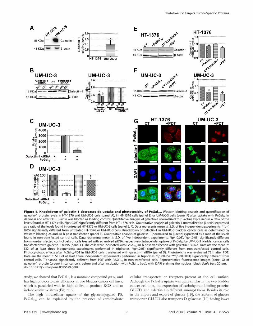
study, we showed that PcGal16 is a nontoxic compound per se, and
has high photocytotoxic efficiency in two bladder cancer cell lines,
which is paralleled with its high ability to produce ROS and to
induce oxidative stress (Figure 6).
The high intracellular uptake of the glycoconjugated PS,
PcGal16, can be explained by the presence of carbohydrate
cellular transporters or receptors present at the cell surface.
Although the PcGal16 uptake was quite similar in the two bladder
cancer cell lines, the expression of carbohydrate-binding proteins
GLUT1 and galectin-1 is different amongst them. Besides its role
in the import and export of glucose [19], the isoform of glucose
transporter GLUT1 also transports D-galactose [19] having lower
Figure 4. Knockdown of galectin-1 decreases de uptake and phototoxicity of PcGal16. Western blotting analysis and quantification ofgalectin-1 protein levels in HT-1376 and UM-UC-3 cells (panel A), in HT-1376 cells (panel E) or UM-UC-3 cells (panel F) after uptake with PcGal16 indarkness and after PDT. b-actin was blotted as loading control. Quantitative analysis of galectin-1 (normalized to b -actin) expressed as a ratio of thelevels found in HT-1376 cells. *(p,0.05) significantly different from HT-1376 cells. Quantitative analysis of galectin-1 (normalized to b-actin) expressedas a ratio of the levels found in untreated HT-1376 or UM-UC-3 cells (panel E, F). Data represents mean 6 S.D. of five independent experiments. *(p,0.05) significantly different from untreated HT-1376 or UM-UC-3 cells. Knockdown of galectin-1 in UM-UC-3 bladder cancer cells as determined byWestern blotting 24 and 48 h post-transfection (panel B). Quantitative analysis of galectin-1 (normalized to b-actin) expressed as a ratio of the levelsfound in non-transfected control cells. Data represents mean 6 S.D. of five independent experiments. *(p,0.05), $(p,0.05) significantly differentfrom non-transfected control cells or cells treated with scrambled siRNA, respectively. Intracellular uptake of PcGal16 by UM-UC-3 bladder cancer cellstransfected with galectin-1 siRNA (panel C). The cells were incubated with PcGal16 48 h post-transfection with galectin-1 siRNA. Data are the mean 6S.D. of at least three independent experiments performed in triplicates. *(p,0.05) significantly different from non-transfected control cells.Photocytotoxic effects after PcGal16-PDT in UM-UC-3 cells transfected with galectin-1 siRNA (panel D). Phototoxicity was evaluated 72 h after PDT.Data are the mean 6 S.D. of at least three independent experiments performed in triplicates. *(p,0.05), ***(p,0.0001) significantly different fromcontrol cells. $(p,0.05), significantly different from PDT with PcGal16 in non-transfected cells. Representative fluorescence images (panel G) ofgalectin-1 protein (green) in cancer cells before and after incubation with PcGal16 (red), with DAPI staining the nucleus (blue). Scale bars 20 mm.doi:10.1371/journal.pone.0095529.g004
Phototoxic Pc Targets Tumor-Specific Proteins
PLOS ONE | www.plosone.org 9 April 2014 | Volume 9 | Issue 4 | e95529
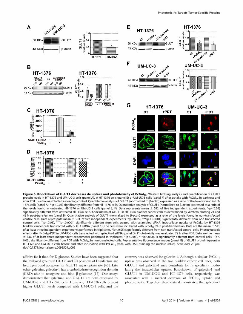
affinity for it than for D-glucose. Studies have been suggested that
the hydroxyl groups in C1, C3 and C4 positions of D-galactose are
hydrogen bond acceptors for GLUT1 sugar uptake site [43]. Like
other galectins, galectin-1 has a carbohydrate-recognition domain
(CRD) able to recognize and bind b-galactose [17]. Our assays
demonstrated that galectin-1 and GLUT1 are both expressed by
UM-UC-3 and HT-1376 cells. However, HT-1376 cells present
higher GLUT1 levels compared with UM-UC-3 cells, and the
contrary was observed for galectin-1. Although a similar PcGal16
uptake was observed in the two bladder cancer cell lines, both
GLUT1 and galectin-1 may contribute for its specificity modu-
lating the intracellular uptake. Knockdown of galectin-1 and
GLUT1 in UM-UC-3 and HT-1376 cells, respectively, was
associated with a marked decrease of PcGal16 uptake and
phototoxicity. Together, these data demonstrated that galectin-1
Figure 5. Knockdown of GLUT1 decreases de uptake and phototoxicity of PcGal16. Western blotting analysis and quantification of GLUT1protein levels in HT-1376 and UM-UC-3 cells (panel A), in HT-1376 cells (panel E) or UM-UC-3 cells (panel F) after uptake with PcGal16 in darkness andafter PDT. b-actin was blotted as loading control. Quantitative analysis of GLUT1 (normalized to b-actin) expressed as a ratio of the levels found in HT-1376 cells (panel A). *(p,0.05) significantly different from HT-1376 cells. Quantitative analysis of GLUT1 (normalized to b-actin) expressed as a ratio ofthe levels found in untreated HT-1376 or UM-UC-3 cells (panel E, F). Data represents mean 6 S.D. of five independent experiments. *(p,0.05)significantly different from untreated HT-1376 cells. Knockdown of GLUT1 in HT-1376 bladder cancer cells as determined by Western blotting 24 and48 h post-transfection (panel B). Quantitative analysis of GLUT1 (normalized to b-actin) expressed as a ratio of the levels found in non-transfectedcontrol cells. Data represents mean 6 S.D. of five independent experiments. *(p,0.05), ***(p,0.0001) significantly different from non-transfectedcontrol cells. $(p,0.05), $$$(p,0.0001) significantly different from cells treated with scrambled siRNA. Intracellular uptake of PcGal16 by HT-1376bladder cancer cells transfected with GLUT1 siRNA (panel C). The cells were incubated with PcGal16 24 h post-transfection. Data are the mean 6 S.D.of at least three independent experiments performed in triplicates. *(p,0.05) significantly different from non-transfected control cells. Photocytotoxiceffects after PcGal16-PDT in UM-UC-3 cells transfected with galectin-1 siRNA (panel D). Phototoxicity was evaluated 72 h after PDT. Data are the mean6 S.D. of at least three independent experiments performed in triplicates. *(p,0.05), ***(p,0.0001) significantly different from control cells. *(p,
0.05), significantly different from PDT with PcGal16 in non-transfected cells. Representative fluorescence images (panel G) of GLUT1 protein (green) inHT-1376 and UM-UC-3 cells before and after incubation with PcGal16 (red), with DAPI staining the nucleus (blue). Scale bars 20 mm.doi:10.1371/journal.pone.0095529.g005
Phototoxic Pc Targets Tumor-Specific Proteins
PLOS ONE | www.plosone.org 10 April 2014 | Volume 9 | Issue 4 | e95529

and GLUT1 contribute for the efficacy of PDT mediated by
PcGal16.
Interestingly, although the similar uptake of PcGal16 by UM-
UC-3 and HT-1376 cells, the phototoxicity induced 24 h after
PDT was higher in UM-UC-3 cells than in HT-1376 cells. Such
lack of association between uptake and phototoxicity has been
described [44,45]. We investigated whether the higher phototox-
icity observed in UM-UC-3 cells was due to higher production of
ROS and/or higher oxidative damage compared with that in HT-
1376 cells. As expected, the ability of PcGal16 to produce ROS
was higher in UM-UC-3 than in HT-1376 cells.
In PDT, it has been described that ROS can be generated by
two photochemical reactions [46,47]. In type-II photochemical
reactions, the excited PS in its triplet state can transfer its energy to
molecular oxygen leading to the formation of singlet oxygen.
Type-I photochemical reactions happen when an excited PS reacts
with a biological substrate forming radicals and radical ions.
Treatment with ROS quenchers demonstrated that in UM-UC-3
cells, singlet oxygen should have a high effect since cell death was
highly reduced with quenchers of singlet oxygen (sodium azide and
histidine). Further studies are needed to gain insight into the
contribution of specific ROS in PcGal16-mediated cell death after
PDT.
Interestingly, we observed that PDT with PcGal16 has a long-
term phototoxic effect in both cancer cell lines. Cytotoxicity assays
(MTT, trypan blue and TUNEL assays) performed 72 h after
PDT demonstrated that UM-UC-3 cells were not able to recover.
Moreover, in HT-1376 cells there was a marked induction of cell
death occurring from 24 to 72 h after PDT with PcGal16. The
three distinct cytotoxic methods used in the present work are
widely applied in the study of cell death: MTT (indicator of
metabolic activity), trypan blue staining (indicator of membrane
integrity loss occurring in necrosis or in late stages of apoptosis)
and TUNEL assay (indicator of DNA fragmentation, a key factor
of apoptosis). Cell death in PDT may occur by apoptosis or
necrosis, or even by a combination of the two mechanisms [2]. A
more specific and comprehensive study is needed to understand
the specific cell death pathways induced after PDT with PcGal16 in
the bladder cancer cells used in this study. The different cell death
obtained 24 h after PDT in UM-UC-3 and HT-1376 cells can be
partially explained by the different amount of ROS present in both
cells lines after irradiation. In addition, the resistance exhibited by
HT-1376 cells could be due to the presence of efficient protective
mechanisms, at least in the first stages after photodynamic
treatment. Cytoprotective mechanisms initiated by cancer cells
after PDT are well-known [47]. The increase of antioxidant
molecules (e.g. gluthathione, vitamin C and vitamin E) [48] and the
induction of genes encoding proteins involved in apoptosis or in
the repair of lesions [49] are two of the well-known cytoprotective
mechanisms induced after PDT. Another one is based on the
equilibrium between photo-oxidative impairment of cells by ROS
versus elimination of ROS by the activity of cellular antioxidant
enzymes. Recent studies have shown that PDT caused increased-
antioxidant enzymes activity and expression [50]. Thus, PDT
efficacy can be influenced by the antioxidant response of the
enzymes SOD, the GSH system and CAT.
Figure 6. Hypothetic illustration of phototoxicity of PcGal16 in human bladder cancer cells. The uptake of PcGal16 by bladder cancer cellsis modulated by the presence of carbohydrate-binding proteins present at the cell surface (i.e. GLUT1 and galectin-1). PcGal16 is a nontoxiccompound per se, and has high photocytotoxic efficiency against bladder cancer cell lines. Treatment with ROS quenchers demonstrated that celldeath in bladder cancer cells is mediated by the production of ROS after PDT. Immediately after PDT with PcGal16 there is an increase on the activityof antioxidant enzymes (SOD, CAT and GR antioxidant enzymes). The photoactivated PcGal16 co-localizes with galectin-1 and GLUT1and reduces theirlevels.doi:10.1371/journal.pone.0095529.g006
Phototoxic Pc Targets Tumor-Specific Proteins
PLOS ONE | www.plosone.org 11 April 2014 | Volume 9 | Issue 4 | e95529
Our data demonstrated that after PDT with PcGal16 there was
an increase in the activity of SOD, CAT and GR antioxidant
enzymes in both cell lines, being higher in HT-1376 than in UM-
UC-3 cells. This higher antioxidant defense of HT-1376 cells
against ROS can explain the results obtained 24 h after treatment.
However, it is hypothesized that this was not maintained for 72 h
after PDT since for this time point there was a massive cell death.
This not only suggests that in this cell line there is a temporal
relationship between ROS levels and cell death, but shows that
antioxidant enzymes activity is of greater importance in protecting
HT-1376 cells for at least 24 h after PDT with PcGal16. Regarding
the activity of antioxidant enzymes, in HT-1376 cells it was also
observed a decrease in the activity of GST, which is an enzyme
implicated in cells defense against oxidation products. This
enzyme has been described as protecting cells from DNA
desintegration and drug toxicity [51]. GST isoforms are overex-
pressed in multidrug resistant tumors having an important role in
tumors drug resistance by direct detoxification or inhibition of the
MAP kinase pathway [51]. Thus, the higher cell death observed in
HT-1376 cells 72 h after treatment can be also related with the
activity of GST. A decrease in the activity of GST can be
associated with DNA fragmentation and cell death 72 h after
treatment.
Understanding the role of galactose moieties in the recognition
of the PS by cancer cells may allow the investigation and
development of more focused therapeutic strategies. Thus, we
investigated whether PcGal16 could be directly recognized by
specific carbohydrate-binding proteins present at the plasma
membrane. Consistently, the photoactivated PcGal16 was shown
to co-localize and reduce the levels of the plasma membrane
proteins galectin-1 and GLUT1. Moreover, the immunofluores-
cence and Western Blotting studies demonstrated that, although its
non-dark toxicity, PcGal16 decreases the levels of galectin-1 and
GLUT1 proteins. A plausible explanation for the decreased levels
of the galactose binding proteins, galectin-1 and GLUT1, after
incubation with PcGal16 can be the masking of the epitope, which
can block antibody-epitope binding due to changes in protein
conformation or, eventually, endocytosis of these proteins and
subsequent degradation. Thus, the changes observed in the levels
of galectin-1 and GLUT1 could be induced directly by the binding
of PcGal16 to the carbohydrate-binding proteins and/or indirectly
by the generation of ROS after PDT with PcGal16.
Although significant progress has been made in research related
with the role of galectins in cancer, the information underlying the
molecular mechanisms that control the expression of these
proteins in tumour cells is scarce. The interaction of PSs with
galectins (namely galectin-1 and galectin-3) has been studied by
spectroscopic studies [52] and molecular modeling analysis [6,27];
however, they have not been validated by in vitro studies. As far as
we know, there are no in vitro reports indicating whether PSs can
modulate the expression of carbohydrate-binding proteins such as
galectin-1 and GLUT1. Knowing that galectin-1 expression is
correlated with cell metastatic potential [18,53] and contributes to
tumor progression and resistance after conventional cancer
therapeutic modalities [18], the ability of PcGal16 to reduce the
levels of galectin-1 after its uptake and/or photoactivation
prompted us to envisage PcGal16 as a potential candidate for
cancer treatment.
Knowing that the overexpression of GLUTs is involved in
tumor glycolysis - one of the biochemical ‘‘hallmarks’’ of cancer -
the efficiency of PcGal16 as an efficient anti-cancer PS is also
evidenced by its ability to reduce GLUT1. GLUT1 is an attractive
target to consider in the development of new PSs because it is
lower expressed in normal-epithelial tissues or benign epithelial
cell tumors when compared with human cancer cells [54]. The
function of GLUT1 in the tumorogenesis process has been
demonstrated by in vitro and in vivo studies, where the overexpres-
sion of GLUT1 antisense resulted in the inhibition of HL60
leukaemia cells proliferation and MKN-45 derived xenografs,
respectively [55,56]. Based on the results of the current study, we
envisage PcGal16 as a promising therapeutic agent for the
treatment of bladder cancer. Further studies are warranted to
investigate the selectivity and photototoxicity of this PS in an in vivo
model of bladder cancer, to contribute to a possible impact on
clinical practice.
Supporting Information
Figure S1 Chemical structures of free phthalocyanine PcF16 and
galacto-dendrimer phthalocyanine PcGal16.
(TIF)
Figure S2 PcGal16 is non-toxic in darkness, PcF16 is non-toxic in
darkness and after PDT. Non-dark toxicity of various concentra-
tions of PcGal16 in HT-1376 and UM-UC-3 cells (panel A). Non-
dark toxicity was assessed using the MTT colorimetric assay 24,
48, and 72 h after treat HT-1376 and UM-UC-3 cells (panel B).
Toxicity of PcF16 at 5 mM in darkness and after PDT (panel C) in
HT-1376 and UM-UC-3 cells. The toxicity was assessed using the
MTT colorimetric assay 24 h after treat HT-1376 and UM-UC-3
cells. Data are the mean 6 S.D. of at least three independent
experiments performed in triplicates.
(TIF)
Acknowledgments
We grateful appreciate the help of Dr. Celia Gomes (IBILI-FMUC,
Portugal) with the spectrofluorometric measurements in IVIS Lumina XR
equipment.
We thank Dr. Filomena Botelho (IBILI-FMUC, Portugal) for the
generous supply of HT-1376 human bladder cancer cells.
P. Pereira and S. Silva acknowledge fellowships from FCT (SFRH/BD/
85941/2012 and SFRH/BPD/64812/2009, respectively).
We appreciate the help of Dr. Celia Gomes (IBILI-FMUC, Portugal)
with the spectrofluorometric measurements in IVIS Lumina XR
equipment.
Author Contributions
Conceived and designed the experiments: PP RF JT. Performed the
experiments: PP SS. Analyzed the data: PP RF JT. Contributed reagents/
materials/analysis tools: PP SS. Wrote the paper: PP RF JT CFR JC.
References
1. Allison RR, Sibata CH (2010) Oncologic photodynamic therapy photosensitiz-
ers: a clinical review. Photodiagnosis Photodyn Ther 7:61–75.
2. Almeida RD, Manadas BJ, Carvalho AP, Duarte CB (2004) Intracellular
signaling mechanisms in photodynamic therapy. Biochim Biophys Acta
1704:59–86.
3. Soares ARM, Tome JPC, Neves MGPMS, Tome AC, Cavaleiro JAS, et al.
(2009) Synthesis of water-soluble phthalocyanines bearing four or eight D-
galactose units, Carbohyd Res 334:507–510.
4. Silva JN, Silva AMG, Tome JPC, Ribeiro AO, Domingues MRM, et al. (2008)
Photophysical properties of a photocytotoxic fluorinated chlorinconjugated to
four b-cyclodextrins. Photochem Photobiol Sci 7:834–843.
5. Pereira PMR, Carvalho JJ, Silva S, Cavaleiro JAS, Schneider RJ, et al. (2014)
Porphyrin conjugated with serum albumins and monoclonal antibodies boosts
efficiency in targeted destruction of human bladder cancer cells, Org Biomol
Chem 12:1804–1811.
Phototoxic Pc Targets Tumor-Specific Proteins
PLOS ONE | www.plosone.org 12 April 2014 | Volume 9 | Issue 4 | e95529

6. Zheng G, Graham A, Shibata M, Missert JR, Oseroff AR, et al. (2001) Synthesis
of beta-galactose-conjugated chlorins derived by enyne metathesis as galectin-specific photosensitizers for photodynamic therapy. J Org Chem 66:8709–8716.
7. Soares ARM, Neves MGPMS, Tome AC, Iglesias-de la Cruz MC, Zamarron A,
et al. (2012) Glycophthalocyanines as Photosensitizers for Triggering Mitotic:Catastrophe and Apoptosis On Cancer Cells. Chem Res Toxicol 25:940–951.
8. Daly R, Vaz G, Davies AM, Senge MO, Scanlan EM (2012) Synthesis andbiological evaluation of a library of glycoporphyrin compounds. Chemistry
18:14671–14679.
9. Vedachalam S, Choi BH, Pasunooti KK, Ching KM, Lee K, et al. (2011)Glycosylated porphyrin derivatives and their photodynamic activity in cancer
cells. MedChemComm 2:371–377.10. Ernst B, Magnani JL (2009) From carbohydrate leads to glycomimetic drugs.
Nat Rev Drug Discov 8:661–677.11. Lourenco LMO, Tome JPC, Domingues MRM, Domingues P, Costa PJ, et al.
(2009) Synthesis and differentiation of alpha- and beta-glycoporphyrin
stereoisomers by electrospray tandem mass spectrometry. Rapid CommunMass Spectrom 23:3478–3483.
12. Figueira F, Pereira PMR, Silva S, Cavaleiro JAS, Tome JPC (2014) Porphyrinsand Phthalocyanines decorated with dendrimers: Synthesis and biomedical
applications. Curr Org Synth In press.
13. Gary-Bobo M, Vaillant O, Maynadier M, Basile I, Gallud A, et al. (2013)Targeting multiplicity: the key factor for anti-cancer nanoparticles. Curr Med
Chem 20:1946–1955.14. Lourenco LMO, Neves MGPMS, Cavaleiro JAS, Tome JPC (2014) Synthetic
approaches to glycophthalocyanines. Tetrahedron In press.15. David A (2010) Carbohydrate-based Biomedical Copolymers for Targeted
Delivery of Anticancer Drugs. Isr J Chem 50:204–219.
16. Sharon N, Lis H (1989) Lectins as cell recognition molecules. Science 246:227–234.
17. Liu FT, Rabinovich GA (2005) Galectins as modulators of tumour progression.Nat Rev Cancer 5:29–41.
18. Cindolo L, Benvenuto G, Salvatore P, Pero R, Salvatore G, et al. (1999)
Galectin-1 and galectin-3 expression in human bladder transitional-cellcarcinomas. Int J Cancer 84:39–43.
19. Carruthers A, DeZutter J, Ganguly A, Devaskar SU (2009) Will the originalglucose transporter isoform please stand up! Am J Physiol Endocrinol Metab
297:E836–E848.20. Fernandes R, Carvalho AL, Kumagai A, Seica R, Hosoya K, et al. (2004)
Downregulation of retinal GLUT1 in diabetes by ubiquitinylation. Mol Vis
10:618–628.21. Fernandes R, Hosoya K, Pereira P (2011) Reactive oxygen species downregulate
glucose transport system in retinal endothelial cells. Am J Physiol-Cell Ph 300:C927–C936.
22. Silva AMG, Tome AC, Neves MGPMS, Cavaleiro JAS, Perrone D, et al. (2005)
Porphyrins in 1,3-dipolar cycloadditions with sugar azomethine ylides. Synthesisof pyrrolidinoporphyrin glycoconjugates. Synlett 0857–0859.
23. Choi CF, Huang JD, Lo PC, Fong WP, Ng DKP (2008) Glycosylated zinc(II)phthalocyanines as efficient photosensitisers for photodynamic therapy.
Synthesis, photophysical properties and in vitro photodynamic activity. OrgBiomol Chem 6:2173–2181.
24. Iqbal Z, Hanack M, Ziegler T (2009) Synthesis of an octasubstituted galactose
zinc(II) phthalocyanine. Tetrahedron Lett 50:873–875.25. Lee PPS, Lo PC, Chan EYM, Fong WP, Ko WH, et al. (2005) Synthesis and in
vitro photodynamic activity of novel galactose-containing phthalocyanines.Tetrahedron Lett 46:1551–1554.
26. Park YK, Bold B, Cui BC, Bai JQ, Lee WK, et al. (2008) Binding affinities of
carbohydrate-conjugated chlorins for galectin-3. B Korean Chem Soc 29:130–134.
27. Pandey SK, Zheng X, Morgan J, Missert JR, Liu TH, et al. (2007)Purpurinimide carbohydrate conjugates: effect of the position of the carbohy-
drate moiety in photosensitizing efficacy. Mol Pharm 4:448–464.
28. Singh S, Aggarwal A, Thompson S, Tome JPC, Zhu X, et al. (2010) Synthesisand photophysical properties of thioglycosylated- chlorins, isobacteriochlorins
and bacteriochlorins for bioimaging and diagnostics. Bioconjugate Chem21:2136–2146.
29. Fujimoto K, Miyata T, Aoyama Y (2000) Saccharide-directed cell recognitionand molecular delivery using macrocyclic saccharide clusters: Masking of
hydrophobicity to enhance the saccharide specificity. J Am Chem Soc
122:3558–3559.30. Ribeiro AO, Tome JPC, Neves MGPMS, Tome AC, Cavaleiro JAS, et al.
(2006) [1,2,3,4-tetrakis(alpha/beta-D-galactopyranos-6-yl)-phthalocyaninato]-zinc(II): a water-soluble phthalocyanine. Tetrahedron Lett 47:9177–9180.
31. Gillies ER, Frechet JMJ (2005) Dendrimers and dendritic polymers in drug
delivery. Drug Discov Today 10:35–43.
32. Wolinsky JB, Grinstaff MW (2008) Therapeutic and diagnostic applications of
dendrimers for cancer treatment. Adv Drug Deliv Rev 60:1037–1055.
33. Klajnert B, Rozanek M, Bryszewska M (2012) Dendrimers in photodynamic
therapy. Curr Med Chem 19:4903–4912.
34. Silva S, Pereira PMR, Silva P, Paz FA, Faustino MA, et al. (2012) Porphyrin and
phthalocyanine glycodendritic conjugates: synthesis, photophysical and photo-
chemical properties. Chem Commun (Camb) 48:3608–3610.
35. Ballardini R, Colonna B, Gandolfi MT, Kalovidouris SA, Orzel L, et al. (2003)
Porphyrin-containing glycodendrimers. Eur J Org Chem 2:288–294.
36. Wang ZJ, Chauvin B, Maillard P, Hammerer F, Carez D, et al. (2012)
Glycodendrimeric phenylporphyrins as new candidates for retinoblastoma PDT:
Blood carriers and photodynamic activity in cells. J Photochem Photobiol B
115:16–24.
37. Ballut S, Makky A, Loock B, Michel JP, Maillard P, et al. (2009) New strategy for
targeting of photosensitizers. Synthesis of glycodendrimeric phenylporphyrins,
incorporation into a liposome membrane and interaction with a specific lectin.
Chem Comm 2:224–226.
38. Bancirova M (2011) Sodium azide as a specific quencher of singlet oxygen
during chemiluminescent detection by luminol and Cypridina luciferin
analogues. Luminescence 26:685–688.
39. Aruoma OI, Halliwell B, Hoey BM, Butler J (1989) The antioxidant action of N-
acetylcysteine: its reaction with hydrogen peroxide, hydroxyl radical, superoxide,
and hypochlorous acid. Free Radic Biol Med 6:593–597.
40. Sies H (1993) Strategies of antioxidant defense. Eur J Biochem 215:213–219.
41. Weydert CJ, Cullen JJ (2010) Measurement of superoxide dismutase, catalase
and glutathione peroxidase in cultured cells and tissue. Nat Protoc 5:51–66.
42. Lotan R, Raz A (1988) Lectins in cancer cells. Ann N Y Acad Sci 551:385–398.
43. Barnett JEG, Holman GD, Chalkley RA, Munday KA (1975) Evidence for two
asymmetric conformational states in the human erythrocyte sugar-transport
system. Biochem J 145:417–429.
44. Hirohara S, Obata M, Ogata S, Ohtsuki C, Higashida S, et al. (2005) Cellular
uptake and photocytotoxicity of glycoconjugated chlorins in HeLa cells.
J Photoch Photobio B 78:7–15.
45. Laville I, Pigaglio S, Blais JC, Doz F, Loock B, et al. (2006) Photodynamic
efficiency of diethylene glycol-linked glycoconjugated porphyrins in human
retinoblastoma cells. J Med Chem 49:2558–2567.
46. Plaetzer K, Krammer B, Berlanda J, Berr F, Kiesslich T (2009) Photophysics
and photochemistry of photodynamic therapy: fundamental aspects. Laser Med
Sci 24:259–268.
47. Buytaert E, Dewaele M, Agostinis P (2007) Molecular effectors of multiple cell
death pathways initiated by photodynamic therapy. Biochimica et biophysica
acta 1776:86–107.
48. Sattler UGA, Mueller-Klieser W (2009) The anti-oxidant capacity of tumour
glycolysis. International journal of radiation biology 85:963–971.
49. Oleinick NL, Morris RL, Belichenko I (2002) The role of apoptosis in response
to photodynamic therapy: what, where, why, and how. Photochemical &
photobiological sciences : Official journal of the European Photochemistry
Association and the European Society for Photobiology 1:1–21.
50. Saczko J, Kulbacka J, Chwilkowska A, Pola A, Lugowski M, et al. (2007)
Cytosolic superoxide dismutase activity after photodynamic therapy, intracellu-
lar distribution of Photofrin II and hypericin, and P-glycoprotein localization in
human colon adenocarcinoma. Folia Histochem Cyto 45:93–98.
51. Townsend DM, Tew KD (2003) The role of glutathione-S-transferase in anti-
cancer drug resistance. Oncogene 22:7369–7375.
52. Bogoeva VP, Varriale A, John CM, D’Auria S (2010) Human galectin-3
interacts with two anticancer drugs. Proteomics 10:1946–1953.
53. Chiariotti L, Berlingieri MT, De Rosa P, Battaglia C, Berger N, et al. (1992)
Increased expression of the negative growth factor, galactoside-binding protein,
gene in transformed thyroid cells and in human thyroid carcinomas. Oncogene
7:2507–2511.
54. Younes M, Lechago LV, Somoano JR, Mosharaf M, Lechago J (1996) Wide
expression of the human erythrocyte glucose transporter Glut1 in human
cancers. Cancer Res 56:1164–1167.
55. Chan JYW, Kong SK, Choy YM, Lee CY, Fung KP (1999) Inhibition of glucose
transporter gene expression by antisense nucleic acids in HL-60 leukemia cells.
Life Sci 65:63–70.
56. Noguchi Y, Saito A, Miyagi Y, Yamanaka S, Marat D, et al. (2000) Suppression
of facilitative glucose transporter 1 mRNA can suppress tumor growth. Cancer
Letters 154:175–182.
Phototoxic Pc Targets Tumor-Specific Proteins
PLOS ONE | www.plosone.org 13 April 2014 | Volume 9 | Issue 4 | e95529