galileo-cerec integration for guided implant...
TRANSCRIPT

Galileos-Cerec Integration for Guided Implant Surgery
www.drsmanda.com
manda.com


Cad/Cam Scanner/Milling Chamber
Milling Chamber
Excessive powdering of preparation
Circa 1993
Galileos-Cerec Integration for Guided Implant Surgery

Monitor screen view of scan Virtual view of restoration
Circa 1993
Galileos-Cerec Integration for Guided Implant Surgery

Finished restoration Milled on 2 axis
In 1993 there was a steeper leaning curve due to limitations of the software and milling capabilities

Galileos-Cerec Integration for Guided Implant Surgery
www.drsmanda.com
manda.com

Combining 3D Conebeam / CT Scan and Cerec
Technology
Known Radiographic
Marker Scan
Fiduciary
Markers
Cerec Virtual Crown Virtual Crown, Implant
and Soft Tissue
Superimposed on CT Scan
Implant
Crown
Soft
Tissue

Digital Intraoral Scan
Omnicam Intraoral Camera CEREC Acquisition Unit
Intraoral Camera
Roll Camera from Occlusal to Buccal then Lingual
Galileos-Cerec Integration for Guided Implant Surgery

• CEREC - Sirona
Galileos-Cerec Integration for Guided Implant Surgery

Radiographic Imaging
www.drsmanda.com
manda.com

Radiographic Imaging
Radiographic Templates
Galileos-Cerec Integration for Guided Implant Surgery
Classic Guide- PROS-Very accurate implant guide, Multiple complex implant placement CONS-Involves shipping to lab for fabrication (longer turn around), Cost
OPTIGUIDE- PROS-Very accurate implant guide, Send case via email (cuts turnaround time)
Cons-Requires Intraoral scanner, Cost
Off-Site Lab Fabricated
Chairside Fabricated
CEREC Guide-Pros- Can fabricate in 20 minutes, inexpensive guide Cons- Utilized for 3-4 implants

Scan Patient with Guide in place Digitally Plan Case
Guide Completed and shipped in 3 weeks
Ship to SICAT LAB
Conventional Diagnostic Model Required
Classic Guide
1 2
5
3
4

OPTIGUIDE
Optical Scan (Optiguide) Instead of Conventional Diagnostic Cast Scan Patient with Guide in place
Digitally Plan Case Guide Completed and shipped in 1 week
Email Digital Scan and Plan to SICAT LAB
Radiographic Template with Bite Material
2
4
1 3
6 5

The Anatomy of a Classic Guide
1) Radiographic Bite Template
2) Radiopaque Acrylic Tooth Form
3) Thermoplast Arch Form www.drsmanda.com
manda.com

Radiographic Template
Radiopaque Bite Material Radiopaque Duplicate Denture
The Anatomy of a Classic Guide
Radiographic Template

Galileo-Cerec Integration Work Flow
drsmanda.com CBCT Scan with Radiographic Template in place


Omnicam Intraoral Scan
Fractured Lateral Incisor
Digital Planning of Surgical Guide
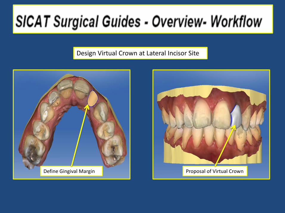
Design Virtual Crown at Lateral Incisor Site
Define Gingival Margin Proposal of Virtual Crown

Radiographic Markers
Intraoral Scan superimposed
Cerec Virtual Crown
Digital Implant placement
Merging of CBCT Scan and the Intraoral Scan

Implant Planning Prosthetic and Surgically Driven
Cerec Scan of gingiva and virtual crown
Digital Implant Placement
Reference Body with Radiographic Markers
Digital Implant Placement
Cerec Scan of gingiva and virtual crown
Radiographic Marker

Implant Planning Prosthetic and Surgically Driven
Cerec Scan of gingiva and virtual crown
Digital Implant Placement
File Emailed to SICAT LAB
Guide shipped to Clinic
Titanium Access for Implant keys

www.drsmanda.com
manda.com
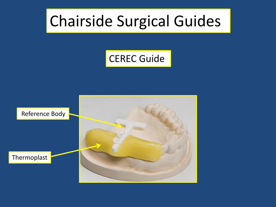
Chairside Surgical Guides
CEREC Guide
Reference Body
Thermoplast
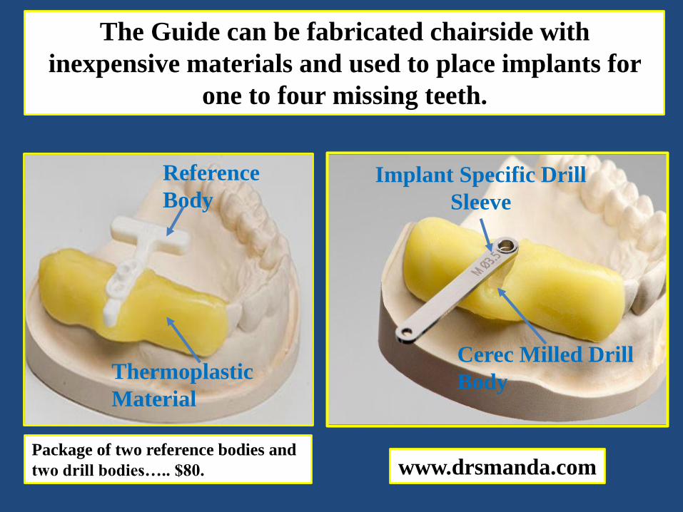
The Guide can be fabricated chairside with
inexpensive materials and used to place implants for
one to four missing teeth.
Reference
Body
Thermoplastic
Material
Implant Specific Drill
Sleeve
Cerec Milled Drill
Body
www.drsmanda.com Package of two reference bodies and
two drill bodies….. $80.

Reference Body:
Radiopaque Markers
Medium is most
commonly used size.
Concave tissue surface
Center reference body in edentulous space.
Thermoplastic material
Reference Body Diagnostic Cast
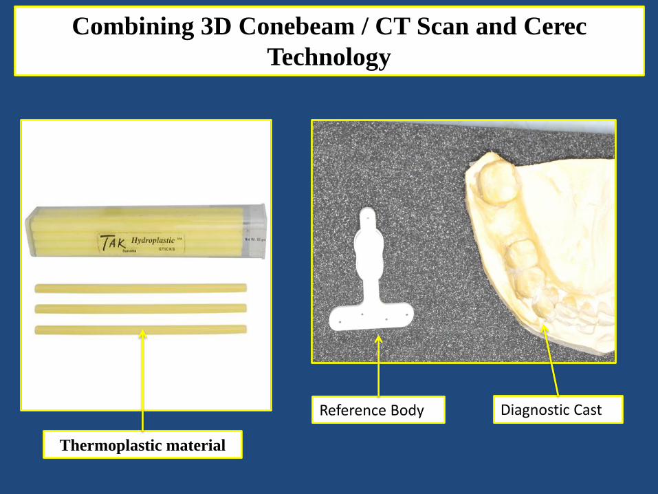
Combining 3D Conebeam / CT Scan and Cerec
Technology

Thermoplastic material
softens and turns clear
in 120° water.
Adapt material to edentulous space and
adjacent teeth.
Center reference body in space and press
to place, making contact with ridge.
Adapt material to sides of reference body.
www.drsmanda.com

Material adapted to
adjacent teeth.
Reference body in contact
with tissue of ridge.
Material adapted to
sides of reference body.
www.drsmanda.com
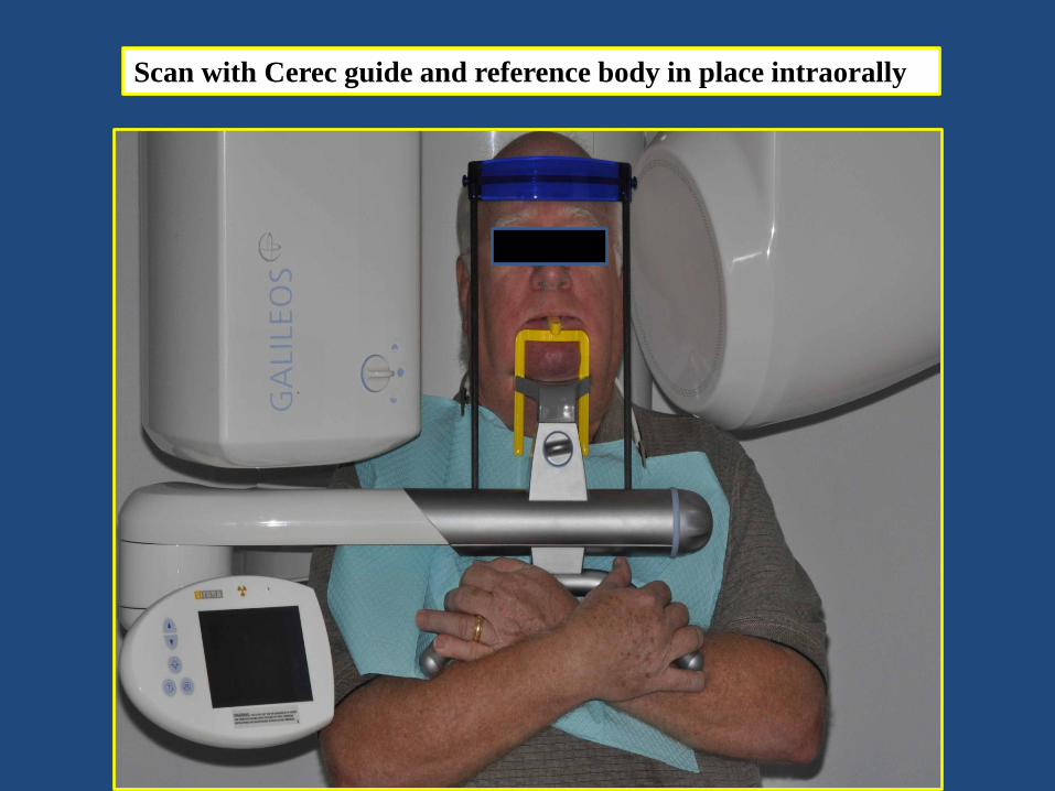
Scan with Cerec guide and reference body in place intraorally:

Radiopaque fiduciary markers embedded in Reference Body will be
used by Sidexis software for orientation.
Combining Galileos 3D Conebeam / CT Scan and
Cerec® Technology

Lateral
View Coronal
View
Crossectional
A/P View
Software allows for viewing in three dimensions.
www.drsmanda.com

Locate and highlight the Mandibular nerve:

Implant selection using software library of multiple implant companies:
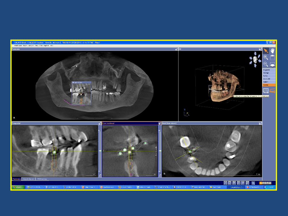

Center implant position within outline of
reference body to insure accurate fabrication of
drill body.
Implant
Positioning: Outline of
Reference Body

Implant
Positioning:
Guidelines: 1. Implant positioned in sound bone.
2. Avoid nerve by at least 2 mm.
3. Minimum of 2mm bone thickness on buccal and lingual.
4. Remain 3.5 mm away from adjacent teeth.

Draw Gingival Margin Virtual Crown Design
Omnicam
Design and Export Of Virtual Crown

Design and Export Of Virtual Crown
Export Design as .CDT File
Load .CDT File onto CBCT
Software

Import Virtual Crown to CBCT Software

Exporting Surgical Plan to Omnicam

Exporting Surgical Plan to Omnicam
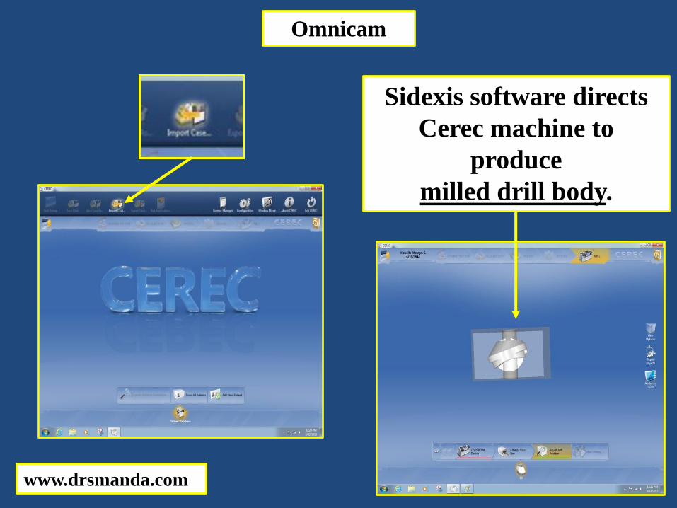
Sidexis software directs
Cerec machine to
produce
milled drill body.
www.drsmanda.com
Omnicam

Milled Drill Bodies:
Tissue Surface
Implant Drill Guide
Channel
Vertical Stop
Orientation
Projection

Orientation Notch
Projection
Drill body produced in Cerec milling chamber.
Drill body snapped into
Cerec Guide.
Reference
body
removed.

Guided Implant Surgery
www.drsmanda.com
manda.com

Implant
site
Tissue punch used with
drill sleeve to enter tissue
while protecting the milled
drill body.

Drill sleeves are specific to
implant manufacturer and
size.
Sleeves fit into drill
body on stent and
guide drill bits to
pre-determined
angle and depth.

Tissue punch technique
provides:
1. Atraumatic entry into
implant site.
2. Minimal bleeding.
3. Minimal post-operative
discomfort.
Initial trephine drill guided to full depth.

Drill sleeves and drill bits of progressively larger sizes
are used until implant can be placed at correct angle
and pre-determined depth.
Multi-purpose fixture mount holds implant while its driven into bone.

Multi-purpose fixture mount.

Careful
planning………..
…….leads to ideal implant
positioning.

Guided implant surgery
produces………….
………excellent results.
Zirconia Screw Retained Hybrid Implant Crown

www.drsmanda.com
manda.com

Digital Implant Impressioning

Hybrid Custom Abutments
Orientation Notch
Ti-Base Scan Body
Orientation Recess
Digital Implant Impressioning

Hybrid Custom Abutment
• Provides precise implant location transfer • Prefabricated rotation locks in zirconium block and on Ti-Base • Metal to metal screw connection
Digital Implant Impressioning

Hybrid Custom Abutments
Zirconia Blocks Screw Access Hole Magnified View of Intaglio Surface of Milled Zirconia Crown
Orientation Recess
Digital Implant Impressioning

Custom Healing Abutment Custom Healing Abutment removed
Gingival Emergence at Implant Fixture Level
Hybrid Screw Retained Custom Abutment Crown
Digital Implant Impressioning

Ti-Base* inserted at fixture level Scan Body* inserted over Ti-Base
*Ti-Base is the fixture level connection to the implant body *Scan Body is the orientation marker for the software
Digital Implant Impressioning
Hybrid Screw Retained Custom Abutment Crown
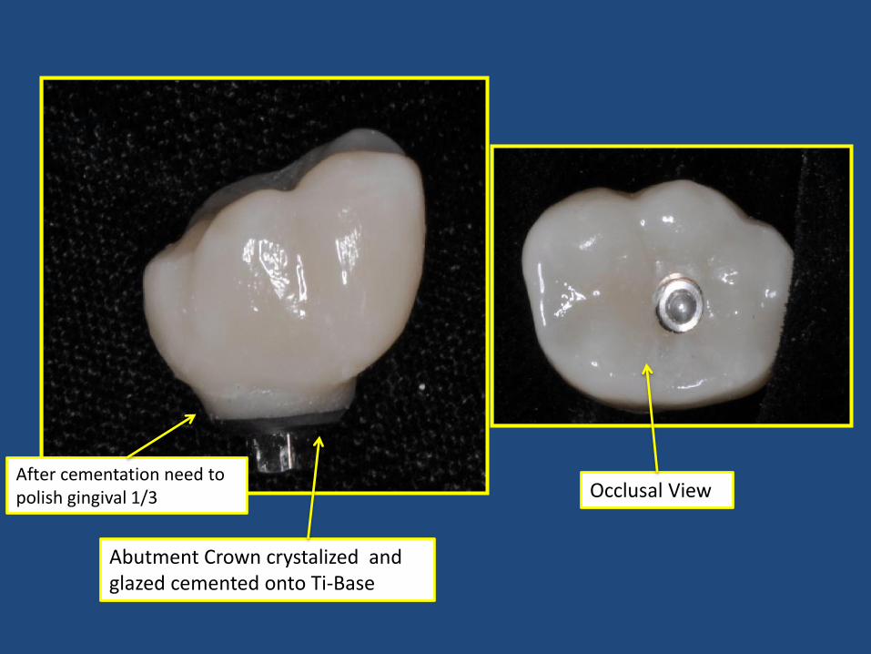
Abutment Crown crystalized and glazed cemented onto Ti-Base
After cementation need to polish gingival 1/3 Occlusal View

Natural Emergence Profile Zirconia Hybrid Custom Screw Retained Implant Crown
Digital Implant Impressioning
Hybrid Screw Retained Custom Abutment Crown

Digital Implant Impressioning
Hybrid Custom Abutments
Healing Abutment
www.drsmanda.com
manda.com

Digital Implant Impressioning
Hybrid Custom Abutments

Digital Implant Impressioning
Scan of the TiBase
Scan of the Scan Body
Digital Implant Impressioning
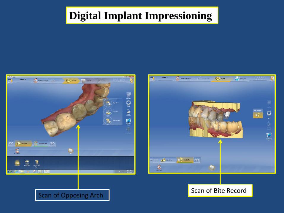
Scan of Opposing Arch Scan of Bite Record
Digital Implant Impressioning
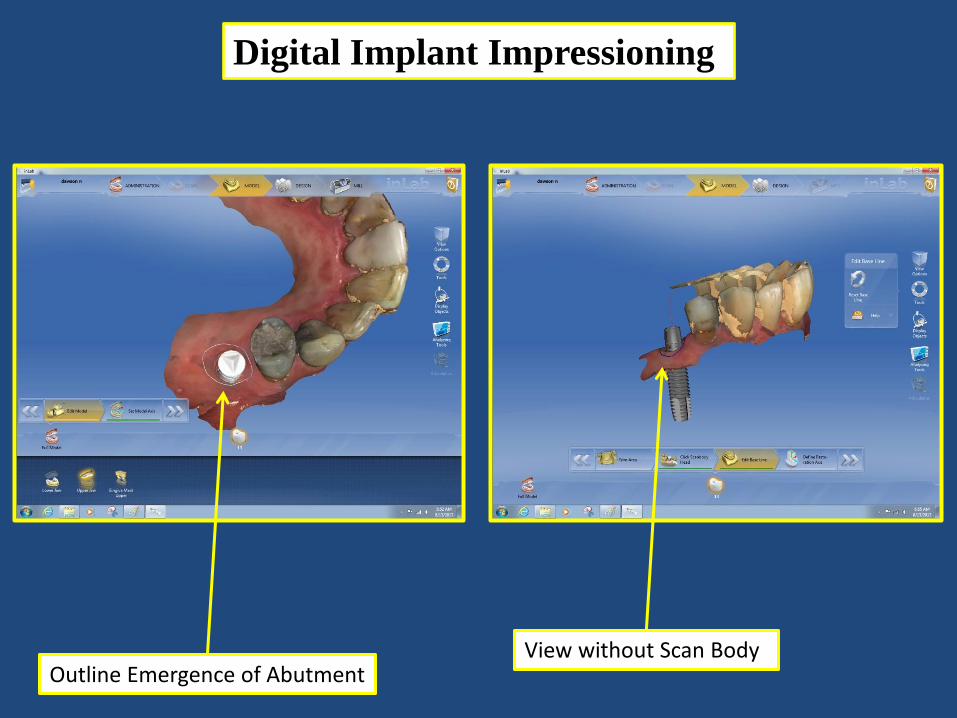
Outline Emergence of Abutment View without Scan Body

Digital Implant Impressioning
Digital View of Crown Proposal Digital View of Crown Proposal Without Gingival Mask

Digital Implant Impressioning
Custom Abutment Crown
TiBase
Interproximal Contact Strength

Digital Implant Impressioning
Split Custom Abutment from Crown
Custom Abutment
Crown
Milling Preview of Custom Abutment

Digital Implant Impressioning
Milling Preview of Crown

Digital Implant Impressioning
TiBase
Custom Abutment
Crown

Milled Zirconia Abutment
Milled Zirconia Abutment Cemented Crown
www.drsmanda.com
manda.com

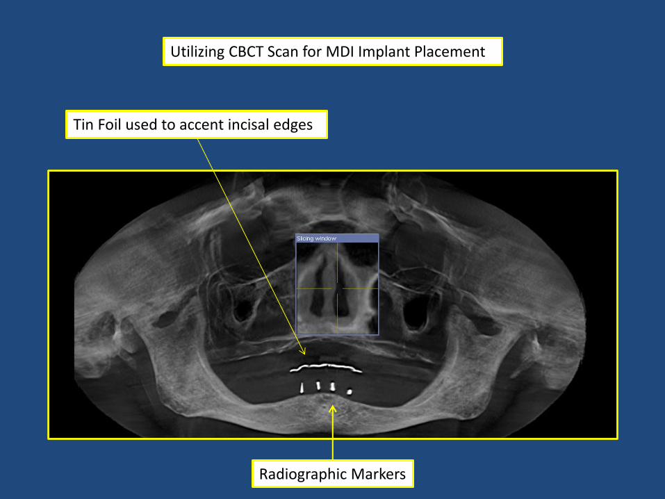
Utilizing CBCT Scan for MDI Implant Placement
Edentulous Mandibular Arch

Acrylic duplicate of Mandibular Denture
Gutta Percha placed as radiographic markers for proposed implant placement in drilled channels
Utilizing CBCT Scan for MDI Implant Placement
Radiographic Markers
Tin Foil used to accent incisal edges

Tin Foil used to accent incisal edges
Gutta Percha placed as radiographic markers for proposed implant placement in drilled channels
Lateral View of Mandible

Tin Foil used to accent incisal edges Gutta Percha placed as radiographic markers for proposed implant placement in drilled channels
Occlusal View of Mandible

Cross Sectional view isolating the mental foramen
First Radiographic Marker 6mm anterior to mental nerve
Tin Foil used to accent incisal edges

Intaglio View with guide holes
Duplicate Mandibular Denture
Occlusal View with guide holes

Duplicate Denture with Guide Holes positioned for Surgery

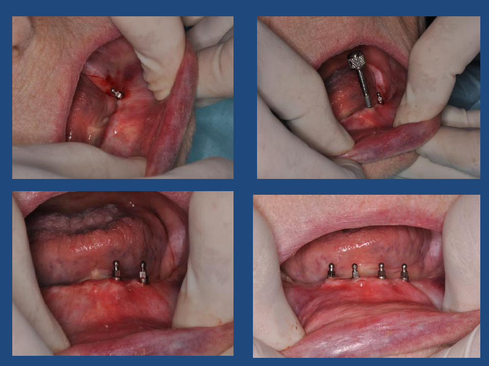
Prepared Sites for implants
Implant Twist Drill guided by access holes

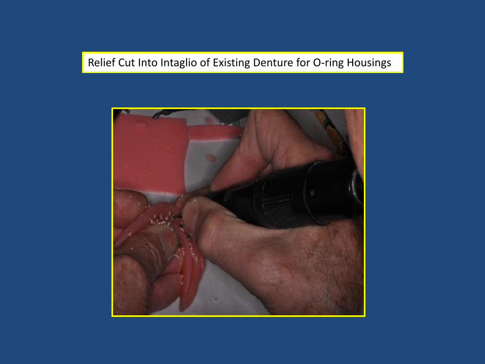
Relief Cut Into Intaglio of Existing Denture for O-ring Housings

Shim Material placed over Implants for block out purposes
O-Ring Housings placed

Silicone Pressure Paste utilized for confirming fit of O-ring Housings
Shim Material
Acrylic Resin Pick-Up of O-Ring Housings

Intaglio of Denture Refined with Acrylic Resin

Final CBCT Scan of Implants


Dental Material List
Intra-Oral Scanner Omnicam- Sirona
CBCT Scanner Galileos- Sirona
Radiographic Bite Plate
Galileos- Sirona www.scicat.com
Cerec Optiguide Patterson Dental- Hydro plastic Material (TAK) Reference Body (Sirona) CEREC Guide Block (Sirona)
CEREC Guide Kit
Implant Drill Sleeve Sirona – www.sironausa.com
Implant System Zimmer Dental MDI

Dental Material List
Ti-Base Kit Patterson Dental – includes Ti-Base and Scan Body
Zirconia Patterson Dental – Sirona Incoris
Lithium Disilicate Porcelain
Patterson Dental – Ivoclar Emax
Maximized Adhesive Dentistry
Implant Drill Sleeve Sirona – www.sironausa.com
Implant System Zimmer Dental MDI 3M
Patterson Dental – Danville Micro etcher Monobond Plus Silane (Ivoclar) All-in-One Unidose bonding agent (Kerr) NX3 Dual Cure Resin Cement (Kerr)
www.drsmanda.com
manda.com