gamma knife radiosurgery for arteriovenous malformations
TRANSCRIPT

Clinical and Translational Science Institute Centers
6-11-2018
Gamma Knife Radiosurgery for Arteriovenous Malformations Gamma Knife Radiosurgery for Arteriovenous Malformations
Using a Four- Dimensional Dynamic Volume Computed Using a Four- Dimensional Dynamic Volume Computed
Tomography Angiography Planning System as an Alternative to Tomography Angiography Planning System as an Alternative to
Traditional Catheter Angiogram Traditional Catheter Angiogram
Christopher P. Cifarelli West Virginia University
John A. Vargo West Virginia University
Todd Tenenholz West Virginia University
Joshua D. Hack West Virginia University
Grenaville Guthrie West Virginia University
See next page for additional authors
Follow this and additional works at: https://researchrepository.wvu.edu/ctsi
Part of the Medicine and Health Sciences Commons
Digital Commons Citation Digital Commons Citation Cifarelli, Christopher P.; Vargo, John A.; Tenenholz, Todd; Hack, Joshua D.; Guthrie, Grenaville; and Carpenter, Jeffrey S., "Gamma Knife Radiosurgery for Arteriovenous Malformations Using a Four- Dimensional Dynamic Volume Computed Tomography Angiography Planning System as an Alternative to Traditional Catheter Angiogram" (2018). Clinical and Translational Science Institute. 908. https://researchrepository.wvu.edu/ctsi/908
This Article is brought to you for free and open access by the Centers at The Research Repository @ WVU. It has been accepted for inclusion in Clinical and Translational Science Institute by an authorized administrator of The Research Repository @ WVU. For more information, please contact [email protected].

Authors Authors Christopher P. Cifarelli, John A. Vargo, Todd Tenenholz, Joshua D. Hack, Grenaville Guthrie, and Jeffrey S. Carpenter
This article is available at The Research Repository @ WVU: https://researchrepository.wvu.edu/ctsi/908

Received 05/21/2018 Review began 05/23/2018 Review ended 05/30/2018 Published 06/11/2018
© Copyright 2018Cifarelli et al. This is an open accessarticle distributed under the terms ofthe Creative Commons AttributionLicense CC-BY 3.0., which permitsunrestricted use, distribution, andreproduction in any medium,provided the original author andsource are credited.
Gamma Knife Radiosurgery forArteriovenous Malformations Using a Four-Dimensional Dynamic Volume ComputedTomography Angiography Planning Systemas an Alternative to Traditional CatheterAngiogramChristopher P. Cifarelli , John A. Vargo , Todd Tenenholz , Joshua D. Hack , GrenavilleGuthrie , Jeffrey S. Carpenter
1. Neurological Surgery, West Virginia University School of Medicine/Ruby Memorial Hospital,Morgantown, USA 2. Department of Radiation Oncology, West Virginia University School of Medicine,Morgantown, USA 3. Department of Radiation Oncology, West Virginia University, Ruby MemorialHospital, Morgantown, USA 4. Radiology, West Virginia University School of Medicine/Ruby MemorialHospital, Morgantown, USA 5. Radiology, West Virginia University School of Medicine/Ruby MemorialHospital, Morgantown , USA
Corresponding author: Christopher P. Cifarelli , [email protected] Disclosures can be found in Additional Information at the end of the article
AbstractBackgroundGamma knife radiosurgery (GKRS) remains a critical intervention in the long-termmanagement of arteriovenous malformations (AVMs). For planning a treatment, identificationof the nidus is essential, and it is dependent on high-resolution blood flow imaging, usually inthe form of a traditional angiogram. The development of dynamic 320-slice computedtomography (CT) angiography has offered a noninvasive alternative to intra-arterialfluoroscopic imaging, and it is capable of providing equivalent temporal resolution. In thisstudy, we describe the feasibility of using four-dimensional CT angiography (4D-CTA) in GKRSplanning for AVM treatment and a comparative analysis with a traditional angiogram.
MethodsA retrospective review was performed on AVM patients treated via GKRS with a 4D-CTA priorto the day of treatment, on the day of treatment, or with a day-of-treatment angiogram.Treatment times, along with total times in the Leksell® coordinate frame G, were obtained fromthe medical records. The frame-on time was calculated by subtracting the treatment time fromthe total time starting from application to removal, and the statistical analysis was performedacross groups using analysis of variance (ANOVA). All treatments were performed on thePerfexion™ model with a dynamic flow imaging procured via a 320-slice CT scanner ortraditional angiography platform.
ResultsSome 27 patients underwent a total of 29 GKRS procedures for AVM treatment at ourinstitution between September 2011 and January 2017. Mean age at the time of treatment was35.5 (6-65) years, and male:female ratio was 5:4. Some 12 patients had 4D-CTA performed prior
1 2 3 2
4 5
Open Access OriginalArticle DOI: 10.7759/cureus.2788
How to cite this articleCifarelli C P, Vargo J A, Tenenholz T, et al. (June 11, 2018) Gamma Knife Radiosurgery for ArteriovenousMalformations Using a Four-Dimensional Dynamic Volume Computed Tomography Angiography PlanningSystem as an Alternative to Traditional Catheter Angiogram. Cureus 10(6): e2788. DOI10.7759/cureus.2788

to the day of treatment, eight patients had the same CTA completed after frame placement onthe day of treatment, while seven patients underwent traditional angiography. The meanframe-on times of each group were 190, 336, and 426 minutes, respectively (p < 0.0001). Noprocedures were aborted based on the image quality.
Conclusions4D-CTA is an effective tool in identifying the AVM nidus for GKRS planning. These studies canbe performed prior to the day of treatment, allowing for a significant reduction in frame-ontime and eliminating the risk of angiogram complication on the day of GKRS.
Categories: Radiation Oncology, NeurosurgeryKeywords: arteriovenous malformation, gamma knife, radiosurgery, four-dimensional cta
IntroductionThe appropriate treatment of cerebral arteriovenous malformations (AVMs) has been thesubject of considerable debate over the past decade [1]. Risk stratification analysis ofunruptured versus ruptured AVMs has prompted an in-depth discussion regarding not only thebest treatment modality options, but also the primary determination to offer any form oftreatment [1-2]. Despite these controversies, gamma knife radiosurgery (GKRS) continues toserve a critical function in the primary treatment of the newly diagnosed AVMs as well as theresidual disease following surgical intervention [3-4].
Paramount to the success of GKRS treatment of AVMs is precise identification and obliterationof the nidus. The classical approach to such a treatment requires a two-dimensional (2D)cerebral angiogram after application of the Leksell® coordinate frame G (Elekta AB, Stockholm,Sweden), followed by a contrast-enhanced three-dimensional (3D) magnetic resonance image(MRI). In a patient population that has already completed a diagnostic digital subtractionangiography (DSA), this treatment-associated study is not only redundant from an imagingperspective, but also exposes the patients to the risks of additional contrast agent, catheter portsite injury, as well as intracranial hemorrhage, albeit at historically low rates [5-6]. Modernimaging advances, including dynamic CT angiography on 320- and 640-slice four-dimensional(4D) scanners, offer a novel mechanism for capture of blood flow imaging with temporalresolution rivaling fluoroscopic capture and offer a safe and effective diagnostic alternative totraditional angiography [7]. Originally employed in cardiac imaging, the use of four-dimensional CT angiography (4D-CTA) has evolved to encompass diagnosis of cerebral AVMsand dural arteriovenous (AV) fistulas [8-9]. In addition, to avoid any potential DSA-associatedcomplications, these 4D-CTA image sets provide the AVM nidal target in a 3D space for directtransfer into the planning software.
In the present study, we review a single-institutional experience using 4D-dynamic volume CTAin GKRS of AVMs as an alternative to catheter-based angiography for the day of service (DOS)flow study used in treatment planning and identify a novel workflow for reducing the timerequired for the patients to remain immobilized in the Leksell® coordinate frame G.
Materials And MethodsStudy design and patient populationAn IRB-approved retrospective review of the patients who received GKRS treatment for cerebralAVMs from 2011 to 2017 was performed. All the patients had a confirmatory diagnostic flowstudy (DSA, CTA) performed prior to GKRS. Spetzler-Martin grades were assigned to each AVM
2018 Cifarelli et al. Cureus 10(6): e2788. DOI 10.7759/cureus.2788 2 of 12

by a neuroradiologist. Three separate workflows were used for GKRS imaging and treatment(Figure 1).
FIGURE 1: Radiosurgery AVM treatment workflow.Patients were treated by one of three mechanisms, with either an angiogram or 4D-CTAperformed after frame placement on the day of service (DOS angiogram, DOS CTA) or with aCTA performed within four weeks prior to the day of treatment (pre-DOS CTA).
Two of the treatment paradigms included all DOS imaging (DSA or CTA) while the third distinctworkflow included a pre-DOS CTA performed within the preceding 30 days of treatment.
AVM treatment imaging Dynamic flow imaging was performed for treatment targeting using either the Aquilion ONE(Toshiba Corporation, Tokyo, Japan) 320-slice CT scanner or the Artis Angiography (SiemensHealthcare, Malvern, PA) platform. The patients undergoing a 4D-CTA on the Aquilion ONEscanner were imaged using 80-120 kV, 100-240 mAs, 320 slice × 0.5 mm collimation withacquisition of three to four frames per second. Whole brain volumes were acquired andreviewed by the neuroradiologist and the treatment team (neurosurgeon/radiation oncologist)for identification of the earliest filling phase of the AVM nidus (Figure 2).
2018 Cifarelli et al. Cureus 10(6): e2788. DOI 10.7759/cureus.2788 3 of 12

FIGURE 2: Time-resolved CTA (4D-CTA) for nidus identification.A representative case of treatment planning in an 11-year-old patient using pre-DOS 4D-CTA toidentify the earliest filling phase (A) through last peak arterial phase and maximal intensityprojection (MIP) images (B and C, respectively).
For patients undergoing cerebral angiogram on the DOS, right common femoral artery accesswas secured using a 5 Fr catheter via a standard Seldinger technique. The AVM visualizationwas achieved following manual injection of a contrast agent and biplanar image acquisition(two to five frames per second) on the Siemens Artis angiography system.
GKRS technique Leksell® coordinate frame G placement was performed with either moderate sedation orgeneral anesthetic. All patients were imaged with a DOS MRI/MRA and either a diagnosticcerebral angiogram or a 4D-CTA. The digital imaging and communications in medicine(DICOM) formatted images were uploaded to the GammaPlan® software (Elekta AB, Stockholm,Sweden) where a conformal target representing the nidus was developed using the dynamicflow study (Figure 3A). A volumetric noncontrast CT head was performed for skull definitionand used for image fusion with CTA when 4D-CTA was acquired (Figure 3B). Target contouringand refinement with a gadolinium-enhanced MRI was used as a confirmation of nidus location(Figure 3C). All treatments were completed on the Perfexion™ Gamma Knife system (Elekta AB,Stockholm, Sweden) utilizing 4, 8, and 16 mm collimators. Frames were removed immediatelyat the completion of the treatment, regardless of the level of anesthesia.
2018 Cifarelli et al. Cureus 10(6): e2788. DOI 10.7759/cureus.2788 4 of 12

FIGURE 3: Sequential target delineation and contouring forGKRS treatment.The DICOM source volume images from the earliest filling phase of the 4D-CTA are used forcontour development in GammaPlan® (A). On the day of service, the 4D-CTA, and theassociated target contour, are co-registered to the volumetric CT head (B). The postgadoliniumT1 and time of flight (TOF) MRI sequences are used for contour verification and furtherrefinement as needed (C).
Calculation of “in frame” timeThe precise time of frame application and removal were noted in each medical record. The totaltreatment beam-on time was subtracted from the total frame application time to determine the“in frame” time for each patient, independent of the treatment length and expressed inminutes. All cases for the treatment of AVM were treated as the first case of the day,eliminating any excess "in frame" time that could occur when multiple patients were treatedwith GKRS within the same day.
Determination of obliteration The follow-up imaging was performed at regular intervals (three to six months) with acombination of CTA, MRA, or a catheter angiogram. The time to obliteration (TTO) wasdetermined by calculating from the DOS of GKRS to the date of the first imaging studydemonstrating absence of fistulous flow. Formal angiograms were used as the gold-standardconfirmatory examination. In cases where the preceding CTA or MRA appeared to demonstrateobliteration and this was subsequently by a catheter angiogram, the date of the preceding testwas used for calculation.
Statistical analysisData are expressed as mean and standard error of the mean for the continuous variables of age,target volume, maximal dose, margin dose, and “in frame” time. Statistical significance was
2018 Cifarelli et al. Cureus 10(6): e2788. DOI 10.7759/cureus.2788 5 of 12

determined via one-way analysis of variance (ANOVA) with multiple comparisons with post-hoc analysis (Fisher’s LSD) using GraphPad Prisim V6.07 (GraphPad Software Inc, LaJolla, CA).Statistical significance was determined at the level of p < 0.05.
ResultsPatient demographics and radiosurgical treatment parametersSome 27 patients underwent a total of 29 GKRS procedures for AVM treatment at ourinstitution between September 2011 and January 2017. Mean age at the time of treatment was35.5 (6-65) years, and the overall male:female ratio was 5:4. Spetzler-Martin grades rangedfrom I to V. Analysis performed on the subsets of patients according to the three treatmentworkflow groups is summarized in Table 1. No statistically significant differences were notedamongst these groups (p > 0.05). The maximal dose received across all the patient workflowcategories ranged from 32.1 to 47 Gy, while the mean margin doses ranged from 16 to 23 Gywith all prescriptions at the 50% isodose line, both without any significant differences acrossthe workflow categories (Table 1).
Pre-DOS CTA DOS CTA DOS Angiogram p
Patient demographics N = 12 N = 8 N = 7
Mean age (years) 32.4 (11-55) 43.1 (17-63) 27.4 (6-65) 0.16
Gender (M:F) 2:1 1:1 3:4 0.591
Target volume (cm3) 4.65 ± 1.39 7.25 ± 1.89 6.54 ± 1.90 0.867
Spetzler-Martin grade II-7; III-3; IV-2 I-1; II-3; III-2; IV-2 I-1; II-1; III-3; IV-1; V-1 0.176
Treatment parameters
Maximal dose (Gy) 37.2 (32.1-40.5) 39.2 (36.1-44.9) 38.1 (32.2-47.0) 0.503
Margin dose (Gy) 18.5 (16-20) 19.3 (18-22) 18.9 (16-23) 0.674
Frame time (minutes) 190 ± 13.5 336 ± 31.3 426 ± 37.8 <0.0001
TABLE 1: Patient demographics and GKRS treatment data.No significant differences were seen in the patient or AVM characteristics across each different workflow group based on ANOVA.The only significant difference (p < 0.0001) was seen in the calculated "in-frame" time, consisting of the total frame time minus thebeam-on treatment time.
Reduction of “in frame” timeThe AVM patients treated with a GKRS using a pre-DOS CTA for the identification of the nidaltarget had a significant reduction of “in frame” time (190 ± 13.5 minutes) compared to a DOSangiogram (426 ± 37.8 minutes; p < 0.0001) and DOS CTA (336 ± 31.3 minutes; p = 0.0016).Furthermore, a comparison of DOS CTA and DOS angiogram did demonstrate a more modestnonsignificant reduction of mean “in frame” time by 89.3 min (p = 0.0816) (Table 2).
2018 Cifarelli et al. Cureus 10(6): e2788. DOI 10.7759/cureus.2788 6 of 12

Frame time (min) Mean difference (DOS Angio Ref.) p 95% CI
Pre-DOS CTA 190 ± 13.5 235 <0.0001 146-324
DOS CTA 336 ± 31.3 89.3 0.0816 -9.95-189
DOS Angiogram 426 ± 37.8 Ref. Ref.
TABLE 2: Statistical analysis of frame times.Using the traditional DOS angiogram approach as the reference, the mean difference in adjusted frame time was found to besignificantly less (p < 0.0001) in the group of patients with pre-DOS imaging and pre-planning.
General anesthetic timeA total of five out of the total 27 patients reviewed received general anesthesia for frameplacement and subsequent GKRS treatment, including 57% of pediatric patients. Of thosepatients receiving general anesthesia, the patients in the pre-DOS CTA group had a mean “inframe” time and general anesthetic time, of 193 ± 29 minutes while the DOS angiogram grouphad an anesthetic exposure time of 261 ± 69 minutes, a difference that did not reach statisticalsignificance (p = 0.471).
Time to obliteration of AVMOf the 27 patients treated with GKRS for AVM, follow-up imaging data was available for 23patients, with 11 patients demonstrating complete obliteration. Among those with radiographicevidence of obliteration, the time from treatment to obliteration was not significantly differentamong the treatment groups, with an overall mean of 885 days (2.4 years) (Table 3).
Mean time to obliteration (days ± SEM) n p
Pre-DOS CTA 890 ± 123 5
0.985DOS CTA 904 ± 261 3
DOS Angiogram 861 ± 86 3
TABLE 3: Time to obliteration.Of the 23 patients with continued follow-up, a total of 11 had radiographic evidence of obliteration [angiogram (five), CTA (five), orMRA (one)], without significant difference in the time to obliteration.
DiscussionThe successful use of GKRS within a wide scope of intracranial diagnoses is directlyproportional to the precision with which the treatment dose is delivered and the accuracy of thepre-procedural images in identification of the target. In this regard, both Perfexion™ andICON™ Gamma Knife systems (Elekta AB, Stockholm, Sweden) boast a QA precision check ofless than 0.4 mm, while the latter is variable across imaging modalities. The need for volumetricdata in AVM target delineation has traditionally required CT and MRI acquisition with
2018 Cifarelli et al. Cureus 10(6): e2788. DOI 10.7759/cureus.2788 7 of 12

correlation of the flow data from angiography [10-11]. Unfortunately, the 2D data from the DSAis of limited use in developing the 3D plan, even with precise co-registration via the Leksell®coordinate frame G [12]. Prior studies have attempted to determine the capacity of MRangiography as a potential replacement for the nidal data provided by DSA. While severalstudies found that medium-sized AVMs with compact niduses were adequately demarcated withMRI/MRA only, others found greater utility in MRI as an adjunctive imaging modalitydetermining that DSA remained necessary for accurate targeting based on variances in plans toknown target locations [13-14].
The development of the 4D-CTA using 640-slice acquisition allows for image capture thatapproaches that of biplanar fluoroscopic images, reaching 0.3-3 frames/second in comparisonto 3-10 frames/second in DSA [15-16]. Coupled with the ability to rapidly image the entirevolume of the head over 16 cm, the 4D-CTA remains a powerful diagnostic tool for a variety ofintracranial pathologies [17-18]. Our work expands that role by assessing the feasibility ofperforming a high-resolution dynamic CT angiogram as an alternative to the traditionalcatheter-based angiography, specifically in the context of treatment planning of GKRS forAVMs.
Vascular imaging with temporally resolved 3D CT angiography is not a novel technique, havingbeen established as an effective method for cardiovascular imaging, including coronary arteryevaluation [19-20]. With regard to diagnostic cerebrovascular imaging, 4D-CTA has been shownto be a reliable means of identification of AVMs, dural AV fistula, as well as aneurysms [18, 21-22]. Here, we present the first series of AVM patients treated with GKRS using 4D dynamicvolume CTA angiography for nidus localization and treatment targeting, either before or afterthe placement of the Leksell® Coordinate Frame G on the day of GKRS treatment. Previously,our institution has reported a single case in which the 4D-CTA was used as a DOS study fortreatment planning, including the use of titanium frame pins, creating a significant imageartifact [23]. This artifact has been well documented by other groups utilizing only CT-basedtreatment planning as an alternative to MRI [24-25]. As there is no frame present in the pre-DOS CTA images, any artifact potentially obscuring nidus identification has been subsequentlyavoided.
With the development of the ICON system, gamma knife radiosurgery has experienced its firsttrend toward frameless treatment strategies, as already embraced by Linac-basedplatforms [26-27]. As more patients receive treatment via this system, we are likely to seefurther reduction in the need for frame-based radiosurgery, especially in large lesions alreadytreated with dose- or volume-fractionated SRS [28-29]. In our study, treatment remains aframe-based procedure, but the use of the 4D-CTA as part of the planning process in advance offrame placement demonstrates a significant reduction of “in frame” time. Although notquantifiable, we were also provided increased time for image analyses with CTAs performedprior to the day of service, insuring that all members of the treatment team, including theneuroradiologists, had the opportunity to review the images and develop a consensus opinionon the optimal target site.
In addition to the reduction in nontreatment frame time, we examined the effect of the pre-DOS CTA planning on the amount of general anesthetic provided during treatment. Of the 27patients treated, only five received general anesthesia, including nearly 60% of the pediatricAVM patients reviewed in this study. Across the treatment groups, pre-DOS CTA and DOS-angiogram, we did find a reduction in mean general anesthetic time (193 ± 29 minutes versus261 ± 69 minutes). Yet, these data were not significant, likely based on the limited total numberof patients (n = 5). Given the link between procedure duration and risks of anestheticcomplications in pediatric patients undergoing general anesthesia for radiotherapy cases, thisreduction in total anesthesia time may importantly reduce such risks [30].
2018 Cifarelli et al. Cureus 10(6): e2788. DOI 10.7759/cureus.2788 8 of 12
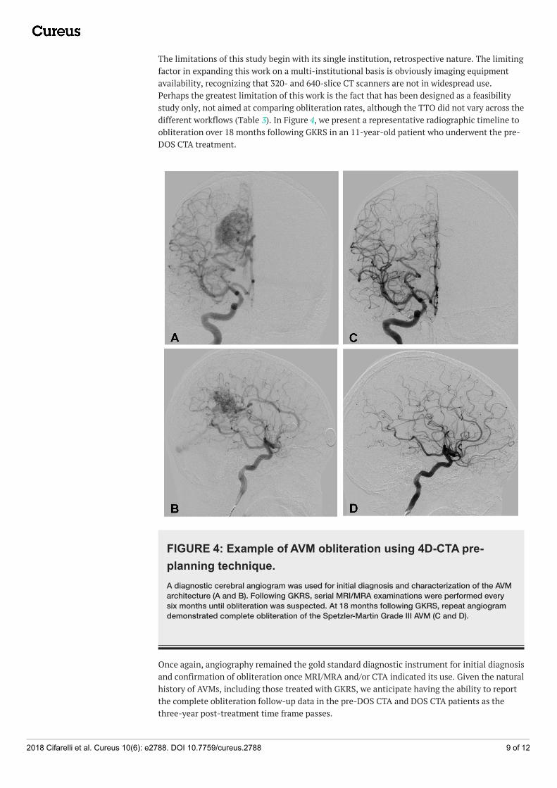
The limitations of this study begin with its single institution, retrospective nature. The limitingfactor in expanding this work on a multi-institutional basis is obviously imaging equipmentavailability, recognizing that 320- and 640-slice CT scanners are not in widespread use.Perhaps the greatest limitation of this work is the fact that has been designed as a feasibilitystudy only, not aimed at comparing obliteration rates, although the TTO did not vary across thedifferent workflows (Table 3). In Figure 4, we present a representative radiographic timeline toobliteration over 18 months following GKRS in an 11-year-old patient who underwent the pre-DOS CTA treatment.
FIGURE 4: Example of AVM obliteration using 4D-CTA pre-planning technique.A diagnostic cerebral angiogram was used for initial diagnosis and characterization of the AVMarchitecture (A and B). Following GKRS, serial MRI/MRA examinations were performed everysix months until obliteration was suspected. At 18 months following GKRS, repeat angiogramdemonstrated complete obliteration of the Spetzler-Martin Grade III AVM (C and D).
Once again, angiography remained the gold standard diagnostic instrument for initial diagnosisand confirmation of obliteration once MRI/MRA and/or CTA indicated its use. Given the naturalhistory of AVMs, including those treated with GKRS, we anticipate having the ability to reportthe complete obliteration follow-up data in the pre-DOS CTA and DOS CTA patients as thethree-year post-treatment time frame passes.
2018 Cifarelli et al. Cureus 10(6): e2788. DOI 10.7759/cureus.2788 9 of 12

This study does not support the use of 4D-CTA as a replacement imaging modality in lieu oftraditional cerebral angiography for lesion diagnosis. We recognize that the accuratecharacterization of the complex architecture of the majority of AVMs often requires the use oftraditional catheter angiography, especially in higher grade Spetzler-Martin lesions. Moreover,the test of cure in our patient population remains catheter-based angiography. Yet, despite thecontinued importance of DSA in AVM management, we assert that the data support the utilityof 4D-CTA in GKRS planning for AVM and would benefit from further study. Finally, the use ofa DOS volumetric CT scan for image merging with the pre-DOS CTA may be applicable to AVMstreated via additional SRS treatment platforms, such as CyberKnife. Unlike the DOS angiogramthat utilizes gamma frame data assimilation, the skull contours used in our work presumablywould be applicable to frameless devices, although our preference for optimal treatmentoutcomes remains GKRS at our institution.
Conclusions4D-CTA offers a reliable means of nidus delineation for GKRS planning in AVM treatment.Performing 4D-CTA prior to the day of treatment offers a significant reduction in frame time.Preliminary results demonstrate similar times to obliteration among CTA-assisted planningcases and those using traditional catheter-based angiograms.
Additional InformationDisclosuresHuman subjects: Consent was obtained by all participants in this study. West VirginiaUniversity Office of Research Integrity and Compliance issued approval 1408383213. ActionDate 07/17/2017 To Christopher Cifarelli From WVU Office of Research Integrity andCompliance Approval Date 08/07/2014 Expiration Date 07/16/2018 Subject AcknowledgementLetter Exempt Initial Protocol Review Protocol Number 1408383213R001 Title The use of 4DCT Angiography in pre-operative Gamma Knife treatment planning The above-referenced studywas reviewed by the West Virginia University Institutional Review Board IRB and was grantedexemption in accordance with 45 CFR 46.101. • This research study was granted an exemptionin accordance with Research on existing data, documents, records, pathological specimens, ordiagnostic specimens [45 CFR 46.101(4)]. In accordance with the Health Insurance Portabilityand Accountability Act, a waiver of research authorization has been granted. Please fulfill thesubject accounting requirements associated with the granting of this waiver. All exemptions areonly good for three years. If this research extends more than three years beyond the approveddate, then the researcher will have to request another exemption. The following documentshave been acknowledged for use in this study and are available in the WVU+kc system:Documents reviewed and/or approved as part of this submission: HIPAA WaiverForm_Blank_GK.pdf: 2014-08-03-04:00 4DCTA_AVM_Data Sheet.xlsx: 2014-08-03-04:00Documents for use in this study have been acknowledged and are available in the WVUkcsystem in the Notes and Attachments section of your protocol. The Office of Research Integrityand Compliance is here to provide assistance to you from the initial submission of an IRBprotocol and all subsequent activity. Please feel free to contact us by phone at 304.293.7073with any question you may have. Thank you. Animal subjects: All authors have confirmed thatthis study did not involve animal subjects or tissue. Conflicts of interest: In compliance withthe ICMJE uniform disclosure form, all authors declare the following: Payment/services info:All authors have declared that no financial support was received from any organization for thesubmitted work. Financial relationships: All authors have declared that they have nofinancial relationships at present or within the previous three years with any organizations thatmight have an interest in the submitted work. Other relationships: All authors have declaredthat there are no other relationships or activities that could appear to have influenced thesubmitted work.
2018 Cifarelli et al. Cureus 10(6): e2788. DOI 10.7759/cureus.2788 10 of 12
References1. Mohr JP, Parides MK, Stapf C, et al.: Medical management with or without interventional
therapy for unruptured brain arteriovenous malformations (ARUBA): a multicentre, non-blinded, randomised trial. Lancet. 2014, 383:614-621. 10.1016/S0140-6736(13)62302-8
2. Pollock BE, Flickinger JC, Lunsford LD, Maitz A, Kondziolka D: Factors associated withsuccessful arteriovenous malformation radiosurgery. Neurosurgery. 1998, 42:1239-1244.10.1097/00006123-199806000-00020
3. Ding D, Starke RM, Kano H, et al.: Radiosurgery for cerebral arteriovenous malformations in arandomized trial of unruptured brain arteriovenous malformations (ARUBA)-eligible patients:a multicenter study. Stroke. 2016, 47:342-349. 10.1161/STROKEAHA.115.011400
4. Yen CP, Ding D, Cheng CH, Starke RM, Shaffrey M, Sheehan J: Gamma knife surgery forincidental cerebral arteriovenous malformations. J Neurosurg. 2014, 121:1015-1021.10.3171/2014.7.JNS131397
5. Dawkins AA, Evans AL, Wattam J, et al.: Complications of cerebral angiography: a prospectiveanalysis of 2,924 consecutive procedures. Neuroradiology. 2007, 49:753-759. 10.1007/s00234-007-0252-y
6. Kaufmann TJ, Huston J, 3rd, Mandrekar JN, Schleck CD, Thielen KR, Kallmes DF:Complications of diagnostic cerebral angiography: evaluation of 19,826 consecutive patients .Radiology. 2007, 243:812-819. 10.1148/radiol.2433060536
7. Li Q, Lv F, Yao G, Li Y, Xie P: 64-section multidetector CT angiography for evaluation ofintracranial aneurysms: comparison with 3D rotational angiography. Acta Radiol. 2014,55:840-846. 10.1177/0284185113506138
8. Alnemari A, Mansour TR, Bazerbashi M, Buehler M, Schroeder J, Gaudin D: Dynamic four-dimensional computed tomography angiography for neurovascular pathologies. WorldNeurosurg. 2017, 105:1034-1011. 10.1016/j.wneu.2017.06.022
9. Wang H, Ye X, Gao X, Zhou S, Lin Z: The diagnosis of arteriovenous malformations by 4D-CTA: a clinical study. J Neuroradiol. 2014, 41:117-123. 10.1016/j.neurad.2013.04.004
10. Lunsford LD, Niranjan A, Kano H, Kondziolka D: The technical evolution of gamma kniferadiosurgery for arteriovenous malformations. Prog Neurol Surg. 2013, 27:22-34.10.1159/000341625
11. Sadler LR, Jungreis CA, Lunsford LD, Trapanotto MM: Angiographic technique to precedegamma knife radiosurgery for intracranial arteriovenous malformations. Am J Neuroradiol.1990, 11:1157-1161.
12. Spiegelmann R, Friedman WA, Bova FJ: Limitations of angiographic target localization inplanning radiosurgical treatment. Neurosurgery. 1992, 30:619-623. 10.1097/00006123-199204000-00026
13. Bednarz G, Downes B, Werner-Wasik M, Rosenwasser RH: Combining stereotacticangiography and 3D time-of-flight magnetic resonance angiography in treatment planningfor arteriovenous malformation radiosurgery. Int J Radiat Oncol Biol Phys. 2000, 46:1149-1154. 10.1016/S0360-3016(99)00530-1
14. Kondziolka D, Lunsford LD, Kanal E, Talagala L: Stereotactic magnetic resonance angiographyfor targeting in arteriovenous malformation radiosurgery. Neurosurgery. 1994, 35:585-590.10.1227/00006123-199410000-00002
15. D'Orazio F, Splendiani A, Gallucci M: 320-Row detector dynamic 4D-CTA for the assessmentof brain and spinal cord vascular shunting malformations. A technical note. Neuroradiol J.2014, 27:710-717. 10.15274/NRJ-2014-10096
16. Suzuki K, Abe K, Maruyama T, et al.: The role of 4D CT angiography for preoperativescreening in patients with intracranial tumors. Neuroradiol J. 2016, 29:168-173.10.1177/1971400916638353
17. Kortman HG, Smit EJ, Oei MT, Manniesing R, Prokop M, Meijer FJ: 4D-CTA in neurovasculardisease: a review. Am J Neuroradiol. 2015, 36:1026-1033. 10.3174/ajnr.A4162
18. Willems PW, Brouwer PA, Barfett JJ, terBrugge KG, Krings T: Detection and classification ofcranial dural arteriovenous fistulas using 4D-CT angiography: initial experience. Am JNeuroradiol. 2011, 32:49-53. 10.3174/ajnr.A2248
19. Hein PA, Romano VC, Lembcke A, May J, Rogalla P: Initial experience with a chest painprotocol using 320-slice volume MDCT. Eur Radiol. 2009, 19:1148-1155. 10.1007/s00330-008-1255-8
2018 Cifarelli et al. Cureus 10(6): e2788. DOI 10.7759/cureus.2788 11 of 12

20. Sheth T, Amlani S, Ellins ML, et al.: Computed tomographic coronary angiographicassessment of high-risk coronary anatomy in patients with suspected coronary artery diseaseand intermediate pretest probability. Am Heart J. 2008, 155:918-923.10.1016/j.ahj.2007.11.035
21. Beijer TR, van Dijk EJ, de Vries J, Vermeer SE, Prokop M, Meijer FJ: 4D-CT angiographydifferentiating arteriovenous fistula subtypes. Clin Neurol Neurosurg. 2013, 115:1313-1316.10.1016/j.clineuro.2012.12.015
22. Shankar JJ, Lum C, Chakraborty S, Dos Santos M: Cerebral vascular malformations: time-resolved CT angiography compared to DSA. Neuroradiol J. 2015, 28:310-315.10.1177/1971400915589682
23. Turner RC, Lucke-Wold BP, Josiah D, et al.: Stereotactic radiosurgery planning based on time-resolved CTA for arteriovenous malformation: a case report and review of the literature. ActaNeurochir Wien. 2016, 158:1555-1562. 10.1007/s00701-016-2874-5
24. Attia A, Tatter SB, Weller M, et al.: CT-only planning for gamma knife radiosurgery in thetreatment of trigeminal neuralgia: methodology and outcomes from a single institution. J MedImaging Radiat Oncol. 2012, 56:490-494. 10.1111/j.1754-9485.2012.02403.x
25. Park KJ, Kano H, Berkowitz O, et al.: Computed tomography-guided gamma knife stereotacticradiosurgery for trigeminal neuralgia. Acta Neurochir Wien. 2011, 153:1601-1609.10.1007/s00701-011-1026-1
26. Kamath R, Ryken TC, Meeks SL, Pennington EC, Ritchie J, Buatti JM: Initial clinicalexperience with frameless radiosurgery for patients with intracranial metastases. Int J RadiatOncol Biol Phys. 2005, 61:1467-1472. 10.1016/j.ijrobp.2004.08.021
27. Minniti G, Scaringi C, Clarke E, Valeriani M, Osti M, Enrici RM: Frameless linac-basedstereotactic radiosurgery (SRS) for brain metastases: analysis of patient repositioning using amask fixation system and clinical outcomes. Radiat Oncol. 2011, 6:158. 10.1186/1748-717X-6-158
28. Franzin A, Panni P, Spatola G, et al.: Results of volume-staged fractionated gamma kniferadiosurgery for large complex arteriovenous malformations: obliteration rates and clinicaloutcomes of an evolving treatment paradigm. J Neurosurg . 2016, 125:104-113.10.3171/2016.7.GKS161549
29. Seymour ZA, Sneed PK, Gupta N, et al.: Volume-staged radiosurgery for large arteriovenousmalformations: an evolving paradigm. J Neurosurg. 2016, 124:163-174.10.3171/2014.12.JNS141308
30. Anghelescu DL, Burgoyne LL, Liu W, et al.: Safe anesthesia for radiotherapy in pediatriconcology: St. Jude Children's Research Hospital experience, 2004-2006. Int J Radiat Oncol BiolPhys. 2008, 71:491-497. 10.1016/j.ijrobp.2007.09.044
2018 Cifarelli et al. Cureus 10(6): e2788. DOI 10.7759/cureus.2788 12 of 12