gastric helicobacters in domestic animals and nonhuman ... · hamsters, ruminants, horses, and...
TRANSCRIPT

CLINICAL MICROBIOLOGY REVIEWS, Apr. 2009, p. 202–223 Vol. 22, No. 20893-8512/09/$08.00�0 doi:10.1128/CMR.00041-08Copyright © 2009, American Society for Microbiology. All Rights Reserved.
Gastric Helicobacters in Domestic Animals and Nonhuman Primatesand Their Significance for Human Health
Freddy Haesebrouck,* Frank Pasmans, Bram Flahou, Koen Chiers, Margo Baele, Tom Meyns,Annemie Decostere, and Richard Ducatelle
Department of Pathology, Bacteriology and Avian Diseases, Faculty of Veterinary Medicine, Ghent University, Salisburylaan 133,B-9820 Merelbeke, Belgium
INTRODUCTION .......................................................................................................................................................202GASTRIC NON-H. PYLORI HELICOBACTER NOMENCLATURE: THE NEED FOR
CLARIFICATION ..............................................................................................................................................203GASTRIC HELICOBACTERS IN DOMESTIC ANIMALS AND NONHUMAN PRIMATES: AN
OVERVIEW .........................................................................................................................................................204Gastric Helicobacters Associated with Pigs ........................................................................................................204Gastric Helicobacters Associated with Dogs and Cats......................................................................................210Gastric Helicobacters Associated with Rabbits ..................................................................................................211Gastric Helicobacters Associated with Ferrets ...................................................................................................211Gastric Helicobacters Associated with Hamsters...............................................................................................212Gastric Helicobacters Associated with Ruminants ............................................................................................212Gastric Helicobacters Associated with Horses ...................................................................................................212Gastric Helicobacters Associated with Nonhuman Primates ...........................................................................212
NON-H. PYLORI HELICOBACTER-ASSOCIATED GASTRIC DISEASES IN HUMANS: ZOONOSES? .........213GASTRIC DISEASE IN HUMANS INFECTED WITH NON-H. PYLORI HELICOBACTER SPECIES .......214TRANSMISSION OF GASTRIC NON-H. PYLORI HELICOBACTER SPECIES..............................................215VIRULENCE FACTORS OF GASTRIC NON-H. PYLORI HELICOBACTER SPECIES .................................215STUDYING GASTRIC HELICOBACTER INFECTIONS IN DOMESTIC ANIMALS: WHAT MIGHT IT
TEACH US ABOUT THESE INFECTIONS IN HUMANS? ........................................................................217CONCLUSIONS .........................................................................................................................................................217REFERENCES ............................................................................................................................................................217
INTRODUCTION
It was first reported in 1984 that gastric ulcer disease inhumans is caused by a bacterial infection (141). The causativeagent, Helicobacter pylori, has also been associated with gastri-tis, peptic ulcer disease, gastric adenocarcinoma, and mucosa-associated lymphoid tissue (MALT) lymphoma (132, 174, 215).This bacterium is very successful in the way that it colonizes thehuman stomach, since in developing countries, more than 80%of the population is infected with H. pylori, even at young age.In developed countries, the prevalence of H. pylori generallyremains under 40% and is considerably lower in children andadolescents than in adults and elderly people (132, 181).
Various tests have been developed for the diagnosis of H.pylori infections (reviewed in reference 132). For routine diag-nostic purposes, histology or culture of biopsies from patientswho have undergone endoscopy and urea breath testing aremost often used. On histology, H. pylori bacteria are identifiedon the basis of their typical localization and their characteristic,slightly curve-shaped morphology. In 0.2 to 6% (depending onthe literature source and the geographical region) of thesebiopsies, however, bacteria with a different, typically long spi-ral-shaped morphology are found. These spiral-shaped non-H.pylori helicobacters were first described in 1987 (46). They
were originally referred to as “Gastrospirillum hominis” (143).Analysis of the 16S rRNA gene of these uncultivated organ-isms resulted in their classification in the genus Helicobacter.They were provisionally named “H. heilmannii” after the Ger-man pathologist Konrad Heilmann, who first studied the pa-thology associated with these microorganisms (101). “H. hei-lmannii” has also been associated with gastritis (40), gastriculcers (41), and gastric MALT lymphoma (153) but not withgastric adenocarcinoma. Further research on “H. heilmannii”has been seriously hampered by the very fastidious nature ofthese microorganisms. Even today, to our knowledge, only two“H. heilmannii” strains have been cultured from human tissue(2, 127). Long spiral-shaped helicobacters have also been dem-onstrated in the stomachs of different animal species. A sum-mary of these helicobacters is given in Table 1.
This article aims to provide an overview of Helicobacterspecies naturally colonizing the stomachs of food-producinganimals, pet animals, and nonhuman primates. First, the verycomplex and confusing nomenclature used to designate non-H.pylori Helicobacter species colonizing the human stomach isconsidered. Thereafter, an overview of helicobacters coloniz-ing the stomachs of domestic animals and nonhuman primatesis presented, and their possible pathogenic significance fortheir animal hosts is discussed. The main aim of this article,however, is to have a closer look at the significance of thesemicroorganisms for human health: should they be consideredzoonotic agents, what are the disease signs in infected humans,
* Corresponding author. Mailing address: Faculty of VeterinaryMedicine, Salisburylaan 133, 9820 Merelbeke, Belgium. Phone: 32 9264 74 30. Fax: 32 9 264 74 94. E-mail: [email protected].
202
on March 31, 2020 by guest
http://cmr.asm
.org/D
ownloaded from

how are they transmitted, and what is known about their vir-ulence factors? The article ends with some thoughts on whatthe study of gastric Helicobacter infections in animals mightteach us about these infections in humans.
GASTRIC NON-H. PYLORI HELICOBACTERNOMENCLATURE: THE NEED
FOR CLARIFICATION
Since the description of H. pylori, the number of species inthe genus Helicobacter has rapidly expanded. Today, a largenumber of non-H. pylori Helicobacter species in a wide varietyof animals and humans have been described, and the genusHelicobacter contains at least 32 species with validly publishednames (http://www.bacterio.cict.fr/h/helicobacter.html). Thefrequent changes in nomenclature of non-H. pylori helico-bacters colonizing the stomachs of humans have causedquite a lot of confusion, not only among clinicians but alsoamong bacteriologists. Today, there is a serious problem intrying to reach international agreement on this complex andexpanding group of microorganisms, all of which have incommon their tightly coiled morphology and their difficultyto culture in vitro.
After the renaming of “Gastrospirillum hominis” as “H. hei-lmannii,” further genetic analysis of the 16S rRNA gene re-vealed two types that differed by more than 3% in their nucle-otide sequence, which prompted the subclassification of thenon-H. pylori helicobacters into “H. heilmannii” type 1 and “H.heilmannii” type 2. Sequencing of the 23S rRNA-encodinggenes also makes it possible to distinguish between the twotypes (48).
“H. heilmannii” type 1 is both morphologically and geneti-cally identical to a bacterium colonizing the stomachs of pigs(43, 166) that was first designated “Gastrospirillum suis” (146,184). Almost 10 years later, sequencing of the 16S rRNA gene,fluorescent in situ hybridization (FISH), and electron mi-
croscopy showed that these organisms belong to the genusHelicobacter and are sufficiently different from all existingspecies to constitute a new taxon. Because at that time thisspecies could not be thoroughly characterized due to thelack of pure in vitro isolates, the organism was described as“Candidatus Helicobacter suis” (43). Only recently have invitro cultures been obtained, resulting in the description ofH. suis as a species (14).
The situation with regard to “H. heilmannii” type 2 is evenmore complex. This type represents not a single Helicobacterspecies but rather a group of species, including three helico-bacters that have been isolated from the stomachs of cats anddogs, namely, H. felis, H. bizzozeronii, and H. salomonis. To add tothe confusion, one uncultivable species detected in the stomachsof humans, wild felids, dogs, and cats was named “CandidatusHelicobacter heilmannii” (166). Two other closely related species,one of which was isolated from a dog and the other from a cat,have been described as H. cynogastricus and H. baculiformis, re-spectively (15, 233). However, to date no information is availableabout the presence of these bacteria in humans.
Differences in morphology between different gastric non-H.pylori Helicobacter species have been described (Table 2), butthis is not an accurate method for species identification. It hasbeen stated that periplasmic fibrils wrapped around the cellbody are a typical feature of H. felis (134). However, H. cyno-gastricus also possesses a periplasmic fibril running along theexternal side of the helix, and both species are tightly coiledorganisms (233). H. salomonis is less tightly coiled and doesnot have periplasmic fibrils (115). H. baculiformis is a large,slender to slightly spiral rod with periplasmic fibrils (15).“Candidatus Helicobacter heilmannii,” H. bizzozeronii, andH. suis are morphologically very similar. These microorgan-isms do not possess periplasmic fibrils and show very tightcoils (14, 92, 166).
Sequencing of the 16S and 23S rRNA-encoding genes allowsdifferentiation of H. suis from the other gastric non-H. pylori
TABLE 1. Helicobacter species naturally colonizing the stomachs of animals and their pathogenic significance for humans
Helicobacter species Natural host (prevalence, %)Associated withgastric disease
in humansReference(s)
H. suis Pig (60–80 in slaughter pigs), macaque (NAb),mandrill monkey (NA)
Yes 14, 42, 45, 167, 227, 230
H. felis Dog (47), cat (63), rabbit (2–9), cheetah (NAc) Yes 45, 134, 166, 227, 230, 231, 234H. bizzozeronii Dog (70), cat (35) Yes 45, 92, 227, 230H. salomonis Dog (9), cat (2), rabbit (0–4) Yes 45, 115, 227, 230“Candidatus Helicobacter heilmannii” Dog (20–100), cat (20–100), wild felidae
(NAc), nonhuman primates (66)Yes 166, 227, 230
H. baculiformis Cat (NA) No 15H. cynogastricus Dog (NA) No 233“Candidatus Helicobacter bovis” Cattle (NA) Yes 44, 45H. mustelae Ferret (0–100) No 66, 67, 68, 69, 70, 71, 85H. aurati Syrian hamster (50–100) No 177H. nemestrinaea Macaque (NA) No 24H. acinonychis Cheetah (low), tiger (NA) No 60, 224H. cetorum Whales (NA), dolphins (NA) No 97H. muridarum Mice (0–62) No 86, 136, 183
a Later heterotypic synonym of H. pylori (219).b NA, not available.c Terio et al. (224) and Morner et al. (155) found the gastric mucosa of 75% of cheetahs and 68% of free-ranging lynx to be colonized with “pet carnivore-associated”
helicobacters. Those studies do not allow differentiation between H. felis, H. bizzozeronii, H. salomonis, H. baculiformis, H. cynogastricus, and “Candidatus Helicobacterheilmannii”.
VOL. 22, 2009 GASTRIC HELICOBACTERS IN DOMESTIC ANIMALS AND PRIMATES 203
on March 31, 2020 by guest
http://cmr.asm
.org/D
ownloaded from

Helicobacter species mentioned above, but it cannot distinguishbetween H. felis, H. bizzozeronii, H. salomonis, H. cynogastricus,H. baculiformis, and “Candidatus Helicobacter heilmannii”(15, 48, 233). For differentiation between these species, se-quencing of the Hsp60 gene (149), the urease A and B genes(161, 166), and the gyrB gene (95) is useful, as is whole-cellprotein profiling (228) if pure in vitro cultures are available.
Phylogenetic trees for the gastric helicobacters discussed inthis review are shown in Fig. 1 and 2. Figure 1 is based on 16SrRNA gene sequence similarity data and Fig. 2 on the partialureA and ureB gene sequences. The sequences that have beendetected in human stomachs are also indicated.
In the literature, gastric infections with spiral-shaped bacte-ria in humans are often referred to as “H. heilmanni” or “H.heilmannii-like organism” infections. However, at present, thename “H. heilmannii” cannot be used as a species name, ac-cording to taxonomical rules. To avoid confusion, we proposeto use the term “gastric non-H. pylori helicobacters” to desig-nate these spiral-shaped bacteria when only results of histopa-thology or crude taxonomic data are available and to reservetrue species designations for those situations in which the spe-cies is defined.
To nonbacteriologists, the changes in “H. heilmannii” no-menclature may appear unwieldy and unnecessary. However, itshould be kept in mind that several important traits, includingpathogenicity and antimicrobial susceptibility, may vary de-pending on the bacterial species. At present it is not knownwhether certain non-H. pylori Helicobacter species are moreoften associated with a certain disease outcome in humansthan others.
GASTRIC HELICOBACTERS IN DOMESTIC ANIMALSAND NONHUMAN PRIMATES: AN OVERVIEW
A summary of gastric lesions described in domestic animalsand nonhuman primates naturally or experimentally infectedwith helicobacters is presented in Table 3. Below, infectionswith gastric helicobacters in pigs, dogs, cats, rabbits, ferrets,hamsters, ruminants, horses, and nonhuman primates are con-sidered. Guinea pigs and Mongolian gerbils are also often kept
as pets, but natural infections with gastric helicobacters havenot been described in these animal species.
Gastric Helicobacters Associated with Pigs
The main Helicobacter species colonizing the stomachs ofpigs is H. suis. Its prevalence at slaughter age in most reportsis 60% or more. H. suis causes gastritis in experimentally andnaturally infected pigs (87, 104, 147, 173, 185). It has also beenassociated with ulcers of the nonglandular part of the stomach(18, 28, 185, 190), although the exact role of H. suis in porcinegastric pathology remains to be elucidated. Indeed, Grasso etal. (87), Melnichouk et al. (145), Park et al. (173), and Szerediet al. (222) did not find this association. These discrepanciesmight be due to differences in laboratory techniques for dem-onstration of Helicobacter, different sampling practices, or dif-ferences in virulence between different H. suis strains. In anycase, in a recent study carried out by our research group,gastric ulcers were induced in pigs experimentally infected withH. suis (T. Meyns, R. Ducatelle, B. Flahou, K. Chiers, F.Pasmans, and F. Haesebrouck, submitted for publication). Inthis study, 6-week-old piglets that were free of H. suis wereused. Nine piglets were intragastrically inoculated with a pureculture of H. suis, while five sham-inoculated piglets were usedas controls. All piglets were fed a finely ground diet. Hyper-keratosis and ulcer formation were clearly present in the gas-tric nonglandular mucosa of all H. suis-inoculated pigs, whilenone of the sham-inoculated piglets developed gastric lesions.
Hyperkeratosis and ulceration of the nonglandular part ofthe stomach have been reported in many countries. Up to 80%of the market pigs in Australia (189) and 60% of the sows (106)in The Netherlands showed gastric lesions. Hessing et al. (106)found gastric ulcers in 10 to 15% of the sows.
The stomach mucosa of pigs can be divided into a glandularpart (cardiac gland zone, fundic gland zone, and antrum withpyloric glands) and a nonglandular part, the latter being asmall rectangular area around the esophageal opening. It isalso called the pars esophagea of the stomach and is covered bya stratified squamous epithelium (Fig. 3). After experimentalinfection, H. suis colonizes mainly the antrum and the fundic
TABLE 2. Differential characteristics of gastric Helicobacter species associated with domestic animals and nonhuman primatesa
Characteristic H.baculiformis
H.cynogastricus
H.bizzozeronii H. felis H.
salomonisH.
pylori H. suis“CandidatusHelicobacterheilmannii”
H.mustelae
H.nemestrinaeb
“CandidatusHelicobacter
bovis”
H.aurati
Length (�m) 10 10–18 5–10 5–7.5 5–7 2.5–5 2.3–6.7 5–10 2 ND 1.5–2.5 4–8Cell width (�m) 1 0.8–1.0 0.3 0.4 0.8–1.2 0.5–1.0 0.9–1.2 0.5–0.6 0.5 ND 0.3 0.6Nitrate reduction � � � � � � � ND � � ND �Urease � � (�) (�) � � � � � � � �Alkaline phosphate
hydrolysis� � V V V � � ND � � ND �
�-Glutamyltranspeptidase
� � � � � � � ND � ND ND �
Indoxyl acetatehydrolysis
� � (�) (�) (�) (�) � ND � � ND �
Growth at 42°C � � V V � (�) � ND V � ND �Growth on 1% glycine � � (�) � � � � ND � � ND �Periplasmic fibril � � � � � � � � � � � �No. of flagella/cell 11 6–12 10–20 14–20 10–23 4–8 4–10 10–20 4–8 4–8 �4 7–10Distribution of flagella BP BP BP BP BP MP BP BP LP BP ND BP
a Data were obtained from references 14, 15, 44, 47, 68, 92, 93, 115, 134, 166, 177, and 233. All taxa are positive for catalase production and possess sheathed flagella.�, 100% of strains positive; �, 0% of strains positive; (�), 80 to 94% of strains positive; �, 7 to 33% of strains positive; V, 42 to 66% of strains positive; ND, notdetermined; BP, bipolar; MP, monopolar; LP, lateral polar.
b Later heterotypic synonym of H. pylori (219).
204 HAESEBROUCK ET AL. CLIN. MICROBIOL. REV.
on March 31, 2020 by guest
http://cmr.asm
.org/D
ownloaded from

gland zone and, to a lesser extent, the cardiac gland zone (103).H. suis DNA was also detected in the pars esophagea by PCR(190), but bacteria were not detected in the nonglandular partof the stomach by microscopic examination (103). Ulcerationof the porcine gastric nonglandular mucosa may result in de-creased feed intake, a decrease in daily weight gain, and evensudden death (10), thus leading to significant economic losses.There is little doubt that this disease can cause pain and dis-comfort.
The nonglandular region and the cardiac gland zone, to-gether representing almost 50% of the stomach, have a pHrange of between 5 and 7 due to the presence of saliva andcardiac gland bicarbonate secretions (109). The distal compart-ment, composed of the fundic and pyloric glands, ensures post-prandial pepsin digestive enzymatic activity through acid se-cretion. Pepsin activity is only possible at the low pH of thedistal compartment. It has been suggested that no mixing ofluminal content takes place between the proximal and thedistal stomach compartments and that the porcine stomachnormally maintains these two compartments with distinct pHand enzymatic conditions (58). Anything contributing to abreakdown in the segregation of the proximal and distal com-
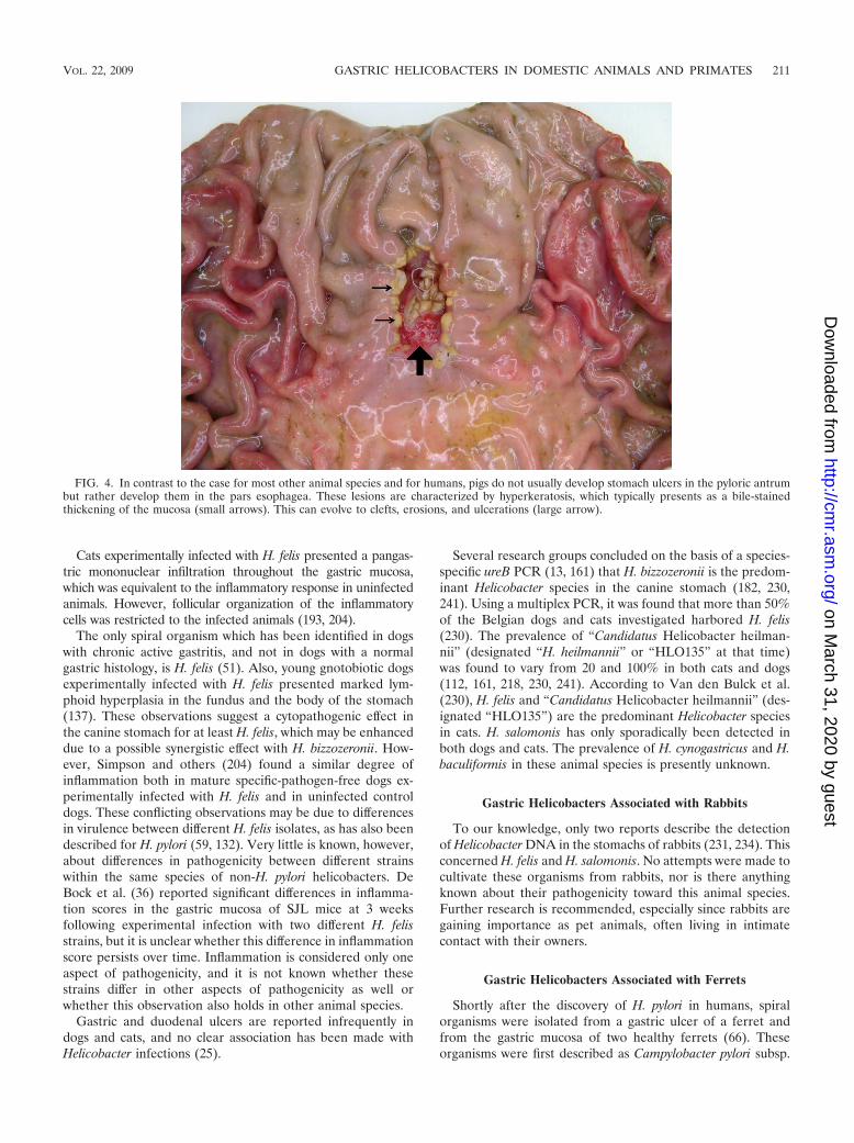
partments may allow the stratified squamous epithelium of thenonglandular region to come into contact with the luminalcontent of the distal part with acid, bile (refluxed from theduodenum), and pepsin. Chronic insult of the nonglandularregion will eventually lead to ulceration (Fig. 4).
Ulceration in the nonglandular stomachs of pigs is a diseaseof complex etiology in which multiple factors are involved,including dietary and stress factors. Small particle size of feed,interruption of feed intake, and presence of highly fermentablecarbohydrates in the diet promote ulcera (10). In general, allconditions increasing the fluidity of the stomach contents maycause a breakdown of the pH gradient between the proximaland the distal parts of the stomach and may play a role in ulcerdevelopment (77). An infection with H. suis may result insecretion of excessive amounts of gastric acid, leading to in-creased contact of the nonglandular part of the stomach withhydrochloric acid. In the fundic gland regions of pigs experi-mentally or naturally infected with H. suis, these microorgan-isms were found in close contact with parietal cells, whichmight indicate that the bacterium may have an impact on thesehydrochloric acid-producing cells (103). An H. suis infectionresults in gastritis, which is mainly localized in the antrum (103,
FIG. 1. Phylogenetic tree based on the near-complete 16S rRNA gene sequences from gastric Helicobacter species and other closely relatedbacteria. The sequences were aligned using the CLUSTAL W program (225), and a phylogenetic tree was constructed using the neighbor-joiningmethod (191) via the PHYLIP package (64). DNADIST was used for distance analysis (126). Bootstrap values (for branches present in more than50 out of 100 resamplings of the data) are indicated at the nodes. Original names found in the Entrez Nucleotide database (NCBI) are shown inparentheses. Sequences marked with an asterisk are derived from bacteria demonstrated to be present in the stomachs of humans.
VOL. 22, 2009 GASTRIC HELICOBACTERS IN DOMESTIC ANIMALS AND PRIMATES 205
on March 31, 2020 by guest
http://cmr.asm
.org/D
ownloaded from

147). In H. pylori infections in humans, increased acid produc-tion has been associated with antral predominant gastritis(132). In a recent study, Sapierzynski et al. (192) demonstratedthat an H. suis infection in pigs results in an increased numberof gastrin-producing cells and a decreased number of soma-tostatin-producing cells. Since gastrin stimulates and soma-tostatin inhibits the secretion of hydrochloric acid by parietalcells, this may also result in excessive acid production. How-ever, Silva et al. (202) did not find increased postprandialserum gastrin concentrations in pigs with ulceration of the parsesophagea.
Krakowka et al. (129) isolated a curve-shaped Helicobacterspecies from naturally infected young piglets that was differentfrom the tightly coiled H. suis. This microorganism is morpho-logically similar to but antigenically different from H. pylori. Asfar as we know, no genomic data on this H. pylori-like bacte-rium have been published yet. Gnotobiotic piglets experimen-
tally inoculated with this microorganism developed ulcers ofthe pars esophagea (130).
Hanninen et al. (93, 94) demonstrated that spindle-shapedmicroorganisms that had been isolated from the stomachs andfeces of pigs and that had tufts of sheathed flagella at both endsand external fibrils outside the cell belonged to the species H.bilis and H. trogontum. These are enterohepatic helicobactersthat were originally isolated from mice and rats, respectively,and were provisionally called “flexispira.” The pathogenicity ofthese microorganisms for pigs is unknown, and their main siteof colonization is most probably the lower intestinal tract. Theyare urease positive, which may help them to survive duringpassage through the stomach. It remains to be determinedwhether they are able to colonize the porcine stomach, as hasbeen shown for some other urease-positive enterohepatic he-licobacters in other animals, such as H. aurati in Syrian ham-sters (177) and H. muridarum in mice (183).
FIG. 2. Phylogenetic tree based on the partial ureA and ureB gene sequences from gastric Helicobacter species and other closely related bacteria.The sequences were aligned using the CLUSTAL W program (225), and a phylogenetic tree was constructed using the neighbor-joining method(191) via the PHYLIP package (64). DNADIST was used for distance analysis (126). Bootstrap values (for branches present in more than 50 outof 100 resamplings of the data) are indicated at the nodes. Original names found in the Entrez Nucleotide database (NCBI) are shown inparentheses. Sequences marked with an asterisk are derived from bacteria demonstrated in the stomach of humans.
206 HAESEBROUCK ET AL. CLIN. MICROBIOL. REV.
on March 31, 2020 by guest
http://cmr.asm
.org/D
ownloaded from

TABLE 3. Summary of gastric lesions in different animal species naturally or experimentally infected with helicobacters
Animalspecies Helicobacter Infection Gastric gross lesion(s) Gastric histological lesion(s) Localization of Helicobacter sp. Reference(s)
Pig H. suis Natural Surface redness NDa Antrum, fundus 87None ND Antrum, fundus 104Mucosa redness and edema,
occasional erosions andhemorrhage
Antrum, fundus: diffusemononuclear cell infiltrationwith occasional neutrophilicinfiltrate and lymphoid follicles
Antrum, fundus: in mucus, inlumen of the pits, in mucosalsurface; positive correlationbetween presence of bacteriaand pyloric gastritis
147
Fundus: proliferation of gastricfolds with occasionalnecrosis, severe mucosalcongestion; pars esophagea:hyperkeratosis
Diffuse lymphocytic infiltrationand lymphoid follicles inlamina propria
Antrum: in mucus, gastric pits,and lumen of gastric glands
173
Pars esophagea: hyperkeratosiswith yellow discoloration orchronic ulcers
Pars esophagea: increasedthickness of epithelium,elongation of papillae,parakeratosis, balloon cells orchronic peptic ulcers (layers ofnecrosis, numerousinflammatory cells, andgranulation tissue andfibrosis); antrum: mild diffusemononuclear cell infiltration inlamina propria with multiplelymphocytic aggregates orlymphoid follicles
Antrum, fundus, cardia;positive correlation betweenpresence of bacteria andlesions in pars esophagea
185
Pars esophagea ulcerativegastritis
Pars esophagea: erosion ofsurface epithelium, necrosis,mixed inflammatory infiltrate,and granulation tissue
Antrum, fundus: in mucouslayer and foveolae,occasionally in lumen ofgastric glands; positivecorrelation between presenceof bacteria and lesions inpars esophagea
18
Pars esophagea ulcerativegastritis
ND Pars esophagea: positivecorrelation between presenceof bacteria and lesions inpars esophagea
28
Pars esophagea ulcerativegastritis
Diffuse mononuclear cellinfiltration in propria mucosa
Antrum: positive correlationbetween presence of bacteriaand lesions in parsesophagea
190
Experimental Pars esophagea: no,preulcerative, and ulcerativelesions
Antrum: mild diffusemononuclear infiltration inlamina propria with multiplelymphocytic aggregates orlymphoid follicles
Antrum: in mucus overlying thesurface epithelium and in thesurface foveola; fundus: inglandular foveola extendinghalfway down the gastric pitsand often in close associationwith mucus-producing cellsand parietal cells
103
Curve-shaped bacteriamorphologically similarto H. pylori
Natural None Antrum, fundus, cardia: diffusemononuclear cell infiltration inlamina propria with multiplelymphoid follicles, andoccasional neutrophilicinfiltrates and exudation intothe glandular lumens
Antrum, fundus, cardia; inclose apposition to thegastric epithelia and in thegastric mucus
129
Experimental Gastroesophageal ulceration,glandular mucosal ulcers,lymphoid follicles, excessluminal mucus, and mucosaledema
Pars esophagea: pepticulceration; antrum, cardia,fundus: diffuse mononuclearcell infiltration in laminapropria with multiple lymphoidfollicles
Cardia, antrum: extracellularly 130
Dog H. felis Natural ND Chronic active gastritis: diffuselymphoplasmic infiltration,lymphocytic aggregates, andoccasional neutrophilicinfiltration
Fundus, corpus, antrum: inmucus adjacent to surfaceepithelium, glandular laminaadjacent to parietal cells, andgastric pits
51
Experimental No lesions Fundus, antrum: diffusemononuclear cell infiltration inlamina propria with multiplevariable-sized lymphoid follicles
Fundus, corpus, antrum: inmucous layer on mucosalsurface within gastric pitsand glandular lumen,occasionally intracellular inglandular epithelial cells
137
Continued on following page
VOL. 22, 2009 GASTRIC HELICOBACTERS IN DOMESTIC ANIMALS AND PRIMATES 207
on March 31, 2020 by guest
http://cmr.asm
.org/D
ownloaded from

TABLE 3—Continued
Animalspecies Helicobacter Infection Gastric gross lesion(s) Gastric histological lesion(s) Localization of Helicobacter sp. Reference(s)
No lesions Antrum: mild diffuselymphoplasmacyticinflammation of lamina propria
Fundus, corpus, antrum: insuperficial gastric mucouslayer, in gastric glands andparietal cells
204
NHPHb Natural Mucosal reddening, edema,erosions, and ulcerations
Fundus: glandular degenerationwith accumulation oflymphocytes and neutrophilicgranulocytes, edema, fibrosis,diffuse lymphoplasmacyticinfiltrates, and lymphoidfollicles in lamina propria
Fundus: in mucus covering thesurface epithelium, thegastric pits, the glandularlumina, and the parietalcells; presence of very highnumbers of bacteria wasdirectly related to the no. oflymphoid follicles
105
Cat NHPH Natural Mucosal reddening, edema,erosions, and ulcerations
Fundus: glandular degenerationwith accumulation oflymphocytes and neutrophilicgranulocytes, edema, fibrosis,diffuse lymphoplasmacyticinfiltrates, and lymphoidfollicles in lamina propria
Fundus: in mucus covering thesurface epithelium, thegastric pits, the glandularlumina, and the parietalcells; increased bacterialcolonization was directlyrelated to the no. oflymphoid follicles, fibrosis,lesions of surface epithelium,and glandular degeneration
105
ND Antrum: diffuse, mixedsubglandular leukocyticinfiltrates and multiplelymphoid nodules in the laminapropria
Antrum: in the canaliculi orcytoplasm of viable parietalcells
171
ND Antrum: moderate lymphoidfollicles in lamina propria;antrum and fundus: moderatemononuclear infiltrates inlamina propria
Antrum, fundus: at themucosal surface, in thelumina of gastric glands, andin cytoplasm of parietal cellsof the fundus
193
H. felis Experimental ND Antrum: mild diffuselymphoplasmacytic andeosinophilic infiltration,lymphoid follicular hyperplasia,and mild fibrosis of laminapropria
Antrum, fundus: at themucosal surface, in thelumina of gastric glands, andin cytoplasm of parietal cellsof the fundus
193
H. pylori Experimental ND Antrum: mild diffuselymphoplasmacytic andgranulocytic infiltration, severelymphoid follicular hyperplasia,and mild fibrosis of laminapropria
Antrum, fundus: at themucosal surface, in thelumina of gastric glands, andin cytoplasm of parietal cellsof the fundus
193
Ferret H. mustelae Natural Gastritis, peptic ulcer Proximal antrum: mononuclearcell infiltrates in superficiallayer of lamina propria, mucusdepletion and occasionalneutrophilic infiltrate, glandnecrosis and regeneration;distal antrum: diffusemononuclear cell infiltrationand mucus depletion; fundus:mononuclear cell infiltrates insuperficial layer of laminapropria, mucus depletion, andoccasional neutrophilicinfiltrate
Proximal antrum: at surfaceand lumen of foveola andoccasionally in deepglandular lumen; distalantrum: at surface andlumen of foveola andoccasionally in superficialglandular lumen; fundus: atsurface and lumen of foveolaadjacent to the inflammation
70, 140
Pyloric adenocarcinoma Antrum: multifocal segmentalglandular proliferation andsurface erosion, multifocalmucosal lymphoid aggregates,mixed inflammatory cellinfiltration, and multifocalfibrosis
Antrum: in the lumen ofgastric pits, adherent toapical surface of the mucousepithelium
75
Gastric lymphoma High- and low-grade B-celllymphoma
Antrum, fundus: within themucosal glands
63
Continued on following page
208 HAESEBROUCK ET AL. CLIN. MICROBIOL. REV.
on March 31, 2020 by guest
http://cmr.asm
.org/D
ownloaded from

TABLE 3—Continued
Animalspecies Helicobacter Infection Gastric gross lesion(s) Gastric histological lesion(s) Localization of Helicobacter sp. Reference(s)
Experimental ND Antrum: focal minimallymphocytic infiltrates withmoderate numbers ofeosinophils and neutrophils;fundus: superficial gastritisconsisting of lymphocytes andoccasional neutrophils
Antrum, fundus: on the surfaceof the gastric epitheliumwithin the mucous layer andwithin gastric pits
72
H. mustelae � MMNGc Experimental Pyloric adenocarcinoma ND Antrum: epithelial surfaces ofthe neck glands
75
Hamster H. aurati Natural ND Distal antrum: diffuselymphoplasmacyticinflammation, scatteredheterophils and eosinophils,and goblet cell hyperplasia
Antrum: within gastric pits orglands
176
Pyloric adenocarcinoma Distal antrum: locally extensivechronic gastritis withintestinal metaplasia andoccasional well-differentiatedand moderately pleomorphictubular to tortuous gastricglands
Antrum: within gastric glands 159
Cattle “Candidatus Helicobacterbovis”
Natural ND ND Distal antrum: in mucus layerand proximal gastric crypts
44
Horse NHPH Natural No lesions, gastritis or gastriculcers
Gastric mucosa: loss ofcontinuity with submucosaexposure and edema;parakeratotic hyperkeratosis;lymphoplasmocyticmononuclear infiltrate
Glandular and nonglandularstomach near margo plicatus
31
Nonhumanprimates
H. pylori Natural No lesions, superficial gastritis Distal fundus, antrum: superficialerosions, marked mononuclearand polynuclear infiltration
Antrum: proximity to themucosal epithelial cells orin the lumen of the gastricpits
57
None No lesions or diffuselymphoplasmacytic infiltrationof the lamina propria,occasional neutrophilicinfiltrates and lymphoidfollicles
Antrum: superficial mucosa 55
ND Antrum: diffuselymphoplasmacytic infiltrationin lamina propria, prominentlymphoid follicles, andoccasional glandular epithelialhyperplasia, patchy necrosis,attenuation of glandularepithelium, and neutrophilicinfiltrates
Antrum: gastric pits and theupper portions of gastricglands, often in intimateassociation with theepithelial cell surface
138
Localized to multifocalreddening of mucosa
Antrum, cardia:lymphoplasmacytic infiltrates,gastric gland epithelialhyperplasia, reduction inmucin content of surface andgland epithelia, increasedlymphoid follicles, minorinfiltrates of neutrophils insuperficial lamina propria andgastric glands, and occasionalerosions
Antrum, cardia: mucosalregions of inflammation;located in gastric pits andupper portions of gastricglands, often associated withepithelial cell surface
187
Experimental ND Antrum: chronic-active gastritis,marked atrophy of the mucosa,microerosions and loss ofmucus from superficialepithelial cells
Antrum 56
Continued on following page
VOL. 22, 2009 GASTRIC HELICOBACTERS IN DOMESTIC ANIMALS AND PRIMATES 209
on March 31, 2020 by guest
http://cmr.asm
.org/D
ownloaded from

Gastric Helicobacters Associated with Dogs and Cats
The majority of Helicobacter infections of the canine and felinegastric mucosa are mixed infections of various Helicobacter spe-cies, including H. felis, H. bizzozeronii, H. salomonis, and “Candi-datus Helicobacter heilmannii.” Recently, one additional specieswas isolated from the stomach of a dog, namely, H. cynogastricus(233), and one additional species was isolated from the stomachof a cat, namely, H. baculiformis (15).
In dogs, spiral-shaped bacteria are commonly found in thestomach. They are present in 67 to 86% of clinically healthy
dogs and in 61 to 100% of dogs presenting chronic vomiting(105, 112). In cats, spiral-shaped organisms have been detectedin 41 to 100% of the animals investigated, with a slightly higherrate in animals presenting chronic vomiting (62, 80, 105, 112,161, 171, 172, 236, 243). Bridgeford et al. (23) hypothesizedthat gastric Helicobacter species may be a cause of feline gastriclymphoma.
The pathogenic significance of gastric Helicobacter species indogs and cats remains enigmatic and may be Helicobacter spe-cies or even strain dependent.
TABLE 3—Continued
Animalspecies Helicobacter Infection Gastric gross lesion(s) Gastric histological lesion(s) Localization of Helicobacter sp. Reference(s)
NHPH Natural No lesions, superficial gastritis No lesions to occasionalmononuclear and polynuclearinfiltration
Fundus: mucus covering thesurface of epithelial cells, inthe lumina of the gastricglands, and overlying parietalcells
57
None No lesions or diffuselymphoplasmacytic infiltrationof the lamina propria,occasional neutrophilicinfiltrates and lymphoidfollicles
Fundus: gastric pits, superficialglands or on the surfaceepithelium,
55
ND Fundus: minor scatteredaggregates of lymphocytes, lownumbers of plasma cells, andoccasional lymphoid follicles
Fundus: in the gland lumens,sometimes attached to theepithelial cell surface, andwithin the cytoplasm ofparietal cells
138
Localized to multifocalreddening of mucosa
Fundus: no lesions Fundus: in gland lumens,parietal cells, and surfacemucus
187
a ND, not described.b NHPH, non-H. pylori Helicobacter species.c MNNG, N-methyl-N-nitro-N�-nitrosoguanidine.
FIG. 3. In the normal porcine stomach there is a small rectangular area around the cardia which is covered by a slightly keratinized squamousepithelium presenting as a white, slightly irregular surface on visual inspection. This area is named the pars esophagea, since the epithelium issimilar to that of the esophagus.
210 HAESEBROUCK ET AL. CLIN. MICROBIOL. REV.
on March 31, 2020 by guest
http://cmr.asm
.org/D
ownloaded from
Cats experimentally infected with H. felis presented a pangas-tric mononuclear infiltration throughout the gastric mucosa,which was equivalent to the inflammatory response in uninfectedanimals. However, follicular organization of the inflammatorycells was restricted to the infected animals (193, 204).
The only spiral organism which has been identified in dogswith chronic active gastritis, and not in dogs with a normalgastric histology, is H. felis (51). Also, young gnotobiotic dogsexperimentally infected with H. felis presented marked lym-phoid hyperplasia in the fundus and the body of the stomach(137). These observations suggest a cytopathogenic effect inthe canine stomach for at least H. felis, which may be enhanceddue to a possible synergistic effect with H. bizzozeronii. How-ever, Simpson and others (204) found a similar degree ofinflammation both in mature specific-pathogen-free dogs ex-perimentally infected with H. felis and in uninfected controldogs. These conflicting observations may be due to differencesin virulence between different H. felis isolates, as has also beendescribed for H. pylori (59, 132). Very little is known, however,about differences in pathogenicity between different strainswithin the same species of non-H. pylori helicobacters. DeBock et al. (36) reported significant differences in inflamma-tion scores in the gastric mucosa of SJL mice at 3 weeksfollowing experimental infection with two different H. felisstrains, but it is unclear whether this difference in inflammationscore persists over time. Inflammation is considered only oneaspect of pathogenicity, and it is not known whether thesestrains differ in other aspects of pathogenicity as well orwhether this observation also holds in other animal species.
Gastric and duodenal ulcers are reported infrequently indogs and cats, and no clear association has been made withHelicobacter infections (25).
Several research groups concluded on the basis of a species-specific ureB PCR (13, 161) that H. bizzozeronii is the predom-inant Helicobacter species in the canine stomach (182, 230,241). Using a multiplex PCR, it was found that more than 50%of the Belgian dogs and cats investigated harbored H. felis(230). The prevalence of “Candidatus Helicobacter heilman-nii” (designated “H. heilmannii” or “HLO135” at that time)was found to vary from 20 and 100% in both cats and dogs(112, 161, 218, 230, 241). According to Van den Bulck et al.(230), H. felis and “Candidatus Helicobacter heilmannii” (des-ignated “HLO135”) are the predominant Helicobacter speciesin cats. H. salomonis has only sporadically been detected inboth dogs and cats. The prevalence of H. cynogastricus and H.baculiformis in these animal species is presently unknown.
Gastric Helicobacters Associated with Rabbits
To our knowledge, only two reports describe the detectionof Helicobacter DNA in the stomachs of rabbits (231, 234). Thisconcerned H. felis and H. salomonis. No attempts were made tocultivate these organisms from rabbits, nor is there anythingknown about their pathogenicity toward this animal species.Further research is recommended, especially since rabbits aregaining importance as pet animals, often living in intimatecontact with their owners.
Gastric Helicobacters Associated with Ferrets
Shortly after the discovery of H. pylori in humans, spiralorganisms were isolated from a gastric ulcer of a ferret andfrom the gastric mucosa of two healthy ferrets (66). Theseorganisms were first described as Campylobacter pylori subsp.
FIG. 4. In contrast to the case for most other animal species and for humans, pigs do not usually develop stomach ulcers in the pyloric antrumbut rather develop them in the pars esophagea. These lesions are characterized by hyperkeratosis, which typically presents as a bile-stainedthickening of the mucosa (small arrows). This can evolve to clefts, erosions, and ulcerations (large arrow).
VOL. 22, 2009 GASTRIC HELICOBACTERS IN DOMESTIC ANIMALS AND PRIMATES 211
on March 31, 2020 by guest
http://cmr.asm
.org/D
ownloaded from

mustelae (68). Later they were designated C. mustelae (69) and,finally, H. mustelae (85). Only a minority of ferrets youngerthan 6 weeks are colonized by this bacterium, in contrast toapproximately 100% of the adult ferrets (67). This indicatesthat widespread colonization occurs after weaning, and itseems to persist throughout the adult life of the ferret (71).
In ferrets naturally infected with H. mustelae, often only asuperficial gastritis is present in the corpus region, where thesebacteria colonize the mucosal surface (140). In the antrum,however, a diffuse mononuclear gastritis is observed with in-flammatory cells often occupying the full thickness of the mu-cosa (72). In this stomach region, H. mustelae colonizes thesurface, gastric pits, and superficial portion of the glands (72).A retrospective study revealed that persistent colonization withH. mustelae over time increases the severity of gastric disease(71).
Gastric and, to a lesser extent, duodenal ulcers have beenreported in ferrets infected with H. mustelae (66, 70), and theincidence of gastric ulceration in this animal species variesbetween 1.4 and 35% (3, 4). However, since the prevalence ofH. mustelae is very high in adult ferrets, long-term observationsof experimentally infected pathogen-free ferrets are needed toelucidate the exact role of H. mustelae infection in the devel-opment of peptic ulcer disease (211).
Fox and coworkers (75) reported on the presence of H.mustelae in the pyloric mucosa of two ferrets suffering frompyloric adenocarcinoma. In both cases, the invasion of neo-plastic tubules into the deep submucosa was described. Anincreased epithelial cell proliferation has also been detected inthe gastric mucosa of ferrets infected with H. mustelae. Thismay play a role in the development of gastric tumors (247).Gastric MALT lymphoma has also been described in ferretsinfected with H. mustelae (63). Replacement of normal epithe-lium by uniform populations of lymphoid cells was seen, withinvasion and destruction of the gastric glands. These lympho-mas arose in the antrum, where H. mustelae-induced gastritis ismost severe. However, for both types of gastric malignancy,evidence remains circumstantial, and the role of H. mustelae inthe development of gastric tumors needs to be confirmed(211).
Gastric Helicobacters Associated with Hamsters
H. aurati has been isolated from the stomachs of hamsters.Several not-further-characterized Helicobacter spp. have alsobeen reported to be present in the stomachs of these animals(159, 176), but no further information is available on thesespecies.
H. aurati has been isolated from the inflamed stomachs andceca of adult Syrian hamsters. Various features, such as thefusiform shape and the presence of periplasmic fibrils, allowmorphological discrimination between H. aurati and the threeother helicobacters that have thus far been identified in ham-sters, namely, H. cholecystus (76), H. mesocricetorum (203), andH. cinaedi (26, 240). The presence of urease activity also dis-tinguishes H. aurati from these three enterohepatic Helico-bacter species. The preferential colonization site of H. aurati inhamsters is probably the intestinal tract, particularly the ce-cum, with subsequent spreading of this bacterial agent to thestomach in selected animals. The coprophagic behavior of
hamsters may play a role in this gastric colonization by H.aurati. At present, the exact role of H. aurati in gastric diseaseof hamsters has not yet been fully clarified, although the or-ganism has been identified in hamsters suffering from chronicgastric inflammation and intestinal metaplasia (176, 177). Thesame authors reported the presence of another helical, urease-negative Helicobacter species, as well as a smaller, urease-negative Campylobacter sp. in the stomachs of these hamsters.Likewise, Nambiar et al. (159) reported a case of gastritis-associated adenocarcinoma and intestinal metaplasia in a Syr-ian hamster naturally infected with different Helicobacterspecies, including H. aurati. They suggested that chronic Heli-cobacter-associated gastritis in hamsters may develop into aninfiltrative gastric adenocarcinoma, similar to what has beendescribed in chronic H. pylori infections in humans. There areno indications that H. aurati is of zoonotic significance.
Gastric Helicobacters Associated with Ruminants
Gastric ulcers regularly occur in calves and adult cattle, withan incidence varying between 2 and 87% (52, 96, 117, 163, 206,239). “Candidatus Helicobacter bovis” has been demonstratedin the pyloric part of the abomasums of calves and adult cattlebut has not yet been cultivated in vitro (44). Although it ishighly prevalent in bovines (reference 88 and unpublished re-sults), its involvement in gastric disease in cattle is presentlyunknown. In contrast, Helicobacter DNA was not detected inthe abomasums of 70 goats, using a genus-specific PCR (88).
Gastric Helicobacters Associated with Horses
Gastric ulcers are common in horses, and their incidence inracehorses in active training may exceed 90% (20, 156). Vari-ous stress factors, diet, management, and training practices areregarded as potential risk factors (31, 156). The proximal halfof a horse’s stomach is entirely lined with a stratified squamousepithelium resembling the pars esophagea of the porcine stom-ach. The more distal portion is the glandular part. Ulcers aremost frequently seen close to the junction between the non-glandular and the glandular parts of the stomach (31). In somestudies, Helicobacter-like organisms or their DNA have beendetected in the stomachs of horses, but their role in develop-ment of gastric ulcers remains speculative. Contreras et al. (31)detected Helicobacter-like DNA in the gastric mucosa of 11thoroughbred racehorses. Sequencing of the 16S rRNA generevealed 99% similarity with H. pylori, but all samples werenegative when tested with H. pylori-specific PCR assays target-ing the cagA and glmM genes, which might indicate that theDNA was from a Helicobacter species different from H. pylori.It remains to be determined whether horses may indeed beinfected with a gastric Helicobacter species specifically associ-ated with this animal host. Attempts to isolate these microor-ganisms from the horse’s stomach should be made, both invitro and in vivo by intragastric inoculation of specific-patho-gen-free mice (166).
Gastric Helicobacters Associated with Nonhuman Primates
In several studies, nonhuman primates were used as modelsfor human H. pylori infections (111, 128, 209, 210, 212). Cap-
212 HAESEBROUCK ET AL. CLIN. MICROBIOL. REV.
on March 31, 2020 by guest
http://cmr.asm
.org/D
ownloaded from

tive rhesus monkeys (Macaca mulatta) are commonly infectedwith H. pylori (54). The rhesus monkey model, therefore, pro-vides an opportunity to examine natural acquisition of H. pyloriusing an experimental setup that closely resembles human in-fection. Socially housed rhesus monkeys rapidly acquire H.pylori infection. Newborns from infected dams are more com-monly infected than those from uninfected dams, particularlyduring the peripartum period, suggesting that close contactduring this time facilitates oral-oral transmission (213, 214).Once acquired, infection is associated with chronic gastritisthat resembles that seen in humans.
The number of reports dealing with natural infections withgastric non-H. pylori helicobacters in nonhuman primates islimited, and at present, a gastric Helicobacter species specifi-cally associated with these animals has not yet been described.Bronsdon et al. (24) isolated and described H. nemestrinaefrom the stomach of a pigtailed macaque (Macaca nemestrina).This microorganism is able to grow at 42°C and possessesbipolar flagella, which is different from the case for H. pyloristrains isolated from humans. Based on the sequencing ofseven housekeeping genes and two flagellin genes, H. nemes-trinae was later shown to be an atypical H. pylori strain (219).H. nemestrinae should therefore be considered a later hetero-typic synonym of H. pylori.
In the stomachs of rhesus monkeys (Macaca mulatta), gastricnon-H. pylori helicobacters which were not identified to thespecies level have been observed in the mucus covering thesurface epithelial cells, in the lumina of the gastric glands, andin close contact with parietal cells (57). These microorganismswere able to invade and on occasion to damage parietal cells,while apparently causing hyperchlorhydria. This is in contrastto the case for H. pylori, which caused gastritis in these animalswithout modifying the acid output (57). Long, spiral-shapedbacteria have also been reported in the stomachs of baboons(Popio hamadryas). This was associated with gastritis byMackie and O’Rourke (138) but not by others (34, 35). Non-H.pylori helicobacters, without clarification about the species,have been described to be naturally present in the stomachs ofup to 100% of cynomolgus monkeys from many different geo-graphic regions (55, 187). These microorganisms were found inthe superficial portions of the gastric epithelium, most fre-quently in the fundic region. The bacteria were located in thegastric pits, in the superficial glands, or on the surface epithe-lium. However, no correlation was observed between the pres-ence of these bacteria and the infiltration of lymphoplasma-cytic cells and inflammatory lesions in these gastric tissues (55).
H. suis has been demonstrated in the stomachs of two man-drill monkeys (Papio sphinx), two cynomolgus monkeys (Ma-caca fasicularis), and one rhesus macaque (Macaca mulatta)from a zoo (167). One isolate, first described as “H. heilman-nii” (114) and later identified as “Candidatus Helicobacterheilmannii” (158), was obtained by intragastric inoculation ofmice with gastric tissue from a cynomolgus monkey (Macacafascicularis). No information on the pathogenic significance ofthese Helicobacter species for nonhuman primates is available,and the source of infection remains to be determined.
Although it is clear that nonhuman primates may be infectedwith different types of gastric helicobacters, little informationon these bacteria and their interactions with these hosts isavailable. Since these animals are closely related to humans,
further research is wanted. Monkeys might serve as a possiblereservoir for human infections.
NON-H. PYLORI HELICOBACTER-ASSOCIATED GASTRICDISEASES IN HUMANS: ZOONOSES?
Although some data in the literature indicate that animals,including cats, dogs, and sheep, occasionally may be infectedwith H. pylori (53, 74, 226), it is unlikely that animals play animportant role in the transmission of this microorganism tohumans. Moreover, it cannot be excluded that in some of thesecases H. pylori-like organisms, but not H. pylori itself, wereinvolved. H. pylori has been demonstrated by culture and PCRmethods in the gastric mucosa of specific-pathogen-free labo-ratory cats in one study (90). This observation may be relatedto an anecdotic anthroponosis, especially since these bacteriahave not been identified in stray cats (62).
Human infections with non-H. pylori Helicobacter organisms,however, most likely originate from animals, although the fin-gerprinting of Helicobacter species present in the human andanimal gastric mucosa should be considered in order to fullyunderstand the zoonotic hazard originating from different an-imal species.
O’Rourke et al. (166) demonstrated that 16S rRNA genesequences and partial ureA and ureB gene sequences fromthree human and four porcine non-H. pylori Helicobacterstrains isolated in vivo by inoculation in specific-pathogen-freemice showed a very high degree of homology, �99.3%. Thisled to the conclusion that they represent the same species, laterdescribed as H. suis (14, 166).
Another human non-H. pylori Helicobacter strain, also iso-lated in vivo in specific-pathogen-free mice, could be easilydifferentiated from H. suis. Urease gene sequence analysisdemonstrated that it clustered with helicobacters from domes-tic and exotic feline species. These microorganisms were des-ignated “Candidatus Helicobacter heilmannii” (166).
Two urease-based PCRs, one developed by O’Rourke et al.(166) and another one by Neiger et al. (161), detected only“Candidatus Helicobacter heilmannii” DNA and not DNAfrom pure in vitro cultures of H. suis, H. felis, H. salomonis, H.bizzozeronii, H. baculiformis, and H. cynogastricus strains (un-published results) and can therefore be considered species-specific. Using the test described by Neiger et al. (161), Chish-olm and Owen (27) demonstrated the presence of CandidatusHelicobacter heilmannii DNA in one of 113 gastric biopsiesfrom human patients with dyspeptic symptoms.
Trebesius et al. (227) used FISH and partial 16S ribosomalsequencing for analyzing 89 gastric biopsy samples from hu-mans in Germany with histological evidence of non-H. pylorihelicobacters. Five short 16S rRNA-directed probes of about20 nucleotides were used in FISH. In total, 71 (80%) of thesesamples hybridized with a probe designated Hhe-1, which rec-ognizes fragments of the 16S rRNA gene of H. suis. The 16Sribosomal gene sequences of the former “H. heilmannii” type 2are highly related, and results obtained with the other probesare therefore more difficult to interpret. Two probes (Hhe2and Hhe4) are identical to fragments of the 16S rRNA gene of“Candidatus Helicobacter heilmannii,” leading to the conclu-sion that DNA of this helicobacter was detected in 17 (19%) ofthe samples. Probe Hhe5 recognizes the 16S rRNA gene of H.
VOL. 22, 2009 GASTRIC HELICOBACTERS IN DOMESTIC ANIMALS AND PRIMATES 213
on March 31, 2020 by guest
http://cmr.asm
.org/D
ownloaded from

felis, H. bizzozeronii and H. salomonis. Five samples (6%) hy-bridized with this probe. Finally, one sample hybridized withprobe Hhe3. It is possible that this probe recognizes a not-yet-described Helicobacter species, since the sequence obtainedfrom HHLO-3 (GenBank accession no. AY014859) shows lessthan 97% similarity with any of the known Helicobacter species.More research is needed to confirm this, however.
De Groote et al. (45) used three PCR assays targeting the16S rRNA gene for screening of paraffin-embedded gastricbiopsy specimens of 101 patients with chronic active gastritisand histological evidence of a non-H. pylori Helicobacter infec-tion. Fourteen samples tested positive in a PCR assay thatspecifically detected H. suis DNA. DNA of “Candidatus Heli-cobacter bovis” was detected in one sample. Samples of 49patients tested positive in a third assay that simultaneously de-tected H. felis, H. bizzozeronii, H. salomonis, “Candidatus Helico-bacter heilmannii,” H. bacculiformis, and H. cynogastricus.
Van den Bulck et al. (230) studied the presence of Helico-bacter species in 123 gastric biopsies of humans from Belgiumand Germany with histological evidence of a non-H. pyloriHelicobacter infection, using a multiplex PCR based on thetRNA intergenic spacers, the urease gene, and the 16S rRNAgene (13). In 37% of the samples, H. suis was detected. Indescending order, H. salomonis (21%), H. felis (15%), “Can-didatus Helicobacter heilmannii” (8%, designated HLO135 bythe authors), and H. bizzozeronii (4%) were found.
The data presented above show that H. suis is the mostprevalent gastric non-H. pylori Helicobacter species in humansand that there are strong indications that pigs may be a sourceof infection for humans. Apart from the stomachs of pigs andhumans, H. suis has also been detected in the stomachs ofmacaques and mandrill monkeys, as has been demonstrated by16S rRNA gene and urease gene sequencing (167), and in onecat (230).
Dogs, cats, and perhaps also pet rabbits may serve as asource of infection in humans with H. felis, “Candidatus Heli-cobacter heilmannii,” and H. bizzozeronii. Anecdotal reports ofthe presence of the same Helicobacter species in the stomach ofa person and his favorite pet animal further point in the samedirection. Several reports indeed suggest the transmission ofgastric non-H. pylori spiral bacteria from dogs to humans (39,116, 226) or from cats to humans (50, 107, 133, 217, 235). In1999, Andersen et al. (2) succeeded in the isolation of a non-H.pylori helicobacter strain from human gastric mucosa. Later,phenotypic analysis, sequencing of the 16S rRNA gene, DNA-DNA hybridization analysis, and whole-cell protein profilingrevealed that this isolate belongs to the species H. bizzozeronii(116). Recently, another in vitro isolate was obtained from ahuman gastric non-H. pylori Helicobacter infection. Despite thelow prevalence of H. bizzozeronii in human biopsies, polyphasicidentification analysis revealed that this isolate also belongedto this species (127). In the study by Van den Bulck et al. (230),H. salomonis represented 21% of the human samples. Thefrequent identification of H. salomonis in human gastric biop-sies, however, is in contrast to its rare identification in petcarnivore samples, thus inclining us to suspect additionalsources of infection. In none of the studies described abovewere tests that specifically detect H. baculiformis and H. cyno-gastricus included, and it is presently not known whether these
recently described species are able to colonize the humanstomach.
GASTRIC DISEASE IN HUMANS INFECTED WITHNON-H. PYLORI HELICOBACTER SPECIES
Studying the effects of non-H. pylori helicobacters in humansis complicated by the fact that these infections are uncommonand it seems likely that there may be variation among non-H.pylori helicobacters in their ability to cause inflammation ordisease in humans. In addition, the presence of H. pylori mustbe excluded in order to assess the effects of the species underconsideration. De Groote et al. (45) detected H. pylori DNA in7 of 64 and Van den Bulck et al. (230) in 6 of 89 human gastricbiopsy specimens that were positive for non-H. pylori helico-bacters. Human H. pylori and non-H. pylori Helicobacter coin-fections have also been identified by histology (113), but thepossible significance of this in terms of disease developmenthas not been determined.
Non-H. pylori Helicobacter infections of the human stomachare consistently accompanied by active chronic gastritis. Thelesions, however, appear less severe than those associated withH. pylori (216). Acute gastritis is also occasionally observed(246). There may be glandular atrophy or intestinal metaplasiaof the fundic mucosa, but these lesions are less common withnon-H. pylori Helicobacter than with H. pylori infections (216).Gastric erosions located mainly in the antrum (22, 41, 50, 199,221, 246) and duodenal ulcers (21, 81, 118, 194) have also beenreported in association with non-H. pylori Helicobacter infec-tions. Furthermore, these infections have been associated withlow-grade MALT lymphoma of the stomach, and the risk ofdeveloping MALT lymphoma is higher with non-H. pylori he-licobacters than with H. pylori (153, 154). Both the gastritis andthe MALT lymphomas have been reported to resolve afterclearance of the non-H. pylori Helicobacter infections, furtherunderlining the causal relationship (154).
Clinical symptoms associated with non-H. pylori helico-bacters in humans can be characterized by atypical complaintssuch as acute or chronic epigastric pain and nausea. Otheraspecific symptoms include hematemesis, recurrent dyspepsia,irregular defecation frequency and consistency, vomiting,heartburn, and dysphagia, often accompanied by a decreasedappetite (50, 81, 101, 123, 148, 164, 194, 199, 221, 235, 238, 245,246). Some people infected with non-H. pylori helicobacters donot present obvious clinical signs (142).
Inspection of the gastric mucosa of people infected with longspiral bacteria through endoscopy reveals a variety of lesions,ranging from a normal to slightly hyperemic mucosa to muco-sal edema and to multiple erosions and ulcerations in theantrum or in the duodenum (50, 81, 194, 199, 221, 235,244, 246).
Histologically, the inflammation induced by non-H. pylorihelicobacters in the gastric tissue is generally characterized bylymphocytic exudation into gastric foveolae, sometimes ad-mixed with plasma cells. In some cases, lymphocytes are orga-nized into lymphoid aggregates. Epithelial mucus is occasion-ally depleted (65, 113, 123, 154, 164, 194).
In human patients presenting severe pathology and clinicalsymptoms associated with the presence of non-H. pylori heli-cobacters, treatment is indicated, although the efficacy of such
214 HAESEBROUCK ET AL. CLIN. MICROBIOL. REV.
on March 31, 2020 by guest
http://cmr.asm
.org/D
ownloaded from

treatment is not always easy to determine due to the lack ofrandomized trials. Such trials are difficult to organize in view ofthe low frequency of these infections in humans. In practice,treatment regimens identical to those used for H. pylori havebeen prescribed. Triple therapy using combinations of a protonpump inhibitor and two antimicrobial agents selected fromclarithomycin, metronidazole, amoxicillin, and tetracyclinemay be effective (39, 81, 123, 221, 235).
Because of the low number of in vitro isolates available, verylittle data exist on the antimicrobial susceptibility and acquiredresistance of gastric non-H. pylori Helicobacter species. Deter-mination of MICs of various antimicrobials against in vitroisolates indicated that acquired resistance to metronidazolemay occur in H. bizzozeronii and H. felis strains of animal origin(232). Experimental H. felis infections in mice showed thatseveral therapies using only one antimicrobial were effective inonly 25 to 70% of the animals tested, while triple therapy usingmetronidazole, tetracycline, and bismuth subcitrate eradicatedH. felis from all the animals (49). In a BALB/c mouse model itwas shown that treatment with ampicillin/omeprazole results inthe suppression of H. suis. Differences in sensitivity were seenbetween different H. suis isolates, which might indicate ac-quired antimicrobial resistance (102).
TRANSMISSION OF GASTRIC NON-H. PYLORIHELICOBACTER SPECIES
Few data are available on how non-H. pylori Helicobacterspecies are transmitted among their hosts.
Hellemans et al. (104) screened stomach samples of pigs ofdifferent ages from different herds for the presence of H. suis.The prevalence of the infection was very low in suckling piglets,increased rapidly after weaning, and reached 90% in the adultboars and sows. The low degree of infection before weaningdespite the high prevalence in adult pigs and thus most prob-ably in the dams of the suckling piglets may indicate maternalprotection through antibodies or other antibacterial factorspresent in sow milk (89), which disappears at weaning. Theregrouping of animals at the time of weaning most probablyfavors the spread of the microorganism from the few pigletsinfected before weaning to noninfected animals. Another fac-tor that may enhance H. suis colonization in weaned pigs is thatafter weaning the lactobacilli disappear temporarily from thestomach. An antagonistic effect of lactobacilli against H. pylori(1, 119, 200) and H. felis (30) infection has been demonstratedin a murine model. The fact that the H. suis infection persistedin the adult boars and sows indicates that any natural immuneresponse against this microorganism did not lead to its clear-ance. Despite numerous attempts, we were not able to detectH. suis in feces of infected pigs, which may indicate that fecal-oral spread between pigs and from pigs to humans is limited.Transmission of H. suis may be oral-oral via saliva or gastric-oral via vomitus, but this remains to be investigated. Persistentstomach colonization with H. suis can be achieved in laboratorymice (102), and it may be worthwhile to determine whetherwild mice can act as vectors or even as reservoirs of theinfection.
Hardly any data on the transmission of non-H. pylori Heli-cobacter infections in dogs and cats are available in the litera-ture. Hanninen et al. (91) described the transmission of H.
salomonis from a dam to her puppies, as well as betweeninfected and noninfected pups, which was proved by the similarpattern found for all cultured isolates using pulsed-field gelelectrophoresis. Transmission is suspected to be through oral-oral or gastric-oral contact, as nursing dogs have very intimatecontact with their offspring and puppies eat material vomitedby the dam. Helicobacter DNA was detected in the oral cavitiesof dogs (186). Lee et al. (135) found that gastric mucus derivedfrom Helicobacter-infected cats was highly infectious for mice,while rectal contents were not. Moreover, the isolation proce-dure starting from fecal material was unsuccessful (91). Theseobservations may indicate that fecal-oral transmission is lessimportant.
Fecal-oral transmission has, however, been suggested for H.mustelae (73). In a group of 36 ferrets, H. mustelae was isolatedfrom the feces of 11 animals. A correlation was found withperiods of transient hypochlorhydria, also seen in experimen-tally infected animals (72), which may allow larger numbers ofH. mustelae to exit the stomach. Keeping in mind the ease bywhich ferrets vomit, oral-oral and gastric-oral contact may alsoplay a role in transmission of this bacterium (71).
It is not exactly known how gastric helicobacters are trans-mitted from animals to humans, but most likely it occursthrough direct contact. Living in close proximity to dogs, cats,and especially swine has indeed been identified as a significantrisk factor for these infections (108, 144, 220). The intensity ofcontact with animals is thought to be important as well, since ahigher incidence of these infections has been noted in pigfarmers, the staff of pig slaughterhouses, and people havingintensive contact with pet animals (217, 221, 235, 246). It isremarkable that H. suis is the most prevalent gastric non-H.pylori Helicobacter species in humans (27, 227, 230). This mightindicate that the infectivity in humans of cat- or dog-relatedstrains is less than that of H. suis.
It is not known whether, besides direct contact with animals,other routes of transmission of non-H. pylori helicobacters areof importance. Recently, it was shown that gastric helico-bacters can survive in water for more than 4 days, a fact whichmay suggest a possible role for water in the transmission ofHelicobacter species between hosts (11). There are no dataavailable on the survival of H. suis on carcasses of slaughteredpigs, and it remains to be determined whether raw or under-cooked pork meat might be a source of infection for humans.
It is also not known how frequently transmission of non-H.pylori helicobacters from animals to humans occurs. Only in alow percentage of human patients with severe gastric com-plaints are long spiral-shaped bacteria found at microscopicexamination of gastric biopsies. However, it is possible that thisrepresents only the tip of the iceberg, and it cannot be excludedthat infections with these bacteria often pass while remainingunapparent or result in mild disease signs which are not furtherexamined (142).
VIRULENCE FACTORS OF GASTRIC NON-H. PYLORIHELICOBACTER SPECIES
Most of the research concerning Helicobacter virulence fac-tors and the evoked host response has been done with H. pylori,and much less information is available about the virulencemechanisms of non-H. pylori helicobacters. Although some
VOL. 22, 2009 GASTRIC HELICOBACTERS IN DOMESTIC ANIMALS AND PRIMATES 215
on March 31, 2020 by guest
http://cmr.asm
.org/D
ownloaded from

virulence factors of these bacteria may indeed be similar tothose described for H. pylori, there may also be differences.
H. pylori is a diverse pathogen, and several bacterial viru-lence factors are considered to play a role in pathogenesis ofinfections with this agent. The key enzymes and proteins foundin H. pylori that are important for colonization include theurease system, alpha carbonic anhydrase, sheathed flagella, thepH taxis tlpB gene, arginase, and several adhesins. Key viru-lence factors also include the cag pathogenicity island (cagPAI) and the vacuolating toxin VacA. Describing these viru-lence factors in detail goes beyond the scope of this article, andreaders are referred to recent reviews dealing with this subject(1a, 100, 132, 152, 160, 179).
All gastric Helicobacter species require a family of genes thatare involved in the production of urease. This enzyme consistsof two subunits, UreA and UreB. It hydrolyzes urea to ammo-nia and carbon dioxide and is an important mechanism ofsurvival that is required for colonization of the stomach. Theammonia produced neutralizes the hydrochloric acid of thestomach, creating a neutral microenvironment around the bac-terium. Urease is localized mainly in the cytoplasm but alsobecomes associated with the surface of the viable bacteria afterautolysis of surrounding bacteria (131, 139, 178). In vitro, thisautolysis occurs at the culture stage when the growth of H.pylori ceases (78). Although urease has been associated withthe outer membrane (16), the concentration of surface-boundurease is probably too low to contribute to acid resistance(197). The proton-gated urea channel UreI regulates the rateof the urea entry into the cytoplasm and is required for acidsurvival and gastric infection (151, 205, 237). When UreI isactivated by an acidic pH of the medium, urease moves fromthe inner portion to the outer portion of H. pylori, closer to thesource of urea, so presumably ammonia production occurs ator near the inner membrane (110). In H. felis, a second ureasesystem, UreA2B2, has been detected (180), but its functionand regulation are currently unknown. One study showed thatan H. pylori strain unable to produce functional urease was ableto colonize and damage the gastric mucosa of Mongolian ger-bils (150). This seems to contrast with results from many otherstudies stating that urease is essential for colonization of thestomachs of several animals, including gnotobiotic piglets (61).An isogenic urease-negative mutant of H. mustelae (208) pro-duced no detectable urease and failed to colonize the ferretstomach (5). In any case, the role of urease seems not to belimited to colonization. Ammonia is probably also used as anitrogen source (242) and is thought to assist in damaging themucosal barrier, thereby releasing nutrients for the bacteriumand maintaining the inflammation process (180, 207). In vitroexperiments have shown that H. pylori urease is capable ofactivating peripheral blood mononuclear leukocytes and mu-cosal macrophages, resulting in production of proinflammatorycytokines (98, 99). Moreover, the H. pylori urease B subunit isable to induce NF-�B activation and interleukin-8 production(19). Urease may also play a role in chemotaxis (157).
Motility is essential for stomach colonization by helico-bacters, allowing them to move toward the gastric mucosa(122, 170), which has a neutral pH. Gastric helicobacters pos-sess monopolar, bipolar, or peritrichous bundles of 2 to 23flagella. The flagella consist of a body, hook, and flagellarfilament. The filament is composed of two flagellin subunits,
namely, the predominant FlaA and the minor FlaB. It works asa propeller and is covered by a sheath which is suspected toplay a role in acid protection, masking of antigens, and maybeadhesion (120). The basal body of the flagellum is embedded inthe bacterial cell wall and contains proteins required for rota-tion and chemotaxis. The hook links the body and the filament.H. mustelae mutants defective in hook production are nonmo-tile and devoid of flagellar filaments (169). H. mustelae flaAflaB double mutants are completely nonmotile and unable tocolonize the ferret, whereas single flaA and flaB mutants havedecreased motility (6, 121). These single mutant strains werestill able to initially colonize the ferret’s stomach at a low leveland establish persistent infection, with increasing numbers oforganisms over time (6).
Using a microscope slide-based pH gradient assay, it hasbeen shown that H. pylori displays pH-tactic behavior. In re-sponse to hydrochloric acid, the microorganism moves awayfrom the strong acid. The chemotaxis receptor TlpB is requiredfor this pH taxis, and tlpB mutants are defective for mousecolonization (33). Homologous genes in gastric non-H. pylorihelicobacters have not yet been described, although it seemslikely that similar mechanisms exist, allowing them to escapefrom the highly acidic stomach lumen.
Once highly motile H. pylori bacteria have escaped pH stress,motility decreases or ceases. The ability of H. pylori to stopswimming in response to a neutral pH environment mightoptimize attachment to gastric epithelial cells (33). While thebacteria can persist deep in the mucus layer, they also attachtightly to gastric epithelial cells via a number of adhesins (162).Nothing is known about the adhesins of non-H. pylori helico-bacters, and information about specific localizations of thesemicroorganisms in the stomach is limited. Gerbils have beenexperimentally infected with H. felis and H. bizzozeronii as amodel for the study of pathogenesis and virulence mechanisms.Transmission electron microscopy revealed H. felis bacteriaoften in close proximity of parietal cells, in contrast to what wasfound for H. bizzozeronii (38). A close apposition between themicrovillus membrane of parietal cells and the outer mem-brane of the bacteria at the level of the periplasmic fibrils wasregularly seen. Bacteria were also found surrounded by ne-crotic debris of parietal cells. H. suis cells were found in closeproximity of mucus-producing epithelial cells and parietal cellsof the stomachs of experimentally infected pigs. This has alsobeen seen in naturally infected pigs, and in these animals thebacteria were also found inside the canaliculi of the parietalcells (103). H. mustelae adheres firmly to the gastric epithe-lium, and only few bacteria are seen lying in the mucus (165).The exact mechanisms promoting its adhesion remain un-known. Most strains of H. mustelae agglutinate red blood cellsfrom various hosts (223). Probably, more than one receptor isinvolved. In H. mustelae-infected ferrets, the gastric mucosalhydrophobicity is reduced, which is correlated with the degreeof mucosal inflammation (84). This may promote the attach-ment of H. mustelae, which is thought to be mainly hydrophilic.H. mustelae binds to the same receptor lipids as H. pylori,particularly phosphatidylethanolamine (82). Adhesion to eu-karyotic cells in vitro correlates with the amount of phosphati-dylethanolamine present (83). Clyne et al. (29) showed thatflagella do not play a direct role in promoting adherence of H.mustelae to gastric epithelial cells.
216 HAESEBROUCK ET AL. CLIN. MICROBIOL. REV.
on March 31, 2020 by guest
http://cmr.asm
.org/D
ownloaded from

Several virulence factors and genes are thought to be im-portant in the pathogenesis of H. pylori infections once contactwith the host cell epithelium is established, including VacAand the cag PAI (1a, 17, 100, 132, 160, 179, 229). The cag PAIencodes a type IV secretion system, which forms a syringe-likestructure capable of penetrating gastric epithelial cells anddelivering CagA into the host cells. Once delivered inside thecell, CagA becomes phosphorylated on tyrosine residues,which are present in EPIYA motifs, resulting in morphologicalepithelial cell changes (162, 198). H. pylori strains that deliverCagA with more phosphorylation motifs are able to inducemore severe cytoskeletal changes and are most often associ-ated with gastric cancer (7, 12). The vacuolating toxin VacAplays a role in the development of vacuoles in epithelial cellsand in the induction of apoptosis of these cells (32, 132). It isinvolved in the osmotic swelling of endosomes into vacuoles(79) and is encoded by the vacA gene, which possesses severalpolymorphic sites, namely, the signal region, the midregionand the intermediate region. For the first two regions, H. pyloristrains with vacA type s1/m1 have been shown to be associatedwith duodenal and gastric ulceration and gastric adenocarci-noma, whereas the intermediate region is an important markerfor H. pylori strains associated with gastric adenocarcinoma(188). Recombinant strains with altered toxicity can emergeduring human infection, both for vacA and the cag PAI (9,124). Moreover, these two genes are thought to downregulateeach other’s effects on epithelial cells, raising the possibility ofavoiding excessive cellular damage (8). Although homologousgenes have not been found in gastric non-H. pylori helico-bacters, H. felis, in particular, induces extensive apoptosis andnecrosis of parietal cells in experimentally infected gerbils (37).H. felis and H. suis induce apoptosis in the murine gastricepithelial cell line GSM06 and in the human gastric adenocar-cinoma cell line AGS (unpublished results). Gamma-glutamyl-transpeptidase, which has been associated with the inductionof apoptosis in gastric epithelial cells and the inhibition ofT-cell proliferation by H. pylori (125, 195, 201), is also pro-duced by gastric non-H. pylori Helicobacter species (Table 2).
H. mustelae produces an array of surface rings which havenot yet been described in other Helicobacter species. Theserings are composed of the Helicobacter surface ring (Hsr) pro-tein, comprising approximately 25% of the total envelope pro-tein of H. mustelae (168). An Hsr-deficient mutant strain wasable to colonize the ferret stomach, but cultures from mutant-dosed ferrets showed reduced levels of bacteria (175). More-over, animals inoculated with the Hsr-negative strain showedreduced gastric inflammation compared to ferrets infectedwith the wild-type strain. This underlines the impact of thesesurface rings on the long-term survival of H. mustelae in theferret stomach.
STUDYING GASTRIC HELICOBACTER INFECTIONS INDOMESTIC ANIMALS: WHAT MIGHT IT TEACH US
ABOUT THESE INFECTIONS IN HUMANS?
One of the main reasons for the lack of knowledge of themain characteristics and bacterium-host interactions of non-H.pylori helicobacters colonizing the human stomach is that thesebacteria are infrequently cultured in vitro, if at all, from gastricbiopsies taken from infected humans. Hence, there are two
major drawbacks to this approach: the difficulty of obtainingfresh human non-H. pylori spiral bacterium-positive stomachbiopsies and the small size of these samples. Tackling thisproblem from a veterinary perspective may be helpful. Indeed,all known gastric non-H. pylori Helicobacter species infectinghumans, except “Candidatus Helicobacter heilmannii” and“Candidatus Helicobacter bovis,” have now been isolated fromthe stomachs of animals and can be cultivated in vitro (14, 92,115, 134). This will facilitate the study of bacterium-host inter-actions and will make it possible to determine the antimicro-bial susceptibilities of these microorganisms. Whole-genomesequencing of non-H. pylori helicobacters may be used fordetecting genes homologous to H. pylori virulence genes. TheH. mustelae and H. suis genome have recently been completed,but results have not yet been published. Studying the geneticdiversity of different isolates belonging to a single species maymake it possible to determine whether certain genotypes aremore often associated with disease in animals and humans thanother genotypes. The availability of pure isolates should alsoenable the development of typing methods, such as multilocussequence typing, which can be directly applied on gastric tissue.These techniques can then be used to determine whether an-imal and human strains are clonally related. It can indeed notbe excluded at this time that gastric non-H. pylori Helicobacterstrains infecting humans may be somewhat different fromthose found in animals.
CONCLUSIONS
There are clear indications that gastric helicobacters otherthan H. pylori can cause disease in humans. These tightly coiledmicroorganisms comprise at least five different Helicobacterspecies. Diagnostic methods enabling the identification ofthese bacteria to the species level are needed to help clarify theepidemiology and pathology of these infections in humans.Evidence is accumulating that especially pigs, dogs, and catsconstitute reservoir hosts for gastric Helicobacter species withzoonotic potential. The recent successes with in vitro isolationof these fastidious microorganisms from domestic animalsopen new perspectives for developing typing techniques thatcan be directly applied on gastric biopsies from humans. Thesetechniques should make it possible to determine whether an-imal and human strains belonging to the same Helicobacterspecies are clonally related. The availability of in vitro isolatesalso opens new perspectives for better understanding thepathogenesis of non-H. pylori Helicobacter-associated gastricpathology and for developing treatment and prevention mea-sures.
REFERENCES
1. Aiba, Y., N. Suzuki, A. M. Kabir, A. Takagi, and Y. Koga. 1998. Lacticacid-mediated suppression of Helicobacter pylori by the oral administrationof Lactobacillus salivarius as a probiotic in a gnotobiotic murine model.Am. J. Gastroenterol. 93:2097–101.
1a.Algood, H. M. S., and T. L. Cover. 2006. Helicobacter pylori persistence: anoverview of interactions between H. pylori and immune defenses. Clin.Microbiol. Rev. 19:597–613.
2. Andersen, L. P., A. Norgaard, S. Holck, J. Blom, and L. Elsborg. 1996.Isolation of a “Helicobacter heilmannii”-like organism from the humanstomach. Eur. J. Clin. Microbiol. Infect. Dis. 15:95–96.
3. Andrews, P. L., O. Illman, and A. Mellersh. 1979. Some observations ofanatomical abnormalities and disease states in a population of 350 ferrets(Mustela furo L.). Z. Verstierkd. 21:346–353.
VOL. 22, 2009 GASTRIC HELICOBACTERS IN DOMESTIC ANIMALS AND PRIMATES 217
on March 31, 2020 by guest
http://cmr.asm
.org/D
ownloaded from

4. Andrews, P. L., O. Illman, and R. D. Wynne. 1976. Gastric ulceration in theferret (Mustela furo L.). Z. Verstierkd. 18:285–291.
5. Andrutis, K. A., J. G. Fox, D. B. Schauer, R. P. Marini, J. C. Murphy, L.Yan, and J. V. Solnick. 1995. Inability of an isogenic urease-negative mutantstrain of Helicobacter mustelae to colonize the ferret stomach. Infect. Im-mun. 63:3722–3725.
6. Andrutis, K. A., J. G. Fox, D. B. Schauer, R. P. Marini, X. Li, L. Yan, C.Josenhans, and S. Suerbaum. 1997. Infection of the ferret stomach byisogenic flagellar mutant strains of Helicobacter mustelae. Infect. Immun.65:1962–1966.
7. Argent, R. H., M. Kidd, R. J. Owen, R. J. Thomas, M. C. Limb, and J. C.Atherton. 2004. Determinants and consequences of different levels of CagAphosphorylation for clinical isolates of Helicobacter pylori. Gastroenterology127:514–523.
8. Argent, R. H., R. J. Thomas, D. P. Letley, M. G. Rittig, K. R. Hardie, andJ. C. Atherton. 2008. Functional association between the Helicobacter pylorivirulence factors VacA and CagA. J. Med. Microbiol. 57:145–150.
9. Aviles-Jimenez, F., D. P. Letley, G. Gonzalez-Valencia, N. Salama, J.Torres, and J. C. Atherton. 2004. Evolution of the Helicobacter pylori vac-uolating cytotoxin in a human stomach. J. Bacteriol. 186:5182–5185.
10. Ayles, H. L., R. M. Friendship, and E. O. Ball. 1996. Effect of dietaryparticle size on gastric ulcers, assessed by endoscopic examination, andrelationship between ulcer severity and growth performance of individuallyfed pigs. Swine Health Prod. 4:211–216.
11. Azevedo, N. F., C. Almeida, I. Fernandes, L. Cerqueira, S. Dias, C. W.Keevil, and M. J. Vieira. 2008. Survival of gastric and enterohepatic Heli-cobacter species in water: implications for transmission. Appl. Environ.Microbiol. 74:1805–1811.
12. Azuma, T., A. Yamakawa, S. Yamazaki, K. Fukuta, M. Ohtani, Y. Ito, M.Dojo, Y. Yamazaki, and M. Kuriyama. 2002. Correlation between variationof the 3� region of the cagA gene in Helicobacter pylori and disease outcomein Japan. J. Infect. Dis. 186:1621–1630.
13. Baele, M., K. Van den Bulck, A. Decostere, P. Vandamme, M. L. Hanninen,R. Ducatelle, and F. Haesebrouck. 2004. Multiplex PCR assay for differ-entiation of Helicobacter felis, H. bizzozeronii, and H. salomonis. J. Clin.Microbiol. 42:1115–1122.
14. Baele, M., A. Decostere, P. Vandamme, L. Ceelen, A. Hellemans, K. Chiers,R. Ducatelle, and F. Haesebrouck. 2008. Isolation and characterization ofHelicobacter suis sp. nov. from pig stomachs. Int. J. Syst. Evol. Microbiol.58:1350–1358.
15. Baele, M., F. Haesebrouck, P. Vandamme, K. Van den Bulck, I. Gruntar, J.Mehle, J. Mast, R. Ducatelle, and A. Decostere. 2008. Helicobacter baculi-formis sp. nov. isolated from feline stomach mucosa. Int. J. Syst. Evol.Microbiol. 58:357–364.
16. Baik, S. C., K. M. Kim, S. M. Song, D. S. Kim, J. S. Jun, S. G. Lee, J. Y.Song, J. U. Park, H. L. Kang, W. K. Lee, M. J. Cho, H. S. Youn, G. H. Ko,and K. H. Rhee. 2004. Proteomic analysis of the sarcosine-insoluble outermembrane fraction of Helicobacter pylori strain 26695. J. Bacteriol. 186:949–955.
17. Baldwin, D. N., B. Shepherd, P. Kraemer, M. K. Hall, L. K. Sycuro, D. M.Pinto-Santini, and N. R. Salama. 2007. Identification of Helicobacter pylorigenes that contribute to stomach colonization. Infect. Immun. 75:1005–1016.
18. Barbosa, A. J. A., J. C. P. Silva, A. M. M. F. Nogueira, E. Paulino, and C. R.Miranda. 1995. Higher incidence of Gastrospirillum sp. in swine with gastriculcer of the pars oesophagea. Vet. Pathol. 32:134–139.
19. Beswick, E. J., I. V. Pinchuk, K. Minch, G. Suarez, J. C. Sierra, Y.Yamaoka, and V. E. Reyes. 2006. The Helicobacter pylori urease b subunitbinds to CD74 on gastric epithelial cells and induces NF-�B activation andinterleukin-8 production. Infect. Immun. 74:1148–1155.
20. Bezdukova, B., P. Jahn, M. Vyskocil, and J. Plachy. 2005. Gastric ulcerationand exercise intensity in standardbred racehorses in Czech Republic. ActaVet. 74:67–71.
21. Borody, T. J., L. L. George, S. Brandl, P. Andrews, N. Ostapowicz, L.Hyland, and M. Devine. 1991. Helicobacter pylori-negative duodenal ulcer.Am. J. Gastroenterol. 86:1154–1157.
22. Boyanova, L., R. Koumanova, E. Lazarova, and C. Jelev. 2003. Helicobacterpylori and Helicobacter heilmannii in children. A Bulgarian study. Diagn.Microbiol. Infect. Dis. 46:249–252.
23. Bridgeford, E. C., R. P. Marini, Y. Feng, N. M. A. Parry, B. Rickman, and J. G. Fox.2008. Gastric Helicobacter species as a cause of feline gastric lymphoma: a viablehypothesis. Vet. Immunol. Immunopathol. 123:106–113.
24. Bronsdon, M. A., C. S. Goodwin, L. I. Sly, T. Chilvers, and F. D. Schoen-knecht. 1991. Helicobacter nemestrinae sp. nov., a spiral bacterium found inthe stomach of a pigtailed macaque (Macaca nemestrina). Int. J. Syst.Bacteriol. 41:148–153.
25. Brown, C. C., D. C. Baker, and I. K. Barker. 2007. Alimentary system, p.1–269. In M. G. Maxie (ed.), Jubb, Kennedy and Palmer’s pathology ofdomestic animals, 5th ed., vol. 2. Elsevier Saunders, New York, NY.
26. Ceelen, L., F. Haesebrouck, R. Ducatelle, and A. Decostere. 2007. Theoccurrence and clinical significance of enterohepatic Helicobacter species inlaboratory rodents. Vlaams Diergen. Tijds. 76:103–116.
27. Chisholm, S. A., and R. J. Owen. 2003. Development and application of a
novel screening PCR assay for direct detection of “Helicobacter heilman-nii”-like organisms in human gastric biopsies in Southeast England. Diagn.Microbiol. Infect. Dis. 46:1–7.
28. Choi, Y. K., J. H. Han, and H. S. Joo. 2001. Identification of novel Helico-bacter species in pig stomachs by PCR and partial sequencing. J. Clin.Microbiol. 39:3311–3315.
29. Clyne, M., T. O. Croinín, S. Suerbaum, C. Josenhans, and B. Drumm. 2000.Adherence of isogenic flagellum-negative mutants of Helicobacter pylori andHelicobacter mustelae to human and ferret gastric epithelial cells. Infect.Immun. 68:4335–4339.
30. Coconnier, M. H., V. Lievin, E. Hemery, and A. L. Servin. 1998. Antago-nistic activity against Helicobacter infection in vitro and in vivo by thehuman Lactobacillus acidophilus strain LB. Appl. Environ. Microbiol. 64:4573–4580.
31. Contreras, M., A. Morales, M. A. Garcia-Amado, M. De Vera, V. Bermudez,and P. Gueneau. 2007. Detection of Helicobacter-like DNA in the gastricmucosa of thoroughbred horses. Lett. Appl. Microbiol. 45:553–557.
32. Cover, T. L., U. S. Krishna, D. A. Israel, and R. M. Peek, Jr. 2003. Inductionof gastric epithelial cell apoptosis by Helicobacter pylori vacuolating cyto-toxin. Cancer Res. 63:951–957.
33. Croxen, M. A., S. Sisson, R. Melano, and P. S. Hoffman. 2006. The Heli-cobacter pylori chemotaxis receptor TlpB (HP0103) is required for pH taxisand for colonization of the gastric mucosa. J. Bacteriol. 188:2656–2665.
34. Curry, A., D. M. Jones, and J. Eldridge. 1987. Spiral organisms in thebaboon stomach. Lancet ii:634–635.
35. Curry, A., D. M. Jones, and P. Skelton-Stroud. 1989. Novel ultrastructuralfindings in a helical bacterium found in the baboon (Papio anubis) stomach.J. Gen. Microbiol. 135:2223–2231.
36. De Bock, M., A. Decostere, K. Van den Bulck, M. Baele, L. Duchateau, F.Haesebrouck, and R. Ducatelle. 2005. The inflammatory response in themouse stomach to Helicobacter bizzozeronii, Helicobacter salomonis and twoHelicobacter felis strains. J. Comp. Pathol. 133:83–91.
37. De Bock, M., A. Decostere, A. Hellemans, F. Haesebrouck, and R.Ducatelle. 2006. Helicobacter felis and Helicobacter bizzozeronii induce gas-tric parietal cell loss in Mongolian gerbils. Microb. Infect. 8:503–510.
38. De Bock, M., K. D’Herde, L. Duchateau, A. Hellemans, A. Decostere, F.Haesebrouck, and R. Ducatelle. 2006. The pathogenic effect of Helicobacterfelis and Helicobacter bizzozeronii on the gastric mucosa in Mongolian ger-bils: a sequential pathological study. J. Comp. Pathol. 135:226–236.
39. De Bock, M., K. Van den Bulck, A. Hellemans, S. Daminet, J. C. Coche,J. C. Debongnie, A. Decostere, F. Haesebrouck, and R. Ducatelle. 2007.Peptic ulcer disease associated with Helicobacter felis in a dog owner. Eur.J. Gastroenterol. Hepatol. 19:79–82.
40. Debongnie, J. C., M. Donnay, and J. Mairesse. 1995. Gastrospirillum homi-nis (“Helicobacter heilmannii”): a cause of gastritis, sometimes transient,better diagnosed by touch cytology? Am. J. Gastroenterol. 90:411–416.
41. Debongnie, J. C., M. Donnay, J. Mairesse, V. Lamy, X. Dekoninck, and B.Ramdani. 1998. Gastric ulcers and Helicobacter heilmannii. Eur. J. Gastro-enterol. Hepatol. 10:251–254.
42. De Groote, D., R. Ducatelle, L. J. Van Doorn, K. Tilmant, A. Verschuuren,and F. Haesebrouck. 2000. Detection of “Candidatus Helicobacter suis” ingastric samples of pigs by PCR: comparison with other invasive diagnostictechniques. J. Clin. Microbiol. 38:1131–1135.
43. De Groote, D., L. J. Van Doorn, R. Ducatelle, A. Verschuuren, F. Haese-brouck, W. G. Quint, K. Jalava, and P. Vandamme. 1999. “CandidatusHelicobacter suis,” a gastric Helicobacter from pigs, and its phylogeneticrelatedness to other gastrospirilla. Int. J. Syst. Bacteriol. 49:1769–1777.
44. De Groote, D., L. J. Van Doorn, R. Ducatelle, A. Verschuuren, K. Tilmant,W. G. V. Quint, F. Haesebrouck, and P. Vandamme. 1999. Phylogeneticcharacterization of ‘Candidatus Helicobacter bovis,’ a new gastric Helico-bacter in cattle. Int. J. Syst. Bacteriol. 49:1707–1715.
45. De Groote, D., L. J. Van Doorn, K. Van Den Bulck, P. Vandamme, M. Vieth,M. Stolte, J. C. Debongnie, A. Burette, F. Haesebrouck, and R. Ducatelle.2005. Detection of non-pylori Helicobacter species in “Helicobacter heilman-nii”-infected humans. Helicobacter 10:398–406.
46. Dent, J. C., C. A. M. McNulty, J. C. Uff, S. P. Wilkinson, and M. W. Gear.1987. Spiral organisms in the gastric antrum. Lancet ii:96.
47. Dewhirst, F. E., J. G. Fox, E. N. Mendes, B. J. Paster, C. E. Gates, C. A.Kirkbride, and K. A. Eaton. 2000. ‘Flexispira rappini’ strains represent atleast 10 Helicobacter taxa. Int. J. Syst. Evol. Microbiol. 50:1781–1787.
48. Dewhirst, F. E., Z. Shen, M. S. Scimeca, L. N. Stokes, T. Boumenna, T.Chen, B. J. Paster, and J. G. Fox. 2005. Discordant 16S and 23S rRNA genephylogenies for the genus Helicobacter: implications for phylogenetic infer-ence and systematics. J. Bacteriol. 187:6106–6118.
49. Dick-Hegedus, E., and A. Lee. 1991. Use of a mouse model to examineanti-Helicobacter pylori agents. Scand. J. Gastroenterol. 26:909–915.
50. Dieterich, C., P. Wiesel, R. Neiger, A. Blum, and I. Corthesy-Theulaz. 1998.Presence of multiple “Helicobacter heilmannii” strains in an individual suf-fering from ulcers and in his two cats. J. Clin. Microbiol. 36:1366–1370.
51. Diker, K. S., R. Haziroglu, M. Akan, S. Celik, and N. Kabakci. 2002. Theprevalence, colonization sites and pathological effects of gastric helico-bacters in dogs. Turk. J. Vet. Anim. Sci. 26:345–351.
218 HAESEBROUCK ET AL. CLIN. MICROBIOL. REV.
on March 31, 2020 by guest
http://cmr.asm
.org/D
ownloaded from

52. Dirksen, G., K. Doll, J. Einhellig, A. Seitz, G. Rademacher, W. Breitner,and W. Klee. 1997. Labmagen geschwuren beim Kalb: klinische Untersu-chungen und Erfahrungen. Tierarztl. Prax. 25:318–328.
53. Dore, M. P., M. Bilotta, D. Vaira, A. Manca, G. Massarelli, G. Leandro, A.Atzei, G. Pisanu, D. Y. Graham, and G. Realdi. 1999. High prevalence ofHelicobacter pylori infection in shepherds. Digest. Dis. Sci. 44:1161–1164.
54. Drazek, E. S., A. Dubois, and R. K. Holmes. 1994. Characterization andpresumptive identification of Helicobacter pylori isolates from rhesus mon-keys. J. Clin. Microbiol. 32:1799–1804.
55. Drevon-Gaillot, E., M.-F. Perron-Lepage, C. Clement, and R. Burnett.2006. A review of background findings in cynomolgus monkeys (Macacafascicularis) from three different geographical origins. Exp. Toxicol. Pathol.58:77–88.
56. Dubois, A., D. E. Berg, E. T. Incecik, N. Fiala, L. M. Heman-Ackah, G. I.Perez-Perez, and M. J. Blaser. 1996. Transient and persistent experimentalinfection of nonhuman primates with Helicobacter pylori: implications forhuman disease. Infect. Immun. 64:2885–2891.
57. Dubois, A., A. Tarnawski, D. G. Newell, N. Fiala, W. Dabros, J. Stachura,H. Krivan, and L. M. Heman-Ackah. 1991. Gastric injury and invasion ofparietal cells by spiral bacteria in rhesus monkeys. Are gastritis and hyper-chlorhydria infectious diseases? Gastroenterology 100:884–891.
58. Dufresne, L. 1998. Alimentary tract disorders of growing pigs, p. 71–77.Proceedings of the 15th IPVS Congress, Birmingham, England.
59. Dunn, B. E., H. Cohen, and M. J. Blaser. 1997. Helicobacter pylori. Clin.Microbiol. Rev. 10:720–741.
60. Eaton, K. A., F. E. Dewhirst, M. J. Radin, J. G. Fox, B. J. Paster, S.Krakowka, and D. R. Morgan. 1993. Helicobacter acinonyx sp. nov., isolatedfrom cheetahs with gastritis. Int. J. Syst. Bacteriol. 43:99–106.
61. Eaton, K. A., and S. Krakowka. 1994. Effect of gastric pH on urease-dependent colonization of gnotobiotic piglets by Helicobacter pylori. Infect.Immun. 62:3604–3607.
62. El-Zaatari, F. A., J. S. Woo, A. Badr, M. S. Osato, H. Serna, L. M. Licht-enberger, R. M. Genta, and D. Y. Graham. 1997. Failure to isolate Helico-bacter pylori from stray cats indicates that H. pylori in cats may be ananthroponosis—an animal infection with a human pathogen. J. Med. Mi-crobiol. 46:372–376.
63. Erdman, S. E., P. Correa, L. A. Coleman, M. D. Schrenzel, X. Li, and J. G.Fox. 1997. Helicobacter mustelae-associated gastric MALT lymphoma inferrets. Am. J. Pathol. 151:273–280.
64. Felsenstein, J. 1989. PHYLIP—phylogeny inference package (version 3.2).Cladistics 5:164–166.
65. Flejou, J. F., I. Diomande, G. Molas, D. Goldfain, A. Rotenberg, M. Florent,and F. Potet. 1990. Gastrite chronique associee chez l’homme a la presencede germes spirales non-Helicobacter pylori (Gastrospirillum hominis). Gas-troenterol. Clin. Biol. 14:806–810.
66. Fox, J. G., B. M. Edrise, E. B. Cabot, C. Beaucage, J. C. Murphy, and K. S.Prostak. 1986. Campylobacter-like organisms isolated from gastric mucosaof ferrets. Am. J. Vet. Res. 47:236–239.
67. Fox, J. G., E. B. Cabot, N. S. Taylor, and R. Laraway. 1988. Gastriccolonization by Campylobacter pylori subsp. mustelae in ferrets. Infect. Im-mun. 56:2994–2996.
68. Fox, J. G., N. S. Taylor, P. Edmonds, and D. J. Brenner. 1988. Campy-lobacter pylori subsp. mustelae subsp. nov., isolated from the gastric mucosaof ferrets (Mustela putorius furo), and an emended description of Campy-lobacter pylori. Int. J. Syst. Bacteriol. 38:367–370.
69. Fox, J. G., T. Chilvers, C. S. Goodwin, N. S. Taylor, P. Edmonds, L. I. Sly,and D. Brenner. 1989. Campylobacter mustelae, a new species resulting fromthe elevation of Campylobacter pylori subsp. mustelae to species status. Int.J. Syst. Bacteriol. 39:301–303.
70. Fox, J. G., P. Correa, N. S. Taylor, A. Lee, G. Otto, J. C. Murphy, and R.Rose. 1990. Helicobacter mustelae-associated gastritis in ferrets. Gastroen-terology 99:352–361.
71. Fox, J. G., G. Otto, J. C. Murphy, N. S. Taylor, and A. Lee. 1991. Gastriccolonization of the ferret with Helicobacter species: natural and experimen-tal infections. Rev. Infect. Dis. 13(Suppl. 8):S671–S680.
72. Fox, J. G., G. Otto, N. S. Taylor, W. Rosenblad, and J. C. Murphy. 1991.Helicobacter mustelae-induced gastritis and elevated gastric pH in the ferret(Mustela putorius furo). Infect. Immun. 59:1875–1880.
73. Fox, J. G., B. J. Paster, F. E. Dewhirst, N. S. Taylor, L.-L. Yan, P. J.Macuch, and L. M. Chmura. 1992. Helicobacter mustelae isolation fromfeces of ferrets: evidence to support fecal-oral transmission of a gastricHelicobacter. Infect. Immun. 60:606–611.
74. Fox, J. G. 1995. Non-human reservoirs of Helicobacter pylori. Aliment.Pharmacol. Ther. 9(Suppl. 2):93–103.
75. Fox, J. G., C. A. Dangler, W. sager, R. Borkowski, and J. M. Gliatto. 1997.Helicobacter mustelae-associated gastric adenocarcinoma in ferrets (Mustelaputorius furo). Vet. Pathol. 34:225–229.
76. Franklin, C. L., C. S. Beckwith, R. S. Livingston, L. K. Riley, S. V. Gibson,C. L. Besch-Williford, and R. R. Hook. 1996. Isolation of a novel Helico-bacter species, Helicobacter cholecystus sp. nov., from the gallbladders ofSyrian hamsters with cholangiofibrosis and centrilobular pancreatitis.J. Clin. Microbiol. 34:2952–2958.
77. Friendship, R. M. 2006. Gastric ulcers, p. 891–899. In B. E. Straw, J. J.Zimmerman, S. D’Allaire, and D. J. Taylor (ed.), Diseases of swine, 9th ed.Blackwell Publishing, Ames, IA.
78. Fujita, Y., K. Yamaguchi, T. Kamegaya, H. Sato, K. Semura, K. Mutoh, T.Kashimoto, H. Ohori, and T. Mukai. 2005. A novel mechanism of autolysisin Helicobacter pylori: possible involvement of peptidergic substances. He-licobacter 10:567–576.
79. Genisset, C., A. Puhar, F. Calore, M. de Bernard, P. Dell’Antone, and C.Montecucco. 2007. The concerted action of the Helicobacter pylori cytotoxinVacA and of the v-ATPase proton pump induces swelling of isolated en-dosomes. Cell. Microbiol. 9:1481–1490.
80. Geyer, C., F. Colbatzky, J. Lechner, and W. Hermanns. 1993. Occurrenceof spiral-shaped bacteria in gastric biopsies of dogs and cats. Vet. Rec.133:18–19.
81. Goddard, A. F., R. P. Logan, J. C. Atherton, D. Jenkins, and R. C. Spiller.1997. Healing of duodenal ulcer after eradication of Helicobacter heilman-nii. Lancet 349:1815–1816.
82. Gold, B. D., M. Huesca, P. M. Sherman, and C. A. Lingwood. 1993. Heli-cobacter mustelae and Helicobacter pylori bind to common lipid receptors invitro. Infect. Immun. 61:2632–2638.
83. Gold, B. D., M. Dytoc, M. Huesca, D. Philpott, A. Kuksis, S. Czinn, C. A.Lingwood, and P. M. Sherman. 1995. Comparison of Helicobacter mustelaeand Helicobacter pylori adhesion to eukaryotic cells in vitro. Gastroenter-ology 109:692–700.
84. Gold, B. D., P. Islur, Z. Policova, S. Czinn, A. W. Neumann, and P. M.Sherman. 1996. Surface properties of Helicobacter mustelae and ferret gas-trointestinal mucosa. Clin. Investig. Med. 19:92–100.
85. Goodwin, C. S., J. A. Armstrong, T. Chilvers, M. Peters, M. D. Colins, L.Sly, W. McConnel, and W. E. S. Harper. 1989. Transfer of Campylobacterpylori and Campylobacter mustelae to Helicobacter gen. nov. as Helicobacterpylori comb. nov. and Helicobacter mustelae comb. nov., respectively. Int. J.Syst. Bacteriol. 39:397–405.
86. Goto, K., H. Ohashi, A. Takakura, and T. Itoh. 2000. Current status ofHelicobacter contamination of laboratory mice, rats, gerbils, and housemusk shrews in Japan. Curr. Microbiol. 41:161–166.
87. Grasso, G. M., G. Ripabelli, M. L. Sammarco, and A. Ruberto. 1996.Prevalence of Helicobacter-like organisms in porcine gastric mucosa: astudy of swine slaughtered in Italy. Comp. Immunol. Microbiol. Infect. Dis.19:213–217.
88. Gueneau, P., J. Fuenmayor, O. C. Aristimuno, S. Cedeno, E. Baez, N. Reyes, F.Michelangeli, and M. G. Dominguez-Bello. 2002. Are goats naturally resistantto gastric Helicobacter infection? Vet. Microbiol. 84:115–121.
89. Gustafsson, A., A. Hultberg, R. Sjostrom, I. Kacskovics, M. E. Breimer, T.Boren, L. Hammarstrom, and J. Holgersson. 2006. Carbohydrate-depen-dent inhibition of Helicobacter pylori colonization using porcine milk. Gly-cobiology 16:1–10.
90. Handt, L. K., J. G. Fox, I. H. Stalis, R. Rufo, G. Lee, J. Linn, X. Li, and H.Kleanthous. 1995. Characterization of feline Helicobacter pylori strains andassociated gastritis in a colony of domestic cats. J. Clin. Microbiol. 33:2280–2289.
91. Hanninen, M. L., I. Happonen, and K. Jalava. 1998. Transmission of caninegastric Helicobacter salomonis infection from dam to offspring and betweenpuppies. Vet. Microbiol. 62:47–58.
92. Hanninen, M. L., I. Happonen, S. Saari, and K. Jalava. 1996. Culture andcharacteristics of Helicobacter bizzozeronii, a new canine gastric Helicobactersp. Int. J. Syst. Bacteriol. 46:160–166.
93. Hanninen, M. L., R. I. Karenlampi, J. M. K. Koort, T. Mikkonen, and K. J.Bjorkroth. 2005. Extension of the species Helicobacter bilis to include thereference strains of Helicobacter sp. flexispira taxa 2, 3 and 8 and Finnishcanine and feline flexispira strains. Int. J. Syst. Evol. Microbiol. 55:891–898.
94. Hanninen, M. L., M. Utriainen, I. Happonen, and F. E. Dewhirst. 2003.Helicobacter sp. flexispira 16S rDNA taxa 1, 4 and 5 and Finnish porcineHelicobacter isolates are members of the species Helicobacter trogontum(taxon 6). Int. J. Syst. Evol. Microbiol. 53:425–433.
95. Hannula, M., and M. L. Hanninen. 2007. Phylogenetic analysis of Helico-bacter species based on partial gyrB gene sequences. Int. J. Syst. Evol.Microbiol. 57:444–449.
96. Haringsma, P. C., and J. M. V. M. Mouwen. 1992. Mogelijke betekenis vanspirilvormige bacterien bij het ontstaan van lebmaagzweren bij het volwas-sen rund. Tijdschr. Diergeneesk. 117:485–486.
97. Harper, C. G., Y. Feng, S. Xu, N. S. Taylor, M. KInsel, F. E. Dewhirst, B. J.Paster, M. Greenwell, G. Levine, A. Rogers, and J. G. Fox. 2002. Helico-bacter cetorum sp. nov., a urease-positive Helicobacter species isolated fromdolphins and whales. J. Clin. Microbiol. 40:4536–4543.
98. Harris, P. R., H. L. T. Mobley, G. I. Perez-Perez, M. J. Blaser, and P. D.Smith. 1996. Helicobacter pylori urease is a potent stimulus of mononuclearphagocyte activation and inflammatory cytokine production. Gastroenter-ology 111:419–425.
99. Harris, P. R., P. B. Ernst, S. Kawabata, H. Kiyono, M. F. Graham, andP. D. Smith. 1998. Recombinant Helicobacter pylori urease activates primarymucosal macrophages. J. Infect. Dis. 181:783–786.
VOL. 22, 2009 GASTRIC HELICOBACTERS IN DOMESTIC ANIMALS AND PRIMATES 219
on March 31, 2020 by guest
http://cmr.asm
.org/D
ownloaded from

100. Hatakeyama, M. 2008. SagA of CagA in Helicobacter pylori pathogenesis.Curr. Opin. Microbiol. 11:30–37.
101. Heilmann, K., and F. Borchard. 1991. Gastritis due to spiral-shaped bac-teria other than Helicobacter pylori: clinical, histological and ultrastructuralfindings. Gut 32:137–140.
102. Hellemans, A., A. Decostere, F. Haesebrouck, and R. Ducatelle. 2005. Eval-uation of antibiotic treatment for eradication of “Candidatus Helicobactersuis” in a mouse model. Antimicrob. Agents Chemother. 49:4530–4535.
103. Hellemans, A., K. Chiers, A. Decostere, M. De Bock, F. Haesebrouck, andR. Ducatelle. 2007. Experimental infection of pigs with “Candidatus Heli-cobacter suis.” Vet. Res. Commun. 31:385–395.
104. Hellemans, A., K. Chiers, D. Maes, M. De Bock, A. Decostere, F. Haese-brouck, and R. Ducatelle. 2007. Prevalence of “Candidatus Helicobactersuis” in pigs of different ages. Vet. Rec. 161:182–192.
105. Hermanns, W., K. Kregel, W. Breuer, and J. Lechner. 1995. Helicobacter-like organisms: histopathological examination of gastric biopsies from dogsand cats. J. Comp. Pathol. 112:307–318.
106. Hessing, J. J. C., M. J. Geudeke, C. J. M. Scheepens, M. J. M. Tielen,W. G. P. Schouten, and P. R. Wiepkema. 1992. Changes in the mucousmembrane of the oesophageal part of the stomach-prevalence and relationsto stress. Tijdschr Diergeneeskd. 117:445–450.
107. Hiroshi, O., H. Hiroko, H. Yoshikazu, T. Kazuhiko, M. Kenjiro, M. Yasuhiro, andS. Isao. 2008. Acute gastritis associated with invading Helicobacter heilmannii or-ganisms from a previously homeless cat. Clin. Gastroenterol. 42:216–217.
108. Holck, S., P. Ingeholm, J. Blom, A. Norgaard, L. Elsborg, S. Adamsen, andL. P. Andersen. 1997. The histopathology of human gastric mucosa inhab-ited by Helicobacter heilmannii-like (Gastrospirillum hominis) organisms,including the first culturable case. APMIS 105:746–756.
109. Holler, H. 1970. Studies on the secretion of the cardiac gland zone in the pigstomach. II. Studies on the influencing of spontaneous secretion of the isolatedcardiac gland zone, fluid and electrolyte secretion in the isolated small stomachcontaining different fluids. Zentralbl. Veterinarmed. A 17:857–873.
110. Hong, W., K. Sano, S. Morimatsu, D. R. Scott, D. L. Weeks, G. Sachs, T.Goto, S. Mohan, F. Harada, N. Nakajima, and T. Nakano. 2003. MediumpH-dependent redistribution of the urease of Helicobacter pylori. J. Med.Microbiol. 52:211–216.
111. Hornsby, M. J., J. L. Huff, R. J. Kays, D. R. Canfield, C. L. Bevins, and J. V.Solnick. 2008. Helicobacter pylori induces an antimicrobial response in rhe-sus macaques in a cag pathogenicity island-dependent manner. Gastroen-terology 134:1049–1057.
112. Hwang, C. Y., H. R. Han, and H. Y. Youn. 2002. Prevalence and clinicalcharacterization of gastric Helicobacter species infection in dogs and cats inKorea. J. Vet. Sci. 3:123–133.
113. Ierardi, E., R. A. Monno, A. Gentile, R. Francavilla, O. Burattini, L.Maran, and A. Francavilla. 2001. Helicobacter heilmannii gastritis: a histo-logical and immunohistochemical trait. J. Clin. Pathol. 54:774–777.
114. Itoh, T., Y. Yanagawa, M. Singaki, N. Masubuchi, S. Takahashi, and S.Saito. 1994. Isolation of Helicobacter heilmannii like organism from thestomachs of cynomolgus monkeys and colonization of them in mice. Gas-troenterology 106:A99.
115. Jalava, K., M. Kaartinen, M. Utriainen, I. Happonen, and M. L. Hanninen.1997. Helicobacter salomonis sp. nov., a canine gastric Helicobacter sp.related to Helicobacter felis and Helicobacter bizzozeronii. Int. J. Syst. Bac-teriol. 47:975–982.
116. Jalava, K., S. L. On, C. S. Harrington, L. P. Andersen, M. L. Hanninen, andP. Vandamme. 2001. A cultured strain of “Helicobacter heilmannii,” a hu-man gastric pathogen, identified as H. bizzozeronii: evidence for zoonoticpotential of Helicobacter. Emerg. Infect. Dis. 7:1036–1038.
117. Jelinski, M. D., C. S. Ribble, M. Chirino-Trejo, E. G. Clark, and E. D.Janzen. 1995. The relationship between the presence of Helicobacter pylori,Clostridium perfringens type A, Campylobacter spp, or fungi and fatal abo-masal ulcers in unweaned beef calves. Can. Vet. J. 36:379–382.
118. Jhala, D., N. Jhala, J. Lechago, and M. Haber. 1999. Helicobacter heilman-nii gastritis: association with acid peptic diseases and comparison withHelicobacter pylori gastritis. Mod. Pathol. 12:534–538.
119. Johnson-Henry, K. C., D. J. Mitchell, Y. Avitzur, E. Galindo-Mata, N. L. Jones,and P. M. Sherman. 2004. Probiotics reduce bacterial colonization and gastricinflammation in H. pylori-infected mice. Dig. Dis. Sci. 49:1095–102.
120. Jones, A. C., R. P. Logan, S. Foynes, A. Cockayne, B. W. Wren, and C. W.Penn. 1997. A flagellar sheath protein of Helicobacter pylori is identical toHpaA, a putative N-acetylneuraminyllactose-binding hemagglutinin, but isnot an adhesion for AGS cells. J. Bacteriol. 179:5643–5647.
121. Josenhans, C., A. Labigne, and S. Suerbaum. 1995. Comparative ultrastruc-tural and functional studies of Helicobacter pylori and Helicobacter mustelaeflagellin mutants: both flagellin subunits, FlaA and FlaB, are necessary forfull motility in Helicobacter species. J. Bacteriol. 177:3010–3020.
122. Josenhans, C., R. L. Ferrero, A. Labigne, and S. Suerbaum. 1999. Cloningand allelic exchange mutagenesis of two flagellin genes of Helicobacter felis.Mol. Microbiol. 33:350–362.
123. Kaklikkaya, N., O. Ozgur, F. Aydin, and U. Cobanoglu. 2002. Helicobacterheilmannii as causative agent of chronic active gastritis. Scand. J. Infect.Dis. 34:768–770.
124. Kersulyte, D., H. Chalkauskas, and D. E. Berg. 1999. Emergence of re-combinant strains of Helicobacter pylori during human infection. Mol. Mi-crobiol. 31:31–43.
125. Kim, K. M., S. G. Lee, M. G. Park, J. Y. Song, H. L. Kang, W. K. Lee, M. J.Cho, K. H. Rhee, H. S. Youn, and S. C. Baik. 2007. Gamma-glutamyl-transpeptidase of Helicobacter pylori induces mitochondria-mediated apop-tosis in AGS cells. Biochem. Biophys. Res. Commun. 355:562–567.
126. Kimura, M. 1980. A simple method for estimating evolutionary rates ofbase substitutions through comparative studies of nucleotide sequences. J.Mol. Evol. 16:111–120.
127. Kivisto, R., J. Linros, H. Rautelin, and M.-L. Hanninen. 2008. Character-ization of Helicobacter bizzozeronii cultured from human gastric mucosa,abstr. HP-53. Abstracts 8th International Workshop on Pathogenesis andHost Response in Helicobacter Infections, Helsingor, Denmark.
128. Kodama, M., K. Murakami, R. Sato, T. Okimoto, A. Nishizono, and T.Fujioka. 2005. Helicobacter pylori infected animal models are extremelysuitable for the investigation of gastric carcinogenesis. World J. Gastroen-terol. 11:7063–7071.
129. Krakowka, S., S. S. Ringler, J. Flores, R. J. Kearns, K. A. Eaton, and J. A.Ellis. 2005. Isolation and preliminary characterization of a novel Helico-bacter species from swine. Am. J. Vet. Res. 66:938–944.
130. Krakowka, S., M. Rings, and J. A. Ellis. 2005. Experimental induction ofbacterial gastritis and gastric ulcer disease in gnotobiotic swine inoculatedwith porcine Helicobacter-like species. Am. J. Vet. Res. 66:945–952.
131. Krishnamurthy, P., M. Parlow, J. B. Zitzer, N. B. Vakil, H. L. Mobley, M.Levy, S. H. Phadnis, and B. E. Dunn. 1998. Helicobacter pylori containingonly cytoplasmic urease is susceptible to acid. Infect. Immun. 66:5060–5066.
132. Kusters, J. G., A. H. M. van Vliet, and E. J. Kuipers. 2006. Pathogenesis ofHelicobacter pylori infection. Clin. Microbiol. Rev. 19:449–490.
133. Lavelle, J. P., S. Landas, F. A. Mitros, and J. L. Conklin. 1994. Acute gastritisassociated with spiral organisms from cats. Dig. Dis. Sci. 39:744–750.
134. Lee, A., S. L. Hazell, J. O’Rourke, and S. Kouprach. 1988. Isolation of aspiral-shaped bacterium from the cat stomach. Infect. Immun. 56:2843–2850.
135. Lee, A., J. G. Fox, G. Otto, E. Dick-Hegedus, and S. Krakowka. 1991.Transmission of Helicobacter species: a challenge to the dogma of fecal oralspread. Epidemiol. Infect. 107:99–109.
136. Lee, A., M. W. Phillips, J. L. O’Rourke, B. J. Paster, F. E. Dewhirst, G. J.Fraser, J. G. Fox, L. I. Sly, P. J. Romaniuk, T. J. Trust, and S. Kouprach.1992. Helicobacter muridarum sp. nov., a microaerophilic helical bacteriumwith a novel ultrastructure isolated from the intestinal mucosa of rodents.Int. J. Syst. Bacteriol. 42:27–36.
137. Lee, A., S. Krakowka, J. G. Fox, G. Otto, K. A. Eaton, and J. C. Murphy.1992. Role of Helicobacter felis in chronic canine gastritis. Vet. Pathol.29:487–494.
138. Mackie, J. T., and J. L. O’Rourke. 2003. Gastritis associated with Helico-bacter-like organisms in baboons. Vet. Pathol. 40:563–566.
139. Marcus, E. A., and D. R. Scott. 2001. Cell lysis is responsible for the appearanceof extracellular urease in Helicobacter pylori. Helicobacter 6:93–99.
140. Marini, R. P., and J. G. Fox. 1999. Animal models of Helicobacter (ferrets),p. 273–284. In O. Zak and M. A. Sande (ed.), Handbook of animal modelsof infection. Academic Press, London, United Kingdom.
141. Marshall, B. J., and J. R. Warren. 1984. Unidentified curved bacilli in thestomach of patients with gastritis and peptic ulceration. Lancet i:1311–1315.
142. Mazzucchelli, L., C. H. Wilder-Smith, C. Ruchti, B. Meyer-Wyss, and H. S.Merki. 1993. Gastrospirillum hominis in asymptomatic, healthy individuals.Dig. Dis. Sci. 38:2087–2089.
143. McNulty, C. A., J. C. Dent, A. Curry, J. S. Uff, G. A. Ford, M. W. Gear, andS. P. Wilkinson. 1989. New spiral bacterium in gastric mucosa. J. Clin.Pathol. 42:585–591.
144. Meining, A., G. Kroher, and M. Stolte. 1998. Animal reservoirs in thetransmission of Helicobacter heilmannii. Results of a questionnaire-basedstudy. Scand. J. Gastroenterol. 33:795–798.
145. Melnichouk, S. I., R. M. Friendship, C. E. Dewey, R. J. Bildfell, and N. L.Smart. 1999. Helicobacter-like organisms in the stomach of pigs with andwithout gastric ulceration. Swine Health Prod. 7:201–205.
146. Mendes, E. N., D. M. Queiroz, G. A. Rocha, S. B. Moura, V. H. R. Leite, andM. E. F. Fonseca. 1990. Ultrastructure of a spiral micro-organism from piggastric mucosa (“Gastrospirillum suis”). J. Med. Microbiol. 33:61–66.
147. Mendes, E. N., D. M. Queiroz, G. A. Rocha, A. M. Nogueira, A. C. Carvalho,A. P. Lage, and A. J. Barbosa. 1991. Histopathological study of porcinegastric mucosa with and without a spiral bacterium (“Gastrospirillum suis”).J. Med. Microbiol. 35:345–348.
148. Mention, K., L. Michaud, D. Guimber, E. Martin De Lasalle, P. Vincent, D.Turck, and F. Gottrand. 1999. Characteristics and prevalence of Helico-bacter heilmannii infection in children undergoing upper gastrointestinalendoscopy. J. Pediatr. Gastroenterol. Nutr. 29:533–539.
149. Mikkonen, T. P., R. I. Karenlampi, and M. L. Hanninen. 2004. Phyloge-netic analysis of gastric and enterohepatic Helicobacter species based onpartial HSP60 gene sequences. Int. J. Syst. Evol. Microbiol. 54:753–758.
150. Mine, T., H. Muraoka, T. Saika, and I. Kobayashi. 2005. Characteristics ofa clinical isolate of urease-negative Helicobacter pylori and its ability toinduce gastric ulcers in Mongolian gerbils. Helicobacter 10:125–133.
220 HAESEBROUCK ET AL. CLIN. MICROBIOL. REV.
on March 31, 2020 by guest
http://cmr.asm
.org/D
ownloaded from

151. Mollenhauer-Rektorschek, M., G. Hanauer, G. Sachs, and K. Melchers.2002. Expression of UreI is required for intragastric transit and colonizationof gerbil gastric mucosa by Helicobacter pylori. Res. Microbiol. 153:659–666.
152. Montecucco, C., and R. Rappuoli. 2001. Living dangerously: how Helicobacterpylori survives in the human stomach. Nat. Rev. Mol. Cell Biol. 2:457–466.
153. Morgner, A., E. Bayerdorffer, A. Meining, M. Stolte, and G. Kroher. 1995.Helicobacter heilmannii and gastric cancer. Lancet 346:551–552.
154. Morgner, A., N. Lehn, L. P. Andersen, C. Thiede, M. Bennedsen, K. Trebe-sius, B. Neubauer, A. Neubauer, M. Stolte, and E. Bayerdorffer. 2000.Helicobacter heilmannii-associated primary gastric low-grade MALT lym-phoma: complete remission after curing the infection. Gastroenterology118:821–828.
155. Morner, T., C. Brojer, M.-P. Ryser-Degiorgis, D. Gavier-Widen, H.-O.Nilsson, and T. Wadstrom. 2008. Detection of gastric Helicobacter speciesin free-ranging lynx (Lynx lynx) and red foxes (Vulpes vulpes) in Sweden. J.Wildl. Dis. 44:697–700.
156. Murray, M. J. 1994. Gastric ulcers in adult horses. Compendium 16:792–797.
157. Nakamura, H., H. Yoshiyama, H. Takeuchi, T. Mizote, K. Okita, and T.Nakazawa. 1998. Urease plays an important role in the chemotactic motility ofHelicobacter pylori in a viscous environment. Infect. Immun. 66:4832–4837.
158. Nakamura, M., S. Y. Murayama, H. Serizawa, Y. Sekiya, M. Eguchi, S.Takahashi, K. Nishikawa, T. Takahashi, T. Matsumoto, H. Yamada, T.Hibi, K. Tshuchimoto, and H. Matsui. 2007. “Candidatus H. Heilmannii”from a cynomolgus monkey induces gastric mucosa-associated lymphoidtissue lymphomas in C57BL/6 mice. Infect. Immun. 75:1214–1222.
159. Nambiar, P. R., S. Kirchain, and J. G. Fox. 2005. Gastritis-associatedadenocarcinoma and intestinal metaplasia in a Syrian hamster naturallyinfected with Helicobacter species. Vet. Pathol. 42:386–390.
160. Naumann, M., and J. E. Crabtree. 2004. Helicobacter pylori-induced epi-thelial cell signalling in gastric carcinogenesis. Trends Microbiol. 12:29–36.
161. Neiger, R., C. Dieterich, A. Burnens, A. Waldvogel, I. Corthesy-Theulaz, F.Halter, B. Lauterburg, and A. Schmassmann. 1998. Detection and preva-lence of Helicobacter infection in pet cats. J. Clin. Microbiol. 36:634–637.
162. Odenbreit, S. 2005. Adherence properties of Helicobacter pylori: inpact onpathogenesis and adaptation to the host. Int. J. Med. Microbiol. 295:317–324.
163. Ok, M., I. Sen, K. Turgut, and K. Irmak. 2001. Plasma gastrin activity and thediagnosis of bleeding abomasal ulcers in cattle. J. Vet. Med. A 48:563–568.
164. Oliva, M. M., A. J. Lzaenby, and J. A. Perman. 1993. Gastritis associatedwith Gastrospirillum hominis in children. Comparison with Helicobacterpylori and review of the literature. Modern Pathol. 6:513–515.
165. O’Rourke, J., A. Lee, and J. G. Fox. 1992. An ultrastructural study ofHelicobacter mustelae and evidence of a specific association with gastricmucosa. J. Med. Microbiol. 36:420–427.
166. O’Rourke, J. L., J. V. Solnick, B. A. Neilan, K. Seidel, R. Hayter, L. M.Hansen, and A. Lee. 2004. Description of “Candidatus Helicobacterheilmannii” based on DNA sequence analysis of 16S rRNA and ureasegenes. Int. J. Syst. Evol. Microbiol. 54:2203–2211.
167. O’Rourke, J. L., M. F. Dixon, A. Jack, A. Enno, and A. Lee. 2004. GastricB-cell mucosa-associated lymphoid tissue (MALT) lymphoma in an animalmodel of ‘Helicobacter heilmannii’ infection. J. Pathol. 203:896–903.
168. O’Toole, P. W., J. W. Austin, and T. J. Trust. 1994. Identification andmolecular characterization of a major ring-forming surface protein from thegastric pathogen Helicobacter mustelae. Mol. Microbiol. 11:349–361.
169. O’Toole, P. W., M. Kostrzynska, and T. J. Trust. 1994. Non-motile mutantsof Helicobacter pylori and Helicobacter mustelae defective in flagellar hookproduction. Mol. Microbiol. 14:691–703.
170. Ottemann, K. M., and A. C. Lowenthal. 2002. Helicobacter pylori usesmotility for initial colonization and to attain robust infection. Infect. Im-mun. 70:19847–1990.
171. Otto, G., S. H. Hazell, J. G. Fox, C. R. Howlett, J. C. Murphy, J. L.O’Rourke, and A. Lee. 1994. Animal and public health implications ofgastric colonization of cats by Helicobacter-like organisms. J. Clin. Micro-biol. 32:1043–1049.
172. Papasouliotis, K., T. J. Gruffydd-Jones, G. Werrett, P. J. Brown, and G. R.Pearson. 1997. Occurrence of ‘gastric Helicobacter-like organisms’ in cats.Vet. Rec. 140:369–370.
173. Park, J. H., B. J. Lee, Y. S. Lee, and J. H. Park. 2000. Association of tightlyspiraled bacterial infection and gastritis in pigs. J. Vet. Med. Sci. 62:725–729.
174. Parsonnet, J., G. D. Friedman, D. P. Vandersteen, Y. Chang, J. H.Vogelman, N. Orentreich, and R. K. Sibley. 1991. Helicobacter pylori infec-tion and the risk of gastric carcinoma. N. Engl. J. Med. 17:1127–1131.
175. Patterson, M. M., P. W. O’Toole, N. T. Forester, B. Noonan, T. J. Trust, S.Xu, N. S. Taylor, R. P. Marini, M. M. Ihrig, and J. G. Fox. 2003. Failure ofsurface ring mutant strains of Helicobacter mustelae to persistently infect theferret stomach. Infect. Immun. 71:2350–2355.
176. Patterson, M. M., M. D. Schrenzel, Y. Feng, and J. G. Fox. 2000. Gastritisand intestinal metaplasia in Syrian hamsters infected with Helicobacteraurati and two other microaerobes. Vet. Pathol. 37:589–596.
177. Patterson, M. M., M. D. Schrenzel, Y. Feng, S. Xu, F. E. Dewhirst, B. J.Paster, S. A. Thibodeau, J. Versalovic, and J. G. Fox. 2000. Helicobacter
aurati sp. nov., a urease-positive Helicobacter species cultured from gastro-intestinal tissues of Syrian hamsters. J. Clin. Microbiol. 38:3722–3728.
178. Phadnis, S. H., M. H. Parlow, M. Levy, D. Ilver, C. M. Caulkins, J. B.Connors, and B. E. Dunn. 1996. Surface localization of Helicobacter pyloriurease and a heat shock protein homolog requires bacterial autolysis. In-fect. Immun. 64:905–912.
179. Pinto-Santini, D., and N. R. Salama. 2005. The biology of Helicobacterpylori infection, a major risk factor for gastric adenocarcinoma. CancerEpidemiol. Biomarkers Prev. 14:1853–1858.
180. Pot, R. G. J., J. Stoof, P. J. M. Nuijten, L. A. M. de Haan, P. Loeffen, E. J.Kuipers, A. H. M. van Vliet, and J. G. Kusters. 2007. UreA2B2: a secondurease system in the gastric pathogen Helicobacter felis. FEMS Immunol.Med. Microbiol. 50:273–279.
181. Pounder, R. E., and D. Ng. 1995. The prevalence of Helicobacter pyloriinfection in different countries. Aliment. Pharmacol. Ther. 9:33–39.
182. Priestnall, S. L., B. Wiinberg, A. Spohr, B. Neuhaus, M. Kuffer, M. Wiedmann,and K. W. Simpson. 2004. Evaluation of “Helicobacter heilmannii” subtypes in thegastric mucosa of cats and dogs. J. Clin. Microbiol. 42:2144–2151.
183. Queiroz, D. M. M., C. Contigli, R. S. Coimbra, A. M. Nogueira, E. N.Mendes, G. A. Rocha, and S. B. Moura. 1992. Spiral bacterium associatedwith gastric, ileal and caecal mucosa of mice. Lab. Anim. 26:288–294.
184. Queiroz, D. M. M., G. A. Rocha, E. N. Mendes, A. P. Lage, A. C. T.Carvalho, and A. J. A. Barbosa. 1990. A spiral microorganism in the stom-ach of pigs. Vet. Microbiol. 24:199–204.
185. Queiroz, D. M. M., G. A. Rocha, E. N. Mendes, S. B. Moura, A. M. RochaDe Oliveira, and D. Miranda. 1996. Association between Helicobacter andgastric ulcer disease of the pars oesophagea in swine. Gastroenterology111:19–27.
186. Recordati, C., V. Gualdi, S. Tosi, V. Facchini, G. Pengo, M. Luini, K. W.Simpson, and E. Scanziani. 2007. Detection of Helicobacter spp DNA in theoral cavity of dogs. Vet. Microbiol. 119:346–351.
187. Reindel, J. F., A. L. Fitzgerald, M. A. Breider, A. W. Gough, C. Yan, J. V.Mysore, and A. Dubois. 1999. An epizootic of lymphoplasmacytic gastritisattributed to Helicobacter pylori infection in cynomolgus monkeys (Macacafascicularis). Vet. Pathol. 36:1–13.
188. Rhead, J. L., D. P. Letley, M. Mohammadi, N. Hussein, M. A. Mohagheghi,M. Eshagh Hosseini, and J. C. Atherton. 2007. A new Helicobacter pylorivacuolating cytotoxin determinant, the intermediate region, is associatedwith gastric cancer. Gastroenterology 133:926–936.
189. Robertson, L. D., J. M. Accioly, K. M. Moore, S. J. Driesen, D. W. Pethick,and D. J. Hampson. 2002. Risk factors for gastric ulcers in Australian pigsat slaughter. Prev. Vet. Med. 53:293–303.
190. Roosendaal, R., J. H. Vos, T. Roumen, R. Van Vugt, G. Cattoli, A. Bart,H. L. Klaasen, E. J. Kuipers, C. M. Vandenbroucke-Grauls, and J. G.Kusters. 2000. Slaughter pigs are commonly infected with closely relatedbut distinct gastric ulcerative lesion-inducing gastrospirilla. J. Clin. Micro-biol. 38:2661–2664.
191. Saitou, N., and M. Nei. 1987. The neighbor-joining method: a new methodfor reconstructing phylogenetic trees. Mol. Biol. Evol. 4:406–425.
192. Sapierzynski, R., M. Fabisiak, M. Kizerwetter-Swida, and A. Cywinska.2007. Effect of Helicobacter sp infection on the number of antral gastricendocrine cells in swine. Polish J. Vet. Sci. 10:65–70.
193. Scanziani, E., K. W. Simpson, S. Monestiroli, S. Soldati, D. Strauss-Ayali,and F. Del Piero. 2001. Histological and immunohistochemical detection ofdifferent Helicobacter species in the gastric mucosa of cats. J. Vet. Diagn.Investig. 13:3–12.
194. Schildt, J., T. Grimm, M. Radke, and H. Lobeck. 2000. Akute Oberbauch-schmerzen und Hamatemesis. Monatsschr. Kinderheilkd. 148:900–901.
195. Schmees, C., C. Prinz, T. Treptau, R. Rad, L. Hengst, P. Voland, S. Bauer,L. Brenner, R. M. Schmid, and M. Gerhard. 2007. Inhibition of T-cellproliferation by Helicobacter pylori gamma-glutamyl transpeptidase. Gas-troenterology 132:1820–1833.
196. Reference deleted.197. Scott, D. R., E. A. Marcus, D. L. Weeks, and G. Sachs. 2002. Mechanisms
of acid resistance due to the urease system of Helicobacter pylori. Gastro-enterology 123:187–195.
198. Segal, E. D., J. Cha, J. Lo, S. Falkow, and L. S. Tompkins. 1999. Alteredstates: involvement of phosphorylated CagA in the induction of host cel-lular growth changes by Helicobacter pylori. Proc. Natl. Acad. Sci. USA96:14559–14564.
199. Seo, W. J., C. S. Park, Y. J. Cho, K. W. Cha, S. W. Lee, S. T. Lim, Y. H.Sung, and A. R. Baek. 2003. A case of gastric ulcer induced by Helicobacterheilmannii-like organism. Korean J. Gastroenterol. 42:63–66.
200. Sgouras, D. N., E. G. Panayotopoulou, B. Martinez-Gonzalez, K. Petraki, S.Michopoulos, and A. Mentis. 2005. Lactobacillus johnsonii La1 attenuatesHelicobacter pylori-associated gastritis and reduces levels of proinflammatorychemokines in C57BL/6 mice. Clin. Diagn. Lab. Immunol. 12:1378–1386.
201. Shibayama, K., K. Kamachi, N. Nagata, T. Yagi, T. Nada, Y. H. Doi, N.Shibata, K. Yokoyama, K. Yamane, H. Kato, Y. Iinuma, and Y. Arakawa.2003. A novel apoptosis-inducing protein from Helicobacter pylori. Mol.Microbiol. 47:443–445.
202. Silva, J. C., J. L. Santos, and A. J. Barbosa. 2002. Gastrinaemia, tissue gastrin
VOL. 22, 2009 GASTRIC HELICOBACTERS IN DOMESTIC ANIMALS AND PRIMATES 221
on March 31, 2020 by guest
http://cmr.asm
.org/D
ownloaded from

concentration and G cell density in the antral mucosa of swine with andwithout gastric ulcer of the pars oesophagea. J. Comp. Pathol. 126:235–237.
203. Simmons, J. H., L. K. Riley, C. L. Besch-Williford, and C. L. Franklin.2000. Helicobacter mesocricetorum sp. nov., a novel helicobacter isolatedfrom the feces of Syrian hamsters. J. Clin. Microbiol. 38:1811–1817.
204. Simpson, K. W., P. L. McDonough, D. Strauss-Ayali, Y. F. Chang, P.Harpending, and B. A. Valentine. 1999. Helicobacter felis in dogs: effect ongastric structure and function. Vet. Pathol. 36:237–248.
205. Skouloubris, S., J. M. Thiberge, A. Labigne, and H. De Reuse. 1998. TheHelicobacter pylori UreI protein is not involved in urease activity but isessential for bacterial survival in vivo. Infect. Immun. 66:4517–4521.
206. Smith, D. F., L. Munson, and H. N. Erb. 1983. Abomasal ulcer disease inadult dairy cattle. Cornell Vet. 73:213–224.
207. Smoot, D. T., H. L. Mobley, G. R. Chippendale, J. F. Lewison, and J. H.Resau. 1990. Helicobacter pylori urease activity is toxic to human gastricepithelial cells. Infect. Immun. 58:1992–199.
208. Solnick, J. V., C. Sodi Josenhans, S. Suerbaum, L. S. Tompkins, and A.Labigne. 1995. Construction and characterization of an isogenic urease-negative mutant of Helicobacter mustelae. Infect. Immun. 63:3718–3721.
209. Solnick, J. V., D. R. Canfield, S. Yang, and J. Parsonnet. 1999. The rhesus monkey(Macaca mulatta) model of Helicobacter pylori: noninvasive detection and deriva-tion of specific pathogen free monkeys. Lab. Anim. Sci. 49:197–201.
210. Solnick, J. V., D. R. Canfield, L. M. Hansen, and S. Z. Torabian. 2000.Immunization with recombinant Helicobacter pylori urease in specific patho-gen free rhesus monkeys (Macaca mulatta). Infect. Immun. 68:2560–2565.
211. Solnick, J. V., and D. B. Schauer. 2001. Emergence of diverse Helicobacterspecies in the pathogenesis of gastric and enterohepatic diseases. Clin.Microbiol. Rev. 14:59–97.
212. Solnick, J. V., L. M. Hansen, D. R. Canfield, and J. Parsonnet. 2001.Determination of the infectious dose of Helicobacter pylori during primaryand secondary infection in rhesus monkeys (Macaca mulatta). Infect. Im-mun. 69:6887–6892.
213. Solnick, J. V., K. Chang, D. R. Canfield, and J. Parsonnet. 2003. Naturalacquisition of Helicobacter pylori infection in newborn rhesus macaques.J. Clin. Microbiol. 41:5511–5516.
214. Solnick, J. V., J. Fong, L. M. Hansen, K. Chang, D. R. Canfield, and J.Parsonnet. 2006. Acquisition of Helicobacter pylori infection in rhesus ma-caques is most consistent with oral-oral transmission. J. Clin. Microbiol.44:3799–3803.
215. Stolte, M., and S. Eidt. 1993. Healing gastric MALT lymphomas by erad-icating H. pylori? Lancet 342:568.
216. Stolte, M., G. Kroher, A. Meining, A. Morgner, E. Bayerdorffer, and B.Bethke. 1997. A comparison of Helicobacter pylori and H. heilmannii gas-tritis. Scand. J. Gastroenterol. 32:28–33.
217. Stolte, M., E. Wellens, B. Bethke, M. Ritter, and H. Eidt. 1994. Helicobacterheilmannii (formerly Gastrospirillum hominis) gastritis: an infection trans-mitted by animals? Scand. J. Gastroenterol. 29:1061–1064.
218. Strauss-Ayali, D., E. Scanziani, D. Deng, and K. W. Simpson. 2001. Heli-cobacter species infection in cats: evaluation of the humoral immune re-spons and prevalence of gastric Helicobacter species. Vet. Microbiol. 79:253–265.
219. Suerbaum, S., C. Kraft, F. E. Dewhirst, and J. G. Fox. 2002. Helicobacternemestrinae ATCC 49396T is a strain of Helicobacter pylori (Marshall et al.1985) Goodwin et al. 1989, and Helicobacter nemestrinae Bronsdon et al.1991 is therefore a junior heterotypic synonym of Helicobacter pylori. Int. J.Syst. Evol. Microbiol. 52:437–439.
220. Svec, A., P. Kordas, Z. Pavlis, and J. Novotny. 2000. High prevalence ofHelicobacter heilmannii-associated gastritis in a small, predominantly ruralarea: further evidence in support of a zoonosis? Scand. J. Gastroenterol.35:925–928.
221. Sykora, J., V. Hejda, J. Varvarovska, F. Stozicky, F. Gottrand, and K. Siala.2003. Helicobacter heilmannii related gastric ulcer in childhood. J. Pediatr.Gastroenterol. Nutr. 36:410–413.
222. Szeredi, L., G. Palkovics, N. Solymosi, L. Tekes, and J. Mehesfalvi. 2005.Study on the role of gastric Helicobacter infection in gross pathological andhistological lesions of the stomach in finishing pigs. Acta Vet. Hung. 53:371–383.
223. Taylor, N. S., A. T. Hasubski, J. G. Fox, and A. Lee. 1992. Haemaggluti-nation profiles of Helicobacter species that cause gastritis in man and ani-mals. J. Med. Microbiol. 37:299–303.
224. Terio, K. A., L. Munson, L. Marker, B. M. Aldridge, and J. V. Solnick. 2005.Comparison of Helicobacter spp. in cheetahs (Acinonyx jubatus) with andwithout gastritis. J. Clin. Microbiol. 43:229–234.
225. Thompson, J. D., D. G. Higgins, and T. J. Gibson. 1994. CLUSTAL W:improving the sensitivity of progressive multiple sequence alignmentthrough sequence weighting, position-specific gap penalties and weightmatrix choice. Nucleic Acids Res. 22:4673–4680.
226. Thomson, M. A., P. Storey, R. Greer, and G. J. Cleghorn. 1994. Canine-humantransmission of Gastrospirillum hominis. Lancet 343:1605–1607.
227. Trebesius, K., K. Adler, M. Vieth, M. Stolte, and R. Haas. 2001. Specificdetection and prevalence of Helicobacter heilmannii-like organisms in thehuman gastric mucosa by fluorescent in situ hybridization and partial 16Sribosomal DNA sequencing. J. Clin. Microbiol. 39:1510–1516.
228. Vandamme, P., C. S. Harrington, K. Jalava, S. L. W. On. 2000. Misiden-tifying helicobacters: the Helicobacter cinaedi example. J. Clin. Microbiol.38:2261–2266.
229. Van de Bovenkamp, J. H. B., A. M. Korteland-van Male, H. A. Buller,A. W. C. Einerhand, and J. Dekker. 2005. Infection with Helicobacter pyloriaffects all major secretory cell populations in the human antrum. Dig. Dis.Sci. 50:1078–1086.
230. Van den Bulck, K., A. Decostere, M. Baele, A. Driesen, J. C. Debongnie, A.Burette, M. Stolte, R. Ducatelle, and F. Haesebrouck. 2005. Identificationof non-Helicobacter pylori spiral organisms in gastric samples from humans,dogs, and cats. J. Clin. Microbiol. 42:2256–2260.
231. Van den Bulck, K., M. Baele, K. Hermans, R. Ducatelle, F. Haesebrouck,and A. Decostere. 2005. First report on the occurrence of ‘Helicobacterheilmannii’ in the stomach of rabbits. Vet. Res. Commun. 29:271–279.
232. Van den Bulck, K., A. Decostere, I. Gruntar, M. Baele, B. Krt, R. Ducatelle,and F. Haesebrouck. 2005. In vitro antimicrobial susceptibility testing ofHelicobacter felis, H. bizzozeronii, and H. salomonis. Antimicrob. AgentsChemother. 49:2297–3000.
233. Van den Bulck, K., A. Decostere, M. Baele, P. Vandamme, J. Mast, R.Ducatelle, and F. Haesebrouck. 2006. Helicobacter cynogastricus sp. nov., aHelicobacter species isolated from the canine gastric mucosa. Int. J. Syst.Evol. Microbiol. 56:1559–1564.
234. Van den Bulck, K., A. Decostere, M. Baele, M. Marechal, R. Ducatelle, andF. Haesebrouck. 2006. Low frequency of Helicobacter species in the stom-achs of experimental rabbits. Laboratory Animals. 40:282–287.
235. van Loon, S., A. Bart, E. J. den Hertog, P. G. Nikkels, R. H. Houwen, J. E. DeSchryver, and J. H. Oudshoorn. 2003. Helicobacter heilmannii gastritis causedby cat to child transmission. J. Pediatr. Gastroenterol. Nutr. 36:407–409.
236. Weber, A. F., O. Hasa, and J. H. Sautter. 1958. Some observations con-cerning the presence of spirilla in the fundic glands of dogs and cats. Am. J.Vet. Res. 19:677–680.
237. Weeks, D. L., S. Eskandari, D. R. Scott, and G. Sachs. 2000. A H�-gatedurea channel: the link between Helicobacter pylori urease and gastric colo-nization. Science 287:482–285.
238. Wegmann, W., M. Aschwanden, N. Schaub, W. Aenishanslin, and K. Gyr.1991. Gastritis associated with Gastrospirillum hominis—a zoonosis?Schweiz Med. Wochenschr. 121:245–254.
239. Welchman, D. D., and G. N. Baust. 1987. A survey of abomasal ulcerationin veal calves. Vet. Rec. 121:586–590.
240. Whary, M. T., and J. G. Fox. 2004. Natural and experimental Helicobacterinfections. Comp. Med. 54:128–158.
241. Wiinberg, B., A. Spohr, H. H. Dietz, T. Egelund, A. Greiter-Wilke, S. P.McDonough, J. Olsen, S. Priestnall, Y. F. Chang, and K. W. Simpson. 2005.Quantitative analysis of inflammatory and immune responses in dogs withgastritis and their relationship to Helicobacter spp. infection. J. Vet. Intern.Med. 19:4–14.
242. Williams, C. L., T. Preston, M. Hossack, C. Slater, and K. E. McColl. 1996.Helicobacter pylori utilizes urea for amino acid synthesis. FEMS Immunol.Med. Microbiol. 13:87–94.
243. Yamasaki, K., H. Suematsu, and T. Takahashi. 1998. Comparison of gastriclesions in dogs and cats with and without gastric spiral organisms. J. Am.Vet. Med. Assoc. 212:529–533.
244. Yang, H., X. Li, Z. Xu, and D. Zhou. 1995. “Helicobacter heilmannii”infection in a patient with gastric cancer. Dig. Dis. Sci. 40:1013–1014.
245. Yang, H., J. A. Goliger, M. Song, and D. Zhou. 1998. High prevalence ofHelicobacter heilmannii infection in China. Dig. Dis. Sci. 43:1493.
246. Yoshimura, M., H. Isomoto, S. Shikuwa, M. Osabe, K. Matsunaga, K.Omagari, Y. Mizuta, K. Murase, I. Murata, and S. Kohno. 2002. A case ofacute gastric mucosal lesions associated with Helicobacter heilmannii infec-tion. Helicobacter 7:322–326.
247. Yu,J., R. M. Russell, R. N. Salomon, J. C. Murphy, L. S. Palley, and J. G.Fox. 1995. Effect of Helicobacter mustelae infection on ferret gastric epithe-lial cell proliferation. Carcinogenesis 16:1927–1931.
222 HAESEBROUCK ET AL. CLIN. MICROBIOL. REV.
on March 31, 2020 by guest
http://cmr.asm
.org/D
ownloaded from

Freddy Haesebrouck is a veterinarian and Ph.D.from Ghent University, Belgium. In 1988, hebecame professor in veterinary bacteriology andmycology, and since 1993, he has been head ofthe Department of Pathology, Bacteriology andAvian Diseases at the Faculty of VeterinaryMedicine, Ghent University. His expertise re-lates primarily to the study of bacterium-hostinteractions, taxonomy, and antimicrobial resis-tance. The taxonomic line of research is used asan auxiliary to the study of bacterium-host inter-actions (characterization of zoonotic bacteria and bacteria that cause diseasein animals). His study of Helicobacter spp. began in 1995 and was initiallyfocused on porcine gastric disease. Other studies focused on canine and felinegastric Helicobacter species and enterohepatic helicobacters of poultry andhorses. The cultivation method for these bacteria was optimized. This re-sulted in the isolation and description of new Helicobacter species H. cyno-gastricus, H. baculiformis, H. suis, and H. equorum.
Frank Pasmans obtained his degree in veteri-nary medicine in 1998 and a Ph.D. in veterinarysciences in 2002 at Ghent University, Belgium.After three years of postdoctoral positions at theDepartment of Pathology, Bacteriology andAvian Diseases, Ghent University, he was ap-pointed a professor in the same department inthe disciplines of veterinary bacteriology andmycology and diseases of reptiles and amphibi-ans. His research focuses on host-pathogen in-teractions of bacteria and fungi with animals. Inthis respect, gastric non-Helicobacter pylori Helicobacter infections in animalsare of special interest because they represent an exciting new area in whichnearly all aspects of the pathogenesis await discovery. Recent findings confirmthe suspected role of H. suis in gastric disease in swine, rendering this researchhighly relevant for both animal and human health.
Bram Flahou is a veterinarian who gradu-ated at the Faculty of Veterinary Medicine,Ghent University, Belgium. In 2005, hestarted his Ph.D. studies on animal-associ-ated gastric helicobacters at the Depart-ment of Pathology, Bacteriology and AvianDiseases of the same faculty. His researchfocuses on the interactions of these micro-organisms with their hosts, with emphasison H. suis.
Koen Chiers is a veterinarian and Ph.D. fromGhent University, Belgium. In 1995, he be-came an assistant and later an assistant pro-fessor in the Department of Pathology, Bac-teriology and Avian Diseases, Faculty ofVeterinary Medicine, Ghent University. Cur-rently, he is employed as a professor of veter-inary pathology at the same department. In2006, he became a diplomate of the EuropeanCollege of Veterinary Pathologists (ECVP)and the European College of Porcine HealthManagement (ECPHM). Since 2001, he has been involved in severalstudies dealing with animal-associated helicobacters.
Margo Baele studied bioscience engineering atGhent University and graduated in 1997. Afterher studies, she joined the Department of Pa-thology, Bacteriology and Avian Diseases at theFaculty of Veterinary Medicine of the same uni-versity and obtained her Ph.D. in 2001. Duringthis period, she performed taxonomical re-search, developed PCR-based methods for theidentification and typing of bacteria, and de-scribed several new bacterial species. After ob-taining her Ph.D. degree, she joined the Helico-bacter team of the same department and studied the taxonomy of Helicobacterspecies from dogs, cats, and pigs. This research resulted in the description ofseveral new Helicobacter species and the first cultivation of and taxonomicalstudies on Helicobacter suis. Since 2008, Margo Baele has joined the Depart-ment of Research Affairs of Ghent University, advising researchers in apply-ing for and managing European Research projects in the European Frame-work Programme.
Tom Meyns graduated as a veterinarian in 2002at the Faculty of Veterinary Medicine, GhentUniversity, Belgium. In 2007, he obtained aPh.D. degree for his research on Mycoplasmahyopneumoniae infections in pigs, and thereaf-ter, he became a postdoctoral researcher at theDepartment of Pathology, Bacteriology andAvian Diseases, Ghent University. His currentresearch focuses on the role of Helicobacter suisin the development of gastric ulcers in pigs.
Annemie Decostere received her education atthe Faculty of Veterinary Medicine, Ghent Uni-versity, Belgium. She obtained a Master of Sci-ence degree in Aquatic Veterinary Studies atthe Institute of Aquaculture, Stirling University,Scotland. Until December 2006, she was em-ployed full-time as a professor of veterinary bac-teriology and mycology. Since the beginning of2007, she has been working at pharma.be (theBelgian association of the pharmaceutical indus-try) as an Advisor in Public Health and Regula-tory Affairs Manager. She is currently still involved as a visiting professor inteaching and supervising scientific research at the faculty listed above. Heractive involvement in research on helicobacters in domestic animals started in2001. This resulted in her being the promoter of various defended doctoraldissertations on helicobacters in domestic animals, mainly focusing on taxon-omy and clinical significance both in animals and human beings.
Richard Ducatelle is a veterinarian and Ph.D.from Ghent University, Belgium. After a fewyears of working for a Belgian research grantorganization as a research grant evaluator, hejoined Ghent University, where he became pro-fessor of avian pathology, general and specialanimal pathology, and animal hygiene. Cur-rently, he is a full professor of general and spe-cial animal pathology and animal hygiene. Themain focus of his research is on interactions be-tween bacteria and the animal host. For manyyears, he has been interested in the problem of gastric ulcers in domesticanimals. A major breakthrough in this field was the recent isolation of Heli-cobacter suis which, together with a number of other animal helicobacters,appears to play a role in human gastric pathology as well.
223
on March 31, 2020 by guest
http://cmr.asm
.org/D
ownloaded from