gastroenterology 2018;154:1694 clinical liver
TRANSCRIPT

Gastroenterology 2018;154:1694–1705
CLINICALLIVER
CLINICAL—LIVER
Association Between Portosystemic Shunts and IncreasedComplications and Mortality in Patients With Cirrhosis
Macarena Simón-Talero,1 Davide Roccarina,2 Javier Martínez,3,4 Katharina Lampichler,5Anna Baiges,4,6 Gavin Low,7 Elba Llop,8 Michael Praktiknjo,9 Martin H. Maurer,10
Alexander Zipprich,11 Michela Triolo,12 Guillaume Vangrinsven,13 Rita Garcia-Martinez,4,14,15
Annette Dam,16 Avik Majumdar,2 Carmen Picón,17 Daniel Toth,5 Anna Darnell,18
Juan G. Abraldes,19 Marta Lopez,8 Guido Kukuk,20 Aleksander Krag,16 Rafael Bañares,4,14
Wim Laleman,13 Vincenzo La Mura,12,21 Cristina Ripoll,11 Annalisa Berzigotti,22
Jonel Trebicka,9,23 Jose Luis Calleja,8 Puneeta Tandon,19 Virginia Hernandez-Gea,4,6
Thomas Reiberger,24 Agustín Albillos,3,4 Emmanuel A. Tsochatzis,2 Salvador Augustin,1,4 andJoan Genescà,1,4 for the Baveno VI-SPSS group from the Baveno Cooperation
1Liver Unit, Department of Internal Medicine, Hospital Universitari Vall d’Hebron, Vall d’Hebron Institut de Recerca, UniversitatAutònoma de Barcelona, Barcelona, Spain; 2Sheila Sherlock Liver Unit and University College London Institute for Liver andDigestive Health, Royal Free Hospital and University College London, London, UK; 3Department of Gastroenterology andHepatology, Hospital Universitario Ramón yCajal, Instituto Ramón yCajal de Investigación Sanitaria, Universidad deAlcalá,Madrid,Spain; 4Centro de Investigación Biomédica en Red de Enfermedades Hepáticas y Digestivas, Instituto de Salud Carlos III, Madrid,Spain; 5Department of Biomedical Imaging and Image-Guided Therapy, Medical University of Vienna, Vienna, Austria; 6HepaticHemodynamic Laboratory, Liver Unit, Hospital Clinic, Institut d’Investigacions Biomèdiques August Pi i Sunyer, Universitat deBarcelona, Barcelona, Spain; 7Department of Radiology, University of Alberta, Edmonton, Alberta, Canada; 8Liver Unit, HospitalU. Puerta deHierro, UniversidadAutónomadeMadrid,Madrid, Spain; 9Department of InternalMedicine I, University of Bonn, Bonn,Germany; 10Department of Radiology, Inselspital, University of Bern, Bern, Switzerland; 11First Department of Internal Medicine,Martin Luther University Halle-Wittenberg, Halle (Saale), Germany; 12Internal Medicine, IRCCS San Donato, Department ofBiomedical Sciences for Health, University of Milan, San Donato Milanese, Milan, Italy; 13Department of Gastroenterology andHepatology, University Hospitals Leuven, Leuven, Belgium; 14Liver Unit, Hospital General Universitario Gregorio Marañón,Universidad Complutense, Madrid, Spain; 15Instituto de Investigacion Sanitaria GregorioMarañon,Madrid, Spain; 16Department ofGastroenterology and Hepatology, Odense University Hospital, Odense, Denmark; 17Department of Radiology, HospitalUniversitario Ramón y Cajal, Instituto Ramón y Cajal de Investigación Sanitaria, Universidad de Alcalá, Spain; 18Department ofRadiology, Hospital Clinic, Universitat de Barcelona, Barcelona, Spain; 19Cirrhosis Care Clinic, Division of Gastroenterology (LiverUnit), Centre of Excellence for Gastrointestinal Inflammation and Immunity Research, University of Alberta, Edmonton, Canada;20DepartmentofRadiology,University ofBonn,Bonn,Germany; 21CentrodiRicercaCoordinata “A.M.eA.Migliavaccaper loStudioe la Cura delle Malattie del Fegato,” Fondazione IRCCS Cà Granda Ospedale Maggiore Policlinico, University of Milan, Milan, Italy;22Hepatology, Inselspital, University of Bern, Bern, Switzerland; 23European Foundation for Study of Chronic Liver Failure,Barcelona, Spain; 24Division ofGastroenterology andHepatology, ViennaHepaticHemodynamic Lab,Medical University of Vienna,Vienna, Austria
42%
64%72%
MELD 6-9CHILD A
MELD 10-13CHILD B
MELD ≥14 CHILD C
%SPSS
ALL PATIENTS
MORE HE
MELD 6-9 / CHILD-PUGH A
MORE COMPLICATIONS AND MORTALITY
Spleen
Kidney
Portal vein
SMV
SV
SPSS
LRV
SPSSLiver cirrhosis

EDITOR’S NOTES
BACKGROUND AND CONTEXT
The presence of SPSS has been associated with recurrentor persistent hepatic encephalopathy (HE), but theirprevalence in patients with cirrhosis remains unclear.
NEW FINDINGS
SPSS are very frequent in liver cirrhosis, and theirpresence increases considerably as liver functiondeteriorates. HE is more frequent in SPSS patients,independently of liver function. Patients with good liverfunction and SPSS develop more complications andhave a lower transplant-free survival.
LIMITATIONS
The study was retrospective. Some data were notavailable in all patients. Imaging tests were onlyevaluated at one time point, with lack of informationabout radiological improvement or deteriorationaccording to the disease course.
IMPACT
In patients with preserved liver function, SPSS identifiespatients with a higher risk of worse outcomes andshould be considered an important imaging biomarker incirrhosis.
Abbreviations used in this paper: CI, confidence interval; CSPH, clinicallysignificant portal hypertension; CT, computed tomography; EGD, esoph-agogastroduodenal; GI, gastrointestinal; HCC, hepatocellular carcinoma;HE, hepatic encephalopathy; HR, hazard ratio; HRS, hepatorenal syn-drome; HVPG, hepatic venous pressure gradient; IQR, interquartile range;L-SPSS, large spontaneous portosystemic shunts; LRV, left renal vein;MELD, Model for End-Stage Liver Disease; MRI, magnetic resonanceimaging; mRS, modified Rankin Scale; SBP, spontaneous bacterial peri-tonitis; SD, standard deviation; SMV, superior mesenteric vein; SPSS,spontaneous portosystemic shunts; S-SPSS, small spontaneous porto-systemic shunts; SV, splenic vein; TE, transient elastography; TIPS,transjugular intrahepatic portosystemic shunt; W-SPSS, without sponta-neous portosystemic shunts.
Most current article
© 2018 by the AGA Institute0016-5085/$36.00
https://doi.org/10.1053/j.gastro.2018.01.028
May 2018 Portosystemic shunts in cirrhosis 1695
CLINICAL
LIVE
R
See editorial on page 1569.
BACKGROUND & AIMS: Spontaneous portosystemic shunts(SPSS) have been associated with hepatic encephalopathy (HE).Little is known about their prevalence among patients withcirrhosis or clinical effects. We investigated the prevalence andcharacteristics of SPSS in patients with cirrhosis and theiroutcomes. METHODS: We performed a retrospective study of1729 patients with cirrhosis who underwent abdominalcomputed tomography or magnetic resonance imaging analysisfrom 2010 through 2015 at 14 centers in Canada and Europe.We collected data on demographic features, etiology of liverdisease, comorbidities, complications, treatments, laboratoryand clinical parameters, Model for End-Stage Liver Disease(MELD) score, and endoscopy findings. Abdominal images werereviewed by a radiologist (or a hepatologist trained by a radi-ologist) and searched for the presence of SPSS, defined asspontaneous communications between the portal venous sys-tem or splanchnic veins and the systemic venous system,excluding gastroesophageal varices. Patients were assigned togroups with large SPSS (L-SPSS, �8 mm), small SPSS (S-SPSS,<8 mm), or without SPSS (W-SPSS). The main outcomes werethe incidence of complications of cirrhosis and mortalityaccording to the presence of SPSS. Secondary measurementswere the prevalence of SPSS in patients with cirrhosis and theirradiologic features. RESULTS: L-SPSS were identified in 488(28%) patients, S-SPSS in 548 (32%) patients, and no shunt(W-SPSS) in 693 (40%) patients. The most common L-SPSS wassplenorenal (46% of L-SPSS). The presence and size of SPSSincreased with liver dysfunction: among patients with MELDscores of 6–9, 14% had L-SPSS and 28% had S-SPSS; amongpatients with MELD scores of 10–13, 30% had L-SPSS and 34%had S-SPSS; among patients with MELD scores of 14 or higher,
40% had L-SPSS and 32% had S-SPSS (P < .001 for multiplecomparison among MELD groups). HE was reported in 48% ofpatients with L-SPSS, 34% of patients with S-SPSS, and 20% ofpatients W-SPSS (P < .001 for multiple comparison among SPSSgroups). Recurrent or persistent HE was reported in 52% ofpatients with L-SPSS, 44% of patients with S-SPSS, and 37% ofpatients W-SPSS (P ¼ .007 for multiple comparison among SPSSgroups). Patients with SPSS also had a larger number of portalhypertension-related complications (bleeding or ascites) thanthose W-SPSS. Quality of life and transplantation-free survivalwere lower in patients with SPSS vs without. SPSS were anindependent factor associated with death or liver trans-plantation (hazard ratio, 1.26; 95% confidence interval, 1.06–1.49) (P ¼ .008) in multivariate analysis. When patients werestratified by MELD score, SPSS were associated with HE inde-pendently of liver function: among patients with MELD scoresof 6–9, HE was reported in 23% with L-SPSS, 12% with S-SPSS,and 5% with W-SPSS (P < .001 for multiple comparison amongSPSS groups); among those with MELD scores of 10–13, HE wasreported in 48% with L-SPSS, 33% with S-SPSS, and 23% withW-SPSS (P < .001 for multiple comparison among SPSSgroups); among patients with MELD scores of 14 or more, HEwas reported in 59% with L-SPSS, 57% with S-SPSS, and 48%with W-SPSS (P ¼ .043 for multiple comparison among SPSSgroups). Patients with SPSS and MELD scores of 6–9 were athigher risk for ascites (40.5% vs 23%; P < .001) and bleeding(15% vs 9%; P ¼ .038) than patients W-SPSS and had lowerodds of transplant-free survival (hazard ratio 1.71; 95% con-fidence interval, 1.16–2.51) (P ¼ .006). CONCLUSIONS: In aretrospective analysis of almost 2000 patients, we found 60%to have SPSS; prevalence increases with deterioration of liverfunction. SPSS increase risk for HE and with a chronic course. Inpatients with preserved liver function, SPSS increase risk forcomplications and death. ClinicalTrials.gov ID NCT02692430.
Keywords: Collateral Vessels; Portal Hypertension; AdvancedChronic Liver Disease; Portal Pressure.
Portal hypertension is the main consequence of cirrhosisand is responsible for the majority of severe complications,such as ascites, variceal hemorrhage, and hepatic encepha-lopathy (HE).1,2 These events entail a detriment to quality oflife and are associated with high mortality.3 Furthermore,clinical decompensations often require hospital admissions

1696 Simón-Talero et al Gastroenterology Vol. 154, No. 6
CLINICALLIVER
and close follow-up, implying substantial costs for thehealth care system.4
One of the consequences of portal hypertension is theformation of portosystemic collateral vessels, commonlydefined as “spontaneous portosystemic shunts” (SPSS), asan attempt to decompress the portal venous system.1
However, SPSS represent an insufficient compensatorymechanism, not allowing for an adequate reduction of portalpressure,5 but decreasing hepatic portal–venous perfusion.6
Although SPSS formation has been assumed to be the resultof dilatationofpre-existing vascular channels, research studieshave also implied an active process of neoangiogenesis.7,8
SPSS can be visualized and characterized on abdominalimaging.9 Their presence has been associated with recurrentor persistent HE,10–12 but very few small case–control andcohort studies describe the prevalence of SPSS, either usingultrasound or cross-sectional imaging methods.13–16 More-over, identification of SPSS has potential therapeutic impli-cations; in the last years, large SPSS have been assessed as atherapeutic target by embolization, especially in patientswith preserved liver function.17–19 However, the trueprevalence of SPSS in patients with cirrhosis remainsunclear and whether the presence and size of SPSS arepredictors of complications and mortality has not beensystematically evaluated in large cohorts.
The aims of the present study were to determine theprevalence and characteristics of SPSS in cirrhosis and toassess the impact of SPSS on clinical outcomes and mortality.
Patients and MethodsIn this multicenter international study, data from cirrhotic
patients were retrospectively assessed. Patients were recruitedfrom 14 centers: 5 in Spain, 2 in Germany, 1 in each the UnitedKingdom, Austria, Canada, Switzerland, Italy, Belgium, andDenmark. The protocol conformed to the Declaration of Hel-sinki and was approved by the ethical review boards of eachparticipating center. All authors had access to the study dataand reviewed and approved the final manuscript.
Study Cohort and Data CollectionAll cirrhotic patients older than 18 years who underwent a
contrast-enhanced abdominal computed tomography (CT) oran abdominal magnetic resonance imaging (MRI) for anyreason between 2010 and 2015 were consecutively selected forthe study. If available, CT was the imaging technique of choice.The diagnosis of cirrhosis was based on medical history, liverbiopsy, or unequivocal clinical data with compatible findings onimaging techniques. Exclusion criteria were presence of hepa-tocellular carcinoma (HCC) beyond Milan criteria, previoustransjugular intrahepatic portosystemic shunt (TIPS) or surgi-cal shunt, any medical condition with expected survival offewer than 6 months, presence of neurologic or psychiatricdisorder preventing a proper HE evaluation, and absence ofcritical information in the medical history.
Patients were identified in each center through a searchthat combined reviewing the registry of imaging studiesordered by the Liver Unit, the registry of the RadiologicalService and coded diagnoses that included general terms ascirrhosis or liver disease, restricted to years 2010–2015. All
information was anonymized, coded, and gathered frommedicalrecords and clinical databases in every center. A coded databasewas used for data collection that was centrally processed.
Patients fulfilling inclusion and lacking exclusion criteriahad their medical history reviewed. Date of inclusion wasconsidered the date of CT/MRI and defined as baseline.Demographic characteristics, etiology of liver disease, comor-bidities, previous complications of cirrhosis, and relevanttreatment were recorded. Laboratory and clinical parameterswere collected at baseline. Data from esophagogastroduodenalendoscopy were analyzed, if available, within a 12-monthperiod before or after the CT/MRI. Also, liver stiffness bytransient elastography (FibroScan; Echosens, Paris, France) andhepatic venous pressure gradient (HVPG) was also collectedwhen available in the subgroup of patients with good liverfunction, if the tests had been performed within a 12-monthperiod before or after the imaging. Clinically significant portalhypertension (CSPH) was defined as a HVPG >10 mm Hg. Liverfunction was evaluated at baseline with the Model for End-Stage Liver Disease (MELD) and Child-Pugh scores.20,21 Thedegree of disability and dependence in daily activities wasassessed through the modified Rankin Scale (mRS).17,22 Follow-up was performed by recording all decompensating events andcomplications, including overt HE; ascites; gastrointestinal (GI)bleeding due to portal hypertension; hepatorenal syndrome;spontaneous bacterial peritonitis (SBP); other infections; anddevelopment of HCC from the time of inclusion (baseline) untilliver transplantation, death, or last available during the studyperiod (until 1 year after the inclusion period had finished). HEwas characterized by the grade of its worst episode (accordingto the West-Haven scale23) and its clinical course, defined asepisodic (isolated episodes), recurrent (in case of bouts thatoccur with a time interval of 6 months or less), or persistent(if the pattern of behavioral alterations was permanent).10,11
Radiological Data and DefinitionsAbdominal CT and MRI were reviewed by a radiologist with
expertise in hepatic disease at each center (in 13 of the 14 cen-ters) or by an hepatologist trained by a radiologist (in 1 center)and instructed to search for the presence of SPSS. A predefinedprotocol for imaging analysis was not used. SPSS were consid-ered as spontaneous communications between the portal venoussystem or splanchnic veins and the systemic venous system,excluding gastroesophageal varices. SPSS were classified in largeor small size according to itsmaximumdiameter, with a cut-off at8 mm. This cut-off was chosen because it was the smallest size ofa symptomatic shunt embolized reported in the literature.24
According to the diameter and presence of SPSS, patients wereclassified into 3 groups: large SPSS (L-SPSS), small SPSS (S-SPSS),or without SPSS (W-SPSS). In addition to the SPSS details, otherradiological information was collected (presence of portal orsplanchnic vein thrombosis, spleen size, ascites). Splenomegalywas defined as a longitudinal diameter >13 cm. The result of aDoppler ultrasound that had been performed closest to the CT/MRI was also collected, recording venous portal flow directionand velocity, if available.
OutcomesThe main outcomes were the incidence of complications of
cirrhosis and mortality according to the presence of SPSS.

May 2018 Portosystemic shunts in cirrhosis 1697
Secondary measurements were the prevalence of SPSS incirrhotic patients and the radiologic characteristics of SPSS.
CLINICAL
LIVE
R
Statistical AnalysisIBM SPSS Statistical Software (version 22.0, IBM Corp,
Armonk, NY, 2013) was used for all analysis. Categoricalvariables were compared using the Pearson’s c2 test, quanti-tative variables were compared among groups using theanalysis of variance and Student t test was used for compareunpaired data between 2 groups. Results are presented inpercentage, as mean and SD or as median and interquartilerange (IQR). All reported P values are 2-tailed. P values �.05were considered as statistically significant. For statisticalanalysis of survival, transplantation-free survival wasconsidered. Survival curves were performed with the Kaplan–Meier method and the log-rank test was used to assess dif-ferences between groups. A multivariate analysis was per-formed to estimate the adjusted effect of SPSS using theforward selection method. Variables were included if P valuewas �.1 at univariate analysis. Well-known confounding fac-tors (age, sex, and liver function) were also included in themodels regardless of P value at univariate analysis. Liverfunction was assessed separately as MELD and Child-Pughscore in order to avoid collinearity. Disease duration wasnot included to avoid overfitting and collinearity with age. Theselected potential confounders were assessed in a Cox pro-portional hazards model. After the global analysis, thedifferent outcomes were stratified by MELD score to analyzethe effect of liver function. Patients were divided and classi-fied in 3 MELD subgroups (according to tertiles, using per-centiles 33 and 66 as cut-offs). Child-Pugh stages A, B, and Cwere also used for the same purpose, but MELD was priori-tized over Child-Pugh for being more objective and notincluding portal hypertensive parameters, such as ascites andHE (both outcome parameters).
Figure 1. Flow diagram ofparticipants in the study.HCC: Hepatocellular car-cinoma; TIPS: Transjugularintrahepatic portosystemicshunt; L-SPSS: Largespontaneous portosyste-mic shunt; S-SPSS: SmallSPSS; W-SPSS: WithoutSPSS.
ResultsFrom a total of 2978 patients who were assessed for
eligibility (Figure 1), 1729 patients were included in thestudy and 1249 patients were excluded. L-SPSS were iden-tified in 488 patients (28%), S-SPSS in 548 patients (32%),and no shunt was identified in 693 patients (W-SPSS: 40%).Distribution of SPSS across different centers is shown inSupplementary Table 1. The median follow-up was 21months (IQR 30; minimum 1 day, maximum 84 months):L-SPSS, 16 months (IQR 27; minimum 1 day, maximum 79months); S-SPSS, 18 months (IQR 25; minimum 1 day,maximum 84 months); and W-SPSS, 28 months (IQR 34;minimum 1 day, 84 months) (P < .001).
Baseline Characteristics and PreviousComplications
Baseline characteristics and previous decompensatingevents of the study cohort are shown in Table 1. Alcohol wasthe main etiology in L-SPSS group, while hepatitis C virusinfection was mostly found in W-SPSS group. Among the 2most predominant types of L-SPSS (Table 2), alcoholiccirrhosis was mainly associated with paraumbilical shunts(53% of patients with paraumbilical shunts had alcoholiccirrhosis), and less with splenorenal shunts (37%). Patientshad no differences in the distribution of comorbidities.Statistical differences in liver function were found: patientswith L-SPSS had higher MELD scores and belonged moreoften to Child-Pugh B and C classes (SupplementaryFigure 1) than patients with S-SPSS, and both had worseliver function compared to the W-SPSS group. Biochemicalparameters also showed higher serum levels of bilirubinand international normalized ratio and lower levels of al-bumin, hemoglobin, and platelet count in L-SPSS, followed

Table 1.Demographic and Clinical Characteristics of the Patients Included in the Study Distributed According to the Presenceof L-SPSS, S-SPSS, or W-SPSS
Characteristic Total (n ¼ 1729) L-SPSS (n ¼ 488) S-SPSS (n ¼ 548) W-SPSS (n ¼ 693) P value
Age, y, mean (SD) 59 (12) 58 (12)x 59 (12) 60 (12)z .001Sex, male, % 71 66 75 71 .116Hypertension, % 33 31 35 34 .472Diabetes, % 30 33 30 27z .050Etiology, %
Alcohol 36 43 40y 29z <.001Hepatitis C virus 28 21 24y 36z <.001Cholestatic diseases 9 11 9 8 .046Other 27 25 27 27 .141
MELD (median, IQR) 11 (7) 13 (7)x 11 (7)y 9 (5)z <.001Child-Pugh, % <.001
A (n ¼ 712) 45 32x 40y 58z
B (n ¼ 575) 36 42 38y 31z
C (n ¼ 299) 19 25 23y 12z
Previous decompensations, %HE 18 32x 19y 8z <.001Ascites 46 57 55y 32z <.001GI hemorrhage 20 25 26y 11z <.001SBP 7 10 9y 3z <.001HRS 3 3 4y 1 .057HCC 11 11 11 12 .512
Endoscopy (n ¼ 981), %Esophageal varices 67 71 71y 59z <.001Large size varices 40 38 44 37 .824Gastric varices 7 10 7 4z .046Portal gastropathy 56 59 62y 48z .003
Analytical parameters, mean (SD)Bilirubin, mg/dL 2.40 (3.52) 2.98 (4.17)x 2.37 (3.35) 2.02 (3.08)z <.001Albumin, g/dL 3.40 (0.72) 3.23 (0.68)x 3.34 (0.68)y 3.56 (0.76)z <.001INR 1.40 (0.44) 1.48 (0.43)x 1.41 (0.45)y 1.33 (0.43)z <.001Creatinine, mg/dL 0.94 (0.58) 0.95 (0.70) 0.96 (0.58) 0.92 (0.47) .451Platelets, �103/mm3 116.5 (67.8) 93.6 (52.3)x 115.9 (64.4)y 133.2 (75.0)z <.001Hemoglobin, g/dL 12.1 (2.4) 11.7 (2.3) 11.8 (2.4)y 12.6 (2.3)z <.001
Treatment, %Lactulose or lactitol 29 35x 28y 12z <.001Rifaximin or neomycin 9 17x 10y 2z <.001Diuretics 52 64 57y 40z <.001b-blockers 41 49 46y 31z <.001
NOTE. Continuous variables are presented as mean (SD) if normally distributed and median (IQR) if not. Significant differencesamong the 3 groups are reported as P value.x,y,zStatistical differences (P � .05) between groups are indicated as:xFor comparison between L-SPSS and S-SPSS.yFor comparison between S-SPSS and W-SPSS.zFor comparison between L-SPSS and W-SPSS.
1698 Simón-Talero et al Gastroenterology Vol. 154, No. 6
CLINICALLIVER
by S-SPSS and W-SPSS. Patients from the L-SPSS group hadexperienced more complications of cirrhosis and weretreated more frequently with liver-related drugs. Data from1590 patients submitted to an esophagogastroduodenalendoscopy were available, but only those performed12 months before or after the CT were analyzed (981patients). Patients with SPSS had a higher prevalence ofesophageal varices, gastric varices, and portal-hypertensivegastropathy, but without differences in terms of variceal size.
Radiological CharacteristicsAmong the 1729 patients studied, 1630 contrast-
enhanced abdominal CTs and 99 abdominal MRIs were
examined. The main reason for performing the imagingstudy was the assessment of a hepatic nodule found byultrasound (29%), followed by characterization of theunderlying liver disease (28%). The 2 techniques allowedidentifying L-SPSS in a similar proportion (28% with CT,34% with MRI; P ¼ .16).
The most common type of L-SPSS identified was sple-norenal (46%), followed by paraumbilical (27%) (Table 2).The mean diameter was 14 mm, with a minimum of 8 mm(according to the study definition) and a maximum of 50mm. More than one L-SPSS was identified in 9% of theL-SPSS group. More than one-third of patients (37%)with L-SPSS also had detectable small collaterals, withparaumbilical veins being the most common type described
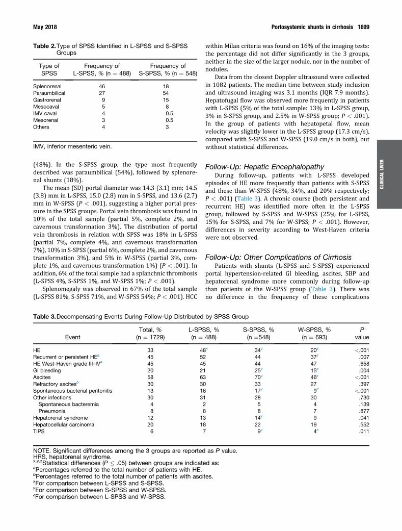
Table 2.Type of SPSS Identified in L-SPSS and S-SPSSGroups
Type ofSPSS
Frequency ofL-SPSS, % (n ¼ 488)
Frequency ofS-SPSS, % (n ¼ 548)
Splenorenal 46 18Paraumbilical 27 54Gastrorenal 9 15Mesocaval 5 8IMV caval 4 0.5Mesorenal 3 0.5Others 4 3
IMV, inferior mesenteric vein.
May 2018 Portosystemic shunts in cirrhosis 1699
CLINICAL
LIVE
R
(48%). In the S-SPSS group, the type most frequentlydescribed was paraumbilical (54%), followed by splenore-nal shunts (18%).
The mean (SD) portal diameter was 14.3 (3.1) mm; 14.5(3.8) mm in L-SPSS, 15.0 (2.8) mm in S-SPSS, and 13.6 (2.7)mm in W-SPSS (P < .001), suggesting a higher portal pres-sure in the SPSS groups. Portal vein thrombosis was found in10% of the total sample (partial 5%, complete 2%, andcavernous transformation 3%). The distribution of portalvein thrombosis in relation with SPSS was 18% in L-SPSS(partial 7%, complete 4%, and cavernous transformation7%), 10% in S-SPSS (partial 6%, complete 2%, and cavernoustransformation 3%), and 5% in W-SPSS (partial 3%, com-plete 1%, and cavernous transformation 1%) (P < .001). Inaddition, 6% of the total sample had a splanchnic thrombosis(L-SPSS 4%, S-SPSS 1%, and W-SPSS 1%; P < .001).
Splenomegaly was observed in 67% of the total sample(L-SPSS 81%, S-SPSS 71%, and W-SPSS 54%; P < .001). HCC
Table 3.Decompensating Events During Follow-Up Distributed
EventTotal, %(n ¼ 1729)
L-SPS(n ¼
HE 33 48Recurrent or persistent HEa 45 52HE West-Haven grade III–IVa 45 45GI bleeding 20 21Ascites 58 63Refractory ascitesb 30 30Spontaneous bacterial peritonitis 13 16Other infections 30 31
Spontaneous bacteremia 4 2Pneumonia 8 8
Hepatorenal syndrome 12 13Hepatocellular carcinoma 20 18TIPS 6 7
NOTE. Significant differences among the 3 groups are reportedHRS, hepatorenal syndrome.x,y,zStatistical differences (P � .05) between groups are indicateaPercentages referred to the total number of patients with HE.bPercentages referred to the total number of patients with ascixFor comparison between L-SPSS and S-SPSS.yFor comparison between S-SPSS and W-SPSS.zFor comparison between L-SPSS and W-SPSS.
within Milan criteria was found on 16% of the imaging tests:the percentage did not differ significantly in the 3 groups,neither in the size of the larger nodule, nor in the number ofnodules.
Data from the closest Doppler ultrasound were collectedin 1082 patients. The median time between study inclusionand ultrasound imaging was 3.1 months (IQR 7.9 months).Hepatofugal flow was observed more frequently in patientswith L-SPSS (5% of the total sample: 13% in L-SPSS group,3% in S-SPSS group, and 2.5% in W-SPSS group; P < .001).In the group of patients with hepatopetal flow, meanvelocity was slightly lower in the L-SPSS group (17.3 cm/s),compared with S-SPSS and W-SPSS (19.0 cm/s in both), butwithout statistical differences.
Follow-Up: Hepatic EncephalopathyDuring follow-up, patients with L-SPSS developed
episodes of HE more frequently than patients with S-SPSSand these than W-SPSS (48%, 34%, and 20% respectively;P < .001) (Table 3). A chronic course (both persistent andrecurrent HE) was identified more often in the L-SPSSgroup, followed by S-SPSS and W-SPSS (25% for L-SPSS,15% for S-SPSS, and 7% for W-SPSS; P < .001). However,differences in severity according to West-Haven criteriawere not observed.
Follow-Up: Other Complications of CirrhosisPatients with shunts (L-SPSS and S-SPSS) experienced
portal hypertension-related GI bleeding, ascites, SBP andhepatorenal syndrome more commonly during follow-upthan patients of the W-SPSS group (Table 3). There wasno difference in the frequency of these complications
by SPSS Group
S, %488)
S-SPSS, %(n ¼548)
W-SPSS, %(n ¼ 693)
Pvalue
x 34y 20z <.00144 37z .00744 47 .65825y 15z .00470y 46z <.00133 27 .39717y 9z <.00128 30 .7305 4 .1398 7 .877
14y 9 .04122 19 .5529y 4z .011
as P value.
d as:
tes.

1700 Simón-Talero et al Gastroenterology Vol. 154, No. 6
CLINICALLIVER
between L-SPSS and S-SPSS groups. Overall, 6% of patientsrequired a TIPS during follow-up; W-SPSS patients neededa TIPS in a significantly lower rate than both SPSS groups.The percentage of non-SBP infections and the developmentof HCC (relapse and new diagnosis) did not differ amonggroups.
Decompensating Events According tothe Type of Collateral
According to the type of L-SPSS found, there were nodifferences in the kind of decompensating event thatpatients presented (Supplementary Table 2). Gastric variceswere more often found in patients with gastrorenal shunts,an association that has been reported previously.25 Never-theless, no differences were observed in the prevalence andsize of esophageal varices across the different types of SPSS.
Figure 2. Probability of transplant-free survival in all patientsincluded in the study categorized according to the presenceof spontaneous portosystemic shunts (SPSS) using Kaplan-Meier curves. Log-rank test: P < .001. L-SPSS: LargeSPSS; S-SPSS: Small SPSS; W-SPSS: Without SPSS.
Performance Status and SurvivalWith regard to performance status, a higher proportion
of patients from W-SPSS group were autonomous (mRS0–1) compared to S-SPSS and L-SPSS (88%, 80%, and 75%,respectively), while the rate of patients with limited activ-ities (mRS 2–3: 12%, 19%, and 23%) or disability (mRS 4–5:0%, 1% and 2%) was larger in the L-SPSS group (P < .001).
Transplant-free survival was significantly higher in theW-SPSS group, compared to S-SPSS and L-SPSS group (log-rank test, P < .001). At the end of the follow-up period, 416patients of the 1729 included had died (L-SPSS 38%, S-SPSS29%, and W-SPSS 32%) and 239 had received a livertransplant (L-SPSS 36%, S-SPSS 34%, and W-SPSS 30%).The hazard ratio (HR) for death/liver transplantation was1.36 (95% confidence interval [CI], 1.13–1.64) for S-SPSSand 1.60 (95% CI, 1.33–1.93) for L-SPSS (Figure 2). Themost common causes of death recorded were liver failure(33%), infections (22%), and HCC (14%), without statisticaldifferences among groups.
The univariate analysis of baseline characteristicsbetween patients alive at the end of follow-up and patientsdead/received transplant is shown in SupplementaryTable 3. Variables significantly associated with the outcomeand entered into the multivariate model were age, sex, dia-betes mellitus, platelet count, MELD score, HCC, and presenceof SPSS. Supplementary Table 4 represents the results of themultivariate analysis for mortality/liver transplant: age,MELD score, a diagnosis of HCC and presence of SPSS wereindependent predictors of transplantation-free survival.
Analysis by Liver FunctionAnalysis of the data was performed stratifying patients
by MELD strata (tertiles) in order to avoid the possibleeffect that the distribution of liver function could have hadon the results. Patients were divided in 3 similar groupsaccording to their MELD score, using percentiles 33 and 66as the cut-off points: the first group included scores from6 to 9; the second group, from 10 to 13 and the third group,from 14 onward. Although MELD score seems more suitableto stratify patients according to liver function for outcome
analyses including HE, we also performed the analysisstratifying by Child-Pugh stage.
The independent effect of etiology in the prevalence ofSPSS (higher prevalence of alcoholic cirrhosis) was lost inthe 2 higher MELD groups, however, it was maintainedin the MELD 6–9 group. HE remained more frequent inpatients with L-SPSS, independently on their liver functionstrata, as shown in Table 4. Similar results were obtainedstratifying by Child-Pugh stage (Supplementary Table 5).Among patients with HE, a recurrent or persistent coursewas identified with more frequency in SPSS patients withworse liver function (MELD score �14).
Regarding other complications, the presence of SPSS wasassociated with a higher risk of portal hypertension-relatedGI bleeding and a high rate of ascites in patients with pre-served liver function (MELD score 6–9) (Table 4) or Child-Pugh A patients (data not shown). Related to this, a moreextensive analysis of markers of portal hypertension wasperformed with the available information in the group ofpatients with Child-Pugh A (Supplementary Table 6). Asseen, SPSS patients presented more indicators of portalhypertension, including HVPG values and presence of CSPH,than W-SPSS patients. On the other hand, presence of SPSShad an effect on outcomes independent of presence of CSPH.Patients with SPSS and CSPH significantly developed moredecompensating events (34 of 50 patients, 68%) thanpatients without SPSS and with CSPH (12 of 27 patients,44%) (P ¼ .047; odds ratio, 2.66; 95% CI, 1.01–6.97).

Table 4.Presence of Episodes of HE With a Recurrent or Persistent Course and Grade III–IV From West-Haven Criteria, andOther Decompensating Events During Follow-Up, According to Presence of SPSS and Liver Function Subgroups(MELD Score Tertiles)
Episodes of HE L-SPSS (n ¼ 488) S-SPSS (n ¼ 548) W-SPSS (n ¼ 693) P value
MELD 6–9 23x 12y 5z <.001MELD 10–13 48x 33y 23z <.001MELD �14 59 57 48z .043
Recurrent or persistent HE L-SPSS with HE (n ¼ 234) S-SPSS with HE (n ¼ 186) W-SPSS with HE (n ¼ 139) P value
MELD 6–9 54 29 47 .790MELD 10–13 45 51 29 .177MELD �14 55 42 36z .013
West-Haven scale: Grade III-IV L-SPSS with HE (n ¼ 234) S-SPSS with HE (n ¼ 186) W-SPSS with HE (n ¼ 139) P value
MELD 6–9 35 29 47 .482MELD 10–13 45 40 43 .753MELD �14 46 51 51 .438
GI bleeding L-SPSS (n ¼ 488) S-SPSS (n ¼ 548) W-SPSS (n ¼ 693) P value
MELD 6–9 18 13 9z .038MELD 10–13 22 30 20 .444MELD �14 21 30 21 .847
Ascites L-SPSS (n ¼ 488) S-SPSS (n ¼ 548) W-SPSS (n ¼ 693) P value
MELD 6–9 40 41y 23z <.001MELD 10–13 53 70 56 .751MELD �14 77 95 80 .211
NOTE. Results are shown as percentages. Significant differences among the 3 groups are reported as P value.x,y,zStatistical differences (P � .05) between groups are indicated as:xFor comparison between L-SPSS and S-SPSS.yFor comparison between S-SPSS and W-SPSS.zFor comparison between L-SPSS and W-SPSS.
May 2018 Portosystemic shunts in cirrhosis 1701
CLINICAL
LIVE
R
Performance status results showed a higher percentageof limitation or disability in L-SPSS patients compared toS-SPSS and W-SPSS, in the subgroup of patients with goodliver function (MELD 6-9) (Supplementary Table 7).
Transplant-free survival in the 2 subgroups of patientswith MELD �10 was not significantly different betweenSPSS patients (L-SPSSþS-SPSS) and W-SPSS patients(Figure 3B and 3C). However, in the subgroup withthe lowest MELD, differences were observed (log-rank testP ¼ .019): HR for death/liver transplantation was 1.57(95% CI, 1.08–2.30) in SPSS (L-SPSSþS-SPSS) with respectto W-SPSS group (Figure 3A). Individual HR for L-SPSS andS-SPSS are shown in Supplementary Table 8. The multi-variate analysis including factors related to death/livertransplantation (age, HCC, and SPSS; SupplementaryTable 8) in this subgroup showed that the presence ofHCC (HR, 4.34; 95% CI, 2.88–6.54; P < .001) and SPSS (HR,1.71; 95% CI, 1.16–2.51; P ¼ .006) were independentlyassociated with mortality and liver transplantation. Similarresults were obtained by analyzing the subgroup of patientswith Child-Pugh A; as seen in Supplementary Figure 2,transplantation-free survival was better in W-SPSS patients(HR for death/transplantation of 1.41 [95% CI, 1.04–1.91]
in SPSS patients) and the multivariate analysis also showedthat HCC (HR 4.06; 95% CI, 2.91–5.67), diabetes mellitus(HR 1.38; 95% CI, 1.01–1.88) and SPSS (HR 1.49; 95% CI,1.09–2.02) were independently associated with death/transplantation.
DiscussionThis is the first study that evaluates a large cohort of
patients with cirrhosis to determine whether the presenceof SPSS correlates with clinical events during the course ofthe disease. Our results suggest that SPSS might develop as aconsequence of a progressive increase in portal pressureand their presence identifies cirrhotic patients at higher riskfor more complications and worse outcomes.
The current study shows, first of all, that SPSS are veryfrequent in liver cirrhosis. In the present series, 60% ofcirrhotic patients had some type of SPSS detected byimaging. Among L-SPSS, the type most often identified wassplenorenal, followed by paraumbilical. This is in line withthe results of previous small studies performed usingultrasound.26–28 Our study allows diagnosing other SPSSthat can be identified more easily using cross-sectional
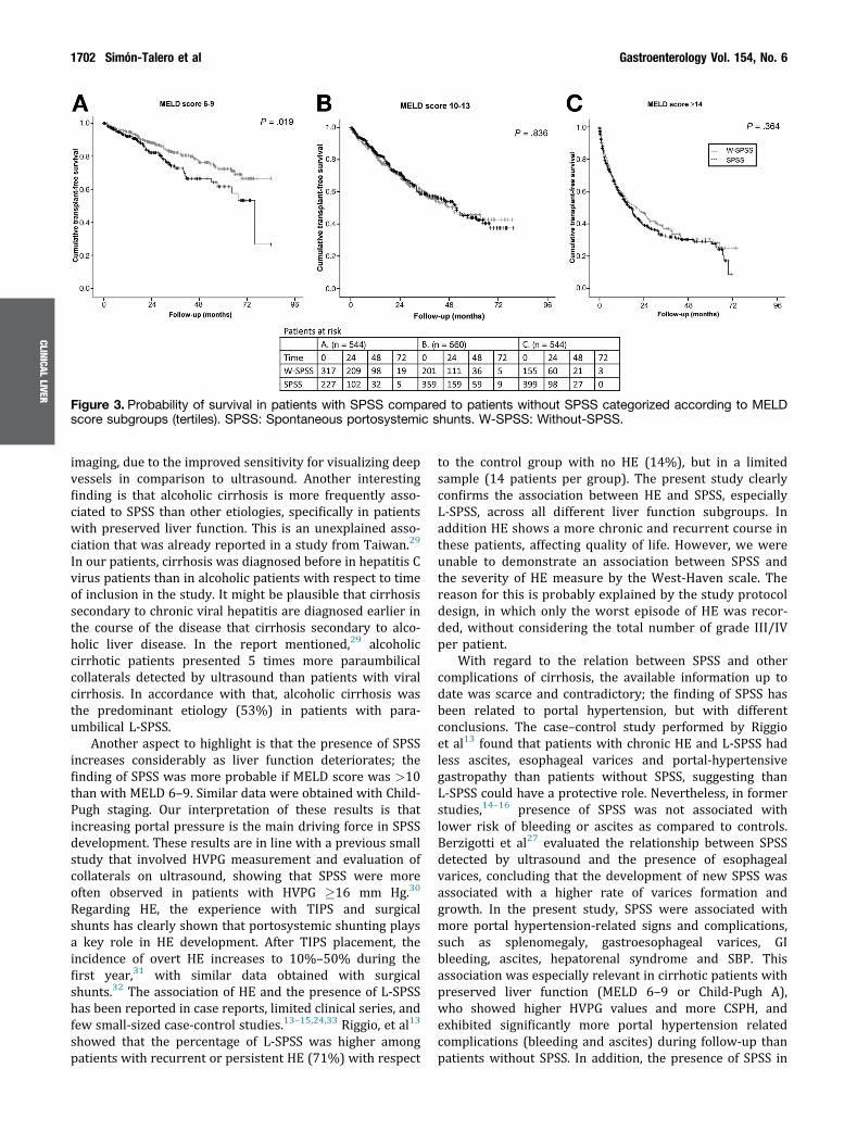
Figure 3. Probability of survival in patients with SPSS compared to patients without SPSS categorized according to MELDscore subgroups (tertiles). SPSS: Spontaneous portosystemic shunts. W-SPSS: Without-SPSS.
1702 Simón-Talero et al Gastroenterology Vol. 154, No. 6
CLINICALLIVER
imaging, due to the improved sensitivity for visualizing deepvessels in comparison to ultrasound. Another interestingfinding is that alcoholic cirrhosis is more frequently asso-ciated to SPSS than other etiologies, specifically in patientswith preserved liver function. This is an unexplained asso-ciation that was already reported in a study from Taiwan.29
In our patients, cirrhosis was diagnosed before in hepatitis Cvirus patients than in alcoholic patients with respect to timeof inclusion in the study. It might be plausible that cirrhosissecondary to chronic viral hepatitis are diagnosed earlier inthe course of the disease that cirrhosis secondary to alco-holic liver disease. In the report mentioned,29 alcoholiccirrhotic patients presented 5 times more paraumbilicalcollaterals detected by ultrasound than patients with viralcirrhosis. In accordance with that, alcoholic cirrhosis wasthe predominant etiology (53%) in patients with para-umbilical L-SPSS.
Another aspect to highlight is that the presence of SPSSincreases considerably as liver function deteriorates; thefinding of SPSS was more probable if MELD score was >10than with MELD 6–9. Similar data were obtained with Child-Pugh staging. Our interpretation of these results is thatincreasing portal pressure is the main driving force in SPSSdevelopment. These results are in line with a previous smallstudy that involved HVPG measurement and evaluation ofcollaterals on ultrasound, showing that SPSS were moreoften observed in patients with HVPG �16 mm Hg.30
Regarding HE, the experience with TIPS and surgicalshunts has clearly shown that portosystemic shunting playsa key role in HE development. After TIPS placement, theincidence of overt HE increases to 10%–50% during thefirst year,31 with similar data obtained with surgicalshunts.32 The association of HE and the presence of L-SPSShas been reported in case reports, limited clinical series, andfew small-sized case-control studies.13–15,24,33 Riggio, et al13
showed that the percentage of L-SPSS was higher amongpatients with recurrent or persistent HE (71%) with respect
to the control group with no HE (14%), but in a limitedsample (14 patients per group). The present study clearlyconfirms the association between HE and SPSS, especiallyL-SPSS, across all different liver function subgroups. Inaddition HE shows a more chronic and recurrent course inthese patients, affecting quality of life. However, we wereunable to demonstrate an association between SPSS andthe severity of HE measure by the West-Haven scale. Thereason for this is probably explained by the study protocoldesign, in which only the worst episode of HE was recor-ded, without considering the total number of grade III/IVper patient.
With regard to the relation between SPSS and othercomplications of cirrhosis, the available information up todate was scarce and contradictory; the finding of SPSS hasbeen related to portal hypertension, but with differentconclusions. The case–control study performed by Riggioet al13 found that patients with chronic HE and L-SPSS hadless ascites, esophageal varices and portal-hypertensivegastropathy than patients without SPSS, suggesting thanL-SPSS could have a protective role. Nevertheless, in formerstudies,14–16 presence of SPSS was not associated withlower risk of bleeding or ascites as compared to controls.Berzigotti et al27 evaluated the relationship between SPSSdetected by ultrasound and the presence of esophagealvarices, concluding that the development of new SPSS wasassociated with a higher rate of varices formation andgrowth. In the present study, SPSS were associated withmore portal hypertension-related signs and complications,such as splenomegaly, gastroesophageal varices, GIbleeding, ascites, hepatorenal syndrome and SBP. Thisassociation was especially relevant in cirrhotic patients withpreserved liver function (MELD 6–9 or Child-Pugh A),who showed higher HVPG values and more CSPH, andexhibited significantly more portal hypertension relatedcomplications (bleeding and ascites) during follow-up thanpatients without SPSS. In addition, the presence of SPSS in

May 2018 Portosystemic shunts in cirrhosis 1703
CLINICAL
LIVE
R
patients with CSPH was associated to higher rate of com-plications compared to W-SPSS patients with CSPH. Thus,the finding of SPSS in patients with good liver functionprobably identifies a subgroup of patients with moreadvanced portal hypertension, who are more likely todevelop complications and might have a worse prognosis. Itis worth to mention that regarding the risk of complicationsrelated to portal hypertension, patients with L-SPSS andS-SPSS seem to behave similarly, with a similar incidence ofcomplications during follow-up.
Even more important, however, is the associationbetween SPSS and decreased transplant-free survival.Although there is a clear relationship between the presenceof SPSS and liver function, SPSS were independently asso-ciated to mortality/transplant on multivariate analysis.Moreover, it is precisely in the subset of patients with lowMELD (score 6–9) or Child-Pugh A, in which this associationwith lower survival was more remarkable. Therefore, in thesubgroup of cirrhotic patients with preserved liver function,the presence of SPSS is a prognostic marker for a higher riskof complications and lower survival. These patients wouldprobably benefit from a closer surveillance and moreintensive therapy.
Few reports have been published about the character-istics of collaterals in cirrhosis. Some of them have sug-gested an association among the type of SPSS and thepredominant kind of complication.25 Anatomically, sple-norenal and gastrorenal shunts have been linked morefrequently with gastro-esophageal varices, and an increasedrisk of bleeding.28 Paraumbilical shunts, that drain into theexternal iliac vein, without feeding the esophageal venousarea, have been associated with less variceal bleeding andmore ascites,34,35 while their relation with HE remainedquestionable.36 These results were not confirmed in otherseries.37 In this large cohort, an association between thetype of complication and SPSS was not observed, exceptfor a higher percentage of gastric varices in gastrorenalshunts, an association already reported.25,27 As explained,HE was more frequent in L-SPSS, indicating that thediameter of the shunt plays a role in this complication, butportal-hypertensive complications results were similar inpatients with L-SPSS and S-SPSS, suggesting that both areindicators of severe portal hypertension.
Our results have the limitations of a retrospective study,mainly originated from data retrieval by reviewing medicalcharts. Some data, such as HVPG, transient elastography,ultrasound, or endoscopy results, were not available in allpatients. In addition, the lack of a predefined systematicprotocol for imaging analysis might explain differences ofSPSS prevalence among centers. Finally, imaging tests wereonly evaluated at one time point and a prospective longi-tudinal study should be performed to analyze data aboutradiological improvement or deterioration according to thedisease course.
There are several strengths of the study. Participantsinvolved were all from tertiary-care university hospitals,with a protocolized management of cirrhotic patients. Thisis the largest cohort ever reported about SPSS with dataprovided from 14 hospitals, from 9 different countries,
allowing the generalization of the results. The review of theimaging tests by expert radiologists is also an added value.Finally, the stratified analysis by MELD score andChild-Pugh class is an important element of the studyeliminating the confounding factor of liver function in therelationship between SPSS and clinical outcomes.
In conclusion, SPSS are frequent in patients withcirrhosis, with splenorenal collaterals found to be themost common type of L-SPSS. The prevalence of SPSSincreases as liver function deteriorates, probably as aconsequence of worsening portal hypertension, butwithout achieving an effective protection against itscomplications. Recurrent or persistent HE is morefrequent in patients with SPSS, independently of liverfunction. Patients with good liver function and SPSSdevelop more portal hypertension-related complications(GI bleeding and ascites) and have a lowertransplantation-free survival. In patients with preservedliver function, SPSS therefore identifies patients with ahigher risk of worse outcomes, and should be consideredan important imaging biomarker in cirrhosis.
Supplementary MaterialNote: To access the supplementary material accompanyingthis article, visit the online version of Gastroenterology atwww.gastrojournal.org, and at http://doi.org/10.1053/j.gastro.2018.01.028.
References
1. Garcia-Tsao G, Abraldes JG, Berzigotti A, et al. Portalhypertensive bleeding in cirrhosis: Risk stratification,diagnosis, and management: 2016 practice guidance bythe American Association for the study of liver diseases.Hepatology 2017;65:310–335.
2. Franchis R de; Baveno VI Faculty. Expanding consensusin portal hypertension: report of the Baveno VIConsensus Workshop: Stratifying risk and individualizingcare for portal hypertension. J Hepatol 2015;63:743–752.
3. Reverter E, Tandon P, Augustin S, et al. A MELD-basedmodel to determine risk of mortality among patients withacute variceal bleeding. Gastroenterology 2014;146:412–419.e3.
4. Poordad FF. Review article: the burden of hepatic en-cephalopathy. Aliment Pharmacol Ther 2007;25(Suppl 1):3–9.
5. Garcia-Tsao G, Sanyal AJ, Grace ND, et al. Preventionand management of gastroesophageal varices and vari-ceal hemorrhage in cirrhosis. Hepatology 2007;46:922–938.
6. Akahane T, Iwasaki T, Kobayashi N, et al. Changes inliver function parameters after occlusion of gastrorenalshunts with balloon-occluded retrograde transvenousobliteration. Am J Gastroenterol 1997;92:1026–1030.
7. Fernandez M, Vizzutti F, Garcia-Pagan JC, et al. Anti-VEGF receptor-2 monoclonal antibody preventsportal-systemic collateral vessel formation in portalhypertensive mice. Gastroenterology 2004;126:886–894.

1704 Simón-Talero et al Gastroenterology Vol. 154, No. 6
CLINICALLIVER
8. Angermayr B, Fernandez M, Mejias M, et al. NAD(P)Hoxidase modulates angiogenesis and the development ofportosystemic collaterals and splanchnic hyperaemia inportal hypertensive rats. Gut 2007;56:560–564.
9. Córdoba J. New assessment of hepatic encephalopathy.J Hepatol 2011;54:1030–1040.
10. American Association for the Study of Liver Diseases,European Association for the Study of the Liver. Hepaticencephalopathy in chronic liver disease: 2014 practiceguideline by the European Association for the Study ofthe Liver and the American Association for the Study ofLiver Diseases. J Hepatol 2014;61:642–659.
11. Vilstrup H, Amodio P, Bajaj J, et al. Hepatic encepha-lopathy in chronic liver disease: 2014 Practice Guidelineby the American Association for the Study of Liver Dis-eases and the European Association for the Study of theLiver. Hepatology 2014;60:715–735.
12. Sherlock S, Summerskill WH, White LP, et al. Portal-systemic encephalopathy; neurological complications ofliver disease. Lancet 1954;267:454–457.
13. Riggio O, Efrati C, Catalano C, et al. High prevalence ofspontaneous portal-systemic shunts in persistenthepatic encephalopathy: a case-control study. Hepatol-ogy 2005;42:1158–1165.
14. Lam KC, Juttner HU, Reynolds TB. Spontaneousportosystemic shunt: relationship to spontaneousencephalopathy and gastrointestinal hemorrhage. DigDis Sci 1981;26:346–352.
15. Ohnishi K, Sato S, Saito M, et al. Clinical and portalhemodynamic features in cirrhotic patients having a largespontaneous splenorenal and/or gastrorenal shunt. Am JGastroenterol 1986;81:450–455.
16. Aseni P, Beati C, Brambilla G, et al. Does large sponta-neous portal systemic shunt in cirrhosis protect from therisk of gastroesophageal bleeding? J Clin Gastroenterol1986;8:235–238.
17. Laleman W, Simon-Talero M, Maleux G, et al. Emboli-zation of large spontaneous portosystemic shunts forrefractory hepatic encephalopathy: a multicenter surveyon safety and efficacy. Hepatology 2013;57:2448–2457.
18. Lynn AM, Singh S, Congly SE, et al. Embolization ofportosystemic shunts for treatment of medicallyrefractory hepatic encephalopathy. Liver Transplant2016;22:723–731.
19. An J, Kim KW, Han S, et al. Improvement in survivalassociated with embolisation of spontaneous portosys-temic shunt in patients with recurrent hepatic encepha-lopathy. Aliment Pharmacol Ther 2014;39:1418–1426.
20. Malinchoc M, Kamath PS, Gordon FD, et al. A model topredict poor survival in patients undergoing transjugularintrahepatic portosystemic shunts. Hepatology 2000;31:864–871.
21. Pugh RN, Murray-Lyon IM, Dawson JL, et al. Transectionof the oesophagus for bleeding oesophageal varices.Br J Surg 1973;60:646–649.
22. Bruno A, Akinwuntan AE, Lin C, et al. Simplified modifiedrankin scale questionnaire: reproducibility over the tele-phone and validation with quality of life. Stroke 2011;42:2276–2279.
23. Conn HO, Leevy CM, Vlahcevic ZR, et al. Comparison oflactulose and neomycin in the treatment of chronicportal-systemic encephalopathy. A double blindcontrolled trial. Gastroenterology 1977;72:573–583.
24. Sakurabayashi S, Sezai S, Yamamoto Y, et al. Emboli-zation of portal-systemic shunts in cirrhotic patients withchronic recurrent hepatic encephalopathy. CardiovascIntervent Radiol 1997;20:120–124.
25. Moubarak E, Bouvier A, Boursier J, et al. Portosystemiccollateral vessels in liver cirrhosis: a three-dimensionalMDCT pictorial review. Abdom Imaging 2012;37:746–766.
26. Zardi EM, Uwechie V, Caccavo D, et al. Portosystemicshunts in a large cohort of patients with liver cirrhosis:detection rate and clinical relevance. J Gastroenterol2009;44:76–83.
27. Berzigotti A, Merkel C, Magalotti D, et al. New abdominalcollaterals at ultrasound: a clue of progression of portalhypertension. Dig Liver Dis 2008;40:62–67.
28. Herbay A von, Frieling T, Häussinger D. Color Dopplersonographic evaluation of spontaneous portosystemicshunts and inversion of portal venous flow in patientswith cirrhosis. J Clin Ultrasound JCU 2000;28:332–339.
29. Chen C-H, Wang J-H, Lu S-N, et al. Comparison ofprevalence for para-umbilical vein patency in patientswith viral and alcoholic liver cirrhosis. Am J Gastroenterol2002;97:2415–2418.
30. Berzigotti A, Rossi V, Tiani C, et al. Prognostic value of asingle HVPG measurement and Doppler-ultrasoundevaluation in patients with cirrhosis and portal hyper-tension. J Gastroenterol 2011;46:687–695.
31. Riggio O, Nardelli S, Moscucci F, et al. Hepaticencephalopathy after transjugular intrahepatic portosys-temic shunt. Clin Liver Dis 2012;16:133–146.
32. Spina G, Santambrogio R. The role of portosystemicshunting in the management of portal hypertension.Baillieres Clin Gastroenterol 1992;6:497–515.
33. Córdoba J, Olivé G, Alonso J, et al. Improvement ofmagnetic resonance spectroscopic abnormalities but notpallidal hyperintensity followed amelioration of hepaticencephalopathy after occlusion of a large spleno-renalshunt. J Hepatol 2001;34:176–178.
34. Mostbeck GH, Wittich GR, Herold C, et al. Hemodynamicsignificance of the para-umbilical vein in portal hyper-tension: assessment with duplex US. Radiology 1989;170:339–342.
35. Dömland M, Gebel M, Caselitz M, et al. Comparisonof portal venous flow in cirrhotic patients with andwithout paraumbilical vein patency using duplex-sonography. Ultraschall Med Stuttg Ger 1980;2000(21):165–169.
36. Del Piccolo F, Sacerdoti D, Amodio P, et al. Centralnervous system alterations in liver cirrhosis: the role ofportal-systemic shunt and portal hypoperfusion. MetabBrain Dis 2003;18:51–62.
37. Aagaard J, Jensen LI, Sørensen TI, et al. Recanalizedumbilical vein in portal hypertension. AJR Am J Roent-genol 1982;139:1107–1110.

May 2018 Portosystemic shunts in cirrhosis 1705
Received July 24, 2017. Accepted January 15, 2018.
Reprint requestsAddress requests for reprints to: Joan Genescà, MD, Liver Unit, Department ofInternal Medicine, Hospital Universitari Vall d’Hebron, Universitat Autònoma deBarcelona, Pg. Vall d’Hebron, 119-129, 08035 Barcelona, Spain. e-mail:[email protected]; fax: þ34932746068. Salvador Augustin, MD, LiverUnit, Department of Internal Medicine, Hospital Universitari Vall d’Hebron,Universitat Autònoma de Barcelona, Pg. Vall d’Hebron, 119-129, 08035Barcelona, Spain. e-mail: [email protected]; fax: þ34932746068.
AcknowledgmentsBaveno VI-SPSS group: Sergi Quiroga, Dominic Yu, Luis Téllez, MattiasMandorfer, Juan Carlos Garcia-Pagan, Claudia Berbel, Jose Ferrusquia,Michel Ble, Mari Angeles Garcia-Criado, Ernest Belmonte, Michael Ney,Cristina Margini, Stefania Casu, Giuseppe Murgia, Christiane Ludwig, MartinRönsch, Dietrich Stoevesandt, Laura Carrion, and Enrique Ramón Botella.Author contributions: Study concept and design: JG, MS-T, SA, EAT, AA,
TR, VH-G, PT, JGA, JLC, JT, AB, CR, AZ, VLM, WL, RB, AK. Acquisition ofdata: MS-T, DR, JM, KL, GL, EL, MP, MHM, MT, GV, RG-M, AD, AM, CP,
DT, AD, ML, GK. Analysis and interpretation of data: JGA, MS-T, SA, EAT,AA, TR, VH-G, PT, JGA, JEC, EL, JT, AB, CR, AZ, VL, WL, RB, RG-M, AK.Drafting of the manuscript: MS-T, SA, JG. Critical revision of the manuscriptfor important intellectual content: TR, PT, AB, EAT, AA, VH-G, JGA, JC, EL,JT, CR, AZ, VLM, WL, RB, RG-M, AK.
Conflicts of interestThe authors disclose no conflicts.
FundingJoan Genescà is a recipient of a Research Intensification grant from Instituto deSalud Carlos III. The study was partially funded by grants PI14/00331, PI15/00066, and PI17/00310 from Instituto de Salud Carlos III, Spain, andco-funded by European Union (ERDF/ESF, “Investing in your future”). Centrode Investigación Biomédica en Red de Enfermedades Hepáticas y Digestivasis supported by Instituto de Salud Carlos III, Spain. Jonel Trebicka is arecipient of grants from the Deutsche Forschungsgemeinschaft (SFB TRR57),Cellex and European Commission H2020. Wim Laleman was supported bythe Gilead Sciences Research Scholars Program in Liver Disease. RitaGarcia-Martinez is a recipient of the grant JR 14/00019 from Instituto deSalud Carlos III, Spain.
CLINICAL
LIVE
R

Supplementary Figure 1. Presence and size of SPSS according to MELD score subgroups (A) and Child-Pugh classes (B).Data are shown as percentages. L-SPSS: Large-Spontaneous portosystemic shunt; S-SPSS: Small-SPSS; W-SPSS: Without-SPSS.
Supplementary Figure 2. Probability of transplant-free sur-vival for W-SPSS and SPSS (L-SPSS and S-SPSS) in Child-Pugh A patients. SPSS: Spontaneous portosystemic shunt;W-SPSS: Without-SPSS.
1705.e1 Simón-Talero et al Gastroenterology Vol. 154, No. 6
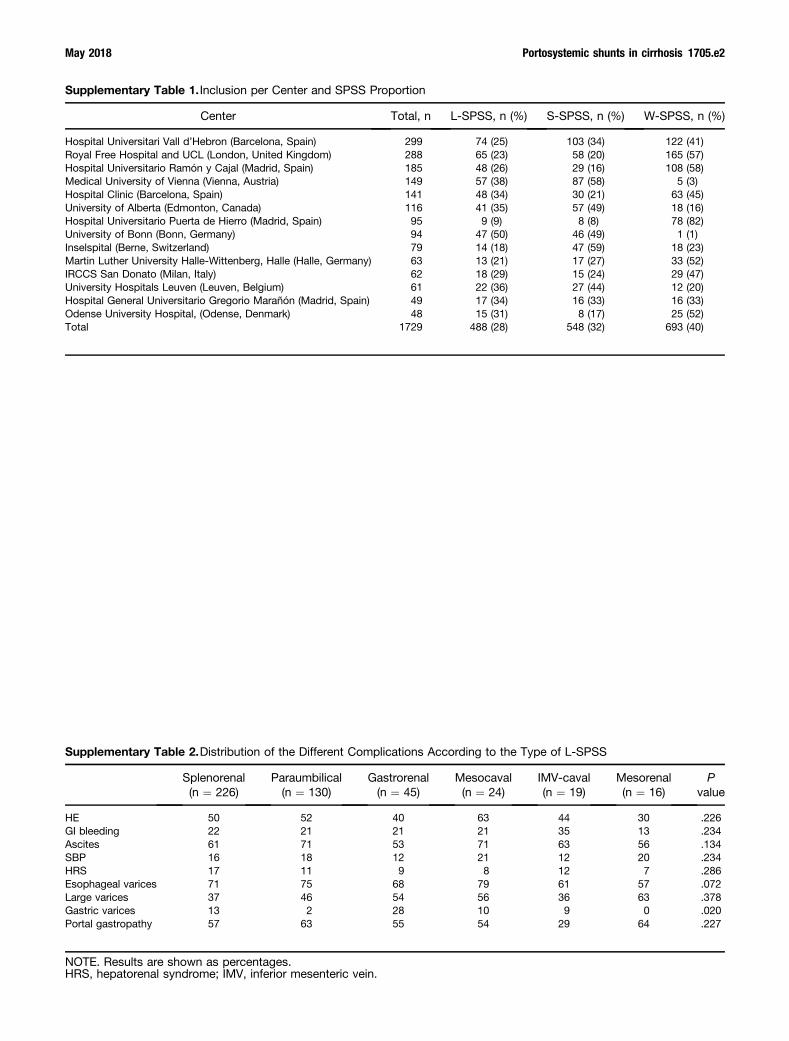
Supplementary Table 1. Inclusion per Center and SPSS Proportion
Center Total, n L-SPSS, n (%) S-SPSS, n (%) W-SPSS, n (%)
Hospital Universitari Vall d’Hebron (Barcelona, Spain) 299 74 (25) 103 (34) 122 (41)Royal Free Hospital and UCL (London, United Kingdom) 288 65 (23) 58 (20) 165 (57)Hospital Universitario Ramón y Cajal (Madrid, Spain) 185 48 (26) 29 (16) 108 (58)Medical University of Vienna (Vienna, Austria) 149 57 (38) 87 (58) 5 (3)Hospital Clinic (Barcelona, Spain) 141 48 (34) 30 (21) 63 (45)University of Alberta (Edmonton, Canada) 116 41 (35) 57 (49) 18 (16)Hospital Universitario Puerta de Hierro (Madrid, Spain) 95 9 (9) 8 (8) 78 (82)University of Bonn (Bonn, Germany) 94 47 (50) 46 (49) 1 (1)Inselspital (Berne, Switzerland) 79 14 (18) 47 (59) 18 (23)Martin Luther University Halle-Wittenberg, Halle (Halle, Germany) 63 13 (21) 17 (27) 33 (52)IRCCS San Donato (Milan, Italy) 62 18 (29) 15 (24) 29 (47)University Hospitals Leuven (Leuven, Belgium) 61 22 (36) 27 (44) 12 (20)Hospital General Universitario Gregorio Marañón (Madrid, Spain) 49 17 (34) 16 (33) 16 (33)Odense University Hospital, (Odense, Denmark) 48 15 (31) 8 (17) 25 (52)Total 1729 488 (28) 548 (32) 693 (40)
Supplementary Table 2.Distribution of the Different Complications According to the Type of L-SPSS
Splenorenal(n ¼ 226)
Paraumbilical(n ¼ 130)
Gastrorenal(n ¼ 45)
Mesocaval(n ¼ 24)
IMV-caval(n ¼ 19)
Mesorenal(n ¼ 16)
Pvalue
HE 50 52 40 63 44 30 .226GI bleeding 22 21 21 21 35 13 .234Ascites 61 71 53 71 63 56 .134SBP 16 18 12 21 12 20 .234HRS 17 11 9 8 12 7 .286Esophageal varices 71 75 68 79 61 57 .072Large varices 37 46 54 56 36 63 .378Gastric varices 13 2 28 10 9 0 .020Portal gastropathy 57 63 55 54 29 64 .227
NOTE. Results are shown as percentages.HRS, hepatorenal syndrome; IMV, inferior mesenteric vein.
May 2018 Portosystemic shunts in cirrhosis 1705.e2

Supplementary Table 3.Univariate Analysis for theIdentification of Predictors atBaseline (Time 0) of Mortality orLiver Transplantation at the End ofFollow-Up
VariableRegressioncoefficient
Pvalue
HR(95% CI)
Age 0.006 .069 1.01 (1.00–1.01)Sex 0.177 .045 1.19 (1.00–1.42)Time of diagnosis
of cirrhosisa0.01 .084 1.01 (0.99–1.02)
Etiology: HCV –0.009 .915 0.99 (0.84–1.17)Etiology: Alcohol 0.077 .349 1.08 (0.92–1.27)Hypertension –0.43 .612 0.96 (0.81–1.13)Diabetes mellitus 0.19 .027 1.20 (1.02–1.42)Platelets <150 � 109/mm3 0.430 <.001 1.54 (1.25–1.88)MELD score 0.115 <.001 1.12 (1.11–1.14)Child-Pugh score 0.886 <.001 1.36 (1.31–1.40)HCC 0.649 <.001 1.91 (1.55–2.36)S-SPSS 0.307 .001 1.36 (1.13–1.64)L-SPSS 0.471 <.001 1.60 (1.33–1.93)SPSS (S þ L) 0.387 <.001 1.47 (1.26–1.73)
aDuration of cirrhosis after initial diagnosis.
Supplementary Table 4.Multivariate (Cox) Analysis ofFactors Related to Death/LiverTransplantation
VariableRegressioncoefficient
Pvalue
HR(95% CI)
Age 0.02 <.001 1.02 (1.01–1.02)Sex 0.13 .171 1.14 (0.95–1.36)Diabetes mellitus 0.12 .163 1.13 (0.95–1.34)MELD score 0.13 <.001 1.14 (1.12–1.15)HCC 0.82 <.001 2.25 (1.80–2.81)Platelets <150 � 109/mm3 0.23 .036 1.26 (1.02–1.57)SPSS (S þ L) 0.23 .008 1.26 (1.06–1.49)
NOTE. L-SPSS was an independent factor related to death orliver transplantation, with a HR 1.32 (95% CI, 1.08–1.61;P ¼ .006). The HR for S-SPSS was 1.20 (95% CI, 0.98–1.46;P ¼ .071).
Supplementary Table 5.Presence of Episodes of HE, With a Recurrent or Persistent Course and Grade III–IV From West-Haven Criteria During Follow-Up, According to Presence of SPSS and Liver Function Subgroups(Child-Pugh Class)
Episodes of HE L-SPSS (n ¼ 488) S-SPSS (n ¼ 548) W-SPSS (n ¼ 693) P value
Child A 28x 10 7z <.001Child B 50 41y 29z <.001Child C 78 68 61z .010
Recurrent or persistent HE L-SPSS with HE (n ¼ 234) S-SPSS with HE (n ¼ 186) W-SPSS with HE (n ¼ 139) P value
Child A 51 40 52 .984Child B 50 47 33 .074Child C 57 44 37z .035
West-Haven scaleGrade III-IV
L-SPSS with HE (n ¼ 234) S-SPSS with HE (n ¼ 186) W-SPSS with HE (n ¼ 139) P value
Child A 44 11 44 .804Child B 38 47 45 .398Child C 51 49 55 .798
NOTE. Results are shown as percentages.x,y,zStatistical differences (P � .05) between groups are indicated as:xFor comparison between L-SPSS and S-SPSS.yFor comparison between S-SPSS and W-SPSS.zFor comparison between L-SPSS and W-SPSS.
1705.e3 Simón-Talero et al Gastroenterology Vol. 154, No. 6

Supplementary Table 6.Markers of Portal Hypertension (Platelet Count, Spleen Size, Rate of Varices and Portal Gastropathyon EGD Endoscopy, TE, HVPG, and Percentage of CSPH) According to Presence of SPSS inChild-Pugh A Patients
Child-Pugh A (n ¼ 712) L-SPSS (n ¼ 144) S-SPSS (n ¼ 196) W-SPSS (n ¼ 372) P value
Platelets, �109/mm3, mean (SD) 95.7 (49.8)x 114.9 (58.6)y 133.9 (65.7)z <.001Spleen diameter, cm, mean (SD) 16 (3)x 14 (3)y 13 (3)z <.001
EGD endoscopy (n ¼ 371) L-SPSS (n ¼ 81) S-SPSS (n ¼ 108) W-SPSS (n ¼ 182) P value
Esophageal varices, % 74 68y 47z <.001Gastric varices, % 7 7 3 .068Portal gastropathy, % 46 58 41 .180
TE (n ¼ 150) L-SPSS (n ¼ 19) S-SPSS (n ¼ 40) W-SPSS (n ¼ 91) P value
Liver stiffness, KPa, median (IQR) 19 (24)x 27 (27)y 18 (17) .002
HVPG measurement (n ¼ 106) L-SPSS (n ¼ 23) S-SPSS (n ¼ 31) W-SPSS (n ¼ 52) P value
HVPG (mmHg), mean (SD) 15 (5)x 19 (7)y 11 (6)z <.001CSPH, n (%) 20 (87) 30 (97)y 27 (52)z <.001
NOTE. Number of subjects available for every marker is indicated at the beginning of in every row. Three patients with L-SPSShad no CSPH: 1 patient had primary biliary cholangitis, 1 patient with mixed alcohol and hepatitis C etiology was abstinent andon b-blockers, and finally 1 patient with hepatitis C was also on b-blockers. One patient with S-SPSS and no CSPH was anabstinent alcoholic patient on b-blockers.EGD, esophagogastroduodenal; TE, transient elastography.x,y,zStatistical differences (P � .05) between groups are indicated as:xFor comparison between L-SPSS and S-SPSS.yFor comparison between S-SPSS and W-SPSS.zFor comparison between L-SPSS and W-SPSS.
Supplementary Table 8.Univariate (Cox) Analysis of FactorsRelated to Death or LiverTransplantation in Patients WithPreserved Liver Function (MELDScore 6–9)
Regressioncoefficient
Pvalue
HR(95% CI)
Age 0.02 .050 1.02 (1.00–1.03)Sex –0.01 .983 0.10 (0.67–1.48)Time of diagnosis
of cirrhosisa0.21 .208 1.02 (0.99–1.06)
Etiology: HCV 0.01 .995 1.00 (0.68–1.47)Etiology: alcohol 0.34 .105 1.41 (0.93–2.13)Hypertension 0.21 .283 1.23 (0.84–1.80)Diabetes mellitus 0.29 .147 1.33 (0.90–1.97)Platelets <150 � 109/mm3 0.10 .619 1.12 (0.74–1.65)HCC 1.46 <.001 4.31 (2.89–6.43)S-SPSS 0.49 .024 1.64 (1.07–2.53)L-SPSS 0.37 .185 1.45 (0.84–2.52)SPSS (S þ L) 0.45 .019 1.57 (1.08–2.30)
HCV, hepatitis C virus.aDuration of cirrhosis after initial diagnosis.
Supplementary Table 7.Quality of Life (Modified RankinScale) According to Presence ofSPSS and MELD Score Subgroups
VariableL-SPSS(n ¼ 488)
S-SPSS(n ¼ 548)
W-SPSS(n ¼ 693)
Pvalue
MELD 6–9Autonomous 84x 92 95z
Limitation 13 8 5 .001Disability 3 0 0
MELD 10–13Autonomous 82 82 88Limitation 17 17 11 .071Disability 1 1 1
MELD �14Autonomous 69 71 75Limitation 29 27 25 .188Disability 2 2 0
NOTE. Results are shown as percentages.x,zStatistical differences (P � .05) between pairs of value areindicated as:xFor comparison between L-SPSS and S-SPSS.zFor comparison between L-SPSS and W-SPSS.
May 2018 Portosystemic shunts in cirrhosis 1705.e4