gastroenterology - university of manitoba...describe the partitioning of the cloaca and formation of...
TRANSCRIPT

Notes compiled for Pediatrics
Gastroenterology (Med I, Block 6, GI)

Class number Class Name Type Department Instructor Pages
GI 002 Development of GI tract I L AN Dr. M Torchia 3-11 of 35
GI 003 Development of GI tract II L AN Dr. M Torchia 4-11 of 35
GI 044 Infectious Diarrhea and Food Poisoning L ID Dr. J Embil 12-19 of 35
GI 046 Gastrointestinal Infections T6 ID Dr. J Embil 20 of 35
GI 057 Congenital Pediatric Disorders A PD Dr. S Pushpa 21-30 of 35
GI 064 Acquired Pediatric Disorders L PD Dr. S Pushpa 22-35 of 35

Development of the Gl Tract University of Manitoba Faculty of Medicine Med II / Gl 02 Dr. M. GTorchia 2008 - 2009
Objectives
1. Understand how the incorporation of the dorsal yolk sac during embryo folding results in the
formation of the early Gl tract 2. List the derivatives of each of the foregut, midgut, and hindgut and their major blood supply 3. Describe the process of stomach rotation and development of the lesser sac 4. Describe how herniation, rotation, reduction, and fixation each act to produce the final position of
the gut 5. Understand development of Gl tract innervation 6. Describe the partitioning of the cloaca and formation of the anal canal 7. Understand the basic principles involved in the histogenesis of the intestine, liver, and pancreas 8. Describe the specific malformations of Gl tract that result in omphalocele, intestinal stenosis,
Meckel diverticulum, annular pancreas, biliary atresia, tracheoesophageal fistula, anorectal agenesis, pyloric stenosis, Hirschsprung's disease
References:
The Developing Human: Clinically Oriented Embryology (6th Ed.); Moore & Persaud, Chapter 12, pp
271-302
Overview:
The development of the Gl system begins during the second and third weeks during head, tail, and
lateral body folding. Folding results in the delineation (pinching off) of the Gl tract from the yolk sac. The foregut is initially sealed by the oropharyngeal membrane and the hindgut is closed by the cloacal plate (proctodeal membrane). Beginning in week four, inductive interactions between the epithelium of the developing Gl tract tube and the surrounding mesenchyme form the digestive/endocrine glands including the liver and pancreas. The spleen develops independently from proliferating mesenchyme cells. Beginning in the fifth week, the tubular portion of the gut grows very rapidly resulting in mechanical stresses that cause a series of folds and twists to produce the final
orientation of the stomach and intestine. This rapid growth also causes the developing bowel to herniate into the body stalk (umbilical cord) until the abdominal cavity is sufficiently large to contain the bowel (by the tenth week.) With reduction of the herniated bowel, the mesentery of some of organs and portions of bowel are forced against the posterior abdominal wall resulting in retroperitoneal locations. Blood and nerve supplies to the organs develop by local angiogenesis and the migration of neural crest cells, respectively. The complexity of the formation of the Gl system results in significant potential for malformations including agenesis, atresia, and duplication.
3 of 35

Development of the GI Tract II University of Manitoba Faculty of Medicine
Med II / GI 03 (L) Dr. M. G. Torchia 2008 - 2009
Objectives Continuation of GI #02
1. pyloric stenosis, omphalocele, incomplete rotation and volvulus of the midgut 2. intestinal stenosis and atresia, and Mechel's 3. imperforate anus and anorectal agenesis with fistula 4. see accompanying clinical problems and notes)
4 of 35
Torchia, 2007
Development of the GI Tract
Dr. Mark G. Torchia
Department of Surgery
**Figures Reference: The Developing Human, 7th ed., Moore and Persaud.
Torchia, 2007
Applications of Embryology
“Anatomical and functional relationships”
“Congenital abnormalities”
“Development of new therapies aimed at inducing embryonic developmental capacities”
Torchia, 2007
Illustrative case
A 48y previously very healthy man
Non-radiating, waxing and waning epigastric abdominal pain of 12h duration, with associated nausea and anorexia
Normal abdominal sounds; normal exam
Pain continued at home in evening
Found dead in bedroom next morning
Torchia, 2007
Illustrative case – Appendicitis ?
The classic history of pain beginning in the periumbilical region and migrating to the right lower quadrant occurs in only 50 percent of patients.
Torchia, 2007
Objectives
Define the importance of the incorporation of the dorsal yolk sac
Describe derivatives of the foregut, midgut, and hindgut and their major blood supply
Explain the development of the lesser sac during stomach rotation
Describe how herniation, rotation, reduction, and fixation produce the final position of the gut
Torchia, 2007
Objectives
List the origin of GI tract innervation
Describe the partitioning of the cloaca and formation of the anal canal
Review histogenesis of the liver and pancreas
Describe specific malformations of GI tract
5 of 35

Torchia, 2007
Recurring Themes
Inductive interactions between gut epithelium and the surrounding mesenchyme
Cell proliferation followed by apoptosis
Symmetry followed by asymmetry
Tissue/organ growth leads to movement
Torchia, 2007
Primordial Gut (Fig. 5-1)
Formed by the envelopment of the dorsal yolk sac into the embryo during folding (head, tail, lateral)
Endoderm forms endothelium
Torchia, 2007
Embryo Folding - Membranes
Proximal oropharyngeal membrane and distal cloacal membrane
Communicates with yolk sac
Torchia, 2007
Division of Embryonic Gut
Foregut - oral cavity, esophagus, stomach, prox. duodenum, liver, biliary app, pancreas (celiac trunk)
Midgut - distal duodenum, small intestine, cecum, appendix, ascending and right transverse colon (superior mesenteric artery)
Hindgut - left transverse colon, descending colon, rectum, superior anal canal
(inferior mesenteric artery)
Torchia, 2007
Foregut - Esophagus
proximal 33% striated muscle
pharyngeal arch mesenchyme
distal 66% smooth muscle
splanchnic mesenchyme
Torchia, 2007
Foregut - Esophagus
vascular supply SHARED (inf thyroid, bronchial, aortic branches, phrenic, L gastric)
trachea forms as an outpouch of esophagus
6 of 35

Torchia, 2007
Foregut –Stomach (Fig. 12-2
tubular form enlarges dorsally
greater curvature
suspended - ventral and dorsal mesentery
dorsal = greater omentum with lesser sac
Torchia, 2007
Developing Stomach (Fig 12.2)
90 degree clockwise rotations
semi-transverse position with greater curvature on L side (nausea/vomiting)
Torchia, 2007
Developing Duodenum (Fig 12.2)
tube rotates with stomach and grows to form a C-shape
Torchia, 2007
Developing Gut (Fig. 12-6)
endothelial hyperplasia (obliteration) followed by apoptosis and vacuolation
lumen by end of week 8
Torchia, 2007
Foregut – Liver & Biliary Apparatus
hepatic diverticulum grows into ventral mesentery cranial portion - liver
caudal portion – gallbladder, cystic duct, bile duct
hepatic cells endoderm
endothlial cells of venous sinusoids
mesenchyme (ventral)
hematopoietic (6 wk)
Kupffer cells (macrophages - reticuloendothelial cells)
Torchia, 2007
Foregut - Pancreas
two pancreatic buds (ventral and dorsal mesentery)
dorsal portion is larger
ventral portion rotates with duodenum to fuse with dorsal portion
ventral portion brings duct with it to form main pancreatic duct
parenchyma from endoderm
7 of 35

Torchia, 2007
Nodal Cilia
Cilia on nodal cells
sweep counterclockwise
(Kartegener’s syndrome
Primary Ciliary Dyskinesia -
situs inversus)
Torchia, 2007
Division of Embryonic Gut
Foregut - oral cavity, esophagus, stomach, prox. duodenum, liver, biliary, pancreas (celiac trunk)
Midgut - distal duodenum, small intestine, cecum, appendix, ascending and right transverse colon (superior mesenteric artery)
Hindgut - left transverse colon, descending colon, rectum, superior anal canal (inf. mesenteric a.)
Torchia, 2007
Midgut
Elongates to form a long U-shaped midgut loop
yolk stalk attached between cranial/caudal limbs
Abdominal cavity volume small (liver/kidneys)
midgut herniates into umbilical cord (wk 6)
Midgut rotates CCW 90o then 180o
axis of superior mesenteric artery
Cranial loop grows much faster (small intestine)
Torchia, 2007
Midgut Loop Rotation (Fig 12-13)
Torchia, 2007
Midgut
Reduction of herniated bowel (wk 10)
Small bowel returns first; cecum last
Presses mesentery of ascending colon, (descending colon (H) and duodenum (F) against posterior abdominal wall (retroperitoneal)
Appendix position depends on growth of ascending colon (retrocecal, pelvic, etc.)
Torchia, 2007
Division of Embryonic Gut
Foregut - oral cavity, esophagus, stomach, prox. duodenum, liver, biliary, pancreas (celiac trunk)
Midgut - distal duodenum, small intestine, cecum, appendix, ascending and right transverse colon (sup. mesenteric a.)
Hindgut - left transverse colon, descending colon, rectum, superior anal canal
(inferior mesenteric artery)
8 of 35

Torchia, 2007
Hindgut
Cloaca separated into urinary/rectal divisions by caudal growth of urorectal septum (complete 7 wk)
Anal membrane ruptures wk 9 (amniotic sac)
Anal canal – pectinate line superior 66% hindgut (inferior mesenteric artery
+ autonomic nerves)
inferior 33% - surface ectoderm (internal pudendual artery + inferior rectal nerve - sensory)
Torchia, 2007
Congenital Defects
Torchia, 2007
Tracheoesophageal Fistula/Atresia(Fig. 11-5)
1:2500, male>>female, coughing, choking, regurg, aspiration
Torchia, 2007
Pyloric Stenosis (Hypertrophy)(Fig. 12-4)
1:150 Male, 1:750 female, projectile vomiting
Torchia, 2007
Annular Pancreas
Vomiting, poor nutrition, duodenal obstruction, chronic pancreatitis, few symptoms
Torchia, 2007
Omphalocele – non-closure umbilicus
(Fig 12-7)
1:6000, herniated bowel, peritoneum+amnion
9 of 35

Torchia, 2007
Meckel’s Diverticulum (Fig. 12-21)
3:100, male>>female, remnant yolk sac, ectopic tissue<90 cm from ileocecal junction
Torchia, 2007
Hirschsprung Disease
Abdominal distension, absent bowel movements, poor nutrition, surgery, mild cases
Torchia, 2007
What you should know:
Incorporation of the dorsal yolk sac
Derivatives of the foregut, midgut, and hindgut and major blood supply
Stomach rotation and development of the lesser sac
Herniation, rotation, reduction, and fixation produce the final position of the gut
Torchia, 2007
What you should know:
Origin of GI tract innervation
Partitioning of the cloaca and formation of the anal canal
Histogenesis of the liver and pancreas
Specific malformations of GI tract
Torchia, 2007
Thank you
Torchia, 2007
MCQ evaluation
1) The rectum and urogenital sinus are formed during the division of the cloaca by the:
a) developing urinary bladder
b) remnant of the yolk sac
c) cloacal membrane
d) urorectal septum
e) Meckel’s diverticulum
10 of 35

Torchia, 2007
MCQ evaluation
2) Non-reduction of the physiological midgut hernia results in formation of:
a) a Meckel’s diverticulum
b) an inguinal hernia
c) an omphalocele
d) a subhepatic cecum
e) a megacolon
Torchia, 2007
MCQ evaluation
3) In the embryo, the duodenum is formed from:
a) foregut
b) midgut
c) hindgut
d) foregut and midgut
e) midgut and cranial portions of the hindgut
Torchia, 2007
MCQ evaluation
4) Formation of the primordial gut during the fourth week results from:
1) caudal budding of oropharyngeal membrane
2) incorporation of the yolk sac into the embryo
3) cranial budding from GI mesenchyme
4) involution of the splanchnic mesoderm
5) none of the above
11 of 35

Infectious Diarrhea and Food Poisoning University of Manitoba Faculty of Medicine Med II / GI 44 Dr. J. Embil 2008 - 2009
Objectives: 1. Describe the elements of the epidemiological triangle and their attributes as they relate to enteric
infections. 2. Describe the features of the normal alimentary tract that form a barrier to infection. 3. Describe the pathogenesis and clinical findings associated with non-inflammatory diarrhea,
inflammatory diarrhea and diarrhea due to penetrating organisms. 4. Describe the difference between enteroinvasion, enteroadhesion and mucosal damage as
mechanisms of diarrhea and food poisoning 5. Describe the different types of food poisoning in terms of etiology and mechanism 6. Discuss the pathogenesis, diagnosis and treatment of pseudomembranous colitis. Outline: 1. Definition and Classification of Diarrhea 2. Host Factors that Form a Barrier to Infection: Personal hygiene, barriers of gastrointestinal tract
(gastric acid, intestinal motility, normal bowel flora, immunoglobulin secretion) 3. Etiology: Infectious (bacterial, viral, protozoal, parasitic) 4. Factors Associated with Increased Morbidity and Mortality: Malnutrition immunosuppression, lack
of rehydration, overwhelming infection, invasive pathogen 5. Pathogenic Mechanisms of Diarrhea: Mucosal adherence, toxin production (neurotoxin,
enterotoxin, cytotoxin), mucosal invasion 6. Clinical Features: Watery diarrhea, dysenteric syndrome, enteric fever, enteric fever-like
syndrome 7. What is food poisoning and how is it caused 8. Pseudomembranous Colitis 9. Diagnostic Evaluation 10. Prevention 11. Management Suggested Reading: 1. Cecil Essentials of Medicine, 6th Edition. 2. Medical Microbiology and Immunology, Levinson and Jawetz 4th Edition 3. Essentials of Pediatrics, Behrman and Kliegman, 2nd Edition 4. Mitchell JE, Skelton MN. Diarrheal Infection. Am Fam Phys 1988; 37: 195-207 5. Armstrong D, Cohn J (editors). Infectious Diseases, 1st Edition. Chapter 35: Enteritis,
Enterocolitis and Infectious Diarrhea Syndromes. 1999, Mosby, New York. 6. Your favorite text book about Medical Microbiology and Infectious Diseases 1. Definition and Classification of Diarrhea: As in all other pathologic conditions, an interaction between the host, the microorganism and the environment exists in infectious diarrhea.
12 of 35

Host Agent Environment
Acute diarrhea is a self-limiting condition resulting from an alteration in bowel habit with an increase in
frequency and decrease in consistency of stool. More specifically, it is associated with an increased stool
weight (> 200 g/day – normal, 50-150 g/day).
The adult digestive tract generally receives approximately 10 liters of fluid per day. 80% of the fluid is
absorbed in the small intestine while the remaining 20% is reabsorbed in the colon. Diarrhea results whenever
there is an imbalance between secretion and absorption of fluids and electrolytes. The mucosa of the small
intestine is made up of absorptive, villous surfaces where nutrients, electrolytes and fluids are absorbed and
crypt cells where electrolytes and fluids are secreted. When the surface villi are destroyed or absorption is
inhibited, then that result is electrolyte and fluid secretion into the bowel movement. Intestinal water absorption
and secretion are passive and follow an osmotic gradient created by the active transport of sodium.
Table 1: Classification of Diarrhea Diarrhea is summarized according to its type and mechanism Secretory: Caused by an increased secretion or decreased absorption of sodium and chloride leaving them in the bowel lumen. This, in turn, increases the volume of water in the intestinal lumen. It is characterized by large volume, watery diarrhea without blood or pus and there is little response to fasting. It may be mediated by bacterial toxins (Vibrio cholera) Figure 1, bile salt enteropathy and fatty acid-induced diarrhea. Solute gap absent. Osmotic: Increased non-absorbable molecules in the bowel lumen leading to a decrease in the absorption of solute from the bowel. This results in watery stool without blood or pus but improves with fasting. An increased solute gap is present. Examples included lactose intolerance, carbohydrate malabsorption and magnesium laxatives. Inflammatory: This is caused by destruction of the bowel mucosa leading to impaired absorption and associated with an outpouring of blood and mucous. The frequently associated symptoms include small, frequent, stools with blood and pus. There is associated fever. Examples include enteroinvasive pathogens such as Entamoeba histolytica (amebiasis) and Shigellia dysenteriae (shigellosis). Decreased Absorptive Surface: Here there is decreased reabsorption of electrolytes as a consequence of a shortened, absorptive surface also known as “short gut” syndrome. This may be due to a bowel resection, or enteric fistula, whereby solutes and other material bypass the absorptive area. The characteristics are variable and the diarrhea may improve with fasting. Motility Disorders: This is a complex condition caused by either increased motility with decreased time for absorption of electrolytes and nutrients. This may be due to hyperthyroidism or irritable bowel syndrome. Decreased bowel motility with bacterial overgrowth may be observed in conditions such as scleroderma and diabetes. This results in a malabsorption syndrome. Figure 1: Diarrhea due to secretory mechanism mediated by toxin Figure 1 demonstrates the mechanism of toxigenic diarrhea. The outpouring of water and electrolytes is due to the action of toxin stimulating adenyl cyclase receptors, with activation of cyclic adenosine monophosphate.
Figure 2: Diarrhea Due to Viral or Bacterial Enteroinvasion
13 of 35

Figure 2 demonstrates viral or bacterial enteroinvasion. Here there is loss of mature absorptive surfaces resulting in immature secretory surfaces. 2. Host Factors: After exposure to a pathogen, the manifestation and severity of diarrheal disease depend upon the status of the host, specifically, personal hygiene, barriers of the gastrointestinal tract (gastric acid, intestinal motility, normal bowel microflora and immunoglobulin secretion). Extremes of age play a role in the susceptibility to enteric infections. Rotavirus, enteropathogenic E coli frequently cause infectious diarrhea in children but are uncommon in older adults. Infectious agents lead to more significant complications in those at the extremes of age. Acquisition of nearly all forms of diarrheal disease depend upon the ingestion of specific microorganisms. Most intestinal pathogens originate from other mammalian species although reptiles are colonized with Salmonella spp. The most frequent mode of transmission is fecal-oral. Poor personal hygiene and proper sanitation are important factors in the fecal-oral route of disease transmission. The gastrointestinal tract of the healthy host is capable of reducing the virulence of the majority of ingested pathogens. Virtually all ingested bacteria are destroyed within 30 minutes in a normal gastric pH, whereas more than one hour is required for pathogen destruction in achlorhydric individuals or individuals who ingest acid neutralizing agents or gastric acid reducing agents. The natural motility of the gastrointestinal tract promotes the proper distribution and flow of resident bowel flora, this also facilitates the rapid passage of pathogens to the gastrointestinal tract and aids in fluid reabsorption. Drugs that slow peristalsis such as opiates and anticholinergics may delay the transit and excretion of pathogens and may lead to significant complications. The immune system of the host is important as intestinal secretions of antigens and specific immunoglobulin have bactericidal, neutralizing or opsonic effect on pathogens. Hereditary or acquired alterations in intestinal secretion of immunoglobulins will put the host at increased risk. Hypogammaglobulinemia may be associated with an increased incidence of diarrheal illnesses. Although host factors are important, microorganisms are also important, specifically the infectious dose in humans of Shigella spp is low, 10-100 bacteria compared to Salmonella spp which requires thousands of microorganisms to cause disease in humans. 3. Etiology It is important to note that although diarrhea may be caused by infectious agents such as bacteria, viruses, protozoa and parasites, other etiology such as toxic ingestion’s, laxatives, osmotically acting agents, neuroendocrine abnormalities, neoplasms, inflammatory bowel disease, ischemia and gastrointestinal hemorrhage, may lead to acute diarrhea. It is critical to maintain an open mind in approaching the patient with both acute and chronic diarrhea. 4. Factors Associated with Increased Morbidity and Mortality (Table 2):
Table 2: The major factors that translate to a higher death rate associated with diarrhea include host, therapeutic and microbial factors, which can be further broken down into:
Malnutrition
Complications of diarrhea, including dehydration, pneumonia, sepsis and hemolytic uraemic syndrome
Infection by an agent more likely to cause dehydration such as Vibrio cholera and rotavirus (infants)
Infection by invasive pathogens such as Shigella spp and failure to receive adequate rehydration or antimicrobial therapy for illnesses requiring active intervention.
Oral rehydration programs in developing countries have reduced morbidity and overall mortality rates from diarrheal illness. This supportive measure, however, has not eliminated diarrhea as a significant cause of death in children.
14 of 35

5. Pathogenic Mechanisms of Diarrhea (Table 3): Table 3: Pathogenic Mechanisms Mucosal Adherence: Here the microorganisms attach to and colonize the intestinal mucosa. Examples include enteropathogenic E coli (EPEC). This may lead to alterations in ion permeability of the membrane (increased secretion or reduced absorption) resulting in a secretory diarrhea. Toxin Production: A variety of different microorganisms can produce different toxins leading to diarrhea.
Neurotoxins: These are proteins which may activate the autonomic nervous system. The most frequently encountered is Staphylococcal enterotoxin, this leads to food poisoning associated with violent nausea, vomiting and diarrhea with rapid convalescence. The incubation period is 1-8 hours. The characteristic history is that of large groups of individuals attending communal gatherings such as church picnics or wedding receptions. Foods may be contaminated by S. aureus from a food worker with a paronychia or other localized S aureus infection. Bacillus cereus can produce 2 distinct forms of food poisoning mediated by toxins. The emetic type is associated with rice dishes. The classic history is that of someone consuming rice in a Chinese restaurant followed by an acute onset of vomiting 1-6 hours after ingestion. The diarrheal form is associated with the consumption of meat and sauce dishes and having an incubation of 1-24 hours. Clostridium botulinum may lead to an acute toxin mediated food poisoning. Table 7 summarizes some of the causes and characteristics of food poisoning. More about food poisoning later…
Enterotoxin: A number of different bacteria produce toxins (Figure 1) which lead to an outpouring of water and electrolytes from the bowel mucosa into the intestinal lumen resulting in a secretory diarrhea.
Cytotoxins: Cytotoxins are defined by their ability to produce damage to the mucosa resulting in an inflammatory colitis usually by the inhibition of protein synthesis. The key example is Shiga toxin which is produced by S. dysenteriae. This toxin is very similar to that of E coli O 157: H7. Clostridium difficile produces two toxins, Toxin A, an enterotoxin, and Toxin B a cytotoxin, they work synergistically to produce significant morbidity and in some cases mortality.
Mucosal Invasiveness: Bacteria such as Shigella spp., Campylobacter spp, Yersinia spp, possess the ability to penetrate into the intestinal mucosa and destroy the epitheal cells causing a dysentery-like syndrome.
6. Clinical Features (Table 4): The pathophysiology mechanisms that produce infectious diarrhea are as follows:
Increased active intestinal secretion of electrolytes causing fluxes of water and ions, mediated by bacterial enterotoxins (watery diarrhea, Figure 1)
Malabsorption of nutrients and electrolytes secondary to damage to the brush-border in either the small or large intestine
Increased intestinal osmolality secondary to saccharidase deficiency when the brush-border is damaged, with resultant lactose intolerance (Figure 2)
Altered intestinal motility
Table 4: Clinical-Pathologic Features and Causes Watery Diarrhea: This is a non-inflammatory process (absence of fecal leukocytes). This is characterized by
large volume stool and increased number of stools because the colonic reservoir is intact. This condition may
be characterized by nausea and vomiting but other constitutional symptoms such as cramping, and abdominal
pain, arthralgias, myalgias, chills and fever rarely occur. This diarrhea is mediated by bacterial enterotoxins
that alter fluid and electrolyte transport. The prototype of an enterotoxigenic diarrhea is that caused by V.
cholera. After an incubation period of several hours to 5 days, the illness may begin with sudden onset of
15 of 35

profuse, watery diarrhea or anorexia and abdominal discomfort followed by diarrhea. The stool has the
characteristic “rice water” appearance because of the mucous content and may have a mild fishy odor.
Tenesmus is absent and vomiting may occur several hours after the onset of diarrhea. Signs and symptoms of
cholera result from the severity of dehydration caused by the food and electrolyte losses from the intravascular
and extravascular spaces into the gut lumen. A small proportion of individuals may proceed very quickly to a
severe hypovolemic shock. Fever is absent. Muscle cramps may occur as a consequence of electrolyte
imbalance. Other microorganisms which may cause a watery diarrhea include rotavirus (most common in
children a Norwalk agent. Giardia lamblia is a protozoan which causes “beaver fever” and causes diarrhea by
altering fluid reabsorption in the proximal small bowel.
Dysenteric Syndrome:
Acute dysentry: This inflammatory or invasive process involves the colon and occasionally the distal
small intestine. Numerous leukocytes in the feces indicate the diffuse colonic inflammation or
invasion of the colonic mucosa. Frequent enteroinvasive pathogens include Shigella spp,
enteroinvasive E. coli, Salmonella spp, Yersinia enterocolitica, C jejuni, V parahaemolyticus.
Individuals inflicted with acute dysenteric syndromes may become very ill and develop complications
such as hemolytic uremic syndrome encountered with E. colli O 157: H7 (Hamburger Disease).
Pseudomembranous colitis: Pseudomembranous colitis is a condition associated with the
administration of antimicrobial agents. Although any antibiotic may lead to this condition, the most
frequently associated clindamycin, amoxicillin and the beta lactams. The onset of this antibiotic
associated diarrhea may be seen anytime after the starting of antibiotics. It may occur during
antibiotic therapy or long after it has been discontinued. The clinical manifestations vary from mild
watery or mucoid green diarrhea to a dysenteric syndrome with bloody diarrhea, high fever, marked
abdominal tenderness and the presence of fecal leukocytes. The proposed pathogenesis of
pseudomembranous colitis include an alteration of the normal bowel flora as a consequence of the
administration of a antimicrobial agent allowing the overgrowth of Clostridium difficile. This
microorganism produces two toxins (Toxin A-enterotoxin, Toxin B-cytotoxin) resulting in colonic
membrane damage. Sigmoidoscopy typically revealed the presence of small, raised
pseudomembranous nodules or plaque. Complications include severe diarrhea, hypovolemic shock,
toxic megacolon, peritonitis, cecal perforation, hemorrhage and sepsis. It is a frequent cause of
diarrhea in hospitalized patients who have received antibiotics.
Enteric Fever: Enteric Fever is an acute systemic illness characterized by fever, headache and abdominal discomfort. This syndrome is characteristically produced by Salmonella typhi and is referred to as Typhoid Fever, however, other Salmonella spp. may cause similar but less severe clinical syndromes. S. typhi is a water and food borne microorganism for which humans are the only natural host. The salmonellae are ingested orally and must traverse the acid barrier as well as various pancreatic enzymes, bile, and intestinal secretions and secretory IgA, which are effective antimicrobial factors. The post-gastrectomy state, hypochlorhydria, altered intestinal motility and prior antibiotic therapy are conditions predisposing to salmonellosis. Other individuals at risk of salmonella infection include those with Sickle Cell Disease, chronic liver disease, immunodeficiency and AIDS. The incubation period S. typhi causing Typhoid Fever is 5-21 days depending upon the inoculum and immune status of the host. The microorganisms make their way to the small bowel where they ultimately replicate in the Peyer’s patches. The microorganisms enter the circulation through the lymphatic route and replicate in the reticuloendothelial cells in the lymph nodes, liver, bone marrow and spleen. The onset of symptoms is gradual and characterized by non-specific symptoms such as fever, headache and abdominal pain. Diarrhea occurs in approximately 50% of individuals. Physical findings include abdominal tenderness, hepatosplenomegaly, evanescent maculopapular rash in the upper abdomen and lower thorax (Rose spots), bradycardia and mental confusion. Complications of enteric fever may be related recurrent bacteremia with dissemination of the
16 of 35

microorganisms. These complications include pneumonia, endocarditis, osteomyelitis, arthritis or meningitis and local complications such as erosion of blood vessels in the Peyer’s patches which may result in gastrointestinal hemorrhage and perforation of the ilium. Yersinia enterocolitica and Campylobacter jejuni may cause an enteric fever-like syndrome indistinguishable from that of Typhoid Fever. Traveler’s’ Diarrhea (Tables 5 and 6): Traveler’s Diarrhea is defined as the passage of 3 or more unformed stools per day in a resident from an industrialized country traveling in or returning from a developing nation. The onset of Traveler’s Diarrhea usually occurs within the first 2 weeks after arrival in a foreign country, most often within the first week of travel. The illness may occur after returning to one’s home. Table 6 demonstrates the most common etiologies according to location. Enterotoxigenic E colli is the most commonly isolated organism of Traveler’s Diarrhea. Traveler’s Diarrhea is almost always a self-limiting illness rarely lasting more than 5 days even when untreated. The diarrhea is accompanied by abdominal pain and cramping and a minority of patients develop a fever. Traveler’s diarrhea may be avoided by simple measures (Table 5) such as avoiding local uncooked delicacies, only consuming fruits with a peel, avoiding raw vegetable which have been rinsed with tap water, consuming bottled water and beverages. Antibiotics may be of benefit only when diarrhea begins.
Table 5: Etiologies of Traveler’s Diarrhea Table 6: Preventing Traveler’s Diarrhea
7. Food poisioning:
Food poisoning can be classified according to the predominant symptoms produced: nausea and vomiting; non-
inflammatory diarrhea; inflammatory diarrhea; neurologic symptoms; systemic or miscellaneous syndromes.
The incubation period after the ingestion of the offending foodstuff may provide clues as to the potential
etiology, as shown in table 7. It is important to remember that food poisoning may be caused by both infectious
and non-infectious etiologies. Food poisoning caused by some bacteria may be associated with only
gastrointestinal symptoms while others may have neurologic symptoms as a major component.
How does Clostridium botulinum cause botulism?
How does C tetani cause tetanus
Why is Staphylococcus aureus food poisoning so severe?
What types of food poisoning would you get if you ate at the following:
Japanese/sushi restaurant
Chinese restaurant
Organic bakery and honey shop
Your well meaning relatives who can preserves
Mary Malone’s diner
Table 7: Food Poisoning
8. Pseudomembranous Colitis:
Some of the details about pseudomembranous colitis (PMC) were previously outlined in the section above
entitled, “Dysenteric Syndromes”. PMC refers to colitis characterized by the formation of a pseudomembrane.
The pseudomembrane is composed of fibrin, mucin, sloughed mucosal epithelial cells, and acute inflammatory
cells. On endoscopy, PMC displays multiple, elevated yellowish white plaques. PMC is the most severe
mucosal change associated with antibiotic associated diarrhea and is virtually always caused by C difficile.
When PMC is associated with antibiotic use, it is almost 100% guaranteed that C difficile is the culprit
17 of 35

pathogen. C difficile produces two toxins, noted above, which differ in their activity and potency in terms of
tissue toxicity. They work synergistically to cause tissue injury. Different laboratory investigations are
necessary to detect the different toxins.
9. Diagnostic Evaluations (Table 8): Table 8: Investigations
Fecal leukocyte test: The presence of leukocytes in fecal material suggests an inflammatory process, whether it is caused by an enteroinvasive pathogen, or an uninvasive colitis such as Crohn’s Disease.
Stool Culture: Stool Culture is indicated for non-hospitalized patients or those who are hospitalized with diarrhea. It is important to specify in the Microbiology Requisition, the exact nature of the patient’s symptoms and whether blood is present. This will help guide the diagnostic evaluation in the laboratory.
Toxin Analysis: In those individuals who have a history of prior antibiotic ingestion and diarrhea, C difficile may be the causative organism. It is important to specify that this is a consideration and, therefore, toxin analysis will be undertaken.
Parasite Evaluation: Once again, it is important to specify to the Microbiology Laboratory what symptoms are present and whether the history of travel or consumption of tainted water exists. The evaluation of stool specimen for parasites and/or their ova can be time consuming. It is important to specify which microorganisms are being contemplated as different techniques may be necessary.
Direct Visualization: Either a proctosigmoidoscopy or colonoscopy may be necessary when all other investigations have not been fruitful. Direct visualization will demonstrate the plaques of pseudomembranous colitis or ulcerations compatible with amebiasis. Direct visualization is helpful to help differentiate between infectious and non-infectious etiologies of diarrhea which has been long-standing.
Upper endoscopy: The history of dysphagia and diarrhea particularly in those who are immunocompromised may be an indication for an evaluation of the upper gastrointestinal tract.
10. Prevention:
Any measure that can be undertaken to prevent the acquisition of diarrheal disease must be emphasized. Simple
measures such as:
1. Handwashing after the handling of infectious or potentially infectious material (diapers, soiled garments,
bedpans) will help minimize the chances of disease transmission. 2. While traveling, only consume cooked foods, avoid drinking unprocessed water, consume bottled water
and beverages, only consume fruits with a peel, avoid raw vegetables which have been rinsed with tap water. Avoid drinking from lakes and streams. Table 6 summarizes the key details of traveling safely.
11. Management: Table 9: Treatment Table 9 summarizes specific treatments according to pathogens for adults and children. It is important to note that this is merely a guide to the treatment of different bacteria and parasites. Should you be faced with an individual with a specific pathogen causing diarrhea, it is important to check a current reference source for the most up to date information.
There are 4 basic principles in the management of individuals who have acute diarrhea. 1. Fluid and electrolyte replacement: This is the cornerstone of therapy for acute diarrhea. It may be life
saving for infants and the elderly. In most cases, oral rehydration therapy is satisfactory, however, in some with profound dehydration and decreased level of consciousness, parenteral rehydration may be indicated.
2. Dietary modification: After an episode of an acute infectious diarrhea which affects the small bowel, the only foods to be avoided are milk products, since transient secondary lactase deficiency is common in diarrheal states. The use of formula diets or elemental diets are rarely indicated because digestive functions are not impaired. For infants, breast milk or lactose-free formula may be administered. A bland diet consisting of boiled starches and cereals such as potatoes, noodles, rice, wheat and oats may be of benefit for older children and adults. When stools are formed, the diet may return to normal.
18 of 35

3. Symptomatic Therapy: Antiperistaltic agents are seldom indicated as they inhibit gastrointestinal motility and may lead to an ileus and some situations may have grave repercussions. It should be avoided in cases of known C difficile disease.
Clostridium difficile may be managed as follows:
First episode: Metronidazole 500 mg po tid for 10 days
Second episode: Metronidazole 500 po tid for 10 days.
Third episode: Vancomycin 125 mg po qid and rifampin 600 po bid for 7 days.
19 of 35

Gastrointestinal Infections University of Manitoba Faculty of Medicine Med II / GI 46 (T) Dr. J. Embil 2008 - 2009
Objectives- 1. develop an approach to diagnosis and management of gastroenteritis in a child utilizing the
information from GI-42 and the reference tests 2. list the characteristics that can be used to distinguish the many different bacterial food poisoning
syndromes 3. appreciate the existence of non-bacterial causes of food-borne illness 4. develop a plan for investigating cases and outbreaks of food poisoning 5. be familiar with procedures required to prevent food-borne illness References:
Cecil Essentials of Medicine, 6th edition. Medical Microbiology and Immunology, Levinson and Jawetz, 4th edition. Essentials of Pediatrics, Behrman & Kliegman, 2nd edition.
20 of 35

Congenital Pediatric Disorders University of Manitoba Faculty of Medicine Med II / GI 57 (L) Dr. S. Moroz 2008 - 2009
Objectives 1. Be aware of the congenital disorders involving the gastrointestinal tract 2. Be aware of the clinical manifestations of these disorders 3. Be aware of aberrations in normal embryologic development and how they lead to the congenital
disorders described 4. Be aware of some clinical investigative methods used to diagnose these disorders 5. Be aware of surgical or medial therapy to correct these disorders
21 of 35

Objectives
1. Be aware of common symptoms of clinical problems of the gastro-intestinal problems of children
2. Be aware of common reasons for these symptoms 3. Be aware of the basic approaches to diagnostic investigation of these problems 4. Be aware of the basic approaches to surgical or medical management of these problems
Acquired Pediatric Disorders University of Manitoba Faculty of Medicine
Med II/ GI064 Dr. S. Moroz
2008-2009
22 of 35

7/16/2009
PEDIATRIC GASTROENTEROLOGY
University of ManitobaMedicine 2
Dr. S. P. Moroz, Pediatric GI
May 14, 2008
Learning Objectives
• Important Pediatric G.I. Conditions
– Case Examples
• Pathophysiology
• Clinical Presentation
• Investigation
• Interactive
CONGENITAL DISORDERS OF THE GASTROINTESTINAL TRACT
• Mechanical Obstruction– Failure of lumen to develop (atresia, stenosis)
– Failure of lumen to recanalize (duodenal atresia)
– Occlusion of lumen with meconium (cystic fibrosis)
– Incomplete rotation of bowel during development
• Functional Obstruction– Dysmotility due to abnormal innervation
Case 1
• Newborn
• Mother had polyhydramnios
• Baby has had difficulty swallowing from birth
• Baby chokes on own secretions
Case 1
• What are some possible explanations for this problem?
• What investigations would you consider?
Case 1
• Differential Diagnosis
– Meconium aspiration
– Cardiac anomaly
– Esophageal atresia with or without fistula
– Other
• Pneumonia
• Sepsis
23 of 35

7/16/2009
Esophageal AtresiaTracheoesophageal Fistula
• Failure of separation of esophagus and trachea
• 1:3000-1:4500 births
• Rarely familial
• 5 types
• Signs and symptoms– Depend on type
– Respiratory distress
– Inability to feed
– Choking during feeding
– Other anomalies
Types
Investigationslateral chest x-ray
Investigationslateral chest x-ray with water soluble contrast
Case 2
• Newborn infant with bilious emesis since birth
• Maternal polyhydramnios
• Infant has abdominal distension
What are the possible explanations for this problem?
What investigations would you perform?
Case 2presentation suggests bowel obstruction distal to the Ampulla of Vater
Differential Diagnosis
– Bowel atresia or stenosis• Pyloric, duodenal, small bowel
– Meconium ileus or plug
– Malrotation with volvulus
– Abnormal intrinsic bowel innervation
• e.g. Hirschsprung
24 of 35

7/16/2009
Look for the Bubbles
• Single Bubble
• Double Bubble
• Multiple Bubbles
Single Bubblesuggests gastric outlet obstruction
Double Bubblesuggests duodenal obstruction Many Bubbles
suggests more distal obstruction
• Jejunal or Ileal atresia
• Meconium Ileus
Meconium Ileusresected bowel
Thick meconium
25 of 35

7/16/2009
Intestinal Malrotation / Volvulus Malrotationbarium enema
Volvuluscongested ischemic small bowel
Case 3respiratory distress from birth
Case 3abdomen and chest exposed at autopsy
Case 3chest wall removed
26 of 35

7/16/2009
Case 3liver removed
Case 3bowel reduced into abdomen
Case 3
• Diaphragmatic hernia
– Usually left side through Foramen of Bochdalek
– Survival dependent on lung development
– Associated with other anomalies
– Herniation may occur later in life if defect is small
Case 4
• Newborn infant
• Fails to pass meconium during 1st 24 hrs
• Abdomen distended
• Emesis
What are some possible explanations for this problem?
Case 4
• Differential Diagnosis
– Anal stenosis/Imperforate anus
– Rectal atresia
– Hirschsprung’s disease
Case 4Anorectal anomalies
27 of 35

7/16/2009
Case 4 Case 4
Case 4
• Hirschsprung’s disease
– Due to defective development of intramural nerve plexus
– Usually affects distal colon but extent variable
– Clinical features:• History
– Delayed meconium passage
• Rectal exam
– Snug anal canal
– Empty rectum
Case 4
• Hirschsprung’s disease
– Investigations• Abdominal x-ray
– Colon full of stool, rectum empty
• Barium enema
– Dilated colon, transistion zone
• Anorectal manometry
– Hypertonic anal sphincter
– Failure internal sphincter to relax with rectal distension
• Rectal biopsy-definitive test
– Absent intramural ganglion cells
– Abnormal nerve plexuses
Hirschsprung’s diseasebarium enema
Case 5
• Male infant
• Projectile emesis shortly after feeding
• Onset 6 weeks of age
• Emesis not bile-stained
• Losing weight, borderline hydration
28 of 35

7/16/2009
Case 5
• What is most likely diagnosis?
• What physical findings would be most helpful to confirm the diagnosis?
• What investigations would you consider?
• How would you treat this child?
Case 5
• Hypertrophic pyloric stenosis
– Etiology unknown
– 1:500 births
– Occasionally familial
– Often affects 1st born male infants
– Emesis causes dehydration, metabolic hypochloremic alkalosis and failure to thrive
– Visible gastric waves following feeds
– Palpable pyloric “tumor”
Case 5Pyloric Stenosis
• Investigations
– Abdominal x-ray• Single bubble, wavy gastric gas pattern
– Abdominal ultrasound• Hypertrophied pylorus
– Barium study• Narrow pyloric canal (string sign)
• Antral beak sign
• Gastric waves
Pyloric StenosisUltrasound
Pyloric StenosisBarium study
Pyloric StenosisEndoscopy
Pyloric Stenosis Normal Pylorus
29 of 35
7/16/2009
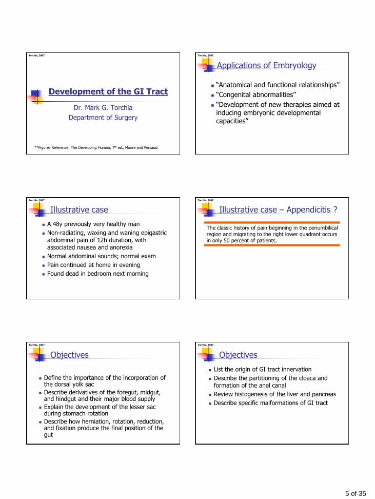
Pyloric StenosisPyloromyotomy
ACQUIRED DISORDERS OF THE GASTROINTESTINAL TRACT
Case 1
• Previously healthy 1 yr old
• 48 hrs emesis, diarrhea, fever
• Sib with similar problem 1 wk earlier
What is most likely cause of this child’s illness?
What concerns would you have about this child?
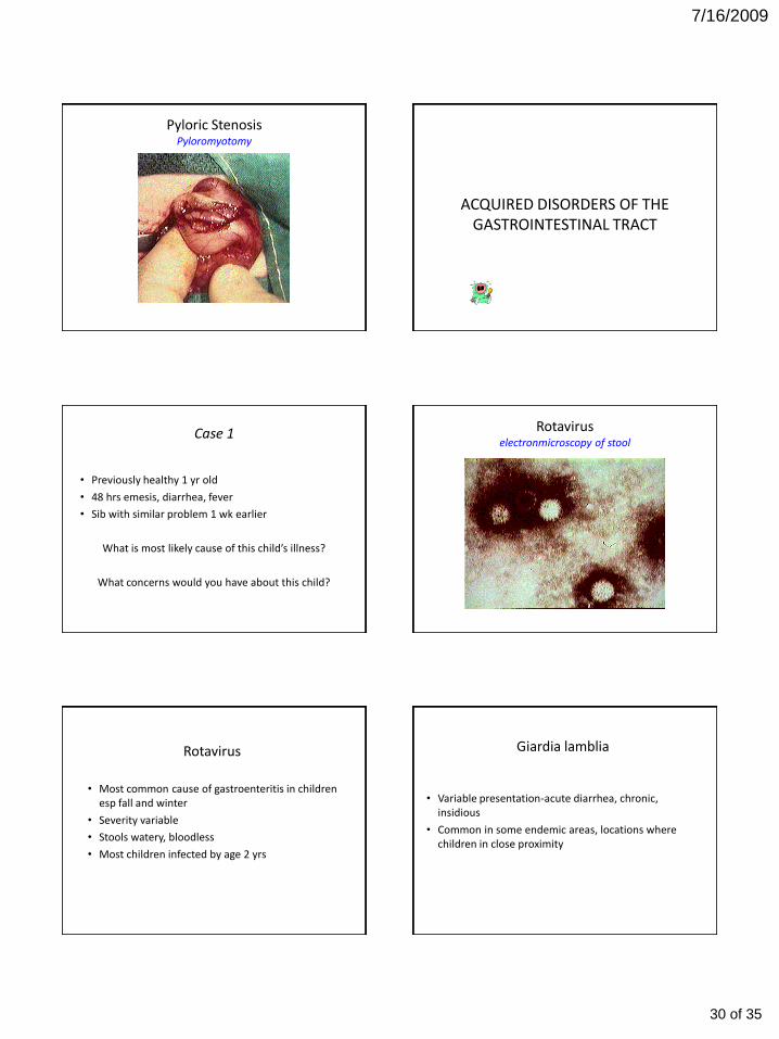
Rotaviruselectronmicroscopy of stool
Rotavirus
• Most common cause of gastroenteritis in children esp fall and winter
• Severity variable
• Stools watery, bloodless
• Most children infected by age 2 yrs
Giardia lamblia
• Variable presentation-acute diarrhea, chronic, insidious
• Common in some endemic areas, locations where children in close proximity
30 of 35
7/16/2009
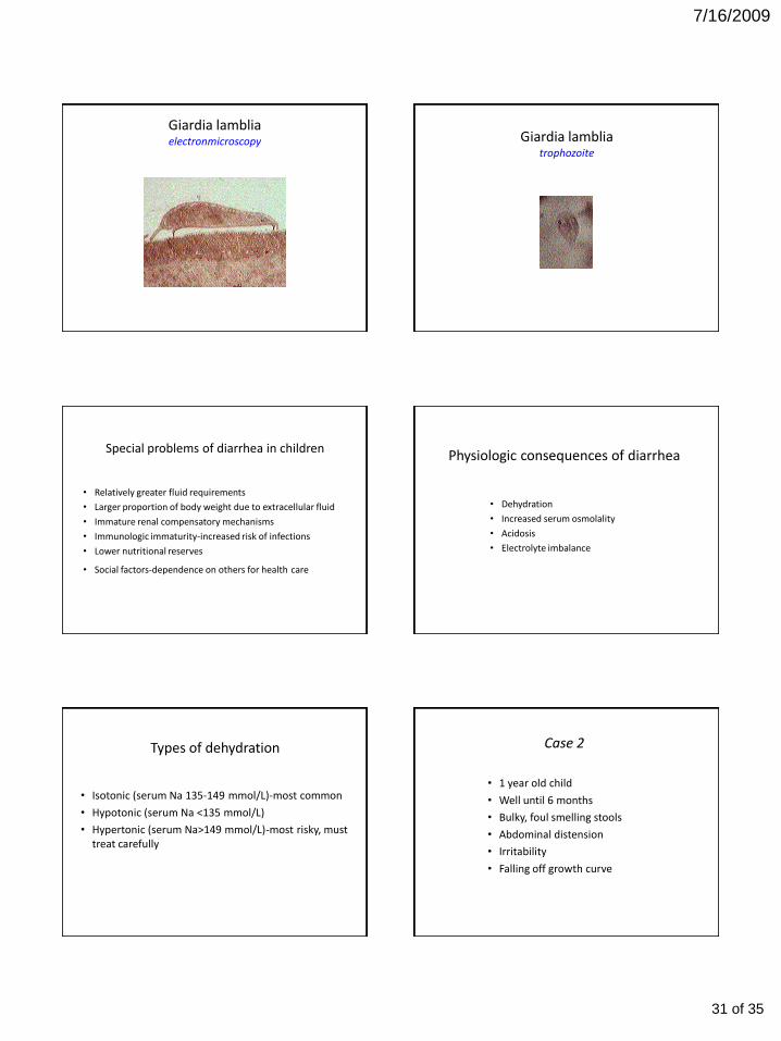
Giardia lambliaelectronmicroscopy Giardia lamblia
trophozoite
Special problems of diarrhea in children
• Relatively greater fluid requirements
• Larger proportion of body weight due to extracellular fluid
• Immature renal compensatory mechanisms
• Immunologic immaturity-increased risk of infections
• Lower nutritional reserves
• Social factors-dependence on others for health care
Physiologic consequences of diarrhea
• Dehydration
• Increased serum osmolality
• Acidosis
• Electrolyte imbalance
Types of dehydration
• Isotonic (serum Na 135-149 mmol/L)-most common
• Hypotonic (serum Na <135 mmol/L)
• Hypertonic (serum Na>149 mmol/L)-most risky, must treat carefully
Case 2
• 1 year old child
• Well until 6 months
• Bulky, foul smelling stools
• Abdominal distension
• Irritability
• Falling off growth curve
31 of 35

7/16/2009
Case 2
Case 2
What is the most likely diagnosis?
Celiac disease
• Variable manifestations
• May be asymptomatic
• Detected by endomysial and transglutaminase antibodies
• Usually HLA DQ2 or DQ8 positive
• Confirmed by intestinal biopsy
• Treated by lifelong gluten-free diet
Celiac diseaseintestinal biopsy
Normal Celiac
Case 3
• Previously well infant
• Sudden onset presumed abdominal pain
• Draws up knees
• Emesis
• Lethargy
• Bloody stool
Case 3
What are some of the causes of intestinal bleeding in children?
What is the most likely diagnosis in this child?
What investigations would you do?
32 of 35
7/16/2009
Causes of intestinal bleeding
• Upper (hematemesis)
– Gastroesophageal reflux (sign of esophagitis)
– Varices (portal hypertension-hepatic, prehepatic)
– Peptic ulcer (usually with pain)
Causes of intestinal bleeding
• Lower (hematochezia)
– Bacterial infections
– Necrotizing enterocolitis
– Polyps
– Intussusception
– Meckel’s diverticulum
– Inflammatory bowel disease
Intussusception
• Most common at 6-24 months of age
• Idiopathic
• May follow viral infection
• Acute pain, emesis, +/- bloody stool (currant jelly)
• Lethargy
• Risk of ischemia, perforation if treatment delayed
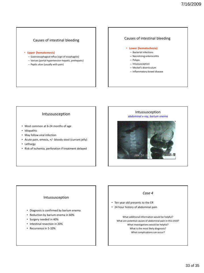
Intussusceptionabdominal x-ray, barium enema
Intussusception
• Diagnosis is confirmed by barium enema
• Reduction by barium enema in 60%
• Surgery needed in 40%
• Intestinal resection in 20%
• Recurrence in 5-10%
Case 4
• Ten year old presents to the ER
• 24 hour history of abdominal pain
What additional information would be helpful?
What are potential causes of abdominal pain in this child?
What investigations would be helpful?
What is the most likely diagnosis?
What complications can occur?
33 of 35

7/16/2009
Case 4helpful clinical information
• History– Onset and character of pain– Location and migration of pain– Aggravating and relieving factors– Associated symptoms
• Physical examination– Inspection– Auscultation– Palpation
• Tendernes• Guarding• Referred and rebound tenderness
– Percussion
Causes of abdominal pain
• Appendicitis
• Intussusception
• Crohn’s disease
• Cholelithiasis/cholecystitis
• Pancreatitis
• Peptic ulcer
• Functional-most common
• Extraintestinal-pneumonia, diabetes, abd. wall
Investigations for abdominal pain
• CBC, ESR
• Pancreatic enzymes
• Liver function studies
• Urinalysis/culture
• Imaging– Abdominal x-ray
– Barium studies
– Ultrasound
– CT
• Endoscopy
Case 4Appendicitis
• Very common
• 12-14 yrs of age most common
• Hx and px most important
• May need observation to clarify diagnosis
• Perforation more likely in younger children or more prolonged course
• Antibiotic therapy may be necessary
• Early surgical treatment best
Complications of appendicitis
• Perforation
• Peritonitis
• Leakage from appendiceal stump
Case 5
• 2 month old infant
• Jaundice since birth
• Clay colored stools, dark urine
• Hepatosplenomegaly
34 of 35

7/16/2009
Case 5hepatosplenomegaly Case 5
Do you think this child’s jaundice is “physiologic”?
What are the possible causes for this child’s jaundice?
What investigations would you perform?
Causes of cholestasis in infants
• Extrahepatic biliary atresia
• Choledochal cyst
• Intrahepatic biliary dysgenesis
• Neonatal hepatitis
– Infection
– Metabolic
– Familial
– Idiopathic
• Iatrogenic (TPN)
Investigation of cholestatic liver disease
• Liver function studies
– Confirms cholestasis
– Assesses impact on liver function
• Infection screen
– STORCH, Hepatitis A, B, C
• Metabolic studies
Investigation of cholestatic liver disease (cont’d)
• Diagnostic imaging– Abdominal ultrasound
– Biliary scan
• Needle biopsy liver
• Laparotomy– Biopsy
– Cholangiogram
– Repair atresia
THE END
35 of 35