gastrointestinal bleeding d. salem thaniah associated consultant gastroenterology king khalid...
TRANSCRIPT
Gastrointestinal bleeding
D. Salem ThaniahAssociated consultant gastroenterology
King Khalid University
Ms S
• 86-year-old woman• Physically active• She presented to the emergency department
after falling in her bathroom.
Almadi et al. JAMA 2011;306:2367-74.

Ms S
• Had been feeling epigastric discomfort • It was episodic in nature and mild in intensity • There were no provocative or palliative
factors.• A few hours prior to her fall she had been
feeling lightheaded with some weakness.
Almadi et al. JAMA 2011;306:2367-74.

Ms S
• When standing up she felt dizzy and fell to the ground but did not lose consciousness.
• She was transported to the hospital by ambulance.
Almadi et al. JAMA 2011;306:2367-74.

Ms S
• In 2008, she developed atrial fibrillation for which she was treated with warfarin.
• Hypertension• osteoarthritis.
Almadi et al. JAMA 2011;306:2367-74.

Her current medications
• Aspirin, 81 mg orally once daily• Extended-release diltiazem, 120mg orally
once daily• Voltarin, 50mg orally PRN• Vitamin D, 10 000 IU orally once daily• Warfarin, 7.5 mg orally once daily.
Almadi et al. JAMA 2011;306:2367-74.
Family Hx
• Both of her parents had gastric ulcers.
Almadi et al. JAMA 2011;306:2367-74.

O/E
• She was found to be diaphoretic• Pulse of 103/min and regular• Blood pressure of 108/68 mm Hg. • No orthostatic measurements were obtained
on presentation. • Her abdominal examination revealed no
abnormalities • But her rectal examination revealed melena.
Almadi et al. JAMA 2011;306:2367-74.

Definitions
• Upper GI bleed – arising from the esophagus, stomach, or proximal duodenum
• Mid-intestinal bleed – arising from distal duodenum to ileocecal valve
• Lower intestinal bleed – arising from colon/rectum

Stool color and origin/pace of bleeding
• Guaiac positive stool– Occult blood in stool – Does not provide any localizing information– Indicates slow pace, usually low volume bleeding
• Melena– Very dark, tarry, pungent stool– Usually suggestive of UGI origin (but can be small
intestinal, proximal colon origin if slow pace)• Hematochezia
– Spectrum: bright red blood, dark red, maroon – Usually suggestive of colonic origin (but can be UGI origin
if brisk pace/large volume)

Causes of UGIB
Gibson et al. Gastrointest Endosc Clin N Am 2011;21:583-96.

History and Physical
History• Localizing symptoms• History of prior GIB• NSAID/aspirin use• Liver disease/cirrhosis• Vascular disease• Aortic valvular disease,
chronic renal failure• AAA repair• Radiation exposure• Family history of GIB
Physical Examination• Vital signs, orthostatics• Abdominal tenderness• Skin, oral examination• Stigmata of liver disease• Rectal examination
– Objective description of stool/blood
– Assess for mass, hemorrhoids– No need for guaiac test

Spurting Blood
Gralnek et al. N Engl J Med 2008;359:928-37.

Non-bleeding Visible Vessel
Gralnek et al. N Engl J Med 2008;359:928-37.

Flat, Pigmented Spot
Gralnek et al. N Engl J Med 2008;359:928-37.
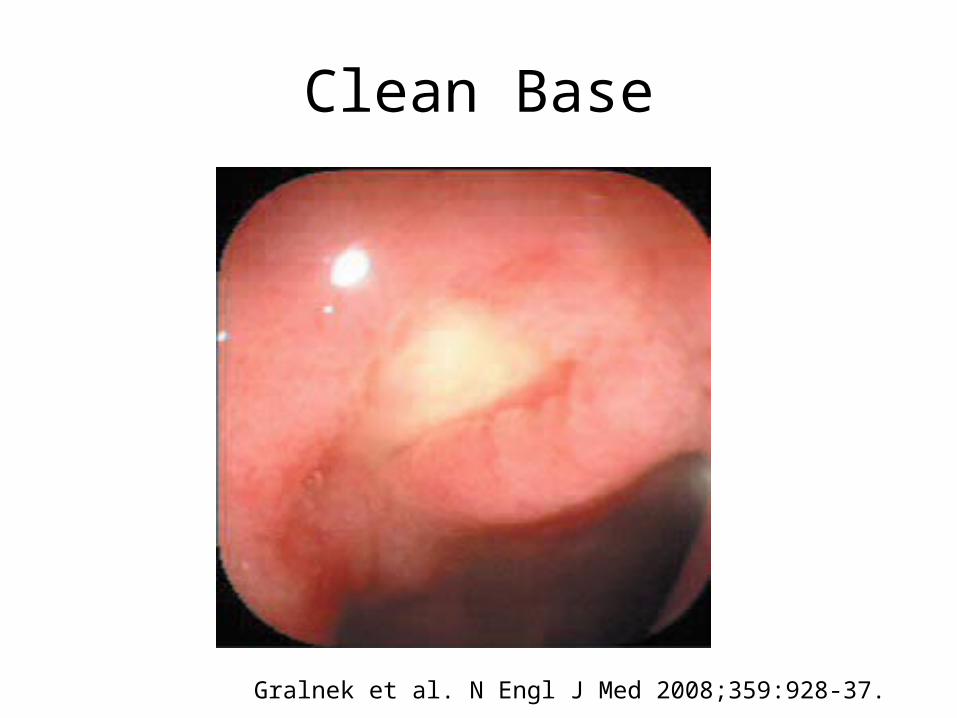
Clean Base
Gralnek et al. N Engl J Med 2008;359:928-37.
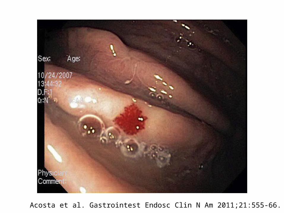
Acosta et al. Gastrointest Endosc Clin N Am 2011;21:555-66.

Acosta et al. Gastrointest Endosc Clin N Am 2011;21:555-66.

Initial Assessment• Always remember to assess A,B,C’s• Assess degree of hypovolemic shock

Pre-endoscopic management
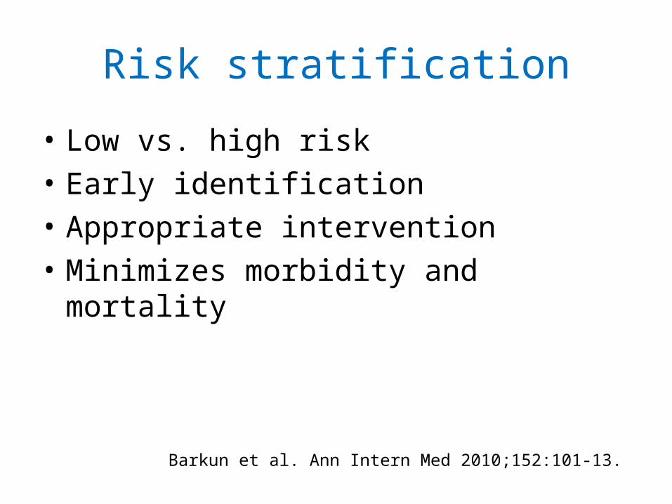
Risk stratification
• Low vs. high risk• Early identification• Appropriate intervention• Minimizes morbidity and mortality
Barkun et al. Ann Intern Med 2010;152:101-13.

Scores

Bardou et al. Nat Rev Gastroenterol Hepatol 2012;9:97-104.
History
Physical
CBC
Urea
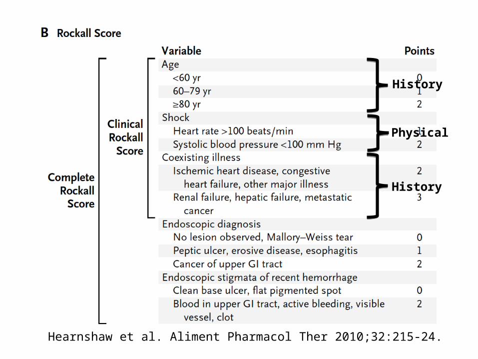
Hearnshaw et al. Aliment Pharmacol Ther 2010;32:215-24.
History
History
Physical

Ms S
• 2 intravenous accesses established and she
• Received crystalloids and was observed in a monitored setting.
Almadi et al. JAMA 2011;306:2367-74.

Laboratory investigations
• Hemoglobin level 7.6 g/dL (compared with 13.7 g/dL a month prior to her presentation)
• White blood cell count 9000/μL• Platelet count of 151,103/μL• INR was 3 • Urea level 21 mmol/L (High)• Electrolyte, creatinine, and liver enzyme levels
were otherwise normal.Almadi et al. JAMA 2011;306:2367-74.
YOU ARE NOT ALONE
Gastroenterology Interventional Rad.
Intensive Care Surgery

IV Fluid Resuscitation

IV Fluid Resuscitation
Then

IV Fluid Resuscitation
Then

IV Fluid Resuscitation
Then

IV Fluid Resuscitation
Then

Blood Transfusion

Blood transfusions
• Should be administered to a patientwith a hemoglobin level of 70 g/L or less
• Rarely indicated when the level is > 100 g/L • Almost always indicated when the level is < 60
g/L. • Target level of 70 to 90 g/L
Barkun et al. Ann Intern Med 2010;152:101-13.

Blood transfusions cont.
• Based on underlying condition hemodynamic status, and markers of tissue hypoxia
• Based on the patient’s risk for complications from inadequate oxygenation
Barkun et al. Ann Intern Med 2010;152:101-13.
Patients receiving anticoagulants
Correction of coagulopathy is recommended
Endoscopy should not be delayed for a high INR unless the INR is supratherapeutic
Barkun et al. Ann Intern Med 2010;152:101-13.

Pre-endoscopic pharmacological therapy

Pre-endoscopy PPI
• Reduces the proportion of patients with high risk endoscopic stigmata (“downstages” lesion)
• Decreases need for endoscopic therapy
• Has not been shown to reduce rebleeding, surgery, or mortality rates
N Engl J Med 2007;356:1631
Endoscopic treatment required:Omeprazole – 19% (23% of PUD)Placebo – 28% (37% of PUD)
High riskHigh risk Low riskLow risk

Ms S
• Transfused with 2 units of packed red blood cells
• Given 5 mg of vitamin K subcutaneously• Transfused with 1000 U of prothrombin
complex concentrate (a mixture of clotting factors II, VII, IX, and X and proteins C and S). She was given 80 mg of pantoprazole intravenously as a bolus followed by an infusion of 8 mg/h while awaiting endoscopy

Endoscopic management

Timing and need for early endoscopy
• Definition of early endoscopy – Ranges from 2 to 24 hours AFTER INITIAL
PRESENTATION
• May need to be delayed or deferred:– Active acute coronary syndromes – Suspected perforation
Barkun et al. Ann Intern Med 2010;152:101-13.

When is Endoscopic Therapy Required?
• ~80% bleeds spontaneously resolve• Endoscopic stigmata of recent hemorrhage
Stigmata Continued/rebleeding rate
Active bleeding 55-90%
Nonbleeding visible vessel 40-50%
Adherent clot Variable, depending on underlying lesion: 0-35%
Flat pigmented spot 7-10%
Clean base < 5%
major


Kovacs et al. Gastrointest Endosc Clin N Am 2011;21:681-96.

Kovacs et al. Gastrointest Endosc Clin N Am 2011;21:681-96.

Kovacs et al. Gastrointest Endosc Clin N Am 2011;21:681-96.

Kovacs et al. Gastrointest Endosc Clin N Am 2011;21:681-96.

Pharmacological therapy

Nonvariceal UGIB –Post-endoscopy management
• Patients with low risk ulcers can be fed promptly, put on oral PPI therapy.
• Patients with ulcers requiring endoscopic therapy should receive PPI gtt x 72 hours– Significantly reduces 30 day rebleeding rate vs
placebo (6.7% vs. 22.5%)– Note: there may not be major advantage with high
dose over non-high dose PPI therapy
N Engl J Med 2000;343:310Arch Intern Med 2010;170:751

• Determine H. pylori status in all ulcer patients• Discharge patients on PPI (once to twice daily),
duration dictated by underlying etiology and need for NSAIDs/aspirin
• In patients with cardiovascular disease on low dose aspirin: restart as soon as bleeding has resolved– RCT demonstrates increased risk of rebleeding (10% v
5%) but decreased 30 day mortality (1.3% v 13%)
Nonvariceal UGIB –Post-endoscopy management
Ann Intern Med 2010;152:1
Not dying is more important than not rebleeding
Not dying is more important than not rebleeding

Management of continued or recurrent bleeding

• Re endoscopy• Angiography • surgery
Percutaneous or transcatheter arterial embolization
• Technical success range from 52% to 98%• Recurrent bleeding in about 10% to 20%• Complications include
– Bowel ischemia– Secondary duodenal stenosis– Gastric, hepatic, and splenic infarction
• A second attempt at endoscopic therapy remains the preferred strategy
Barkun et al. Ann Intern Med 2010;152:101-13.

Angiography
Where available, percutaneous embolization can be considered as an alternative to surgery for patients for whom endoscopic therapy has
failed.
Barkun et al. Ann Intern Med 2010;152:101-13.

H pylori
• Patients with bleeding peptic ulcers should be tested for H. pylori – Receive eradication therapy if present– Confirmation of eradication
• Negative H. pylori diagnostic tests obtained in the acute setting should be repeated
Barkun et al. Ann Intern Med 2010;152:101-13.

Bardou et al. Nat Rev Gastroenterol Hepatol 2012;9:97-104.

When to go to surgery?

Conclusions Resuscitation should be initiated prior to any
diagnostic procedure Gastrointestinal endoscopy allows visualization of
the stigmata, accurate assessment of the level of risk and treatment of the underlying lesion
Intravenous PPI therapy after endoscopy is crucial to decrease the risk of cardiovascular complications and to prevent recurrence of bleeding
Helicobacter pylori testing should be performed in the acute setting
Bardou et al. Nat Rev Gastroenterol Hepatol 2012;9:97-104.

Thank you