gastrointestinal bleeding scintigraphy in the early 21st...
TRANSCRIPT
C O N T I N U I N G E D U C A T I O N
Gastrointestinal Bleeding Scintigraphy in the Early21st Century
Erin Grady1
1Section of Nuclear Medicine, Department of Radiology, Christiana Care Health System, Newark, Delaware
Learning Objectives: On successful completion of this activity, participants should be able to describe (1) diagnostic uses of gastrointestinal bleedingscintigraphy; (2) proper methodology for performing the procedure; (3) the importance of correlative and hybrid imaging; (4) interpretive criteria to make aclinical diagnosis; (5) special considerations in children; and (6) imaging of Meckel diverticula.
Financial Disclosure: The author of this article has indicated no relevant relationships that could be perceived as a real or apparent conflict of interest.
CME Credit: SNMMI is accredited by the Accreditation Council for Continuing Medical Education (ACCME) to sponsor continuing education for physicians.SNMMI designates each JNM continuing education article for a maximum of 2.0 AMA PRA Category 1 Credits. Physicians should claim only creditcommensurate with the extent of their participation in the activity. For CE credit, SAM, and other credit types, participants can access this activity throughthe SNMMI website (http://www.snmmilearningcenter.org) through February 2019.
Gastrointestinal bleeding scintigraphy performed with 99mTc-
labeled autologous erythrocytes or historically with 99mTc-sulfur col-
loid has been a clinically useful tool since the 1970s. This article
reviews the history of the techniques, the different methods of radio-labeling erythrocytes, the procedure, useful indications, diagnostic
accuracy, the use of SPECT/CT and CT angiography to evaluate
gastrointestinal bleeding, and Meckel diverticulum imaging. The
causes of pediatric bleeding are discussed by age.
Key Words: GI bleeding scintigraphy or scan; radiolabeled red
blood cells or erythrocytes; SPECT; SPECT/CT; CTA; pediatric GIbleeding by age; Meckel’s diverticulum
J Nucl Med 2016; 57:252–259DOI: 10.2967/jnumed.115.157289
It was in 1977 that Alavi et al., at the University of Pennsylvania,originally described evaluating gastrointestinal bleeding with scinti-graphic methods using 99mTc-sulfur colloid (1). Also in 1977, at theUniversity of Copenhagen, Miskowiak et al. described the use of99mTc-human serum albumin for gastrointestinal bleeding (2). Sub-sequently, in 1979 at the Massachusetts General Hospital, Winzelberget al. described diagnosing and localizing gastrointestinal bleed-ing with 99mTc-erythrocytes (3). Other techniques for labeling redblood cells with greater efficiency were developed later, but thepractice of evaluating gastrointestinal bleeding with scintigraphictechniques had already become established in clinical practice.Gastrointestinal bleeding is one of the major causes of death in the
United States, with mortality ranging from 10% to 30% (4). In light ofthe significant mortality, timely diagnosis and evaluation are critical, asemergent intervention may be needed. Bleeding may originate
from the upper or lower gastrointestinal tract. Upper gastrointestinalbleeding—bleeding originating in any portion of the tract up to theligament of Treitz at the duodenal flexure—annually affects 50–150of every 100,000 adults (4) and causes 20,000 deaths in the UnitedStates (5). Lower gastrointestinal bleeding originates distal to theligament of Treitz and is fairly common, accounting for approxi-mately 21% of gastrointestinal bleeding overall (6) but is usuallyself-limited. The prevalence of lower gastrointestinal bleeding in-creases by more than 200 times between the ages of 30 and 90 y (7).Approximately 21 of every 100,000 adults in the United Statesrequire hospitalization for lower gastrointestinal bleeding annually(4). Upper and lower gastrointestinal bleeding require differentclinical approaches, but although each has its usual signs thesource of bleeding can be difficult to distinguish clinically. Upperbleeding generally presents with hematemesis (either red or coffee-ground emesis) or melena. Some melena, however, may be related tothe ascending colon. Hematochezia is most often due to a lowerbleed, but a brisk upper bleed could have a similar appearance.Noninvasive imaging and other tests assist the clinician in deter-mining the appropriate treatment.Scintigraphy is indicated for evaluation of overt gastrointestinal
bleeding. Per the SNMMI guidelines for gastrointestinal bleedingscintigraphy (GIBS), the goal of the examination is to determinewhether the patient is actively bleeding, to localize the bleeding bowelsegment, and to estimate the rate of blood loss (7). All of these allowfor treatment planning and risk stratification (8). GIBS does best in themid to lower gastrointestinal tract. Occult bleeding identified byguaiac fecal testing is not an appropriate indication: the microscopicblood; slow, intermittent bleeding; or low-volume blood identified instool is below the scintigraphic detection limit (7).
TECHNIQUE
Before initiation of scanning, it is important to learn more aboutthe patient and the patient’s symptoms. Understanding clinicaldescriptions of gastrointestinal bleeding such as overt or occult ishelpful in elucidating patient history from our clinical colleagues(Table 1) (8). Areas to explore include the clinical signs of gas-trointestinal bleeding; the color of the blood; the physical examinationresults, including rectal examination or nasogastric lavage, if performed;the results of prior imaging, endoscopy, or colonoscopy; whether the
Received Nov. 2, 2015; revision accepted Dec. 10, 2015.For correspondence or reprints contact: Erin Grady, Department of
Radiology, Section of Nuclear Medicine, Christiana Care Health System,4755 Ogletown-Stanton Rd., Newark, DE 19718.E-mail: [email protected] online Dec. 17, 2015.COPYRIGHT © 2016 by the Society of Nuclear Medicine and Molecular
Imaging, Inc.
252 THE JOURNAL OF NUCLEAR MEDICINE • Vol. 57 • No. 2 • February 2016
by on June 18, 2018. For personal use only. jnm.snmjournals.org Downloaded from
patient is stable enough to come to the imaging department; andwhether a portable scanner is available. Good intravenous access iskey; the patient should have 1 or 2 large-bore intravenous lines andfluid resuscitation products available on demand on entry to thenuclear medicine area. It is important to note whether the patienthad any prior bowel or abdominal surgery and whether the surgerywas recent; patients who recently underwent barium evaluationshould be excluded from GIBS as the findings may be obscured(7). Knowing the patient’s medications is helpful, especially whentroubleshooting the cause of poor erythrocyte labeling. A list of med-ications and other substances contributing to poor labeling is availablein Table 2 (9–15).
99mTc-sulfur colloid has a short circulating half-life of 3 min andan equally quick extraction by the reticuloendothelial system (liver,spleen, and bone marrow) (11). Imaging is generally performed for20–30 min with 99mTc-sulfur colloid, decreasing the opportunity tovisualize the classically intermittent lower gastrointestinal bleed. Thehigh level of background activity in the liver and spleen can obscureupper-gastrointestinal bleeding sources. For these reasons, multiplestudies have found 99mTc-erythrocytes to be superior (16–18). Inother countries, 99mTc-human serum albumin diethylenetriami-nepentaacetic acid is used for the diagnosis of gastrointestinalbleeding (19). All these radiopharmaceuticals assist in compart-mental localization. Each of them images the vascular compart-
ment, although some tracers stay in the vascular compartmentlonger than others.There are 3 ways to label erythrocytes: in vivo, modified in vivo,
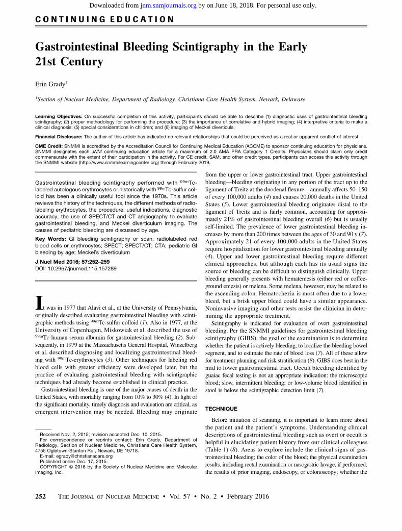
and in vitro. In the in vivo method, no blood is withdrawn from thepatient. The patient receives an intravenous injection of stannouspyrophosphate, which is allowed to circulate for a few minutes,followed by intravenous injection of 99mTc-pertechnetate. This tech-nique is generally not preferred because it has the lowest labelingefficiency, but it is reserved for patients who will not receive bloodproducts for religious reasons (11,20). The modified in vivo method,also known as the “in vivtro” method, begins similarly to the in vivomethod with intravenous injection of stannous pyrophosphate; blood issubsequently withdrawn from the patient and mixed with 99mTc-pertechnetate. This method has a somewhat higher labeling efficiency(11,20). In the in vitro method, blood is withdrawn from the patientand a cold kit containing stannous pyrophosphate and a few othercomponents is used; 99mTc-pertechnetate is added. This methodhas the highest labeling efficiency, which improves the target-to-background ratio and decreases the likelihood that free pertechnetatewill interfere with interpretation of the results. Care must be taken toensure that each patient receives back his or her own radiolabeledblood product.The erythrocyte labeling techniques are summarized in Table 3. All
have one thing in common: use of a redox reaction (Fig. 1) that results
TABLE 1Definitions of Various Types of Gastrointestinal Bleeding (8)
Term Definition
Overt or acute gastrointestinal bleeding Visible bleeding in the form of hematemesis, melena, or hematochezia
Occult or chronic gastrointestinal bleeding Bleeding not apparent to patient and presenting as anemia or on fecal occult
blood testing
Obscure gastrointestinal bleeding Recurrent bleeding of uncertain source after or despite upper or lower endoscopy
Hematemesis Vomiting of blood
Melena Dark, tarry/sticky feces containing partially digested blood
Hematochezia Passage of fresh blood per anus, usually in or within stools
TABLE 2Causes of Interference with Erythrocyte Radiolabeling, Leading to Free 99mTc-Pertechnetate (9–15)
Cause Mechanism of disrupted radiolabeling
Methyldopa Oxidation of stannous ion; decrease in reduction
Hydralazine Oxidation of stannous ion; decrease in reduction
Quinine Possible antibody to red blood cells
Doxorubicin Lowered labeling efficiency in proportion to concentration of drug
Iodinated contrast medium Decrease in stannous reduction; alteration of 99mTc binding
Chocolate Unknown
Tobacco Oxidation of stannous ion, possible damage to red blood cell plasma membrane or
possible chelating action on stannous or pertechnetate ions (mechanism relates to
reactive oxygen species present)
Heparin Formation of complexes with 99mTc-pertechnetate in presence of stannous ion,
causing renal excretion
Too much or too little stannous ion Alteration of 99mTc-pertechnetate reduction
Recent blood transfusion Unknown
Sickled red blood cells Impaired labeling due to abnormal hemoglobin structure
GIB SCINTIGRAPHY IN THE EARLY 21ST CENTURY 253
by on June 18, 2018. For personal use only. jnm.snmjournals.org Downloaded from

in labeling of the erythrocytes at the b-chain of the hemoglobin (3). Inthis redox reaction, Tc71O4
2 is the oxidizing agent and the stannousion (Sn21) is the reducing agent (11,20,21).After the patient’s erythrocytes have been labeled and readminis-
tered, a dynamic acquisition of the abdomen and pelvis is per-formed with a 1282 matrix. The dynamic images are acquired for10–20 s/frame, with an optional initial angiographic phase ob-tained for 1–3 s/frame (7). The dynamic images can be reframedor summed on an as-needed basis to increase the informationdensity per frame. Acquiring static images other than for trou-bleshooting is not recommended, as cine visualization is key tointerpretation (22). If a dual-head g-camera is used for evaluation,acquiring the images with both camera heads may enhance visualiza-tion of a rectal bleed (7). The scanning duration is not standardized butshould be long enough to allow for intermittent bleeding, which is aparticular possibility in the large bowel. An imaging interval of 1–4 h,depending on camera availability, is reasonable (7), although somestudies indicate that 1–2 h would be optimal (23,24).
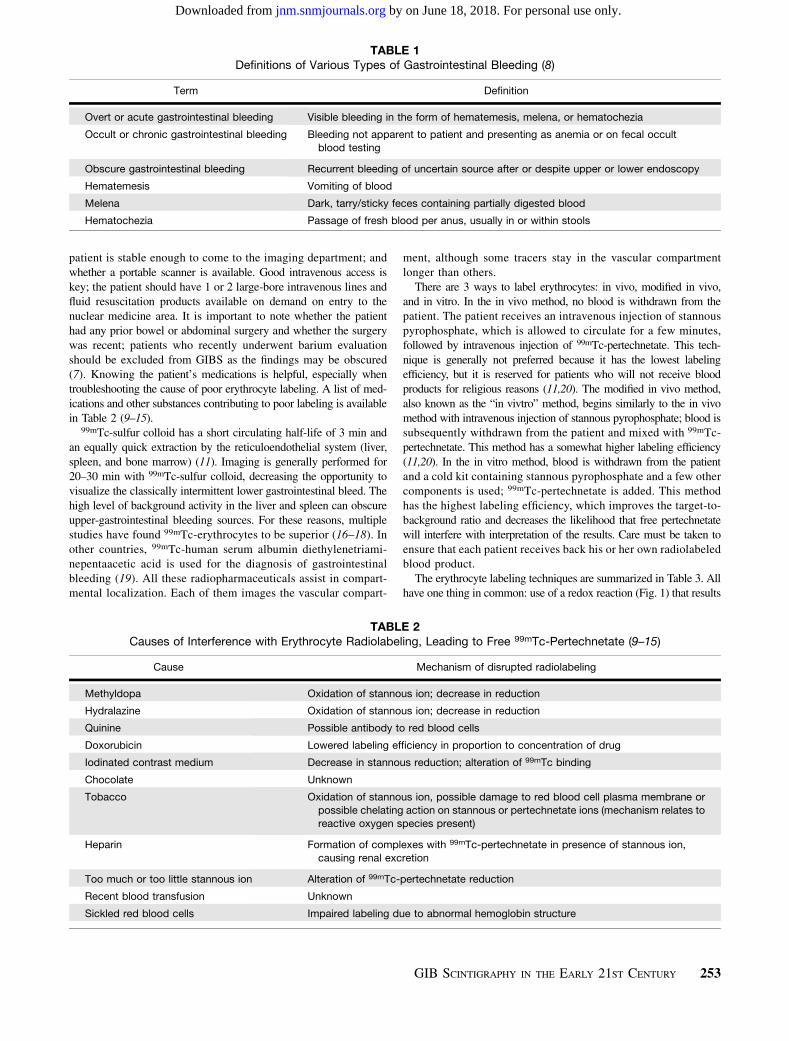
NORMAL BIODISTRIBUTION AND INTERPRETATION
The normal biodistribution of 99mTc-radiolabeled erythrocytes in-cludes the cardiac blood pool, vasculature, liver, spleen, penile circu-lation, and usually mildly in the kidneys and urinary bladder (Fig. 2).The scintigraphic results will be positive only if the patient is
actively bleeding at the time of imaging. To diagnose a gastrointestinal
bleed, 4 criteria need to be met: the focus of extravascular activityshould start in a region where there was no abnormal activitybefore, should increase in intensity over time, should move in eitheran anterograde or a retrograde fashion, and should conform to thebowel (11). When identifying the site of bleeding, it is important toknow the major feeding vessel. The major feeding vessels are de-termined embryologically: perfusing the foregut, midgut, and hind-gut. The portion of the foregut that is visualized on GIBS is thestomach through the second part of the duodenum and is perfusedby the celiac trunk; the branches of the celiac trunk include the leftgastric artery, common hepatic artery, and splenic artery. The mid-gut is perfused by the superior mesenteric artery, from the duodenalpapilla through most of the transverse colon; the branches of thesuperior mesenteric artery include the inferior pancreaticoduodenalartery, intestinal arteries, ileocolic artery, right colic artery, andmiddle colic artery. The hindgut is perfused by the inferior mesen-teric artery, from the remaining transverse colon through the supe-rior portion of the anal canal; the branches of the inferior mesenteric
TABLE 3Methods of Labeling Erythrocytes with 99mTc and Labeling Efficiency (3,11,20,21)
Method Description and considerations Efficiency
In vivo Patient is injected intravenously with 1 mg of stannous pyrophosphate, which
circulates for 20 min, followed by intravenous injection of 555–1,110 MBq of 99mTc-
pertechnetate. This technique is generally not recommended because of its low
labeling efficiency but is reserved for patients who will not receive blood productsfor religious reasons.
75%–80%
Modified in vivo Patient is injected intravenously with 1 mg of stannous pyrophosphate, whichcirculates for 20 min. Vial of blood is mixed with 555–1,110 MBq of 99mTc-
pertechnetate and allowed to incubate for 10 min before intravenous injection into
patient.
85%–90%
In vitro Vial of blood is withdrawn from patient and added to vial containing stannous
pyrophosphate. After 5 min, vial A containing sodium hypochlorite is added to
destroy extracellular Sn21. Vial B containing citrate buffer is then added. 99mTc-
pertechnetate (555–1,110 MBq) is added and incubated before intravenousadministration to patient.
$97%
FIGURE 1. Stannous reduction method (3).
FIGURE 2. Normal biodistribution of 99mTc-labeled erythrocytes.
Heart (H), vascular structures (V), liver (L), spleen (S), and penis (P) are
labeled. We see no intraluminal activity to suggest presence of active
gastrointestinal bleed.
254 THE JOURNAL OF NUCLEAR MEDICINE • Vol. 57 • No. 2 • February 2016
by on June 18, 2018. For personal use only. jnm.snmjournals.org Downloaded from

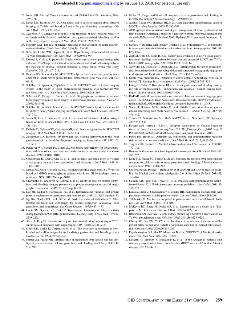
artery include the left colic artery, sigmoid branches, and superiorrectal artery (11,25). Examples of bleeding at these 3 major sites areillustrated in Figures 3–5. Localizing the main arterial distributionis helpful when planning angiographic intervention. Variceal bleed-ing is also an important finding but does not have an arterial source.Some institutions perform delayed dynamic imaging, which may
not accurately localize the origin of bleeding unless the 4 criteria aremet. Blood urea nitrogen and creatinine tests can be helpful, as ablood urea nitrogen–to–creatinine ratio of 25 or greater indicates agreater likelihood of a positive result on delayed imaging (26).Some work has suggested a prognostic benefit to delayed GIBS(27). Digital subtraction has been found helpful in this setting (7).
PITFALLS AND PEARLS
Several potential false-positive findings can occur in GIBS. Redblood cells can localize at a site other than that of the worrisomegastrointestinal bleeding, such as in the case of splenosis, pancreaticpseudocysts, or nonenteric bleeding/hematoma (7,8,28). Otherphysiologic activity can occasionally confuse the interpreter; thisactivity is usually fixed. Previously reported sources of error includerenal activity from a morphologically normal kidney, a transplantedor horseshoe kidney, urine in the bladder or urine contamination,a urinary diversion, a dilated abdominal aorta, bowel ischemia,hepatic hemangioma, vascular collaterals such as caput medusaor dilated mesenteric veins, angiodysplasia, the left ovarian ar-tery and gallbladder in patients with renal failure, the penis, theuterus, and a uterine leiomyoma (7,20). If the patient has hadrecent bowel or other abdominal surgery, prominent activity canbe seen related to normal postoperative hyperemia. Inflammatorybowel diseases such as Crohn disease, a diverticular abscess, andhypervascular neoplasms may also make interpretation difficult (7,20).Relying on all 4 criteria to positively diagnose a gastrointestinal
bleed will often clarify whether the patient has active gastrointestinal
bleeding or another process masquerading as bleeding. The trueetiology of certain findings may be clarified by performing staticimaging in the lateral or oblique projection, SPECT, or SPECT/CTor by correlating the images with a prior CT scan. In particular, staticimages of the neck with attention to the thyroid and salivary glandsare often obtained to exclude the presence of free 99mTc-pertechnetate
FIGURE 3. Example of bleeding originating from branch of celiac ar-
tery. Focus of increasing intensity is identified in upper abdomen, moves
in anterograde fashion, and conforms to bowel. Distribution of focus is
suggestive of gastric bleed (arrows).
FIGURE 4. Example of bleeding originating from branch of superior
mesenteric artery. Focus of increasing intensity is identified in lower
abdomen at midline (arrows) and shows anterograde and retrograde
movement conforming to bowel lumen. Focus crosses midline several
times and thus is most compatible with small-bowel bleed.
FIGURE 5. Example of bleeding originating from branch of inferior
mesenteric artery. Focus of increasing intensity is identified in left upper
quadrant and shows anterograde movement. Given its distribution in
periphery of abdomen (arrows), focus is typical of large-bowel bleed
originating in descending colon.
GIB SCINTIGRAPHY IN THE EARLY 21ST CENTURY 255
by on June 18, 2018. For personal use only. jnm.snmjournals.org Downloaded from

if it is suspected on the basis of abdominal or pelvic GIBS. In patientswho are being treated with exogenous thyroid hormone or who re-ceived prior treatment with 131I-sodium iodide or thyroid suppression,the thyroid may not be visualized despite the presence of free per-technetate. Similarly, salivary activity can be reduced by a host ofmedical conditions, medications, or prior therapies (e.g., Sjögren syn-drome, Parkinson disease, antihistamines, diuretics, antipsychotics,chemotherapy, prior surgical disruption, external-beam radiationtherapy, and 131I-sodium iodide therapy). Areas of free pertechnetateseen on scintigraphic images include the stomach, salivary glands,thyroid gland, and choroid plexus (29). A lateral image of the pelviscan be helpful to exclude the presence of a rectal bleed and clarifywhat may be only physiologic penile activity.
SPECT AND SPECT/CT
In addition to clarifying unclear sites of radiolabeled erythrocytes,SPECT or SPECT/CT has been applied to further define the locationof the bleeding—useful in therapeutic planning, particularly angiog-raphy or surgery. When this technique is used regularly, dynamicimages are generally performed for 10- to 15-min intervals andchecked. When a suggestive focus of labeled erythrocytes is identi-fied, SPECT or SPECT/CT can be performed (30). The SPECT/CTscan can be shortened to a 15-min acquisition to arrive at a morerapid diagnosis (7). SPECT/CT has also been found helpful inbleeding sites that are difficult to localize (31–35). In particular,investigators have found that SPECT or SPECT/CT can increasethe sensitivity and specificity of bleeding-site localization (30).Accurate localization can help to streamline therapy and achievean earlier therapeutic response. In the event of rapid intraluminalgastrointestinal bleeding during the SPECT/CT examination, local-ization might be impaired. Referencing the SNMMI procedureguideline for SPECT/CT imaging is recommended (36).
SENSITIVITY OF GIBS
GIBS will detect a bleeding rate of 0.05–0.2 mL/min (3,37). Thesensitivity of GIBS has been reported to be 93% and the specificity95% (16,17), although some investigators have cited lower rates(38,39). The variation in sensitivity is likely related to the lack of agold standard, but detection rates clearly increase when the study isperformed as intended—when the patient is actively bleeding.There is research indicating which characteristics of positive
GIBS can be predictive of positivity on a subsequent angiographicexamination. Mehta et al. found that positive GIBS within 12 minof scanning correlated with a positive angiogram (40). Chamarthyet al. found that visualization of bleeding early, within the firstframe of imaging, correlated best with a positive angiogram (41).Prompt performance of GIBS, early time to positivity on GIBS,
relative intensity of activity, and prompt performance of angiogra-phy subsequent to GIBS were found to lead to a positive angiogramby Lee et al. (42). Ng et al. found that “immediate” visualizationof bleeding on GIBS, defined as 2 min or less, was associatedwith a positive angiogram (43). In addition, Gupta et al. found thatgrading the intensity of bleeding may be helpful in predicting an-giographic positivity (44). To provide a patient with the best of care,timely evaluation by angiography is important after obtaining GIBSresults positive for a gastrointestinal bleed.
GIBS FOR SURGICAL PLANNING
Several articles have evaluated GIBS in the setting of surgicalplanning. The opinions are mixed: some reported GIBS to be effec-tive as the sole means for surgical planning (45,46), others suggestedthat it is useful only as a screening tool before angiography (47), andstill others called it “useless” for surgical planning (48). Given thisdisagreement, it is uncertain how well GIBS performs for surgicalplanning in all centers. None of these papers evaluated surgical plan-ning with GIBS SPECT/CT, which enhances localization, sensitivity,and specificity. This could be a topic for further investigation.
CT ANGIOGRAPHY (CTA) VERSUS GIBS
Some centers are starting to move away from GIBS and towardCTA for evaluation of active gastrointestinal bleeding. In CTA, 3discrete CT scans of the abdomen and pelvis are generallyacquired: unenhanced phase (some institutions do not obtain thisphase), contrast-enhanced arterial-phase, and delayed-phase. Wa-ter and hyperdense oral contrast agent are avoided. The CT slicesare quite thin, on the order of 1–2 mm (49). When referencingappropriateness criteria for lower gastrointestinal bleeding, bothGIBS and CTA are ranked similarly according to the AmericanCollege of Radiology (50).Although there is controversy about whether GIBS or CTA is
best, many have attributed the rapid adoption of CTA to severalfactors, including faster diagnosis in patients who are clinicallydeteriorating (51), ability to delineate congenital and other vascu-lar abnormalities that could affect the angiographic approach (52),greater diagnostic accuracy (53), and a relatively similar reportedsensitivity of 0.3–1 mL of extravasated blood (54). Some draw-backs include higher cost and limited time of evaluation (55).Although the Appropriateness Criteria of the American College ofRadiology rate the relative radiation level of absorbed dose to thepatient as the same for GIBS and CTA, the radiation dose to thepatient is higher with CTA (Table 4) (56). The iodinated contrastload is also greater (there is no nephrotoxic aspect to GIBS),although with greater localization less contrast agent might beused on a more selective angiographic evaluation or intervention.
TABLE 4Comparison of Whole-Body Absorbed Radiation Dose Evaluated by GIBS vs. CTA (56,72)
Technique Dose (mSv)
Pediatric GIBS with 80–784 MBq of 99mTc-labeled red blood cells 0.559–5.488
Adult GIBS with 555–1,110 MBq of 99mTc-labeled red blood cells 3.885–7.77
CTA protocoled for gastrointestinal bleeding without initial unenhanced CT phase 18.2–28*
CTA protocoled for gastrointestinal bleeding with initial unenhanced CT phase 26.8–42*
*Iterative reconstruction CT will be lower.
256 THE JOURNAL OF NUCLEAR MEDICINE • Vol. 57 • No. 2 • February 2016
by on June 18, 2018. For personal use only. jnm.snmjournals.org Downloaded from

Both false-positives and false-negatives can be seen in the settingof GIBS and CTA (7,57).
PEDIATRIC GIBS
Upper gastrointestinal bleeding has several different etiologiesthat vary by location of bleeding and age of patient: in newborns to1-mo-olds, it can be related to milk protein sensitivity, coagulopathy,stress gastritis or ulcer, vitamin K deficiency, swallowed maternalblood, or vascular anomaly; in 1- to 2-mo-olds, to stress gastritisor ulcer, acid-peptic disease, gastrointestinal duplications, gastricor esophageal varices, duodenal or gastric webs, bowel obstruc-tion, or vascular anomaly; and in children older than 2 mo andadolescents, to acid-peptic disease, caustic ingestion, bowel obstruc-tion, Crohn disease, Dieulafoy lesions, or Mallory-Weiss tears (58).Lower gastrointestinal bleeding also has different etiologies by age.
In newborns to 1-mo-olds, it can be related to necrotizing enteroco-litis, allergic proctocolitis, Hirschsprung disease, hemorrhagic disease,or malrotation with volvulus; in 1- to 2-mo-olds, to anal fissure,infectious colitis, allergic proctocolitis, Meckel diverticulum, Hirsch-sprung disease, intestinal duplication, lymphonodular hyperplasia, andintussusception; in 2- to 5-y-olds, to anal fissures, infectious colitis,polyps, Meckel diverticulum, Henoch-Schönlein purpura, hemolyticuremic syndrome, or lymphonodular hyperplasia; and in childrenolder than 5 y, to anal fissures, infectious colitis, polyps, inflam-matory bowel disease, and Henoch-Schönlein purpura (58).The recommended administered activity is based on the Euro-
pean Association of Nuclear Medicine pediatric dosage card, whichuses a baseline activity of 56 MBq multiplied by a weight-basedfactor and a minimum administered activity of 80 MBq (7,59). Theimaging technique otherwise generally follows that for adults.GIBS is preferred in the pediatric population because of the
lower absorbed radiation dose, and CTA should be avoided (60). IfSPECT/CT is used, the milliampere-seconds settings should beappropriate for patient size and age (34).
MECKEL DIVERTICULUM IMAGING
In 1809, Johan Friedrick Meckel first comprehensively describedthe diverticulum that was subsequently to be named for him (26).Bleeding from a Meckel diverticulum can potentially occur at anyage but is far more common in children. We recall the “rule of 2s”for Meckel diverticulum: 2 feet from the end of the small intestine,2 inches in length, 2% of the population, 2 times more common inmales, and presenting in the first 2 decades of life and often within
the first 2 years. The most common congenital cause of a Meckeldiverticulum is failed closure of the omphalomesenteric duct, mostcommonly in the distal ileum. Approximately 10%–60% of thesediverticula contain ectopic mucosa: most commonly gastric but alsopancreatic or duodenal (61). Irritation from the gastric acid andpepsin produced in an ectopic location will lead to bleeding (62).Other lesions with ectopic mucosa can include enteric duplications,gastrogenic cysts, and duplication cysts (63).In preparation for the examination, the patient should fast for
3–4 h for best sensitivity, although this is not required (64). 99mTc-pertechnetate is the radiopharmaceutical of choice as it localizesto gastric mucosa. This agent’s mechanism of localization is viaboth the parietal cells and the mucin-secreting cells of the gastricmucosa (11). A Meckel scan assists in localizing the abnormaltissue in preparation for surgical removal. Per the North AmericanConsensus Guidelines for pediatric radiopharmaceutical adminis-tered doses, the dosing of 99mTc-pertechnetate is recommended at1.85 MBq/kg, with a minimum of 9.25 MBq (65). There are a fewcold pharmaceuticals that can be used to enhance visualization ofa Meckel diverticulum (Table 5) (11,61).
TABLE 5Pharmaceuticals that Augment Visualization of Meckel Diverticulum (11,58)
Pharmaceutical Dosing/timing Effect
Cimetidine (other H2 blockers, such as
famotidine, ranitidine, or proton pump
inhibitors, can also be used but have
different dosing)
20 mg/kg/d orally for 2 d in children or
10–20 mg/kg/d for 2 d in neonates
Inhibits release of 99mTc-pertechnetate
by intraluminal cells, thus increasing
and prolonging uptake
Glucagon 50 μg/kg intravenously 10 min after
administration of 99mTc-pertechnetate
Slightly reduces gastric activity of 99mTc-
pertechnetate and suppressesperistaltic activity
Pentagastrin (no longer recommended inUnited States secondary to side
effects)
6 μg/kg subcutaneously 20–30 minbefore 99mTc-pertechnetate
administration
Increases gastric mucosal uptake of99mTc-pertechnetate, thus increasing
target-to-background ratio
FIGURE 6. Typical imaging appearance of Meckel diverticulum. Focus
(arrows) increases in intensity over time, similar to level of gastric uptake.
GIB SCINTIGRAPHY IN THE EARLY 21ST CENTURY 257
by on June 18, 2018. For personal use only. jnm.snmjournals.org Downloaded from

Images are acquired dynamically at a frame rate of 30–60 s witha 1282 matrix, lasting for a minimum of 30 min. Imaging for60 min can be performed when clinical suspicion is high andthe initial 30-min images are negative (61). Postvoiding imagesor a urinary catheter can be helpful should the diverticulum beobscured by excreted activity in the urinary bladder. A dose offurosemide (1 mg/kg intravenously) may assist in clearing a prom-inent renal collecting system or ureter (61).On imaging, a Meckel diverticulum is identified as a focus of
activity in the lower abdomen or upper pelvis that generally appears atthe time of stomach visualization and increases as stomach activityincreases (Fig. 6) (11,20). False-positive results can occur from in-tussusception, volvulus, abscess, appendicitis, neoplasm, angiodyspla-sia, Crohn disease, ulcerative colitis, or other forms of colitis (66–70).Table 6 provides more information on gastrointestinal and nongas-trointestinal causes of false-positive results on Meckel scans.SPECT/CT has been described as a troubleshooting technique in
Meckel diverticulum, excluding artifacts and assisting in surgicalplanning (71). Once again, SPECT/CT should be performed with aCT amperage appropriate for patient size and age (34,61).
CONCLUSION
Scintigraphic imaging of gastrointestinal bleeding remains impor-tant in nuclear medicine. The current techniques and interventionshave been described in this review. Because gastrointestinal bleedingcan be lethal in certain instances, prompt evaluation and direction ofcare is important. Nuclear medicine can contribute significantly topatient management.
REFERENCES
1. Alavi A, Dann RW, Baum S, et al. Scintigraphic detection of acute gastrointes-
tinal bleeding. Radiology. 1977;124:753–756.
2. Miskowiak J, Munch O, Nielsen SL, et al. Abdominal scintiphotography with99mtechnetium-labeled albumin in acute gastrointestinal bleeding: an experimental
study and a case-report. Lancet. 1977;2:852–854.
3. Winzelberg GG, McKusick KA, Strauss HW, et al. Evaluation of gastrointestinal
bleeding by red blood cells labeled in vivo with technetium-99m. J Nucl Med.
1979;20:1080–1086.
4. Jairath V, Hearnshaw S, Brunskill SJ, et al. Red cell transfusion for the
management of upper gastrointestinal haemorrhage. Cochrane Database Syst
Rev. 2010;9:CD006613.
5. El-Tawil AM. Trends on gastrointestinal bleeding and mortality: where are we
standing? World J Gastroenterol. 2012;18:1154–1158.
6. Navuluri R, Kang L, Patel J, et al. Acute lower gastrointestinal bleeding. Semin
Intervent Radiol. 2012;29:178–186.
7. Dam HQ, Brandon DC, Graham VV, et al. The SNMMI procedure standard/
EANM practice guideline for gastrointestinal bleeding scintigraphy 2.0. J Nucl
Med Technol. 2014;42:308–317.
8. Kim BSM, Li BT, Engel A, et al. Diagnosis of gastrointestinal bleeding: a
practical guide for clinicians.World J Gastrointest Pathophysiol. 2014;5:467–478.
9. Drug–radiopharmaceutical drug interactions. University of Arkansas for Medical
Sciences website. http://nuclearpharmacy.uams.edu/resources/Interactions.
htm. Accessed December 14, 2015.
10. Braga AC, Oliveria MB, Feliciano GD, et al. The effects of drugs on the labeling
of blood elements with technetium-99m. Curr Pharm Des. 2000;6:1179–1191.
11. Ziessman H, O’Malley J, Thrall J. Nuclear Medicine: The Requisites. 4th ed.
Philadelphia, PA: Elsevier Saunders; 2013:307–321.
12. Vidal MV, Gutfilen B, da Fonseca LM, et al. Influence of tobacco on the labeling
of red blood cells and plasma proteins with technetium-99m. J Exp Clin Cancer
Res. 1998;17:41–46.
13. Bustani H, Colavolpe C, Imbert-Joscht I, et al. Chocolate intake associated with
failed labeling of 99mTc red blood cells. J Nucl Med Technol. 2009;37:107–110.
14. Kawabe J, Higashiyama S, Torli K, et al. Poor labeling of Tc-99m red blood cells
in vivo in a radionuclide intestinal bleeding study of a patient who had recently
undergone frequent blood transfusions. Clin Nucl Med. 2003;28:911–912.
15. Sampson CB. Complications and difficulties in radiolabelling blood cells: a re-
view. Nucl Med Commun. 1996;17:648–658.
16. Siddiqui AR, Schauwecker DS, Wellman HN, et al. Comparison of technetium-99m
sulfur colloid and in vitro labeled technetium-99m RBCs in the detection of gastro-
intestinal bleeding. Clin Nucl Med. 1985;10:546–549.
17. Bunker SR, Lull RJ, Tanasescu DE, et al. Scintigraphy of gastrointestinal hem-
orrhage: superiority of Tc-99m red blood cells over Tc-99m sulfur colloid. AJR.
1984;143:543–548.
18. Bunker SR, Brown JM, McAuley RJ, et al. Detection of gastrointestinal bleeding
sites: use of in vitro technetium Tc99m-labeled RBCs. JAMA. 1982;247:789–792.
19. Kotani K, Kawabe J, Higashiyama S, et al. Diagnostic ability of 99mTc-HAS-
DTPA scintigraphy in combination with SPECT/CT for gastrointestinal bleeding.
Abdom Imaging. 2014;39:677–684.
20. Henkin RE, Bova D, Dillehay GL, et al. Nuclear Medicine. 2nd ed. Philadelphia,
PA: Mosby Elsevier. 2006:988–993.
21. Ultratag RBC package insert. Mallinckrodt website. http://www2.mallinckrodt.
com/WorkArea/DownloadAsset.aspx?id5654. Published October 2015. Ac-
cessed December 11, 2015.
22. Maurer AH. Gastrointestinal bleeding and cine-scintigraphy. Semin Nucl Med.
1996;26:43–50.
23. Winzelberg GG, Froelich JW, McKusick KA, et al. Radionuclide localization of
lower gastrointestinal hemorrhage. Radiology. 1981;139:465–469.
24. Markisz JA, Front D, Royal HD, et al. An evaluation of Tc-99m-labeled red
blood cell scintigraphy for the detection and localization of gastrointestinal
bleeding sites. Gastroenterology. 1982;83:394–398.
TABLE 6False-Positive Results on Meckel Scanning (11,58)
Related to gastrointestinal tract Not related to gastrointestinal tract
Peptic ulcer Hydronephrosis
Barrett esophagus Aneurysm of abdominal vessel
Retained gastric antrum Calyceal diverticulum
Duplication cyst of ileum Anterior sacral meningomyelocele
Small-bowel obstruction Hemangioma
Appendicitis Lymphoma
Intussusception Ectopic kidney
Inflammatory bowel diseases (e.g., Crohndisease or ulcerative colitis)
Recent laparoscopic surgery (hyperemia at periumbilicalport site)
Carcinoid of small bowel
Volvulus
Small-bowel bleeding not related to Meckel diverticulum
258 THE JOURNAL OF NUCLEAR MEDICINE • Vol. 57 • No. 2 • February 2016
by on June 18, 2018. For personal use only. jnm.snmjournals.org Downloaded from
25. Netter FH. Atlas of Human Anatomy. 6th ed. Philadelphia, PA: Saunders; 2014:
259–271.
26. Lewis DH, Jacobson AF. BUN/Cr ratios: aid to decision making about delayed
imaging in Tc-99m red blood cell scans for gastrointestinal hemorrhage. Clin
Nucl Med. 1998;23:201–204.
27. Jacobson AF. Cerqueria: prognostic significance of late imaging results in
technetium-99m-labeled red blood cell gastrointestinal bleeding studies
with early negative images. J Nucl Med. 1992;33:202–207.
28. Howarth DM. The role of nuclear medicine in the detection of acute gastroin-
testinal bleeding. Semin Nucl Med. 2006;36:133–146.
29. Stern SS, Leslie WD, Dupont JO, et al. Hepatobiliary clearance of intravenous
Tc-99m pertechnetate. Clin Nucl Med. 1995;20:962–964.
30. Dolezal J, Vizda J, Kopacova M. Single-photon emission computed tomography
enhanced Tc-99m-pertechnetate disodium-labeled red blood cell scintigraphy in
the localization of small intestine bleeding: a single-centre twelve-year study.
Digestion. 2011;84:207–211.
31. Bentley BS, Tulchinsky M. SPECT/CT helps in localization and guiding man-
agement of small bowel gastrointestinal hemorrhage. Clin Nucl Med. 2014;39:
94–96.
32. Schillaci O, Spanu A, Tagliabue L, et al. SPECT/CT with a hybrid imaging
system in the study of lower gastrointestinal bleeding with technetium-99m
red blood cells. Q J Nucl Med Mol Imaging. 2009;53:281–289.
33. Schillaci O, Filippi L, Danieli R, et al. Single-photon emission computed
tomography/computed tomography in abdominal diseases. Semin Nucl Med.
2007;37:48–61.
34. Schillaci O, Danieli R, Manni C, et al. Is SPECT/CTwith a hybrid camera useful
to improve scintigraphic imaging interpretation? Nucl Med Commun. 2004;25:
705–710.
35. Yama N, Ezoe E, Kimura Y, et al. Localization of intestinal bleeding using a
fusion of Tc-99m-labeled RBC SPECT and x-ray CT. Clin Nucl Med. 2005;30:
488–489.
36. Delbeke D, Coleman RE, Guiberteau MJ, et al. Procedure guideline for SPECT/CT
imaging 1.0. J Nucl Med. 2006;47:1227–1234.
37. Zuckerman DA, Bocchini TP, Birnbaum EH. Massive hemorrhage in the lower
gastrointestinal tract in adults: diagnostic imaging and intervention. AJR. 1993;161:
703–711.
38. Pennoyer WP, Vignati PV, Cohen JL. Mesenteric angiography for lower gastro-
intestinal hemorrhage: are there any predictors for a positive study? Dis Colon
Rectum. 1997;40:1014–1018.
39. Gunderman R, Leef J, Ong K, et al. Scintigraphic screening prior to visceral
arteriography in acute lower gastrointestinal bleeding. J Nucl Med. 1998;39:
1081–1083.
40. Mehta AS, Amin A, Masse N, et al. Predicting positive angiograms by 99mTc-red
blood cell (RBC) scintigraphy in patients with lower GI hemorrhage: time to
positivity. JVIR. 2015;26(suppl):S102.
41. Chamarthy M, Shapoval A, Golowa Y, et al. Utility of positive nuclear gastro-
intestinal bleeding imaging parameters to predict subsequent successful angio-
graphic localization. JVIR. 2011;22(suppl):S31.
42. Lee JH, Rashid S, Hargreaves EL, et al. Differentiating variables that predict
positive angiograms in gastrointestinal hemorrhage. JVIR. 2013;24(suppl):S137.
43. Ng DA, Opelka FG, Beck DE, et al. Predictive value of technetium Tc 99m-
labeled red blood cell scintigraphy for positive angiogram in massive lower
gastrointestinal hemorrhage. Dis Colon Rectum. 1997;40:471–477.
44. Gupta SM, Spencer RP, Chak SP. Significance of intensity of delayed activity
during technetium-99m-RBC gastrointestinal bleeding study. J Nucl Med. 1991;32:
2249–2252.
45. Alavi A, Ring EJ. Localization of gastrointestinal bleeding: superiority of 99mTc
sulfur colloid compared with angiography. AJR. 1981;137:741–748.
46. Dusold R, Burke K, Carpentier W, et al. The accuracy of technetium-99m-
labeled red cell scintigraphy in localizing gastrointestinal bleeding. Am J
Gastroenterol. 1994;89:345–348.
47. Hunter JM, Pezim ME. Limited value of technetium 99m-labeled red cell scin-
tigraphy in localization of lower gastrointestinal bleeding. Am J Surg. 1990;159:
504–506.
48. Miller TA. Tagged red blood cell imaging to localize gastrointestinal bleeding: is
it really that helpful? Gastroenterology. 1992;102:733.
49. Jaeckle T, Stuber G, Hoffman HK, et al. Acute gastrointestinal bleeding: value of
MDCT. Abdom Imaging. 2008;33:285–293.
50. ACR appropriateness criteria: radiologic management of lower gastrointestinal
tract bleeding. American College of Radiology website. https://acsearch.acr.org/
docs/69457/Narrative/. Published 2006. Updated 2014. Accessed December 11,
2015.
51. Geffroy Y, Rodallec MH, Boulay-Coletta I, et al. Multidetector CT angiography
in acute gastrointestinal bleeding: why, when and how. Radiographics. 2011;31:
E35–E46.
52. Zink SI, Ohki SK, Stein B, et al. Noninvasive evaluation of active lower gastro-
intestinal bleeding: comparison between contrast enhanced MDCT and 99mTc-
labeled RBC scintigraphy. AJR. 2008;191:1107–1114.
53. Jacovides CL, Nadolski G, Allen SR, et al. Arteriography for lower gastrointes-
tinal hemorrhage role of preceding abdominal computed tomographic angiogram
in diagnosis and localization. JAMA Surg. 2015;150:650–656.
54. Kuhle WG, Shelman RG. Detection of active colonic hemorrhage with use of
helical CT: findings in a swine model. Radiology. 2003;228:743–752.
55. Laing CJ, Tobias T, Rosenblum DI, et al. Acute gastrointestinal bleeding: emerg-
ing role of multidetector CT angiography and review of current imaging tech-
niques. Radiographics. 2007;27:1055–1070.
56. RADAR medical procedure radiation dose calculator and consent language gen-
erator. The RAdiation Dose Assessment Resource website. http://www.doseinfo-
radar.com/RADARDoseRiskCalc.html. Accessed December 11, 2015.
57. Stuber T, Hoffman MHK, Stuber G, et al. Pitfalls in detection of acute gastro-
intestinal bleeding with multi-detector row helical CT. Abdom Imaging. 2009;34:
476–482.
58. Treves ST. Pediatric Nuclear Medicine/PET. 3rd ed. New York, NY: Springer;
2007:192–208.
59. Dosage card (version 1.2.2014). European Association of Nuclear Medicine
website. http://www.eanm.org/docs/EANM_Dosage_Card_040214.pdf?
PHPSESSID5nb6bln4nl6sdts2632a5qrop60. Accessed December, 2015.
60. Fahey FH, Treves ST, Adelstein SJ. Minimizing and communicating radiation
risk in pediatric nuclear medicine. J Nucl Med Technol. 2012;40:13–24.
61. Turgeon DK, Bennet JL. Meckel’s diverticulum. Am J Gastroenterol. 1990;85:
777–781.
62. Brayton D. Gastrointestinal bleeding of unknown origin. Am J Dis Child. 1964;107:
288–292.
63. Kong MS, Huang SC, Tzen KY, Lin JN. Repeated technetium-99m pertechnetate
scanning for children with obscure gastrointestinal bleeding. J Pediatr Gastro-
enterol Nutr. 1994;18:284–287.
64. Spottswood SE, Pfluger T, Bartold SP, et al. SNMMI and EANM practice guide-
line for Meckel diverticulum scintigraphy 2.0. J Nucl Med Technol. 2014;42:
163–169.
65. Gelfand MJ, Parisi MT, Treves ST, et al. Pediatric radiopharmaceutical admin-
istered doses: 2010 North American consensus guidelines. J Nucl Med. 2011;52:
318–322.
66. Lunia S, Lunia C, Chandramouly B, Chodos RB. Radionuclide meckelogram with
particular reference to false-positive results. Clin Nucl Med. 1979;4:285–288.
67. Tulchinsky M. Meckel’s scan: pitfall in patients with active small bowel bleed-
ing. Clin Nucl Med. 2006;31:814–816.
68. McKevitt EC, Baerg JE, Nadel HR, et al. Laparoscopy as a cause of a false-
positive Meckel’s scan. Clin Nucl Med. 1999;24:102–104.
69. Blackmon KN, Rao AG. Ectopic kidney mimicking a Meckel’s diverticulum on
Tc-99m pertechnetate scan. Clin Nucl Med. 2011;36:e228–e230.
70. Cherng SC, Fan YM, Yu CY, et al. Incidental accumulation of technetium-99m
pertechnetate in pediatric Burkitt’s lymphoma with tumor-induced intussuscep-
tion. Clin Nucl Med. 2004;29:264–265.
71. Papathanassiou D, Leihn JC, Meneroux B, et al. SPECT-CT of Meckel divertic-
ulum. Clin Nucl Med. 2007;32:218–220.
72. Kulkarni C, Moorthy S, Sreekumar K, et al. In the workup of patients with
obscure gastrointestinal bleed, does 64-slice MDCT have a role? Indian J Radiol
Imaging. 2012;22:47–53.
GIB SCINTIGRAPHY IN THE EARLY 21ST CENTURY 259
by on June 18, 2018. For personal use only. jnm.snmjournals.org Downloaded from

Doi: 10.2967/jnumed.115.157289Published online: December 17, 2015.
2016;57:252-259.J Nucl Med. Erin Grady Gastrointestinal Bleeding Scintigraphy in the Early 21st Century
http://jnm.snmjournals.org/content/57/2/252This article and updated information are available at:
http://jnm.snmjournals.org/site/subscriptions/online.xhtml
Information about subscriptions to JNM can be found at:
http://jnm.snmjournals.org/site/misc/permission.xhtmlInformation about reproducing figures, tables, or other portions of this article can be found online at:
(Print ISSN: 0161-5505, Online ISSN: 2159-662X)1850 Samuel Morse Drive, Reston, VA 20190.SNMMI | Society of Nuclear Medicine and Molecular Imaging
is published monthly.The Journal of Nuclear Medicine
© Copyright 2016 SNMMI; all rights reserved.
by on June 18, 2018. For personal use only. jnm.snmjournals.org Downloaded from