gatingofagprotein-sensitivemammaliankir3.1prokaryotic ...kir3.1 chimera in planar lipid bilayers and...
TRANSCRIPT
Gating of a G protein-sensitive Mammalian Kir3.1 ProkaryoticKir Channel Chimera in Planar Lipid Bilayers*□S
Received for publication, June 3, 2010, and in revised form, September 13, 2010 Published, JBC Papers in Press, October 11, 2010, DOI 10.1074/jbc.M110.151373
Edgar Leal-Pinto‡1, Yacob Gomez-Llorente§1,2, Shobana Sundaram‡1, Qiong-Yao Tang‡,Tatyana Ivanova-Nikolova‡, Rahul Mahajan‡3, Lia Baki‡, Zhe Zhang‡, Jose Chavez§, Iban Ubarretxena-Belandia§4,and Diomedes E. Logothetis‡5
From the ‡Department of Physiology and Biophysics, School of Medicine, Virginia Commonwealth University, Richmond, Virginia23298 and the §Department of Structural and Chemical Biology, Mount Sinai School of Medicine, New York, New York 10029
Kir3 channels control heart rate and neuronal excitabilitythrough GTP-binding (G) protein and phosphoinositide sig-naling pathways. These channels were the first characterizedeffectors of the �� subunits of G proteins. Because we cur-rently lack structures of complexes between G proteins andKir3 channels, their interactions leading to modulation ofchannel function are not well understood. The recent crystalstructure of a chimera between the cytosolic domain of amammalian Kir3.1 and the transmembrane region of a pro-karyotic KirBac1.3 (Kir3.1 chimera) has provided invaluablestructural insight. However, it was not known whether thischimera could form functional K� channels. Here, we achievedthe functional reconstitution of purified Kir3.1 chimera in pla-nar lipid bilayers. The chimera behaved like a bona fide Kirchannel displaying an absolute requirement for PIP2 andMg2�-dependent inward rectification. The channel could alsobe blocked by external tertiapin Q. The three-dimensional re-construction of the chimera by single particle electron micros-copy revealed a structure consistent with the crystal structure.Channel activity could be stimulated by ethanol and activatedG proteins. Remarkably, the presence of both activated G� andG�� subunits was required for gating of the channel. Theseresults confirm the Kir3.1 chimera as a valid structural andfunctional model of Kir3 channels.
GTP-binding (G)6 protein-sensitive potassium (K�) chan-nels comprise the third subfamily of inwardly rectifying (Kir)
channels, so called as they conduct more current in the in-ward than outward direction. Like all Kir family members,Kir3 channels depend on phosphoinositides to maintain theiractivity (1–3). Kir3 channels are unique among other Kirmembers in that their activity is stimulated by the �� subunitsof G proteins (G��) (4, 5). Indeed, a wide variety of G protein-coupled receptors activate Kir3 channels, including the M2-muscarinic, opioid, 5-HT serotonin, A1-adenosine, �2-adre-nergic, D2-dopamine, and GABAB receptors (6). Kir3channels play an important role in human physiology as theycan control heart rate and neuronal excitability (7).A number of comprehensive Kir reviews summarize nu-
merous background studies on this type of K� channels (e.g.Refs. 7, 8). Four mammalian Kir3 members have been identi-fied (Kir3.1–3.4) (9–12). Kir channels consist of a pore (P)region flanked by two transmembrane domains (M1 and M2).A recent crystallographic structure of Kir2.2 (13) confirmed asimilar architecture for the transmembrane portion of amammalian Kir channel compared with bacterial channels,such as the KcsA, KirBac1.1, and KirBac3.1 (14–16). Highresolution structures of a chimera (hereafter referred to as theKir3.1 chimera) between the cytosolic region of Kir3.1 and thetransmembrane region of a prokaryotic Kir channel (Kir-Bac1.3) have indeed captured one of the putative cytosolicgates (the G-loop gate) in two states, seemingly “open” and“closed” (17). Structures of complexes of Kir3 channel intra-cellular domains (18–20) or the Kir3.1 chimera with the G��subunits have not yet been elucidated, presumably because oftheir low stability.Kir3.1 channels do not form functional homomers and they
localize poorly to the cell surface (e.g. Ref. 21). Yet, they po-tentiate the activity of other Kir3 channels upon assemblyinto heteromeric complexes (e.g. Refs.12, 22). Kir3 channels,other than Kir3.1, also exist as homotetramers (e.g. Kir3.2 orKir3.4) (11, 23), albeit exhibiting lower activity than whenfound in heteromeric complexes with Kir3.1. Specific pointmutations in a pore helix position of Kir3.1 (F137S or Kir3.1*)and Kir3.4 (S143T) have yielded potentiated homomeric cur-rents with qualitatively similar properties to the wild-typeheteromeric Kir3.1/3.4 currents (23, 24). Kir3 channels arehighly expressed in heart (Kir3.1, Kir3.4) and brain (Kir3.1,Kir3.2, Kir3.3).Phosphoinositides regulate the activity of many different
ion channels and transporters (e.g. 25–26). Phosphoinositidedependence of Kir channels has been studied extensively (25–
* This work was supported, in whole or in part, by National Institutes ofHealth Grant R01 HL59949 (to D. E. L.) and National Science FoundationGrant MCB-0546087 (to I. U.-B.).
□S The on-line version of this article (available at http://www.jbc.org) con-tains supplemental Figs. S1–S5.
1 These authors contributed equally to this work.2 Supported by a postdoctoral fellowship from the Ministerio de Educacion
y Ciencia.3 Supported by an National Institutes of Health F30 award.4 To whom correspondence may be addressed: Dept. of Structural and
Chemical Biology, Mount Sinai School of Medicine, New York, NY 10029.E-mail: [email protected].
5 To whom correspondence may be addressed: Dept. of Physiology andBiophysics, VCU School of Medicine, Richmond, VA 23298. E-mail:[email protected].
6 The abbreviations used are: G, GTP-binding; PIP2, phosphatidylinositol4,5-bisphosphate; DDM, dodecyl maltoside; GTP�S, guanosine 5�-3-O-(thio)triphosphate; EM, electron microscopy; PL, proteoliposomes; PE,phosphatidylethanolamine; PS, phosphatidylserine; FSC, Fourier shellcorrelation.
THE JOURNAL OF BIOLOGICAL CHEMISTRY VOL. 285, NO. 51, pp. 39790 –39800, December 17, 2010© 2010 by The American Society for Biochemistry and Molecular Biology, Inc. Printed in the U.S.A.
39790 JOURNAL OF BIOLOGICAL CHEMISTRY VOLUME 285 • NUMBER 51 • DECEMBER 17, 2010
at VIV
A, V
A C
omm
onwealth U
niv, on January 2, 2011w
ww
.jbc.orgD
ownloaded from
http://www.jbc.org/content/suppl/2010/10/06/M110.151373.DC1.htmlSupplemental Material can be found at:

27). A model emerging from such studies proposes that open-ing of the cytosolic gates occurs as the cytosolic domains ofKir channels get tethered to the plasma membrane by virtueof electrostatic interactions between the acidic phosphoi-nositides and basic binding pockets on the channel surfacenear the inner leaflet of the lipid bilayer (26, 28). High affinityof channel-PIP2 interactions correlates strongly with highchannel activity (29). It has been suggested that channel-PIP2interactions affect the cytosolic G-loop gate (19) and that mu-tations that cause disease alter channel-PIP2 interactions (25,29). Furthermore ethanol has been shown to activate Kir3channels (6, 7).The structure of the Kir3.1 chimera (17) is the first high
resolution Kir3 structure that contains both cytosolic andtransmembrane channel domains. The chimera contains theKir3.1 residues Lys41–Trp82 (N terminus) and Phe181–Leu371(bottom of M2 and C terminus) and the KirBac1.3 residuesPhe45–Ala127 (transmembrane domains and extracellularloops). Thus, the chimera is missing the transmembrane do-mains (Asn83–Met180) and the last 129 C-terminal residues(Ile372–Thr501) of Kir3.1. The lack of functional expression orreconstitution of activity of this chimeric channel casteddoubt as to its usefulness in being utilized as a model for Kir3structure and function studies.Here, we aimed to functionally reconstitute the purified
Kir3.1 chimera in planar lipid bilayers and to test its sensitiv-ity to molecules that modulate Kir3 activity, such as phos-phoinositides, ethanol and G proteins. A three-dimensionalreconstruction of the Kir3.1 chimera by single particle elec-tron microscopy was consistent with the crystal structure.Purified Kir3.1 chimera displayed activity only in the presenceof phosphatidylinositol 4,5-bisphosphate (PIP2). Ethanol stim-ulated the activity of the Kir3.1 chimera, consistent with itseffect on wild-type Kir3 currents. Interestingly, the activity ofthe Kir3.1 chimera was inhibited rather than stimulated bynanomolar concentrations of G�� or G�-GDP or G�-GTP�S.Yet, activity of the Kir3.1 chimera was stimulated in the pres-ence of both activated G-protein subunits (i.e. G�-GTP�S andG��). Such stimulation recovered approximately half of thePIP2-induced activity that had been inhibited by the individ-ual G protein subunits or the heterotrimeric complex. Theseresults pave the way for future electrophysiology and struc-tural studies of the Kir3.1 chimera in complex with the G pro-tein subunits aimed at understanding the molecular basis ofKir3 channel regulation.
EXPERIMENTAL PROCEDURES
Expression and Purification of the Kir3.1 Chimera—Theexpression and purification of the Kir3.1 chimera was carriedout following a previously described protocol (17) with thefollowing modifications: 1) Following incubation with throm-bin to remove the His tag the sample was run over a high-affinity cobalt resin (Clontech) for the second time to elimi-nate uncleaved material and impurities. 2) Size-exclusionchromatography was carried out using a Sephacryl S-200 gelfiltration column equilibrated with buffer (8 mM Bis-Tris, pH6.5, 120 mM KCl, 3 mM DTT, and 5 mM DDM) containing thedetergent dodecyl maltoside (DDM). The identity of the
Kir3.1 chimera was confirmed by in-gel digestion and massspectrometry. The yield was �0.5 mg of pure Kir3.1 chimeraper liter of Escherichia coli culture.Electron Microscopy—The Kir3.1 chimera that eluted in
peak B (supplemental Fig. S1A) was diluted (1/20) in gel filtra-tion buffer. A 2-�l aliquot of the dilution was adsorbed ontoglow-discharged carbon-coated copper grids, and negativelystained with 2% uranyl acetate. The specimen was imaged in aJeol 2100F FEG transmission electron microscope at 200 kVunder low dose conditions, using a 2k � 2k pixel CCD cameraat the equivalent calibrated magnification of 63,450. To avoidbiases generated during manual particle selection, we em-ployed a strategy that involves: automated particle selection(30), followed by statistical analysis, alignment and classifica-tion (31, 32). An initial dataset of 51,000 particles was auto-matically selected from 130 CCD images using EMAN (30).The software Xmipp (31, 32) was employed to extract parti-cles in 64 � 64 images, to normalize them and to performstatistical analysis. Following normalization �5% of the initialimages were discarded using purely statistical criteria basedon the standard deviation of the dataset. The contrast transferfunction of the images was estimated using CTFFIND3 (33)and corrected using Bsoft (34). Subsequently, the particleswere grouped into 24 different defocus groups to performclassification and heterogeneity analysis. Alignment of im-ages, two-dimensional and three-dimensional maximum like-lihood classification, and reconstruction were performed us-ing Xmipp. To obtain representative families of theheterogeneity present in the specimen, we carried out fivesuccessive rounds of MLF2D (multireference two-dimen-sional alignment using maximum-likelihood in Fourierspace), a maximum likelihood algorithm included in theXmipp package (31, 32). During this process, particle imageswith an irregular background, close neighbors, overlappingparticles and aggregates were discarded to yield a homogene-ous dataset of 19,300 particles. Heterogeneity analysis andthree-dimensional reconstruction was carried out usingMLF3D (35). To this end, an initial volume was generated bythe common lines method using EMAN and without impos-ing any symmetry. This volume was then filtered to a resolu-tion of 80 Å, and its gray scale corrected according to the pro-tocol recommended by the developers of MLF3D. From thismodel, 3 initial seeds were generated (using different subsetsfrom the 19,300 particle dataset) to serve as initial volumes forMLF3D. Following 25 iterations, in which no symmetry wasimposed, the two most populated volumes (containing 52 and34% of the particles) were used to generate 4 new initial seedsfor a new round of MLF3D. The resulting four volumes werevery similar and their back-projections were comparable toreference-free average classes solved by MLF2D. The firstvolume, containing 35% of the particle images (6,900 out of19,300) and an estimated resolution of 24 Å, was selected tocalculate the final reconstruction by imposing 4-fold symme-try around the z axis. The 0.5 criterion of the Fourier shellcorrelation (FSC) was employed to estimate the resolution ofthe final map.Reconstitution into Planar Lipid Bilayers—Bilayer exper-
iments were performed as described (36, 37). Briefly, puri-
Functional Characterization of a Kir3 Chimera
DECEMBER 17, 2010 • VOLUME 285 • NUMBER 51 JOURNAL OF BIOLOGICAL CHEMISTRY 39791
at VIV
A, V
A C
omm
onwealth U
niv, on January 2, 2011w
ww
.jbc.orgD
ownloaded from
fied Kir3.1 chimera was used to form proteoliposomes(PLs) by sonicating the purified protein with a 1:1 mixtureof bovine brain phosphatidylethanolamine (PE: 10 mg/ml)and phosphatidylserine (PS: 10 mg/ml). The experimentalapparatus consisted of two 1-ml buffer chambers separatedby a Teflon film that contained a single 50–100-�m hole.A lipid bilayer was formed by “painting” the hole with a 1:1mixture of PE and PS dissolved in n-decane to a final con-centration of 50 �g/ml. This resulted in formation of ahigh-resistance seal between the two cups. For these stud-ies, the Cis side was defined as the chamber connected tothe voltage-holding electrode, and all voltages are refer-enced to the Trans (ground) chamber. Stability of the bi-layer was determined by clamping the voltage at variouslevels. If a resistance was �100 G�, the noise �0.2 pA, andthe bilayer was stable 5 �l aliquots of PLs containing theKir3.1 chimera were added to the Trans side of the cham-ber and stirred for 5 min. When channel activity was ob-served, PLs were washed from the Trans chamber to limitfurther channel incorporation. The orientation of theKir3.1 chimera insertion was with the intracellular surfacefacing the Cis side of the bilayer. We attribute our highsuccess of channel insertion at the appropriate orientationto the asymmetry of the lipids used (38). Records were fil-tered at 10 kHz, unless otherwise indicated. Channelevents with an open time greater than 2.0 ms and a noiselevel at the open state less than two times the backgroundnoise were further filtered at 1 kHz for further analysis (seebelow). All experiments were performed with either sym-metrical buffered solutions or with ionic gradients to ex-amine channel selectivity. Exact composition of solutionsused in each experiment is described below. For all of theexperiments (Kir3.1 chimera or oocytes membranes) thesolutions were similar containing in mM: 150 KCl, 1 CaCl2,1 MgCl2, 1% Chaps, 10 Tris-Hepes, pH 7.4. The solutionswere symmetrical in both sides of the chamber unless oth-erwise indicated.Electrophysiological Data and Statistical Analysis—The
bilayer channel events were analyzed using the Clampfit mod-ule (version 9.2.1.9) of pClamp (Axon Inc.). The software de-termines the valid channel transitions (i.e. openings and clos-ings), based on 50% threshold crossing methods. If multiplechannel events are observed in a single patch/recording,the total number of functional channels (N) in the patch can beestimated from the number of peaks in the all point amplitudehistogram. In such cases, the product of number of channels(N) and the open probability (Po) can be used to measure thechannel activity in the patch. In the records shown in Fig. 5,C–F, NPo was obtained in 10 s sequential intervals through-out the experiment. For each condition, the reagent indicatedwas added to either the Cis or Trans side of the bilayer as in-dicated and the resulting NPo was normalized to the corre-sponding NPo for PIP2. Percent NPo data were pooled to-gether in the summary graphs shown in 5D, F, which plotmean � S.E. values. Statistical significance shown for theseplots was obtained using one-way ANOVA analysis inMicroCal Origin 7.5.
RESULTS
Electron Microscopy and Three-dimensional Reconstruction ofthe Kir3.1 Channel Chimera
The Kir3.1 chimera was expressed in E. coli and purified(supplemental Fig. S1) in DDM, following a protocol de-scribed by Nishida et al. (17). To ensure that the purified pro-tein to be employed in our electrophysiology experimentsretained its tetrameric assembly and structural integrity, weperformed a three-dimensional reconstruction of the chimerausing single particle electron crystallography. Fig. 1A displaysa representative field view showing abundant globular parti-cles with a diameter of �100 Å (see “Experimental Proce-dures”). The remaining subset of particle images (19,300 par-ticles, �38% of the initial dataset) displayed a very highcorrelation in terms of size and background. The reference-free class averages generated for this subset appeared as dif-ferent projections from the same object (Fig. 1B), and dis-played an enrichment of lateral orientations (supplementalFig. S2A). This subset of 19,300 particle images was employedfor three-dimensional reconstruction and refinement, cou-pled with heterogeneity analysis (35). Fig. 1C illustrates thereconstructed volume after applying a 4-fold symmetry paral-lel to the z axis. Back-projections of the three-dimensionalreconstruction correspond closely with reference-free classaverages (supplemental Fig. S2B), indicating consistency be-tween the reconstructed structure and the particle dataset.The final structure (Fig. 1C) was determined to a resolution of24 Å based on the 0.5 criterion of the FSC (supplemental Fig.S2C). Fig. 1D displays a cut-away view to show the fitting ofthe crystal structure of the Kir3.1 chimera tetramer (PDBcode: 2QKS) solved by Nishida et al. (17).
Overall, our EM map was consistent with the x-ray struc-ture of the tetrameric chimera, indicating that the proteinemployed in our experiments was indeed tetrameric. Notably,the cytoplasmic region of the x-ray structure fitted very well(manual fitting using CHIMERA (39)) within the envelope ofour reconstruction. In the transmembrane region a semi-spherical additional mass could be observed likely corre-sponding to DDMmolecules arranged concentrically aroundthe transmembrane helices of the chimera. We note thatcomparable features due to bound detergent have been ob-served in single particle EM structures of detergent-solubi-lized membrane proteins both under negative stain (40) andin vitreous ice (41).
Functional Reconstitution of the Kir3.1 Chimera and ItsCharacteristic Properties
Unsuccessful Attempts—Nishida et al. (17) concluded theirstructural study of the Kir3.1 chimera unable to obtain func-tional reconstitution of this protein. They had attemptedfunctional reconstitution in planar lipid membranes consis-ting of POPE:POPG lipids in a 3:1 ratio and speculated severalpotential reasons for the lack of function. 1) There could havebeen an unmet lipid requirement. 2) The chimera could havebeen non-functional as a homomultimer, because Kir3.1 isnormally functional as a heteromultimer with other membersof the Kir3 family. 3) The chimera might have lacked the
Functional Characterization of a Kir3 Chimera
39792 JOURNAL OF BIOLOGICAL CHEMISTRY VOLUME 285 • NUMBER 51 • DECEMBER 17, 2010
at VIV
A, V
A C
omm
onwealth U
niv, on January 2, 2011w
ww
.jbc.orgD
ownloaded from
proper coupling between the cytoplasmic and transmembranepores; and 4) the prokaryotic transmembrane domain of thechimera could have been problematic for functional reconsti-tution into the bilayer system, as single channel activity hadnot been demonstrated for any of the prokaryotic Kirchannels.Because Kir3.1 is found as a complex with Kir3.4 in atrial
cells giving rise to KAch, we first set out to test whether theKir3.1 chimera could be functionally expressed in Xenopuslaevis oocytes or HEK-293 cells, either by itself or in complexwith Kir3.4 subunits. Injection into Xenopus oocytes of theKir3.1 chimera mRNA alone (supplemental Fig. S3, B and F)or together with Kir3.4 mRNA (supplemental Fig. S2, D andF) yielded no significantly increased currents compared withthe muscarinic type 2 receptor (M2R control) injected alone(supplemental Fig. S3, A and F) or together with twice theamount of Kir3.4 (Kir3.4 control) (supplemental Fig. S3, Cand F). In contrast, co-injection of wild-type Kir3.1 and Kir3.4mRNAs resulted in significantly higher currents than any ofthe homomeric subunit injections alone, consistent with pre-vious results (supplemental Fig. S3, E and F) (12, 42).
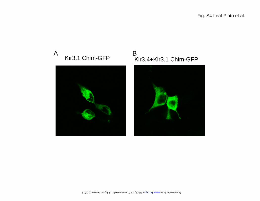
Tagging of the C-terminal cytoplasmic tail of the Kir3.1chimera with EGFP (Kir3.1 Chim-GFP) and transfectingHEK-293 cells, revealed lack of cell surface expression (sup-plemental Fig. S4A), similar to that previously observed withKir3.1-GFP alone (e.g. 24). Even co-transfection of Kir3.4failed to alter cell surface expression of the Kir3.1 Chim-GFP(supplemental Fig. S4B), in sharp contrast with Kir3.1-GFPshown previously (21). These results are consistent with theinterpretation that the Kir3.1 chimera neither produces func-tional homomeric channels nor it localizes to the cell surface.In addition, the chimera failed to show potentiated currents
when expressed together with Kir3.4 and to be localized tothe cell surface, suggesting a possible failure to associate withKir3.4. Indeed, the Kir3.1 chimera is missing the 40 aminoacid residues of N-terminal end of Kir3.1 that have beenshown previously to be critical for heteromeric assembly withKir3.4 and cell surface localization (21). Following these un-successful attempts to attain functional expression from theKir3.1 chimera in two different cell systems, we focused ourefforts in reconstitution studies into planar lipid bilayers, ex-ploring further the potential problems discussed by Nishidaet al. (17).Successful Functional Reconstitution in Planar Lipid
Bilayers—Bilayer experiments were performed as previouslydescribed (36, 37). Briefly, the affinity purified Kir3.1 chi-mera was used to form PLs by sonicating at 80 KHz for 1min with a 1:1 mixture of bovine brain PE (10 mg/ml) andPS (10 mg/ml). For these studies, the Cis (intracellular)side was defined as the chamber connected to the voltage-holding electrode and all voltages were referenced to theTrans (ground or extracellular) chamber. The insertion ofchannels in the bilayer was assessed by the presence ofclear current transitions from a level of zero current (see“Experimental Procedures”).Fig. 2A shows that when, along with the purified Kir3.1 chi-
mera contained in perfused PLs, diC8 phosphatidylinositol-bisphosphate (PIP2) was added to the Cis but not the Transside of the bilayer, clear channel activity was obtained. Addi-tion of 50 nM of the naturally occurring arachidonyl-stearyl(AASt) PIP2 to the Cis side of the bilayer resulted in �100%channel open probability (n 36). The water-soluble, eight-carbon long analog of PIP2, diC8-PIP2, although less effectivethan AASt PIP2 (43) has proven very useful, as it allows con-
FIGURE 1. Single particle EM of the Kir3.1 channel chimera. A, representative field view of the negatively stained Kir3.1 chimera. The inset displays a gal-lery of characteristic particles. B, reference-free class averages produced by MLF2D, after five cycles of alignment and classification, to produce a working setof 19,300 particles. The percentage of particle images in each class is denoted. Out of 20 possible class averages, the particles in the dataset populatedmainly seven classes. C, three-dimensional structure of the Kir3.1 chimera filtered to a resolution of 24 Å. Different views of the calculated isosurface con-toured at 3 sigma are shown. D, fitting of the x-ray structure of the Kir3.1 channel chimera (PDB code: 2QKS) inside a cutaway of the volume. The locationsof the transmembrane (TM) and cytoplasmic regions (CYT) are given. The region filled with dots around the TM region is suggested to correspond to DDMdetergent molecules.
Functional Characterization of a Kir3 Chimera
DECEMBER 17, 2010 • VOLUME 285 • NUMBER 51 JOURNAL OF BIOLOGICAL CHEMISTRY 39793
at VIV
A, V
A C
omm
onwealth U
niv, on January 2, 2011w
ww
.jbc.orgD
ownloaded from
struction of dose-response relationships and estimation ofrelative apparent affinities of ion channels to PIP2. The Kir3.1chimera showed a relatively high apparent affinity for diC8-PIP2 (EC50 16.75 � 0.50 �M) (Fig. 2B). In comparison, anal-ogous experiments using inside-out patches from Xenopusoocytes expressing the full-length active homomer Kir3.1*yielded lower apparent affinities (EC50 30.6 � 3.8 �M, n 3–5), suggesting that the Kir3.1 chimera experiences strongerinteractions with PIP2 than Kir3.1*. Scavengers of PIP2, suchas poly-Lysine (Fig. 2C) or PIP2 antibody (PIP2 Ab, Fig. 2D)inhibited diC8 PIP2-stimulated channel activity when appliedto the Cis side but not the Trans side. Similar results usingpolylysine or PIP2 Ab were obtained when the Kir3.1 chimerahad been activated by AASt-PIP2. These results confirmedthat Nishida et al. were unable to functionally reconstitute theKir3.1 chimera in lipid bilayers due to an unmet lipid require-ment, namely the presence of PIP2.The Kir3.1 chimera exhibited biophysical properties char-
acteristic of Kir channels. Single-channel currents showedintraburst kinetics exhibiting one closed-time (�c 0.31 ms)
and two open-time (�o1 1.6 ms, �02 29.7 ms) components(Fig. 3B) and a 29.3 � 6.7 pS unitary conductance (Fig. 3C).Inward rectification was obtained in the presence of intracel-lular Mg2� (Fig. 3, A and C, right panel), while a linear cur-rent-voltage relationship was obtained in the absence of intra-cellular Mg2� (Fig. 3C, left panel). 100 nM of the peptideTertiapin Q, which blocks potently Kir3 and Kir1.1 currents(44–46), added to the Trans but not to the Cis side, inhibitedthe activity of the Kir3.1 chimera (Fig. 3D). Both the internalMg2�-dependent rectification and the external block by Ter-tiapin Q are defining characteristics of Kir channels.Because Kir3 activation by ethanol is thought to result by
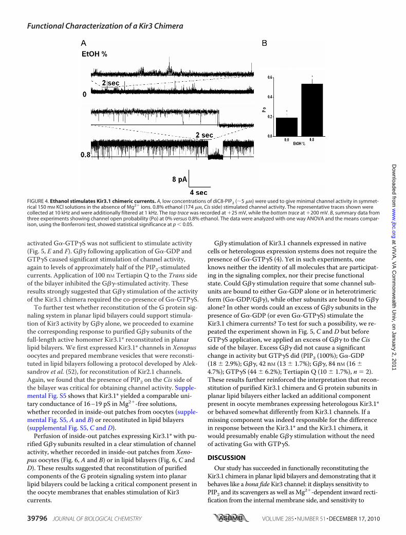
association of the alcohol in a physical binding pocket of thechannel (47), we tested the effects of ethanol on the activity ofthe Kir3.1 chimera. Fig. 4A shows a representative record,where 0.8% ethanol, a concentration shown to stimulate Kir3currents (48, 49), stimulated the activity of the Kir3.1 chi-mera. The significant stimulatory effect of ethanol on Kir3.1chimeric currents (n 3) (Fig. 4B) is an additional functionalcharacteristic of Kir3 channels preserved in the chimera.
FIGURE 2. Functional reconstitution of the Kir3.1 chimera requires PIP2. A, 2-s traces of the Kir3.1 chimera reconstituted in a planar lipid bilayer in which20 �M diC8-PIP2 was added from the Trans side (top) or the Cis side (bottom). B, dose-response curve of DiC8-PIP2 applied from the Cis side. Data points rep-resent mean � S.E. from four experiments ran in triplicate were fitted to the equation: y B � xn/{(k)n � xn}, where y Po; x [PIP2]; B 1.07 � 0.05; EC50,k 16.75 � 0.50; Hill coefficient, n 5.54 � 0.77; chi2 0.00214. C, holding potential values (left), 10-s representative traces (middle), and open probabilityvalues (right) of the reconstituted chimera in the presence of 20 �M diC8-PIP2. Control (upper trace), 300 �g/ml polylysine added to the Trans side (middletrace) or to the Cis side (lower trace), n 3. D, holding potential values (left), 20-s representative traces (center) and open probability values (right) of theKir3.1 chimera activity in the presence of 20 �M diC8-PIP2. Control (upper trace), 1:200 dilution PIP2 Ab added to the Trans side (middle) or 1:200 dilution PIP2Ab added to the Cis side, n 3.
Functional Characterization of a Kir3 Chimera
39794 JOURNAL OF BIOLOGICAL CHEMISTRY VOLUME 285 • NUMBER 51 • DECEMBER 17, 2010
at VIV
A, V
A C
omm
onwealth U
niv, on January 2, 2011w
ww
.jbc.orgD
ownloaded from

Another defining characteristic of Kir3 channels is theirsensitivity to the �� subunits of G proteins (4). All Kir3 sub-units expressed as homomers or heteromers are activated bythe G�� subunits (50, 51). Nanomolar concentrations of puri-fied G�� (G�1�2) failed to stimulate the activity of the Kir3.1chimera either in the absence of PIP2 (n 7) or in the pres-ence of PIP2 at concentrations that produced submaximalactivity (n 6). Instead, nanomolar concentrations of G��caused an inhibition of Kir3.1 chimera channel activity byincreasing the interburst intervals of unitary channel activity(Fig. 5A). The IC50 for G�� inhibition was rather high(48.97 � 5.91 nM), possibly related to its high PIP2 apparentaffinity (Fig. 5B). A similar inhibition (25–30% reduction inPo) was obtained by 16 nM G��, whether the Kir3.1 chimerawas activated maximally by diC8-PIP2 or AASt-PIP2 (n 5).
We proceeded to test whether application of purified G�-GDP affected activity of the Kir3.1 chimera induced by 14.5�M diC8-PIP2. 40 nM G�i1-GDP caused an inhibition of chan-
nel activity (Fig. 5, C and D) similar to that caused by 42 nMG�� applied before G�-GDP (not shown). Subsequent appli-cation of the complementary G-protein subunit did not havea significant further effect (Fig. 5, C and D, G�� followingG�-GDP). Addition of 100 �M GTP�S caused robust channelactivation to levels approximately half of the PIP2-stimulatedcurrents. 100 nM Tertiapin Q applied to the Trans side of thebilayer inhibited the GTP�S-stimulated activity. This resultsuggested the Kir3.1 chimera could be activated by GTP�S ina manner similar to Kir3 channels expressed in native or het-erologous systems (4, 12). Moreover, these experiments sug-gested that activation of Kir3.1 chimera by G proteins re-quired that both subunits were in the active form.To further assess this requirement of channel activity for
both active G protein subunits, we tested whether active G�subunits (G�-GTP�S) affected the Kir3.1 chimera activity.Again G�-GDP inhibited diC8 PIP2-stimulated activity andapplication of GTP�S had no further effect, suggesting that
FIGURE 3. Functional properties of Kir3.1 chimera are consistent with a Mg2�-dependent inwardly rectifying channel, sensitive to block by extra-cellular Tertiapin Q. A, 1-min representative traces of the Kir3.1 chimera reconstituted in a planar lipid bilayer. B, intra-burst time constants for the experi-ment depicted in A. The open-time histogram (upper) was best fitted with a 2-component exponential and the closed-time histogram (lower) was best fit-ted with a one-component exponential (see text and figure for values). C, current-voltage relationship for the Kir3.1 chimera reconstituted in the absence ofMg2� (left) or in the presence of 1 mM Mg2�, n 3. D, 0.5 s representative traces of the chimera reconstituted under the same conditions as in A in the pres-ence of 20 �M diC8-PIP2 (control, upper trace) or when 100 nM Tertiapin Q was added to the Trans side (middle trace) or to the Cis side (lower trace),n 4.
Functional Characterization of a Kir3 Chimera
DECEMBER 17, 2010 • VOLUME 285 • NUMBER 51 JOURNAL OF BIOLOGICAL CHEMISTRY 39795
at VIV
A, V
A C
omm
onwealth U
niv, on January 2, 2011w
ww
.jbc.orgD
ownloaded from
activated G�-GTP�S was not sufficient to stimulate activity(Fig. 5, E and F). G�� following application of G�-GDP andGTP�S caused significant stimulation of channel activity,again to levels of approximately half of the PIP2-stimulatedcurrents. Application of 100 nM Tertiapin Q to the Trans sideof the bilayer inhibited the G��-stimulated activity. Theseresults strongly suggested that G�� stimulation of the activityof the Kir3.1 chimera required the co-presence of G�-GTP�S.To further test whether reconstitution of the G protein sig-
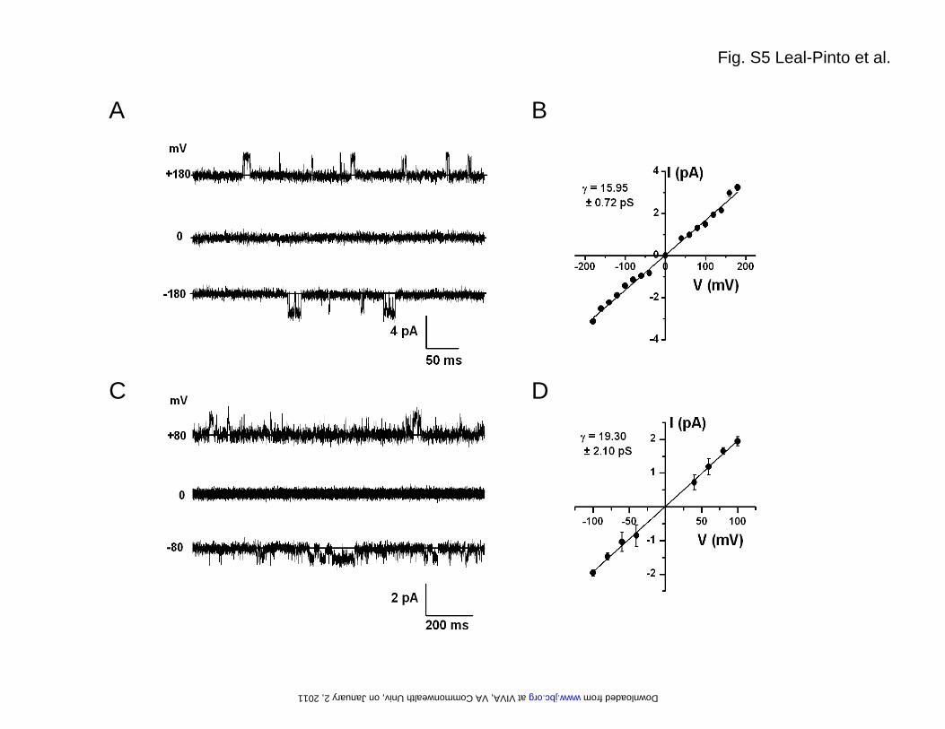
naling system in planar lipid bilayers could support stimula-tion of Kir3 activity by G�� alone, we proceeded to examinethe corresponding response to purified G�� subunits of thefull-length active homomer Kir3.1* reconstituted in planarlipid bilayers. We first expressed Kir3.1* channels in Xenopusoocytes and prepared membrane vesicles that were reconsti-tuted in lipid bilayers following a protocol developed by Alek-sandrov et al. (52), for reconstitution of Kir2.1 channels.Again, we found that the presence of PIP2 on the Cis side ofthe bilayer was critical for obtaining channel activity. Supple-mental Fig. S5 shows that Kir3.1* yielded a comparable uni-tary conductance of 16–19 pS in Mg2�-free solutions,whether recorded in inside-out patches from oocytes (supple-mental Fig. S5, A and B) or reconstituted in lipid bilayers(supplemental Fig. S5, C and D).Perfusion of inside-out patches expressing Kir3.1* with pu-
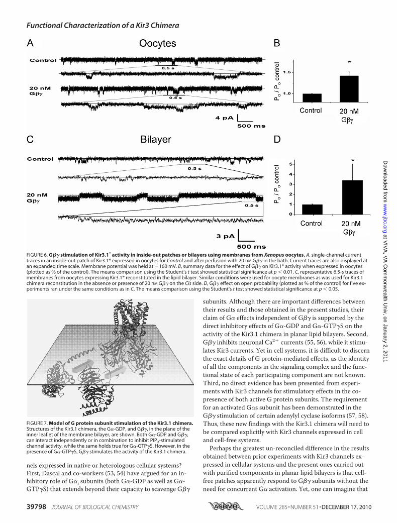
rified G�� subunits resulted in a clear stimulation of channelactivity, whether recorded in inside-out patches from Xeno-pus oocytes (Fig. 6, A and B) or in lipid bilayers (Fig. 6, C andD). These results suggested that reconstitution of purifiedcomponents of the G protein signaling system into planarlipid bilayers could be lacking a critical component present inthe oocyte membranes that enables stimulation of Kir3currents.
G�� stimulation of Kir3.1 channels expressed in nativecells or heterologous expression systems does not require thepresence of G�-GTP�S (4). Yet in such experiments, oneknows neither the identity of all molecules that are participat-ing in the signaling complex, nor their precise functionalstate. Could G�� stimulation require that some channel sub-units are bound to either G�-GDP alone or in heterotrimericform (G�-GDP/G��), while other subunits are bound to G��alone? In other words could an excess of G�� subunits in thepresence of G�-GDP (or even G�-GTP�S) stimulate theKir3.1 chimera currents? To test for such a possibility, we re-peated the experiment shown in Fig. 5, C and D but beforeGTP�S application, we applied an excess of G�� to the Cisside of the bilayer. Excess G�� did not cause a significantchange in activity but GTP�S did (PIP2 (100%); G�-GDP(18 � 2.9%); G��, 42 nM (13 � 1.7%); G��, 84 nM (16 �4.7%); GTP�S (44 � 6.2%); Tertiapin Q (10 � 1.7%), n 2).These results further reinforced the interpretation that recon-stitution of purified Kir3.1 chimera and G protein subunits inplanar lipid bilayers either lacked an additional componentpresent in oocyte membranes expressing heterologous Kir3.1*or behaved somewhat differently from Kir3.1 channels. If amissing component was indeed responsible for the differencein response between the Kir3.1* and the Kir3.1 chimera, itwould presumably enable G�� stimulation without the needof activating G� with GTP�S.
DISCUSSION
Our study has succeeded in functionally reconstituting theKir3.1 chimera in planar lipid bilayers and demonstrating that itbehaves like a bona fideKir3 channel: it displays sensitivity toPIP2 and its scavengers as well asMg2�-dependent inward recti-fication from the internal membrane side, and sensitivity to
FIGURE 4. Ethanol stimulates Kir3.1 chimeric currents. A, low concentrations of diC8-PIP2 (�5 �M) were used to give minimal channel activity in symmet-rical 150 mM KCl solutions in the absence of Mg2� ions. 0.8% ethanol (174 �M, Cis side) stimulated channel activity. The representative traces shown werecollected at 10 kHz and were additionally filtered at 1 kHz. The top trace was recorded at �25 mV, while the bottom trace at �200 mV. B, summary data fromthree experiments showing channel open probability (Po) at 0% versus 0.8% ethanol. The data were analyzed with one way ANOVA and the means compar-ison, using the Bonferroni test, showed statistical significance at p � 0.05.
Functional Characterization of a Kir3 Chimera
39796 JOURNAL OF BIOLOGICAL CHEMISTRY VOLUME 285 • NUMBER 51 • DECEMBER 17, 2010
at VIV
A, V
A C
omm
onwealth U
niv, on January 2, 2011w
ww
.jbc.orgD
ownloaded from

block by Tertiapin Q from the external surface. Channel activitydisplayed an absolute requirement for PIP2, the lack of whichpotentially explains previous unsuccessful attempts to function-ally reconstitute it (17). At PIP2 concentrations that producedsubmaximal Kir3.1 chimeric activity, ethanol stimulated channelactivity consistent with its effects on wild-type Kir3 currents.A number of unexpected findings were obtained with the G
protein subunit gating of the Kir3.1 chimera. First, both G�-GDP as well as G�-GTP�S caused inhibition of PIP2-stimu-lated currents in the complete absence of the G�� subunits.Second, and most unexpectedly, the G�� subunits also inhib-
ited the PIP2-stimulated Kir3.1 chimera currents. Third, thecombination of G�-GDP and G�� subunits proved incapableof stimulating channel activity, even when the G�� subunitswere used in stoichiometric excess. In contrast, the fourthfinding showed that G�� subunits were effective in stimulat-ing activity of the Kir3.1 chimera in the co-presence of G�-GTP�S. This result suggests that the Kir3.1 chimera requiresboth activated G protein subunits in order to have its activitystimulated (Fig. 7).How do these results of G protein subunit effects on the
Kir3.1 chimera compare with previous studies on Kir3 chan-
FIGURE 5. G protein regulation of Kir3.1 chimeric currents. A, G�� concentrations (left), 30-s representative traces (center) and open probability (right) forthe chimera in the presence of 20 �M diC8-PIP2 at the G�� concentrations depicted on the left. The traces shown come from the same experiment andwere collected at 10 kHz and were additionally filtered at 1 kHz for final analysis. B, G�� dose-response on the open probability of the Kir3.1 chimera (n 5)reconstituted under the same conditions as in A. Data points were fitted to the equation: y B � xn/{(k)n � xn} where, y Po; x [G��]; B 0.87 � 0.08;EC50, k 48.97 � 5.91; Hill coefficient, n 3.31 � 1.06; chi2 0.00865. C, representative NPo of the Kir3.1 chimera as a function of time for the entire ex-periment (n 4). The bars at the top indicate the sequential addition of PIP2 (14.5 �M), G�-GDP (40 nM), G�� (42 nM), GTP�S (100 �M), and Tertiapin Q (100nM). All additions except for Tertiapin were added to the Cis side of the chamber. D, bar graph of the mean NPo (� S.E.) for the time interval between se-quential additions of PIP2, G�-GDP, G��, GTP�S, and Tertiapin Q (n 4). Asterisk (*) indicates significance level of 0.05 (see “Experimental Procedures”).E, representative NPo of Kir3.1 chimera as a function of time for an entire experiment (n 3). The bars at the top indicate the sequential addition of PIP2,G�-GDP, GTP�S, G��, and Tertiapin Q (concentrations were similar to those indicated in C). All reagents except Tertiapin Q were added to the Cis side of thechamber. F, bar graph of the mean NPo (�S.E.) for the time interval between sequential additions of PIP2, G�, GTP�S, G��, and Tertiapin Q (n 3). All re-agents except Tertiapin Q were added to the Cis side of the chamber. The asterisk (*) indicates significance level of 0.05.
Functional Characterization of a Kir3 Chimera
DECEMBER 17, 2010 • VOLUME 285 • NUMBER 51 JOURNAL OF BIOLOGICAL CHEMISTRY 39797
at VIV
A, V
A C
omm
onwealth U
niv, on January 2, 2011w
ww
.jbc.orgD
ownloaded from
nels expressed in native or heterologous cellular systems?First, Dascal and co-workers (53, 54) have argued for an in-hibitory role of G�i subunits (both G�-GDP as well as G�-GTP�S) that extends beyond their capacity to scavenge G��
subunits. Although there are important differences betweentheir results and those obtained in the present studies, theirclaim of G� effects independent of G�� is supported by thedirect inhibitory effects of G�-GDP and G�-GTP�S on theactivity of the Kir3.1 chimera in planar lipid bilayers. Second,G�� inhibits neuronal Ca2� currents (55, 56), while it stimu-lates Kir3 currents. Yet in cell systems, it is difficult to discernthe exact details of G protein-mediated effects, as the identityof all the components in the signaling complex and the func-tional state of each participating component are not known.Third, no direct evidence has been presented from experi-ments with Kir3 channels for stimulatory effects in the co-presence of both active G protein subunits. The requirementfor an activated G�s subunit has been demonstrated in theG�� stimulation of certain adenylyl cyclase isoforms (57, 58).Thus, these new findings with the Kir3.1 chimera will need tobe compared explicitly with Kir3 channels expressed in celland cell-free systems.Perhaps the greatest un-reconciled difference in the results
obtained between prior experiments with Kir3 channels ex-pressed in cellular systems and the present ones carried outwith purified components in planar lipid bilayers is that cell-free patches apparently respond to G�� subunits without theneed for concurrent G� activation. Yet, one can imagine that
FIGURE 6. G�� stimulation of Kir3.1* activity in inside-out patches or bilayers using membranes from Xenopus oocytes. A, single-channel currenttraces in an inside-out patch of Kir3.1* expressed in oocytes for Control and after perfusion with 20 nM G�� in the bath. Current traces are also displayed atan expanded time scale. Membrane potential was held at 160 mV. B, summary data for the effect of G�� on Kir3.1* activity when expressed in oocytes(plotted as % of the control). The means comparison using the Student’s t test showed statistical significance at p � 0.01. C, representative 6.5-s traces ofmembranes from oocytes expressing Kir3.1* reconstituted in the lipid bilayer. Similar conditions were used for oocyte membranes as was used for Kir3.1chimera reconstitution in the absence or presence of 20 nM G�� on the Cis side. D, G�� effect on open probability (plotted as % of the control) for five ex-periments ran under the same conditions as in C. The means comparison using the Student’s t test showed statistical significance at p � 0.05.
FIGURE 7. Model of G protein subunit stimulation of the Kir3.1 chimera.Structures of the Kir3.1 chimera, the G�-GDP, and G��, in the plane of theinner leaflet of the membrane bilayer, are shown. Both G�-GDP and G��,can interact independently or in combination to inhibit PIP2-stimulatedchannel activity, while the same holds true for G�-GTP�S. However, in thepresence of G�-GTP�S, G�� stimulates the activity of the Kir3.1 chimera.
Functional Characterization of a Kir3 Chimera
39798 JOURNAL OF BIOLOGICAL CHEMISTRY VOLUME 285 • NUMBER 51 • DECEMBER 17, 2010
at VIV
A, V
A C
omm
onwealth U
niv, on January 2, 2011w
ww
.jbc.orgD
ownloaded from

if endogenous G protein-coupled receptors were part of thesignaling complex (missing in our bilayer reconstitution ex-periments), they might cause partial G protein subunit activa-tion through their basal and agonist-independent activity. Insuch a scenario, G�� stimulation could be coupled to a con-current G� activation for stimulation of Kir3 currents. Alter-natively, the replacement of the Kir3.1 transmembrane do-mains by the corresponding KirBac1.3 region and/or the lackof the last 129 amino acid residues of Kir3.1 could be respon-sible for this difference. These and other predictions born outfrom the present experiments with the Kir3.1 chimera modelwill undoubtedly stimulate new experiments with Kir3 chan-nels expressed in native and heterologous cell systems, as wellas in planar lipid bilayers.To date, progress in structural studies of Kir3-G�� com-
plexes has been hampered in part by the fact that even thoughcrystal structures of G�� dimers exist (e.g., Ref. 59) “func-tional” Kir3 channels of known structures had not been re-ported. Various crystallographic studies have provided struc-tural information on Kir3 channels. Two of them lacked thetransmembrane region describing the soluble cytoplasmicpores of eukaryotic Kir3.1 and Kir3.2 at 1.8 and 2.3Å, re-spectively (18, 20). The third one, the Kir3.1 chimera at2.2Å, provided the Kir3.1 cytosolic pore in the context of aprokaryotic transmembrane domain (17). We believe thatour single-particle electron microscopy work on the Kir3.1chimera paves the way for structural studies of Kir3.1 chi-mera-G�� complexes using hybrid approaches (53), basedon the fitting of existing crystal structures of the individualcomponents to three-dimensional electron microscopymaps. Together with other structural approaches, such asx-ray crystallography and computational chemistry, struc-tural models of complexes between Kir3 channels and Gproteins will allow a greater in depth study of G proteinregulation of effectors.The present studies suggest that both G protein subunits
are critically involved in the regulation of the activity of theKir3.1 chimera. Thus it is important that future studies aim todiscern the structural interrelationships of the entire signalingcomplex, which may confer the stability necessary for suc-cessful structural studies.
Acknowledgments—We thank Motohiko Nishida and RoderickMacKinnon for sharing the Kir3.1 chimera construct. We are alsograteful to Dr. James Wells (University of Toronto) for useful discus-sions, Dr. David Stokes (New York University) for constructive feed-back on the manuscript, and members of the Logothetis and Ubar-retxena laboratories for helpful discussions throughout the project.We also thank the New York Structural Biology Center for use of itselectron microscopy facilities.
REFERENCES1. Huang, C. L., Feng, S., and Hilgemann, D. W. (1998) Nature 391,
803–8062. Sui, J. L., Petit-Jacques, J., and Logothetis, D. E. (1998) Proc. Natl. Acad.
Sci. U.S.A. 95, 1307–13123. Logothetis, D. E., Lupyan, D., and Rosenhouse-Dantsker, A. (2007)
J. Physiol. 582, 953–965
4. Logothetis, D. E., Kurachi, Y., Galper, J., Neer, E. J., and Clapham, D. E.(1987) Nature 325, 321–326
5. Reuveny, E., Slesinger, P. A., Inglese, J., Morales, J. M., Iniguez-Lluhi,J. A., Lefkowitz, R. J., Bourne, H. R., Jan, Y. N., and Jan, L. Y. (1994) Na-ture 370, 143–146
6. Yamada, M., Innabe, A., and Kurachi, Y. (1998) Pharmacol. Rev. 50,723–760
7. Hibino, H., Inanobe, A., Furutani, K., Murakami, S., Findlay, I., and Ku-rachi, Y. (2010) Physiol. Rev. 90, 291–366
8. Stanfield, P. R., Nakajima, S., and Nakajima, Y. (2002) Rev. Physiol. Bio-chem. Pharmacol. 145, 47–179
9. Kubo, Y., Reuveny, E., Slesinger, P. A., Jan, Y. N., and Jan, L. Y. (1993)Nature 364, 802–806
10. Dascal, N., Lim, N. F., Schreibmayer, W., Wang, W., Davidson, N., andLester, H. A. (1993) Proc. Natl. Acad. Sci. U.S.A. 90, 6596–6600
11. Lesage, F., Duprat, F., Fink, M., Guillemare, E., Coppola, T., Lazdunski,M., and Hugnot, J. P. (1994) FEBS Lett. 353, 37–42
12. Krapivinsky, G., Gordon, E. A., Wickman, K., Velimirovic, B., Krapivin-sky, L., and Clapham, D. E. (1995) Nature 374, 135–141
13. Tao, X., Avalos, J. L., Chen, J., and MacKinnon, R. (2009) Science 326,1668–1674
14. Doyle, D. A., Cabral, J. M., Pfeutzner, R. A., Kuo, A., Gulbis, J. M., Co-hen, S. L., Chait, B. T., and Mackinnon, R. (1998) Science 280, 69–77
15. Kuo, A., Gulbis, J. M., Antcliff, J. F., Rahman, T., Lowe, E. D., Zimmer, J.,Cuthbertson, J., Ashcroft, F. M., Ezaki, T., and Doyle, D. A. (2003) Sci-ence 300, 1922–1926
16. Kuo, A., Domene, C., Johnson, L. N., Doyle, D. A., and Venien-Bryan, C.(2005) Structure 13, 1463–1472
17. Nishida, M., Cadene, M., Chait, B. T., and MacKinnon, R. (2007) EMBOJ. 26, 4005–4015
18. Nishida, M., and MacKinnon, R. (2002) Cell 111, 957–96519. Pegan, S., Arrabit, C., Zhou, W., Kwiatkowski, W., Collins, A., Slesinger,
P. A., and Choe, S. (2005) Nat. Neurosci. 8, 279–28720. Inanobe, A., Matsuura, T., Nakagawa, A., and Kurachi, Y. (2007) Chan-
nels 1, 39–4521. Mirshahi, T., and Logothetis, D. E. (2004) J. Biol. Chem. 279,
11890–1189722. Lesage, F., Guillemare, E., Fink, M., Duprat, F., Heurteaux, C., Fosset,
M., Romey, G., Barhanin, J., and Lazdunski, M., (1995) J. Biol. Chem.270, 28660–28667
23. Chan, K. W., Sui, J. L., Vivaudou, M., and Logothetis, D. E. (1996) Proc.Natl. Acad. Sci. U.S.A. 93, 14193–14198
24. Vivaudou, M., Chan, K. W., Sui, J. L., Jan, L. Y., Reuveny, E., and Logoth-etis, D. E. (1997) J. Biol. Chem. 272, 31553–31560
25. Logothetis, D. E., Petrou, V. I., Adney, S. K., and Mahajan, R. (2010)Pflugers Arch. 460, 321–341
26. Logothetis, D. E., and Nilius, B. (2007) Pflugers Arch. 455, 1–327. Logothetis, D. E., Jin, T., Lupyan, D., and Rosenhouse-Dantsker, A.
(2007) Pflugers Arch. 455, 83–9528. Stansfeld, P. J., Hopkinson, R., Ashcroft, F. M., and Sansom, M. S. (2009)
Biochemistry 48, 10926–1093329. Lopes, C. M., Zhang, H., Rohacs, T., Jin, T., Yang, J., and Logothetis,
D. E. (2002) Neuron 34, 933–94430. Ludtke, S. J., Baldwin, P. R., and Chiu,W. (1999) J. Struct. Biol. 128, 82–9731. Marabini, R., Masegosa, I. M., San, Martin, M. C., Marco, S., Fernandez,
J. J., de, la, Fraga, L. G., Vaquerizo, C., and Carazo, J. M. (1996) J. Struct.Biol. 116, 237–240
32. Scheres, S. H., Nunez-Ramírez, R., Sorzano, C. O., Carazo, J. M., andMarabini, R. (2008) Nat. Protoc. 3, 977–990
33. Mindell, J. A., and Grigorieff, N. (2003) J. Struct. Biol. 142, 334–34734. Heymann, J. B. (2001) J. Struct. Biol. 133, 156–16935. Scheres, S. H., Nunez-Ramírez, R., Gomez-Llorente, Y., San, Martín, C.,
Eggermont, P. P., and Carazo, J. M. (2007) Structure 15, 1167–117736. Leal-Pinto, E., London, R. D., Knorr, B. A., and Abramson, R. G. (1995) J.
Membr. Biol. 146, 123–13237. Leal-Pinto, E., Tao, W., Rappaport, J., Richardson, M., Knorr, B. A., and
Abramson, R. G. (1997) J. Biol. Chem. 272, 617–62538. Rostovtseva, T. K., Kazemi, N., Weinrich, M., and Bezrukov, S. M.
Functional Characterization of a Kir3 Chimera
DECEMBER 17, 2010 • VOLUME 285 • NUMBER 51 JOURNAL OF BIOLOGICAL CHEMISTRY 39799
at VIV
A, V
A C
omm
onwealth U
niv, on January 2, 2011w
ww
.jbc.orgD
ownloaded from
(2006) J. Biol. Chem. 281, 37496–3750639. Pettersen, E. F., Goddard, T. D., Huang, C. C., Couch, G. S., Greenblatt,
D. M., Meng, E. C., and Ferrin, T. E. (2004) J. Comput. Chem. 25,1605–1612
40. Rubinstein, J. L. (2007)Methods 41, 409–41641. Muench, S. P., Huss, M., Song, C. F., Phillips, C., Wieczorek, H., Trinick,
J., and Harrison, M. A. (2009) J. Mol. Biol. 386, 989–99942. Chan, K. W., Langan, M. N., Sui, J. L., Kozak, J. A., Pabon, A., Ladias,
J. A., and Logothetis, D. E. (1996) J. Gen. Physiol. 107, 381–39743. Rohacs, T., Chen, J., Prestwich, G. D., and Logothetis, D. E. (1999) J. Biol.
Chem. 274, 36065–3607244. Jin, W., and Lu, Z. (1998) Biochemistry 37, 13291–1329945. Jin, W., Klem, A. M., Lewis, J. H., and Lu, Z. (1999) Biochemistry 38,
14294–1430146. Jin, W., and Lu, Z. (1999) Biochemistry 38, 14286–1429347. Aryal, P., Dvir, H., Choe, S., and Slesinger, P. A. (2009) Nat. Neurosci.
12, 988–99548. Kobayashi, T., Ikeda, K., Kojima, H., Niki, H., Yano, R., Yoshioka, T., and
Kumanishi, T. (1999) Nat. Neurosci. 2, 1091–1097
49. Lewohl, J. M., Wilson, W. R., Mayfield, R. D., Brozowski, S. J., Morrisett,R. A., and Harris, R. A. (1999) Nat. Neurosci. 2, 1084–1090
50. He, C., Zhang, H., Mirshahi, T., and Logothetis, D. E. (1999) J. Biol.Chem. 274, 12517–12524
51. He, C., Yan, X., Zhang, H., Mirshahi, T., Jin, T., Huang, A., and Logoth-etis, D. E. (2002) J. Biol. Chem 277, 6088–6096
52. Aleksandrov, A., Velimirovic, B., and Clapham, D. E. (1996) Biophys. J.70, 2680–2687
53. Schreibmayer, W., Dessauer, C. W., Vorobiov, D., Gilman, A. G., Lester,H. A., Davidson, N., and Dascal, N. (1996) Nature 380, 624–627
54. Rubinstein, M., Peleg, S., Berlin, S., Brass, D., and Dascal, N. (2007)J. Physiol. 581, 17–32
55. Ikeda, S. R. (1996) Nature 380, 255–25856. Herlitze, S., Garcia, D. E., Mackie, K., Hille, B., Scheuer, T., and Catterall,
W. A. (1996) Nature 380, 258–26257. Tang, W. J., and Gilman, A. G. (1991) Science 254, 1500–150358. Gao, B. N., and Gilman, A. G. (1991) Proc. Natl. Acad. Sci. U.S.A. 88,
10178–1018259. Gaudet, R., Bohm, A., and Sigler, P. B. (1996) Cell 87, 577–588
Functional Characterization of a Kir3 Chimera
39800 JOURNAL OF BIOLOGICAL CHEMISTRY VOLUME 285 • NUMBER 51 • DECEMBER 17, 2010
at VIV
A, V
A C
omm
onwealth U
niv, on January 2, 2011w
ww
.jbc.orgD
ownloaded from

SUPPLEMENTAL MATERIAL
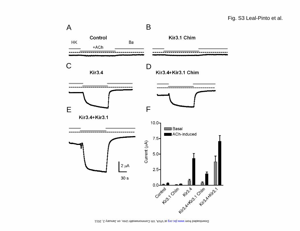
EXPERIMENTAL PROCEDURES Channel expression in Xenopus laevis oocytes and preparation of membranes. For expression in Xenopus laevis oocytes, the coding sequence of the Kir3.1 chimera was PCR-amplified and inserted between the BamH1 and HindIII restriction sites of the dual-function expression vector pXooM (58). Kir3.1, Kir3.1*, Kir3.4 and M2R cDNAs were in the pGEMHE plasmid vector. Synthesis of capped cRNAs was performed by in vitro transcription using the mMESSAGE mMACHINE T7 kit (Ambion, Austin, TX). Injected oocytes (50l/oocyte) were kept at 19 °C for 2 days to allow for expression of the heterologous proteins and subsequently used for electrophysiological recordings or preparation of membranes. For isolation of the membrane fraction, oocytes were homogenized in 5mM Tris-HCl pH: 8.0, 1mM EDTA, 1mM EGTA containing 10mM NaVO4, 10mM NaF and complete protease inhibitor cocktail (Roche, Indianapolis, IN), homogenates were centrifuged for 5min at 5000 RPM (4o C) and supernatants were subsequently ultracentrifuged at 100,000g for 40min at 4°C in a Beckman Maxima Ultracentrifuge (Beckman, Brea, CA). The pellets were re-suspended by sonicating them with a 1:1 mixture of bovine brain PE (10 mg/ml) and PS (10 mg/ml) and reconstituted in lipid bilayers as described for the Kir3.1 chimera. Two-electrode voltage clamp: Recombinant Kir3 channels and M2 receptors were expressed in Xenopus laevis oocytes as previously described (22, 55). cRNA was produced with T7 RNA polymerase using the Ambion kit. In all experimental groups, cRNA of the M2 receptors was injected at 1 ng/oocyte and the ratio of M2R to the combination of all Kir3 constructs injected was maintained at 1 2 ׃. Recordings in Xenopus laevis oocytes were performed 2–4 days following cRNA injection. Whole-oocyte currents were measured by conventional two-microelectrode voltage clamp with a GeneClamp 500 amplifier (Axon Instruments/Molecular Devices, Sunnyvale, CA). Electrodes were filled with 3 M KCl dissolved in 1% agarose to prevent the leakage of KCl into the oocytes. The electrodes had a resistance of less than 1 M. Oocytes were constantly perfused with a high-potassium solution (HK) containing in mM: 96 KCl, 1.8 CaCl2, 1 MgCl2, 5 HEPES (pH 7.4). M2 receptors expressed in the oocytes were activated by perfusion with high-potassium solution, containing 5 µM ACh (ACh). At the end of each experiment, high-potassium solution containing 5 mM BaCl2 (Ba) was used to inhibit the Kir3 currents. Current amplitudes were measured at –100 mV. Data acquisition and analysis were achieved using pClamp8 (Axon Instruments/Molecular Devices, Sunnyvale, CA) and Origin 6 (Microcal/Origin, Clemente, CA) software. Single-channel recording in oocytes: The cRNA of Kir3.1* was injected at 5-10 ng/50 nl for single channel recordings. Single channel currents were recorded from devitellinized oocytes under a standard excised inside-out patch-clamp configuration (56) with an Axopatch 200 amplifier (Axon Instruments/Molecular Devices, Sunnyvale, CA). Single-channel currents were sampled at 5 kHz and filtered at 1 kHz. pClamp was used to drive stimulus protocols and digitize currents. A standard pipette solution, ND96K and FVPP + 20 mM NaCl, was used in both solutions to prevent channel rundown. Recordings were performed 3-5 days following injection. Recording pipettes with a resistance of 1~1.5 MΏ in the both solution were frequently employed. G and short chain diC8 PIP2 were applied to the intracellular side of excised patches. All experiments were performed at room temperature (~22-25o
C). Data analysis. Single-channel current amplitude was determined by generating amplitude histograms for selected segments and fitted to Gaussian functions. Single-channel conductance values were determined by the slope of current-voltage (I-V) curve between –200 to + 200mV, where I-V data could be well fitted to a linear line. Channel open probability (Po) was determined with continuous recordings of at least 2,000ms (total 1-3min) using pClamp software. The channel number used was the maximal number of simultaneous channel openings observed. Confocal fluorescence microscopy: The C terminus of the Kir3.1-chimera was tagged with enhanced green fluorescent protein (Clontech, Mountain View, CA) by subcloning into pEGFP-N3 plasmid vector.
at VIV
A, V
A C
omm
onwealth U
niv, on January 2, 2011w
ww
.jbc.orgD
ownloaded from

For mammalian cell expression, HEK-293 cells were grown on coverslips and transfected using lipofection with Kir3.1 chimera-EGFP alone or Kir3.1 chimera-EGFP and Kir3.4. One day after the transfection, cells were fixed in 2.5% paraformaldhyde. Coverslips were washed and mounted onto coverslides using ProLong Gold antifade reagent (invitrogen, Carlsbad, CA). Cells were visualized using a Leica TCS2 AOBS laser scanning confocal microscope.
FIGURE LEGENDS Fig. S1. Purification of the Kir3.1 chimera. (A) In the final step of the purification, the eluate obtained from cobalt affinity chromatography was run over a Sephacryl S-200 gel filtration column to yield a chromatogram containing three peaks. Peak A eluted at the exclusion volume (36 ml) of the column. (B) A 15% SDS-PAGE analysis of selected fractions stained with coomassie blue shows that peak B contains the majority of the chimera. Two main bands marked by arrows can be observed. As assessed by in gel-digestion and mass spectrometry, both bands correspond to the Kir3.1 chimera. The faster band migrates with an apparent molecular weight of 34 kDa, which matches well with the calculated mass (36 kDa) of the chimera monomer. The upper band migrates with an apparent molecular weight above the 212 kDa marker and likely corresponds to the chimera tetramer. In-gel digestion and mass spectrometry confirmed the identity of both bands. Fig. S2. Electron microscopy. (A) Plot of the Euler angle distribution of the particles used in the 3D reconstruction. Every particle is represented by a triangle in a tilted transparent semi-sphere. Particles are grouped by the default angular step (10 deg) used by the reconstruction algorithm. Two regions lacked complete angular coverage: The first one (surrounded by a dotted line) is the result of the 4-fold symmetry (not applied during the reconstruction process) of the volume. The second one (top of the sphere) coincides with the z-axis and it is due to enrichment of the experimental particle images in a lateral orientation. The lack of angular coverage in this region could cause a decrease in the resolution of the volume along this z-axis. (B) Gallery of experimental class averages (same as in Fig. 1D) generated by MLF2D compared to selected backprojections (with white Gaussian noise added and boxed in dotted lines) from the final 3D reconstruction. (C) The Fourier shell correlation (FSC) suggests a resolution of 24 Å according to the 0.5 criterion. Fig. S3. Expression in Xenopus oocytes. (A) Xenopus oocytes were injected with M2R-cRNA alone (Control), or (B) coinjected with cRNA for M2R (1 ng) in combination with cRNA for Kir3.1 chimera (2 ng,); (C) Kir3.4 (2 ng); (D) Kir3.4 (1 ng) and Kir3.1 chimera (1 ng); and (E) Kir3.4 (1 ng) and Kir3.1 (1ng). All currents were recorded at -100 mV. Basal currents were recorded in the presence of HK solution. M2Rs were activated with 5 μM ACh. Fig. S4. Expression in HEK-293 cells. Kir3.1 chimera was C-terminally tagged with EGFP. Localization was monitored by confocal microscopy for (A) Kir3.1 chimera-GFP alone and (B) co-expression with Kir3.4. Fig. S5. Single-channel conductance of Kir3.1* measure in inside-out patches or bilayers using membranes from Xenopus oocytes. (A) Single-channel current traces from an inside-out patch at –180, 0 and +180 mV for GIRK1* when expressed in oocytes. (B) Current-voltage relationships for GIRK1* when expressed in oocytes. Single-channel conductance was 15.95 ± 0.72 ps (n = 7). (C) Single-channel current traces for Kir3.1* expressed in oocyte membranes and reconstituted in a lipid bilayer. Membrane potentials were held as indicated at -80, 0, and +80 mV. (D) Current-voltage relationships for Kir3.1* obtained in bilayer. Single-channel conductance was 19.30 ± 2.10 ps (n = 3). Key words: G protein K+ channel, Kir3.1 chimera, G subunits, PIP2, electron microscopy
at VIV
A, V
A C
omm
onwealth U
niv, on January 2, 2011w
ww
.jbc.orgD
ownloaded from
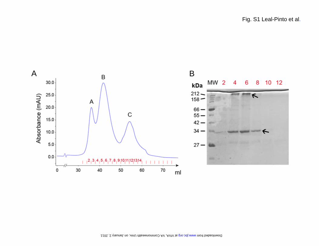
Fig. S1 Leal-Pinto et al.
A B
at VIVA, VA Commonwealth Univ, on January 2, 2011 www.jbc.org Downloaded from
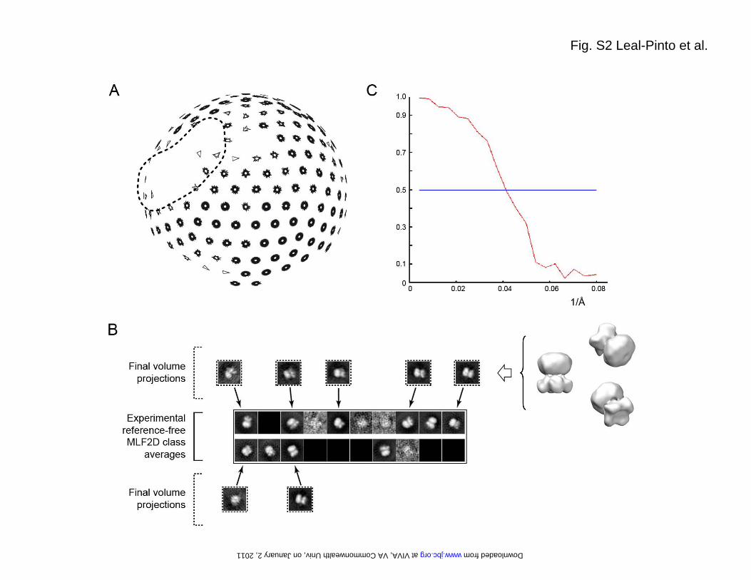
Fig. S2 Leal-Pinto et al.
at VIVA, VA Commonwealth Univ, on January 2, 2011 www.jbc.org Downloaded from
A B
C D
E F
Fig. S3 Leal-Pinto et al.
at VIVA, VA Commonwealth Univ, on January 2, 2011 www.jbc.org Downloaded from
AKir3.1 Chim-GFP Kir3.4+Kir3.1 Chim-GFP
B
Fig. S4 Leal-Pinto et al.
at VIVA, VA Commonwealth Univ, on January 2, 2011 www.jbc.org Downloaded from
A
D
Fig. S5 Leal-Pinto et al.
C
B
D
at VIVA, VA Commonwealth Univ, on January 2, 2011 www.jbc.org Downloaded from