ge healthcare logiq p5 premium · logiq p5 premium the ge logiq p5 premium is an economical shared...
TRANSCRIPT

GE Healthcare
Logiq P5 Premium

LOGIQ P5PREMIUMThe GE Logiq P5 premium is an economical
shared service ultrasound machine that is unique
in it’s price range by offering deep support for all
applications from Cardiac to Pediatric, from 4D
Obstetrics to Surgical or Elastography with an
astounding 29 compatible transducers spread
across every possible application. If it can be
done with ultrasound, the Logiq P5 can do it.

APPLICATION1. Abdominal
2. Obstetrical
3. Gynecological
4. Cardiac
5. Musculoskeletal
6. Vascular
7. Urological
8. Small Parts
9. Superficial
10. Pediatric
11. Neonatal
12. Transcranial
13. Endocavitary
14. Transesophageal
15. Transrectal
16. Transvaginal
IMAGING MODES1. B-Mode
2. M-Mode
3. Crossbeam
4. SRI
5. Elastography
6. Anatomical M Mode
7. Color Flow Mode
8. Split Screen
9. Quad Screen
10. B-Flow
11. Steered CW Doppler
12. Dedicated CW Doppler
13. Coded Contrast Imaging
14. 3D/4D Volume Modes
15. Directional Power Doppler
16. PW Doppler with High PRF
17. Anatomical M-Mode
18. CW Doppler
19. Coded Contrast Imaging
17. Real-time Triplex Mode
FEATURES1. Hard Disk partition of 50GB
2. CINE Memory Frames
3. Real-time Triplex Mode
4. ATO (Auto Tissue Optimization)
5. ACO (Auto CFM Optimization)
6. ASO (Auto Spectrum Optimization)
7. Coded harmonic Imaging
8. Coded Excitation
9. Virtual Convex
10. Patient Information Database
11. Image Archive on Hard Drive and DVD
12. Easy Backup for Media
13. Report Designer
14. Vascular Calcs
15. Cardiac Calcs
16. OB Calcs
17. Fetal Trending
18. Multi Gestational Calcs
19. Hip Dysplasia Calcs
FEATURES CONTINUED20. Gynecological Calcs
21. Urological Calcs
22. Renal Calcs
23. Network Storage
24. Auto Focus
25. MpegView
26. Key Macro
27. Virtual Convex with X-Beam
28. ED Report
29. Wide Field of View on convex
30. 15” TFT LCD monitor with Flexible Monitor Arm
31. Quick Save
32. B-Steer
33. Real-time Auto Doppler Calculations
34. TruAccess, Raw Data Processing
35. Simultaneous Display on BE9CS probe
36. Crossbeam
37. Directional Power Doppler Imaging
38. High Pulse Repetition Frequency (HPRF)
39. Speckle Reduction Imaging (SRI)
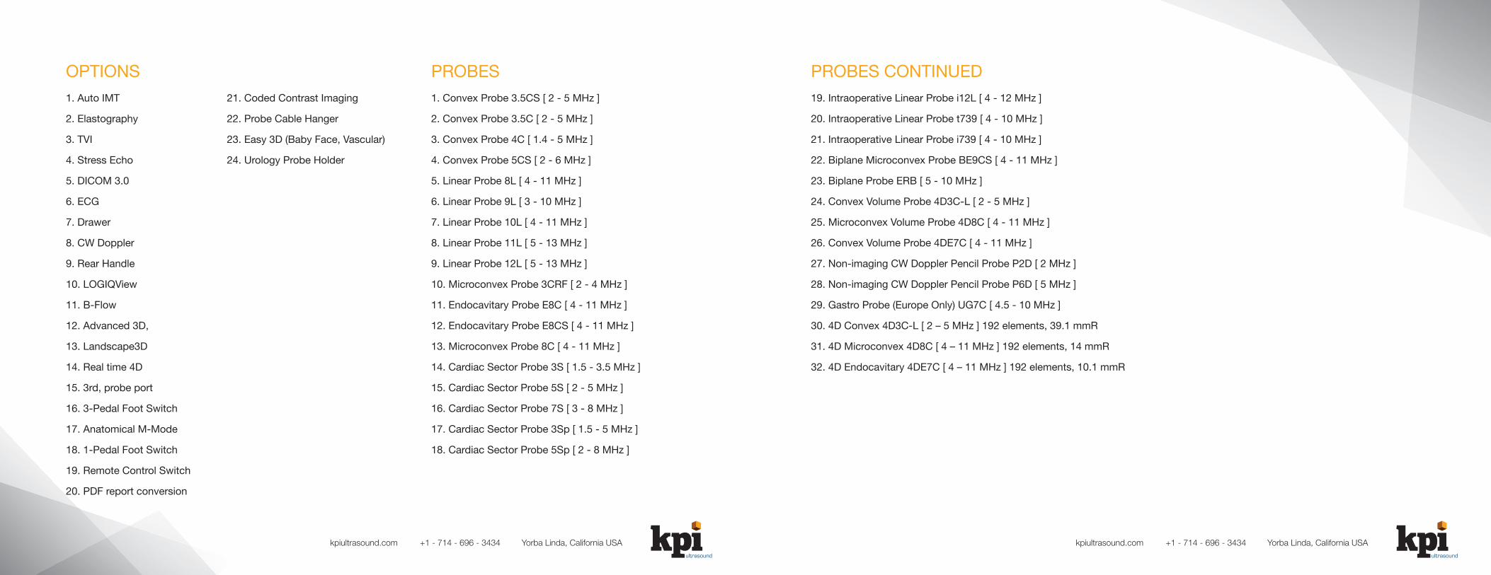
OPTIONS1. Auto IMT
2. Elastography
3. TVI
4. Stress Echo
5. DICOM 3.0
6. ECG
7. Drawer
8. CW Doppler
9. Rear Handle
10. LOGIQView
11. B-Flow
12. Advanced 3D,
13. Landscape3D
14. Real time 4D
15. 3rd, probe port
16. 3-Pedal Foot Switch
17. Anatomical M-Mode
18. 1-Pedal Foot Switch
19. Remote Control Switch
20. PDF report conversion
21. Coded Contrast Imaging
22. Probe Cable Hanger
23. Easy 3D (Baby Face, Vascular)
24. Urology Probe Holder
PROBES1. Convex Probe 3.5CS [ 2 - 5 MHz ]
2. Convex Probe 3.5C [ 2 - 5 MHz ]
3. Convex Probe 4C [ 1.4 - 5 MHz ]
4. Convex Probe 5CS [ 2 - 6 MHz ]
5. Linear Probe 8L [ 4 - 11 MHz ]
6. Linear Probe 9L [ 3 - 10 MHz ]
7. Linear Probe 10L [ 4 - 11 MHz ]
8. Linear Probe 11L [ 5 - 13 MHz ]
9. Linear Probe 12L [ 5 - 13 MHz ]
10. Microconvex Probe 3CRF [ 2 - 4 MHz ]
11. Endocavitary Probe E8C [ 4 - 11 MHz ]
12. Endocavitary Probe E8CS [ 4 - 11 MHz ]
13. Microconvex Probe 8C [ 4 - 11 MHz ]
14. Cardiac Sector Probe 3S [ 1.5 - 3.5 MHz ]
15. Cardiac Sector Probe 5S [ 2 - 5 MHz ]
16. Cardiac Sector Probe 7S [ 3 - 8 MHz ]
17. Cardiac Sector Probe 3Sp [ 1.5 - 5 MHz ]
18. Cardiac Sector Probe 5Sp [ 2 - 8 MHz ]
PROBES CONTINUED19. Intraoperative Linear Probe i12L [ 4 - 12 MHz ]
20. Intraoperative Linear Probe t739 [ 4 - 10 MHz ]
21. Intraoperative Linear Probe i739 [ 4 - 10 MHz ]
22. Biplane Microconvex Probe BE9CS [ 4 - 11 MHz ]
23. Biplane Probe ERB [ 5 - 10 MHz ]
24. Convex Volume Probe 4D3C-L [ 2 - 5 MHz ]
25. Microconvex Volume Probe 4D8C [ 4 - 11 MHz ]
26. Convex Volume Probe 4DE7C [ 4 - 11 MHz ]
27. Non-imaging CW Doppler Pencil Probe P2D [ 2 MHz ]
28. Non-imaging CW Doppler Pencil Probe P6D [ 5 MHz ]
29. Gastro Probe (Europe Only) UG7C [ 4.5 - 10 MHz ]
30. 4D Convex 4D3C-L [ 2 – 5 MHz ] 192 elements, 39.1 mmR
31. 4D Microconvex 4D8C [ 4 – 11 MHz ] 192 elements, 14 mmR
32. 4D Endocavitary 4DE7C [ 4 – 11 MHz ] 192 elements, 10.1 mmR
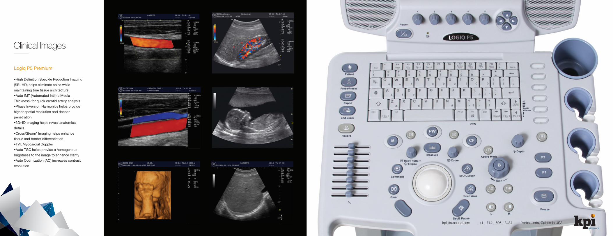
Clinical Images
Logiq P5 Premium
•High Definition Speckle Reduction Imaging
(SRI-HD) helps eliminate noise while
maintaining true tissue architecture
•Auto IMT (Automated Intima Media
Thickness) for quick carotid artery analysis
•Phase Inversion Harmonics helps provide
higher spatial resolution and deeper
penetration
•3D/4D imaging helps reveal anatomical
details
•CrossXBeam* Imaging helps enhance
tissue and border differentiation
•TVI, Myocardial Doppler
•Auto TGC helps provide a homogenous
brightness to the image to enhance clarity
•Auto Optimization (AO) increases contrast
resolution
