ge powerpoint template - health and social care in ... · after a 2 minute wait, perform the usual...
TRANSCRIPT

GE Mammography
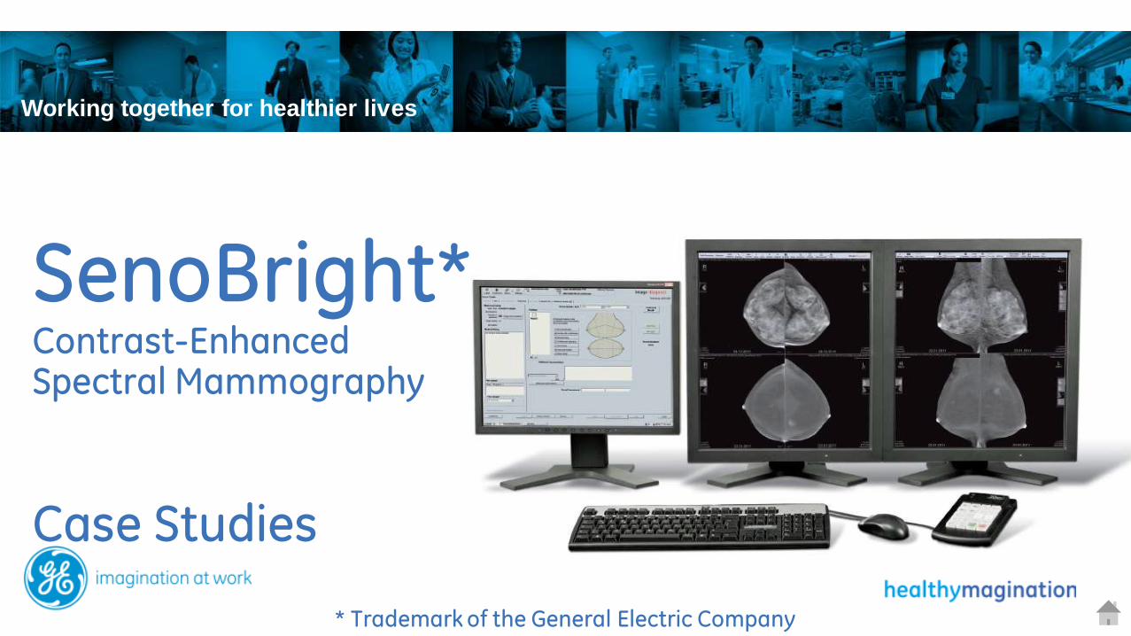
SenoBright* Contrast-Enhanced Spectral Mammography
* Trademark of the General Electric Company
Working together for healthier lives

MR and Ultrasound procedures and volumes vary widely by institution.This example is based on an average population
Breast Imaging pathway
Early health saves lives
GE Screening tools include:
Senographe Essential
GE Diagnosis tools include: SenoBright CESM
GE Biopsy tools include:
Stereotaxy
Is it real? Where is it? What is it? What next?
Verification Localization Analysis Action plan
Is there something?
Detection
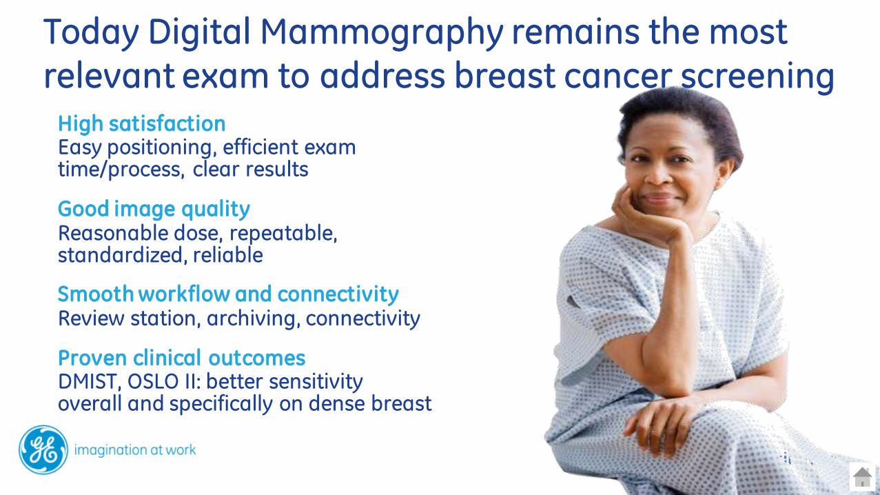
High satisfaction Easy positioning, efficient exam time/process, clear results
Good image quality Reasonable dose, repeatable, standardized, reliable
Smooth workflow and connectivity Review station, archiving, connectivity
Proven clinical outcomes DMIST, OSLO II: better sensitivity overall and specifically on dense breast
CESM
Today Digital Mammography remains the most relevant exam to address breast cancer screening
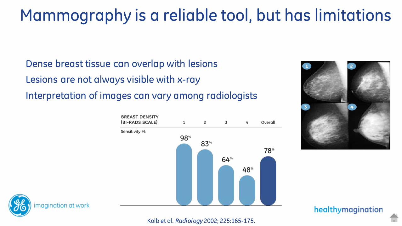
Kolb et al. Radiology 2002; 225:165-175.
Dense breast tissue can overlap with lesions
Lesions are not always visible with x-ray
Interpretation of images can vary among radiologists
CESM
Mammography is a reliable tool, but has limitations
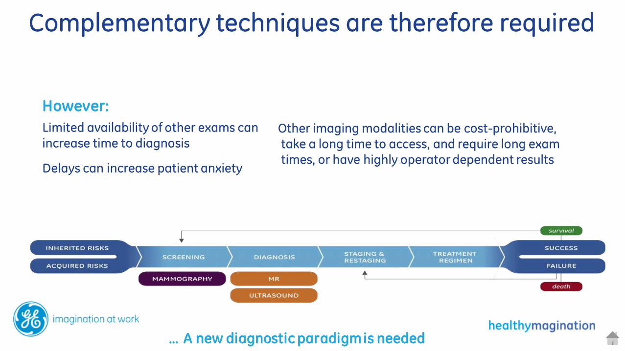
However:
Limited availability of other exams can
increase time to diagnosis
Delays can increase patient anxiety
Other imaging modalities can be cost-prohibitive, take a long time to access, and require long exam times, or have highly operator dependent results
… A new diagnostic paradigm is needed
CESM
Complementary techniques are therefore required

Iodine injection – Contrast agent can be used to highlight areas of unusual blood flow patterns
Modalities l ike MR and CT use contrast-enhanced imaging to highlight suspicious areas
CESM
Contrast-Enhanced areas drive increased suspicion
Idea: use contrast agents in digital mammography to provide contrast-enhanced images

Detection of unusually high blood flow can be used to increase suspicion
Iodine injection – Contrast agent can be used to highlight areas of
unusual blood flow
SenoBright
Two images per view are provided. The first uses standard mammographic techniques and represents tissue density. The second is a contrast-enhanced image in exactly the same position.
SenoBright uses multiple x-ray exposures to reduce background signal, effectively highlighting contrast enhanced areas.

Perform additional tests right away:
same equipment, same staff, same day
Clinically proven results:
As an adjunct to inconclusive mammography and ultrasound
Help reduce patient anxiety
By performing follow-up tests quickly
CESM
SenoBright. Know more now Before
After

Perform additional tests right away
same equipment, same staff, same day
Leverage a new problem-solving exam for inconclusive mammography
Helps reduce anxiety related to waiting
SenoBright. Follow up faster.

Quick learning curve
because images are acquired in standard
views
Easy correlation
With standard mammography results
Easy Communication
Surgeons and specialists can get what they
need in familiar mammography views
The information you need, in the context you know.
SenoBright. Stay in Context.

Easily fits into your practice
Administer intravenous iodine injection exactly like injected CT exams
After a 2 minute wait , perform the usual 4 mammo views in 5 minutes
Mammography Standards Respected
Dedicated quality control protocol using the usual FFDM phantom
DICOM compatible
CESM
SenoBright. Deploy with ease.
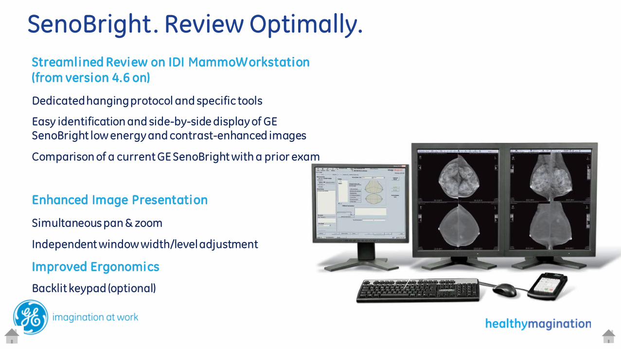
Streamlined Review on IDI MammoWorkstation (from version 4.6 on)
Dedicated hanging protocol and specific tools
Easy identification and side-by-side display of GE SenoBright low energy and contrast-enhanced images
Comparison of a current GE SenoBright with a prior exam
Enhanced Image Presentation
Simultaneous pan & zoom
Independent window width/level adjustment
Improved Ergonomics
Backlit keypad (optional)
CESM
SenoBright. Review Optimally.

SenoBright – Technology Fundamentals
Energy and Iodine Attenuation
• Iodine attenuation has a k-edge at 33KeV (dashed line in the above graph)
•Take exposures below and above that edge
•Process to suppress background tissue/highlight contrast uptake
•Recombined image represents iodine
Spectral Shaping of X-rays
• Generator/Tube capable of 49KV
• Fast KV switching to avoid motion artifacts
• Multi-layer High-Energy filter minimizes dose
• high-energy exposure adds ~20% average glandular dose.
GE Mammography
SenoBright* Contrast-Enhanced Spectral Mammography
Case Studies
* Trademark of the General Electric Company
Working together for healthier lives

79 yo w palpable mass on left breast, original mammography
SenoBright – Case 1 Mammographically equivocal (occult?) lesion
Images courtesy of Mikawa Breast Center, Mikawa, Japan
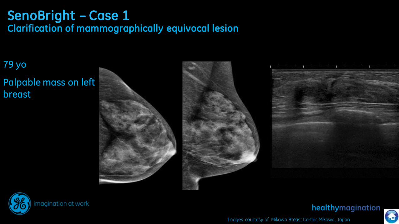
SenoBright – Case 1 Clarification of mammographically equivocal lesion
79 yo
Palpable mass on left breast
Images courtesy of Mikawa Breast Center, Mikawa, Japan

79 yo w palpable mass on left breast
SenoBright contrast-enhanced images clearly localize the lesion
SenoBright – Case 1 Clarification of mammographically equivocal lesion
CC @ 2min
Low Energy Contrast-Enhanced
MLO @ 4 min
Contrast-Enhanced Low Energy
Images courtesy of Mikawa Breast Center, Mikawa, Japan
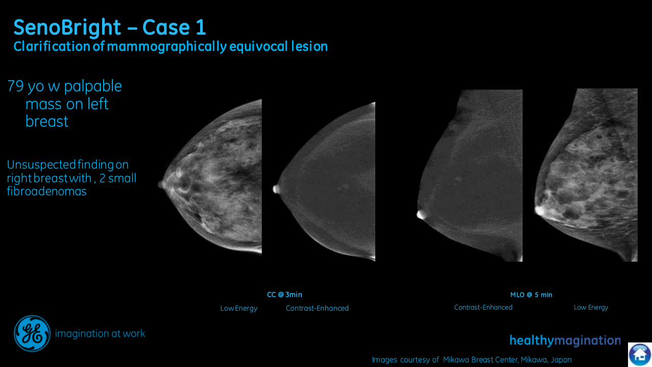
79 yo w palpable mass on left breast
Unsuspected finding on right breast with , 2 small fibroadenomas
SenoBright – Case 1 Clarification of mammographically equivocal lesion
CC @ 3min
Low Energy Contrast-Enhanced
MLO @ 5 min
Contrast-Enhanced Low Energy
Images courtesy of Mikawa Breast Center, Mikawa, Japan

SenoBright – Case 2 Mammographically heterogenous density
RCC L CC R MLO L MLO
72 y/o, screening mammo
Previous operation for a benign nodule on the right external superior quadrant .
Clinical exam reveals a dense area at 9 o’clock (right) and 3 o’clock (left)
Images courtesy of Centre D’imagerie Medicale, Creil , France
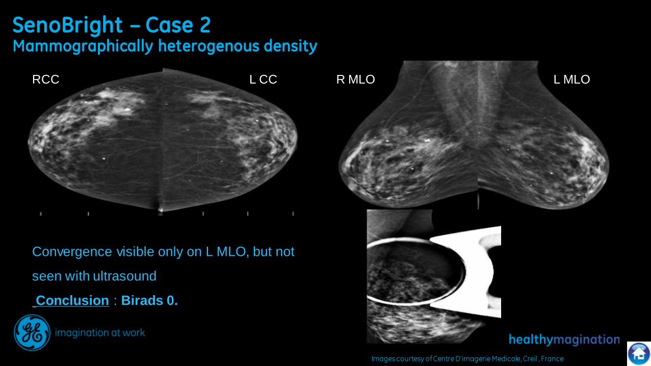
SenoBright – Case 2 Mammographically heterogenous density
Images courtesy of Centre D’imagerie Medicale, Creil , France
RCC L CC R MLO L MLO
Convergence visible only on L MLO, but not
seen with ultrasound
Conclusion : Birads 0.
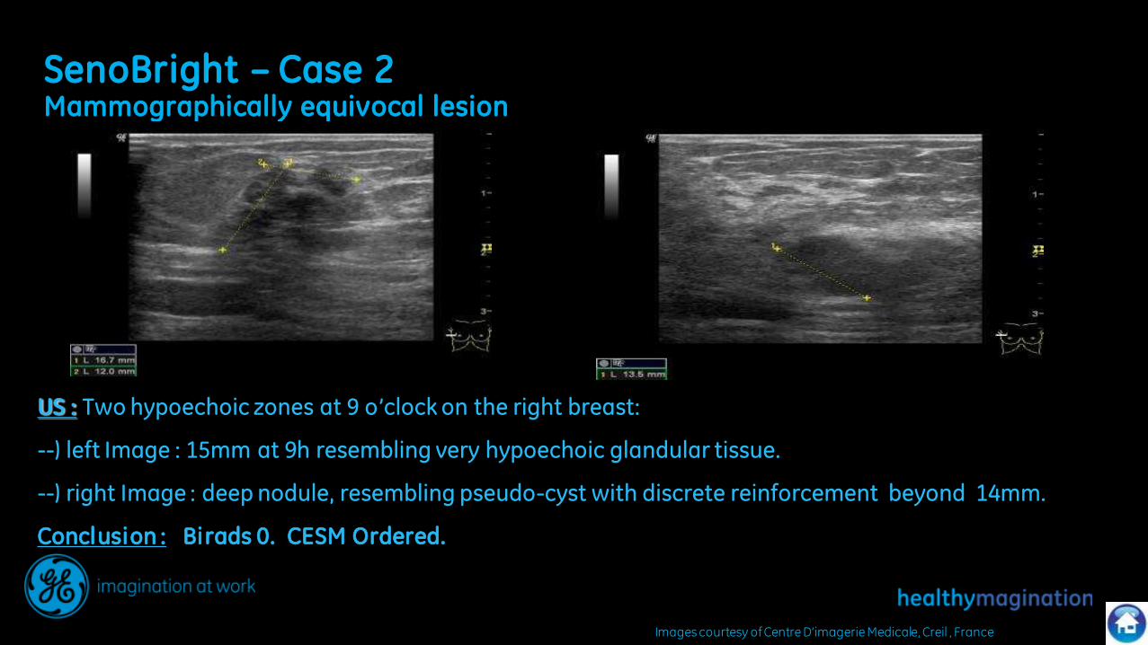
SenoBright – Case 2 Mammographically equivocal lesion
US : Two hypoechoic zones at 9 o’clock on the right breast:
--) left Image : 15mm at 9h resembling very hypoechoic glandular tissue.
--) right Image : deep nodule, resembling pseudo-cyst with discrete reinforcement beyond 14mm.
Conclusion : Birads 0. CESM Ordered.
Images courtesy of Centre D’imagerie Medicale, Creil , France

SenoBright – Case 2 Mammographically equivocal lesion
CESM : Two disctinct contrast enhanced zones that correspond to the ultrasound imaging.
Superficial « cloudy » enhancement
Deeper nodular enhancement
--) Birads 4. Next step : U/S guided micro-biopsy showed:
Superficial zone : ductal hyperplasia without atypia
Profound zone : Invasive ductal carcinoma
1st
2nd
Images courtesy of Centre D’imagerie Medicale, Creil , France

SenoBright – Case 3 Mammographically equivocal lesion
Images courtesy of Armentieres Hospital, France

SenoBright – Case 3 Mammographically equivocal lesion
Images courtesy of Armentieres Hospital, France
Contrast-enhanced images reveal the lesion by hiding the cyst
Biopsy : Infiltrating ductal carcinoma

Large cluster of microcalcifications noted on screening mammogram
SenoBright – Case 4
Images courtesy of Mikawa Breast Center, Mikawa, Japan

SenoBright– Case 4
Large cluster of microcalcifications noted on screening mammogram
Images courtesy of Mikawa Breast Center, Mikawa, Japan

SenoBright – Case 5 Mammographically equivocal lesion
57 years old, presented to screening
Normal clinical exam
Image of convergence lower quadrant right breast.
Images courtesy of Centre D’imagerie Medicale, Creil , France
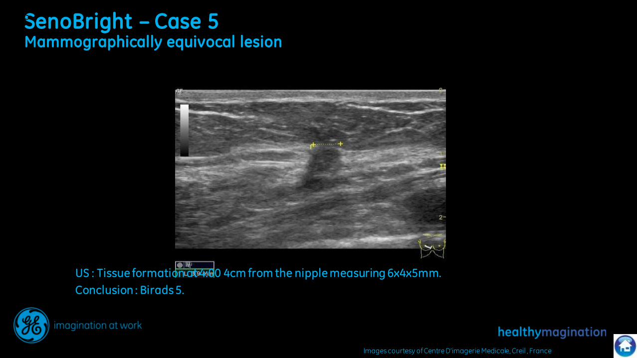
SenoBright – Case 5 Mammographically equivocal lesion
Images courtesy of Centre D’imagerie Medicale, Creil , France
US
US : Tissue formation at 4:00 4cm from the nipple measuring 6x4x5mm.
Conclusion : Birads 5.
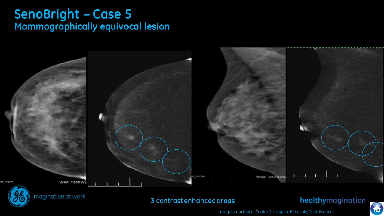
SenoBright – Case 5 Mammographically equivocal lesion
Images courtesy of Centre D’imagerie Medicale, Creil , France
3 contrast enhanced areas

SenoBright – Case 5 Mammographically equivocal lesion
Images courtesy of Centre D’imagerie Medicale, Creil , France
Second look US : Visualisation of the second contrast-enhanced area found under SenoBright.
Spiculated appearance 19mm in front of the primary suspected lesion.
Third contrast-enhanced area not visualized
Biopsy: Three IDCs, 12mm, 6mm, and 1mm diameters
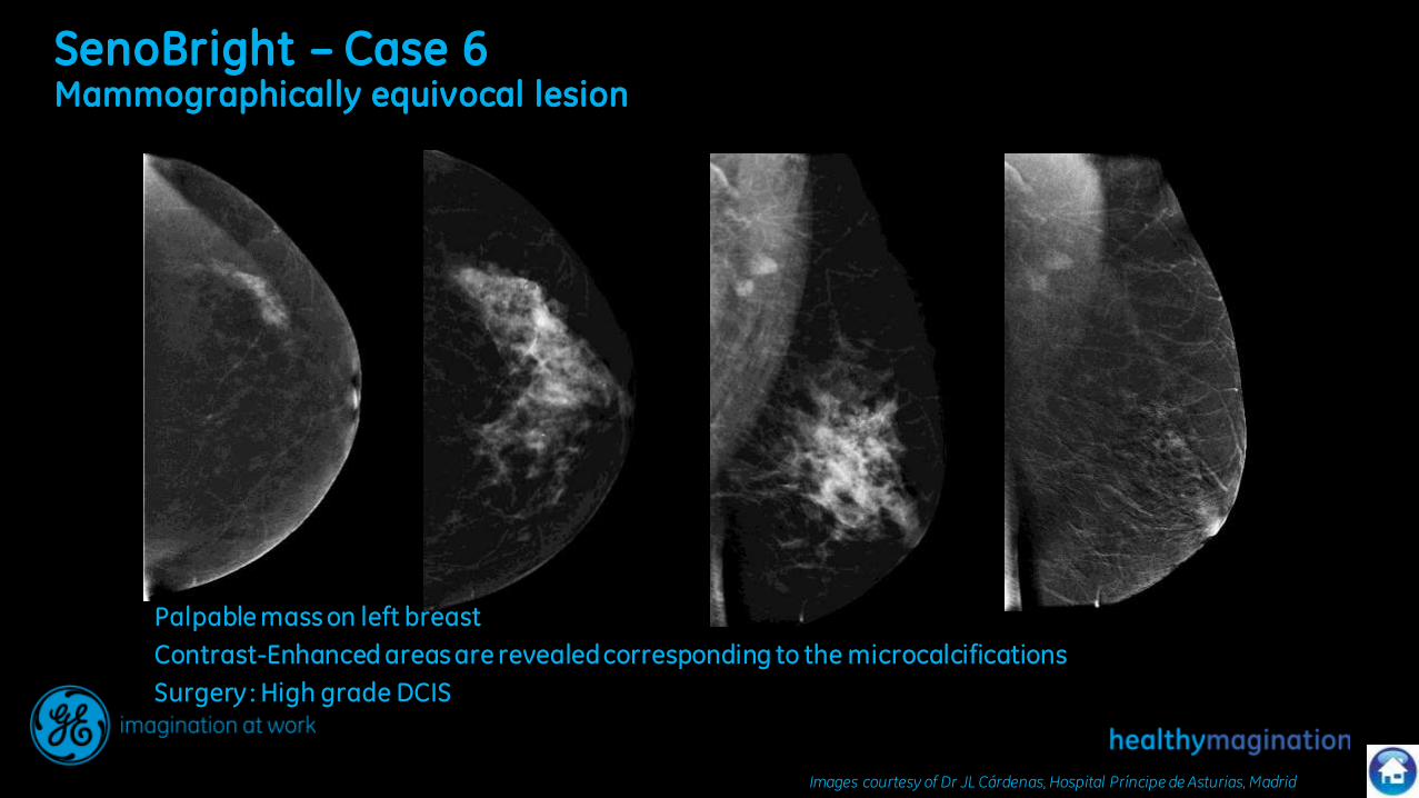
SenoBright – Case 6 Mammographically equivocal lesion
Images courtesy of Dr JL Cárdenas, Hospital Príncipe de Asturias, Madrid
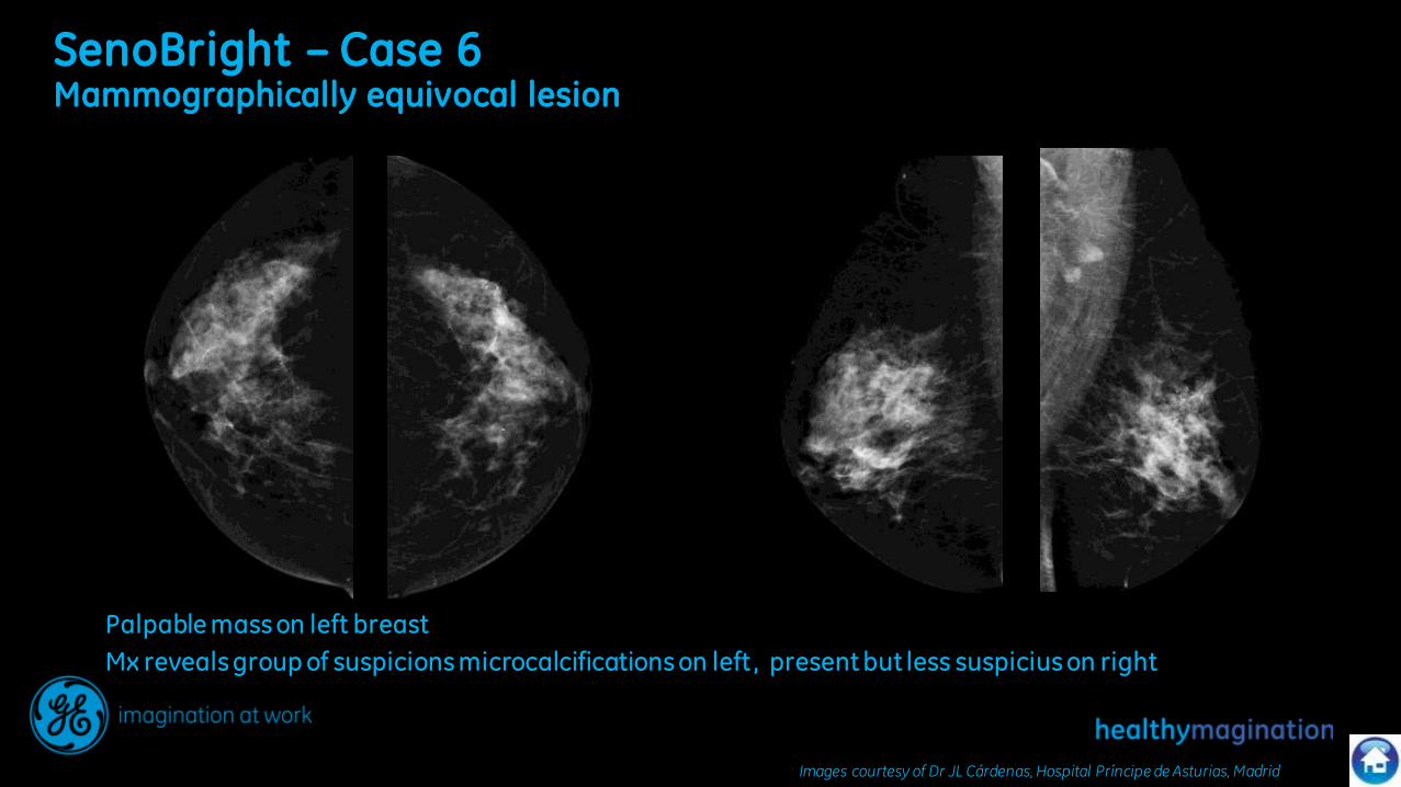
Palpable mass on left breast
Mx reveals group of suspicions microcalcifications on left , present but less suspicius on right
SenoBright – Case 6 Mammographically equivocal lesion
Palpable mass on left breast
Contrast-Enhanced areas are revealed corresponding to the microcalcifications
Surgery : High grade DCIS
Images courtesy of Dr JL Cárdenas, Hospital Príncipe de Asturias, Madrid

SenoBright – Case 6 Mammographically equivocal lesion
Palpable mass on left breast
Contrast-Enhanced areas are also revealed on the right breast
Surgery : IDC
Images courtesy of Dr JL Cárdenas, Hospital Príncipe de Asturias, Madrid
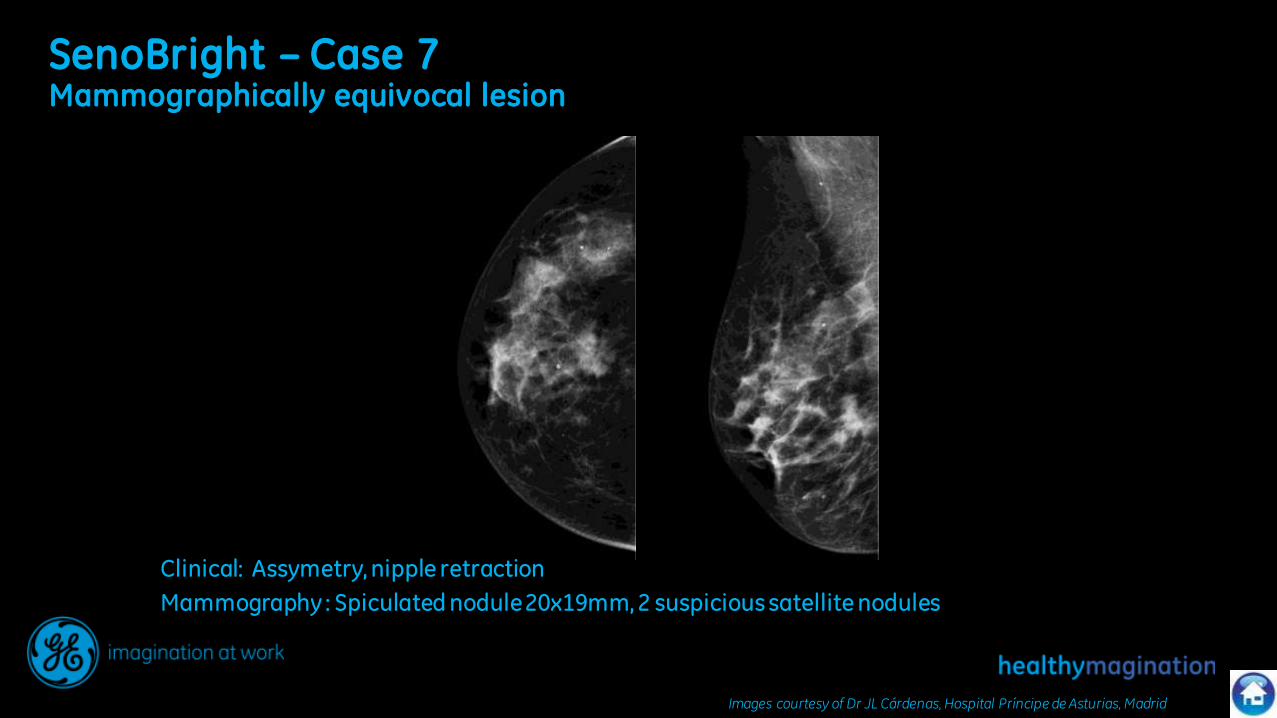
SenoBright – Case 7 Mammographically equivocal lesion
Clinical: Assymetry, nipple retraction
Mammography : Spiculated nodule 20x19mm, 2 suspicious satellite nodules
Images courtesy of Dr JL Cárdenas, Hospital Príncipe de Asturias, Madrid

SenoBright – Case 7 Mammographically equivocal lesion
Clinical: Assymetry, nipple retraction
Mammography : Spiculated nodule 20x19mm, 2 suspicious satellite nodules
Images courtesy of Dr JL Cárdenas, Hospital Príncipe de Asturias, Madrid
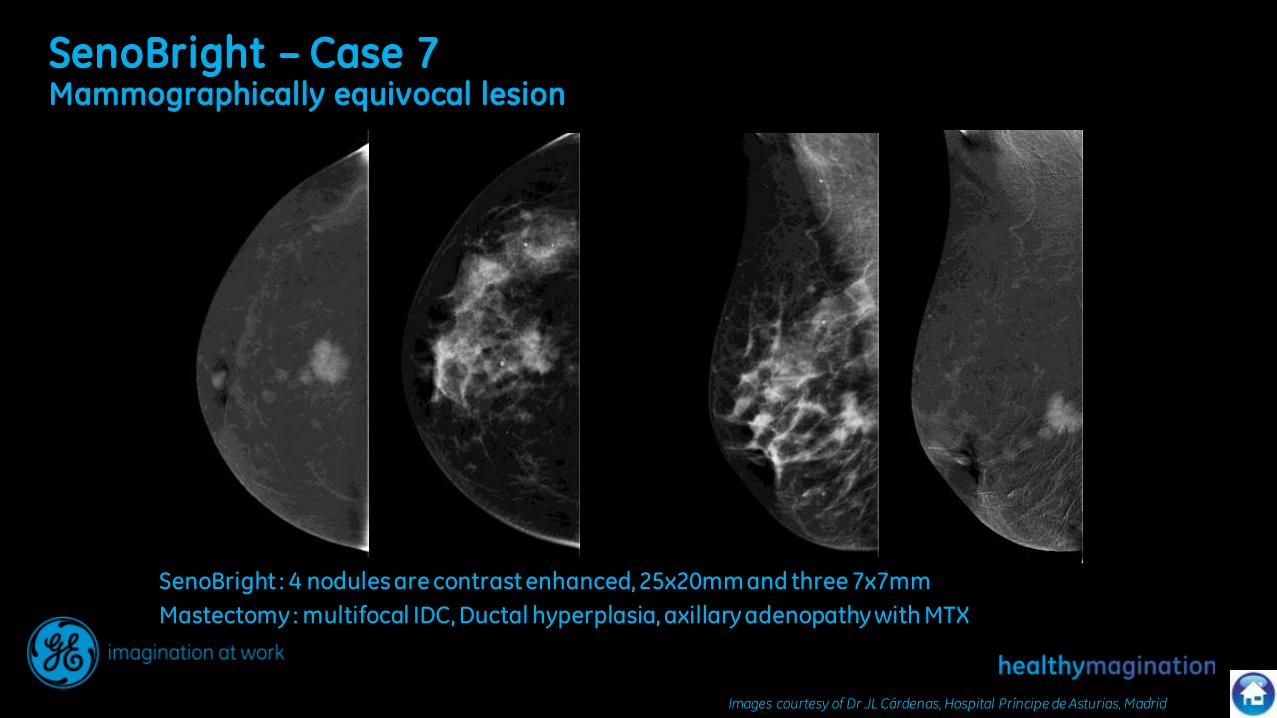
SenoBright – Case 7 Mammographically equivocal lesion
SenoBright : 4 nodules are contrast enhanced, 25x20mm and three 7x7mm
Mastectomy : multifocal IDC, Ductal hyperplasia, axillary adenopathy with MTX
Images courtesy of Dr JL Cárdenas, Hospital Príncipe de Asturias, Madrid

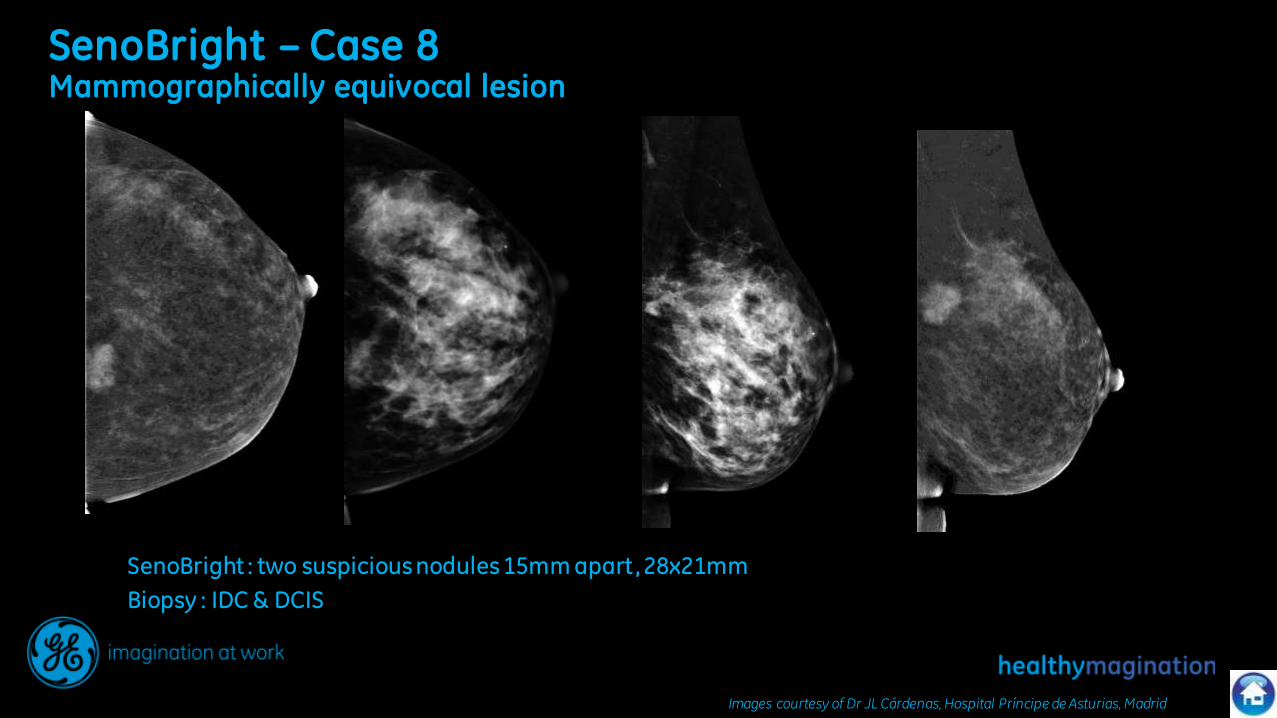
SenoBright – Case 8 Mammographically equivocal lesion
SenoBright : 24x24mm nodule, and 6mmx3.5mm
Biopsy : IDC & DCIS, malignant lymph node
Images courtesy of Dr JL Cárdenas, Hospital Príncipe de Asturias, Madrid
SenoBright – Case 8 Mammographically equivocal lesion
SenoBright : two suspicious nodules 15mm apart, 28x21mm
Biopsy : IDC & DCIS
Images courtesy of Dr JL Cárdenas, Hospital Príncipe de Asturias, Madrid