gec/estro-eau recommendations on temporary brachytherapy
TRANSCRIPT

GEC/ESTRO-EAU recommendations on temporary brachytherapyusing stepping sources for localised prostate cancer
Gyorgy Kovacsa,*, Richard Potterb, Tillmann Lochc, Josef Hammerd,Inger-Karine Kolkman-Deurlooe, Jean J.M.C.H. de la Rosettef, Hagen Bertermanng
aInterdisciplinary Brachytherapy Centre, University Hospital Schleswig-Holstein Campus Kiel, Arnold Heller Str 9, D-24105 Kiel, GermanybUniversity Clinic for Radiotherapy and Radiobiology, Vienna General Hospital, Vienna, Austria
cDepartment of Urology, University of the Saarland, Homburg, GermanydDepartment of Radiotherapy, Barmherzige Schwestern Hospital and St Vincenc Clinic, Linz, Austria
eDivision of Clinical Physics, Department of Radiation Oncology, Erasmus MC, Daniel Den Hoed Cancer Center, Rotterdam, The NetherlandsfDepartment of Urology, AMC, Amsterdam, The Netherlands
gDepartment of Urology, City Hospital, Kiel, Germany
Received 3 August 2004; accepted 2 September 2004Available online 22 October 2004
Abstract
Background and purpose: The aim of this paper is to present the GEC/ESTRO-EAU recommendations for template and transrectalultrasound (TRUS) guided transperineal temporary interstitial prostate brachytherapy using a high dose rate iridium-192 stepping source anda remote afterloading technique. Experts in prostate brachytherapy developed these recommendations on behalf of the GEC/ESTRO and ofthe EAU. The paper has been approved by both GEC/ESTRO steering committee members and EAU committee members.
Patients and methods: Interstitial brachytherapy (BT) to organ confined prostate cancer can be applied as a boost treatment in combinationwith external beam radiation therapy (EBRT) using a proper number of BT fractions in curative intent. Temporary transperineal BT alone orin combination with EBRT are feasible as a palliative/salvage treatment modality because of local recurrence, however, without large clinicalexperience. The use of temporary BT as a monotherapy is subject of ongoing clinical research.
Results: Recommendations for pre-treatment investigations, patient selection, equipment and facilities, the clinical team, the implantprocedure (treatment planning and needle implantation) dose and fractionation, reporting, management of side effects and follow-up aregiven.
Conclusions: These recommendations are intended to be technically and advisory in nature, but the ultimate responsibility for the medicaldecision rests with the treating physician. Although, this paper represents the consensus of an interdisciplinary group of experts, TRUS andtemplate guided temporary transperineal interstitial implants in prostate cancer are a constantly evolving field and the recommendations aresubject to modifications as new data become available.q 2004 Elsevier Ireland Ltd. All rights reserved.
Keywords: Prostate cancer; HDR-brachytherapy; Transrectal ultrasound; Stepping source technology; Afterloading; Recommendations
1. Introduction
The proportion of patients treated by brachytherapy israpidly increasing over the past years, and both permanentseed implants and temporary afterloading techniques playan important role in the treatment of localised prostate
cancer [2,6,19,30,56]. To achieve an appropriate quality forpermanent seed implants and to synchronise activities in thismultidisciplinary field GEC-ESTRO/EAU/EORTC rec-ommendations for permanent implants were previouslypublished [2]. The following recommendations on tempor-ary transperineal prostate brachytherapy, contain a sum-mary of the experience in prostate BT of a small group ofbrachytherapy experts and urologists on behalf of the GEC/ESTRO-EAU Prostate Brachytherapy Group (PROBATE);
0167-8140/$ - see front matter q 2004 Elsevier Ireland Ltd. All rights reserved.
doi:10.1016/j.radonc.2004.09.004
Radiotherapy and Oncology 74 (2005) 137–148
www.elsevier.com/locate/radonline
* Corresponding author.
they are intended to form a basis for an appropriate use ofthis treatment modality.
Remote temporary afterloading brachytherapy hasseveral advantages:
accurate positioning of the source by first implantingnon-active guide needles,possibility to choose the source positions over the lengthof the needle,no target movement during radiation,stepping source technology allowing for dose andvolume adaptation due to adjustment of source dwelllocations and times according to 3D imaging basedindividual dose prescription before irradiation [68].
Temporary brachytherapy using a stepping source doesnot need any source preparation time and there is fullradiation protection. It also allows fractionated irradiationand volume optimisation of the target dose distribution. IfBT and complementary EBRT are the chosen treatment,the costs of temporary BT are low. Introducing a remoteafterloading technique combined with the technologicaldevelopments in sonography devices, such as transrectalultrasound (TRUS), as well as treatment planning softwaredevelopments result in an appropriate target delineationand guidance of the needles [37,52]. A high quality of thetreatment planning and delivery of dose to the target can beachieved [22].
Disadvantage of temporary BT-implants:
temporary BT-implants require usually a fractionatedschedule which results in more work load per patient.
Temporary transperineal implants for prostate cancerhave been applied since the mid 80’s in an increasingnumber of centres worldwide. Clinical experience has beenbuilt up progressively, and the technique appears nowadaysto be safe and effective [4,9,20,23,49,50,52,53].
However, the value of temporary BT and its clinicalbenefit compared to low-dose rate BT have not beendemonstrated in randomised clinical studies. To allowrelevant comparative information on clinical results, it isessential that patient data and treatment parameters aredescribed in a similar way for permanent and for temporaryimplants.
An important radiobiological issue is the evaluation ofbiological weighting factors to apply to compensate for thedifferences in dose rate and time factors. Some models havebeen proposed but their validity and relevance for thetreatment of prostatic adenocarcinoma need to be furtherassessed [70]. Furthermore, as the available radiobiologicaldata do not allow definitive conclusions about the relativeefficacy of temporary BT compared to other radiationtreatment modalities, comparison of clinical data isrequired. However, radiobiological data and considerationsindicate that a low alpha/beta value (!2 Gy) might be
appropriate for the relative value of temporary brachyther-apy with a high fraction dose. If such values are appropriate,the relative efficacy of temporary BT with a high-dose perfraction would be pronounced [11,21,35,36]. High-dose-rate brachytherapy might be then expected to produceresults comparable to or better than those from low-dose-rate implants. However, using temporary implants exclu-sively for treatment of localised prostate cancer has not beentested clinically in a sufficient amount of patients withappropriate follow-up. Delivering the total dose exclusivelyin a few very high-dose fractions (one or two) is notrecommended because of radiobiological disadvantages,e.g. inadequate tumour reoxygenation and normal tissuedamage [63]. In any case, the application and evaluation ofbiological weighting factors is recommended for compari-son of results and will be guided by the increase of clinicalexperience with longer follow-up.
Developments in remote afterloading brachytherapy(temporary BT) devices and technology as well as intransrectal sonography resulted in highly sophisticated toolsin the field of interstitial treatment of localised prostatecancer. There is a wide consensus today, that TRUS guidedtransperineal template implant techniques represent thestandard of interstitial prostate brachytherapy with anaccurate needle placement [4,7,12,27,60].
Due to the excellent dose distribution of BT implantsusing a stepping source and anatomy related doseoptimisation (adaptation of dwell locations and dwelltimes to the target and no impairment from oedema, fromsource migration, and from prostate movement during theshort time interval of the boost application) and due to thesignificant low costs of remote afterloading treatment, BTboosts with a stepping source in combination with EBRTseems to be challenging treatment option for a selectedgroup of patients (Tables 1 and 2).
Long-term follow-up data confirm that temporary BTboost combined with EBRT represents a successfultreatment choice and results in excellent bNED, localcontrol and survival rates (Table 2). The treatment seems tobe especially advantageous for patients in intermediate andpoor prognostic groups (iPSAO10 ng/ml or GleasonO7 orStageOT2a).
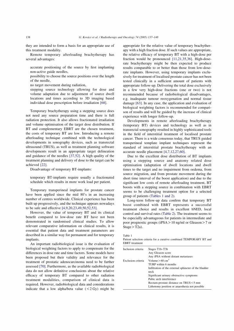
Table 1
Patient selection criteria for a curative combined TEMPORARY BT and
EBRT treatment
Inclusion criteria Stages T1b–T3bAny Gleason score
Any iPSA without distant metastases
Exclusion criteria VolumeO60 cm3
TURP within 6 monthsInfiltration of the external sphincter of the bladder
neck
Significant urinary obstructive symptomsPubic arch interference
Rectum-prostate distance on TRUS!5 mm
Lithotomy position or anaesthesia not possible
G. Kovacs et al. / Radiotherapy and Oncology 74 (2005) 137–148138

Some of following paragraphs (in chapter 1 and 2) referto issues not different for permanent or interstitial implants.If appropriate, recommendations as published previously forpermanent implants were taken, they are particularlyindicated and referred to [2].
1.1. Pre-treatment investigations
For comparison of treatment outcome, it is essential thatpre-treatment investigations be identical for patients treatedwith permanent implants and temporary BT for prostaticcarcinoma. Therefore, the recommendations for temporaryBT should be identical to those for permanent seedimplantations [2].
‘All patients should have a history and general physicalexamination to assess their suitability to the treatment.Local and if needed (initial PSAO10 ng/ml) systemicstaging should be completed’ [2].
1.2. PSA
‘Initial PSA should be recorded in all patients’ [2].
1.3. Transrectal ultrasound (TRUS)
TRUS of the prostate should be performed on all patientsby an expert (usually the urologist) to more accuratelyassess the local extent of disease. In the hand of experiencedusers, TRUS is still one of the most sensitive imagingmethods to determine local extent of prostate cancer [5,17,33,42,45,62], especially if the histologic biopsy informationis present. Imaging of the zonal anatomy of the prostategland is an important factor in defining different target areaswithin the gland [5,10,22,25,53]. Also the knowledge ofextra-capsular disease and its localisation are extremelyuseful for treatment planning [4,10,22,25,39,53]. Addition-ally, TRUS may serve as a substitute for CT in most patientsregarding the detection of pubic arch interference [71].
1.4. Prostate biopsy
‘All patients should have TRUS guided biopsy provenadenocarcinoma It is usual to take six to 12 biopsy coreswith ultrasound guidance’ [2]. The use of TRUS improves
the biopsy results and can utilise valuable information fortreatment planning. The percentage of positive biopsiesseems to be a strong prognostic factor [41,47,64].
1.5. Bone scan
If the probability of bone metastases is!5% according tothe Partin tables, bone scan is not obligatory [58].
1.6. CT scan/conventional MRI of the pelvis(not mandatory)
These imaging methods have a moderate value inassessing the local extent of prostate tumours. PSA valuesaccording to the Partin tables give more information onmicrometastatic disease. MRI is useful in detecting nodalenlargement.
1.7. Pelvic MRI with endorectal coil (not mandatory)
This is one of the most sensitive imaging investigation toassess local extent of prostate cancer [2]. The knowledge ofextra-capsular disease and its localisation is extremelyimportant for further treatment decisions; however, TRUS isthe general practice in local staging.
1.8. Surgical lymph node staging (not mandatory)
There is controversy about the benefit of surgical lymphnode sampling, in particular, in patients with poorprognostic factors [20,23]. Temporary BT may be used incombination with external beam radiation therapy (EBRT)and regional lymph nodes then represent a potential target ofradiation treatment. However, there is no clear evidencefrom clinical trials, showing advantage or disadvantage ofsurgical lymph node sampling with regard to therapeuticoutcome.
1.9. Urodynamic studies
Lower urinary tract symptoms (LUTS) have to beevaluated before therapy and the patient has to completean International Prostate Symptom Score (IPSS) or AUAsymptom-scoring sheet. ‘Maximum urinary flow rate
Table 2Results of temporary BT boost in the treatment of localised prostate cancer
n/nT3 mPSA mFU mG bNED (%) PSA def. G3 GU/GI
Borghede et al. [9] 50/13 n.d. 45 7 78 !1–2 ng/ml 8%
Dinges et al. [20] 82/61 14 24 n.d. 53 !1 ng/ml 7%
Kovacs et al. [39] 189/63 16 78 7 78 !1 ng/ml 2.7/2.9%Martinez et al. [50] 161/21 9.9 34 7 67 ASTRO cons. 5%
Mate et al. [53] 104/11 8.1 46 6 60 ASTRO cons. n.d.
Temporary BT, transperineal TRUS guided prostate brachytherapy with a stepping source; n/nT3, total number of patients/number of T3 patients; mPSA, mean
initial PSA (ng/ml); mFU, mean follow-up (months); mG, mean Gleason score; bNED, biochemical freedom of recurrence; PSA def., PSA definition of
biochemical freedom of recurrence; G3 GU/GI, EORTC/RTOG scale G3 side effect genitourinary/gastrointestinal; n.d., no data available.
G. Kovacs et al. / Radiotherapy and Oncology 74 (2005) 137–148 139

and post voidal residual urine should be measured andreported in patients with significant symptoms’ [2]. TheIPSS score should be below 12.
2. Patient selection
The most important prognostic factors with the highestimpact on disease free survival are initial PSA, Gleasonscore (or WHO grade) and stage. For functional outcome,the initial prostate volume and lower urinary tractsymptoms, best characterised by the IPSS score, providethe best guide. Indication for treatment is histologicallyproven localised or locally advanced prostate adenocarci-noma with a volume of the gland smaller than 60 cm3 andwith a distance to the rectal mucosa greater than 5 mm. Aninterval from the last surgical intervention (like TURP)longer than 6 months is advisory to reduce the chance ofincontinence as severe late normal tissue injury. Further-more, TRUS or MRI should exclude infiltration of theexternal sphincter or bladder neck. Patients withO50%positive probes of sextant biopsy seem to be associated withhigher rates of 5-year PSA failure following permanentimplant monotherapy [41]. Combined EBRT and temporaryBT boost is effective in low-risk patients (T2a, initial PSA!10 ng/ml, Gleason scoreZ!6) but these patients do alsowell with permanent brachytherapy alone, so for this patientcohort seed treatment may represent an adequate option. Iflow-risk patients are treated with a combined EBRT andinterstitial brachytherapy boost schedule, dose and treat-ment time usually do not vary from other prognostic groups,however, total treatment time is longer than usingpermanent implants. So far there is no prospectiverandomised study available comparing these differenttypes of radiation treatment. The highest advantage ofEBRTCtemporary BT seems to be given in intermediate-and high-risk patients (OT2a or PSAO10 ng/ml orGleasonO6). According to retrospective evaluations oftreatment results in different prognostic groups (two factors)in high/intermediate risk cases, the long-term treatmentresults of combined EBRT and permanent (LDR) bra-chytherapy or temporary BT as a local boost seem to befavourable [9,23,49,50,53,59,65,66]. Temporary BT aloneis not yet a standard treatment, but subject to clinicalinvestigations [51,73].
2.1. The role of PSA
Prostate specific antigen (PSA) is a strong prognosticfactor and initial PSA (iPSA) correlates with outcome [74].There is consensus in literature (‘ASTRO consensus’), thatthe use of three consecutive values reduces the risk offalsely declaring biochemical failure due to ‘bouncing’PSAs. This phenomenon results when sequential PSAdeterminations show one or two rises followed by a falland a subsequent rise again [1]. Many authors [16,59,69]
have shown, that in patients treated with conventionalEBRT with iPSAO10 ng/ml, the PSA failure rate was 50%or higher. Patients with iPSAO10 ng/ml have a higherprobability in both, in extra-capsular invasion as well asin the treatment failure rate and, therefore, they are notsuitable for interstitial treatment alone. In these cases, it isadvisory to combine brachytherapy treatment with externalbeam radiotherapy [59,60]. PSA values for selection ofpatients for temporary brachytherapy may vary from !10toO20 ng/ml, in any case they should be !50 ng/mlbecause of high risk of distant spread in this subgroup.
2.2. The role of Gleason score
The most common used grading systems are the GleasonScore System and the WHO System. Grading has beenshown to be predictive of the metastatic potential and of theprognosis of the patient. Patients with Gleason score of 6 orless have a favourable prognosis, and do well withbrachytherapy alone (usually permanent implants), patientswith Gleason score of 7 have an intermediate prognosis, andpatients with Gleason score 8–10 a poor prognosis.Temporary brachytherapy in combination with externalbeam therapy is often considered for patients with inter-mediate or poor prognosis as indicated by Gleason score. Ithas been also found that tumour grademay not be an accuratepredictor of the overall clinical course. This may be due tosampling error as well as to importance of other prognosticfactors, such as volume, stage and PSA level [14,15,34,65].
2.3. The role of clinical stage
In the pre-PSA era, clinical stage determined by digitalrectal examination (DRE) was found to be a strong predictorof local control. It is now generally accepted, that DRE isassociated with a high rate of underestimating true extent ofprostate tumours. The incidence of unsuspected capsularpenetration ranges from 25 to 65%. PSA and Gleason/WHOscore are more important in defining prognostic groups thanclinical stage.
There is a considerable degree of uncertainty regardingthe value of imaging modalities for clinical staging. Severalstudies showed that transrectal ultrasound (TRUS) is helpfulin detecting extra-prostatic extension with an accuracy of50–60% [5,14,17,33,45,62]. The guidelines of the AJCC1998 Staging System for Prostate Cancer [72] imply thatpalpable T2 lesions should be classified as T3-stage in caseof TRUS or MRI indicating extra-capsular or seminalvesicle invasion. Patients presenting with clinical stageT2a–c or T3 are regarded as candidates for temporarybrachytherapy.
2.4. The role of urinary outflow, IPSS
The patient symptom score before treatment seems to beone of the most important predictors of urinary morbidity
G. Kovacs et al. / Radiotherapy and Oncology 74 (2005) 137–148140

after treatment. Patients with residual urine volumeO50 cm3, IPSSO12 and Qmax!15 cm3/s are high riskcandidates for treatment related impairment.
2.5. The role of prostate volume
In general, interstitial implants in prostate volumeslarger than 60 cm3 are associated with a higher risk of sideeffects A prostate volumeO60 cm3 is usually not eligiblefor implantation. Pre-implant TUR is certainly notindicated for downsizing, as TUR represents one of themost important predictive factors contributing to seriousurinary morbidity. Hormonal treatment for downsizing (dueto reducing benign prostate hyperplasia volume) isindicated before brachytherapy in case of large volumeprostate. Prostate volume reduction of 30% can be achievedafter 3 months of hormonal cytoreduction treatment [7].Patients with prostate larger than 60 cm3 have also a highprobability of pubic arch interference, complicating needleimplantation.
3. Contra-indications
Combined EBRT and temporary BT is not indicated(a) in patients with a life expectancy shorter than 5 years,(b) in patients with regional and/or distant metastaticdisease, (c) in patients with!6 months interval to TURP,(d) in patients with a large prostatic volume defect after aprevious TURP, (e) if distance between tumour and rectalmucosa!5 mm, (f) if patient has general contraindicationsfor appropriate anaesthesia and/or operative treatment,(g) if it is to be expected, that treatment cannot becompleted because of technical problems due to anatomicalabnormalities, (h) if there are contraindications for comp-lementary external beam treatment in case of combinedEBRTCtemporary BT was indicated.
4. Potential indications and investigational treatments
4.1. HDR-brachytherapy alone
In patients with localised prostate cancer (T1b, T2a)presenting with favourable prognostic factors (iPSA!10 ng/ml, Gleason max. 6) who have an 80% or greaterprobability of localised disease, temporary BT alone may beperformed using appropriate fractionation to reach theoptimal therapeutic ratio (investigational treatment) [51,73].For patients with higher Gleason scores and higher PSAvalues, the risk of disease outside the prostate capsuleincreases. In these situations, temporary BT alone is notindicated.
4.2. HDR-BT for salvage after failure of surgery, primaryhormonal treatment or EBRT
Salvage implantation in locally progressive prostatecancer after radical prostatectomy, with or without adjuvantEBRT, represents a feasible method. In locally recurrentdisease EBRT has been shown to have some impact on thecourse of disease [13,55]. Very limited experiences arepublished regarding salvage permanent BT [3,48,67]. Earlyreports claim the feasibility of salvage temporary BT with orwithout complementary EBRT [46,57], however, suchprocedure should be applied only within prospective clinicaltrials. In case of implantation local disease has to be provenby biopsy and has to be visible by TRUS for targetdefinition.
Biopsy proven local failure after EBRT in principle issuitable for salvage implantation. However, the risk of sideeffects is significantly higher than for brachytherapy as firstline treatment and for brachytherapy as salvage treatmentafter radical prostatectomy (RPE). Furthermore, there ishardly any experience reported. Therefore, such procedureis not recommended in general and should be restricted toexperienced hands and prospective protocols.
In the case of failures after primary hormonal mono-therapy in localised prostate cancer, temporary implantswith complementary EBRT is feasible, however, clinicalresults are not yet analysed.
4.3. Neo- and adjuvant hormone therapy (HT)
Hormonal treatment has a significant role in reducingprostate volume before treatment (‘down-sizing’) due toreducing BPH (benign prostate hyperplasia) volume of thegland.
The role of short course neo-adjuvant hormonal therapycombined with EBRT and temporary BT (dose escalationprotocol) is under investigation. So far there is no significantadvantage of short hormonal treatment observed in doseescalation studies (total biologic effective doseO70 Gy)with regard to long-term results [24,50]. On the other hand,some groups using EBRT combined with HT and applyingmuch lower total radiation doses showed a significantbenefit for such combination [8,28,40].
5. Preconditions for temporary (HDR) brachytherapy
5.1. Equipment
Equipment for temporary BT for prostate is in some partsimilar to the equipment needed for permanent seedimplants. The most common technique to perform prostateimplants is the transperineal technique, guided by transrec-tal ultrasound. Minimum requirements are: (a) adequateTRUS unit with template and an adequate treatmentplanning software, (b) a TRUS fixation and stepping unit
G. Kovacs et al. / Radiotherapy and Oncology 74 (2005) 137–148 141

with adequate fixation avoiding movements (c) an implan-tation room and brachytherapy suite with adequate shieldingto perform the HDR treatment, according to nationalradiation protection rules, (d) an afterloading unit with ahigh-dose-rate stepping source, (e) a fluoroscopy device toperform images in treatment position for radiographiccontrol of needle (f) a monitor system for patientobservation during the performance of brachytherapy.
CT or MRI after implantation may be additionally usedfor treatment planning using other implantation techniques(flexible templates and/or the use of flexible needles), whichare also described in literature. However, these techniqueshave only been reported with brief follow-up and incom-plete outcome data for patients [18,29].
5.2. Facilities
Brachytherapy has to be performed in a centre, which islicensed for high-dose-rate stepping source BT treatments.The treatment room has to fulfil national regulations inradiation protection for performance of temporary BT.There has to be access to anaesthesia and sterilisationfacilities.
5.3. Radiation protection
For temporary BT of prostate cancer the generalradiation protection rules concerning operation theatre,patient handling and performance of temporary BT are inprincipal the same as for other types of remote afterloading(e.g. in gynaecology). Depending on local facilities,treatment is performed in a dedicated room for temporaryBT or in a shielded operating theatre. However, it isrecommended to perform the active phase of brachytherapytreatment without additional patient transportation.
The average photon energy of the iridium-192 source isover 300 keV and makes the situation with regard toradiation protection completely different from permanentimplants where iodine-125 and palladium-103 seeds areused with a photon energy less than 30 kV. From the nursingpoint of view the main difference is, that seeds remain in thebody of the patient, while temporary BT patients do notcarry any activity outside the treatment room.
5.4. The clinical team
HDR brachytherapy needs an experienced team toperform treatment planning and delivery, and to controlall issues necessary for a successful clinical treatment Theinterdisciplinary team should be experienced in prostateinterventional procedures, in TRUS (urologist, radiologist,or radiation oncologist), and in interstitial HDR brachyther-apy and should in principle consist of a radiotherapist, aurologist, and a medical physicist. The urologist shouldevaluate the urological status (clinical tumour stage,prostate volume, IPSS, urinary flow and residual urine
volume) and exclude together with the radiation oncologistcontraindications for transperineal TRUS guided bra-chytherapy. These findings need to be discussed withinthe interdisciplinary team. Documentation of the bra-chytherapy treatment must be performed according tonational standards. It is helpful, by starting with this specialtreatment modality to have a radiotherapist experienced inprostate temporary brachytherapy, on-site during the first3–5 implant procedures. In addition, a centre or courses onTRUS and/or brachytherapy should have been visited.
6. Implant procedure
6.1. Treatment planning
Temporary BT by using a stepping source offers thepossibility to deliver a high boost dose to a well definedvolume with high precision and a rapid decrease of dose tonearby critical structures, which are transprostatic urethra,rectal wall and bladder base/bladder outlet. Transprostaticurethra is visible (for example, with a Foley catheter placedinto the bladder) on TRUS images, rectal wall will bedefined as the muscle layer outside the mucosa. Since thereare different both critical structures (urethra) and targetareas within the prostate, it seems to be advisory to definedifferent planning target volumes (PTV) within this organ[10,25,53]. The detectable tumour and the peripheral zonerepresent the regions with the highest tumour load, whereasthe transitional zone and the central zone usually have amuch lower tumour infiltration rate.
The prostate clinical target volume (prostate CTV 1) willbe represented by the whole prostate gland visible on 5 mmseparated TRUS images, often without safety margins,‘prostate surface’ (Figs. 1–4). There are different target andtreatment philosophies in literature: for the whole gland(CTV 1) some groups apply a homogenous needledistribution using inter needle spacing of 10–15 and 5 mmspace from the prostate circumference followed by plannedhot-spots in visible tumour infiltration areas (tumor CTV 3),as well as well-placed low-dose areas according to criticalstructures [10,53]. Other groups use the same homogenousneedle distribution within the prostate gland and ahomogenous dose distribution [20] and CT based post-implant planning containing ‘hot’ areas [22,29] withoutrelation to a specific intraprostatatic CTV (Figs. 2 and 3).Finally, some groups use a low number of needles (mean 8)placed into the peripheral zone, which is the most frequentplace of tumour origin and indicate beside the wholeprostate CTV 1 a CTV related to the peripheral zone (CTV2) [25] (Fig. 1). Independent from the geometry and numberof inserted needles the fraction dose on the prostate surface(CTV 1) is very similar in the majority of groups: 6–10 Gy(range 3–10). Due to the planned inhomogenities, theisodose encompassing visible tumour infiltration areas(CTV 3) and the peripheral zone (CTV 2) may differ
G. Kovacs et al. / Radiotherapy and Oncology 74 (2005) 137–148142

considerably depending on the technique applied: it is, e.g.15–20 Gy per fraction in Fig.4 (w200% of the dose inCTV 1) and !125% in Fig. 3.
Since no remarkable differences in both reported out-come and side effects have been reported so far betweendifferent treatment planning philosophies if similar riskgroups are compared, the importance of these target dose
inhomogeneities is not yet well understood. Regardingfraction size in a dedicated target recent radiobiologicalfindings (low alpha/beta for prostate cancer) suggestthat hypofractionation in HDR may have its opportunitiesfor widening the therapeutic window, but also has itslimits [63].
Urethral doses!10 Gy/fraction and rectal doses!6 Gy/-fraction are well tolerable at a certain point or in a limitedvolume, which should be precisely stated. They have to bekept within the accepted overall tolerance levels of theseorgans [10,20,25,50,53]. It is advisory to keep the maximal
Fig. 1. Needle (source) positioning (A)—low number of needles; target is
the peripheral zone. U, course of the transprostatic urethra (critical organ);
CTV2, peripheral zone, representing the border of the anatomical region inthe prostate with highest tumour load; N, planned needle positions; CTV1,
capsule of the prostate, representing the border of the prostate as clinical
target.
Fig. 2. Needle (source) positioning (b)—high number of needles; target is
the prostate capsule. U, course of the transprostatic urethra (critical organ);
CTV1, (whole prostate gland as clinical target; N, planned needle positions;(Courtesy of G. Edmundson, William Beaumont Hospital, Royal Oak,
Michigan, USA).
Fig. 3. Typical dose distribution using high number of needles (see Fig. 2).
D, prescribed dose for CTV 1 (100%); CTV1, prostate gland as target; N,
needles; (Courtesy of G. Edmundson, William Beaumont Hospital, Royal
Oak, Michigan, USA).
Fig. 4. Typical dose distribution using low number of needles (see Fig. 1).
D, reference isodose (15 Gy); CTV1, capsule of the prostate, representingthe border of the prostate as clinical target; CTV2, peripheral zone,
representing the border of the anatomical region in the prostate with highest
tumour load; D1, prescribed dose for CTV1 (10 Gy); D2, prescribed dose
for CTV2 (15 Gy); U, marked urethra; N, needles.
G. Kovacs et al. / Radiotherapy and Oncology 74 (2005) 137–148 143

urethral dose per fraction under 120% of the MTD [53].There are no literature data on temporary BT-inducederectal dysfunction, but in permanent brachytherapy theradiation dose delivered to 50% to the bulb of the penis isrecommended to be !50 Gy to minimise post-treatmentpotency impairment [54]. Evaluation of potency rate relatedto bulb dose in combined EBRTCtemporary BT isnecessary in cohorts with long follow-up (min. 10 years).
Treatment planning can be performed as pre-planningsome days before implantation or as intra-operative on-lineplanning in the operation theatre or as a CT—based post-implant procedure. However, on-line planning seems to beadvantageous in terms of patient comfort. A treatmentplanning system has to be available, which has beenappropriately tested before first application. As mentionedabove, there are different methods of applied needlegeometry and target dose homogeneity, as well asdifferences in target definition [4,10,20,38,39,49,53].Beside the prostate capsule (CTV 1) the peripheral zoneshould be delineated (CTV 2) and, if possible, regionsinfiltrated by macroscopic tumour (CTV 3) as these seem tobe the most important boost target volumes inside theprostate. For pre-treatment planning purposes, the patienthas to be in a lithotomy position identical to that used for theimplant procedure. Axial TRUS images in steps of 5 mmdistance from base to apex of prostate using the steppingunit are necessary, nowadays supplemented by coronal andsagittal views. The co-ordinates of the template appear oneach section and this can be used as imaging basis for the 3Dpre-treatment planning procedure defining the coordinatesof the needles and the source dwell times and sourcepositions. In treatment situation, identical position of patientand prostate has to be achieved. In case of relevantdeviations (on-line) corrections can be performed. The useof a stepping source and inactive guide needles inafterloading technique allow for planning needle positionsand dwell times dependent on the specific demands with theneedles in place. If there is, for example, macroscopicinfiltration of the latero-posterior capsule left a higher dosemay be applied in this area by prolonging dwell times orincreasing dwell locations. This will result in a higher dosein this area (CTV 3 for GTV) by increasing the high-dosevolume around one or more needles (‘boost-in-booststrategy’).
The physicist/dosimetrist and radiation therapist calcu-late the dwell times and dwell positions for each needle toprovide an appropriate dose distribution for the CTV(s) withlower doses to the urethra, bladder base and rectal wall.
Placement of needles first to peripheral locations isadvocated. The use of on-line treatment planning and laterplacement of further needles according to the actualiseddosimetry data was introduced by some of the temporary BTgroups. Consequent use of intra-operative on-line treatmentplanning resulted in a significant reduction of the operatordependence and led to a higher uniformity of treatmentplans [22].
6.2. Needle application
For the application of needles, general or spinalanaesthesia is needed. The patient is placed in lithotomyposition and a Foley catheter is placed into the urinarybladder. If a pre-planning method is used, the position of thepatient and the template position should reproduce thepositioning identical to the pre-plan images. Contrastmedium gives visualisation of the bladder on fluoroscopy.The sonography unit with its first plane at the base of theprostate is in the planned position of the needle tips. Theneedles are inserted transperineally under direct sono-graphic control. If the needle tip position deviates more than3 mm compared to the pre-planning coordinates on thetemplate, on-site planning has to be performed according tothe new needle coordinates. After completing the implant, ifin vivo dosimetry is to be performed, positioning of the invivo dosimetry catheters into the transprostatic urethra andthe rectum follows under fluoroscopy control. Orthogonalradiographs of the implant are recommended for documen-tation purposes. In the procedure of needle placement it isimportant to know, that usually the first source position is2.5 mm backwards from the tip of the needle. With deflatingthe water balloon of the probe, or extraction of the motorunit of the TURS device one can achieve additional distancebetween the basal needle line and the rectal mucosaresulting in a lower rectal dose as seen on US. Performingthe active phase of the treatment not immediately after theimplantation procedure, regular control of needle geometry(e.g. X-rays) and, if necessary, modifications in dosimetry isrecommended before each fraction. After completing thetreatment, the needles and the dosimetry devices can beremoved, followed by tamponation/compression of theperineal region. After 6–8 h, the Foley catheter can beremoved.
6.3. Brachytherapy dose and fractionation
It is recommended that adequate information be recordedto give a consistent description of dose and fractionation inthe implant. Different fractionation schemes with differenttarget volumes (prostate capsule, peripheral zone, TRUSvisible tumour volume) are reported in literature (Table 3).The most common prescribed temporary BT fraction dosescovering the whole prostate are 6–10 Gy per fraction (range3–10) to the prostate surface with a total brachytherapy doseof 12–20 Gy in 2–4 fractions combined with a conventionalfractionated EBRT of 45–54 Gy, applied in 6–7 weeks. Forthe peripheral zone prescribed doses go up to 15–20 Gy perfraction. Due to different application techniques, e.g., a newimplantation procedure for each fraction or one implant andfractionated loading of needles, the reported inter-fractiontimes vary from a few hours to 14 days in literature. Thisinterfraction time and the overall treatment time should beclearly stated although the clinical relevance of thisdifferences is unknown due to lack of prospective trials
G. Kovacs et al. / Radiotherapy and Oncology 74 (2005) 137–148144
and to the fact that retrospective studies have not yet shownany significant differences in outcome. For assessment ofthe total biological effect of dose and fractionation the use ofa biological model is recommended [11].
6.4. Recording and reporting EBRTCstepping sourcetemporary brachytherapy
The ICRU recommendations for recording and reportingbrachytherapy applications should be followed as much aspossible [32]: GTV, CTV, total reference air kerma(TRAK); description of implantation technique and loadingpattern; prescribed dose (PD) per fraction and total dose,treated volume (TV), minimum target dose (MTD), meancentral dose (MCD), high-dose volumes (HDV), homogen-eity index (HI); time dose pattern.
It has to be taken into account that implantation of aprostate is a procedure covering different targets andapplying different target doses due to zonal anatomy relateddifferences, possible tumour locations within the prostategland and different implantation techniques and loadingpatterns used. Therefore, it is advisory to define differenttarget areas within the gland and to record and reporttreatment parameters as outlined above related to theoverall prostate CTV (CTV 1), to the peripheral zoneCTV (CTV 2), and to areas where gross disease is detectable(CTV 3).
In case of different CTVs inside the prostate the termsMCD, HI and HDV are not applicable straightforward asdefined by ICRU 58 for one target volume. Reportingthese parameters has then to include a detailed descriptionof the way the dose points have been positioned to definethe MCD: dose points, e.g. can be different for thedifferent CTVs.
In addition, parameters which have been proven to beuseful for reporting permanent prostate implants should beapplied as they seem to be valid and reliable and also enablea better comparison between the different brachytherapyprocedures: D90, D100, V100, V150, V200 [2].
The exclusive use of dose volume histogram (DVH) doesnot give enough information on anatomic distribution ofapplied dose within the prostate gland as a single target.Using one target (the prostate gland) and one minimumtarget dose, allows only the description of dose on thesurface of the prostate, but does not take into account thatthere are significant differences (70–250%) in differentareas of the gland with high or low tumour load.
The dose to organs at risk (bladder base, urethra, rectalwall) should be part of the report. The dose should be relatedto fixed points and/or fixed volumes, even if there is nogeneral agreement on certain points or fixed volumes atpresent. There are, e.g. suggestions to indicate the dose D(2 cm3) for the most exposed 2 cm3 of rectum or bladder,and D (0.1 cm3) for the most exposed 0.1 cm3 of the urethraor D1 for 1% of the contoured prostatic urethra.
The time dose pattern should include:
dose rate and dose per fraction of the target dose(D100, D90) for CTV 1, CTV 2 and CTV 3,number and duration of the fractions,time interval between fractions and the overall time.
The physical absorbed dose should always be reportedbut the dose should also be expressed using weightingfactors for different doses per fraction so that an overall totaldose comparable to conventional fractionation can be given.This information allows intercomparison of differenttreatment schedules with regard to their biological efficacy.
6.5. Management of side effects
Common acute side effects after prostate brachytherapyoccurring within 90 days after treatment are perineal pain,urinary retention, dysuria, cystitis or proctitis. Late sideeffects usually result in different grades of proctitis, cystitis,urethral strictures, incontinence and changes in potency. Inreporting adverse side effects, the RTOG/EORTC score iscommonly used in radiation oncology [61], the IPSS and thesexual function score in urology. After high qualitytreatment the rate of grade 3 late side effects should bebelow 5%, which is comparable with results of 3D-conformal radiotherapy [40,44]. Quality of life (QL) aftertransperineal TRUS and template guided prostate bra-chytherapyCEBRT seems not to be significantly impairedon the long term [26]. Some significant decreases have beenreported in the first months after permanent brachytherapyand radical prostatectomy, but not after external beamtreatment. One year after HRQOL scores were notsignificantly different from the baseline measurements forany group [43]. The rate of potency impairment is usuallyreported to be around 35–40% in conformal EBRT patientspresented with stable disease, median 31 months aftertreatment [44]. Similar rate was observed after temporaryBTCEBRT treatments, however, prospective observationsare needed [26].
Table 3Prostate target dose variations in temporary BT of localised prostate cancer
EBRT #BT fx Gy/fx Target
Borghede et al. [9] 50 2 15 Tumor volume
(CTV3)
Dinges et al. [20] 45 2 10 Prostate capsule(CTV1)
Kovacs et al. [39] 40/50 2 15 Peripheral zone
(CTV2)
Martinez et al. [49] 45 3 5.5–10.5 Prostate capsule(CTV1)
Mate et al. [53] 50.4 4 3–4 Prostate capsule
(CTV1)
Temporary BT, transperineal TRUS guided prostate brachytherapy with astepping source; EBRT, external beam radiation dose (Gy); # BT fx,
number of brachytherapy fractions; Gy/fx, dose of one brachytherapy
fraction.
G. Kovacs et al. / Radiotherapy and Oncology 74 (2005) 137–148 145

In case of acute low urinary symptoms, alpha-blockersand non-steroidal anti-inflammatory drugs are helpful.Temporary post-implant urinary retention is usually due topost-implant oedema and has to be treated by catheterisa-tion. Late injury should be treated usually in aconservative way. Transurethral resection of the prostateshould be avoided at least within 6 months before and inthe first year after the treatment to avoid unnecessaryincontinence [2,31].
6.6. Follow-up
Patients should be seen 6 weeks after completing thetreatment by the radiotherapist and urologist to check thelevel of acute reactions. Afterwards, they have to be seenquarterly for the first year and half-yearly up to 5 years andthen annually. Follow-up should include clinical examin-ation, history, PSA and record of treatment related sideeffects (including status of potency and IPSS) usingvalidated scoring systems, also a TURS is required.
7. Conclusion
Numerous groups have shown that high qualitytreatment planning and performance of temporary HDRbrachytherapy combined with EBRT leads to goodtreatment results in patients with localised prostate cancer,and is particularly recommended for patients withintermediate and high risk disease. Significant professionalskill and a well functioning co-operation within anexperienced interdisciplinary team are mandatory. Pub-lished outcome data (5–8 years follow-up) are encoura-ging and increasingly provided by different groups activein this field. Due to the natural history of prostate cancerwith slow growth in the majority of cases, however, evenlonger follow-up is recommended for final conclusions(10–15 years).
There is growing consensus on these recommendationsas outlined by the GEC-ESTRO/EAU group whichinclude patient work up with defined pre-treatmentinvestigations, patient selection based on individual riskassessment, contra-indications and potential indications,preconditions for temporary brachytherapy, implantprocedure with target volume definition, treatmentplanning and needle placement and a common languagefor recording and reporting treatment. These recommen-dations should be followed as closely as possible toensure a high quality of temporary brachytherapytreatment and to further evolve this field with rapidlydeveloping imaging, application, and software technologyand clinical experience. Outcome should be continuouslyevaluated within prospective clinical trials and comparedto other treatment modalities based on the parameters asrecommended.
References
[1] American Society for Therapeutic Radiology and Oncology Con-
sensus Panel. Consensus statement: guidelines for PSA following
radiation therapy. Int J Radiat Oncol Biol Phys 1997;37:1035–41.
[2] Ash D, Flynn A, Battermann J, et al. ESTRO/EAU/EORTC
recommendations on permanent seed implantation for localized
prostate cancer. Radiother Oncol 2000;57:315–21.
[3] Battermannn JJ. Feasibility of permanent implants for prostate cancer
after previous radiotherapy in the true pelvis. Radiother Oncol 2000;
57:297–300.
[4] Bertermann H, Brix F. Ultrasonically guided interstitial high dose rate
brachytherapy with Ir-192: technique and preliminary results in
locally confined prostate cancer. In: Martinez AA, Orton CF,
Mould RF, editors. Brachytherapy HDR and LDR: remote after-
loading state of the art. Leersum, The Netherlands: Nucletron
International BV; 1990. p. 281–303.
[5] Bertermann H. Transrektale Sonographie von Prostata und Samen-
blasen. T-Klassifikation. Landsberg: Ecomed; 1997 p. 25–59.
[6] Blasko JC, Grimm PD, Ragde H. Brachytherapy and organ
preservation in the management of carcinoma of the prostate. Semin
Radiat Oncol 1993;3:240–9.
[7] Blasko JC, Ragde H, Schumacher D. Transperineal percutaneous
iodine 125 implantations for prostatic carcinoma using transrectal
ultrasound and template guidance. Endocurie Hypertherm Oncol
1987;3:131–9.
[8] Bolla M, Gonsales D, Warde P, et al. Improved survival in patients
with locally advanced prostate cancer treated with radiotherapy and
goserelin. N Engl J Med 1997;337:295–300.
[9] Borghede G, Hedelin H, Holmang S, et al. Combined treatment with
temporary short-term high dose rate iridium-192 brachytherapy and
external beam radiotherapy for irradiation of localized prostatic
carcinoma. Radiother Oncol 1997;44:237–44.
[10] Borghede G, Hedelin H, Holmang S, Johansson KA, Sernbo G,
Mercke C. Irradiation of localized prostatic carcinoma with a
combination of high dose rate iridium-192 brachytherapy and external
beam radiotherapy with three target definitions and dose levels inside
the prostate gland. Radiother Oncol 1997;44:245–50.
[11] Brenner DJ, Martinez AA, Edmundson GK, et al. Direct evidence that
prostate tumors show high sensitivity to fractionation (low alpha/beta
ratio), similar to late-responding normal tissue. Int J Radiat Oncol
Biol Phys 2002;52:6–13.
[12] Bruggmoser G, Nanko N, Hempel M, Heitz R, Wannenmacher M.
Equipment for rapid and reliable perineal implantation of I-125 seeds
in prostatic cancer with reduced radiation exposure. Strahlenther
Onkol 1987;163:593–4.
[13] Cadeddu JA, Partin AW, De Wese TL, Walsh PC. Long term results
of radiation therapy for prostate cancer recurrence following radical
prostatectomy. J Urol 1998;159:173–8.
[14] Chodak GW, Wald V, Parmer E. Comparison of digital examination
and transrectal ultrasonography for the diagnosis of prostate cancer.
J Urol 1986;135:951–4.
[15] Cookson MS, Fleshner NE, Soloway SM, et al. Correlation between
Gleason score of needle biopsy and radical prostatectomy specimen:
accuracy and clinical implications. J Urol 1997;157:559–62.
[16] Davis JW, Kolm P, Wright GL, et al. The durability of external beam
radiation therapy for prostate cancer: can it be identified? J Urol 1999;
162:758–61.
[17] De la Rosette JJMC, Aarnink RG. New developments in ultrasono-
graphy for the detection of prostate cancer. J Endourol 2001;15:
93–104.
[18] Demanes DJ, Rodrigues RR, Altieri GA. High dose rate prostate
brachytherapy: the California endocurietherapy (CET) method.
Radiother Oncol 2000;57:289–96.
[19] Devlin PM, Brus CR, Kazakin J, et al. First national survey on the use
of high dose rate afterloading brachytherapy for carcinoma of the
G. Kovacs et al. / Radiotherapy and Oncology 74 (2005) 137–148146
prostate. Proceedings of the 10th international brachytherapy
conference. Madrid, Spain; 11–14 November, 2000, p. 60. [Abstract
#30].
[20] Dinges S, Deger S, Koswig S, et al. High dose rate interstitial
brachytherapy combined with external beam irradiation for localized
prostate cancer—a prospective study. Radiother Oncol 1998;48:
197–203.
[21] D’Sousa WD, Thames HD. Is the alpha/beta ratio for prostate cancer
low? Int J Radiat Oncol Biol Phys 2001;51:1–3.
[22] Edmundson GK, Di Yan RT, Martinez AA. Intraoperative optimis-
ation of needle placement and dwell times for conformal prostate
brachytherapy. Int J Radiat Oncol Biol Phys 1995;33:1257–63.
[23] Galalae RM, Kovacs G, Schultze J, et al. Long term outcome after
elective irradiation on the pelvic lymphatics and local dose escalation
using high-dose-rate brachytherapy for locally advanced prostate
cancer. Int J Radiat Oncol Biol Phys 2002;52:81–90.
[24] Galalae R, Martinez AA, Mitchell C, Gustafson GS, Kovacs G. No
benefit at 5 years from a brief course of neoadjuvant androgen
deprivation for prostate cancer patients treated with a conformal high
dose rate brachytherapy boost (Abstract). Int J Radiat Oncol Biol Phys
2001;51:319.
[25] Galalae R, Kovacs G, Loch T, et al. Anatomy-related and transrectal
sonography-guided interstitial high-dose-rate brachytherapy com-
bined with elective irradiation of the pelvic lymphatics for localized
prostate cancer: the Kiel experience. In: Wiegel T, Heicapell R,
Miller K, Hinkelbein W et al, editors. Controversies in uro-oncology.
Front Radiat Ther Oncol, vol. 36. Basel: Karger; 2002. p. 183–90.
[26] Galalae R, Riemer B, Rzehak P, et al. Is health related quality of life
status a new prognostic factor for survival in men with localized
prostate cancer? Measurement following high dose rate brachytherapy
and teletherapy using a prostate specific instrument (Abstract)
Radiother Oncol 2000;55:47.
[27] Hammer J. Iodine-125 seed implantation in the treatment of prostatic
carcinoma. Strahlenther Onkol 1991;167:63–81.
[28] Hinkelbein W. Adjuvant or therapeutic androgen suppression in
locoregional advanced prostatic carcinoma (RTOG 85-31) [Article in
German]. Strahlenther Onkol 1998;174:385–6.
[29] Hoskin PJ. High dose rate brachytherapy boost treatment in radical
radiotherapy for prostate cancer. Radiother Oncol 2000;57:285–8.
[30] Hsu JI, Pickett B, Shinohara K, Krieg R, Roach III M, Phillips T.
Normal tissue dosimetric comparison between HDR prostate implant
boost and conformal external beam radiotherapy boost: potential for
dose escalation. Int J Radiat Oncol Biol Phys 2000;46:851–8.
[31] Hu K, Wallner K. Rectal and urinary morbidity in patients undergoing
prostate I-125 implant (Abstract). Int J Radiat Oncol Biol Phys 1997;
39:292.
[32] ICRU Report 58. Dose and volume specification for reporting
interstitial brachytherapy, International Commission on Radiation
Units and Measurements, ICRU, Bethesda, Maryland, USA; 1997.
[33] Ismail M, Gomella LG. Ultrasound for prostate imaging and biopsy.
Curr Opin Urol 2001;11:471–7.
[34] Johnstone PA, Riffenburgh R, Saunders EL, et al. Grading
inaccuracies in diagnostic biopsies revealing prostatic adenocarci-
noma: implications for definitive radiation therapy. Int J Radiat Oncol
Biol Phys 1995;32:479–82.
[35] King CR, DiPetrillo TA, Waser DE. Optimal radiotherapy for prostate
cancer: predictions for conventional external beam IMRT, and
brachytherapy from radiobiologic models. Int J Radiat Oncol Biol
Phys 2000;46:165–72.
[36] King CR, Fowler J. A simple analytic derivation suggests that prostate
cancer alpha/beta ratio is low. Int J Radiat Oncol Biol Phys 2001;51:
213–4.
[37] Kini VR, Edmundson GK, Vicini FA, Jaffray DA, Gustafson G,
Martinez AA. Use of three-dimensional radiation therapy planning
tools and intraoperative ultrasound to evaluate high dose rate prostate
brachytherapy implants. Int J Radiat Oncol Biol Phys 1999;43:571–8.
[38] Kovacs G, Galalae R, Wirth B, et al. Optimisation of interstitial
brachytherapy by a new implant technique in the treatment of prostate
cancer (ger). Strahlenther Onkol 1995;171:685–8.
[39] Kovacs G, Galalae R, Loch T, et al. Treatment outcome following
high dose rate brachytherapy and external beam radiation in localized
prostate cancer—the Kiel experience (ger.). In: Roth SL,
Ackermann R, Bock W-J, Gabbert HE, Ganzer U, Gobel U,
Haussinger D, Haas R, Roher H-D, Schulte KW, editors. Klinische
Onkologie 2000/‘01. Dusseldorf: Tumorzentrum Dusseldorf e.V;
2000. p. 343–7.
[40] Lawton CA, Winter K, Murray K, et al. Updated results of the phase
III Radiation Therapy Oncology Group (RTOG) trial 85-31 evaluating
the potential benefit of androgen suppression following standard
radiation therapy for unfavourable prognosis carcinoma of the
prostate. Int J Radiat Oncol Biol Phys 2001;49:937–46.
[41] Lee AK, Schultz S, Renshaw AA, Richie JP, D’Amico AV.
Optimizing patient selection for prostate monotherapy. Int J Radiat
Oncol Biol Phys 2001;49:673–7.
[42] Lee F, Siders DB, Torp-Pedersen ST. Prostate cancer: transrectal
ultrasound and pathology comparison: a preliminary study of outer
gland (peripheral and central zones) and inner gland (transition zone)
cancer. Cancer 1987;67:1132–6.
[43] Lee WR, Hall C, McQuellon RP, Case LD, McCullogh DL. A
prospective quality-of-life study in men with clinically localized
prostate carcinoma treated with radical prostatectomy, external beam
radiotherapy, or interstitial brachytherapy. Int J Radiat Oncol Biol
Phys 2001;51:614–23.
[44] Lilleby W, Fossa SD, Waehre HR, Olsen DR. Long-term morbidity
and quality of life in patients with localized prostate cancer
undergoing definitive radiotherapy or radical prostatectomy. Int
J Radiat Oncol Biol Phys 1999;43:735–43.
[45] Loch T, Bertermann H, Stockle M. Technische und anatomische
Grundlagen des transrektalen Ultraschalls (TRUS) der Prostata.
Urologe B 2000;40:475–83.
[46] Loch T, Kovacs G, Nurnberg N, et al. High dose rate brachytherapy
(HDR-BT) for local recurrence after radical prostatectomy: a new
treatment modality. Radiother Oncol 1999;51:S10.
[47] Loch T, McNeal JE, Stamey TA. Interpretation of bilateral positive
biopsies in prostate cancer. J Urol 1995;154:1078–83.
[48] Loening SA, Tuner JW. Use of percutaneous transperineal Au-198
seeds to treat recurrent prostate adenocarcinoma after failure of
definitive radiotherapy. Prostate 1993;23:283–90.
[49] Martinez AA, Gonzalez J, Stromberg J, et al. Conformal prostate
brachytherapy: initial experience of a phase I/II dose-escalating trial.
Int J Radiat Oncol Biol Phys 1995;33:1019–27.
[50] Martinez AA, Kestin LL, Stromberg JS, et al. Internim report of image
guided conformal high dose rate brachytherapy for patients with
unfavourable prostate cancer: the William Beaumont phase II dose
escalating trial. Int J Radiat Oncol Biol Phys 2000;47:343–52.
[51] Martinez AA, Pataki I, Edmundson G, et al. Phase II prospective study
of the use of conformal high-dose-rate brachytherapy as monotherapy
for the treatment of favourable stage prostate cancer: a feasibility
report. Int J Radiat Oncol Biol Phys 2001;49:61–9.
[52] Martinez AA, Stitt JA, Speiser BL, et al. Conformal interstitial HDR
brachytherapy. In: Perez CA, Brady LW, editors. Principles and
practice of radiation oncology. 3rd ed. Philadelphia: Lippincott-
Raven; 1998. p. 576–82.
[53] Mate TP, Gottesman JE, Hatton J, Gribble M, van Hollebeke L. High
dose rate afterloading 192 iridium prostate brachytherapy: feasibility
report. Int J Radiat Oncol Biol Phys 1998;41:525–33.
[54] Merrick GS, Wallner K, Buttler WM, Galbreath RW, Lief JH,
Benson ML. A comparison of radiation dose to the bulb of the penis in
men with and without prostate brachytherapy—induced erectile
dysfunction. Int J Radiat Oncol Biol Phys 2001;50:597–604.
[55] Morris MM, Dallow KC, Zietmann AL, et al. Adjuvant and salvage
irradiation following radical prostatectomy for prostate cancer. Int
J Radiat Oncol Biol Phys 1997;38:731–6.
G. Kovacs et al. / Radiotherapy and Oncology 74 (2005) 137–148 147
[56] Nag S, Beyer D, Friedland J, et al. American Brachytherapy
Society (ABS) recommendations for transperineal permanent
brachytherapy of prostate cancer. Int J Radiat Oncol Biol Phys
1999;44:789–99.
[57] Niehoff P, Loch T, Galalae R, et al. HDR brachytherapy treatment for
local recurrence after radical prostatectomy: first results and
experiences. Radiother Oncol 2000;55:44.
[58] Partin AW, Kattan MW, Subong EN, et al. Combination of prostate-
specific antigen, clinical stage, and Gleason score to predict
pathological stage of localized prostate cancer. A multi-institutional
update. JAMA 1997;277:1445–51.
[59] Ragde H, Elgamal AA, Snow PB, et al. Ten-year disease free survival
after transperineal sonography-guided iodine-125 brachytherapy with
or without 45-gray external beam irradiation in the treatment of
patients with clinically localized, low to high Gleason grade prostate
carcinoma. Cancer 1998;83:989–1001.
[60] Riccabona M, Hammer J, Schorn A. Percutaneous, perineal,
ultrasound-controlled implantation of 125iodine in prostatic cancer:
technics, report of initial experiences and comparison with the
retropubic method of implantation. Urologe [A] 1987;26:15–21.
[61] Rubin P, Constine LS, Fajardo LF, et al. Late effects consensus
conference: RTOG/EORTC. Radiother Oncol 1995;35:5–7.
[62] Sanchez-Chapado M, Angulo JC, Ibarburen C, et al. Comparison of
digital rectal examination, transrectal ultrasonography, and multicoil
magnetic resonance imaging for preoperative evaluation of prostate
cancer. Eur Urol 1997;32:140–9.
[63] Simina P, Schneider ChJ, Fowler J. The optimal fraction size in high
dose rate brachytherapy: dependency on tissue repair kinetics and
low-dose rate. Int J Radiat Oncol Biol Phys 2002;52:844–9.
[64] Steinberg DM, Sauvageot J, Piantadosi S, et al. Correlation of needle
biopsy and radical prostatectomy Gleason grade in academic and
community settings. Am J Surg Pathol 1997;21:566–76.
[65] Stromberg JS, Martinez A, Benson R, et al. Improved local control
and survival for surgically staged patients with locally advanced
prostate cancer treated with up-front low-dose rate iridium-192prostate implantation and external beam irradiation. Int J Radiat
Oncol Biol Phys 1994;28:171–7.
[66] Stromberg JS, Martinez A, Horwitz EM, et al. Conformal high dose
rate iridium-192 boost brachytherapy in locally advanced prostatecancer: superior prostate specific antigen response compared with
external beam treatment. Cancer J Sci Am 1997;3:346–52.
[67] Teh BS, Berner BM, Carpenter LS, et al. Permanent gold-198 implantfor locally recurrent adenocarcinoma of the prostate after failing
initial radiotherapy. J Brachyther Int 1998;14:233.
[68] Thomadsen BR, Houdek PV, Edmundson G, van der Laarse R,
Kolkmann-Deurloo IKK, Visser AG. Treatment planning andoptimisation. In: Nag S, editor. High dose rate (HDR) brachytherapy.
A textbook. Future Publishing Company; 1994. p. 19–145.
[69] Vicinci FA, Vijay RK, Edmundson G, Gustavson GS, Stromberg J,
Martinez AA. A comprehensive review of prostate cancer brachyther-apy: defining an optimal technique. Int J Radiat Oncol Biol Phys 1999;
44:483–91.
[70] Visser AG, van der Aardweg GJMJ, Levendag PC. Pulsed dose rateand fractionated high dose rate brachytherapy: choice of brachyther-
apy schedules to replace low dose rate treatments. Int J Radiat Oncol
Biol Phys 1996;34:497–505.
[71] Wallner K, Ellis W, Russsel K, Cavanagh W, Blasko J. Use of TRUSto predict pubic arch interference of prostate brachytherapy. Int
J Radiat Oncol Biol Phys 1999;43:583–5.
[72] Yarbro JW, Page DL, Fielding LP, Partridge EE, Murphy GP.
American Joint Committee on Cancer Prognostic Factors ConsensusConference. Cancer 1999;86:2436–46.
[73] Yoshioka Y, Nose T, Yoshida T, et al. High-dose-rate interstitial
brachytherapy as a monotherapy for localized prostate cancer:
treatment description and preliminary results of a phase I/II clinicaltrial. Int J Radiat Oncol Biol Phys 2000;48:675–81.
[74] Zagars GK, Pollack A, von Eschenbach AC. Prostate cancer and
radiation therapy: the message covered by serum prostate specificantigen. Int J Radiat Oncol Biol Phys 1995;33:23–5.
G. Kovacs et al. / Radiotherapy and Oncology 74 (2005) 137–148148