gene amplification in leishmania - beverleybeverleylab.wustl.edu/pdfs/048. beverley gen amp in leish...
TRANSCRIPT

Annu. Rev. Microbiol. 1991.45.’417-44Copyright © 1991 by Annual Reviews Inc. All rights reserved
GENE AMPLIFICATION INLEISHMANIA
Stephen M. Beverley
Department of Biological Chemistry and Molecular Pharmacology, Harvard MedicalSchool, Boston, MA 02115
KEY WORDS: drug resistance, karyotypic change, P-glycoprotein, DNA rearrangement,prot~3zo~an parasites
CONTENTS
INTRODUCTION ..................................................................................... 417A Leishmania Primer ............................................................................ 418The Leishmania Genome and Circular DNAs ............................................... 419
MULTIPLE MECHANISMS OF DRUG RESISTANCE ...................................... 420GENES AMPLIFIED IN RESPONSE TO DRUG PRESSURE ............................. 421
Dihydrofolate Reductase-Thymidylate Synthase ............................................. 421The H Region: a Complex Multiple Drug-Resistance Element .......................... 422Tunicamycin and Glycosyltransferase ......................................................... 425ODC, IMPDH, and Classic MDR ............................................................. 425
MISCELLANEOUS AMPLIFIED GENES ....................................................... 426T and D DNAs .................................................................................... 426Miniexon ............................................................................................ 427Subchromosomal Amplifications ................................................................ 427What Leads to the Emergence of Apparently Nonfunctional Amplified
DNAs? ................................................................................. 428FREQUENCY OF GENE AMPLIFICATION ................................................... 429STRUCTURE OF EXTRACHROMOSOMAL AMPLIFIED DNAs ........................ 429
Simple, Time-lnvariant Direct and Inverted Amplifications .............................. 430Unstable and Stable Amplification ............................................................ 430Recurrence of Rearrangements Involving Repetitive DNA Sequences .................. 431Functional Genetic Elements Within Amplified DNAs ..................................... 431Comparisons with Amplified DNAs in Cultured Mammalian Cells ..................... 432Three Chromosomal Types of Amplification ................................................. 432
MECHANISM OF GENE AMPLIFICATION ................................................... 433ROLE OF GENE AMPLIFICATION IN LEISHMANIA BIOLOGY
AND EVOLUTION ..................................................................... 436Clinical Drug Resistance ........................................................................ 436Amplified Genes in Leishmania: a Bridge Between Prokaryotic Resistance
Factors and Mammalian Gene Amplifications ................................. 437Role of Amplification in Shaping the Parasite Genome ................................... 438
PROSPECTUS ......................................................................................... 439
417
0066-4227/91/1001-0417502.00
www.annualreviews.org/aronlineAnnual Reviews
Ann
u. R
ev. M
icro
biol
. 199
1.45
:417
-444
. Dow
nloa
ded
from
arj
ourn
als.
annu
alre
view
s.or
gby
Was
hing
ton
Uni
vers
ity L
ibra
ry, D
anfo
rth
Cam
pus
on 1
1/03
/07.
For
per
sona
l use
onl
y.

418 BEVERLEY
INTRODUCTION
Trypanosomatid protozoa provide amazingly fertile ground for the discoveryof novel molecular phenomena such as RNA editing (102), trans-splicing(16), and bent DNA (77) that have subsequently been observed in metazoans.Studies of drug-resistant Leishmania spp. have similarly yielded new per-spectives on the mechanism of gene amplification, in which a limited portionof the genome selectively increases in copy number. De novo circular ampli-fications mediating drug resistance, including direct and inverted amplifica-tion, were first discovered in Leishmania spp. (7). These parasites continue offer simple model systems for the study of gene amplification in highereukaryotes, including cultured mammalian cells (reviewed in 51, 93-96, 105,106).
Since gene amplification in Leishmania spp. was last reviewed (10),progress in this field has been rapid. Amplified DNAs have been modified togenerate prototypic DNA transfection vectors for Leishmania spp. (60),which can be used to directly test gene function. This approach has permittedthe identification of the drug resistance genes contained within amplifiedDNA (H. L. Callahan & S. M. Beverley, in preparation) and will allowidentification of cis-acting elements mediating gene expression, replication,and maintenance. Current data indicate that gene amplification is widespreadin drug-resistant Leishmania, occurring in many different species in responseto a wide spectrum of compounds. One amplification, the H region, is acomplex drug-resistance element widely conserved during Leishmania spp.evolution, encoding at least two different drug-resistance genes. The regionappears to be an amplification-prone segment of the Leishmania spp. genomethat combines elements of prokaryotic drug resistance factors and geneamplification. Surprisingly, gene amplification is a common phenomenon inunselected laboratory stocks, although the role of most of these amplificationsis often unknown.
A Leishmania PrimerThe eukaryotic Leishmania is a genus of pathogenic protozoan parasitesbelonging to the family Trypanosomatidae (Order Kinetoplastida). Dependingupon the particular species, Leishmania infection can result in a mildcutaneous lesion, a disfiguring mucocutaneous disease, or a fatal visceralinfection. Approximately 10 million cases are estimated worldwide, but thisfigure is probably an underestimate (119). Current methods for treatment the parasite involve pentavalent antimony complexes; however, these drugsare antiquated and better chemotherapeutic agents are urgently needed (2, 30,119). Several studies of gene amplification in fact arose from investigationsfocused upon proteins that may prove to be excellent targets for selectivechemotherapy in the future.
www.annualreviews.org/aronlineAnnual Reviews
Ann
u. R
ev. M
icro
biol
. 199
1.45
:417
-444
. Dow
nloa
ded
from
arj
ourn
als.
annu
alre
view
s.or
gby
Was
hing
ton
Uni
vers
ity L
ibra
ry, D
anfo
rth
Cam
pus
on 1
1/03
/07.
For
per
sona
l use
onl
y.

LEISHMANIA AMPLIFICATION 419
Leishmania are digenetic parasites with two basic life stages. An insectvector, the phlebotomine sand fly, transmits the flagellated promastigotestage. After introduction by the bite of the fly, promastigotes are taken up intothe phagolysosome of macrophages. Despite the hostile cellular environment,the infective promastigotes resist the action of complement, hydrolytic en-zymes, and oxidizing agents, and in some manner mitigate the response of theimmune system. Differentiation into the aflagellate amastigote stage ensues,followed by growth and cell division. The developmental cycle is completewhen a feeding sand fly takes up cells containing amastigotes, which thendifferentiate back into promastigotes in the fly gut. Promastigotes are grownin defined or semidefined media in vitro, and quantities are generally notlimiting (58). Colonies can readily be obtained on the surface of semisolidmedia, greatly facilitating genetic analysis and the recovery of drug resistantmutants.
The Leishmania Genome and Circular DNAs
One of the attractive features of Leishmania spp. is the small size of itsgenome, approximately 50,000 kb (75); current data indicate that the parasiteis diploid at most loci (10, 31, 56, 57, 75). Because amplified DNA oftenconstitutes as much as 5-10% of total parasite DNA (27), amplified DNAfragments are readily detected as abundant DNA fragments in restriction-enzyme digests of total genomic DNA and can be directly isolated andmolecularly cloned (10, 27). Many Leishmania spp. amplifications arise asextrachromosomal circular DNAs, so preparative quantities can be obtainedby biochemical fractionations such as CsC1 density gradient centrifugation,alkaline lysis, or differential NaC1/SDS precipitation (7, 32, 117).
Leishmania spp. contain 25-30 small chromosomes that are readily separ-able by pulsed field electrophoresis (28, 42, 44, 91, 98, 113). Amplifiedsupercoiled circular DNAs exhibit migration properties distinct from those oflinear DNAs, such as pulse time-independent absolute mobility (pulse time-dependent relative to linear markers) (4, 42). In many apparatuses, super-coiled circular DNAs tend to migrate along a somewhat different track thanthe linear chromosomes because of the variable response of DNAs of differingtopology under alternating electric fields of different strengths (4). Thesediagnostic properties have been used to establish the circularity of LeishmaniaDNA amplifications, and can be manipulated to purify circular DNAs that arefree of the chromosomes (4, 22, 42). Large (> 200 kb), nicked, relaxed, concatenated circular DNAs are frequently trapped in the sample well andmust be distinguished from amplified DNA that has integrated into largerlinear chromosomes. One can use -/-irradiation to introduce limited numbersof double-strand breaks, thereby releasing the circular molecules as linearDNAs whose size can readily be measured (5, 88, 90, 112).
www.annualreviews.org/aronlineAnnual Reviews
Ann
u. R
ev. M
icro
biol
. 199
1.45
:417
-444
. Dow
nloa
ded
from
arj
ourn
als.
annu
alre
view
s.or
gby
Was
hing
ton
Uni
vers
ity L
ibra
ry, D
anfo
rth
Cam
pus
on 1
1/03
/07.
For
per
sona
l use
onl
y.
420 BEVERLEY
MULTIPLE MECHANISMS OF DRUG RESISTANCE
Most amplifications studied in Leishmania spp. arose in response to multiplerounds of stepwise drug selection. Schimke et al (96) have suggested that, mammalian cells, the stepwise selection protocol, which employs relativelysmall increases in drug pressure, greatly increases the likelihood of geneamplification. No data addressing this point have been obtained for Leish-mania species. Lines derived by multiple serial steps often contain multiple ¯alterations contributing to drug resistance, although this effect depends great-ly upon the particular drug and cell line studied (96). Selection with methotre-xate (MTX) has yielded clonal lines of Leishmania major possessing twodifferent functional gene amplifications [dihydrofolate reductase-thymidylatesynthase (DHFR-TS) and the H region] and alterations in MTX uptake, well as lines exhibiting various combinations of these three mutations (36,38). These data suggest that even when gene amplification is a potentialresistance mechanism, it is not necessarily the favored event. For example,selections of clonal derivatives of L. major have yielded DHFR-TS or Hamplifications in about 30~40% of the MTX-resistant lines (S. M. Beverley,J. Cordingley, & D. D. Rogers, in preparation). MTX-resistant lines ofLeishmania donovani and Leishmania mexicana amazonensis did not exhibitgene amplification (64, 100), nor did MTX-resistant Leishmania tarentolaeselected from a line lacking pre-existing H amplification (117). Thus, anygiven resistant line may fail to include amplification in its spectrum ofresistance mechanisms.
One often-overlooked factor is the effect of the specific culture mediumutilized on the spectrum of drug-resistance mutations obtained. In MTXselections, the external folate concentration can modulate the potency ofMTX over a factor of 100,000 in Leishmania spp., whereas the effect ofexternal folate is small in mammalian cells (64, 89). Whether this potencymodulation affects the type of resistance mechanisms elicited is unknown.However, the recovery of mutants exhibiting severe deficiencies in MTX andfolate uptake could probably be enhanced in several commonly utilized mediathat contain folate levels nearly 1000 times normal physiological con-centrations (64, 117). Similarly, to obtain sensitivity to low concentrations mycophenolic acid, an inhibitor of inosine monophosphate dehydrogenase(IMPDH), Wilson et al (118) used a defined medium containing hypoxanthineas the sole source of purine (trypanosomatids are obligate purine auxotrophs);resistant mutants then arose by amplification of the IMPDH structural gene.
The occurrence of multiple resistance mechanisms means that one mustdemonstrate a causal association between molecular alterations such as geneamplification and inferred biochemical mechanism of resistance. Prior to theavailability of genetic tests, most workers relied upon correlative studies,
www.annualreviews.org/aronlineAnnual Reviews
Ann
u. R
ev. M
icro
biol
. 199
1.45
:417
-444
. Dow
nloa
ded
from
arj
ourn
als.
annu
alre
view
s.or
gby
Was
hing
ton
Uni
vers
ity L
ibra
ry, D
anfo
rth
Cam
pus
on 1
1/03
/07.
For
per
sona
l use
onl
y.

LEISHMANIA AMPLIFICATION 421
associating amplifications with phenotypes in resistant lines obtained bydifferent methods, with different drugs, or in different species (33, 37, 53,62, 63, 88, 117). Surprisingly, this kind of analysis also revealed the presenceof several amplifications whose presence does not correlate with drug resist-ance. These apparently nonfunctional amplifications are discussed in a latersection and underscore the need for functional analysis.
The advent of stable transfection and expression vectors for Leishmaniaspp. (26, 60, 69, 71) and methods of specific gene targeting (31) will enablefunctional tests of the genes encoded within amplifications. These techniqueshave already enabled direct demonstration of the functional role of the Hregion in drug resistance in both L. major and L. tarentolae (H. L. Callahan& S. M. Beverley, in preparation). As the use of this powerful technology isexplored, progress will be rapid on many of the questions raised in thisreview.
GENES AMPLIFIED IN RESPONSE TODRUG PRESSURE
Several drug-resistant Leishmania spp. have been obtained, but for many ofthese the causal biochemical mechanism or molecular changes associated withresistance have not been characterized. This section considers only thosedrug-selected lines in which amplification of genes known or strongly sus-pected to mediate resistance has been observed.
Dihydrofolate Reductase-Thytnidylate Synthase
MTX is a stoichiometric inhibitor of DHFR from most sources, and MTX-resistant mutants in many species frequently exhibit overproduction of DHFRas well as structural alterations and reductions in MTX accumulation. Ineukaryotes, overproduction is mediated by amplification of the DHFR struc-tural gene. Many MTX-resistant lines of L. major selected for resistance toMTX show elevated DHFR activity and amplification of the structural genecontained within a segment of DNA termed the R region (7, 9, 10, 27; S. M.Beverley, J. Cordingley, & D. D. Rogers, in preparation). In Leishmaniaspp. and all protozoan parasites studied to date, DHFR is encoded in a fusionpolypeptide that contains the structural gene for thymidylate synthase (TS)appended to the carboxy terminus (9, 49). DHFR-TS amplification has thusbeen observed in lines resistant to the TS inhibitor 10-propargyl-5,8-deazafolate (CB3717) (41), Because MTX-resistant lines always exhibit addi-tional metabolic alterations, a transfection-based approach was employed totest whether DHFR-TS amplification alone was sufficient to confer MTXresistance. A multicopy molecular construct containing the DHFR-TS struc-tural gene was introduced into wild-type cells without employing MTX
www.annualreviews.org/aronlineAnnual Reviews
Ann
u. R
ev. M
icro
biol
. 199
1.45
:417
-444
. Dow
nloa
ded
from
arj
ourn
als.
annu
alre
view
s.or
gby
Was
hing
ton
Uni
vers
ity L
ibra
ry, D
anfo
rth
Cam
pus
on 1
1/03
/07.
For
per
sona
l use
onl
y.

422 BEVERLEY
treatment. DHFR-TS transfectants were MTX-resistant, while control trans-fectants were not (86).
Depending upon the specific amplification, upwards of 30 kb of DNAflanking the DHFR-TS structural gene are additionally co-amplified (7, 10,54; S. M. Beverley, J. Cordingley, & D. D. Rogers, in preparation). TheDNAs found within the most commonly amplified 30-kb segment (the pro-totypic R region) (7, 10) are extensively transcribed into at least nine poly-somal polyadenylated RNAs, but sequencing of the genomic copy of severalof these has not revealed significant open reading frames (59, 61). function nor role in drug resistance for these additional RNAs has beenshown.
The H Region: a Complex Multiple Drug-Resistance Element
The first MTX-resistant line of L. major (R1000) contained another DNAamplification in addition to the DHFR-TS/R region amplification, termed theH region amplification (7). Analysis of clonal derivatives revealed that the amplification occurred within the same cells bearing DHFR-TS/R regionamplification, the first example of cells bearing two unrelated amplifications.Initially, the H region was a puzzle: amplification of genes other thanDHFR-TS in antifolate-resistant mutants from other species had not beenreported; no data causally implicated this amplification in MTX resistance;and it might have represented an amplification not functionally mediatingdrug resistance (examples of which are discussed in a later section).
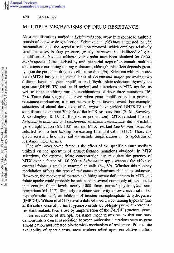
Subsequent studies showed a correlation between drug resistance and Hregion amplification in numerous independent lines and species. First, in-dependent selections of L. major with the drugs primaquine and terbinafine,which are structurally and mechanistically unrelated to MTX, yielded linesbearing only H amplification that were 10- to 20-fold cross-resistant to MTX(38). Second, independent MTX selections of L. major and Leishmaniatropica (including clonal derivatives) have yielded lines bearing H but notDHFR-TS amplification (36, 38, 53; S. M. Beverley, J, Cordingley, & D. D.Rogers, in preparation; Beverley, S. M. Iovannisci, P. F. Kamitsuka, J.Manning, & N. Mukhopadhyay, in preparation). Third, several unselectedlaboratory stocks of the lizard parasite L. tarentolae contained an unselectedamplification that turned out to be the equivalent of the L. major H regionamplification, with extensive sequence homology and the characteristic in-verted repeat structure (88, 117) (Figure 1). These lines exhibited up 20-fold resistance to MTX, and the H region copy number could be elevatedby MTX pressure. Fourth, selection ofL. mexicana amazonensis with sodiumarsenite yielded lines bearing amplification of the H region of this species.These lines were highly cross-resistant to MTX (33, 62). Correspondingly,H-amplified L. major exhibit varying levels of arsenite resistance (37; H. L.Callahan & S. M. Beverley, in preparation).
www.annualreviews.org/aronlineAnnual Reviews
Ann
u. R
ev. M
icro
biol
. 199
1.45
:417
-444
. Dow
nloa
ded
from
arj
ourn
als.
annu
alre
view
s.or
gby
Was
hing
ton
Uni
vers
ity L
ibra
ry, D
anfo
rth
Cam
pus
on 1
1/03
/07.
For
per
sona
l use
onl
y.
LEISHMANIA AMPLIFICATION 423
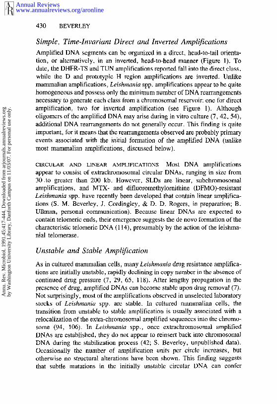
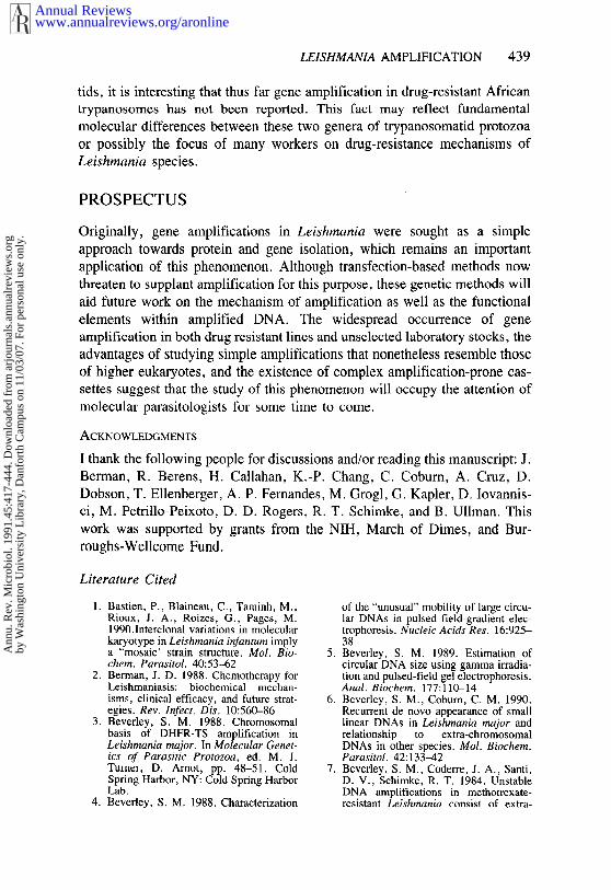
Direct Amplification Inverted Amplification
Figure 1 Amplified DNA structures characterized in Leishmania species. The boxes indicaterepetitive DNA sequences whose orieatations are indicated by small arrows and the heavy linesflanking chromosomal DNA.
The data summarized above reveal a clear correlation of H amplificationwith drug resistance, especially MTX resistance. However, the use of linesobtained by serial stepwise selection introduced an. element of uncertaintyresulting from the possibility of multiple mechanisms of MTX resistancediscussed earlier. One approach to this problem was to focus on lines selectedwith structurally unrelated drugs because the MTX resistance of these linescould probably be attributed solely to the H region amplification. Biochemicalstudies of the MTX resistant, H-amplified L. major obtained by terbinafineand primaquine selection revealed no alterations in MTX uptake, accumula-tion, or efflux (37) or increased MTX hydrolase activity (38). In contrast,alterations in MTX uptake were observed in all MTX-selected lines includingthose lacking 1-1 region amplification (36, 64). Thus far, no biochemicalchange likely to mediate MTX resistance has been associated with H regionamplification, suggesting that this region of DNA encodes a novel mode ofresistance.
Molecular studies of the H-amplified lines of L. major have shown that the45-kb H region is transcribed into at least 20 polyadenylated RNAs andencodes at least one protein (35, 83). The relative levels of these molecules not vary in response to heat shock or drug pressure (35). Interestingly, homologue of the P-glycoprotein or multiple drug-resistance (MDR) genesimplicated in multiple drug resistance in cultured mammalian cells (47) hasbeen identified within the H amplification of both L. tarentolae (ltpgA) (83)and L. major (ImpgpA) (H. L. Callahan & S. M. Beverley, in preparation).
www.annualreviews.org/aronlineAnnual Reviews
Ann
u. R
ev. M
icro
biol
. 199
1.45
:417
-444
. Dow
nloa
ded
from
arj
ourn
als.
annu
alre
view
s.or
gby
Was
hing
ton
Uni
vers
ity L
ibra
ry, D
anfo
rth
Cam
pus
on 1
1/03
/07.
For
per
sona
l use
onl
y.

424 BEVERLEY
Some P-glycoproteins mediate resistance by increasing drug export, andP-glycoprotein relatives in Drosophila melanogaster control the accumulationof pterin derivatives (34, 82) (recall that MTX is,~ pterin derivative). ever, a comparable role for P-glycoprotein genes in MTX resistance seemsunlikely because (a) MTX accumulation was~ not ~altered in the H-amplifiedlines selected by unrelated drugs (37); (b) MTX resistance was not reversedby verapamil, which does reverse the toxicity of many P-glycoprotein sub-strates (37; H. L. Callahan & S. M. Beverley, in preparation); and (c) H-amplified L. tarentolae was not resistant to a variety of compounds normal-ly considered to be substrates of the mammalian P-glycoprotein, such aspuromycin, vincristine, or adriamycin (83).
H REGION AMPLIFICATION CONFERS DRUG RESISTANCE Recently DNAtransfection approaches have been used to assess the coding potential of the Hregion (H. L. Callahan & S. M. Beverley, in preparation). A series of DNAfragments encompassing the wild-type L. major H region were inserted intomulticopy transfection vectors bearing the neomycin (NEO)-resistance gene,introduced into Leishmania by electroporation and G418 selection, and ampli-fied by increasing G418 pressure (60). When tested for drug resistance, single segment of the H region was shown to confer resistance to arsenite bythis protocol, and deletion studies localized the resistance gene to the MDRgene homologue lmpgpA mentioned previously. Consistent with previouswork, lmpgpA constructs did not confer MTX resistance, and preliminary dataindicate that the MTX resistance gene maps to another part of the H region(H. Callahan and S. M. Beverley, unpublished data). These data provide thefirst evidence that the H region encodes drug resistance determinants that arefunctional following gene amplification. Moreover, it suggests that thelmpgpA gene is functionally divergent from P-glycoprotein genes involved inclassical multiple drug resistance (49). Further studies are required to de-termine (a) if lmpgpA mediates arsenite resistance through reductions inarsenite accumulation, as expected for members of the P-glyc6protein genefamily and (b) if point mutations in ImpgpA or other genes confer altered drugresistance specificities as observed in mammalian MDR genes (24). Studiesof the MTX-resistance element encoded by the H region are currently under-way.
Interestingly, reports of a parallel for the association of arsenite and MTXresistance with gene amplification in Leishmania spp. have emerged formetazoans. Treatment of cultured mammalian cells with arsenite increases thefrequency of DHFR amplification (73) and induces the expression of glycoprotein mRNAs (23); however, P-glycoprotein expression alone doesnot confer MTX resistance. Whether these associations are functional orfortuitous is unknown.
www.annualreviews.org/aronlineAnnual Reviews
Ann
u. R
ev. M
icro
biol
. 199
1.45
:417
-444
. Dow
nloa
ded
from
arj
ourn
als.
annu
alre
view
s.or
gby
Was
hing
ton
Uni
vers
ity L
ibra
ry, D
anfo
rth
Cam
pus
on 1
1/03
/07.
For
per
sona
l use
onl
y.

LEISHMANIA AMPLIFICATION 425
While pre-existing amplification of the H region is clearly evident inunselected laboratory stocks of L. tarentolae (88, 117), some controversysurrounds the occurrence of pre-existing H region amplifications in the humanpathogen L. major. One group (53) has reported that two independent stabi-lates of the unselected LT252 line possess H region amplification. However,our (37) studies of an early-passage stabilate of this line, prepared prior selection of the first MTX-resistant line bearing R and H amplification(R1000) (7), have failed to detect any H amplification. Recent work ployed a sensitive PCR amplification method to detect H amplification-specific rearrangements (S. M. Beverley, D. M. Iovannisci, P. F. Kamitsuka,J. Manning, & N. Mukhopadhyay, in preparation). It is possible that theinstability of the H amplification may account for the discrepant findings (7).However, at present the existence of H-region amplification in unselected L.major is uncertain.
Tunicamycin and Glycosyltransferase
Chang and coworkers have studied a series of Leishmania spp. lines selectedwith tunicamycin (TUN), an inhibitor of the microsomal N-acetylglucos-amine-l-phosphate transferase. In most species, this enzyme is the first stepin the dolichol pathway for protein N-glycosylation. In other systems, TUNresistance results from altered uptake, structural alterations, or amplificationsof the glycosyltransferase (68, 74, 99, 108). Although the first two mech-anisms have not been tested in Leishmania spp., every TUN-resistant line ofLeishmania obtained thus far contains a DNA amplification and elevatedlevels of a TUN-sensitive glycosyl transferase activity, regardless of whethermutagenesis was included in the selection process (32, 63, 65, 86). Examina-tion of a variety of independent amplified lines from several species revealedthe presence of a 20-kb homologous region common to all amplifications(63). The common region is transcribed into at least five abundant RNAs,which translate in vitro into several protein bands (63; K.-P. Chang, personalcommunication). The common region may contain the glycosyltransferasestructural gene, but heterologous probing with the yeast gene has proveninconclusive despite early reports (63). The coding potential of the commonregion is currently being tested using DNA sequencing and transfectionstudies. This amplification is interesting ~because" it appears to be associatedwith a small but significant increase in parasite virulence (65, 66), although has not been encountered in unselected virulent strains.
ODC, IMPDH, and Classic MDR
Recent work by Ullman and coworkers suggests that gene amplification hasbeen obtained in response to at least three different agents in L. donovani.They (118) have reported amplification of the L. donovani IMPDH gene in
www.annualreviews.org/aronlineAnnual Reviews
Ann
u. R
ev. M
icro
biol
. 199
1.45
:417
-444
. Dow
nloa
ded
from
arj
ourn
als.
annu
alre
view
s.or
gby
Was
hing
ton
Uni
vers
ity L
ibra
ry, D
anfo
rth
Cam
pus
on 1
1/03
/07.
For
per
sona
l use
onl
y.
426 BEVERLEY
response to mycophenolic acid selection. Vinblastine-resistant lines containan amplification of a P-glycoprotein homologue distinct from the ImpgpAgene present on the H region and show collateral resistance to many of theclassical mammalian P-glycoprotein substrates (47; B. Ullman, personalcommunication). A line selected for resistance to a-difluoromethylornithinehas been obtained that exhibits unstable overproduction of ornithine de-carboxylase (ODC), a phenotype highly suggestive of gene amplification(29). These new amplifications are currently being characterized. Theiroccurrence emphasizes the fact that, as in mammalian cells, gene amplifica-tion in Leishmania spp. is probably a widespread phenomenon underlyingmany instances of drug resistance and is not restricted to any specific structur-al or functional class of compounds.
MISCELLANEOUS AMPLIFIED GENES
The powerful methods available for electrophoretic separation of Leishmaniaspp. chromosomes have revealed a variety of chromosomal alterations involv-ing gene amplification that are distinct from those reported above. Theseinclude the T amplification of L. tarentolae, the D amplification of L. trop-ica, subchromosomal amplifications of the LD- 1/715 class, and amplificationof the miniexon. Current data suggest that such chromosomal changes may becommon (6, 43, 56, 85; S. M. Beverley, J. Cordingley, & D. D. Rogers, inpreparation). The four kinds of amplifications discussed below are those thathave been characterized to date. These amplifications often do not involveDNAs of known function, and in most cases have not been associated withany phenotype.
T and D DNAs
The Trager line of L. tarentolae contains an amplified DNA, designated the Tregion (87). Hybridization studies revealed that the T amplification wasdistinct from other known amplifications, including the H region amplified inother lines ofL. tarentolae (87). The amplified T region contains about 20 of DNA, amplified as a circular dimeric molecule. This amplification isunusually ephemeral in that examination of the Trager line obtained fromother laboratories or following various periods of culture showed widelyvarying levels. T-amplified lines do not show drug resistance.
Electrophoretic surveys revealed the presence of an amplification termed DDNA in an unselected laboratory stock of L. tropica (53). This DNA con-tained approximately 75 kb of DNA and possessed an inverted repeat struc-ture. Hybridization studies revealed that this DNA was distinct from both theH and R regions, and selection of these cells with MTX led to the emergenceof H amplification but no changes in D DNA levels. Thus far, no phenotypehas been ascribed to either T or D DNA amplification.
www.annualreviews.org/aronlineAnnual Reviews
Ann
u. R
ev. M
icro
biol
. 199
1.45
:417
-444
. Dow
nloa
ded
from
arj
ourn
als.
annu
alre
view
s.or
gby
Was
hing
ton
Uni
vers
ity L
ibra
ry, D
anfo
rth
Cam
pus
on 1
1/03
/07.
For
per
sona
l use
onl
y.

LE1SHMANIA AMPLIFICATION 427
Miniexon
Studies of L. major revealed alterations in karyotype arising from amplifica-tions occurring within the locus encoding the miniexon donor RNA (56). ThisRNA provides the 39-nucleotide leader sequence added by trans-splicing ontothe 5’ end of all known protein-coding mRNAs in trypanosomatids (16) and encoded in the genome as a tandem repetitive array of 0.45-kb units inLeishmania spp. (56, 78). Chromosome 2 in L. major contains the miniexonarray, and several lines exhibiting alterations in this chromosome containedexpansions of the miniexon array. Clonal derivatives of the LT252A, in whichthe miniexon amplification was first observed, exhibit a growth advantage(56), although genetic tests are required to determine whether the miniexonamplification itself is responsible for the growth phenotype. Interestingly,amplification of the miniexon array has been observed in several L. majorisolates (56; S. Beverley, unpublished data). Whether this amplificationoccurs in nature or represents an adaptation to in vitro culture is currentlyunknown.
Subchromosomal Amplifications
Karyotypic surveys of numerous Leishmania species have revealed the occa-sional occurrence of relatively small chromosomes (250 kb or less) absent other isolates of the same species (6, 15, 40, 98, 107). These small linearDNAs (SLDs) can be distinguished from normal chromosomes by the fact thatthey occur in multiple copies, up to 40 per cell. In L. donovani, they weretermed the "HU-3" minichromosome (15) or LD-1 (98), and L. major theywere designated the 715-class of SLDs (6). Hybridization studies revealedthat these DNAs from different strains or species are similar but not identicalin size or sequence content (6). Because SLDs occur in some but not allisolates from numerous Leishmania species, researchers initially proposedthat they could represent the result of some form of horizontal transmission.Subsequent studies have revealed that all species of Leishmania contain areservoir copy of the sequences present on the new small chromosomes (6,14). Current data, though limited, are most consistent with the idea that theSLDs represent large, subchromosomal amplifications; as such they musthave acquired at least one new telomere during their formation. Their greatlyincreased copy number suggests that they replicate autonomously, and some-how escape mechanisms normally limiting chromosome number. In-terestingly, Stuart and coworkers (6, 40, 50, 107) have also described multicopy circular DNA, CD-1, which exhibits a complex relationship withcertain SLDs.
Although the antiquity of the SLDs detected in laboratory stocks of L.donovani, L. braziliensis, and L. rnexicana (6, 15, 40, 107) is unknown, thede novo generation of SLDs may have occurred in L. major and L. brazilien-sis (including a WHO reference strain) during routine culture in vitro and
www.annualreviews.org/aronlineAnnual Reviews
Ann
u. R
ev. M
icro
biol
. 199
1.45
:417
-444
. Dow
nloa
ded
from
arj
ourn
als.
annu
alre
view
s.or
gby
Was
hing
ton
Uni
vers
ity L
ibra
ry, D
anfo
rth
Cam
pus
on 1
1/03
/07.
For
per
sona
l use
onl
y.
428 BEVERLEY
two clonal derivatives of L. major undergoing MTX selection. In lines notknown to have.undergone drug pressure that bear SLDs, drug resistance hasnot been observed, and to date no biological role for these SLDs has beendemonstrated. Their recurrent emergence throughout most Leishmania spec-ies is provocative.
What Leads to the Emergence of Apparently NonfunctionalAmplified DNAs?
Why are amplified DNAs commonly observed in unselected laboratory stocksof Leishmania spp., and what is their significance to the parasite? This sectiondiscusses two models.
FUNCTIONAL SELECTION These amplifications may be functional but con-fer a phenotype not yet tested. For example, they may provide some subtlegrowth advantage during adaptation to culture in vitro or conceivably innature. This could provide a directed selective force responsible for themultiple independent occurrence of events such as amplification of theminiexon in L. major or SLDs in many Leishmania species. Recall that in the1970s a variety of amplified DNAs of unknown function and origins weresimilarly observed in cultured mammalian tumor cells (93). Subsequently,many of these amplifications were shown to contain functional oncogenes.
A variant of this model is that these amplifications are part of morecomplex biochemical resistance mechanisms, obligatorily requiring the pres-ence of another unlinked resistance gene. Segregation in the absence ofselective pressure could produce cells bearing only the amplification, whichwould appear to be nonfunctional. The best candidates for this kind of modelwould be those amplifications discovered in unselected laboratory stocks,such as T and D DNA and certain SLDs. Studies of chloroquine-rcsistantmalaria postulated an analogous two-step model (39, 81, 115); however, present this model remains only a possibility for Leishmania species.
NEUTRAL CHROMOSOMAL MUTATIONS Some chromosomal alterationsand amplifications may be approximately functionally neutral, constitutingchromosomal mutations with no functional significance. Because this modelpostulates no intrinsic selective force promoting fixation of these mutations,other mechanisms such as recurrent directed mutation and population forcesinvolving genetic drift must be invoked. Although other creatures exhibitexamples of these forces, their role and presence in Leishmania is unknown.Founder effects contributing to the emergence of variant phenotypes couldarise from multiple serial passages or cloning, (1). Another possibility hitchhiking, i.e. the occurrence of an unlinked advantageous mutation (suchas drug resistance) in a line containing a neutral chromosomal alteration or
www.annualreviews.org/aronlineAnnual Reviews
Ann
u. R
ev. M
icro
biol
. 199
1.45
:417
-444
. Dow
nloa
ded
from
arj
ourn
als.
annu
alre
view
s.or
gby
Was
hing
ton
Uni
vers
ity L
ibra
ry, D
anfo
rth
Cam
pus
on 1
1/03
/07.
For
per
sona
l use
onl
y.

LEISHMANIA AMPLIFICATION 429
amplification. A similar phenomenon, termed periodic selection, has beenreported in bacterial chemostat experiments (52, 67).
FREQUENCY OF GENE AMPLIFICATION
In cultured mammalian cells, .the frequency of gene amplification in drug-selected cells has been estimated to be from 10-3 to less than 10-8; this valuedepends on the specific cell line utilized (primary, established, or tumor), themethods of measurement, and whether positive or negative selection methodsare utilized (96, 110, 120). In Leishmania spp., studies of 20 independentclonal lines of L. major undergoing MTX selection revealed a frequency ofDHFR-TS or H region amplification of about 5 x 10⁻⁸ (S. M. Beverley, J.Cordingley, & D. D. Rogers, in preparation), a value considerably lower thanestimates for DHFR amplification in tumorigenic or cultured mammalian ~ellsusing comparable methods (about 10-5). The value for Leishmania spp. isconsistent with studies showing that the frequency of gene amplification innormal human cells is very low (120). Gene amplification does not generallyreduce the infectivity of Leishmania spp. to animals, cultured macrophages,and sand flies (10, 65, 86), and appears quite compatible with the normalinfectious cycle.
Many of the amplifications currently thought to be unrelated to drugresistance have been detected in drug-selected lines. For example, in the 20clonal derivatives of L. major described above that were selected formethotrexate resistance, 3 independent mutations apparently unrelated tomethotrexate resistance were observed, 2 of which belonged to the 715-classof subchromosomal amplifications (6, 56; S. Beverley, unpublished data).Similarly, in 10 lines maintained over a p~r!od, of at least 6 months, 2mutations unrelated to drug resistance were observed, 1 involving the miniex-on and 1 a SLD (6, 56). Although these data are not directly comparable(since hitchhiking effects will be greater in the drug-selected lines because ofthe low frequency of drug resistance), they provide no evidence for anelevated frequency of the apparently nonfunctional amplifications in L. majorundergoing drug pressure.
STRUCTURE OF EXTRACHROMOSOMALAMPLIFIED DNAs
The,,.~st,~ctures and’properties of several Leishmania spp. amplifications arek~nown in de~.ail. The general features emerging from these studies are de-scrib.e.d.:~bel_ow ~
www.annualreviews.org/aronlineAnnual Reviews
Ann
u. R
ev. M
icro
biol
. 199
1.45
:417
-444
. Dow
nloa
ded
from
arj
ourn
als.
annu
alre
view
s.or
gby
Was
hing
ton
Uni
vers
ity L
ibra
ry, D
anfo
rth
Cam
pus
on 1
1/03
/07.
For
per
sona
l use
onl
y.
430 BEVERLEY
Simple, Time-Invariant Direct and Inverted Amplifications
Amplified DNA segments can be organized in a direct, head-to-tail orienta-tion, or alternatively, in an inverted, head-to-head manner (Figure 1). date, the DHFR-TS and TUN amplifications reported fall into the direct class,while the D and prototypic H region amplifications are inverted. Unlikemammalian amplifications, Leishmania spp. amplifications appear to be quitehomogeneous and possess only the minimum number of DNA rearrangementsnecessary to generate each class from a chromosomal reservoir: one for directamplification, two for inverted amplification (see Figure 1). Althougholigomers of the amplified DNA may arise during in vitro culture (7, 42, 54),additional DNA rearrangements do not generally occur. This finding is quiteimportant, for it means that the rearrangements observed are probably primaryevents associated with the initial formation of the amplified DNA (unlikemost mammalian amplifications, discussed below).
CIRCULAR AND LINEAR AMPLIFICATIONS Most DNA amplificationsappear to consist of extrachromosomal circular DNAs, ranging in size from30 .to greater than 200 kb. However, SLDs are linear, subchromosomalamplifications, and MTX- and difluoromethylornithine (DFMO)-resistantLeishmania spp. have recently been developed that contain linear amplifica-tions (S. M. Beverley, J. Cordingley, & D. D. Rogers, in preparation; B.Ullman, personal communication). Because linear DNAs are expected tocontain telomeric ends, their emergence suggests the de novo formation of thecharacteristic telomeric DNA (114), presumably by the action of the leishma-nial telomerase.
Unstable and Stable Amplification
As in cultured mammalian cells, many Leishmania drug resistance amplifica-tions are initially unstable, rapidly declining in copy number in the absence ofcontinued drug pressure (7, 29, 65, 118). After lengthy propagation in thepresence of drug, amplified DNAs can become stable upon drug removal (7).Not surprisingly, most of the amplifications observed in unselected laboratorystocks of Leishmania spp. are stable. In cultured mammalian cells, thetransition from unstable to stable amplification is usually associated with arelocalization of the extra-chromosomal amplified sequences into the chromo-some (94, 106). In Leishmania spp., once extrachromosomal amplifiedDNAs are established, they do not appear to reinsert back into chromosomalDNA during the stabilization process (42; S. Beverley, unpublished data).Occasionally the number of amplification units per circle increases, butotherwise no structural alterations have been shown. This finding suggeststhat subtle mutations in the initially unstable circular DNA can confer
www.annualreviews.org/aronlineAnnual Reviews
Ann
u. R
ev. M
icro
biol
. 199
1.45
:417
-444
. Dow
nloa
ded
from
arj
ourn
als.
annu
alre
view
s.or
gby
Was
hing
ton
Uni
vers
ity L
ibra
ry, D
anfo
rth
Cam
pus
on 1
1/03
/07.
For
per
sona
l use
onl
y.

LE1SHMANIA AMPLIFICATION 431
stability, or that stability is conferred by factors or mutations acting in trans(11).
The lack of knowledge concerning chromosomal elements mediatingkaryotypic stability in Leishmonia hinders studies of the stability of amplifiedLeishmonia DNA. Mitotic chromosomes do not condense, and althoughstructures resembling kinetochores have been reported in other trypanosoma-tids their role is dubious because their number is considerably less than thenumber of chromosomes detected using pulsed-field electrophoresis (45,104). Whether Leishmania chromosomes possess functional localized centro- ¯meres or whether they are holocentric, with centromeric activity dispersedover the entire chromosome as in nematodes and some arthropods (116) unknown.
Recurrence of Rearrangements Involving RepetitiveDNA Sequences
Frequently, the same amplified DNA structures are observed in independentlines. Six of nine independent amplifications of the DHFR-TS region utilizedthe same site, and six of six independent amplifications of the H region of L.major have the same structure (10, 37; S. M. Beverley, J. Cordingley, & D.D. Rogers, in preparation; Beverley, S. M. Iovannisci, P. F. Kamitsuka, J.Manning, & N. Mukhopadhyay, in preparation). Current data indicate thatthese rearrangements occur within repetitive DNA sequences. The DHFR-TSgene is flanked by two 600-bp elements separated by 30 kb, and the circularamplified genes are preferentially formed by joining of these two elements(10; S. M. Beverley, J. Cordingley, & D. D. Rogers, in preparation) (Figure1). Similarly, the H region of L. major is flanked by two different pairs ofinverted repeats (Figure 1), which are joined during amplification to form thetwo new rearrangements required (S. M. Beverley, D. M. Iovannisci, P. F.Kamitsuka, J. Manning, & N. Mukhopadhyay, in preparation). Similar datawere recently reported for L. tarentolae (84).
Functional Genetic Elements Within Amplified DNAs
The ability of the extrachromosomal amplified DNAs to persist and direct thesynthesis of encoded gene products has led many workers to conclude thatthese are autonomous elements. Kapler et al (60) explicitly tested this conclu-sion in the R region amplification of L. major, where a modified version ofthis region introduced by DNA transfection replicated autonomously anddirected the efficient formation of a correctly trans-spliced RNA. Smallerderivatives of this plasmid are also equivalently functional (71), and as theseDNAs are dissected further the identification of cis-acting elements involvedin DNA replication, segregation, initiation of transcription, and RNA pro-cessing should follow. Origins of replication would be especially interesting
www.annualreviews.org/aronlineAnnual Reviews
Ann
u. R
ev. M
icro
biol
. 199
1.45
:417
-444
. Dow
nloa
ded
from
arj
ourn
als.
annu
alre
view
s.or
gby
Was
hing
ton
Uni
vers
ity L
ibra
ry, D
anfo
rth
Cam
pus
on 1
1/03
/07.
For
per
sona
l use
onl
y.

432 BEVERLEY
to study in an amplification context. Curiously, many Lei~hmania DNAsegments can apparently replicate autonomously (60, 69, 71). Because linearamplifications may have acquired telomeres de novo, characterization ofthese should provide some insight into the structure and mechanism oftelomere formation in Leishmania species.
Comparisons with Amplified DNAs in CulturedMammalian Cells
Comparisons of the structural features of amplified Leishmania genes withthose observed in cultured mammalian cells, summarized in several recentreviews (95, 96, 105), are interesting. One is immediately struck by thecomplexity of amplified mammalian DNA: large tracts of DNA can beamplified, possibly as much as 10 megabases. The structures of these ampli-fied DNAs are usually quite heterogeneous, containing multiple rearrange-ments or novel joints. The structure of the amplified DNA also varies, bothwithin and between different cell lines. As a function of increased drugpressure and time, additional DNA rearrangements accumulate, and occa-sionally smaller amplification units predominate. In contrast, secondary rear-rangements are generally not observed in Leishmania species.
The simplicity of amplification in Leishmania spp. could be attributed tothe small size of the Leishmania genome and chromosomes; a 30-kbamplification in Leishmania is proportionally 1.8 Mb in mammalian cells.Another source could be the propensity of Leishmania spp. to undergohomologous recombination (31, 111), accounting for the frequent occurrenceof rearrangements joining homologous repetitive DNAs. Regardless of thecause, the relative simplicity of amplified DNAs in Leishmania spp. greatlyfacilitates their analysis.
Three Chromosomal Types of Amplification
Pulsed-field methods of chromosomal analysis have permitted an examinationof the fate of the wild-type chromosomal genes during generation of theamplified DNAs. Thus far, three outcomes have been characterized (Figures and 3):
1. In deletional amplification, a copy of.the wild-type chromosomal locus isdeleted, yielding a smaller chromosome. Because Leishmania spp. arediploid, the result is a heterozygous deletion line (10, 31; S. M. Beverley,J. Cordingley, & D. D. Rogers, in preparation).
2. In conservative amplification, no alterations in chromosomal structure orploidy are detected (7, 10, 37, 88; S. M. Beverley, J. Cordingley, & D. D.Rogers, in preparation).
3. In duplicative amplification, several additional gene copies are found
www.annualreviews.org/aronlineAnnual Reviews
Ann
u. R
ev. M
icro
biol
. 199
1.45
:417
-444
. Dow
nloa
ded
from
arj
ourn
als.
annu
alre
view
s.or
gby
Was
hing
ton
Uni
vers
ity L
ibra
ry, D
anfo
rth
Cam
pus
on 1
1/03
/07.
For
per
sona
l use
onl
y.
LE1SHMANIA AMPLIFICATION 433
+ 4"
deletional conservative duplicative
Figure 2 Three chromosomal types of amplification. The thick bars represent flanking chromo-
somal DNA, and the thin lines are the DNA segments that give rise to the extrachromosomal
circular DNA in addition to being either deleted, retained, or duplicated.
inserted into the wild-type locus, yielding a larger chromosome in additionto the ]amplified extrachromosomal copies (S. M. Beverley, J. Cordingley,& D. D. Rogers, in preparation).
Thus far, the majority of events characterized fall into the conservativeclass; only one example each of the deletional and duplicative classes hasbeen observed, both involving DHFR-TS (10; S. M. Beverley, J. Cordingley,& D. D. Rogers, in preparation). Chromosomal changes are evident in somearsenite-resistant L. mexicana amazonensis, but these have not been furthercharacterized (33). Some eases of deletional amplification may have beenmissed because hybridization probes flanking the amplified region are notalways employed to reveal deletion chromosomes obscured in ethidium bro-mide-stained gels.
MECHANISM OF GENE AMPLIFICATION
Several models have emerged from studies of amplification in culturedmammalian cells, including: (a) unscheduled rereplication of the wild-typechromosomal locus during a single cell cycle, followed by resolution into thefinal amplified DNA (51, 92, 94, 106); (b) duplications formed by unequalsister-chromatid exchange (46, 105, 106); and (c) models invoking recom-binations in the absence of rereplication, such as the deletion plus episomemodel in which the wild-type chromosomal locus is converted into an ex-
www.annualreviews.org/aronlineAnnual Reviews
Ann
u. R
ev. M
icro
biol
. 199
1.45
:417
-444
. Dow
nloa
ded
from
arj
ourn
als.
annu
alre
view
s.or
gby
Was
hing
ton
Uni
vers
ity L
ibra
ry, D
anfo
rth
Cam
pus
on 1
1/03
/07.
For
per
sona
l use
onl
y.

434 BEVERLEY
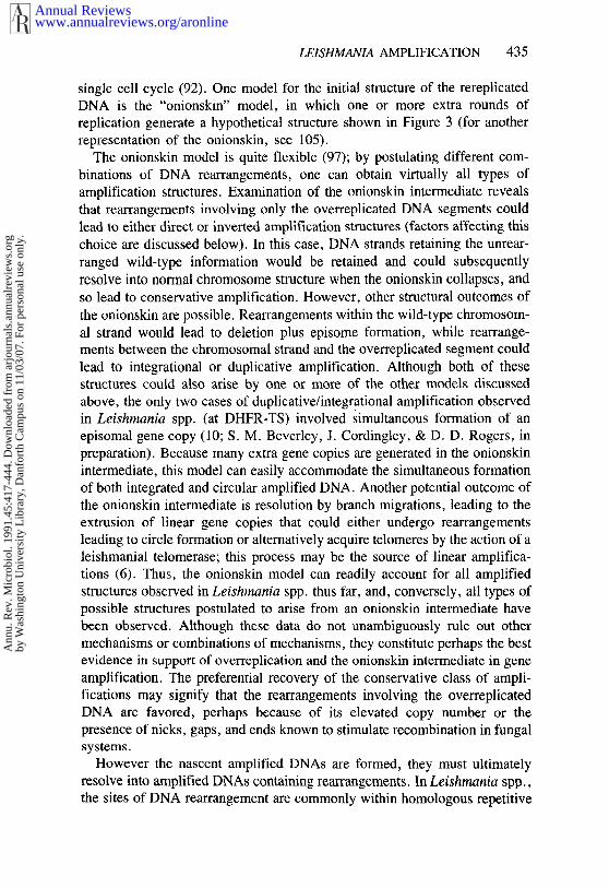
over-replication
potential products
7-ol I
(.deletion
I 7-~1linear
duplication
Figure 3 Formation of the onionskin intermediate and potential products. Overreplication givesrise to the characteristic onionskin structure; both DNA strands are shown in the upper half of thisfigure. The vertical black bars indicate repetitive DNAs that are the sites of DNA rearrangement(see Figure 1 for an expanded view). Potential products formed by resolution of the onionskinintermediate by recombination among the repetitive DNAs or by branch migration and telomereaddition are shown.
trachromosomal plasmid by intrachromosomal recombination (21, 97). Thediversity of amplified structures has led some workers to postulate thatmammalian cells may employ several different mechanisms that vary amongloci or during different amplification steps (105).
If one considers only the Leishmania spp. amplifications that mediate drugresistance whose structures have been characterized, the majority appearconservative: changes in the wild-type chromosomal structure or ploidy areseen only rarely. This observation probably rules out models requiring altera-tion of the parental chromosome structure, such as sister chromatid exchangeor recombination in the absence of rereplication (among others). Therefore,the amplified DNAs are likely generated from extra copies of the chromosom-al locus, which are formed by some kind of rereplication occurring within a
www.annualreviews.org/aronlineAnnual Reviews
Ann
u. R
ev. M
icro
biol
. 199
1.45
:417
-444
. Dow
nloa
ded
from
arj
ourn
als.
annu
alre
view
s.or
gby
Was
hing
ton
Uni
vers
ity L
ibra
ry, D
anfo
rth
Cam
pus
on 1
1/03
/07.
For
per
sona
l use
onl
y.
LE1SHMAN1A AMPLIFICATION 435
single cell cycle (92). One model for the. initial structure of the rereplicatedDNA is the "onionskin" model, in which one or more extra rounds ofreplication generate a hypothetical structure shown in Figure 3 (for anotherrepresentation of the onionskin, see 105).
The onionskin model is quite flexible (97); by postulating different com-binations of DNA rearrangements, one can obtain virtually all types ofamplification structures. Examination of the onionskin intermediate revealsthat rearrangements involving only the overreplicated DNA segments couldlead to either direct or inverted amplification structures (factors affecting thischoice are discussed below). In this case, DNA strands retaining the unrear-ranged wild-type information would be retained and could subsequentlyresolve into normal chromosome structure when the onionskin collapses, andso lead to conservative amplification. However, other structural outcomes ofthe onionskin are possible. Rearrangements within the wild-type chromosom-al strand would lead to deletion plus episome formation, while rearrange-ments between the chromosomal strand and the overreplicated segment couldlead to integrational or duplicative amplification. Although both of thesestructures could also arise by one or more of the other models discussedabove, the only two cases of duplicative/integrational amplification observedin Leishmania spp. (at DHFR-TS) involved simultaneous formation of episomal gene copy (10; S. M. Beverley, J. Cordingley, & D. D. Rogers, inpreparation). Because many extra gene copies are generated in the onionskinintermediate, this model can easily accommodate the simultaneous formationof both integrated and circular amplified DNA. Another potential outcome ofthe onionskin intermediate is resolution by branch migrations, leading to theextrusion of linear gene copies that could either undergo rearrangementsleading to circle formation or alternatively acquire telomeres by the action of aleishmanial telomerase; this process may be the source of linear amplifica-tions (6). Thus, the onionskin model can readily account for all amplifiedstructures observed in Leishmania spp. thus far, and, conversely, all types ofpossible structures postulated to arise from an onionskin intermediate havebeen observed. Although these data do not unambiguously rule out othermechanisms or combinations of mechanisms, they constitute perhaps the bestevidence in support of overreplication and the onionskin intermediate in geneamplification. The preferential recovery of the conservative class of ampli-fications may signify that the rearrangements involving the overreplicatedDNA are favored, perhaps because of its elevated copy number or thepresence of nicks, gaps, and ends known to stimulate recombination in fungalsystems.
However the nascent amplified DNAs are formed, they must ultimatelyresolve into amplified DNAs containing rearrangements. In Leishmania spp.,the sites of DNA rearrangement are commonly within homologous repetitive
www.annualreviews.org/aronlineAnnual Reviews
Ann
u. R
ev. M
icro
biol
. 199
1.45
:417
-444
. Dow
nloa
ded
from
arj
ourn
als.
annu
alre
view
s.or
gby
Was
hing
ton
Uni
vers
ity L
ibra
ry, D
anfo
rth
Cam
pus
on 1
1/03
/07.
For
per
sona
l use
onl
y.
436 BEVERLEY
sequences, "hotspots" often used in multiple independent lines; in contrast, inmammalian cells amplification rearrangements are not generally recurrent norpreferentially associated with homologous repetitive DNAs. This finding mayreflect intrinsic differences in the activity of recombination pathways: Leish-mania spp. and other unicellular eukaryotes appear to possess powerfulsystems mediating homologous recombination among DNAs as much as1,000 times more active than those observed in mammalian cells whenmeasured by the relative frequencies of homologous gene targeting (20, 31,109). These systems could direct DNA rearrangements towards homologousrepeated DNA sequences in Leishmania spp. and give rise to hotspots forDNA rearrangement. One prediction of this model is that genes flanked bydirect repeats should undergo direct-type amplification, while genes flankedby inverted repeats should undergo inverted amplification. The amplificationscharacterized thus far have upheld this prediction: direct amplifications of theR region usually join flanking direct repeats, while inverted amplifications ofthe H region join two flanking sets of inverted repeats (Figure 1), and directamplifications of DNA overlapping a portion of the H region join two directlyrepeating P-glycoprotein genes (84).
Thus, amplification in Leishmania spp. likely occurs by the over-replication/recombination model (3, 92, 94, 95), in which homologous repeti-tive DNA sequences usually direct the sites of recombination. White et al(117) proposed a similar model in which overreplication of the amplifiedregion is initiated by DNA synthesis primed by the repetitive elementsthemselves (as opposed to initiating from an internal element as depicted inFigure 3--possibly a chromosomal origin of replication). Because mostrecombinational mechanisms require strand invasion and subsequent DNAresynthesis, the amplified DNAs are probably ultimately formed by a com-bination of these two mechanisms. The final step in the amplification mech-anism is that the circular DNA, once formed, must increase in copy number.Whether this increase results from unequal segregation or an escape fromnormal mechanisms regulating copy number is currently unknown.
ROLE OF GENE AMPLIFICATION IN LEISHMANIABIOLOGY AND EVOLUTION
Clinical Drug Resistance
Most of the agents used in laboratory studies of drug resistance and geneamplification are not employed in clinical treatment of leishmaniasis,although proteins such as DHFR-TS are likely to be targeted in the future(103). Recent studies of the H region amplification suggest that this regioncould be involved in the parasite’s response to clinically utilized organicantimonial compounds. In addition to conferring resistance to arsenite, over-
www.annualreviews.org/aronlineAnnual Reviews
Ann
u. R
ev. M
icro
biol
. 199
1.45
:417
-444
. Dow
nloa
ded
from
arj
ourn
als.
annu
alre
view
s.or
gby
Was
hing
ton
Uni
vers
ity L
ibra
ry, D
anfo
rth
Cam
pus
on 1
1/03
/07.
For
per
sona
l use
onl
y.

LEISHMANIA AMPLIFICATION 437
expression of the H region lmpgpA gene confers resistance to trivalent anti-monials [Sb(III)] when tested using DNA transfection (H. L. Callahan & M. Beverley, in preparation). Many workers believe that the pentavalentantimonial derivatives are not the active form and are metabolically alteredinto an active species, possibly into Sb(llI); however, definitive evidence hasnot been presented. If the Sb(III) model is correct, the lmpgpA gene couldmodulate the toxicity of clinical antimonials, perhaps by altering Sb(III)accumulation. In a manner consistent with this model, agents such as ver-apamil that are known to inhibit the action of P-glycoprotein family membershave been shown to reverse antimonial resistance in L. donovani (79).Moreover, monoclonal antibodies directed against the mammalian P-glycoprotein recognize proteins in Leishmania spp.--these proteins are moreabundant in some antimonial-resistant lines (48). One might further speculatethat the reportedly pre-existing H region amplifications in human-infectingLeishmania spp. may have been induced by clinical antimony treatment,although this idea has not been proven. Future studies are required to de-finitively address the role of the lmpgpA gene in antimony sensitivity andImpgpA amplification in clinical antimony resistance.
Amplified Genes in Leishmania: a Bridge BetweenProkaryotic Resistance Factors and Mammalian GeneAmplifications
In many ways, amplified Leishmania DNAs occupy a position intermediatebetween the drug-resistance plasmids of prokaryotes and the amplified DNAsof cultured mammalian ceils. Like the prokaryotic plasmids (and somemammalian amplifications), they are generally extrachromosomal circularDNAs, occasionally bearing more than one drug-resistance element. Likemammalian amplifications, they arise from a chromosomal reservoir de novo,and can arise as unstable or stable forms. However, the differences are alsoinstructive: unlike prokaryotic resistance factors, amplified LeishmaniaDNAs do not appear to contain gene functions that facilitate their transferamongst cells. Unlike mammalian amplifications, frequently the same struc-ture is observed in independent amplifications in many Leishmania species.
One model for Leishmania DNA amplification is that these organismscontain within their genome amplification-prone cassettes that participate inan amplification and loss cycle. When subjected to selective stresses, in-formation built into the wild-type genome (such as flanking repeats) directsthe formation of characteristic amplified DNA structures; although most cellsperish, the small proportion undergoing amplification survive. Once theselective pressure abates, the extrachromosomal DNAs are lost, leavingbehind the original locus. This kind of amplification and loss model in-corporates features of both prokaryotic and eukaryotic systems discussed
www.annualreviews.org/aronlineAnnual Reviews
Ann
u. R
ev. M
icro
biol
. 199
1.45
:417
-444
. Dow
nloa
ded
from
arj
ourn
als.
annu
alre
view
s.or
gby
Was
hing
ton
Uni
vers
ity L
ibra
ry, D
anfo
rth
Cam
pus
on 1
1/03
/07.
For
per
sona
l use
onl
y.

438 BEVERLEY
above. However, for most loci we have little information about the obligatoryrole of specific flanking elements in directing the site of DNA amplification,and the amplification structures probably arise from utilization of fortuitouslyplaced repeated sequences.
The H region seems like an excellent candidate for the amplificationcassette model, as this model suggests a reason for the conservation of theunique wild-type structure (shown in Figure 1) in both L. major and L.tarentolae (84; S. M. Beverley, D. M. Iovannisci, P. F. Kamitsuka, J.Manning, & N. Mukhopadhyay, in preparation), which diverged as much as50-100 million years ago (13; K. Nelson & S. M. Beverley, in preparation).The flanking inverted-repeat pairs evolve more rapidly than internal H regionloci and have no known coding potential (S. M. Beverley, D. M. Iovannisci,P. F. Kamitsuka, J. Manning, & N. Mukhopadhyay, in preparation). Thus,their sole role may be in directing the site of H region amplification. The Hregion may then be seen as a reservoir of amplification-based drug resistancemechanisms, encoding at least two functional drug resistance genes (mediat-ing resistance to certain metals and antifolates) and having the coding poten-tial for many more (35) (genes mediating resistance to other agents known induce H region amplification have not yet been sought). Because H regiongenes have been retained as readily amplified DNA segments during evolu-tion, one can postulate that this persistence provides some evolutionaryadvantage to the parasite population. Thus, Leishmania spp. may be seen aspossessing composite drug resistance factors analogous to those of pro-karyotes.
Role of Amplification in Shaping the Parasite Genome
Gene amplification is a mechanism commonly employed during evolution,and it undoubtedly plays a similar role in shaping the parasite genome. Manyworkers have noted that most genes in trypanosomatid protozoa are present inmultiple linked copies, including many genes normally present in one or a fewcopies in other taxa (25). A surprising fact is that in the current literaturesingle-copy genes are the exception. In Leishmania spp., examples of repeat-ed gene families include the miniexon and ribosomal RNA clusters (56, 75,78), a- and /3-tubulins (55, 70), a putative transporter (18), and proteinsrecognized by immune sera such as the surface antigens gp63 (17) and gp46(76), and the 70- and 83-kilodalton (kd) heat shock proteins (72, Amplification of genes provides not only abundant quantitative changes, butalso new substrates for evolution because extra amplified copies can receivepoint mutations and evolve new functions while leaving the original copiesunchanged.
Given the widespread occurrence of amplification in drug-resistant Leish-mania spp., and the widespread occurrence of gene families in trypanosoma-
www.annualreviews.org/aronlineAnnual Reviews
Ann
u. R
ev. M
icro
biol
. 199
1.45
:417
-444
. Dow
nloa
ded
from
arj
ourn
als.
annu
alre
view
s.or
gby
Was
hing
ton
Uni
vers
ity L
ibra
ry, D
anfo
rth
Cam
pus
on 1
1/03
/07.
For
per
sona
l use
onl
y.
LEISHMANIA AMPLIFICATION 439
tids, it is interesting that thus far gene amplification in drug-resistant Africantrypanosomes has not been reported. This fact may reflect fundamentalmolecular differences between these two genera of trypanosomatid protozoaor possibly the focus of many workers on drug-resistance mechanisms ofLeishmania species.
PROSPECTUS
Originally, gene amplifications in Leishmania were sought as a simpleapproach towards protein and gene isolation, which remains an importantapplication of this phenomenon. Although transfeetion-based methods nowthreaten to supplant amplification for this purpose, these genetic methods willaid future work on the mechanism of amplification as well as the functionalelements within amplified DNA. The widespread occurrence of geneamplification in both drug resistant lines and unselected laboratory stocks, the
advantages of studying simple amplifications that nonetheless resemble thoseof higher eukaryotes, and the existence of complex amplification-prone cas-settes suggest that the study of this phenomenon will occupy the attention ofmolecular parasitologists for some time to come.
ACKNOWLEDGMENTS
I thank the following people for discussions and/or reading this manuscript: J.Berman, R. Berens, H. Callahan, K.-P. Chang, C. Coburn, A. Cruz, D.Dobson, T. Ellenberger, A. P. Fernandes, M. Grogl, G. Kapler, D. Iovannis-ci, M. Petrillo-Peixoto, D. D. Rogers, R. T. Schimke, and B. Ullman. Thiswork was supported by grants from the NIH, March of Dimes, and Bur-roughs-Wellcome Fund.
Literature Cited
1. Bastien, P., Blaineau, C., Taminh, M.,Rioux, J. A., Roizes, G., Pages, M.1990.Interclonal variations in molecularkaryotype in Leishmania infantum implya "mosaic’ strain structure. Mol. Bio-chem. Parasitol. 40:53-62
2. Berman, J. D. 1988. Chemotherapy forLeishmaniasis: biochemical ~nechan-isms, clinical efficacy, and future strat-egies. Rev. Infect. Dis. 10:560-86
3. Beverley, S. M. 1988. Chromosomalbasis of DHFR-TS amplification inLeishmania major. In Molecular Genet-ics of Parasitic Protozoa, ed. M. J.Turner, D. Arnot, pp. 48-51. ColdSpring Harbor, NY: Cold Spring HarborLab.
4. Beverley, S. M. 1988. Characterization
of the "unusual" mobility of large circu-lar DNAs in pulsed field-gradient elec-trophoresis. Nucleic Acids Res. 16:925-38
5. Beverley, S. M. 1989. Estimation ofcircular DNA size using gamma irradia-tion and pulsed-field gel electrophoresis.Anal. Biochem. 177:110-14
6. Beverley, S. M., Coburn, C. M. 1990.Recurrent de novo appearance of smalllinear DNAs in Leishmania major andrelationship to extra-chromosomalDNAs in other species. Mol. Biochem.Parasitol. 42:133-42
7. Beverley, S. M., Coderre, J. A., Santi,D. V., Schimke, R. T. 1984. UnstableDNA amplifications in methotrexate-resistant Leishmania consist of extra-
www.annualreviews.org/aronlineAnnual Reviews
Ann
u. R
ev. M
icro
biol
. 199
1.45
:417
-444
. Dow
nloa
ded
from
arj
ourn
als.
annu
alre
view
s.or
gby
Was
hing
ton
Uni
vers
ity L
ibra
ry, D
anfo
rth
Cam
pus
on 1
1/03
/07.
For
per
sona
l use
onl
y.

440 BEVERLEY
chromosomal circles which relocalizeduring stabilization. Cell 38:431 39
8. Deleted in proof9. Beverley, S. M., Ellenberger, T. E.,
Cordingley, J. S. 1986. Primary struc-ture of the gene encoding the bifunction-al dihydrofolate reductase-thymidylatesynthase of Leishmania major. Proc.Natl. Acad. Sci. USA 83:2584-88
10. Beverley, S. M., Ellenberger, T. E.,lovannisci, D. M., Kapler, G. M., Pet-rillo-Peixoto, M., Sina, B. J. 1988.Gene amplification in Leishmania. InThe Biology of Parasitism, ed. P. T.Englund, A. Sher, pp. 431-448. NewYork: Liss
11. Beverley, S. M., Ellenberger, T. E.,Petrillo-Peixoto, M. 1989. Unstable andstable gene amplification in methotre-xate-resistant Leishmania major and na-tural isolates of Leishmania tarentolae.See Ref. 51a, pp. 885-90
12. Deleted in proof13. Beverley, S. M., Ismach, R. B., McMa-
hon-Pratt, D. 1987. Evolution of thegenus Leishmania as revealed by com-parisons of nuclear DNA restrictionfragment patterns. Proc. Natl. Acad.Sci. USA 84:484-88
14. Bishop, R. P. 1990. Extensive homolog-ies between Leishmania donovanichromosomes of markedly differentsize. Mol. Biochem. Parasitol. 38:1-12
15. Bishop, R. P., Miles, M. A. 1987.Chromosome size polymorphisms ofLeishmania donovani. Mol. Biochem.Parasitol. 24:263-72
16. Borst, P. 1986. Discontinuous transcrip-tion and antigenic variation in trypano-somes. Annu. Rev. Biochem. 55:701-32
17. Button, L, L., Russell, D. G., Klein,H., Medina-Acosta, E., Karess, R. E.,McMaster, W. R. 1989. Genes encodingthe major surface glycoprotein in Leish-mania are tandemly linked at a singlechromosomal locus and are consti-tutively transcribed. Mol. Biochem. Pa-rasitol. 32:271-84
18. Cairns, B. R., Collard, M. W., Land-fear, S. M. 1989, Developmentally reg-ulated gene from Leishmania encodes aputative membrane transport protein.Proc. Natl. Acad. Sci. USA 86:7682-86
19. Dcleted in proof20. Capecchi, M. 1990. How efficient can
you get?. Nature 348:109~9.21. Carroll, S. M., DeRose, M. L., Gaud-
ray, P., Moore, C. M., Needham-Vandevanter, D. R., et al. 1988. Doubleminute chromosomes can be producedfrom precursors derived from a chromo-somal deletion. Mol. Cell. Biol. 8:1525-33
22. Chaudhuri, G., Chang, K. P. 1988.Acid protease activity of a major surfacemembrane g|ycoprotein (gp63) fromLeishmania mexicana promastigotes.Mol. Biochem. Parasitol. 27:43-52
23. Chin, K.-V., Tanaka, S., Darlington,G., Pastan, I., Gottesman, M. M. 1990.Heat shock and arsenite increase expres-sion of the multidrug resistance (MDR1)gene in human renals carcinoma cells. J.Biol. Chem. 265:221-26
24. Choi, K. C. J., Chen, M. K., Roninson,I. B. 1988. An altered pattern of cross-resistance in multidrug-resistant humancells results from spontaneous mutationsin the mdrl (P-glycoprotein) gene. Cell53:51%29
25. Clayton, C. E. 1988. The molecular bi-ology of the Kinetoplastidae. Genet.Eng. 7:1-56
26. Cobum, C. M., Otteman, K., McNeely,T., Turco, S., Beverley, S. M. 1991.Stable transfection of a wide range oftrypanosomatids. Mol. Biochem. Para-sitol. In press
27. Coderre, J. A., Beverley, S. M.,Schimke, R. T., Santi, D. V. 1983.Overproduction of a bifunctional thy-midylate synthase-dihydrofolate reduc-tase and DNA amplification in methotre-xate-resistant Leishmania. Proc. Natl.Acad. Sci. USA 80:2132-36
28. Comeau, A. M., Miller, S. I., Wirth, D.F. 1986. Chromosome location of fourgenes in Leishmania. Mol. Biochem.Parasitol. 21:161-69
29. Coons, T., Bitonti, A. J., McCann, P.P., Ullman, B. 1990. a-Difluoromethyl-omithine resistance in Leishmania dono-vani associated with increased omithinedecarboxylase activity. Mol. Biochem.Parasitol. 39:77-89
30. Croft, S. L. 1986. In vitro screens in theexperimental chemotherapy of leish-maniasis and trypanosomiasis. Para-sitol. Today 2:64-69
31. Cruz, A., Beverley, S. M. 1990. Genereplacement in parasitic protozoa. Na-ture 348:171-74
32. Detke, S., Chaudhuri, G., Kink, J. A.,Chang, K. P. 1988. DNA amplificationin tunicamycin-resistant Leishmaniamexicana. Multiple copies of a single 63kilobase supercoiled molecule and theirexpression. J. Biol. Chem. 263:3418-24
33. Detke, S., Katakura, K., Chang, K. P.1989. DNA amplification in arsenite-resistant Leishmania, Exp. Cell Res.180:161-70
34. Dreesen, T. D., Johnson, D. H., Heni-koff, S. 1988. The brown protein ofDrosophila melanogaster is similar tothe white protein and to components of
www.annualreviews.org/aronlineAnnual Reviews
Ann
u. R
ev. M
icro
biol
. 199
1.45
:417
-444
. Dow
nloa
ded
from
arj
ourn
als.
annu
alre
view
s.or
gby
Was
hing
ton
Uni
vers
ity L
ibra
ry, D
anfo
rth
Cam
pus
on 1
1/03
/07.
For
per
sona
l use
onl
y.

LEISHMAN1A AMPLIFICATION 441
active transport complexes. Mol. Cell.Biol. 8:5206-15
35. Ellenberger, T. E. 1989. Geneamplification and pleiotropic drug re-sistance in the human parasite Leish-mania. PhD Thesis. 1:1-196. Boston,MA: Harvaxd Univ.
36. Ellenbcrgcr, T. E., Beverley, S. M.1987. Reductions in methotrcxate andfolate influx in methotrexate-resistantlines of Leishmania major arc in-dependent of R or H region amplifica-tion. J. Biol. Chem. 262:13501-6
37. Ellenberger, T. E., Beverley, S. M.1989. Multiple drug resistance and con-servative amplification of the H regionin Leishmania major. J. Biol. Chem.264:15094-103
38. Ellenberger, T. E., Wright, J. E.,Rosowsky, A., Beverley, S. M. 1989.Wild-type and drug-resistant Leishmaniamajor hydrolyze methotrexate to N 10-methyl-4-deoxy-4-aminopteroate with-out accumulation of methotrexate poly-glutamates. J. Biol. Chem. 264:1596(~66
39. Foote, S. J., Kyle, D. E., Martin, R.K., Oduola, A. M. J., Forsyth, K., et al.1990. Several alleles of the multidrug-resistance gene are closely linked tochloroquine resistance in Plasmodiumfalciparum. Nature 345:255-58
40. Gajendran, N., Dujardin, J.-C., Ray, D.L., Matthyssens, G., Muyldermans, S.,Hammers, R. 1989. Abnormally migrat-ing chromosome identifies Leishmaniadonovani populations. See Ref. 51a, pp.549-47
41. Garvey, E. P., Coderre, J. A., Santi, D.V. 1985. Selection and properties ofLeishmania resistant to 10-propargyl-5,8-dideazafolate, an inhibitor of thy-midylate synthase. Mol. Biochem. Para-sitol. 17:79-91
42. Garvey, E. P., Santi, D. V. 1986. Stableamplified DNA in drug-resistant Leish-mania exists as extrachromosomal cir-cles. Science 233:535-40
43. Giannini, S. H., Curry, S. S., Tesh, R.B., Van der Ploeg, L. H. T. 1990. Size-conserved chromosomes and stability ofmolecular karyotype in cloned stocks ofLeishmania major. Mol. Biochem.Parasitol. 39:9-22
44. Giannini, S. H., Schittini, M., Keithly,J. S., Warburton, P. W., Cantor, C. R.,Van der Ploeg, L. H. T. 1986.Karyotype analysis of Leishmania spe-cies and its use in classification and cli-nical diagnosis. Science 232:762-65
45. Gibson, W. C., Miles, M. A. 1986. Thekaryotype and ploidy of Trypanosomacruzi. EMBO J. 5:1299-305
46. Giulotto, E., Saito, I., Stark, G. R.1986. Structure of DNA formed in thefirst step of CAD gene amplification.EMBO J. 5:2115-21
47. Gottesman, M. M., Pastan, I. 1988. Themultidrug transporter, a double-edgedsword. J. Biol. Chem. 263:12163 66
48. Grogl, M., Martin, R. K., Oduola, A.M. J., Milhous, W. K., Kyle, D. E.1991. Characteristics of multidrug re-sistance in Plasmodium and Leishmania:detection of P-glycoprotein-like com-ponents. Am. J. Trop. Med. Hyg. Inpress
49. Grumont, R., Washtien, W. L., Caput,D., Santi, D. V. 1986. Bifunctionalthymidylate synthase-dihydrofolate re-ductasc from Leishmania: sequencehomology with the corresponding mono-functional proteins. Proc. Natl. Acad.Sci. USA 83:5387-91
50. Hamers, R. N., Gajendran, N. J. C.,Dujardin, K. S. 1989. Circular andlinear forms of small nucleic acids inLeishmania. See Ref. 51a, pp. 985-88
51. I-lamlin, J. L., Milbrandt, J. D., Heintz,N. H., Azizkahn, J. C. 198~. DNA se-quence amplification in mammaliancells. Int. Rev. Cytol. 90:31-82
51a. Hart, D. T., ed. 1989. Leishmaniasis:the Current Status and New Strategiesfor Control. New York: Plenum
52. Hedrick, P. W. 1982. Genetic hitchhik-ing: a new factor in evolution? Biosci-ence 32:845-53
53. Hightower, R. C., Ruiz-Perez, L. M.,Wong, M. L., Santi, 13. V. 1988. Ex-trachromosomal elements in the lowereukaryote Leishmania. J. Biol. Chem.263:16970-76
54. Hightower, R. C., Wong, M. L., Ruiz-Perez, L., Santi, D. V. 1987. Electronmicroscopy of amplified DNA forms inantifolate-resistant Leishmania. ,I. Biol.Chem. 262:14618-24
55. Huang, P. L., Roberts, B. E., Mc-Mahon-Pratt, D., David, J. R., Miller,J. S. 1984. Structure and arrangement ofthe/3-tubulin genes of Leishmania tropi-ca. Mol. Cell. Biol. 4:1372-83
56. Iovannisci, D. M., Beverley, S. M.1989. Structural alterations of chromo-some 2 in Leishmania major as evidencefor diploidy, including spontaneousamplification of the mini-exon array.Mol. Biochem. Parasitol. 34:177-88
57. Iovannisci, D. M., Goebel, D., Allen,K., Kaur, K., Ullman, B. 1984. Geneticanalysis of adenine metabolism in Leish-mania donovani promastigotes. J. Biol.Chem. 259:14617-23
58. Jaffe, C. L., Grimaldi, G., McMahon-Pratt, D. 1984. The cultivation and clon-
www.annualreviews.org/aronlineAnnual Reviews
Ann
u. R
ev. M
icro
biol
. 199
1.45
:417
-444
. Dow
nloa
ded
from
arj
ourn
als.
annu
alre
view
s.or
gby
Was
hing
ton
Uni
vers
ity L
ibra
ry, D
anfo
rth
Cam
pus
on 1
1/03
/07.
For
per
sona
l use
onl
y.
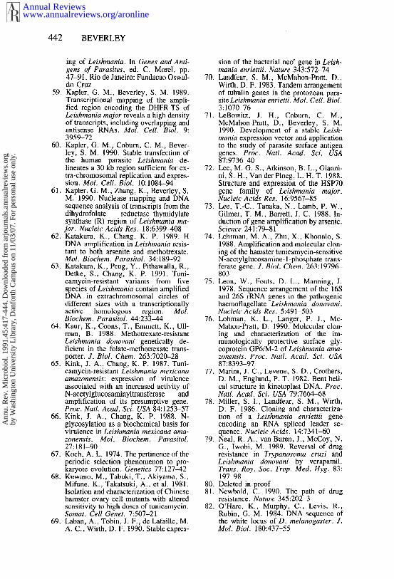
442 BEVERLEY
ing of Leishmania. In Genes and Anti-gens of Parasites, ed. C. Morel, pp.47-91. Rio de Janeiro: Fundacao Oswal-do Cruz
59. Kapler, G. M., Beverley, S. M. 1989.Transcriptional mapping of the ampli-fied region encoding the DHFR-TS ofLeishmania major reveals a high densityof transcripts, including overlapping andantisense RNAs. Mol. Cell. Biol. 9:3959-72
60. Kapler, G. M., Coburn, C. M., Bever-ley, S. M. 1990. Stable transfection ofthe human parasite Leishmania de-lineates a 30 kb region sufficient for ex-tra-chromosomal replication and expres-sion. Mol. Cell. Biol. 10:1084-94
61. Kapler, G. M., Zhang, K., Beverley, S.M. 1990. Nuclease mapping and DNAsequence analysis of transcripts from thedihydrofolate reductase-thymidylatesynthase (R) region of Leishmania ma-jor. Nucleic Acids Res. 18:6399~[08
62. Katakura, K., Chang, K. P. 1989. HDNA amplification in Leishmania resis-tant to both arsenite and methotrexate.Mol. Biochem. Parasitol. 34:189-92
63. Katakura, K., Peng, Y., Pithawalla, R.,Detke, S., Chang, K. P. 1991. Tuni-camycin-resistant variants from fivespecies of Leishmania contain amplifiedDNA in extrachromosomal circles ofdifferent sizes with a transcriptionallyactive homologous region. Mol.Biochem. Parasitol. 44:233~44
64. Kaur, K., Coons, T., Emmett, K., Ull-man, B. 1988. Methotrexate-resistantLeishmania donovani genetically de-ficient in the folate-methotrexate trans-porter. J. Biol. Chem. 263:7020-28
65. Kink, J. A., Chang, K. P. 1987. Tuni-camycin-resistant Leishmania mexicanaamazonensis: expression of virulenceassociated with an increased activity ofN-acetylglucosaminyltransferase andamplification of its presumptive gene.Proc. Natl. Acad. Sci. USA 84:1253-57
66. Kink, J. A., Chang, K. P. 1988. N-glycosylation as a biochemical basis forvirulence in Leishmania mexicana ama-zonensis. Mol. Biochem. Parasitol.27:181-90
67. Koch, A. L. 1974. The pertinence of theperiodic selection phenomenon to pro-karyote evolution. Genetics 77:127-42
68. Kuwano, M., Tabuki, T., Akiyama, S.,Mifune, K., Takatsuki, A., et al. 1981.Isolation and characterization of Chinesehamster ovary cell mutants with alteredsensitivity to high doses of tunicamycin.Somat. Cell Genet. 7:507-21
69. Laban, A., Tobin, J. F., de Lafaille, M.A. C., Wirth, D. F. 1990. Stable expres-
sion of the bacterial neor gene in Leish-mania enriettii. Nature 343:572-74
70. Landfear, S. M., McMahon-Pratt, D,Wirth, D. F. 1983. Tandem arrangementof tubulin genes in the protozoan para-site Leishmania enrietti. Mol. Cell. Biol.3:107~76
71. LeBowitz, J. H., Coburn, C. M.,McMahon-Pratt, D., Beverley, S. M.1990. Development of a stable Leish-mania expression vector and applicationto the study of parasite surface antigengenes. Proc. Natl. Acad. Sci. USA87:973(:~40
72. Lee, M. G. S., Atkinson, B. L., Gianni-ni, S. H., Van der Ploeg, L. H. T. 1988.Structure and expression of the HSP70gene family of Leishmania major.Nucleic Acids Res. 16:9567-85
73. Lee, T.-C., Tanaka, N., Lamb, P. W.,Gilmer, T. M., Barrett, J. C. 1988. In-duction of gene amplification by arsenic.Science 241:79-81
74. Lehrman, M. A., Zhu, X., Khounlo, S.1988. Amplification and molecular clon-ing of the hamster tunicamycin-sensitiveN-acetylglucosamine- 1-phosphate trans-ferase gene. J. Biol. Chem. 263:19796-803
75. Leon, W., Fouts, D. L., Manning, J.1978. Sequence arrangement of the 16Sand 26S rRNA genes in the pathogenichaemoflagellate Leishmania donovani.Nucleic A cids Res. 5:491-503
76. Lohman, K. L., Langer, P. J., Mc-Mahon-Pratt, D. 1990. Molecular clon-ing and characterization of the im-munologically protective surface gly-coprotein GP6/M-2 of Leishmania ama-zonensis. Proc. Natl. Acad. Sci. USA87:8393-97
77. Marini, J. C., Levene, S. D., Crothers,D. M., Englund, P. T. 1982. Bent heli-cal structure in kinetoplast DNA. Proc.Natl. Acad. Sci. USA 79:7664-68
78. Miller, S. I., Landfear, S. M., Wirth,D. F. 1986. Cloning and characteriza-tion of a Leishmania enriettii geneencoding an RNA spliced leader se-quence. Nucleic Acids. 14:7341-60
79. Neal, R. A., van Buren, J., McCoy, N.G., lwobi, M. 1989. Reversal of drugresistance in Trypanosoma cruzi andLeishmania donovani by verapamil.Trans. Roy. Soc. Trop. Med. Hyg. 83:197-98
80. Deleted in proof81. Newbold, C. 1990. The path of drug
resistance. Nature 345:202-382. O’Hare, K., Murphy, C., Levis, R.,
Rubin, G. M. 1984. DNA sequence ofthe white locus of D. melanogaster. J.Mol. Biol. 180:437-55
www.annualreviews.org/aronlineAnnual Reviews
Ann
u. R
ev. M
icro
biol
. 199
1.45
:417
-444
. Dow
nloa
ded
from
arj
ourn
als.
annu
alre
view
s.or
gby
Was
hing
ton
Uni
vers
ity L
ibra
ry, D
anfo
rth
Cam
pus
on 1
1/03
/07.
For
per
sona
l use
onl
y.

LEISHMAN1A AMPLIFICATION 443
83. Ouellette, M., Fase-Fowler, F., Borst,P. 1990. The amplified H circle ofmethotrexate-resistant Leishmania tar-entolae contains a novel P-glycoproteingene. EMBO J. 9:1027-33
84. Ouellette, M., Hettema, E., Wrist, D.,Fase-Fowler, F., Borst, P. 199l. Directand inverted DNA repeats associatedwith P-glycoprotein gene amplificationin drug-resistant Leishmania. EMBO J,In press
85. Pages, M., Bastien, P., Veas, F., Rossi,V., Bellis, M., et al. 1989. Chromo-some size and number polymorphisms inLeishmania infantum suggest amplifica-tion/deletion and possible genetic ex-change. Mol. Biochem. Parasitol.36:161-68
86. Panton, L. J., Tesh, R. B., Nadeau, K.,Beverley, S. M. 1991, A test for geneticexchange in mixed infections of Leish-mania major in the sand fly Phleboto-mus papatasi. J. Protozool. In press
87. Petrillo-Peixoto, M., Beverley, S. M.1989. Amplification of a new region ofDNA in an unselected laboratory stockof Leishmania tarentolae: the T region.J. Protozool. 36:2257-61
88. Petrillo-Peixoto, M. L., Beverley, S. M.1988. Amplified DNAs in laboratorystocks of L. tarentolae: extrachromo-somal circles structurally and functional-ly similar to the inverted H regionamplification of methotrexate-resistantL. major. Mol. Cell. Biol. 8:5188-99
89. Petrillo-Peixoto, M. P., Beverley, S. M.1987. In vitro activity of sulfonamidesand sulfones against Leishmania majorpromastigotes, Antimicrob. Agents Che-mother. 31:1575-78
90. Ruiz, J. C., Choi, K., Hoff, D. D. vo,Roninson, I. B., Wahl, G. M. 1989.Autonomously replicating episomescontain mdrl genes in a multidrug-resistant human cell line. Mol. Cell.Biol. 9:109-15
91. Samaras, N., Spithill, T. W. 1987.Molecular karyotype of five species ofLeishmania and analysis of gene loca-tions and chromosomal rearrangements.Mol. Bioehem. Parasitol. 25:27%91
92. Schirnke, R. T. 1982. Gene amplifica-tion summary. See Ref. 93, pp. 317-33
93. Schimke, R. T., ed. 1982. GeneAmplification. Cold Spring Harbor: ColdSpring Harbor Lab. 339 pp.
94. Schimke, R. T. 1984. Gene amplifica-tion in cultured animal cells. Cell37:705-13
95. Schimke, R. T. 1988. Gene amplifica-tion in cultured cells. J. Biol. Chem.263:5989-92
96. Schimke, R. T., Roos, D. S., Brown, P.C. 1987. Amplification of genes insomatic mammalian cells. Methods En-zymol. 151:85-104
97. Schimke, R. T., Sherwood, S. W., Hill,A. B., Johnston, R. N. 1986. Over-replication and recombination of DNAin higher eukaryotes: potential con-sequences and biological implications.Proc. Natl. Acad. Sci. USA 83:2157-61
98. Scholler, J. K., Reed, S. G., Stuart, K.1986. Molecular karyotype of speciesand subspecies of Leishmania. Mol.Biochem. Parasitol. 20:279-93
99. Scocca, J. R., Hartog, K. O., Krag, S.S. 1988. Evidence of gene amplificationin tunicamycin-resistant Chinese ham-ster ovary cells. Biochem. Biophys. Res.Commun. 156:1063-69
100. Scott, D. A., Coombs, G. H., Sander-son, B. E. 1987. Effects of methotrexateand other antifolates on the growth anddihydrofolate reductase activity ofLeishmania promastigotes. Biochem~Pharmacol. 36:2043-45
101. Shapira, M., Pinelli, E. 1989. Heatshock protein 83 of Leishmania mexica-na amazonensis is an abundant cytoplas-mic protein with a tandemly repeatedgenomic arrangement. Eur. J. Biochem.185:231-36
102. Simpson, L., Shaw, J. 1989. RNA edit-ing and the mitochondrial cryptogenes ofkinetoplastid protozoa. Cell 57:355-66
103. Sirawaraporn, W., Sersrivanich, R.,Booth, R. G., Hansch, C., Neal, R. A.,Santi, D. V. 1988. Selective inhibitionof Leishmania dihydrofolate reductaseand Leishmania growth by 5-benzyl-2,4-diaminopyrimidines. Mol. Biochem. Pa-rasitol. 31:79-86
104. Solari, A. J. 1980. The 3odimensionalfine structure of the mitotic spindle inTrypanosoma cruzi. Chromosoma 78:23%55
105. Stark, G. R., Debatisse, M., Giulotto,E., Wahl, G. M. 1989. Recent progressin understanding mechanisms of mam-malian DNA amplification. Cell57:901-8
106. Stark, G. R., Wahl, G. M. 1984. Geneamplification. Annu. Rev. Biochem.53:447-9 I
107. Stuart, K., Karp, S., Aline, R. Jr.,Smiley, B., Scholler, J., Keithly, J.1989. Small nucleic acids in Leish-mania. See Ref. 51a, pp. 555-62
108. Sudo, T., Onodera, K. 1979. Isolationand characterization of tunicamycin-resistant mutants from Chinese hamsterovary cells. J. Cell. Physiol. 101:149-56
109. Ten Asbroek, A. L. M. A., Ouellette,
www.annualreviews.org/aronlineAnnual Reviews
Ann
u. R
ev. M
icro
biol
. 199
1.45
:417
-444
. Dow
nloa
ded
from
arj
ourn
als.
annu
alre
view
s.or
gby
Was
hing
ton
Uni
vers
ity L
ibra
ry, D
anfo
rth
Cam
pus
on 1
1/03
/07.
For
per
sona
l use
onl
y.

444 BEVERLEY
M., Borst, P. 1990. Targeted insertionof the neomycin phosphotransferasegene into the tubulin gene cluster of Try-panosoma brucei. Nature 348:174-75
110. Tlsty, T. D., Margolin, B. H., Lum, K.1989. Differences in the rates of geneamplification of nontumorigenic andtumorigenic cell lines as measured byLuria-Delbruck fluctuation analysis.Proc. Natl. Acad. Sci. USA 86:9441-45
111. Tobin, J. F., Laban, A., Wirth, D. F.1991. Homologous recombination inLeishmania enrietti. [’roc. Natl. Acad.Sci. USA 88:864-68
112. Van der Bliek, A..M., Lincke, C. R.,Borst, P. 1988. Circular DNA of3T6R50 double minute chromosomes.Nucleic Acids Res. 16:4841-52
113. Van der Ploeg, L. H. T., Coruelissen,A. W. C. A., Barry, J. D., Borst, P.1984. Chromosomes of kinetoplastida.EMBO J. 3:3109-15
114. Van der Ploeg, L. H. T., Liu, A. Y. C.,Borst, P. 1984. Structure of the growingtelomeres of trypanosomes. Cell 36:459-68
115. Wellems, T. E., Panton, L. J., Gluz-man, I. Y., do Rosario, V. E., Gwadz,
R. W., et al. 1990. Chloroquine resis-tance not linked to mdr-like genes in aPlasmodium faleiparum cross. Nature345:253-55
116, White, M. J. D. 1973. Animal Cytologyand Evolution. Cambridge: CambridgeUniv. Press. 961 pp.
117. White, T. C., Fase-Fowler, F., Luenen,H. va, Calafat, J., Borst, P. 1988. TheH circles of Leishmania tarentolae are aunique amplifiable system of oligomericDNAs associated with drug resistance.J. Biol. Chem. 263:16977-83
118. Wilson, K., Collart, F. R., Huberman,E., Stringer, J. R., Ullman. B. 1991.Amplification and molecular cloning ofthe IMP dehydrogenase gene of Leish-mania donovani. J. Biol. Chem. 266:1665-71
119. World Health Organization Expert Com-mittee. 1984. The Leishmaniasis. Gene-va: WHO. 146 pp.
120. Wright, J. A., Smith, H. S., Watt, F.M., Hancock, M. C., Hudson, D. L.,Stark, G. R. 1990. DNA amplification israre in normal human cells. Proc. Natl.Acad. Sci. USA 87:1791-95
www.annualreviews.org/aronlineAnnual Reviews
Ann
u. R
ev. M
icro
biol
. 199
1.45
:417
-444
. Dow
nloa
ded
from
arj
ourn
als.
annu
alre
view
s.or
gby
Was
hing
ton
Uni
vers
ity L
ibra
ry, D
anfo
rth
Cam
pus
on 1
1/03
/07.
For
per
sona
l use
onl
y.

Ann
u. R
ev. M
icro
biol
. 199
1.45
:417
-444
. Dow
nloa
ded
from
arj
ourn
als.
annu
alre
view
s.or
gby
Was
hing
ton
Uni
vers
ity L
ibra
ry, D
anfo
rth
Cam
pus
on 1
1/03
/07.
For
per
sona
l use
onl
y.

Ann
u. R
ev. M
icro
biol
. 199
1.45
:417
-444
. Dow
nloa
ded
from
arj
ourn
als.
annu
alre
view
s.or
gby
Was
hing
ton
Uni
vers
ity L
ibra
ry, D
anfo
rth
Cam
pus
on 1
1/03
/07.
For
per
sona
l use
onl
y.