gene therapy for the central and peripheral nervous system
TRANSCRIPT

GENE THERAPY FOR THE CENTRAL AND PERIPHERAL NERVOUS SYSTEM
EDITED BY : Andrew P. Tosolini and George M. SmithPUBLISHED IN : Frontiers in Molecular Neuroscience

1 May 2018 | Gene Therapy for the CentralFrontiers in Molecular Neuroscience
Frontiers Copyright Statement
© Copyright 2007-2018 Frontiers Media SA. All rights reserved.
All content included on this site, such as text, graphics, logos, button
icons, images, video/audio clips, downloads, data compilations and
software, is the property of or is licensed to Frontiers Media SA
(“Frontiers”) or its licensees and/or subcontractors. The copyright in the
text of individual articles is the property of their respective authors, subject to
a license granted to Frontiers.
The compilation of articles constituting this e-book, wherever published,
as well as the compilation of all other content on this site, is the exclusive
property of Frontiers. For the conditions for downloading and
copying of e-books from Frontiers’ website, please see the Terms for
Website Use. If purchasing Frontiers e-books from other websites
or sources, the conditions of the website concerned apply.
Images and graphics not forming part of user-contributed materials may
not be downloaded or copied without permission.
Individual articles may be downloaded and reproduced in accordance
with the principles of the CC-BY licence subject to any copyright or
other notices. They may not be re-sold as an e-book.
As author or other contributor you grant a CC-BY licence to others to
reproduce your articles, including any graphics and third-party materials
supplied by you, in accordance with the Conditions for Website Use and
subject to any copyright notices which you include in connection with your
articles and materials.
All copyright, and all rights therein, are protected by national and
international copyright laws.
The above represents a summary only. For the full conditions see the
Conditions for Authors and the Conditions for Website Use.
ISSN 1664-8714 ISBN 978-2-88945-475-4
DOI 10.3389/978-2-88945-475-4
About Frontiers
Frontiers is more than just an open-access publisher of scholarly articles: it is a pioneering approach to the world of academia, radically improving the way scholarly research is managed. The grand vision of Frontiers is a world where all people have an equal opportunity to seek, share and generate knowledge. Frontiers provides immediate and permanent online open access to all its publications, but this alone is not enough to realize our grand goals.
Frontiers Journal Series
The Frontiers Journal Series is a multi-tier and interdisciplinary set of open-access, online journals, promising a paradigm shift from the current review, selection and dissemination processes in academic publishing. All Frontiers journals are driven by researchers for researchers; therefore, they constitute a service to the scholarly community. At the same time, the Frontiers Journal Series operates on a revolutionary invention, the tiered publishing system, initially addressing specific communities of scholars, and gradually climbing up to broader public understanding, thus serving the interests of the lay society, too.
Dedication to Quality
Each Frontiers article is a landmark of the highest quality, thanks to genuinely collaborative interactions between authors and review editors, who include some of the world’s best academicians. Research must be certified by peers before entering a stream of knowledge that may eventually reach the public - and shape society; therefore, Frontiers only applies the most rigorous and unbiased reviews. Frontiers revolutionizes research publishing by freely delivering the most outstanding research, evaluated with no bias from both the academic and social point of view.By applying the most advanced information technologies, Frontiers is catapulting scholarly publishing into a new generation.
What are Frontiers Research Topics?
Frontiers Research Topics are very popular trademarks of the Frontiers Journals Series: they are collections of at least ten articles, all centered on a particular subject. With their unique mix of varied contributions from Original Research to Review Articles, Frontiers Research Topics unify the most influential researchers, the latest key findings and historical advances in a hot research area! Find out more on how to host your own Frontiers Research Topic or contribute to one as an author by contacting the Frontiers Editorial Office: [email protected]
2 May 2018 | Gene Therapy for the CentralFrontiers in Molecular Neuroscience
GENE THERAPY FOR THE CENTRAL AND PERIPHERAL NERVOUS SYSTEM
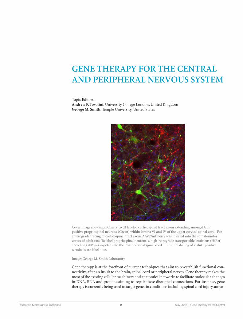
Cover image showing mCherry (red) labeled corticospinal tract axons extending amongst GFP positive propriospinal neurons (Green) within lamina VI and IV of the upper cervical spinal cord. For anterograde tracing of corticospinal tract axons AAV2/mCherry was injected into the somatomotor cortex of adult rats. To label propriospinal neurons, a high-retrograde transportable lentivirus (HiRet) encoding GFP was injected into the lower cervical spinal cord. Immunolabeling of vGlut1 positive terminals are label blue.
Image: George M. Smith Laboratory
Topic Editors: Andrew P. Tosolini, University College London, United KingdomGeorge M. Smith, Temple University, United States
Gene therapy is at the forefront of current techniques that aim to re-establish functional con-nectivity, after an insult to the brain, spinal cord or peripheral nerves. Gene therapy makes the most of the existing cellular machinery and anatomical networks to facilitate molecular changes in DNA, RNA and proteins aiming to repair these disrupted connections. For instance, gene therapy is currently being used to target genes in conditions including spinal cord injury, amyo-
3 May 2018 | Gene Therapy for the CentralFrontiers in Molecular Neuroscience
trophic lateral sclerosis, spinal muscular atrophy, stroke and multiple sclerosis, amongst others. The various delivery routes include viral-vectors, genetically modified cellular implants, naked DNA/RNA, liposomes, Cre-Lox recombination, optogenetics and nanoparticles. In particular, gene therapy aims to restore function by augmenting the expression of neuroprotective/axonal growth-promoting neurotrophic factors (e.g., BDNF, CNTF, NGF and GDNF, etc.). Furthermore, the downstream intracellular signalling pathways after receptor activation can also be targeted (e.g., mTor, MAPK, etc.).
On the other hand, gene therapy can also be used to downregulate and/or remove faulty mutated genes, such as those contributing to disease progression or that inhibit axonal regeneration (e.g., SOD-1, TDP-43, Nogo-A, MAG, OmGP, etc.). Depending on the methodology, these genes, for instance, can be silenced, removed or replaced to alleviate the underlying pathology. As such, gene therapy can transform a largely toxic and inhibitory milieu surrounding a neuronal/axonal insult into a growth-permissive environment that will ultimately aid neuronal survival and functional regeneration. Moreover, gene therapy has the capacity to target non-neuronal cells and can be even used for neuroanatomical tract tracing. Ultimately, the principal outcome of gene therapy is to functionally restore damaged neuronal and/or axonal connections irrespective of the system it is being introduced in to.
This Research Topic is devoted to work using gene therapy for the both the central and/or peripheral nervous system.
Citation: Tosolini, A. P., Smith, G. M., eds. (2018). Gene Therapy for the Central and Peripheral Nervous System. Lausanne: Frontiers Media. doi: 10.3389/978-2-88945-475-4
4 May 2018 | Gene Therapy for the CentralFrontiers in Molecular Neuroscience
Table of Contents
06 Editorial: Gene Therapy for the Central and Peripheral Nervous SystemAndrew P. Tosolini and George M. Smith
10 Non-viral gene therapy that targets motor neurons in vivoMary-Louise Rogers, Kevin S. Smith, Dusan Matusica, Matthew Fenech, Lee Hoffman, Robert A. Rush and Nicolas H. Voelcker
22 Development of non-viral vehicles for targeted gene transfer into microglia via the integrin receptor CD11bMarkus Smolny, Mary-Louise Rogers, Anthony Shafton, Robert A. Rush and Martin J. Stebbing
41 Systemic AAVrh10 provides higher transgene expression than AAV9 in the brain and the spinal cord of neonatal miceYannick Tanguy, Maria G. Biferi, Aurore Besse, Stephanie Astord, Mathilde Cohen-Tannoudji, Thibaut Marais and Martine Barkats
51 Better Targeting, Better Efficiency for Wide-Scale Neuronal Transduction with the Synapsin Promoter and AAV-PHP.BKasey L. Jackson, Robert D. Dayton, Benjamin E. Deverman and Ronald L. Klein
62 Corrigendum: Better Targeting, Better Efficiency for Wide-Scale Neuronal Transduction with the Synapsin Promoter and AAV-PHP.BKasey L. Jackson, Robert D. Dayton, Benjamin E. Deverman and Ronald L. Klein
63 Recombinant Human Myelin-Associated Glycoprotein Promoter Drives Selective AAV-Mediated Transgene Expression in OligodendrocytesGeorg von Jonquieres, Dominik Fröhlich, Claudia B. Klugmann, Xin Wen, Anne E. Harasta, Roshini Ramkumar, Ziggy H. T. Spencer, Gary D. Housley and Matthias Klugmann
77 Neuroprotective Effect of Non-viral Gene Therapy Treatment Based on Tetanus Toxin C-fragment in a Severe Mouse Model of Spinal Muscular AtrophySara Oliván, Ana C. Calvo, Amaya Rando, Mireia Herrando-Grabulosa, Raquel Manzano, Pilar Zaragoza, Eduardo F. Tizzano, Jose Aquilera and Rosario Osta
87 Expressing Constitutively Active Rheb in Adult Dorsal Root Ganglion Neurons Enhances the Integration of Sensory Axons that Regenerate Across a Chondroitinase-Treated Dorsal Root Entry Zone Following Dorsal Root CrushDi Wu, Michelle C. Klaw, Nikolai Kholodilov, Robert E. Burke, Megan R. Detloff, Marie-Pascale Côté and Veronica J. Tom
105 MiR-30b Attenuates Neuropathic Pain by Regulating Voltage-Gated Sodium Channel Nav1.3 in RatsSongxue Su, Jinping Shao, Qingzan Zhao, Xiuhua Ren, Weihua Cai, Lei Li, Qian Bai, Xuemei Chen, Bo Xu, Jian Wang, Jing Cao and Weidong Zang
5 May 2018 | Gene Therapy for the CentralFrontiers in Molecular Neuroscience
120 Biology of adeno-associated viral vectors in the central nervous systemGiridhar Murlidharan, Richard J. Samulski and Aravind Asokan
129 Lentiviral vectors as tools to understand central nervous system biology in mammalian model organismsLouise C. Parr-Brownlie, Clémentine Bosch-Bouju, Lucia Schoderboeck, Rachel J. Sizemore, Wickliffe C. Abraham and Stephanie M. Hughes
141 Non-Viral Nucleic Acid Delivery Strategies to the Central Nervous SystemJames-Kevin Y. Tan, Drew L. Sellers, Binhan Pham, Suzie H. Pun and Philip J. Horner
154 Gene, Stem Cell, and Alternative Therapies for SCA 1Jacob L. Wagner, Deirdre M. O’Connor, Anthony Donsante and Nicholas M. Boulis
166 Motor Neuron Gene Therapy: Lessons from Spinal Muscular Atrophy for Amyotrophic Lateral SclerosisAndrew P. Tosolini and James N. Sleigh
190 Evaluation of Gene Therapy as an Intervention Strategy to Treat Brain Injury from StrokeAmanda J. Craig and Gary D. Housley
199 Adeno Associated Viral Vector Delivered RNAi for Gene Therapy of SOD1 Amyotrophic Lateral SclerosisLorelei Stoica and Miguel Sena-Esteves
206 CRISPR/Cas9: Implications for Modeling and Therapy of Neurodegenerative DiseasesWeili Yang, Zhuchi Tu, Qiang Sun and Xiao-Jiang Li
210 Gene therapy and peripheral nerve repair: a perspectiveStefan A. Hoyng, Fred de Winter, Martijn R. Tannemaat, Bas Blits, Martijn J. A. Malessy and Joost Verhaagen

EDITORIALpublished: 22 February 2018
doi: 10.3389/fnmol.2018.00054
Frontiers in Molecular Neuroscience | www.frontiersin.org February 2018 | Volume 11 | Article 54 |
Edited by:
Hermona Soreq,
Hebrew University of Jerusalem, Israel
Reviewed by:
Shai Berlin,
Technion Israel Institute of Technology,
Israel
*Correspondence:
Andrew P. Tosolini
George M. Smith
Received: 14 January 2018
Accepted: 07 February 2018
Published: 22 February 2018
Citation:
Tosolini AP and Smith GM (2018)
Editorial: Gene Therapy for the Central
and Peripheral Nervous System.
Front. Mol. Neurosci. 11:54.
doi: 10.3389/fnmol.2018.00054
Editorial: Gene Therapy for theCentral and Peripheral NervousSystemAndrew P. Tosolini 1* and George M. Smith 2*
1 Sobell Department of Motor Neuroscience and Movement Disorders, Institute of Neurology, University College London,
London, United Kingdom, 2Department of Neuroscience, Shriners Hospitals Pediatric Research Center, Lewis Katz School of
Medicine, Temple University, Philadelphia, PA, United States
Keywords: gene therapy, CNS, PNS, AAV, lentivirus, non-viral vectors, neurons, glia
Editorial on the Research Topic
Gene Therapy for the Central and Peripheral Nervous System
It is with great pleasure that we present the research topic dedicated to Gene Therapy for the Central(CNS) and Peripheral Nervous System (PNS). Gene therapy is at the cutting-edge of techniquesutilized to develop novel therapeutics to treat insult(s) to the brain, spinal cord and/or peripheralnerves. Indeed, gene therapy can be applied via many different routes and as such can overcome themany obstacles facing oral and systemic delivery of synthetic drugs, thereby permit greater targetingof neural tissue. With this advantage, gene therapy has the potential to (1) correct disease-causingDNAmutations, (2) eliminate toxic RNA/proteins and/or (3) increase the expression of therapeuticproteins. Gene therapy can ameliorate aspects of debilitating neurological diseases and thus, canprovide a platform for functional recovery.
Over the last few years, advances in basic science and technology have enabled enhancedpre-clinical strategies culminating in the emergence of sophisticated treatment options availablefor patients. The most recent gene therapy success is an FDA and EMA approved antisenseoligonucleotide (ASO) that is the first and only treatment option available to treat spinal muscularatrophy. Moreover, ASOs have successfully reduced toxic protein levels in a phase 1/2a clinical trialto treat Huntington’s Disease (HD). This treatment option was considered safe and well toleratedand was granted orphan drug designation by the FDA and EMA. These advances offer great hope topatients, their families, clinicians and basic scientists, and emphasizes the potential of gene therapyto treat “the incurable” neurological diseases/disorders.
In 2013, we launched this research topic to provide a platform to continue the discussion andamalgamate recent advances in gene therapy technology and strategies for the treatment of PNSand CNS disorders. In conclusion, we are proud to present an extremely productive discussionconsisting of 18 articles in total comprised of eight original articles, five full-length reviews, threemini-reviews and one hypothesis and theory article and represents a world-wide collaborationwith submissions received from USA, United Kingdom, Europe, Asia, New Zealand and Australia.This research topic discusses gene therapy strategies to treat neurological disorders includingamyotrophic lateral sclerosis (ALS), spinal muscular atrophy (SMA), spinocerebellar ataxia (SCA),neuropathic pain, stroke, peripheral nerve injury and repair.
The novel studies presented in this research topic focus on improving transduction efficiencyand gene transfer using adeno-associated virus (AAVs) and non-viral methods (Table 1).Tanguy et al compare the transduction efficiency between scAAV9 and AAVrh10 serotypes aftersystemic delivery. von Jonquieres et al. continue to demonstrate glial-specific transduction in the
6

Tosolini and Smith Gene Therapy for CNS/PNS
TABLE 1 | Highlights from the original research published in this research topic.
Article Highlights Model
Rogers et al. • Established a non-viral, antibody-based delivery method to transduce motor neurons in vivo after
intraperitoneal injection.
• PEGylated polyethylenimine (PEI-PEG12) conjugated to a MRL2-antibody carrying DNA to the
neurotrophin receptor p75 (p75NTR) targeted to motor neurons.
• 72 h after injection, ∼25% of lumbar, ∼18% or thoracic and 17% of cervical motor neurons were
transduced.
Wild-type mice
Smolny et al. • Developed a non-viral, antibody-based delivery method for specific gene transfer in microglia in vitro and
in vivo.
• OX42-immunoporter can bind plasmid DNA, and is trafficked to lysosomes in microglia via CD11b
receptor-mediated internalization.
• OX42-immunogenes were specific to microglia and not astrocytes, but did not induce robust gene
expression in vitro and in vivo.
In vitro and Wild-type mice
Tanguy et al. • Compared transduction efficiencies of scAAV9 and AAVrh10 in the brain, spinal, cord and peripheral
nervous tissue after intravenous delivery in neonatal mice.
• AAVrh10 transduction was superior in the medulla, cerebellum, hippocampus, cortex, dorsal spinal cord,
and spinal motor neurons.
• Dose-related transduction efficiency differences were observed in the sciatic nerve.
Wild-type mice
Jackson et al. • For the first time, AAV-PHP.B was demonstrated to transduce the rat CNS.
• After intravenous delivery in neonatal rats AAV-PHP.B was demonstrated to have a higher transduction
efficiency than AAV9 when using the same CBA promoter.
• AAV-PHP.B with a synapsin promoter resulted in an enhanced transduction efficiency and neuronal
specificity that induced TDP-43-like pathology and ALS-like phenotypes.
Wild-type rats
von Jonquieres et al. • Three MAG promoter sizes (0.3, 1.5, and 2.2 kb) were packaged into AAV-cy5 vector and were delivered
into the striatum in wild-type neonates.
• All three promoter sizes exclusively transduced oligodendrocytes.
• Robust and oligodendrocyte-specific long-term GFP expression was reported at 8 months after neonatal
delivery.
In vitro and Wild-type mice
Oliván et al. • Application of a non-toxic, tetanus toxin fragment (TTC) to spinal cord organotypic cultures increased SMN
levels.
• Intramuscular injections of TTC reduced mRNA of autophagy markers (Becn1, Atg5, LC3, and p62) and
pro-apoptotic genes (Bax and Casp3) in the spinal cord and downregulated LC3 and Casp3 expression in
skeletal muscle in SMA mice.
• Intramuscular TTC application is suggested to show a compensatory effect in the expression of certain
genes involved in muscle damage response, oxidative stress and calcium homeostasis in SMA mice.
Ex vivo and SMN17 mice
Wu et al. • Intraganglionic injections of AAV5-caRHEB into cervical DRGs transduced mainly large caliber DRG
neurons.
• ChABC treatment increased the number of regenerating axons through the DREZ irrespective of
DRG-transduction, which resulted in sensory behavioral “responses.”
• caRHEB expression in DRGs after dorsal root crush enhances synaptic formation and/or functional
regeneration into the spinal gray matter.
In vitro and Wild-type mice
Su et al. • miR-30b agomir transfection down-regulated the voltage-gated sodium channel Nav1.3 mRNA that was
stimulated with TNF-α in primary DRG neurons.
• miR-30b overexpression reduced neuropathic pain after spinal nerve ligation, with demonstrated reduction
in Nav1.3 mRNA and protein expression in both DRG neurons and spinal cord.
• miR-30b antagomir activated the Nav1.3 voltage-gated sodium channel.
In vitro and wild-type rats
brain after injecting a chimeric AAV1/2 vector into neonatalstriatum, despite using three differently sized MAG promoters.Jackson et al. combined an engineered AAV-serotype with aneuronal-specific promoter to increase transduction efficiencyand reduce off-target effects after intravenous delivery. Wu et al.increase the intrinsic growth potential of injured sensory axonsusing combinatory treatment involving chondroitinase ABC andAAV-mediated constitutively active GTPase Rheb (Wu et al.).
In addition, Smolny et al. present a non-viral, antibody-based delivery method for microglia-specific gene transfer.Rogers et al. describe spinal motor neuron transductionafter peripheral delivery of plasmid DNA as a PEGylatedpolyethylenimine conjugated antibody. Oliván et al. demonstratethat atoxic-tetanus neurotoxin fragment modifies expressionof autophagy and pro-apoptotic genes in the spinal cord andskeletal muscle. Su et al. show miRNA-mediated suppression of
Frontiers in Molecular Neuroscience | www.frontiersin.org February 2018 | Volume 11 | Article 54 | 7

Tosolini and Smith Gene Therapy for CNS/PNS
TABLE 2 | Highlights from the review, mini-review and hypothesis and theory articles published in this research topic.
Article Highlights Type
Murlidharan et al. • Describes AAV-vector biology, their cellular entry mechanisms and axonal transport profiles of
well-characterized AAV serotypes.
• Discusses the implications of AAV-vector applications (e.g., direct application, intravenous injections, etc.).
• Considers the safety aspects of AAV-mediated applications to the CNS.
Review
Parr-Brownlie et al. • Describes lentiviral vector biology, including modified envelope glycoproteins and the expression of transgenes
under the regulation of cell-selective and inducible promoters.
• Deliberates on the benefit of lentiviral-vectors combined with other techniques such as anatomical tract-tracing,
immunohistochemistry, confocal and electron microscopy.
• Proposes limitations and future perspectives including ways that lentiviral-vectors can contribute to the gene
therapy clinical trials.
Review
Tan et al. • Explores the challenges facing non-viral nucleic acid delivery to the CNS and provides strategies to overcome
them.
• Discusses the advantages and disadvantages of different administration routes of nucleic acid delivery.
• Considers how retrograde axonal transport can be used to deliver non-viral nucleic acids.
Review
Wagner et al. • Describes the epidemiology, molecular pathology and mouse models related to spinocerebellar ataxia-1
(SCA-1).
• Discusses the literature related to stem cell, gene and alternative therapies used to treat SCA-1.
• Identifies the various challenges for gene, stem cell and alternative therapies for SCA-1.
Review
Tosolini and Sleigh • Describes the epidemiology, genetics, classifications and mechanisms causing SMA and ALS/MND and
deliberates on potential commonalities.
• Provides an update on clinical gene therapies for both SMA and ALS/MND.
• Identifies four key areas that ALS/MND gene therapies can learn from the recent success in the SMA gene
therapies including therapeutic targeting, combinational treatment, considering the dose and drug
concentration as well as optimizing the therapeutic timing.
Review
Craig and Housley • Provides a summary of the viral-mediated gene therapy research used to treat stroke.
• Highlights the key areas that gene therapy needs to address to ameliorate stroke including protein synthesis,
delivery site and viral-vectors.
• Identifies therapeutic protein candidates for stroke treatment.
Mini-review
Stoica and Sena-Esteves • Summarizes the literature on AAV-mediated gene therapy studies that reduce SOD1 toxicity to treat
SOD1-related ALS/MND.
• Discusses the current hurdles to be addressed to advance the development of clinical gene therapies such as
non-cell autonomous toxicity, cellular and anatomical targeting and the delivery methods.
• Identifies RNA interference as a successful therapeutic target to ameliorate disease.
Mini-review
Yang et al. • Summarizes the development and application of the CRISPR/CAS9 toolkit.
• Describes the use of CRISPR/Cas9 to generate animal models of neurodegenerative diseases.
• Discusses how CRISPR/Cas9 can be applied to treat animal models of Parkinson’s and Huntington’s Disease.
Mini-review
Hoyng et al. • Summarizes the research on gene therapy in animal models of peripheral nerve repair and identify key future
directions.
• Provides a perspective on the path for clinical translation for PNS-gene therapy for traumatic nerve injuries.
• Addresses efficacy and safety concerns for human applications and identify the ideal patient population for a
proof-of-concept clinical study.
Hypothesis and theory
specific voltage-gated sodium channels can alleviate neuropathicpain.
This research topic also includes a number of full-lengthand mini-reviews (Table 2). Murlidharan et al. describe thebiology of different AAV strains, including their transductionprofiles, cellular tropisms and mechanisms of CNS transport,for increased translational application. Parr-Brownlie et al.review lentiviral-vector approaches to enhance transductionefficiency, mediate cell-specificity, restrict gene expressionspatially and temporally, and discuss limitations and futureprospects. Tan et al. critically analyse the advantages and
disadvantages of strategies using non-viral nucleic acids todeliver therapeutic genes by circumventing the immune responseand thus, appeasing safety concerns potentially associated withviral-mediated gene therapies. Wagner et al. examine the geneand stem cell pre-clinical therapeutic options that preservePurkinje cell health to treat SCA, and suggest that RNAinterference (RNAi) might have great promise. Finally, Tosoliniand Sleigh discuss important considerations learned from thesuccess of a recently FDA- and EMA-approved gene therapyfor SMA to develop viable gene therapies and strategies totreat ALS.
Frontiers in Molecular Neuroscience | www.frontiersin.org February 2018 | Volume 11 | Article 54 | 8

Tosolini and Smith Gene Therapy for CNS/PNS
For the mini-reviews, Yang et al. discuss the “hot topic” of
CRISPR/Cas9 gene editing and how these tools can be applied
to various research models and the development of treatments
for neurodegenerative disease, such as HD and Parkinson’sdisease. Craig and Housley focus on gene therapy approaches for
stroke and discuss injury mechanisms, appropriate timings for
therapeutic intervention and deliberate on candidate therapeutic
proteins as therapeutic options. Stoica and Sena-Esteves deliver asuccinct mini-review of AAV-mediated SOD1-ALS amelioration
strategies and describe the hurdles to overcome for CNS gene
delivery.The Hypothesis and theory submission by Hoyng et al.
summarize the state-of-research for peripheral nerve repair,identify future targets and provide a translational perspective onPNS gene therapy.
This research forum describes many important characteristicsof diverse gene therapy applications for the development oftangible treatment options for different CNS and PNS disorders.Indeed, the advances in gene therapy strategies discussed withinthis research topic give hope that treatment options for manyincurable CNS and PNS disorders are closer to becoming a viableclinical option.
AUTHOR CONTRIBUTION
All authors listed have made a substantial, direct and intellectualcontribution to the work, and approved it for publication.
FUNDING
AT holds a postdoctoral position supported by a WellcomeTrust Senior Investigator Award [107116/Z/15/Z] to GiampietroSchiavo (Institute of Neurology, University College London).This work was supported by National Institute of Health(R01NS103481), Shriners Hospitals for Pediatric Research(SHC-84050) and DOD (SC140089) to GS.
Conflict of Interest Statement: The authors declare that the research was
conducted in the absence of any commercial or financial relationships that could
be construed as a potential conflict of interest.
Copyright © 2018 Tosolini and Smith. This is an open-access article distributed
under the terms of the Creative Commons Attribution License (CC BY). The use,
distribution or reproduction in other forums is permitted, provided the original
author(s) and the copyright owner are credited and that the original publication
in this journal is cited, in accordance with accepted academic practice. No use,
distribution or reproduction is permitted which does not comply with these terms.
Frontiers in Molecular Neuroscience | www.frontiersin.org February 2018 | Volume 11 | Article 54 | 9
ORIGINAL RESEARCH ARTICLEpublished: 14 October 2014
doi: 10.3389/fnmol.2014.00080
Non-viral gene therapy that targets motor neurons in vivoMary-Louise Rogers1*, Kevin S. Smith1, Dusan Matusica 2 , Matthew Fenech1, Lee Hoffman 3,
Robert A. Rush1 and Nicolas H. Voelcker 4
1 Department of Human Physiology, Centre for Neuroscience, Flinders University, Adelaide, SA, Australia2 Department of Anatomy and Histology, Centre for Neuroscience, Flinders University, Adelaide, SA, Australia3 Department of Chemistry and Biochemistry, South Dakota State University, Brookings, SD, USA4 Australian Research Council Centre of Excellence in Convergent Bio-Nano Science and Technology, Mawson Institute, University of South Australia, Adelaide,
SA, Australia
Edited by:
Andrew Paul Tosolini, University ofNew South Wales, Australia
Reviewed by:
Gong Chen, The Pennsylvania StateUniversity, USAGiampietro Schiavo, UCL Institute ofNeurology, UK
*Correspondence:
Mary-Louise Rogers, Department ofHuman Physiology, Centre forNeuroscience, Flinders University,GPO Box 2100, Adelaide, SA 5001,Australiae-mail: [email protected]
A major challenge in neurological gene therapy is safe delivery of transgenes to sufficientcell numbers from the circulation or periphery. This is particularly difficult for diseasesinvolving spinal cord motor neurons such as amyotrophic lateral sclerosis (ALS). Wehave examined the feasibility of non-viral gene delivery to spinal motor neurons fromintraperitoneal injections of plasmids carried by “immunogene” nanoparticles targetedfor axonal retrograde transport using antibodies. PEGylated polyethylenimine (PEI-PEG12)as DNA carrier was conjugated to an antibody (MLR2) to the neurotrophin receptor p75(p75NTR). We used a plasmid (pVIVO2) designed for in vivo gene delivery that producesminimal immune responses, has improved nuclear entry into post mitotic cells andalso expresses green fluorescent protein (GFP). MLR2-PEI-PEG12 carried pVIVO2 andwas specific for mouse motor neurons in mixed cultures containing astrocytes. Whileonly 8% of motor neurons expressed GFP 72 h post transfection in vitro, when theimmunogene was given intraperitonealy to neonatal C57BL/6J mice, GFP specific motorneuron expression was observed in 25.4% of lumbar, 18.3% of thoracic and 17.0% ofcervical motor neurons, 72 h post transfection. PEI-PEG12 carrying pVIVO2 by itself didnot transfect motor neurons in vivo, demonstrating the need for specificity via the p75NTRantibody MLR2. This is the first time that specific transfection of spinal motor neuronshas been achieved from peripheral delivery of plasmid DNA as part of a non-viral genedelivery agent. These results stress the specificity and feasibility of immunogene deliverytargeted for p75NTR expressing motor neurons, but suggests that further improvementsare required to increase the transfection efficiency of motor neurons in vivo.
Keywords: targeted gene delivery, PEI, PEGylation, retrograde transport, immunogenes, p75NTR
INTRODUCTIONTargeted gene therapy has the potential to be developed for dis-eases involving death of motor neurons such as amyotrophic lateralsclerosis (ALS). Motor neurons can be transfected by injectingevery muscle innervated by spinal motor neurons. Transport oftherapy is then by axonal pathways originating from, terminatingin, or passing through the injection site. However, this requiresmany painful injections and even then, it may not be possible toreach all spinal motor neurons (Towne et al., 2011). Alternativelymotor neurons can be difficult to access and transfect from thecirculation or centrally through injections into the cerebrospinalfluid (CSF). Peripheral injections of viral gene therapy have notbeen successful at selectively targeting motor neurons (Towneet al., 2008). The blood brain barrier (BBB) is also effective atkeeping toxins and infectious material out of the central nervoussystem (CNS; Pardridge, 2006). Our group has been developingtargeted gene delivery agents called “immunogenes” with the aimof using them to deliver therapeutic genes to diseased motor neu-rons (Rogers and Rush, 2012). They are composed of antibodiesthat internalize after targeting cell surface receptors and are conju-gated to cationic carriers, able to condense DNA/RNA, forming the
immunogene. Cells that express the cognate cell-surface receptorsof the targeting antibody can therefore be specifically transfectedwith immunogenes in vivo from the circulation (Rogers and Rush,2012).
Antibodies that internalize into target cells are essential forimmunogenes. We previously used an antibody (clone MC192)to the rat common neurotrophin receptor p75 (p75NTR) as atargeting agent (Barati et al., 2006). p75NTR is a receptor highlyexpressed on motor neurons during the embryonic period, downregulated in adulthood (Yan and Johnson, 1988), only to bere-expressed following neuronal injury, including ALS (Lowryet al., 2001). Past research has revealed that p75NTR is retro-gradely trafficked in signaling endosomes in motor neurons whentaken up by at distal terminals (Lalli and Schiavo, 2002), ren-dering this receptor ideally suited to deliver therapeutic genesfor motor neurons. Transport from the periphery to motor neu-rons should be possible using antibodies that target rat p75NTR(Bronfman et al., 2003), i.e., MC192 and pan specific MLR2(Rogers et al., 2006; Matusica et al., 2008). Both have been demon-strated to internalize with the receptor making them ideal targetingagents.
Frontiers in Molecular Neuroscience www.frontiersin.org October 2014 | Volume 7 | Article 80 | 10
Rogers et al. Gene delivery targeting motor neurons
The development of immunogenes as targeted nanocarriersis particularly attractive for diseases such as ALS. In almost allcases of ALS, death occurs within 3–5 years of diagnosis due tothe selective death of motor neurons and there are no effectivetherapies (Turner et al., 2013). We have previously used immuno-genes to deliver therapeutic glial-derived growth factor (GDNF)to injured motor neurons in vivo in neonatal rats (Barati et al.,2006). The rat specific p75NTR antibody MC192 was conju-gated to a cationic polymer poly(L-lysine; PLL) to condenseplasmids expressing GDNF and the immunogene was given intra-muscularly (Barati et al., 2006). Although GDNF rescued motorneurons that innervated injected muscles, this first generationimmunogene could not be used in the circulation to access largerpools of motor neurons (Barati et al., 2006), making it vulnera-ble to rapid degradation. Cytotoxicity in vivo can be associatedwith the surface charge of the polymer (Chollet et al., 2002)and poor stability is associated with interactions with erythro-cytes and serum components such as albumin, lipoproteins orIgG (Rogers and Rush, 2012). These issues can be overcomeby masking the surface charge with agents such as polyethy-lene glycol (PEG). Forming a hydrophilic shell, PEG limits thehydrophobic or electrostatic interactions with the extracellularmedium and prevents binding of the cationic polymer with ery-throcytes and plasma proteins (Chollet et al., 2002; Rogers andRush, 2012). Hence, such measures are required for stealth in thecirculation.
After entering cells, non-viral gene delivery agents must be ableto escape the endosome/lysosomal compartments to deliver theirpayload of DNA or RNA to the nucleus and RNA-induced silencingcomplex (RISC) complex, respectively, (Rogers and Rush, 2012).Our first generation immunogene used PLL that required fuso-genic peptides to escape endosomal/lysososomal compartments ofcells (Navarro-Quiroga et al., 2002). Other DNA/RNA condensingagents such as polyethylenimine (PEI) have more useful proper-ties including a mechanism for endosomal escape. PEI possessesa high cationic charge density due to secondary amino groupsthat enables the endosomal/lysosomal release of complexes dueto the so-called “proton sponge effect” (Boussif et al., 1995; Tangand Szoka, 1997; Lungwitz et al., 2005). PEI unlike PLL also facil-itates the entry of plasmid DNA into the nucleus (Godbey et al.,1999).
Toxicity of intravenously administered cationic polyplexes can-not only be reduced by PEGylation (Merdan et al., 2003; Ogriset al., 2003; Malek et al., 2009) but also when nanoconstructs arealso endowed with antibodies or other targeting moieties (Zhanget al., 2003; Luo et al., 2010; Höbel et al., 2011; Schaffert et al.,2011). This may be reflective of specificity in addition to lowertoxicity because of reduction in charge after conjugation to forexample an antibody. Besides systemic toxicities, cytotoxic effectsare also observed upon polyplex internalization. Since polycationselectrostatically bind and condense DNA, non-specific electro-static binding to any kind of cellular polyanions (e.g., enzymes,mRNA, or genomic DNA) may deregulate the expression profileof housekeeping genes (Godbey et al., 2001) or induce activationof genes involved in apoptosis (Masago et al., 2007). Consequently,characteristics of cationic polyplex formulations such as molecularweight, cationic charge density and the presence of free polymer
also influence their cytotoxicity (Kunath et al., 2003; Boeckle et al.,2004; van Gaal et al., 2011). Accordingly, we hypothesize that anideal candidate for a safe non-viral gene delivery vector is a car-rier with a neutral to slightly negative charge and the capability ofbeing targeted to the cell type required.
In addition to targeting cells from the periphery, we also aimedto improve the expression of transgenes. Methods to improvenuclear import of plasmids are of particular importance in postmitotic cells such as motor neurons. Transfection rates can bepoor in post mitotic cells as there is limited breakdown of thenuclear envelope (Zabner et al., 1995). Therefore, modifying plas-mids to improve nuclear entry is required. One way of achievingthis is to include in the plasmid design a DNA targeting sequence(DTS) that bind to endogenously expressed transcription fac-tors that then act as nuclear localization sequences (NLSs) andimprove nuclear import (Mandke and Singh, 2012). Plasmid vec-tors also often contain sites that can produce innate immuneresponses through unmethylated cytosine guanine bases separatedby only one phosphate (CpGs; Magnusson et al., 2011). Removalof CpGs from the plasmid backbone has been shown to reduceimmune reactions to plasmids and prolong expression in vivo(Magnusson et al., 2011; Davies et al., 2012). Hence, plasmids thatare chosen for in vivo delivery should include DTS and minimalCpGs.
Here, we report on the development and evaluation ofimmunogenes capable of targeting motor neurons in vitro andin vivo. We demonstrate specificity of delivery to motor neuronscan be achieved from peripheral injections using p75NTR anti-body MLR2. We show that nanocarriers comprised of p75NTRantibody MLR2 conjugated to PEGylated PEI can deliver plasmidsto mouse motor neurons in vitro and in vivo. In addition, wedemonstrate gene expression in motor neurons in vivo using plas-mids designed for improved nuclear entry and less immunogenic-ity. Hence, we explore the potential of using p75NTR-targetingimmunogenes as gene therapy.
MATERIALS AND METHODSPREPARATION OF NANOCONSTRUCTSBranched PEI (C24H59N11PEI, molecular weight 25 kDa; SigmaAldrich, Australia) was made to 20 mg/ml in H2O and depro-tonated with HCl to pH 7.0. PEI was then buffer exchanged onPD10 columns (GE, Australia) with 20 mM 4-(2-hydroxyethyl)-1-piperazine ethane sulfonic acid (HEPES; Invitrogen, Aust),250 mM NaCl, and pH 7.9. 50 mg of PEI was PEGylatedwith a branched PEG reagent (Methyl-PEO12)3-PEO4-NHS ester(Thermo Scientific, Rockford, IL, USA) with a molecular weightof 2421 g/mol, (Figure 1). This was achieved at a molar ratioof 10:1 PEG to PEI. The number of PEGs per PEI was analyzedby spectral analysis using a Varian 300 MHz NMR spectrometerNMR with deuterium oxide (D2O) as the solvent indicating onaverage 12 PEG moieties conjugated per PEI, corresponding to6% of amines on the PEI being PEGylated. Hybridoma MLR2 wasgrown and MLR2 purified on protein G column as previouslydescribed (Rogers et al., 2006). Conjugation of PEI-PEG12 orPEI to anti-p75NTR MLR2 was achieved using methods adaptedfrom Blessing et al. (2001) and Germershaus et al. (2006). Briefly,the cross-linker N-succinimidyl 3-(2-pyridyldithio)propionate
Frontiers in Molecular Neuroscience www.frontiersin.org October 2014 | Volume 7 | Article 80 | 11
Rogers et al. Gene delivery targeting motor neurons
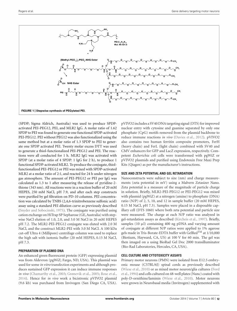
FIGURE 1 | Stepwise synthesis of PEGylated PEI.
(SPDP; Sigma Aldrich, Australia) was used to produce SPDP-activated PEI-PEG12, PEI, and MLR2 IgG. A molar ratio of 1.62SPDP to PEI was found to generate one functional SPDP-activatedPEI-PEG12. PEI without PEG12 was also functionalized using thesame method but at a molar ratio of 1.3 SPDP to PEI to gener-ate one SPDP activated PEI. Twenty molar excess DTT was usedto generate a thiol-functionalized PEI-PEG12 and PEI. The reac-tions were all conducted for 1 h. MLR2 IgG was activated withSPDP (at a molar ratio of 4 SPDP: 1 IgG for 2 h), to produce 1functional SPDP-activated MLR2. To produce the conjugate, thiol-functionalized PEI-PEG12 or PEI was mixed with SPDP-activatedMLR2 at a molar ratio of 2:1, and reacted for 24 h under nitrogengas atmosphere. The amount of PEI-PEG12 or PEI per IgG wascalculated as 1.3 to 1 after measuring the release of pyridine-2-thione (343 nm). All reactions were in a reaction buffer of 20 mMHEPES, 250 mM NaCl, pH 7.9, and after each step constructswere purified by gel filtration on PD-10 columns. PEI concentra-tion was calculated by TNBS (2,4,6-trinitrobenzene sulfonic acid)assay using a standard PEI dilution curve as previously described(Snyder and Sobocinski, 1975). The conjugate was purified usingcation exchange on HiTrap SP Sepharose (GE,Australia) with step-wise NaCl elution of 1.0, 2.0, and 3.0 M NaCl in 20 mM HEPESpH 7.2. The MLR2-PEI-PEG12 conjugate was eluted with 2.0 MNaCl, and the construct MLR2-PEI with 3.0 M NaCl. A 100 kDacut-off Ultra 4 (Millipore) centrifuge column was used to replacethe high salt with isotonic buffer (20 mM HEPES, 0.15 M NaCl,pH 7.3.
PREPARATION OF PLASMID DNAAn enhanced green fluorescent protein (GFP) expressing plasmidwas from Aldevron (pgWiZ; Fargo, ND, USA). This plasmid wasused for some in vitro transfection experiments and although pro-duces sustained GFP expression it can induce immune responsesin vivo (Chamarthy et al., 2003; Grønevik et al., 2005; Rose et al.,2014). Hence for in vivo work a bicistronic pVIVO2 plasmid(9.6 kb) was purchased from Invivogen (San Diego CA, USA).
pVIVO2 includes a SV40 DNA targeting signal (DTS) for improvednuclear entry with cytosine and guanine separated by only onephosphate (CpG) motifs removed from the plasmid backbone toreduce immune reactions in vivo (Davies et al., 2012). pVIVO2also contains two human ferritin composite promoters, FerH(heavy chain) and FerL (light chain) combined with SV40 andCMV enhancers for GFP and LacZ expression, respectively. Com-petent Escherichia coli cells were transformed with pgWiZ orpVIVO2 plasmids and purified using Endotoxin Free Maxi PrepKits (Qiagen) as per the manufacturer’s instructions.
SIZE AND ZETA POTENTIAL AND GEL RETARDATIONNanoconstructs were subject to size (nm) and charge measure-ments (zeta potential in mV) using a Malvern Zetasizer Nano.Zeta potential is a measure of the magnitude of particle chargein solution. Briefly, MLR2-PEI-PEG12 or PEI-PEG12 was mixedwith plasmid (pgWiZ) at a nitrogen (amine) to phosphate (DNA)ratio (N/P) of 2, 5, 10, and 12 in sample buffer (20 mM HEPES,0.15 M NaCl, pH 7.3). Samples were placed in a disposable cap-illary cell (DTS 1060) where both zeta potential and particle sizewere measured. The charge at each N/P ratio was analyzed ingel-retardation assays as described (Kircheis et al., 1997). Briefly,samples (10 μl) containing 400 ng pDNA and varying amountof conjugate at different N/P ratios were applied to 1% agarosegels made in Tris-Borate-EDTA buffer with GelRedTM at 1/10,000(Biotium, Hayward, CA, US) at 100 V for 60 min. The gel wasthen imaged on a using BioRad Gel Doc 2000 transilluminator(Bio-Rad Laboratories, Hercules, CA, USA).
CELL CULTURE AND CYTOTOXICITY ASSAYSPrimary motor neurons (PMN) were isolated from E12.5 embry-onic mouse (C57BL/6J) spinal cords as previously described(Wiese et al., 2010) or as mixed motor neuron/glia cultures (Fordet al., 1994) and cells cultured on 48-well plates (Nunc) coated withpoly-D-ornithine/laminin (Wiese et al., 2010). Motor neuronswere grown in Neurobasal media (Invitrogen) supplemented with
Frontiers in Molecular Neuroscience www.frontiersin.org October 2014 | Volume 7 | Article 80 | 12

Rogers et al. Gene delivery targeting motor neurons
10% horse serum, GlutaMAX, B27 supplement (Invitrogen) and10 nM β-mercaptoethanol and BDNF and CNTF (10 ng/ml; Invit-rogen, Aust) as previously described (Wiese et al., 2010). Plasmidsused for transfection were pgWiZ or pVIVO2 (both express-ing GFP). Motor neurons were transfected in cell culture media(without horse serum or β-mercaptoethanol) for 4 h using thepolyplexes MLR2-PEI, MLR2-PEI-PEG12, PEI-PEG12, and 20 μgof plasmid (pGwiZ or pVIVO2). Transfectants were removed after4 h before replacing with full culture media. Viable motor neu-rons were examined before and after transfection for a total of7 days in five separate wells using a Leica IX70 inverted fluores-cence microscope. Transfection was measured by counting motorneurons expressing GFP detected by microscopy as a percentage ofmotor neurons plated in at least five wells. Mouse NSC34 motorneuron-like cells, human SHSY5Y and fibroblast control cells werecultured as previously described (Rogers et al., 2010; Shepheardet al., 2014). Flow cytometry for determining labeled antibodyspecificity is exactly as described previously (Rogers et al., 2010)using an Accuri C6 Flow Cytometry (BD).
ANTIBODY AND GENE DELIVERY IN C57BL/6J MICEApproval to undertake experiments using C57BL/6J micedescribed in this current study was by the Flinders UniversityAnimal Welfare Committee. Antibody to p75NTR (MLR2) waslabeled with 4 fluorescent dye molecules (Atto-488-NHS-Ester;Sigma) per antibody molecule, as described by the manufacturer.The degree of labeling (DOL) was determined by absorbanceof labeled antibody at 501 and 280 nm with the appropriateextinction coefficients and corrections for DOL. Intraperitonealinjections of labeled antibody or immunogenes were given to new-born C57BL/6J neonatal mice, always in two equal doses. After3–4 days, mice were euthanized and transcardially perfused withPBS containing 1% sodium nitrite, followed by Zamboni’s fixative(4% paraformaldehyde (w/v), 7.5% saturated picric acid (v/v),PBS, pH 7.4). Spinal cords and dorsal root ganglia (DRG) wereremoved and post fixed overnight in Zamboni’s fixative at 4◦C andthen cryoprotected in PBS containing 30% sucrose (w/v). 30 and10 μm sections were cut from spinal cords and DRGs embedded inOCT on a cryostat. Sections were blocked in blocking diluent (PBSwith 10% donkey serum (Sigma-Aldrich), 0.2% Tween-20, 0.02%azide) and antibodies incubated in antibody diluent (PBS with1% donkey serum (Sigma-Aldrich), 0.2% Tween-20, 0.02% azide).Primary antibodies used were rabbit anti homeobox transcriptionfactor 9 (Hb9 used at 1:1000; Abcam, unavailable post 2012); rab-bit anti-Choline Acetyltransferase (ChAT) P3YEB (a generous giftfrom Prof Dr. M. Schemann, Techn Univ Munich, 1:5000), andgoat anti-mouse p75NTR (Sigma; 1 μg/ml) and chicken anti-GFP(Biosensis; 1/500). Secondary antibodies included donkey antisheep-488, donkey anti rabbit-CY3, and donkey anti-chicken-488(Jackson ImmunoResearch Laboratories). All secondary antibod-ies were diluted to 1:800. Imaging was carried out on an OlympusBX50 fluorescence microscope.
RESULTSCONSTRUCTION AND CHEMICAL PROPERTIES OF NANOCONSTRUCTSBranched PEI was used as a DNA condensing agent in thenanoconstructs. Each PEI molecule was PEGylated with 12 PEG
moieties, each being 2.4 kDA in molecular weight (Figure 1). Toengineer specificity of nanoconstructs for motor neurons express-ing the cell surface receptor p75 neurotrophin receptor (p75NTR),PEI-PEG12 was conjugated to a monoclonal antibody p75NTR(MLR2; Rogers et al., 2006) using methods adapted from Blessinget al. (2001) and Germershaus et al. (2006) and shown in Figure 2.The final construct contains a disulfide bond between an amineon the antibody and an amine on the PEI.
Gel retardation was used to monitor electrostatic interactionsbetween cationic amines (Nitrogen) in the PEI and the anionicphosphate group of the plasmid DNA (pgWiZ or pVIVO2). Thisprocedure showed the PEI (N): plasmid (P) DNA ratio requiredto generate a neutral complex. Figure 3 shows that an N/P of 10(lane 7) and 12 (lane 8) retarded the complex MLR2-PEI-PEG12-pVIVO2 in the loading well. This is in contrast to PEI-PEG12-pVIVO2 where the complex was retarded with a N/P of 5 (lane 3),indicating that the full immunogene had a less positive charge thanPEGylated PEI lacking the antibody. Exactly the same results wereobtained if pVIVO2 was replaced with pgWiZ. The charge of theimmunogene was confirmed by measuring zeta potential. Table 1shows that MLR2-PEI-PEG12 complexed to pgWiZ at N/P 12 hada zeta potential of −19.91 ± 1 mV, in contrast to PEI-PEG12complexed to pgWiZ with a zeta potential of 4.8 ± 0.9 mV at N/P12. The size of the MLR2-PEI-PEG12 complexed to plasmid atN/P 12 was 95.3 ± 11 nm, indicating that the DNA was condensed.PEI-PEG12 was 101.1 ± 16.1 nm in size at N/P 12.
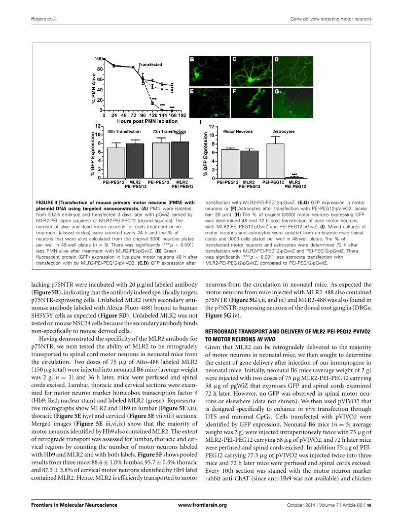
CYTOTOXICITY AND IN VITRO SPECIFICITY OF NANOCONSTRUCTSWe next examined the cytotoxicity and transfection ability ofimmunogenes for motor neurons in vitro. PMN were isolated fromembryonic mice as previously described (Wiese et al., 2010) in 48-well plates and 4 days later transfected with plasmids (pgWiZ orpVIVO2) expressing GFP, using MLR2-PEI or MLR2-PEI-PEG12.We counted viable motor neurons before and after transfection(Figure 4A) for a total of 7 days (n = 3 motor neuron isola-tions in five separate wells). The viability of cells transfected withMLR2-PEI-PEG12-pgWiz was not significantly different than fornon-transfected cells over this time period. 48 h and 72 h posttransfection with MLR2-PEI-PEG12-pGwiZ there were 46.4 ± 3.5and 41.1 ± 0.7% of the original viable motor neurons present.This was not significantly different from control non-transfectedcells where there were 57.7 ± 2.4 and 51.2 ± 2.7% of originalviable motor neurons present at that same time period. However,when PEI was not PEGylated, the number of live motor neu-rons was significantly (p < 0.001) reduced to 14.2 ± 2.6% then1.1 ± 0.35%, 48 and 72 h post transfection with MLR2-PEI-pGwiZ(Figure 4A). There was no significant difference in the percent-age of live motor neurons if pVIVO2 was used in place of pgWiZ(results not shown).
GFP expression in pure motor neurons 48 h after transfection isdemonstrated with pVIVO2 (Figure 4B) or pgWiZ (Figures 4C,D)carried by MLR2-PEI-PEG12. Figure 4C shows GFP expression inthe cell body and processes of a motor neuron and Figure 4Dshows GFP-containing transfected neuronal processes over a bedof non-transfected cells. GFP expression was also observed inmotor neurons after transfection with pVIVO2 carried by PEI-PEG12 (Figures 4E,G), and again there is cell bodies and processes
Frontiers in Molecular Neuroscience www.frontiersin.org October 2014 | Volume 7 | Article 80 | 13

Rogers et al. Gene delivery targeting motor neurons
FIGURE 2 | Stepwise synthesis of the targeted nanoconstructs.
FIGURE 3 | Agarose gel retardation assay of MLR2-PEI-PEG12-pVIVO2
and PEI-PEG12-pVIVO2. Lane 1, 100 bp ladder; lane 2, naked pVIVO2(400 ng); lane 3–5 400 ng pVIVO2 with PEI-PEG12 at N/P 5, 10, and 12;lanes 6–8 400 ng pVIVO2 with MLR2-PEI-PEG12 at N/P 5, 10, and 12.
with GFP and also non-transfected cells. The percentage of motorneurons expressing GFP was determined 48 and 72 h post transfec-tion with MLR2-PEI-PEG12-pgWiz and PEI-PEG12-pgWiz andMLR2-PEI-pgWiz (Figure 4H). Notably, MLR2-PEI-pgWiz didnot produce any GFP possibly because few live motor neuronswere present after 48 h. However, 8.0 ± 0.8% of motor neuronsexpressed GFP 48 h after transfection by MLR2-PEI-PEG12-pgWiz and this did not increase significantly by 72 h (8.3 ± 1.8%).Similarly, 7.0 ± 1.15% of motor neurons expressed GFP 48 h posttransfection with PEI-PEG12-pgWiz and this rose to 8.1 ± 1.5%by 72 h post transfection (Figure 4H).
Mixed cultures of motor neurons and astrocytes were iso-lated from embryonic mice spinal cords (Ford et al., 1994) andtransfected with MLR2-PEI-PEG12-pgWiz or PEI-PEG12-pgWiz.The percentage of motor neurons transfected after 48 h was
Table 1 | Size and zeta potential of nanoconstructs.
Complex N/P Ratio Zeta potential (mV) Particle size (nm)
PEI-PEG12 2 −14.4 ± 4.7 88.7 ± 13.2
PEI-PEG12 5 −0.5 ± 2.9 78.0 ± 15
PEI-PEG12 10 0.7 ± 3.1 75.8 ± 14
PEI-PEG12 12 4.8 ± 0.9 101.1 ± 16.1
MLR2-PEI-PEG12 5 −42.0 ± 0.4 78.3 ± 14
MLR2-PEI-PEG12 10 −32.5 ± 1.3 82.7 ± 10
MLR2-PEI-PEG12 12 −19.9 ± 1.3 95.3 ± 11
6.7 ± 0.32% for MLR2-PEI-PEG12-pGWIZ and 8.0 ± 1.6% forPEI-PEG12-pgWiz. However, 8.0 ± 1.6% of astrocytes were trans-fected with PEI-PEG12-pgWiz and significantly (p < 0.001) less(0.3 ± 0.3%) with MLR2-PEI-PEG12-pgWiz (Figure 4I). Thisdemonstrates the selectivity of MLR2-PEI-PEG12 for motor neu-rons. Figure 4F shows GFP expression in astrocytes 48 h posttransfection with PEI-PEG12-pgWiz.
SPECIFICITY OF ANTI-p75NTR (MLR2) AND RETROGRADE TRANSPORTIN VIVOA key requirement for in vivo gene therapy is specificity to the targetcell population. We used an antibody to p75NTR to target motorneurons and sought to demonstrate specificity and usefulness inneonatal mice where high numbers of motor neurons that expressp75NTR occur. MLR2 was fluorescently labeled with Atto-488)and the specificity of the labeled antibody for p75NTR determinedby flow cytometry. Cells expressing mouse p75NTR (Figure 5A)and human p75NTR (Figure 5C) were incubated with and without20 μg/ml labeled MLR2 and subjected to flow cytometry analy-sis. The shift in mean fluorescence intensity to the right indicatesan increase in the antibody binding to the cells. However, therewas no change in fluorescence intensity after control fibroblasts
Frontiers in Molecular Neuroscience www.frontiersin.org October 2014 | Volume 7 | Article 80 | 14
Rogers et al. Gene delivery targeting motor neurons
FIGURE 4 |Transfection of mouse primary motor neurons (PMN) with
plasmid DNA using targeted nanoconstructs. (A) PMN were isolatedfrom E12.5 embryos and transfected 3 days later with pGwiZ carried byMLR2-PEI (open squares) or MLR2-PEI-PEG12 (closed squares). Thenumber of alive and dead motor neurons for each treatment or notreatment (closed circles) were counted every 24 h and the % ofneurons that were alive calculated from the original 3000 neurons platedper well in 48-well plates (n = 5). There was significantly (∗∗∗p < 0.001)less PMN alive after treatment with MLR2-PEI-pGwiZ. (B) Greenfluorescent protein (GFP) expression in live pure motor neurons 48 h aftertransfection with by MLR2-PEI-PEG12-pVIVO2. (C,D) GFP expression after
transfection with MLR2-PEI-PEG12-pGwiZ. (E,G) GFP expression in motorneurons or (F) Astrocytes after transfection with PEI-PEG12-pVIVO2. (scalebar: 20 μm). (H) The % of original (3000) motor neurons expressing GFPwas determined 48 and 72 h post transfection of pure motor neuronswith MLR2-PEI-PEG12-pGwiZ and PEI-PEG12-pGwiZ. (I). Mixed cultures ofmotor neurons and astrocytes were isolated from embryonic mice spinalcords and 3000 cells plated per well in 48-well plates. The % oftransfected motor neurons and astrocytes were determined 72 h aftertransfection with MLR2-PEI-PEG12-pGwiZ and PEI-PEG12-pGwiZ. Therewas significantly (∗∗∗p < 0.001) less astrocyte transfection withMLR2-PEI-PEG12-pGwiZ, compared to PEI-PEG12-pGwiZ.
lacking p75NTR were incubated with 20 μg/ml labeled antibody(Figure 5B), indicating that the antibody indeed specifically targetsp75NTR-expressing cells. Unlabeled MLR2 (with secondary anti-mouse antibody labeled with Alexia-Fluor-488) bound to humanSHSY5Y cells as expected (Figure 5D). Unlabeled MLR2 was nottested on mouse NSC34 cells because the secondary antibody bindsnon-specifically to mouse derived cells.
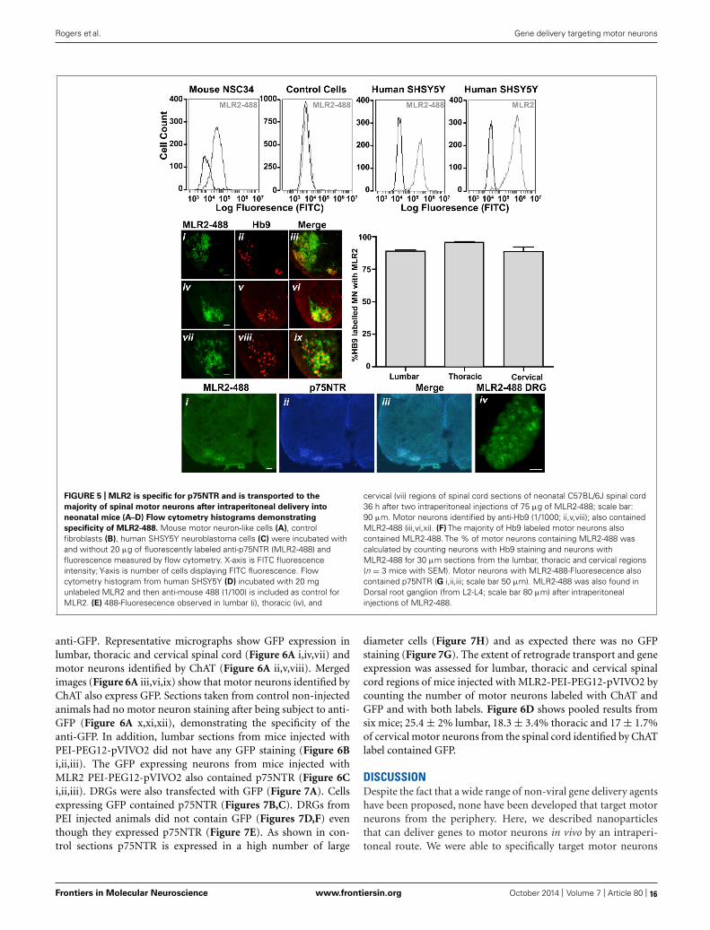
Having demonstrated the specificity of the MLR2 antibody forp75NTR, we next tested the ability of MLR2 to be retrogradelytransported to spinal cord motor neurons in neonatal mice fromthe circulation. Two doses of 75 μg of Atto-488 labeled MLR2(150 μg total) were injected into neonatal B6 mice (average weightwas 2 g; n = 3) and 36 h later, mice were perfused and spinalcords excised. Lumbar, thoracic and cervical sections were exam-ined for motor neuron marker homeobox transcription factor 9(Hb9; Red; nuclear stain) and labeled MLR2 (green). Representa-tive micrographs show MLR2 and Hb9 in lumbar (Figure 5E i,ii),thoracic (Figure 5E iv,v) and cervical (Figure 5E vii,viii) sections.Merged images (Figure 5E iii,vi,ix) show that the majority ofmotor neurons identified by Hb9 also contained MLR2. The extentof retrograde transport was assessed for lumbar, thoracic and cer-vical regions by counting the number of motor neurons labeledwith Hb9 and MLR2 and with both labels. Figure 5F shows pooledresults from three mice; 88.6 ± 1.0% lumbar, 95.7 ± 0.5% thoracicand 87.3 ± 3.8% of cervical motor neurons identified by Hb9 labelcontained MLR2. Hence, MLR2 is efficiently transported to motor
neurons from the circulation in neonatal mice. As expected themotor neurons from mice injected with MLR2-488 also containedp75NTR (Figure 5G i,ii, and iii) and MLR2-488 was also found inthe p75NTR-expressing neurons of the dorsal root ganglia (DRGs;Figure 5G iv).
RETROGRADE TRANSPORT AND DELIVERY OF MLR2-PEI-PEG12-PVIVO2TO MOTOR NEURONS IN VIVOGiven that MLR2 can be retrogradely delivered to the majorityof motor neurons in neonatal mice, we then sought to determinethe extent of gene delivery after injection of our immunogene inneonatal mice. Initially, neonatal B6 mice (average weight of 2 g)were injected with two doses of 75 μg MLR2-PEI-PEG12 carrying58 μg of pgWiZ that expresses GFP and spinal cords examined72 h later. However, no GFP was observed in spinal motor neu-rons or elsewhere (data not shown). We then used pVIVO2 thatis designed specifically to enhance in vivo transfection throughDTS and minimal CpGs. Cells transfected with pVIVO2 wereidentified by GFP expression. Neonatal B6 mice (n = 5; averageweight was 2 g) were injected intraperitonealy twice with 75 μg ofMLR2-PEI-PEG12 carrying 58 μg of pVIVO2, and 72 h later micewere perfused and spinal cords excised. In addition 75 μg of PEI-PEG12 carrying 77.3 μg of pVIVO2 was injected twice into threemice and 72 h later mice were perfused and spinal cords excised.Every 10th section was stained with the motor neuron markerrabbit anti-ChAT (since anti-Hb9 was not available) and chicken
Frontiers in Molecular Neuroscience www.frontiersin.org October 2014 | Volume 7 | Article 80 | 15
Rogers et al. Gene delivery targeting motor neurons
FIGURE 5 | MLR2 is specific for p75NTR and is transported to the
majority of spinal motor neurons after intraperitoneal delivery into
neonatal mice (A–D) Flow cytometry histograms demonstrating
specificity of MLR2-488. Mouse motor neuron-like cells (A), controlfibroblasts (B), human SHSY5Y neuroblastoma cells (C) were incubated withand without 20 μg of fluorescently labeled anti-p75NTR (MLR2-488) andfluorescence measured by flow cytometry. X-axis is FITC fluorescenceintensity; Y-axis is number of cells displaying FITC fluorescence. Flowcytometry histogram from human SHSY5Y (D) incubated with 20 mgunlabeled MLR2 and then anti-mouse 488 (1/100) is included as control forMLR2. (E) 488-Fluoresecence observed in lumbar (i), thoracic (iv), and
cervical (vii) regions of spinal cord sections of neonatal C57BL/6J spinal cord36 h after two intraperitoneal injections of 75 μg of MLR2-488; scale bar:90 μm. Motor neurons identified by anti-Hb9 (1/1000; ii,v,viii); also containedMLR2-488 (iii,vi,xi). (F) The majority of Hb9 labeled motor neurons alsocontained MLR2-488. The % of motor neurons containing MLR2-488 wascalculated by counting neurons with Hb9 staining and neurons withMLR2-488 for 30 μm sections from the lumbar, thoracic and cervical regions(n = 3 mice with SEM). Motor neurons with MLR2-488-Fluoresecence alsocontained p75NTR (G i,ii,iii; scale bar 50 μm). MLR2-488 was also found inDorsal root ganglion (from L2-L4; scale bar 80 μm) after intraperitonealinjections of MLR2-488.
anti-GFP. Representative micrographs show GFP expression inlumbar, thoracic and cervical spinal cord (Figure 6A i,iv,vii) andmotor neurons identified by ChAT (Figure 6A ii,v,viii). Mergedimages (Figure 6A iii,vi,ix) show that motor neurons identified byChAT also express GFP. Sections taken from control non-injectedanimals had no motor neuron staining after being subject to anti-GFP (Figure 6A x,xi,xii), demonstrating the specificity of theanti-GFP. In addition, lumbar sections from mice injected withPEI-PEG12-pVIVO2 did not have any GFP staining (Figure 6Bi,ii,iii). The GFP expressing neurons from mice injected withMLR2 PEI-PEG12-pVIVO2 also contained p75NTR (Figure 6Ci,ii,iii). DRGs were also transfected with GFP (Figure 7A). Cellsexpressing GFP contained p75NTR (Figures 7B,C). DRGs fromPEI injected animals did not contain GFP (Figures 7D,F) eventhough they expressed p75NTR (Figure 7E). As shown in con-trol sections p75NTR is expressed in a high number of large
diameter cells (Figure 7H) and as expected there was no GFPstaining (Figure 7G). The extent of retrograde transport and geneexpression was assessed for lumbar, thoracic and cervical spinalcord regions of mice injected with MLR2-PEI-PEG12-pVIVO2 bycounting the number of motor neurons labeled with ChAT andGFP and with both labels. Figure 6D shows pooled results fromsix mice; 25.4 ± 2% lumbar, 18.3 ± 3.4% thoracic and 17 ± 1.7%of cervical motor neurons from the spinal cord identified by ChATlabel contained GFP.
DISCUSSIONDespite the fact that a wide range of non-viral gene delivery agentshave been proposed, none have been developed that target motorneurons from the periphery. Here, we described nanoparticlesthat can deliver genes to motor neurons in vivo by an intraperi-toneal route. We were able to specifically target motor neurons
Frontiers in Molecular Neuroscience www.frontiersin.org October 2014 | Volume 7 | Article 80 | 16

Rogers et al. Gene delivery targeting motor neurons
FIGURE 6 | MLR2-PEI-PEG12-pVIVO2 but not PEI-PEG12-pVIVO2 is
retrogradely transported to motor neurons in neonatal mice and GFP
expressed. (A) Two doses of 75 μg of MLR2-PEI-PEG12-pVIVO2 carrying58 μg pVIVO2 (N/P 12) was injected into neonatal mice and 72 h later spinalcords excised and examined for GFP expression (n = 5). Motor neurons inthe lumbar (ii), thoracic (iv), and cervical (vii) regions were identified bystaining with ChAT (1/5000) and GFP expression identified with chickenanti-GFP (1/500). Motor neurons that expressed GFP (i,iv,vii) alwayscontained ChAT (iii scale bar: 100 μm; vi scale bar: 100 μm; ix scale bar:50 μm). Motor neurons from control sections of untreated mice identified byChAT (xi), did not contain GFP fluorescence (x) after treatment with chicken
anti-GFP (1/500) scale bar: 50 μm. (B) Two doses of 75 μg ofPEI-PEG12-pVIVO2 carrying 77.3 μg pVIVO2 (N/P 5) was injected intoneonatal mice and 72 h later spinal cords excised and examined for GFPexpression (n = 3). Lumbar sections did not contain GFP fluorescence (i)after treatment with chicken anti-GFP (1/500) and motor neurons wereidentified by ChAT (ii), scale bar: 50 μm. (C) Motor neurons expressing GFPobserved in mice injected with MLR2-PEI-PEG12-pVIVO2 (i) also expressedp75NTR (1 μg/ml goat anti-p75NTR; ii) scale bar: 50 μmm. (B,C) M.(D) Percentage of lumbar, thoracic and cervical motor neurons labeled withGFP and ChAT 48 h after MLR2-PEI-PEG12-pVIVO2 given, i.p. (n = 5 micewith SEM).
by including in our nanoparticle an antibody to p75NTR (MLR2)that binds and internalizes into motor neurons (Matusica et al.,2008).
The ability of p75NTR antibody MLR2 to target our nanopar-ticles to motor neurons from the periphery was shown by labeledMLR2 being observed in the majority of spinal motor neuronsfollowing intraperitoneal administration. MLR2 was labeled withan Atto-488 fluorophore and observed in the majority (near to90%) of motor neurons identified by Hb9 staining, which is spe-cific to the nucleus of developing spinal motor neurons (Arberet al., 1999). These observations indicate that MLR2 is retrogradelytransported to most of the motor neurons after intraperitonealdelivery. The similar percentage of labeling across the lumbar,thoracic and cervical regions is not surprising, since motor neu-rons in all segments of the rodent neonatal spinal cord are knownto express p75NTR (Yan and Johnson, 1988). We also observedlabeled antibody in dorsal root ganglia (DRG). Previous workhas shown the majority of motor neurons and sensory fibers inthe spinal tract can be accessed in an identical manner by intra-venous or intraperitonealy delivered agents that travel retrogradelyin motor neurons and sensory fibers. Hence, intraperitoneal routes
to motor neurons and dorsal root ganglia (DRG) that contain cellbodies of sensory fibers are from the circulation to terminals inthe periphery. This was clearly shown by Alisky et al. (2002) whereboth intraperitoneal and intravenous injections of retrograde trac-ing agent cholera toxin subunit B (CTB) accessed all spinal motorneurons and produced identical staining. Hence, labeled MLR2probably travels to the neuromuscular junctions via the circulationafter intraperitoneal injections.
The nanoparticle comprising MLR2 conjugated to PEGylatedPEI, and the GFP expressing plasmid pVIVO2 transfected motorneurons 72 h post intraperitoneal injections into 5 neonatal mice.25.4% of lumbar, 18.3% of thoracic, and 17.0% of spinal motorneurons were transfected with pVIVO2 identified by GFP expres-sion. When we injected PEGylated PEI carrying pVIVO2, therewere no motor neurons transfected, demonstrating again thatMLR2 antibody is an important component for retrograde trans-port to motor neurons in the spinal cord. This is also demonstratedby the fact there was no transfection in any other type of spinalcord cells when PEGylated PEI carrying pVIVO2 was injectedinto neonatal mice. Specificity and retrograde transport of theimmunogene to motor neurons is by MLR2. Motor neurons were
Frontiers in Molecular Neuroscience www.frontiersin.org October 2014 | Volume 7 | Article 80 | 17

Rogers et al. Gene delivery targeting motor neurons
FIGURE 7 | MLR2-PEI-PEG12-pVIVO2 is retrogradely transported to
dorsal root ganglia (DRG)s in neonatal mice and GFP expressed. Twodoses of 75 μg of MLR2-PEI-PEG12-pVIVO2 carrying 58 μg pVIVO2 (N/P12) was injected into neonatal mice and 72 h L2-L4 DRGs excised andexamined for GFP expression (n = 5). GFP expression was identified withchicken anti-GFP (1/500) and p75NTR with goat anti-p75NTR (1 μg/ml). GFPfluorescence in DRG sections was detected after treatment with chickenanti-GFP (1/500; A) that also contained p75NTR (B,C scale bar: 50 μm). Twodoses of 75 μg of PEI-PEG12-pVIVO2 carrying 77.3 μg pVIVO2 (N/P 5) wasalso injected into neonatal mice and 72 h later spinal cords excised andexamined for GFP expression (n = 3). DRG sections did not contain GFPfluorescence (D) in neurons that also contained p75NTR (E,F; scale bar:50 μm E,F). Control mice non-injected DRG sections did not contain GFP(G) but did contain p75NTR (H,I; scale bar: 50 μm).
identified by ChAT. Since motor neurons identified by ChAT over-lap with Hb9 staining in neonatal mice, this was a valid analysis(Shneider et al., 2009). The low level of transfection observed inmotor neurons in vivo may be explained by the reported inef-ficiency of non-viral gene delivery (Mintzer and Simanek, 2009;Rogers and Rush, 2012). In regards to dosage, we used the samedosage of nanoparticle as we did labeled antibody (75 μg/g bodyweight). Hence since the same amount of antibody (when labeled)can access all the motor neurons, other areas of the nanoparticledelivery may not be optimal. Further improvements to transfec-tion efficiency in vivo can be made. We already have a large payloadfor our nanoparticle, and although PEI is PEGylated, the wholeIgG (MLR2) was not. A way to reduce interactions of the IgGwith the immune system is to use the antibody binding fragments.For example, antibody fragments that lack Fc domains (FAb, Fv,scFv), have reduced interactions with the immune system and non-targeted cells through Fc receptors (Peer and Lieberman, 2011).Indeed, previous work with antibody fragments for tumor target-ing using immunoliposomes carrying plasmid DNA has producedless immune reaction than whole antibodies and more sustainedexpression in vivo (Zhou et al., 2011). Hence, further improve-ments to transfection efficiency may be made by using FAb orscFV of MLR2 instead of the whole IgG.
This is the first report of specific gene delivery to motor neu-rons via the circulation. Previous viral gene delivery attempts
to transfect neonatal mouse motor neurons did not have thespecificity to transfect mouse motor neurons via the circulation(Towne et al., 2008). Towne et al. (2008) tested intravenous deliv-ery of recombinant adeno-associated virus (rAAVs) expressingsmall hairpin RNAs targeting mutant SOD1 in the ALS mousemodel. Although the AAV virus could transfect mouse motor neu-rons from the circulation it was not specific, it also transfectedmost other cell types. Towne et al. (2011) then went on to serotypetheir AAV viral delivery for retrograde transport and gave multi-ple injections to muscle groups innervated by motor neurons inneonatal SOD1 mice. Unfortunately, they could not down regu-late mutant SOD1 enough to improve outcomes in ALS mice. Thiswas suggested to be because not all motor neurons were accessedby intramuscular injections resulting in inconsistent of levels oftransfection across the spinal cord. Notably, approximately 28%of lumbar, 12% of thoracic, and 18% of cervical motor neuronswere transfected in neonatal mice (Towne et al., 2011). It was con-cluded that the lack of improvement after their viral gene therapymight be because it is difficult to access all motor neurons by intra-muscular injections. In contrast, we were able to achieve motorneuron transfection after intraperitoneal injections of immuno-gene. We did not need to inject every muscle group to get specifictransfection of motor neurons. To our knowledge we are the firstgroup to do so. We have shown that you can transfect 25.4% oflumbar, 18.3% of thoracic, and 17.0% of spinal motor neuronsafter delivery of our immunogene Considering our nanoconstructmay not still be optimal, our results are hopeful for developingtargeted therapy.
PEI was used to condense plasmid DNA for gene deliveryin vitro and in vivo. However, PEI was modified by PEGylationto make it “stealth-like” in the circulation. Our data indicatesthat PEGylation reduces the toxicity branched PEI has to puremotor neurons. The viability of motor neurons in vitro subjectto PEI conjugated to p75NTR targeting antibody MLR2 was sig-nificantly poorer than PEGylated PEI conjugated to MLR2. Thisresult was not surprising since previous work has shown PEI with-out modification is toxic (Moghimi et al., 2005) causing cell stressand apoptosis (Godbey et al., 1999; Moghimi et al., 2005). Otherwork has shown modifying PEI by PEGylation reduces cellulartoxicity (Ogris et al., 1999; Malek et al., 2009), presumably byreduction in positive charge (Merdan et al., 2003; Hoskins et al.,2012) and the formation of a hydrophilic corona around thePEI/DNA core (Merdan et al., 2005). Grafting of PEI with PEGchains thus reduces the zeta potential of PEI-based polyplexeseven at high N/P ratio (Merdan et al., 2005). The zeta potential ofour immunogene were negative at the N/P ratio of 12 used in vitroand in vivo (Hoskins et al., 2012). Therefore, our results showingthat PEGylated PEI reduces the zeta potential and toxicity of ourimmunogene in vitro are consistent with the literature.
Although we were able to transfect motor neurons in vivo, ourimmunogene produced a low percentage (∼8%) of transfectionin vitro. This was significantly lower than the 17–25% of motorneurons transfected throughout the spinal cord in vivo. This dis-parity between in vitro and in vivo transfection is not unusualfor stable cationic constructs containing grafted stealth agents andtargeting agents. For example, Höbel et al. (2011) showed thatdespite their relatively low in vitro efficacy, PEI grafted with sugars
Frontiers in Molecular Neuroscience www.frontiersin.org October 2014 | Volume 7 | Article 80 | 18

Rogers et al. Gene delivery targeting motor neurons
showed better in vivo than in vitro profiles and reduced toxicity.Most testing of non-viral gene delivery agents in vitro employscell lines cell that rapidly divide with cell culture reagents that donot accurately mimic the in vivo situation (van Gaal et al., 2011).Furthermore, transfection is often undertaken without serum inthe media, which does not mimic the high perecentage of serumin the circulation. Cationic polyplexes can interact with nega-tively charged blood components (e.g., proteins, erythrocytes),followed by the formation of aggregates. Under these conditions,precipitation can enhance the association of the delivery systemwith the cell surface, which can artificially elevate transfectionrates with agents that are not stable in physiological media. Con-versely, non-viral agents that are stable in physiological mediaoften do not transfect efficiently in cell culture, leading to the con-clusion that such systems are not worthy of further consideration.Another confounding factor with cell culture experiments is thatthe nuclear membrane breaks down during cell division, allowingefficient translocation of DNA into the nucleus of rapidly divid-ing cells that greatly facilitates transfection (Pérez-Martínez et al.,2011). We chose to test our nanoconstructs on PMN which donot divide. Our stable nanoparticle was not only PEGylated butalso cross-linked to an antibody to p75NTR. Previous researchhas found that cross-linking amines in PEI increased the stabil-ity of PEGylated PEI and improved in vivo stability (Neu et al.,2007; Höbel et al., 2011). Therefore, our results producing sig-nificant in vivo transfection is probably reflective of the difficultysimulating in vivo environments in vitro.
PEGylation of PEI decreases the number of amines available forcondensing plasmid DNA. The size of our immunogene complexat neutral charge was small (near 100 nm). This is in contrast toprevious reports where branched PEI nanoconstructs complexedwith DNA can be above 300 nm (Ewe et al., 2014). Conjugation toantibody MLR2 via a disulfide bridge, where amines were furtherreduced in the PEGylated PEI did not significantly increase the sizeof the complex. Previous work has shown that positively chargedparticles with sizes above 200 nm may be recognized and removedby the reticuloendothelial system (RES; Dash et al., 1999; Maleket al., 2009). PEGylation reduces this interaction (Ogris et al., 1999;Merdan et al., 2005; Malek et al., 2009) and also the size of thecomplex.
We observed no obvious off-target effects in the spinal cordand transfection of cells other than motor neurons in the spinalcord with our immunogene. Indeed, we did not observe transfec-tion in any other cell types except for motor neurons. However,some of the p75NTR expressing cells were transfected the DRGs.p75NTR is known to be expressed in DRG cells (Yan and Johnson,1988) and transfection of some of these cells by our immunogeneagain highlights the immunogene travels by receptor mediatedretrograde transport to p75NTR expressing cells. The bicistronicpVIVO2 plasmid we used is specifically designed for in vivo trans-fection. The GFP reporter plasmid (pgWiZ) we used for in vitrotransfections contains CpGs in its backbone that are known toinduce immune response in vivo (Davies et al., 2012). pgWiZ isoften used to improve humoral immune response to plasmid vac-cination in vivo (Chamarthy et al., 2003; Grønevik et al., 2005; Roseet al., 2014). In contrast, pVIVO2 has minimal CpGs in its plasmidbackbone and high levels of constitutive transgene expression has
been reported for this plasmid in vivo (Mandke and Singh, 2012).In addition, pVIVO2 has DTS to improve nuclear entry into postmitotic cells such as motor neurons. We tried delivering pgWiZ tomotor neurons by intraperitoneal injections with our immuno-porter MLR2-PEI-PEG12, but found no significant expressionin vivo. This has led us to us to conclude that plasmid designis an important component of effective non-viral gene deliveryagents.
Specific delivery of genes to motor neurons is highly relevantto therapy of ALS and spinal muscular atrophy (SMA), where cur-rently no effective therapy exists (Sreedharan and Brown, 2013).Our results show that motor neurons can be specifically transfectedwith peripherally administered immunogenes. Targeting motorneurons by use of the p75NTR is not surprising as this receptoris highly expressed in the embryonic period and early neonatallife (Yan and Johnson, 1988). Lentivirus that expressed heavy andlight chains of rat p75NTR antibody (MC192) were recently showntransported retrogradely from the axonal tip to the cell body in anin vitro microfluidic culture model (Eleftheriadou et al., 2014).This is in agreement with our in vivo data where p75NTR anti-body was found throughout the spinal cord after intraperitonealdelivery, indicating retrograde transport from terminals to ven-tral motor neurons throughout the spinal cord. Caution has to betaken with immunogenes use in ALS. p75NTR is down regulated inadulthood and re-expressed in injury, including ALS (Lowry et al.,2001). However, the level of p75NTR re-expression and healthof the motor neurons must be sufficient for retrograde trans-port in the majority of motor neurons. A previous study (Coprayet al., 2003) indicated 5% of L4 Lumbar motor neurons re-expressp75NTR in adult ALS mice at symptomatic age. Further work isneeded to determine if sufficient therapeutic genes can be deliveredin adult ALS animal models via p75NTR targeting immunogenes.Since SMA is a disease often occuring in childhood (Arnold andBurghes, 2013), p75NTR targeting immunogenes could be trialedin SMA mice.
CONCLUSIONOur current research demonstrates the suitability of p75NTRtargeting immunogenes to transfect motor neurons from theperiphery in neonatal mice, but further work is required for use inadult animals.
ACKNOWLEDGMENTSThis project was funded by the Motor Neuron Disease ResearchInstitute of Australia (Mary-Louise Rogers, Robert A. Rush), theFlinders Medical Centre Foundation (Mary-Louise Rogers, RobertA. Rush) and Flinders University Research Scholarship (KevinS. Smith). We thank Prof Neil Cashman (University of BritishColumbia, Canada) for use of the NSC34 cell line. Funding wasalso provided by the Australian Research Council Centre of Excel-lence in Convergent Bio-Nano Science and Technology (projectnumber CE140100036, Nicolas H. Voelcker).
REFERENCESAlisky, J. M., Van De Wetering, C. I., and Davidson, B. L. (2002). Widespread
dispersal of cholera toxin subunit b to brain and spinal cord neurons fol-lowing systemic delivery. Exp. Neurol. 178, 139–146. doi: 10.1006/exnr.2002.8031
Frontiers in Molecular Neuroscience www.frontiersin.org October 2014 | Volume 7 | Article 80 | 19

Rogers et al. Gene delivery targeting motor neurons
Arber, S., Han, B., Mendelsohn, M., Smith, M., Jessell, T. M., and Sockanathan, S.(1999). Requirement for the homeobox gene Hb9 in the consolidation of motorneuron identity. Neuron 23, 659–674. doi: 10.1016/S0896-6273(01)80026-X
Arnold, W. D., and Burghes, A. H. M. (2013). Spinal muscular atrophy: developmentand implementation of potential treatments. Ann. Neurol. 74, 348–362. doi:10.1002/ana.23995
Barati, S., Hurtado, P. R., Zhang, S. H., Tinsley, R., Ferguson, I. A., and Rush, R.A. (2006). GDNF gene delivery via the p75(NTR) receptor rescues injured motorneurons. Exp. Neurol. 202, 179–188. doi: 10.1016/j.expneurol.2006.05.027
Blessing, T., Kursa, M., Holzhauser, R., Kircheis, R., and Wagner, E. (2001). Differentstrategies for formation of pegylated EGF-conjugated PEI/DNA complexes fortargeted gene delivery. Bioconjug. Chem. 12, 529–537. doi: 10.1021/bc0001488
Boeckle, S., Von Gersdorff, K., Van Der Piepen, S., Culmsee, C., Wagner, E.,and Ogris, M. (2004). Purification of polyethylenimine polyplexes highlightsthe role of free polycations in gene transfer. J. Gene Med. 6, 1102–1111. doi:10.1002/jgm.598
Boussif, O., Lezoualc’h, F., Zanta, M., Mergny, M., Scherman, D., Demeneix, B.,et al. (1995). A versatile vector for gene and oligonucleotide transfer into cells inculture and in vivo: polyethylenimine. Proc. Natl. Acad. Sci. U.S.A. 92, 7297–7301.doi: 10.1073/pnas.92.16.7297
Bronfman, F., Tcherpakov, M., Jovin, T., and Fainzilber, M. (2003). Ligand-inducedinternalization of the p75 neurotrophin receptor: a slow route to the signalingendosome. J. Neurosci. 23, 3209–3220.
Chamarthy, S. P., Kovacs, J. R., Mcclelland, E., Gattens, D., and Meng, W. S.(2003). A cationic peptide consists of ornithine and histidine repeats aug-ments gene transfer in dendritic cells. Mol. Immunol. 40, 483–490. doi:10.1016/j.molimm.2003.08.001
Chollet, P., Favrot, M. C., Hurbin, A., and Coll, J.-L. (2002). Side-effects of a systemicinjection of linear polyethylenimine-DNA complexes. J. Gene Med. 4, 84–91. doi:10.1002/jgm.237
Copray, J., Jaarsma, D., Kust, B., Bruggeman, R., Mantingh, I., Brouwer, N.,et al. (2003). Expression of the low affinity neurotrophin receptor p75 in spinalmotoneurons in a transgenic mouse model for amyotrophic lateral sclerosis.Neuroscience 116, 685–694. doi: 10.1016/S0306-4522(02)00755-8
Dash, P. R., Read, M. L., Barrett, L. B., Wolfert, M. A., and Seymour, L. W. (1999).Factors affecting blood clearance and in vivo distribution of polyelectrolytecomplexes for gene delivery. Gene Ther. 6, 643–650. doi: 10.1038/sj.gt.3300843
Davies, L. A., Hyde, S. C., Nunez-Alonso, G., Bazzani, R. P., Harding-Smith, R.,Pringle, I. A., et al. (2012). The use of CpG-free plasmids to mediate persistentgene expression following repeated aerosol delivery of pDNA/PEI complexes.Biomaterials 33, 5618–5627. doi: 10.1016/j.biomaterials.2012.04.019
Eleftheriadou, I., Trabalza, A., Ellison, S., Gharun, K., and Mazarakis, N. (2014).Specific retrograde transduction of spinal motor neurons using lentiviral vec-tors targeted to presynaptic NMJ receptors. Mol. Ther. 22, 1285–1298. doi:10.1038/mt.2014.49
Ewe, A., Schaper, A., Barnert, S., Schubert, R., Temme, A., Bakowsky, U.,et al. (2014). Storage stability of optimal liposome-polyethylenimine complexes(lipopolyplexes) for DNA or siRNA delivery. Acta Biomater. 10, 2663–2673. doi:10.1016/j.actbio.2014.02.037
Ford, T., Graham, J., and Rickwood, D. (1994). Iodixanol: a nonionic iso-osmoticcentrifugation medium for the formation of self-generated gradients. Anal.Biochem. 220, 360–366. doi: 10.1006/abio.1994.1350
Germershaus, O., Merdan, T., Bakowsky, U., Behe, M., and Kissel, T.(2006). Trastuzumab-polyethylenimine-polyethylene glycol conjugates for tar-geting Her2-expressing tumors. Bioconjug. Chem. 17, 1190–1199. doi:10.1021/bc0601119
Godbey, W. T., Wu, K. K., and Mikos, A. G. (1999). Tracking the intracellular pathof poly(ethylenimine)/DNA complexes for gene delivery. Proc. Natl. Acad. Sci.U.S.A. 96, 5177–5181. doi: 10.1073/pnas.96.9.5177
Godbey, W. T., Wu, K. K., and Mikos, A. G. (2001). Poly(ethylenimine)-mediatedgene delivery affects endothelial cell function and viability. Biomaterials 22, 471–480. doi: 10.1016/S0142-9612(00)00203-9
Grønevik, E., Von Steyern, F. V., Kalhovde, J. M., Tjelle, T. E., and Mathiesen, I.(2005). Gene expression and immune response kinetics using electroporation-mediated DNA delivery to muscle. J. Gene Med. 7, 218–227. doi: 10.1002/jgm.650
Höbel, S., Loos, A., Appelhans, D., Schwarz, S., Seidel, J., Voit, B., et al. (2011).Maltose- and maltotriose-modified, hyperbranched poly(ethylene imine)s
(OM-PEIs): physicochemical and biological properties of DNA and siRNAcomplexes. J. Control. Release 149, 146–158. doi: 10.1016/j.jconrel.2010.10.008
Hoskins, C., Wang, L., Cheng, W. P., and Cuschieri, A. (2012). Dilemmas inthe reliable estimation of the in-vitro cell viability in magnetic nanoparticleengineering: which tests and what protocols? Nanoscale Res. Lett. 7, 77. doi:10.1186/1556-276X-7-77
Kircheis, R., Kichler, A., Wallner, G., Kursa, M., Ogris, M., Felzmann, T., et al. (1997).Coupling of cell-binding ligands to polyethylenimine for targeted gene delivery.Gene Ther. 4, 409–418. doi: 10.1038/sj.gt.3300418
Kunath, K., Von Harpe, A., Fischer, D., Petersen, H., Bickel, U., Voigt, K., et al.(2003). Low-molecular-weight polyethylenimine as a non-viral vector for DNAdelivery: comparison of physicochemical properties, transfection efficiency andin vivo distribution with high-molecular-weight polyethylenimine. J. Control.Release 89, 113–125. doi: 10.1016/S0168-3659(03)00076-2
Lalli, G., and Schiavo, G. (2002). Analysis of retrograde transport in motor neuronsreveals common endocytic carriers for tetanus toxin and neurotrophin receptorp75NTR. J. Cell Biol. 156, 233–239. doi: 10.1083/jcb.200106142
Lowry, K., Murray, S., Mclean, C., Talman, P., Mathers, S., Lopes, E., et al.(2001). A potential role for the p75 low-affinity neurotrophin receptor inspinal motor neuron degeneration in murine and human amyotrophic lat-eral sclerosis. Amyotroph. Lateral Scler. 2, 127–134. doi: 10.1080/146608201753275463
Lungwitz, U., Breunig, M., Blunk, T., and Göpferich, A. (2005). Polyethylenimine-based non-viral gene delivery systems. Eur. J. Pharm. Biopharm. 60, 247–266. doi:10.1016/j.ejpb.2004.11.011
Luo, X., Pan, S., Feng, M., Wen, Y., and Zhang, W. (2010). Stability of poly(ethyleneglycol)-graft-polyethylenimine copolymer/DNA complexes: influences of PEGmolecular weight and PEGylation degree. J. Mater. Sci. Mater. Med. 21, 597–607.doi: 10.1007/s10856-009-3903-1
Magnusson, T., Haase, R., Schleef, M., Wagner, E., and Ogris, M. (2011).Sustained, high transgene expression in liver with plasmid vectors using opti-mized promoter-enhancer combinations. J. Gene Med. 13, 382–391. doi:10.1002/jgm.1585
Malek, A., Merkel, O., Fink, L., Czubayko, F., Kissel, T., and Aigner, A. (2009). Invivo pharmacokinetics, tissue distribution and underlying mechanisms of var-ious PEI(-PEG)/siRNA complexes. Toxicol. Appl. Pharmacol. 236, 97–108. doi:10.1016/j.taap.2009.01.014
Mandke, R., and Singh, J. (2012). Cationic nanomicelles for delivery of plas-mids encoding interleukin-4 and interleukin-10 for prevention of autoimmunediabetes in mice. Pharm. Res. 29, 883–897. doi: 10.1007/s11095-011-0616-1
Masago, K., Itaka, K., Nishiyama, N., Chung, U.-I., and Kataoka, K. (2007). Genedelivery with biocompatible cationic polymer: pharmacogenomic analysis oncell bioactivity. Biomaterials 28, 5169–5175. doi: 10.1016/j.biomaterials.2007.07.019
Matusica, D., Fenech, M., Rogers, M., and Rush, R. (2008). Characterization and useof the NSC-34 cell line for study of neurotrophin receptor trafficking. J. Neurosci.Res. 86, 553–565. doi: 10.1002/jnr.21507
Merdan, T., Callahan, J., Petersen, H., Kunath, K., Bakowsky, U., Kopecková, P., et al.(2003). Pegylated polyethylenimine-Fab & apos; antibody fragment conjugatesfor targeted gene delivery to human ovarian carcinoma cells. Bioconjug. Chem.14, 989–996. doi: 10.1021/bc0340767.
Merdan, T., Kunath, K., Petersen, H., Bakowsky, U., Voigt, K. H., Kopecek,J., et al. (2005). PEGylation of poly(ethylene imine) affects stability of com-plexes with plasmid DNA under in vivo conditions in a dose-dependent mannerafter intravenous injection into mice. Bioconjug. Chem. 16, 785–792. doi:10.1021/bc049743q
Mintzer, M. A., and Simanek, E. E. (2009). Nonviral vectors for gene delivery. Chem.Rev. 109, 259–302. doi: 10.1021/cr800409e
Moghimi, S. M., Symonds, P., Murray, J. C., Hunter, A. C., Debska, G.,and Szewczyk, A. (2005). A two-stage poly(ethylenimine)-mediated cytotox-icity: implications for gene transfer/therapy. Mol. Ther. 11, 990–995. doi:10.1016/j.ymthe.2005.02.010.
Navarro-Quiroga, I., Antonio Gonzalez-Barrios, J., Barron-Moreno, F., Gonzalez-Bernal, V., Martinez-Arguelles, D. B., and Martinez-Fong, D. (2002). Improvedneurotensin-vector-mediated gene transfer by the coupling of hemagglutininHA2 fusogenic peptide and Vp1 SV40 nuclear localization signal. Mol. BrainRes. 105, 86–97. doi: 10.1016/S0169-328X(02)00396-0
Frontiers in Molecular Neuroscience www.frontiersin.org October 2014 | Volume 7 | Article 80 | 20

Rogers et al. Gene delivery targeting motor neurons
Neu, M., Germershaus, O., Behe, M., and Kissel, T. (2007). Bioreversibly crosslinkedpolyplexes of PEI and high molecular weight PEG show extended circula-tion times in vivo. J. Control. Release 124, 69–80. doi: 10.1016/j.jconrel.2007.08.009
Ogris, M., Brunner, S., Schüller, S., Kircheis, R., and Wagner, E. (1999). PEGylatedDNA/transferrin-PEI complexes: reduced interaction with blood components,extended circulation in blood and potential for systemic gene delivery. Gene Ther.6, 595–605. doi: 10.1038/sj.gt.3300900
Ogris, M., Walker, G., Blessing, T., Kircheis, R., Wolschek, M., and Wagner, E.(2003). Tumor-targeted gene therapy: strategies for the preparation of ligand-polyethylene glycol-polyethylenimine/DNA complexes. J. Control. Release 91,173–181. doi: 10.1016/S0168-3659(03)00230-X
Pardridge, W. (2006). Molecular Trojan horses for blood-brain barrier drug delivery.Curr. Opin. Pharmacol 6, 494–500. doi: 10.1016/j.coph.2006.06.001
Peer, D., and Lieberman, J. (2011). Special delivery: targeted therapy with smallRNAs. Gene Ther. 18, 1127–1133. doi: 10.1038/gt.2011.56
Pérez-Martínez, F. C., Guerra, J., Posadas, I., and Ceña, V. (2011). Barriers tonon-viral vector-mediated gene delivery in the nervous system. Pharm. Res. 28,1843–1858. doi: 10.1007/s11095-010-0364-7
Rogers, M., Atmosukarto, I., Berhanu, D., Matusica, D., Macardle, P., andRush, R. (2006). Functional monoclonal antibodies to p75 neurotrophinreceptor raised in knockout mice. J. Neurosci. Methods 158, 109–120. doi:10.1016/j.jneumeth.2006.05.022
Rogers, M. L., Bailey, S., Matusica, D., Nicholson, I., Muyderman, H., Pagadala,P. C., et al. (2010). ProNGF mediates death of Natural Killer cells through acti-vation of the p75NTR-sortilin complex. J. Neuroimmunol. 226, 93–103. doi:10.1016/j.jneuroim.2010.05.040
Rogers, M.-L., and Rush, R. A. (2012). Non-viral gene therapy for neurologicaldiseases, with an emphasis on targeted gene delivery. J. Control. Release 157,183–189. doi: 10.1016/j.jconrel.2011.08.026
Rose, L., Mahdipoor, P., Kucharski, C., and Uludag, H. (2014). Phar-macokinetics and transgene expression of implanted polyethylenimine-based pDNA complexes. Biomater. Sci. 2, 833–842. doi: 10.1039/c3bm60200a
Schaffert, D., Kiss, M., Rodl, W., Shir, A., Levitzki, A., Ogris, M., et al. (2011).Poly(I:C)-mediated tumor growth suppression in EGF-receptor overexpressingtumors using EGF-polyethylene glycol-linear polyethylenimine as carrier. Pharm.Res. 28, 731–741. doi: 10.1007/s11095-010-0225-4
Shepheard, S. R., Chataway, T., Schultz, D. W., Rush, R. A., and Rogers, M.L. (2014). The extracellular domain of neurotrophin receptor p75 as a can-didate biomarker for amyotrophic lateral sclerosis. PLoS ONE 9:e87398. doi:10.1371/journal.pone.0087398
Shneider, N. A., Brown, M. N., Smith, C. A., Pickel, J., and Alvarez, F. J. (2009).Gamma motor neurons express distinct genetic markers at birth and requiremuscle spindle-derived GDNF for postnatal survival. Neural Dev. 4, 42. doi:10.1186/1749-8104-4-42
Snyder, S. L., and Sobocinski, P. Z. (1975). An improved 2,4,6-trinitrobenzenesulfonic acid method for the determination of amines. Anal.Biochem. 64, 284–288. doi: 10.1016/0003-2697(75)90431-5
Sreedharan, J., and Brown, R. H. (2013). Amyotrophic lateral sclerosis: problemsand prospects. Ann. Neurol. 74, 309–316. doi: 10.1002/ana.24012
Tang, M. X., and Szoka, F. C. (1997). The influence of polymer structure on theinteractions of cationic polymers with DNA and morphology of the resultingcomplexes. Gene Ther. 4, 823–832. doi: 10.1038/sj.gt.3300454
Towne, C., Raoul, C., Schneider, B. L., and Aebischer, P. (2008). Systemic AAV6delivery mediating RNA interference against SOD1: neuromuscular transductiondoes not alter disease progression in fALS mice. Mol. Ther. 16, 1018–1025. doi:10.1038/mt.2008.73
Towne, C., Setola, V., Schneider, B. L., and Aebischer, P. (2011). Neuroprotection bygene therapy targeting mutant SOD1 in individual pools of motor neurons doesnot translate into therapeutic benefit in fALS mice. Mol. Ther. 19, 274–283. doi:10.1038/mt.2010.260
Turner, M. R., Hardiman, O., Benatar, M., Brooks, B. R., Chio, A., De Carvalho, M.,et al. (2013). Controversies and priorities in amyotrophic lateral sclerosis. LancetNeurol. 12, 310–322. doi: 10.1016/S1474-4422(13)70036-X
van Gaal, E. V. B., Van Eijk, R., Oosting, R. S., Kok, R. J., Hennink, W. E.,Crommelin, D. J. A., et al. (2011). How to screen non-viral gene delivery sys-tems in vitro? J. Controll. Release 154, 218–232. doi: 10.1016/j.jconrel.2011.05.001
Wiese, S., Herrmann, T., Drepper, C., Jablonka, S., Funk, N., Klausmeyer, A., et al.(2010). Isolation and enrichment of embryonic mouse motoneurons from thelumbar spinal cord of individual mouse embryos. Nat. Protoc. 5, 31–38. doi:10.1038/nprot.2009.193.
Yan, Q., and Johnson, E. M. Jr. (1988). An immunohistochemical study of the nervegrowth factor receptor in developing rats. J. Neurosci. 8, 3481–3498.
Zabner, J., Fasbender, A. J., Moninger, T., Poellinger, K. A., and Welsh, M. J. (1995).Cellular and molecular barriers to gene transfer by a cationic lipid. J. Biol. Chem.270, 18997–19007. doi: 10.1074/jbc.270.32.18997
Zhang, Y., Schlachetzki, F., and Pardridge, W. M. (2003). Global non-viral genetransfer to the primate brain following intravenous administration. Mol. Ther. 7,11–18. doi: 10.1016/S1525-0016(02)00018-7
Zhou, Q. H., Fu, A., Boado, R. J., Hui, E. K., Lu, J. Z., and Pardridge, W.M. (2011). Receptor-mediated abeta amyloid antibody targeting to Alzheimer’sdisease mouse brain. Mol. Pharm. 8, 280–285. doi: 10.1021/mp1003515
Conflict of Interest Statement: Author Robert A. Rush is now retired from FlindersUniversity and holds the position of Managing Director at Biosensis Pty. Ltd. whichhas commercial interest involving the MLR2 antibody used in this study. No otherauthors have conflicting interest.
Received: 03 July 2014; paper pending published: 07 August 2014; accepted: 18September 2014; published online: 14 October 2014.Citation: Rogers M-L, Smith KS, Matusica D, Fenech M, Hoffman L, Rush RA andVoelcker NH (2014) Non-viral gene therapy that targets motor neurons in vivo. Front.Mol. Neurosci. 7:80. doi: 10.3389/fnmol.2014.00080This article was submitted to the journal Frontiers in Molecular Neuroscience.Copyright © 2014 Rogers, Smith, Matusica, Fenech, Hoffman, Rush and Voelcker.This is an open-access article distributed under the terms of the Creative CommonsAttribution License (CC BY). The use, distribution or reproduction in other forums ispermitted, provided the original author(s) or licensor are credited and that the originalpublication in this journal is cited, in accordance with accepted academic practice. Nouse, distribution or reproduction is permitted which does not comply with these terms.
Frontiers in Molecular Neuroscience www.frontiersin.org October 2014 | Volume 7 | Article 80 | 21