general musculoskeletal screening: upper extremities€¦ · 1 1 general musculoskeletal screening:...
TRANSCRIPT

1
1
General Musculoskeletal General Musculoskeletal Screening: Upper ExtremitiesScreening: Upper Extremities
Gregory Crovetti, M.D.Gregory Crovetti, M.D.Sports Medicine ProgramSports Medicine Program
West Suburban Health CareWest Suburban Health CareTrinity OrthopaedicsTrinity Orthopaedics
8/27/02 Gregory Crovetti, M.D. 2
General ApproachGeneral Approach
HistoryHistoryInspectionInspectionRange of Motion (ROM)Range of Motion (ROM)PalpationPalpationMuscular and neurological examsMuscular and neurological exams
8/27/02 Gregory Crovetti, M.D. 3
HistoryHistory
An accurate history is essentialAn accurate history is essentialWill give you diagnosis 80Will give you diagnosis 80--90% of time90% of timeHow symptoms started (mechanism of How symptoms started (mechanism of injury)?injury)?Duration of complaint?Duration of complaint?Location, nature of pain, or symptoms?Location, nature of pain, or symptoms?Exacerbating or relieving maneuvers?Exacerbating or relieving maneuvers?

2
8/27/02 Gregory Crovetti, M.D. 4
General InspectionGeneral Inspection
Observe how the patient moves as they Observe how the patient moves as they go into the room or move from chair to go into the room or move from chair to tabletableGeneral appearanceGeneral appearanceBody proportionsBody proportions
8/27/02 Gregory Crovetti, M.D. 5
Inspection of Specific AreaInspection of Specific Area
Look for asymmetry between sidesLook for asymmetry between sidesSwelling Swelling DeformitiesDeformitiesAtrophyAtrophyErythemaErythema
8/27/02 Gregory Crovetti, M.D. 6
Range of Motion (Active)Range of Motion (Active)
Have patient range the jointsHave patient range the jointsWatch for decreased or increased Watch for decreased or increased movement of the joint compared to the movement of the joint compared to the other side as well as the normother side as well as the normWatch for pain with movementWatch for pain with movementListen for crepitus or Listen for crepitus or ““poppingpopping””Watch for abnormal movementsWatch for abnormal movements

3
8/27/02 Gregory Crovetti, M.D. 7
Range of Motion (Passive)Range of Motion (Passive)
Next range the joints passively, Next range the joints passively, comparing the end points to the activecomparing the end points to the activeAgain note any decreased or increased Again note any decreased or increased movementmovementPain with the movement Pain with the movement Crepitus or Crepitus or ““poppingpopping””
8/27/02 Gregory Crovetti, M.D. 8
PalpationPalpation
When palpating a structure, you need When palpating a structure, you need to know the anatomy of that structureto know the anatomy of that structurePalpate for swellingPalpate for swellingPalpate for warmthPalpate for warmthPalpate each area of the structure in Palpate each area of the structure in turn evaluating for pain, and turn evaluating for pain, and abnormalities as compared to the other abnormalities as compared to the other sideside
8/27/02 Gregory Crovetti, M.D. 9
Muscular and NeurologicalMuscular and Neurological
Check the following comparing one side Check the following comparing one side to the other:to the other:–– Grade strength (0Grade strength (0--5)5)–– Grade reflexes (0Grade reflexes (0--4)4)–– Sensory examSensory exam

4
8/27/02 Gregory Crovetti, M.D. 10
Generalized Screening ExamGeneralized Screening Exam
If any If any abnormalities, a abnormalities, a more thorough more thorough exam of the joint exam of the joint needs to be done. needs to be done.
Each joint is:Each joint is:–– Inspected (look for Inspected (look for
abnormalities)abnormalities)–– PalpatedPalpated–– ExaminedExamined
8/27/02 Gregory Crovetti, M.D. 11
Neck: Active Range of MotionNeck: Active Range of Motion
Chin to chest (flexion)Chin to chest (flexion)““look at ceilinglook at ceiling”” (extension)(extension)Chin to each shoulder (lateral rotation)Chin to each shoulder (lateral rotation)Ear to each shoulder (lateral flexion, Ear to each shoulder (lateral flexion, i.e., head tilt)i.e., head tilt)
8/27/02 Gregory Crovetti, M.D. 12
Special Tests for the NeckSpecial Tests for the NeckDekleyn testDekleyn test: head and neck rotation with extension. Tests for vertebral : head and neck rotation with extension. Tests for vertebral artery compression.artery compression.SpurlinSpurlin’’ss: (foraminal compression test): patient extends rotates head to : (foraminal compression test): patient extends rotates head to side, the examiner then applies axial load to the head. Positivside, the examiner then applies axial load to the head. Positive test is e test is when there is pain radiating into arm. Indicates Pressure on a when there is pain radiating into arm. Indicates Pressure on a nerve nerve root. root. Elvey testElvey test: (upper limb tension tests): tests designed to put stress on : (upper limb tension tests): tests designed to put stress on the neurological structures of the upper limb.the neurological structures of the upper limb.
A.A. Median nerve C5,6,7Median nerve C5,6,7B.B. Median nerve, axillary nerveMedian nerve, axillary nerveC.C. Radial nerveRadial nerveD.D. Ulnar nerve C8, T1Ulnar nerve C8, T1
5
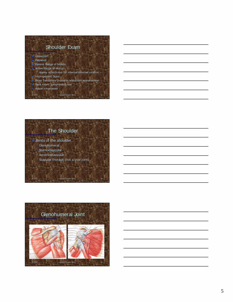
8/27/02 Gregory Crovetti, M.D. 13
Shoulder ExamShoulder Exam
InspectionInspectionPalpationPalpationPassive Range of MotionPassive Range of MotionActive Range of MotionActive Range of Motion–– Appley scratch test for internal/external rotationAppley scratch test for internal/external rotation
Impingement SignsImpingement SignsBicep Tendonitis/Crossarm adduction/apprehensionBicep Tendonitis/Crossarm adduction/apprehensionNeck exam: compression testNeck exam: compression testAdsonAdson’’s manuevers manuever
8/27/02 Gregory Crovetti, M.D. 14
The ShoulderThe Shoulder
Joints of the shoulderJoints of the shoulder–– GlenohumeralGlenohumeral–– SternoclavicularSternoclavicular–– AcromioclavicularAcromioclavicular–– Scapular thoracic (not a true joint)Scapular thoracic (not a true joint)
8/27/02 Gregory Crovetti, M.D. 15
Glenohumeral JointGlenohumeral Joint

6
8/27/02 Gregory Crovetti, M.D. 16
Glenohumeral LigamentsGlenohumeral Ligaments
Folds in the anterior Folds in the anterior capsule produce the capsule produce the superior, middle and superior, middle and inferior glenohumeral inferior glenohumeral ligaments. ligaments. Like the capsule these Like the capsule these ligaments come into ligaments come into play based upon arm play based upon arm position and rotation. position and rotation.
8/27/02 Gregory Crovetti, M.D. 17
Glenoid LabrumGlenoid Labrum–– Glenoid labrum: a Glenoid labrum: a
fibrocartilaginous rim to fibrocartilaginous rim to increase the contact area and increase the contact area and depth of the glenoiddepth of the glenoid
–– Triangular on crossTriangular on cross--section and section and three sides which face the three sides which face the humeral head, joint capsule, humeral head, joint capsule, and glenoid surface and glenoid surface respectivelyrespectively
–– An intact labrum increases An intact labrum increases humeral contact area by 75% humeral contact area by 75% in vertical and 56% in in vertical and 56% in transverse directionstransverse directions
8/27/02 Gregory Crovetti, M.D. 18
ScapulothoracicScapulothoracic
Scapular stabilizing Scapular stabilizing muscles:muscles:–– Trapezius (all three Trapezius (all three
portions)portions)–– Serratus anteriorSerratus anterior–– RhomboidsRhomboids–– Levator scapulaeLevator scapulae–– Pectoralis MinorPectoralis Minor

7
8/27/02 Gregory Crovetti, M.D. 19
Acromioclavicular JointAcromioclavicular Joint
Acromioclavicular Acromioclavicular ligamentligament: resists axial : resists axial rotation and posterior rotation and posterior translationtranslationTrapezoidTrapezoid: is : is anterolateral, resists anterolateral, resists axial compression of the axial compression of the distal end of the clavicledistal end of the clavicleConoidConoid: is : is posteromedial, resists posteromedial, resists anterior and superior anterior and superior translationtranslation
8/27/02 Gregory Crovetti, M.D. 20
Sternoclavicular JointSternoclavicular Joint
These structures still These structures still allow for 35 degrees allow for 35 degrees of elevation, 35 of elevation, 35 degrees of degrees of translation, and 50 translation, and 50 degrees of rotation degrees of rotation at the at the sternoclavicular jointsternoclavicular joint
8/27/02 Gregory Crovetti, M.D. 21
ShoulderShoulder
Palpation of the Palpation of the shoulder includes: shoulder includes: –– Sternoclavicular jointSternoclavicular joint–– Acromioclavicular Acromioclavicular
jointjoint–– Subacromial areaSubacromial area–– Bicipital grooveBicipital groove–– Muscles of the Muscles of the
ScapulaScapula
Have patient place Have patient place each hand:each hand:
1.1. Behind head Behind head (external rotation (external rotation and abduction)and abduction)
2.2. Up the small of the Up the small of the back (internal back (internal rotation)rotation)

8
8/27/02 Gregory Crovetti, M.D. 22
ShoulderShoulder
Rotator cuff:Rotator cuff:–– SupraspinatusSupraspinatus–– InfraspinatusInfraspinatus–– Teres MinorTeres Minor–– SubscapularisSubscapularis
8/27/02 Gregory Crovetti, M.D. 23
8/27/02 Gregory Crovetti, M.D. 24
Special Tests for the ShoulderSpecial Tests for the ShoulderApprehension (crank) testApprehension (crank) test: The arm is abducted to 90 degrees and : The arm is abducted to 90 degrees and laterally rotated. Positive test is when the patient has feelinglaterally rotated. Positive test is when the patient has feeling as if the as if the shoulder may shoulder may ““come out.come out.””Jobe relocation testJobe relocation test: A posterior stress placed to the shoulder in the : A posterior stress placed to the shoulder in the above position will cause relief of pain and apprehension if posabove position will cause relief of pain and apprehension if positive. itive. Rockwood test for anterior instabilityRockwood test for anterior instability: Similar positioning as the crank : Similar positioning as the crank test, but the shoulder is laterally rotated at 0, 45, 90, and 12test, but the shoulder is laterally rotated at 0, 45, 90, and 120 degrees. 0 degrees. Rowe test for anterior instabilityRowe test for anterior instability: Patient supine with hand behind head. : Patient supine with hand behind head. Examiners clenched fist placed behind the humeral head and a dowExaminers clenched fist placed behind the humeral head and a downward nward force is applied to the arm. force is applied to the arm. Fulcrum testFulcrum test: Patient supine arm abducted to 90 degrees, examiners : Patient supine arm abducted to 90 degrees, examiners hand under the glenoid and the arm is laterally rotated.hand under the glenoid and the arm is laterally rotated.Anterior and posterior drawerAnterior and posterior drawer: 0: 0--25% translation (normal), 2525% translation (normal), 25--50% 50% (Grade I), >50% but spontaneously reduces (Grade II), >50% remai(Grade I), >50% but spontaneously reduces (Grade II), >50% remains ns dislocated (Grade III)dislocated (Grade III)

9
8/27/02 Gregory Crovetti, M.D. 25
Special Tests for the ShoulderSpecial Tests for the ShoulderFeagin testFeagin test: arm abducted to 90 elbow straight arm on examiner: arm abducted to 90 elbow straight arm on examiner’’s shoulder, a s shoulder, a don and forward pressure is applied. Positive if apprehension andon and forward pressure is applied. Positive if apprehension and presence of d presence of anteroinferior instability.anteroinferior instability.Clunk testClunk test: Patient supine, examiner hand on the posterior aspect of the : Patient supine, examiner hand on the posterior aspect of the shoulder, other hand hold the humerus above the elbow and abductshoulder, other hand hold the humerus above the elbow and abducts the arm s the arm over the head. Then pushing anteriorly with the hand under the over the head. Then pushing anteriorly with the hand under the shoulder and shoulder and rotating the humerus laterally with the other hand, feel for a grotating the humerus laterally with the other hand, feel for a grind or clunk which rind or clunk which may indicate a tear of the labrum.may indicate a tear of the labrum.Compression rotation testCompression rotation test: Patient supine, elbow flexed and abducted 20 : Patient supine, elbow flexed and abducted 20 degrees, the examiner pushes up on the elbow and rotates the humdegrees, the examiner pushes up on the elbow and rotates the humerus medially erus medially and laterally. Snapping or catching is positive for labral tearand laterally. Snapping or catching is positive for labral tear..Scapular thoracic glide testsScapular thoracic glide tests: To determine the stability of the scapula during : To determine the stability of the scapula during glenohumeral movements.glenohumeral movements.SpeedSpeed’’s tests test: forearm supinated, elbow extended and resistance to forward : forearm supinated, elbow extended and resistance to forward flexion of the shoulder. Positive if tenderness in the bicipitaflexion of the shoulder. Positive if tenderness in the bicipital groove indicating l groove indicating bicipital tendinitis.bicipital tendinitis.
8/27/02 Gregory Crovetti, M.D. 26
Special Tests for the ShoulderSpecial Tests for the ShoulderYergasonYergason’’s tests test: Elbow flexed to 90 degrees, forearm pronated, : Elbow flexed to 90 degrees, forearm pronated, resistance to supination is applied as the patient also laterallresistance to supination is applied as the patient also laterally rotates y rotates the arm. Positive if pain in the bicipital groove and indicatesthe arm. Positive if pain in the bicipital groove and indicates bicipital bicipital tendinitis.tendinitis.Supraspinatus (empty can/ Jobes) testSupraspinatus (empty can/ Jobes) test: The shoulder is forward flexed : The shoulder is forward flexed at 30 degrees, arms straight and thumbs pointing to ground, a at 30 degrees, arms straight and thumbs pointing to ground, a downward force is applied to the arms. Tests for tear or weaknedownward force is applied to the arms. Tests for tear or weakness of ss of the supraspinatus.the supraspinatus.CodmanCodman’’s (drop arm) tests (drop arm) test: shoulder is abducted to 90 degrees and : shoulder is abducted to 90 degrees and patient asked to lower the arm slowly. If drops or is painful, ipatient asked to lower the arm slowly. If drops or is painful, it is t is positive and indicates tear in the rotator cuff.positive and indicates tear in the rotator cuff.Neer impingement testNeer impingement test: Arm is elevated through forward flexion, : Arm is elevated through forward flexion, positive if painful.positive if painful.HawkinsHawkins--Kennedy impingement testKennedy impingement test: Arm is forward flexed to 90 then : Arm is forward flexed to 90 then internally rotated, positive if painful.internally rotated, positive if painful.
8/27/02 Gregory Crovetti, M.D. 27
Special Tests for the ShoulderSpecial Tests for the ShoulderImpingement testImpingement test: Arm is abducted to 90 and full lateral rotation, : Arm is abducted to 90 and full lateral rotation, positive if painful.positive if painful.Military brace (Costoclavicular Syndrome) testMilitary brace (Costoclavicular Syndrome) test: Palpate the radial pulse : Palpate the radial pulse as the shoulder is drawn down and back. Positive if a decreased as the shoulder is drawn down and back. Positive if a decreased pulse pulse and indicates possible thoracic outlet syndrome.and indicates possible thoracic outlet syndrome.Adson ManeuverAdson Maneuver: radial pulse palpated as arm is rotated laterally and : radial pulse palpated as arm is rotated laterally and elbow is extended as the patient extends and rotates head to teselbow is extended as the patient extends and rotates head to test t shoulder.shoulder.Allen testAllen test: Elbow is flexed to 90, shoulder abducted and laterally : Elbow is flexed to 90, shoulder abducted and laterally rotated and patient rotates head away for the test side.rotated and patient rotates head away for the test side.Halstead maneuverHalstead maneuver: Radial pulse felt as arm is pulled down as the : Radial pulse felt as arm is pulled down as the patients neck is hyperextended and rotated to the opposite side.patients neck is hyperextended and rotated to the opposite side.

10
8/27/02 Gregory Crovetti, M.D. 28
The ElbowThe Elbow
Palpation: lateral and medial Palpation: lateral and medial epicondyles, olecranon, radial head, epicondyles, olecranon, radial head, groove on either side of the olecranongroove on either side of the olecranonInspect the carrying angle, and any Inspect the carrying angle, and any nodules or swelling nodules or swelling
8/27/02 Gregory Crovetti, M.D. 29
8/27/02 Gregory Crovetti, M.D. 30
Special Tests for the ElbowSpecial Tests for the ElbowVarus testVarus test: Tests for ligamentous stability of the lateral : Tests for ligamentous stability of the lateral collateral ligamentcollateral ligamentValgus testValgus test: Tests the medial collateral ligament: Tests the medial collateral ligamentCozenCozen’’s tests test: (Lateral Epicondylitis / Tennis elbow test) Patient : (Lateral Epicondylitis / Tennis elbow test) Patient makes fist and pronates the forearm radially deviates and makes fist and pronates the forearm radially deviates and extends the wrist against resistance. Positive if pain in the extends the wrist against resistance. Positive if pain in the lateral epicondyle area. lateral epicondyle area. GolferGolfer’’s elbow tests elbow test: While palpating the medial epicondyle, the : While palpating the medial epicondyle, the forearm is supinated and the elbow and wrist are extended. forearm is supinated and the elbow and wrist are extended. Positive if pain over the medial epicondyle.Positive if pain over the medial epicondyle.TinelTinel’’s of the elbows of the elbow: Percussion of the ulnar nerve in the grove. : Percussion of the ulnar nerve in the grove. Positive if radiating sensation down arm into hand.Positive if radiating sensation down arm into hand.

11
8/27/02 Gregory Crovetti, M.D. 31
Wrist and HandWrist and Hand
Inspect for swelling or deformitiesInspect for swelling or deformitiesPalpate: anatomic snuff box, volar and Palpate: anatomic snuff box, volar and dorsal aspects of the wrist, all joints of dorsal aspects of the wrist, all joints of the fingersthe fingersFlexion, extension, ulnar and radial Flexion, extension, ulnar and radial deviation of the wristdeviation of the wristHave patient make a fist and extend Have patient make a fist and extend and spread the fingers.and spread the fingers.
8/27/02 Gregory Crovetti, M.D. 32
Bones of the WristBones of the Wrist
ScaphoidScaphoidLunateLunateTriquetrumTriquetrumPisiformPisiformTrapeziumTrapeziumTrapezoidTrapezoidCapitateCapitateHamateHamate
8/27/02 Gregory Crovetti, M.D. 33
Anatomy of the ElbowAnatomy of the Elbow

12
8/27/02 Gregory Crovetti, M.D. 34
Nerves of the HandNerves of the Hand
UlnarUlnarRadial Radial MedianMedianPalmar branch of the medianPalmar branch of the median
8/27/02 Gregory Crovetti, M.D. 35
8/27/02 Gregory Crovetti, M.D. 36

13
8/27/02 Gregory Crovetti, M.D. 37
8/27/02 Gregory Crovetti, M.D. 38
Special Tests of Hand and WristSpecial Tests of Hand and WristCascade signCascade sign: Patient flexes the fingers, the tips should all converge towar: Patient flexes the fingers, the tips should all converge toward the d the scaphoid tubercle. If they do not, it may indicate a fracture inscaphoid tubercle. If they do not, it may indicate a fracture in that finger.that finger.Boutonniere deformityBoutonniere deformity: Extension of the MCP and DIP joints and flexion of the : Extension of the MCP and DIP joints and flexion of the PIP joint. This is due to a rupture of the central tendinous sliPIP joint. This is due to a rupture of the central tendinous slip of the extensor p of the extensor hood.hood.SwanSwan--neck deformityneck deformity: Flexion of the MCP and DIP joints, with extension of the : Flexion of the MCP and DIP joints, with extension of the PIP joint. This is due to contracture of the intrinsic muscles. PIP joint. This is due to contracture of the intrinsic muscles. Seen after trauma Seen after trauma or in RA.or in RA.Ulnar driftUlnar drift: Ulnar deviation of the digits most commonly due to RA.: Ulnar deviation of the digits most commonly due to RA.DupuytrenDupuytren’’s contractures contracture: This is due to contracture of the palmar fascia. Most : This is due to contracture of the palmar fascia. Most common in the ring finger or little finger, men more then women,common in the ring finger or little finger, men more then women, ages 50ages 50--70.70.Claw fingersClaw fingers: This deformity is a form a combination of a ulnar and median : This deformity is a form a combination of a ulnar and median nerve palsy. This causes loss of intrinsic muscle function and onerve palsy. This causes loss of intrinsic muscle function and over action of the ver action of the extrinsic extensors. This causes hyperextension of the MCP jointextrinsic extensors. This causes hyperextension of the MCP joints and flexion of s and flexion of the PIP and DIP joints. If the intrinsic function of the hand isthe PIP and DIP joints. If the intrinsic function of the hand is lost, it is then lost, it is then called an intrinsic minus hand.called an intrinsic minus hand.
8/27/02 Gregory Crovetti, M.D. 39
Special Tests of Hand and WristSpecial Tests of Hand and WristTrigger fingerTrigger finger: Results from a thickening of the flexor tendon sheath, causing: Results from a thickening of the flexor tendon sheath, causingsticking of the tendon. At later stages the finger can become ststicking of the tendon. At later stages the finger can become stuck in flexion, uck in flexion, needing to be passively extended. Associated with RA.needing to be passively extended. Associated with RA.BishopBishop’’s Hands Hand: (Benediction Hand) Secondary to ulnar nerve palsy. There is : (Benediction Hand) Secondary to ulnar nerve palsy. There is wasting of the hypothenar, interossei, and the two medial lumbriwasting of the hypothenar, interossei, and the two medial lumbrical muscles. cal muscles. Flexion of the 4Flexion of the 4thth and 5and 5thth fingers is the most noticeable deformity.fingers is the most noticeable deformity.““ZZ”” deformity of the thumbdeformity of the thumb: May be secondary to RA or heredity. The thumb : May be secondary to RA or heredity. The thumb is flexed at the MCP and hyperextended at the IP joint.is flexed at the MCP and hyperextended at the IP joint.DropDrop-- wristwrist: Secondary to radial nerve palsy.: Secondary to radial nerve palsy.Mallet fingerMallet finger: The distal phalanx remains in flexion when the finger is : The distal phalanx remains in flexion when the finger is extended. This is the result of rupture or avulsion of the extenextended. This is the result of rupture or avulsion of the extensor tendon from sor tendon from the distal phalanx.the distal phalanx.ClubbingClubbing: Can be caused by many medical problems such as pulmonary or : Can be caused by many medical problems such as pulmonary or cardiac diseases, as well as genetic.cardiac diseases, as well as genetic.HeberdenHeberden’’s nodess nodes: Swelling of the DIP joints secondary to OA.: Swelling of the DIP joints secondary to OA.BouchardBouchard’’s nodess nodes: Swelling of the PIP joints secondary to RA.: Swelling of the PIP joints secondary to RA.

14
8/27/02 Gregory Crovetti, M.D. 40
Special Tests of Hand and WristSpecial Tests of Hand and WristGanglion cystGanglion cyst: Localized swelling usually on the dorsum of the hand.: Localized swelling usually on the dorsum of the hand.Thumb ulnar collateral ligament testThumb ulnar collateral ligament test: (test for gamekeeper: (test for gamekeeper’’s or skiers or skier’’s s thumb) Valgus stress applied to the MCP joint, if 10thumb) Valgus stress applied to the MCP joint, if 10--20 degrees there is 20 degrees there is most likely a partial tearmost likely a partial tearCarpal Compression testCarpal Compression test: Pressure applied directly to the carpal tunnel : Pressure applied directly to the carpal tunnel for 30 seconds. If positive, indicates carpal tunnel syndrome.for 30 seconds. If positive, indicates carpal tunnel syndrome.FromentFroment’’s signs sign: Patient holds piece of paper between the thumb and : Patient holds piece of paper between the thumb and index paper. If the distal phalanx flexes, it is a positive testindex paper. If the distal phalanx flexes, it is a positive test and and indicates ulnar nerve palsy. If the MCP joint hyperextends, it iindicates ulnar nerve palsy. If the MCP joint hyperextends, it is a s a positive positive JeanneJeanne’’s signs sign and also indicates ulnar nerve palsy.and also indicates ulnar nerve palsy.Allen testAllen test: Tests for competency of the ulnar and radial arteries.: Tests for competency of the ulnar and radial arteries.Anatomic snuffboxAnatomic snuffbox: Lies between the extensor pollicis longus and : Lies between the extensor pollicis longus and extensor pollicis brevis tendons. The scaphoid bone is palpated extensor pollicis brevis tendons. The scaphoid bone is palpated inside inside the box as well as the radial styloid. Pain in the box should inthe box as well as the radial styloid. Pain in the box should indicate dicate scaphoid fracture until proven otherwise.scaphoid fracture until proven otherwise.
8/27/02 Gregory Crovetti, M.D. 41
Special Tests of Hand and WristSpecial Tests of Hand and WristGuyonGuyon’’s canals canal: (pisohamate) Through this canal runs the ulnar nerve. If : (pisohamate) Through this canal runs the ulnar nerve. If compression of the canal occurs, there is sensation lose to the compression of the canal occurs, there is sensation lose to the fingers and fingers and muscle weakness in the hand of ulnar distribution.muscle weakness in the hand of ulnar distribution.>35 degrees indicates a torn ulnar and accessory collateral liga>35 degrees indicates a torn ulnar and accessory collateral ligaments.ments.MurphyMurphy’’s signs sign: Patient makes a fist, if the head of the third metacarpal is l: Patient makes a fist, if the head of the third metacarpal is level evel with the second and fourth metacarpals, it is a sign of a lunatewith the second and fourth metacarpals, it is a sign of a lunate dislocation.dislocation.Retinacular ligament testRetinacular ligament test: Test for the structures around the PIP joint. The : Test for the structures around the PIP joint. The patient is passive, the PIP joint is held in extension and the Dpatient is passive, the PIP joint is held in extension and the DIP is flexed. If the IP is flexed. If the DIP does not flex, the retinacular ligaments (collateral) or capDIP does not flex, the retinacular ligaments (collateral) or capsule is tight. The sule is tight. The PIP joint is the flexed, if the DIP now flexes easily, the retinPIP joint is the flexed, if the DIP now flexes easily, the retinacular ligaments are acular ligaments are tight and the capsule is normal.tight and the capsule is normal.Lunatotiquetral Ballottement (ReaganLunatotiquetral Ballottement (Reagan’’s test)s test): The triquetrum is grasped : The triquetrum is grasped between the thumb and second finger of one hand and the lunate bbetween the thumb and second finger of one hand and the lunate between the etween the thumb and second finger of the other hand. The lunate is then mothumb and second finger of the other hand. The lunate is then moved up and ved up and down, if any laxity, crepitus or pain it indicates a positive tedown, if any laxity, crepitus or pain it indicates a positive test for st for Lunatotriquetral instability.Lunatotriquetral instability.
8/27/02 Gregory Crovetti, M.D. 42
Special Tests of Hand and WristSpecial Tests of Hand and WristWatson (scaphoid shift) testWatson (scaphoid shift) test: The patient: The patient’’s hand is taken into full ulnar deviation s hand is taken into full ulnar deviation and slight extension. With the other hand the thumb is pressed aand slight extension. With the other hand the thumb is pressed against the gainst the distal pole of the scaphoid to prevent it from moving. The patiedistal pole of the scaphoid to prevent it from moving. The patientnt’’s hand is then s hand is then moved radially and slightly flexed. If the dorsal pole of the scmoved radially and slightly flexed. If the dorsal pole of the scaphoid subluxes aphoid subluxes over the dorsal rim of the radius and there is pain, it is a posover the dorsal rim of the radius and there is pain, it is a positive test for itive test for scaphoid and lunate instability.scaphoid and lunate instability.Scaphoid stress testScaphoid stress test: Modification of Watson test in which the patient actively : Modification of Watson test in which the patient actively radial deviates the wrist while scaphoid pressure is applied. Ifradial deviates the wrist while scaphoid pressure is applied. If there is pain and there is pain and a clunk, it is a positive test.a clunk, it is a positive test.““Piano KeyPiano Key”” testtest: Patient: Patient’’s arms are in pronation. Using the index finger while s arms are in pronation. Using the index finger while stabilizing the hand with the other hand the distal ulna is pushstabilizing the hand with the other hand the distal ulna is pushed down. The test ed down. The test is positive if there is pain and difference in mobility comparedis positive if there is pain and difference in mobility compared to the other side. to the other side. This indicates distal radioulnar joint instability.This indicates distal radioulnar joint instability.Axial load testAxial load test: Axial load to the thumb or fingers, if pain or crepitation it : Axial load to the thumb or fingers, if pain or crepitation it is a is a positive test for metacarpal or adjacent carpal bone fracture orpositive test for metacarpal or adjacent carpal bone fracture or joint arthrosis.joint arthrosis.Grind testGrind test: Grabbing the thumb below the metacarpophalangeal joint, an axi: Grabbing the thumb below the metacarpophalangeal joint, an axial al load is applied with rotation. If there is pain the test is posiload is applied with rotation. If there is pain the test is positive and indicates tive and indicates DJG of the metacarpophalangeal or metacarpotrapezial joints.DJG of the metacarpophalangeal or metacarpotrapezial joints.

15
8/27/02 Gregory Crovetti, M.D. 43
Special Tests of Hand and WristSpecial Tests of Hand and WristFinkelstein testFinkelstein test: Tests for De Quervain: Tests for De Quervain’’s or Hoffmanns or Hoffmann’’s disease. A positive test s disease. A positive test indicates a tenosynovitis of the abductor pollicis longus and exindicates a tenosynovitis of the abductor pollicis longus and extensor pollicis tensor pollicis brevis tendons.brevis tendons.Sweater finger signSweater finger sign: When patient makes a fist, if one of the distal phalanx : When patient makes a fist, if one of the distal phalanx (most often the ring finger) does not flex, the test is positive(most often the ring finger) does not flex, the test is positive. It indicates a . It indicates a ruptured flexor digitorum profundus tendon.ruptured flexor digitorum profundus tendon.BunnelBunnel--Littler testLittler test: (Finochietto: (Finochietto--Bunnel test) The patient is passive during the Bunnel test) The patient is passive during the test. The test is for structures around the MCP joint. The MCP test. The test is for structures around the MCP joint. The MCP joint is held in joint is held in extension, while the PIP is flexed. If unable to flex the PIP, textension, while the PIP is flexed. If unable to flex the PIP, the test is positive he test is positive and indicates tight intrinsic muscle or contracture of the jointand indicates tight intrinsic muscle or contracture of the joint capsule. The capsule. The MCP is then slightly flexed, if the PIP now flexes easily it indMCP is then slightly flexed, if the PIP now flexes easily it indicates tight icates tight intrinsic muscles and that the capsule is normal. If the PIP stiintrinsic muscles and that the capsule is normal. If the PIP still does not flex it ll does not flex it indicates a tight joint capsule.indicates a tight joint capsule.TinelTinel’’s signs sign: Positive if tingling into the fingers of the median nerve : Positive if tingling into the fingers of the median nerve distribution, indicating carpal tunnel syndrome.distribution, indicating carpal tunnel syndrome.PhalenPhalen’’s tests test: Position must be held for one minute. If positive indicates ca: Position must be held for one minute. If positive indicates carpal rpal tunnel syndrome. The dorsal aspect of the hands is pushed togethtunnel syndrome. The dorsal aspect of the hands is pushed together to er to maximal flexion of the wrists.maximal flexion of the wrists.
8/27/02 Gregory Crovetti, M.D. 44
CaseCase
7575--year old man comes in for yearly physical.year old man comes in for yearly physical.History of hypertension, elevated lipids, and History of hypertension, elevated lipids, and mild obesitymild obesityHe has taken your advise and started an He has taken your advise and started an exercise program, and now has a complaint exercise program, and now has a complaint of right shoulder pain. of right shoulder pain. What do you want to know?What do you want to know?What do you do next?What do you do next?