general overview of the brain - ohio state university · 2013-10-03 · 1 neuro review ccrn review...
TRANSCRIPT
1
Neuro Review
CCRN Review Cheryl Newton RN,MSN,CCRN,CNRN
Describe the essential aspects of neurological physiology and pathophysiologyIdentify the goals of neurological assessmentAnalyze the nursing care interventions for the patient with neurological diseases
Central Nervous SystemBrain StemSpinal Cord
Peripheral Nervous SystemCranial Nerves (12 pairs)Spinal Nerves (31 pairs)Somatic NS (sensory)Autonomic NS (motor)
Cerebral Hemispheres
Cerebral dominance: right/left
DiencephalonMajor division of cerebrum4 regions
Brain StemCerebellum
BalanceCoordination
CerebrumTwo cerebral hemispheresBroca’s speech area
Basal gangliaProvides circuitry for conscious bodily movements, motor control, and fine body movements
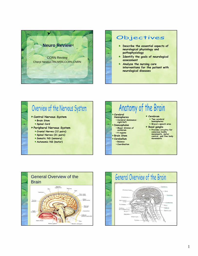
General Overview of the Brain

2
The Brain
4 lobes of cerebral hemispheres
Basal ganglia
Limbic system
Diencephalon
Cerabellum
Brain stem
Lobes of the Brain
FrontalHigher mental function, speech, Broca’s area (articulate speech)
ParietalSensory area, body awareness
TemporalSound, Wernicke’s area (spoken word and written language)
OccipitalVisual, integrates visual stimulation
Diencephalon
ThalamusSensory and motor relay
HypothalamusNeuro-endocrine functionVisceral, autonomic, endocrine, and emotional functionsTemperature, appetite, ADH, sleep
Pituitary glandSecretes and stores hormones
Second major division of the brain Midbrain Pons MedullaContains respiratory and autonomic control centersCoordinates activity of the cerebellum and rest of the brain10 cranial nerves originate from brainstem CN III oculomotor, pupillary constriction CN X Vagus: PNS motor

3
Brain StemMidbrain (III, IV)
Motor and sensory pathways
Location of reticular activating system
Responsible arousal from sleep, wakefulness, focusing of attention
Brain StemPons (V, VI, VII)
Connects cerebral cortex and cerebellumContains motor and sensory pathwaysContains respiratory center
Brain StemMedulla oblongata (VIII, IX, X, XI, XII)
Connect motor and sensory tracts of spinal cord to medullaContains cardiac and respiratory centersContinuous with spinal cord
CerebellumCerebellum
Coordinates muscle movement with sensory inputControls balance and equilibriumInfluences muscle toneAffects locomotion and postureControls non-stereotyped movements Synchronizes muscle action
Complex intricate system that communicates motor and sensory impulses from the brain to body: and body to brain
Tracts (groups of Neuron Fibers) Spinothalamic tract Corticospinal tract
4
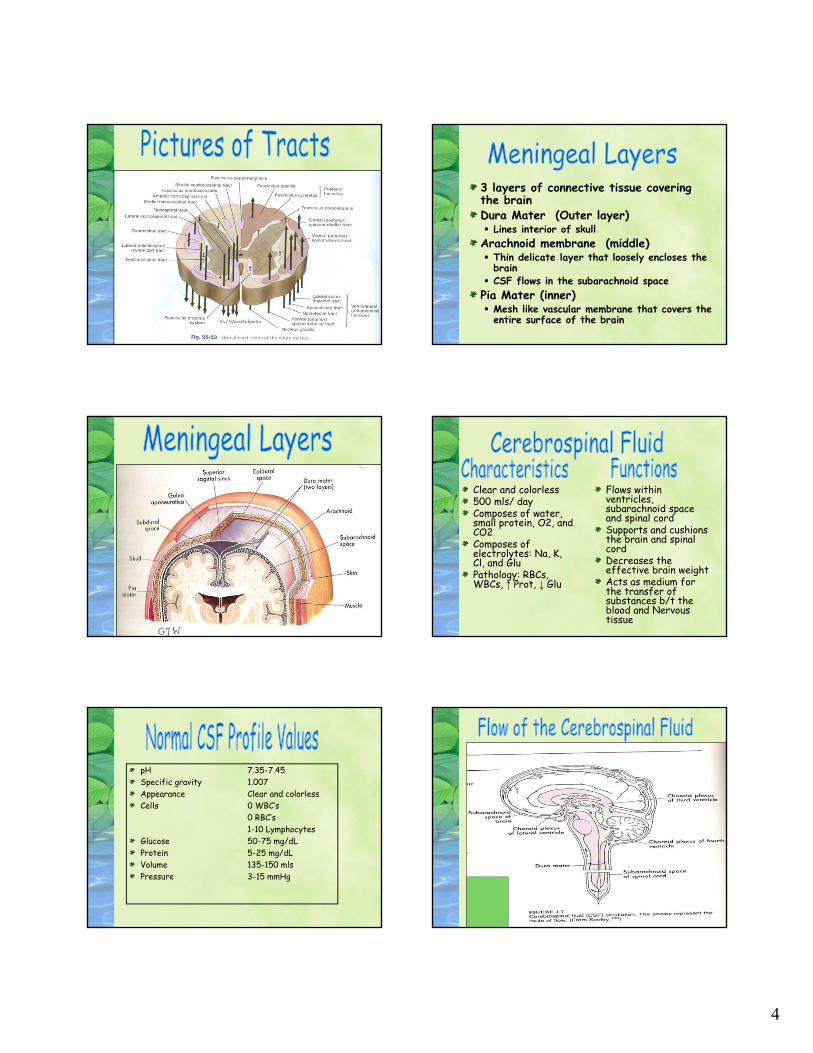
3 layers of connective tissue covering the brainDura Mater (Outer layer) Lines interior of skullArachnoid membrane (middle) Thin delicate layer that loosely encloses the
brain CSF flows in the subarachnoid spacePia Mater (inner) Mesh like vascular membrane that covers the
entire surface of the brain
Clear and colorless 500 mls/ dayComposes of water, small protein, O2, and CO2Composes of electrolytes: Na, K, Cl, and GluPathology: RBCs, WBCs, ↑ Prot, ↓ Glu
Flows within ventricles, subarachnoid space and spinal cordSupports and cushions the brain and spinal cordDecreases the effective brain weightActs as medium for the transfer of substances b/t the blood and Nervous tissue
pH 7.35-7.45Specific gravity 1.007Appearance Clear and colorless Cells 0 WBC’s
0 RBC’s1-10 Lymphocytes
Glucose 50-75 mg/dLProtein 5-25 mg/dLVolume 135-150 mlsPressure 3-15 mmHg
5
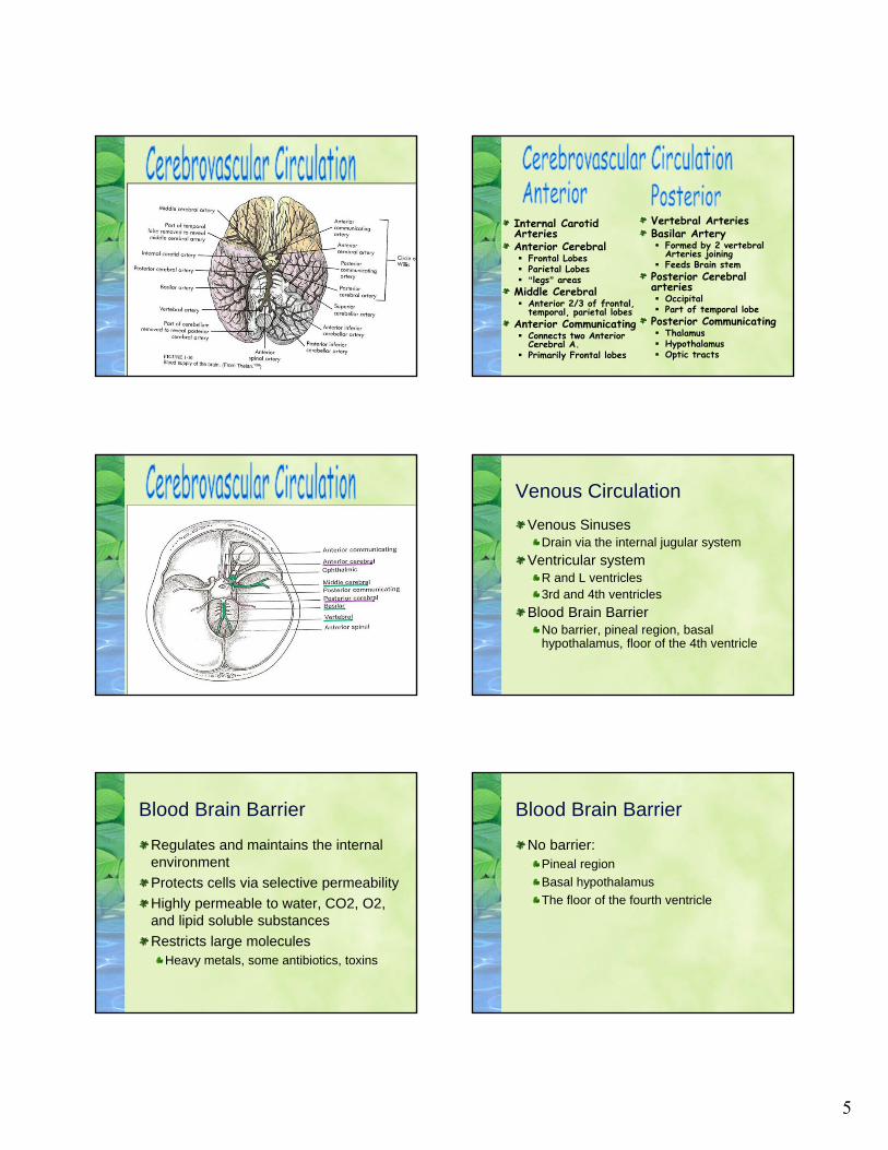
Internal Carotid ArteriesAnterior Cerebral Frontal Lobes Parietal Lobes “legs” areasMiddle Cerebral Anterior 2/3 of frontal,
temporal, parietal lobesAnterior Communicating Connects two Anterior
Cerebral A. Primarily Frontal lobes
Vertebral ArteriesBasilar Artery Formed by 2 vertebral
Arteries joining Feeds Brain stemPosterior Cerebral arteries Occipital Part of temporal lobePosterior Communicating Thalamus Hypothalamus Optic tracts
Venous Circulation
Venous SinusesDrain via the internal jugular system
Ventricular systemR and L ventricles3rd and 4th ventricles
Blood Brain BarrierNo barrier, pineal region, basal hypothalamus, floor of the 4th ventricle
Blood Brain Barrier
Regulates and maintains the internal environment
Protects cells via selective permeability
Highly permeable to water, CO2, O2, and lipid soluble substances
Restricts large moleculesHeavy metals, some antibiotics, toxins
Blood Brain Barrier
No barrier:Pineal region
Basal hypothalamus
The floor of the fourth ventricle

6
Concepts of Neuro-Dynamics
Cerebral Blood Flow (CBF)
Intracranial Volume
Pressure
Compliance
Elastance
Autoregulation
Cerebral Blood Flow
Components are blood and CSF
Flow rate 50-60 mL/100g brain tissue
750 ml/min
Regulated by baroreceptors and chemoreceptors
Intracranial Volume
Volume of the cranial vault is:Brain Mass: 80%
CSF: 10%
Blood Volume: 10%
Monro-Kellie Doctrine
Within the fixed non-distensible skull of the adult, intracranial volume must remain constant to maintain a normal ICP
Monro-Kellie Doctrine
An increase in volume of one compartment must be off set by a decrease in another compartment to keep ICP normal
This is called compensation
Intracranial Pressure
Normal ICP = 0-15 mmHg
Intracranial HTN is considered an ICP >20 mm Hg.
Cerebral Perfusion PressurePressure at which the brain cells are perfused
CPP = MAP - ICP

7
Cerebral Perfusion Pressure
Normal CPP: 60 – 95 mmHg
Ischemia: 50 – 60 mmHg
Cell Death: < 30 mmHg
CBF ceases: 0 mmHg
Autoregulation
Brain’s ability to maintain a constant rate of CBF despite variations in systemic arterial pressure
CBF=blood +CSF50-60ml/100gms of brain tissue
750ml/min
When autoregulation is intact: CBF and cerebral blood volume are independent of MAP, CO, and body activity
Compliance
Refers to the brain’s ability to tolerate increases in volume without sustaining an increase in pressure
C = change in volume/change in pressure
Compliance
Normal ICP: Compliance is high
When C is high: a change in volume results in a small change in pressure
When C is low: a small change in volume results in a large change in pressure
Causes of Increased ICP
Increase in tissue volumeTumorsAbscessEdema
Increase in blood volumeHemorrhageDecreased venous return
Increase CSF volumeObstructionOverproductionDecreased absorption
Compensatory Mechanisms
Displacement of CSF to the Spinal SAS
Increased absorption of CSF
Decreased production of CSF
Displacement of blood volumeVenous blood is shunted away from affected area into the distant venous sinuses
Displacement of brain tissueShift of brain mass

8
Cerebrospinal Fluid (CSF)
FunctionsCushions brain and spinal cordCompensation for changes in ICP
PropertiesClear, colorless, odorless
Xanthochromic=yellow=old RBC
Volume 120 – 150 ml in ventricular system and SASSpecific gravity: 1.007Glucose: 60% of serum glucose level
Most CSF is contained in the spinal cord
Cerebrospinal Fluid
CSF production and reabsorptionFormed by choroid plexus in ventricles
95% is produced in the lateral ventricles
Absorbed via arachnoid villi
Ventricular System
Communication system within brainVentricles
Contain specialized epithelium called choroid plexus
Produce CSF
Lateral ventricles are the largest
The 3rd ventricle lies midline between the 2 lateral ventricles
4th ventricle lies in posterior fossa
Categories of Assessment
Glasgow Coma Scale (3-15)Eye opening (1-4)Verbal response (1-5)Best motor response (1-6)
Muscle strengthGrade 1, 2, 3, 4, 5
Reflexes (+ or -)Babinski’sDTRs
Homonculus-sensory Homonculus-motor
9
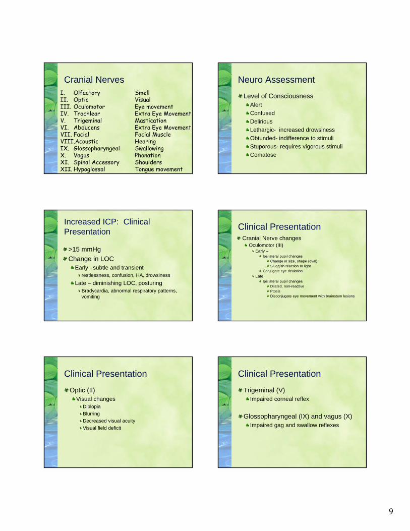
Cranial NervesI. Olfactory SmellII. Optic VisualIII. Oculomotor Eye movementIV. Trochlear Extra Eye MovementV. Trigeminal MasticationVI. Abducens Extra Eye MovementVII. Facial Facial MuscleVIII.Acoustic HearingIX. Glossopharyngeal SwallowingX. Vagus PhonationXI. Spinal Accessory ShouldersXII. Hypoglossal Tongue movement
Neuro Assessment
Level of ConsciousnessAlert
Confused
Delirious
Lethargic- increased drowsiness
Obtunded- indifference to stimuli
Stuporous- requires vigorous stimuli
Comatose
Increased ICP: Clinical Presentation
>15 mmHg
Change in LOCEarly –subtle and transient
restlessness, confusion, HA, drowsiness
Late – diminishing LOC, posturingBradycardia, abnormal respiratory patterns, vomiting
Clinical PresentationCranial Nerve changes
Oculomotor (III)Early –
Ipsilateral pupil changesChange in size, shape (oval)Sluggish reaction to light
Conjugate eye deviation
Late Ipsilateral pupil changes
Dilated, non-reactivePtosisDisconjugate eye movement with brainstem lesions
Clinical Presentation
Optic (II)Visual changes
Diplopia
Blurring
Decreased visual acuity
Visual field deficit
Clinical Presentation
Trigeminal (V)Impaired corneal reflex
Glossopharyngeal (IX) and vagus (X)Impaired gag and swallow reflexes

10
Clinical Presentation
Motor changes
ContralateralDue to compression or pressure on the corticospinal tracts
EarlyParesis, plegia
Lateposturing
Clinical Presentation
VomitingMay occur
Pressure on the vomiting center in the brainstem causes projectile vomiting without nausea
HeadacheIncreasing severity but inconsistent symptom
Seizure May occur
Clinical Presentation
Vital sign changes
Cushing’s triad
Due to pressure on or ischemia of vasomotor center in brainstem
Components
Increased SBP
Widening pulse pressure (DBP normal or decreased)
Bradycardia
Respiratory pattern
Dependent on location of injury
Temperature
Central hyperthermia may occur late r/t pressure on thermoregulatory center in hypothalamus
Increased ICP - Monitoring
Early recognition
With ICP monitoring, intracranial problems can be identified and treatment initiated before clinical signs and symptoms develop
Treatment can be evaluated
Allows monitoring of post-op cerebral edema, infections, or following intracerebral hemorrhage
ICP can be measured
Intraventricular cannula -Ventriculostomy
Intraparenchymal catheter or bolt - Camino
Management of Increased ICP
Goal is to prevent ischemia and herniationMaintain Fluid
Improving MAP to normalize CPPFluids may be limited to keep serum osmolality 290-320Hypotonic solutions avoided: No D5W3% NS (hypertonic saline)Fluids not restricted if vasospasm an issue
Osmotic diuretic therapyMannitol and loop diuretics may help decease the ICPDraw fluid out of swollen brain
CSF drain via ventriculostomy
Management of Increased ICP
Keep CPP > 60 Promote hemodynamic stabilityControl pain and agitationPrevent seizureBarbiturate coma
May be tried if motor responses such as hemiparesis or decorticate or decerebrate positioning occur as a result of cortical and midbrain compression of motor tracts.

11
Nursing Interventions
Good neuro assessment for early identification of increased ICP
Transient increases in ICP may be seen with
Confusion, difficulty in arousal, sluggishness, slight pupillary dilatation, monoplegia or hemiparesis, H/A, aphasia, Cheyne-Stokes respirations or change in VS.
Nursing Interventions
Most nursing procedures have an affect on the ICP
Turning the pt may increase ICP, but can be lessened if the pt is log-rolled with the head in alignment.
If 2 nursing actions both increase ICP, space the actions to allow the ICP to diminish
SuctioningIncrease in ICP may be decreased by pre-oxygenating, limiting suctioning to 10 sec, 1 or 2 passes, and use of less than 120 mm Hg pressure.
Nursing Interventions
Careful monitoring/corrections of electrolytes and osmolality
Monitor for infections and treat feverFever increases cerebral metabolism and may increase the ICP
Use of hypothermia blankets
Maintaining HOB elevation of 30 degrees to promote venous return from the head
Maintain head in neutral position
Nursing Interventions
Maintain a quiet environmentProvide adequate pain medicationsDexamethasone
Decreases cerebral edema
PropranololDecreases metabolic rate/demand
Barbiturate ComaDecreases metabolic demand
Traumatic Brain Injury (TBI)
Concussion, contusion, epidural hematoma, SDH, SAH, IVH, seizure
Etiology: falls and MVCs
DX: CT and MRI
Injury Severity Score (GCS)
TBI
Injury Severity Score (GCS)13-15 mild injury9-12 moderate<=8 severe
Primary InjuryHappens at the time of the trauma
Secondary InjuryInflammatory responseElectrolyte imbalancesHypotension, hypoxia

12
Closed Head Injuries (CHI)
Damage occurs to the cranium without causing a break in the skull
Commonly result fromMVC, falls, and assaults
Alcohol involved
CHI
ConcussionTemporary neural dysfunction
HA, dizziness, irritability, nausea
Transient LOC, post-traumatic amnesia
Contusion
CHI
ContusionMore severe
Bruising of the brain surface, causing neuronal dysfunction
HA, vomiting, restlessness, decreased LOC, aphasia, hemiplegia, sensory deficits
CHI
LacerationTraumatic tear of the cortical surface of the brain
Diffuse Axonal Injury (DAI)Extensive damage to the axons in the white matter
Occurs from high velocity acceleration-deceleration accidents
Hypoxic InjuryOxygen deficit and injury (metabolic or cardiac)
Management of TBI/CHI
Perfuse the brainCPP>65, SBP>90, HOB>30 degrees
Prevent secondary injuryNeuro checksOsmotic diuretics (Mannitol), electrolyte replacement
Decrease brain swelling
SteroidsMinimize cerebral edema
Prevent complicationsSCDs for DVT prophylaxisAntacids and histamine blockersStool softeners
Prevent Valsalva
Skull Fractures
Disruption in the integrity of the cranial vault
Blunt or penetrating trauma
Symptoms includeHA, pain at the site, altered LOC, sensory deficits
3 typesBasilar
Depressed
Linear

13
Skull Fractures
BasilarBase of the skullParanasal sinuses involvement
Rhinorrhea, periorbital ecchymosis (raccoon eyes)
otorrhea, vertigo, nastagmus, and ecchymosis over mastoid bone (Battle’s sign)
Depressed Inward displacement of the outer skullCan occur with open or closed injuries
LinearMost common type of skull fractureNo displacement of bone
Clinical Manifestations
Based on location and type of fractureRhinorrhea
Otorrhea
Vertigo
HA
Nystagmus
Pain at fracture site
Change in consciousness
Management
Relieving pressureRelieving ischemiaPreventing complications
Internal carotid artery injury and CSF leaks are associated with basilar fracturesInfections are associated with depressed fracturesHerniation from increased ICPHemorrhages
Nursing Interventions
Neuro assessment
Monitor for signs of infection
Prevent secondary injury
Intracranial Hematomas
Intracranial hematomas are hemorrhages that produce bleeding into the epidural, subdural, subarachnoid space, or the ventricles
May occur spontaneously or as result of trauma
Up to 15% of all head injuries are SDH
Intracranial Hematomas
Etiology: Usually traumaTypes
Subdural (SDH)May be spontaneousPrevalent in older pts
Epidural (EDH)Associated with linear skull fx
Intracerebral (ICH)Result of GSW or stab, laceration of brain from depressed skull fx, severe acceleration / deceleration injuryBleeding from aneurysm, AV malformation, tumor, rupture of vessel r/t HTN
14
Subdural Hematoma (SDH)
Usually venous, forms slowly, cerebral contusionAccumulates below dura materClassification
AcuteClinical indication occur within 48 hours after injury
SubacuteClinical indication occur within 2 weeks after injury
ChronicClinical indication occur weeks to months after injury
Epidural Hematoma
Usually arterial, may be associated with tearing of arteries from skull fractures
Middle meningeal artery
May be due to venous bleeding, associated with fractures that cross major vascular channels (superior sagittal or transverse sinus)
Accumulates above dura mater
Intracerebral (ICH)
SAH or IVH
Hematoma into brain parenchyma
May be due to bleeding caused by missile injury or severe acceleration / deceleration force
Subarachnoid Hemorrhage
SAH
Bleeding into the subarachnoid space
Common in severe head injuries
Arteriovenous Malformations
Abnormal vascular networkAVMs are a tangled mass of dilated vessels AVMs are congenital lesions that develop in the 4th to 8th week of embryonic lifeMost AVMs do not become symptomatic until the third decade of life
Arteriovenous Malformations:Signs and Symptoms
Hemorrhage is the most common presenting symptom
Seizures
Headache, nausea, vomiting
Hydrocephalus (uncommon)
15
Arteriovenous Malformations
Steal phenomenon: steal from the adjacent brain tissue
Blood is diverted from one area of brain to another due to lower vascularization
Occurs when AVM is large
Symptoms: seizure, HA
Arteriovenous Malformations:Diagnostic Studies
Cerebral angiography: most definitive study reveals the feeding vessels and the draining vessels, size and location
CT scan
MRI
Treatment
Surgery, embolization, Gamma knife
Supportive Care
Prevent secondary injuryNutrition
Skin care
Ulcer prophylaxis
DVT prophylaxis
Cerebral Aneurysms
Dilatation of a cerebral artery resulting from weakness in the arterial wall
95% of all aneurysms occur close to the Circle of Willis at the bifurcations of the internal carotid, middle cerebral, and basilar arteries, and in relation to the anterior and posterior communicating arteries.
Aneurysms
Most occur in the vessels of the anterior portion of the circle of WillisAbout 20% will have multiple aneurysmsRupture of an aneurysm cause SAH in more than 95% of patientsIncreased ICP, hydrocephalus and vasospasm can occur especially with worsening clinical grade
Types of aneurysms
Berry-most commonLooks lie berries
SaccularOut pouching from one wall
FusiformDiffuse enlargement
Charcot-BouchardBrain stem, related to HTN
Mycotic-rareSeptic emboli/endocarditis

16
Etiology
Hemodynamic factors, arteriosclerosis, and breakdown of the internal elastic membrane contribute to the formation and growth of aneurysms HTN not a major factor, genetics is
Familial, Marfan’s, PCKD, neurofibromatosis
Asymptomatic until aneurysm rupturesAssociated with AVMs
Diagnostic Testing
CT scan
MRI
MRA
Angiography
Transcranial doppler
Classification of Aneurysms
Determines the severity of the rupture
Hunt and HessIndex of surgical risk
Fisher ClassificationIndex of spasm risk
GCS
Hunt and Hess
Grade 0: Unruptured aneurysmGrade I: Asymptomatic or slightly symptomaticGrade II: Moderate to severe HA, nuchal
rigidity, third nerve palsyGrade III: Drowsiness, confusion or mild focal
deficitsGrade IV: Stupor, moderate to severe
hemiparesis and posturingGrade V: Deep coma, decerebrate posturing
Aneurysm Treatment
Ventriculostomy placementClipping (OR for surgery)Endovascular CoilingMonitor for complications
InfectionsVasospasmHyponatremia
Monitor and treat electrolytes
Vasospasm
Commonly occurs between 3 and 14 days after rupture (up to 21 days)
Peak incidence between day 5 and 9
The incidence and degree of vasospasm is related to the amount of blood in the subarachnoid space
DX clinically, TCD, angiography
17
Treatment of Vasospasm
Calcium channel blockers such as oral Nimodipine. Usual dose: 60 mg q 4 hours.
Adjust dose for BP
Watch for hypotension
Hypervolemia, hemodilution, and hypertension (Triple H therapy)
Triple H Therapy
Hemodilution and hypervolemia are usually accomplished with a combination of colloid (5% albumin) and crystalloid solutions.Objectives:
Decrease hematocrit by 30%Increase cardiac output to 6.5-8 L/minCVP 8-10 mmHgPAWP 12-16 mmHg
Vasospasm
Can also use Vasopressors if triple H therapy does not increase blood pressure and CPP to desired range
Dopamine
Phenylephrine
Norepinephrine
Cerebrovascular Disease
Third leading cause of morbidity & mortalityAHA efforts to educate community and health care providers…..TIME IS TISSUE…”brain attack”Interventions within 3 hours of onset of signs and symptoms may reverse the cerebral ischemia
Cerebral Vascular Disease Cerebral Vascular Alteration
Sudden or rapid onset of focal Neuro deficits which may be temporary or permanentAcute impairment of blood flow Neuro deficitsTIA---- RINDS--- CVACVA Ischemic (thrombotic, embolic)—85% Hemorrhagic (intracerebral, subarachnoid)—15%
18
Transischemic Attack (TIA)
A temporary disruption in blood flow due to spasm or microemboli
Brief neurological deficit lasting minutes to hours
Symptoms depend on vessel affectedR cerebral hemisphere
L hemianopia, hemiparesis, hemiplegia
L cerebral hemisphereR hemianopia, hemiparesis, hemiplegia, and aphasia
Expressive, receptive, or global
Cerebrovascular Accident
Neurological deficit that is the result of damage or disease in the cerebral blood vesselsAn area of the brain becomes hypoxic due to edema or an obstructed vesselDeficits depend on the area affectedThrombosis, embolism, ICH from ruptured blood vessel (middle cerebral artery)
Cerebrovascular Accident (CVA)or Stroke
ThromboticMost commonHTN is most significant factorDM, CAD, increasing age, men>women
EmbolicCardiac in originEndocarditis, a-fib, valve abnormalities
Hemorrhagic (ICH)Chronic HTN, vascular malformations, coagulopathies, alcohol use, and tumorsSympathomimetic drugs
Cocaine and amphetamines
Symptoms
Numbness or weakness
Confusion
Trouble speaking or understanding
Visual disturbances
Sudden severe HA
Difficulty walking, loss of balance, dizziness
Stroke Management
Time is brain tissue
Blood pressure controlThrombotic therapy (rT-PA)
SBP<185, DBP<110Decrease risk of bleeding
Non-thromboticSBP up to 210, DBP up to 110
Adequate perfusion
Carotid stenosis >70% should be treated
Supportive therapy for ICH
Hemorrhagic Stroke
Hemorrhage25% of strokesBleeding into parenchyma, causing irritation of and pressure on cerebral tissues and nerves, leading to loss of fx and death of neurons.HTN intracranial hemorrhage usually occurs in the basal ganglia, cerebellum or brain stem but may affect more superficial areas of the cerebrum.

19
HemorrhagicIntracerebral
Trauma
Hypertensive rupture of vessel
Vascular intracerebral tumor, thrombolytics, anticoagulants, bleeding disorders
Subarachnoid
Cerebral aneurysm
Accounts for majority of SAH
Most are small
Weakened bulging area on intracranial blood vessel
AV malformation
Tangle of abnormal arteries and veins, artery feeds into vein directly without capillary bed
Always congenital
Causes ischemia of surrounding tissue
Risk Factors for SAH
Smoking
Hypertension
Moderate to heavy alcohol use
Presentation of SAH
Abrupt HA, localized
Usually occurs at night
Worst HA of my life
Associated with nausea, vomiting, sz
ManagementSAHID vasospasm by worsening of neuro status
Vasospasm occurs anytime from the 3rd day post bleed to 2-3 weeks after initial bleed
Administer calcium channel blockersMaintain triple-H therapy (HTN, hypervolemia, hemodilution)
HTN – goal BPVasodilatorsVasopressors
Hypervolemia and hemodilutionColloids and crystalloidsGoal PAOP Goal Hematocrit
Management
Minimize potential for rebleed and promote stabilization of patient
Rebleed occurs most often within 24 hours or a 7-10 days after bleed
Brain Tumors
Act as a space occupying lesion
Tumors are classified by histological features and grade of malignancy
Prognosis is based on histological type, grade of tumor, location, size, the patient’s age, clinical status prior to surgery

20
Types of Brain Tumors
GliomasAstrocytoma: usually grades I to II
Anaplastic astrocytoma
Cerebellar astrocytoma
Optic nerve glioma
Oligodendroglioma: rare slow growing
Gliomas
Glioblastoma Multiforme: 50% of primary brain tumors
Brain Stem Glioma: poor prognosis
Ependymoma: arise from cells lining ventricles
Medulloblastoma: highly malignant
Pineal region tumor: rare
Extra-Axial Tumors
MeningiomaMake up 15-20% off all primary BTArise from meningeal tissueEncapsulatedPrognosis: usually good
Acoustic Neuroma10% of all primary brain tumorsArises from CN VIIISlow growing and benignAffects CN V, IX, X
Pituitary Tumors
Make up 10-15% of all primary BTsArise from pituitary glandTechnically an endocrine tumorSecreting vs non-secreting
Non-secreting: 90% of all pituitary tumorsCompression of optic chiasmVisual changes
Secreting:Prolactin secretingGrowth hormoneACTH secreting
Signs and Symptoms
Depends on location of tumor and how rapidly the tumor grows
Headache
Seizures
Visual changes
Mental status changes
Vomiting
Signs and Symptoms: Frontal Lobe Tumors
Inappropriate behaviorsInattentivenessInability to concentrateLoss of self restraintImpaired recent memoryFlat affect, apathyExpressive aphasiaMotor weakness (usually contralateral hemiparesis)

21
Signs and Symptoms:Parietal Lobe Tumors
Hyperesthesia
Paresthesia: tingling, burning, crawling
Loss of two point discrimination
Unilateral neglect
Signs and Symptoms:Temporal Lobe Tumors
Psychomotor seizures
Homonymous hemianopsia
Receptive aphasia
Alterations in hearing
Signs and Symptoms:Occipital Lobe Tumors
Contralateral homonymous hemianopsia
Visual hallucinations
Seizures with an aura
Signs and Symptoms:Pituitary Tumors
Visual defectsHypopituitarismHeadacheCushing syndromeAcromegalyEndocrine dysfunctionMenstrual dysfunction
Signs and Symptoms:Ventricular Tumors
Hydrocephalus
Headache
Change in LOC
Signs and Symptoms:Cerebellar Tumors
Ataxia
Dizziness
Nystagmus

22
Signs and Symptoms:Brain Stem Tumors
Cranial nerve deficits
Cerebellar dysfunction
Vomiting
Obstructive hydrocephalus
Diagnostic Studies
CT scan
Skull films
Cerebral angiography
MRI
Treatment
Craniotomy
Radiation Therapy
Chemotherapy
Seizures
Abnormal electrical discharge of cerebral neurons
Sensory, motor, or behavioral changes, change in LOC
Ictal period- lethargic and disoriented
Seizures
Generalized Absence-sudden lapse of consciousness-staring spellMyoclonic-sudden, brief muscle jerking on one or more muscle groups. Commonly associated with metabolic, degenerative, and hypoxic causes.Atonic-Sudden loss of muscle tone.Clonic-rhythmic muscle jerking.Tonic-sustained muscle contraction.Tonic-clonic-muscle activity varies between sustained contraction and jerking.
Seizures
PartialOccurs when there is a focal discharge in one area of the cerebral cortex.Simple and complex
Epilepsy = recurrent seizuresStatus epilepticus = continuous or repetitive seizures > 30 min

23
Seizure ManagementControlling the seizure as quickly as possible, preventing recurrence, maintaining patient safety, and identifying the underlying cause!Position patient on side to decrease aspirationProvide supplemental oxygenNothing should be placed in the patient’s mouth during a seizure.EKG monitoring, pulse oximetry, and blood pressure monitoring are required in patients with prolonged seizures.Hypoglycemia can induce seizure activity, so a glucose level is checked immediately and treated as appropriate.
Seizure Management
Lorazepam 0.1 mg/Kg Phenytoin SLOW IV no more than 50 mg over 1 min May cause cardiac dysrhythmias Must check blood level Free DPH level is more accuratePhenytoin PO TF must be stopped 1 hr before and 2 hrs after the dose
Encephalitis
Inflammation of the brain parenchyma
Most commonly due to Herpes Simplex Virus
SymptomsFever, neurologic changes, HA, SZ, stiff neck
Meningitis
Acute inflammation of the meninges of the brain and spinal cordCan be caused by bacteria, viruses, fungi, or parasitesRisk factors: immunocompromise, trauma or surgery, crowded living conditions
Meningitis Symptoms
FeverHeadacheNeck stiffnessIrritabilityVomitingPhotophobiaChange in LOCSeizuresWeaknessCranial nerve signs
Meningitis Diagnosis
LP for pressure and CSF analysisBlood CulturesSputum/ nasopharyngeal culturesEEGHead CTComplications of Meningitis:HydrocephalusCerebral EdemaVasculitis

24
Kernig’s Sign
Patient can not extend leg at the knee when the thigh is flexed because of stiffness of the hamstring muscle
Brudzinski’s sign
Have patient attempt to touch their chin to their chest
Chin to chest causes patient’s hips to flex when the neck is flexed
Meningitis Diagnosis Spinal Cord Injury
Results from:CompressionContusionTrans-section of spinal cord
EtiologyTraumaDisease processes
TumorsRuptured spinal AV malformationInfectious process
AbscessHematoma
Mechanism of Injury: Trauma
HyperflexionChin forced to chestOccurs most often in cervical regionResults from
Sudden decelerationLigament tears, stretching of spinal cordDislocated or subluxation of intervertebral disks
Mechanism of Injury: Trauma
Hyperextension – whiplashHead thrown backOccurs most often in cervical regionForces of acceleration-decelerationResults in backward and downward movement, stretching spinal cord
Rotation injurySpinal cord is rotatedCan involve all parts of vertebral columnTearing spinal ligamentsDisplacement of intervertebral disks, compression of spinal nerve roots
25
Mechanism of Injury: Trauma
Vertical compressionVertebral column is compressed
Occurs primarily in area of T12 – L2
Force applied downward from head
Burst vertebra and intervertebral disks
Mechanism of Injury: Trauma
Penetrating traumaCan occur at any level
Results from a penetrating object
Can be complete or incomplete trans-section of the spinal cord
Clinical Presentation
Depends on type and extent of lesionSubjective
Hx of precipitating event
ObjectiveLevel of lesionComplete versus incomplete
Complete – loss of sensory and motor fx below level of lesion, irreversibleIncomplete –
Spinal shockResults from loss of inhibition of descending tracts
Incomplete SCI
Central Cord SyndromeWeakness in both upper extremities and lower extremities
Weakness is greater in upper extremities
Incomplete SCI
Anterior Cord SyndromeAnterior paralysis
Decreased sensation
Decreased pain
Position, touch, vibration remain intact
Incomplete SCI
Brown-Sequard½ spinal cord is affected
Same side-loss of motor, position, and vibration
Opposite side-loss of pain and temperature sensation

26
Incomplete SCI
Cauda Equina SyndromeInjury to nerve roots below L1
Lower extremity motor loss
Loss of bowel and bladder function
Areflexia
Spinal Shock
May last days to monthsLoss of all motor, sensory and reflex responsesBradycardia and hypotensionLoss of autonomic controlTransient reflex depression below level of injuryFlaccid paralysis below level of injuryParalytic ileusUrinary and fecal retentionImpaired temperature regulationPriapism may occur
Diagnostic
ABGs – evaluation ventilatory and oxygenation status
Spinal x-rays
EMG
CTVisualize fractures
MRIID extent of damage, degree of cord contusion, presence of blood, edema necrosis, disk herniation, tumor
Management
Immediate immobilizationCervical traction with Gardner-Wells or Crutchfield tongs inserted in to skull’s outer table with traction applied
Halo traction
SurgeryDecompression laminectomy
Closed or open fracture reduction
Cervical spinal fusion
Harrington rods – thoracic injury
Log roll
Management
Prevent further spinal cord edemaHigh dose steroids
Protects the neuro-membrane from further destruction and improves blood flow
ContraindicationsInjury more than 8 hours old
Injury below L2
Injury to cauda equina
SUP
Osmotic diuretics
Therapeutic hypothermia
Management
Maintain airway, ventilation and oxygenationTreat pain and discomfortMonitor for and treat spinal shock
Administer colloids, avoid hypotonic solutionAdminister atropine as prescribed for bradycardia