general reading list - psychologymonika/teaching/level4cnp/handouts.pdf · general reading list...
TRANSCRIPT

1
General Reading List
Farah, MJ (1991). Visual Agnosia. MIT Press (Main Library, Short Loan).Harvey, M and Milner, AD (1995). Balint's patient. CognitiveNeuropsychology, 12, 261-281 (Lynda Young).Milner, AD and Goodale, MA (1995). The Visual Brain in Action. OxfordUniversity Press. (Main Library, Short Loan)Milner, AD and Goodale, MA (1993). Visual pathways to perception andaction. Progress in Brain Research, 95, 317-337 (Lynda Young).Robertson, IH and Marshall, JC (1993). Unilateral Neglect: Clinical andExperimental Studies. Lawrence Erlbaum Associates (Main Library).
Lectures 8 and 9 (Parallel visual pathways)Milner, AD and Goodale, MA (1995): The visual brain in action. Oxford UniversityPress (PP 1 -5, PP 20-24, and chapter 2 ). (Main Library, Short Loan)
Livingstone M (1988) Scientific American, 258, 68-75.
Schiller, P.H. and Logothetis, N.K. (1990). The color-opponent and broad-bandchannels of the primate visual system. Trends in Neuroscience, 13, 392-398.
Martin, KAC (1992) Parallel Pathways converge. Current Biology, 2 (10), 555-557.
Mishkin, M. and Ungerleider, LG and Macko, KA (1983). Object vision and spatialvision: two cortical pathways. Trends in Neuroscience, 6, 414-417.
Goodale, MA and Milner, AD (1992). Separate visual pathways for perception andaction. Trends in Neuroscience, 15, 20-25.
Milner, A.D. and Goodale, M.A. (1993). Visual pathways to perception and action.In Progress in Brain Research, Vol.95, (ed. T.P. Hicks, S. Molotchnikoff and T. Ono)Elsevier, Amsterdam, pp. 317-337.
Additional Reading:Maunsell, JHR and Merigan, W (1993): Annual Review of Neuroscience, 16, 369-402.
Livingstone, M and Hubel, DH (1988). Segregation of form, color, movement, anddepth: anatomy, physiology, and perception. Science, 240, 740-749.
Schneider, GE (1967). Two visual systems: brain mechanisms for localization and

2
discrimination are dissociated by tectal and cortical lesions. Science, 163, 895-902.
Taira, M., Mine, S., Georgopoulos, A.P., Mutara, A. and Sakata, H. (1990). Parietalcortex neurons of the monkey related to the visual guidance of hand movements.Experimental Brain Research, 83, 29-36.
Lecture 10 (Blindsight)Milner, AD and Goodale, MA (1995): The visual brain in action. Oxford UniversityPress (OUP) (PP 67-82, PP 85, 86).
Cowey, A and Stoerig, P (1991) Trends in Neuroscience (TINS), 14, 140-145.
Weiskrantz, L. (1986). Blindsight: A Case Study and Implications. Oxford UniversityPress, Oxford. (pp 1-32 and 115-146 and 90-End of Chapter, Main Library)
Weiskrantz, L. (1987). Residual vision in a scotoma. A follow-up study of 'form'discrimination. Brain 110, 77-92.
Fendrich, R. et al (1992) Science, 258, 1489-1491.
Campion, J, Latto, R., and Smith, YM (1983). Is blindsight an effect of scatteredlight, spared cortex, and near-threshold vision? Behavioural Brain Sciences, 6, 423-486 (Main Library).
Perenin, M.-T. and Jeannerod, M. (1975). Residual vision in cortically blindhemifields. Neuropsychologia 13, 1-7. (Main Library)
Perenin, M.-T. and Jeannerod, M. (1978). Visual function within the hemianopic fieldfollowing early cerebral hemidecortication in man: I. spatial localization.Neuropsychologia 16, 1-13. (Main Library)
Perenin, MT and Jeannerod M (1978) Neuropsychologia, 16, 697-708. (MainLibrary)
Lecture 11 (Balint’s Syndrome )Milner, AD and Goodale, MA (1995): The visual brain in action. Oxford UniversityPress (PP 88-101).
Harvey, M. (1995) 'Psychic paralysis of gaze, optic ataxia, spatial disorder of
3
attention'. Translated from Balint (1909) 'Seelenlähmung des "Schauens", optischeAtaxie, räumliche Störung der Aufmerksamkeit'. Cognitive Neuropsychology, 12 (3),265-281.
Ratcliff, G. and Davies-Jones, G.A.B. (1972). Defective visual localization in focalbrain wounds. Brain, 95, 49-60.
Perenin, M.-T. and Vighetto, A. (1988). Optic ataxia: a specific disruption invisuomotor mechanisms. I. Different aspects of the deficit in reaching for objects.Brain 111, 643-674.
Perenin, M.-T. and Vighetto, A. (1983). Optic ataxia: a specific disorder invisuomotor coordination. In: Spatially Oriented Behavior, (ed. A. Hein and M.Jeannerod) Springer-Verlag, New York, pp. 305-326.
Jakobson, L.S., Archibald, Y.M., Carey, D.P. and Goodale, M.A. (1991). Akinematic analysis of reaching and grasping movements in a patient recovering fromoptic ataxia. Neuropsychologia, 29, 803-809.
Holmes, G. (1918). Disturbances of visual orientation. British Journal ofOphthalmology, 2, 449-468, 1918.
Additional Reading:Husain, M and Stein, J (1988), Archives of Neurology, 45, 89-93.
Grafton, S.T., Mazziotta, J.C., Woods, R.P. and Phelps, M.E. (1992). Humanfunctional anatomy of visually guided finger movements. Brain 115, 565-587.
Pierrot-Deseilligny, C., Gray, F. and Brunet, P. (1986). Infarcts of both inferiorparietal lobules with impairment of visually guided eye movements, peripheral visualinattention and optic ataxia. Brain 109, 81-97.
Lectures 12 and 13 (Agnosia)Milner, AD and Goodale, MA (1995): The visual brain in action. Oxford UniversityPress (PP120-144).
Milner, A.D., Perrett, D.I., Johnston, R.S., Benson, P.J., Jordan, T.R., Heeley, D.W.,Bettucci, D., Mortara, F., Mutani, R., Terazzi, E. and Davidson, D.L.W. (1991).Perception and action in "visual form agnosia". Brain , 114, 405-428.

4
Goodale, M.A., Milner, A.D., Jakobson, L.S. and Carey, D.P.(1991) A neurologicaldissociation between perceiving objects and grasping them. Nature, 349, 154-156.
Goodale, M.A., Jakobson, L.S., Milner, A.D., Perrett, D.I., Benson, P.J., & Hietanen,J.K. (1994). The nature and limits of orientation and pattern processing supportingvisuomotor control in a visual form agnosic. Journal of Cognitive Neuroscience, 6(1), 46-56.
Carey, D.P., Harvey, M. and Milner, A.D. (1996). Visuomotor sensitivity for shapeand orientation in a patient with visual form agnosia. Neuropsychologia, 34, 329-338.
Benson, D.F. and Greenberg, J.P. (1969). Visual Form Agnosia. Archives ofNeurology, 20, 82-89.
Farah, MJ (1990) Visual Agnosia, MIT Press. (PP 1-33). Additional Reading:
Adler, A. (1944) Archives of Neurology and Psychiatry, 51, 243-259
Campion, J. and Latto, R. (1985). Apperceptive agnosia due to carbon monoxidepoisoning. An interpretation based on critical band masking from disseminatedlesions. Behavioural Brain Research, 15, 227-240.
Lectures 16 to 19 (Neglect)
Mishkin, M. and Ungerleider, LG and Macko, KA (1983). Object vision and spatial
vision: two cortical pathways. Trends in Neuroscience, 6, 414-417
Posner, M. I., Walker, J.A., Friedrich, F. J. and Rafal, R.D. (1987). How do theparietal lobes direct covert attention? Neuropsychologia, 25, 135-145
Halligan, PW and Marshall, JC (1993). The history and clinical presentation ofneglect. In: IH Robertson and JC Marshall Unilateral Neglect: Clinical andExperimental Studies, Erlbaum.
Wurtz, R., Goldberg, M.E. and Robinson, D.L. (1982) Brain mechanisms of visual
attention. Scientific American, 246, 100-107.

5
Bisiach, E. and Luzzatti, C. (1978). Unilateral neglect of representational space.Cortex, 14, 129-133.
Heilman, K.M. and Valenstein, E. (1979). Mechanisms underlying hemispatialneglect. Annals of Neurology, 5, 166-170
Milner, A.D., Harvey M., Roberts R.C. and Forster, S.V. (1993), Line bisectionerrors in visual neglect: misguided action or size distortion? Neuropsychologia, 31,39-49.
Bisiach, E., Geminiani, G., Berti, A. and Rusconi, M.L. (1990). Perceptual andpremotor factors in unilateral neglect. Neurology 40, 1278-1281
Tegner, R. and Levander, M. (1991). Through a looking glass. A new technique todemonstrate directional hypokinesia in unilateral neglect. Brain, 114, 1943-1951.
Riddoch, M.J. and Humphreys, G.W. (1983). The effect of cueing on unilateral
neglect. Neuropsychologia, 21, 589-599.
6
List of terms to help with revision (this is out of the top of my head, no guarantee
provided)
Agnosia: patients fail to recognise objects that they consciously detect, distinctionbetween apperceptive (D.F.) and associative
Allaesthesia: a contralesional stimulus is reported as ipsilesional
Anosognosia: denial of illness
Balint’s syndrome includes hemineglect, psychic paralysis of gaze, optic ataxia
Blindsight: ability to perform certain tasks (pointing, grasping) with having awarenessof this (damage in V1)
Constructional apraxia: an impairment in combinatory or organising activity in whichdetails must be clearly perceived and in which the relationship among the componentparts must be apprehended (might be due to parietal lesions)
Directional Hypokinesia: deficit to respond with either limb (or head or eyes) tocontralesional side (Heilman)
Dorso lateral geniculate nucleus (dLGN): main subcortical relay station of visual info(Biggest pathway in primates and humans, part of basal ganglia), do not confuse withventral LGN (audition)
Efron test: subject has to judge whether pairs of rectangles are the same or different(All shapes are matched for surface area so that differences in brightness cannot beused as a cue)
Extinction: failure to respond to contralesional stimulus when bilateral stimuli arepresent (sensory) or to move contralesional limb when bilateral movements arerequired (motor)
Hemiakinesia: reluctance to make a movement or delay in initiating a movement(contralesional side) in absence of clinical evidence for weakness
Hemianopia: visual field deficit

7
Hemifield: visual field in relation to retinal coordinates
Hemineglect: failure to respond or act in parts of space (might be due to inferiorparietal lesions, also frontal and subcortical), also called visuo-spatial neglect,unilateral neglect; visual hemi-inattention, spatial agnosia
Hemiplegia: paralysis of one half of the body
Hemispace: visual area in relation to subject’s head/trunk
IT: inferior temporal lobe, endpoint of the ventral stream
LIP: ‘visuomotor’ area in the dorsal stream
M cells (or P alpha or type A) (with large cell bodies)
MT (or V5 when Zeki talks): ‘movement area in the dorsal stream
Optic chiasm: subcortical relay station where visual pathways cross before they reachthe dLGN
Optic ataxia: inability to shape hand appropriately to target demands (might be due tosuperior parietal lesions)
P cells ( or P beta or type B) (medium to small cell bodies) are two types of ganglioncells in the primate retina, the dLGN and visual cortex
Perimetry/Perimeter: procedure/instrument to measure size of the intact visual field
PO: area in the dorsal stream, indicated in localising stimuli in periphery
PPC: posterior parietal cortex, endpoint of the dorsal stream (contains both superiorand inferior parietal lobe)
Psychic paralysis of gaze: inability to perceive more than one object at a time
Pulvinar: part of basal ganglia

8
Superior Colliculi: subcortical relay station of visual information (biggest pathway innon-primates)
Topographical Disorientation: problems with route finding, spatial maps (might bedue to medial temporal lesions)
V1, primary visual cortex or area 17 or striate cortex (all synonyms)
V3a: area in dorsal stream containing orientation selective cells
V4: ‘colour’ area in the ventral stream
Learning Outcomes of the course(Knowledge)
1. To outline the major theories of visualprocessing in the brain and examine someof their strengths and weaknesses.2. To present a new theory of parallelprocessing and contrast it with the existingones.3. To relate higher visual disorders seen inthe clinic to these major theories:Higher visual disorders fascinating:
BlindsightAgnosia
Optic ataxiaNeglect
Learning Outcomes of the course (KeySkills)

9
Professional (Understanding and workingwith special populations)Information finding (articles on readinglist and novel searches)Literacy (essay)Analytic skills (critical evaluation oftheories, methods)Public communication (present issues intutorial, participation in lectures)

10
WHAT SHOULD YOU KNOWALREADY?
Terms of relationship: rostral - caudal,dorsal - ventral, lateral medial; sagittal,coronal, horizontal plane
Visual pathways and types of hemianopia
Thalamus (Pulvinar and LGN)
Superior colliculi
Anatomy of the cortical lobes, especiallytemporal and parietal
Retinotectal pathway: from the retina tothe superior colliculi to extrastriate cortexRetinogeniculate pathway: from retina toLGN to primary visual cortex (striatecortex or V1)
Visual areas after V1:
11
Overview Lecture 8:
1)Definition of Perception
2)Input pathways of the visual system:
3)Discuss the main visual systemshypotheses:
a) Schneider (1969): subcorticalb)Ungerleider and Mishkin (1982):corticalc)Livingstone and Hubel (1988: corticaland subcortical)
12
Function of vision:
- object recognition which in turn allows learning aboutthe structure of our environment (representation of theworld)- allows us to perform skilled actions such as catching aball, grasping an object, walking through a crowdedroom
I will argue that these two functions of vision depend onindependent systems
Definition of Perception:
- Perception refers to a process which allows one toassign meaning and significance to external objects andevents. - Perception provides the foundation for the cognitive lifeof an individual, allowing to construct long-termmemories and models of the environment.

13
Input pathways into the visual system
Two largest pathways: retinotectal and retinogeniculate. In mammals the retinogeniculate pathway is by far themost prominent.Neurones in the dLGN project to cerebral cortex, withalmost all the fibres terminating in V1 (alias striatecortex, alias primary visual cortex). The retinotectal pathway runs from the retina via thesuperior colliculi via the thalamus (pulvinar) to differentextrastriate areas.
Other pathways:- ventral LGN (audition)- Pretectum (papillary light reflexes)- Nucleus of the optic tract (automatic reactions)-Accessory optic tract (optic flow, posture andlocomotion)-Suprachiasmic nucleus (day- night cycle)

14
The main visual systems hypotheses:
Schneider (1969): subcorticalStudied hamsters: argued that the retinal projection to thesuperior colliculus enables organisms to localise astimulus in visual space, while the geniculostriate systemallows them to identify that stimulus.
Lesioned the SC in the Syrian hamster and found a poororienting response to stimuli displayed overhead.Hamsters were not impaired in a visual discriminationtask where they had to push one of two doors withdifferent patterns. They correctly identified the pattern.
Lesions of the visual cortex produced the opposite effect:grossly impaired visual discrimination but intactresponses to overhead stimuli.
Schneider concluded that SC lesioned hamsters candetect visual stimuli (what) but have trouble localisingstimuli in space (where).

15
Ungerleider and Mishkin (1982) cortical:
Claimed that the appreciation of an objects’ qualities(object identification) and of its spatial location (where isit) depends on the processing of information in theinferior temporal (IT) and posterior parietal cortex (PPC)respectively.Same distinction as Schneider between what and wherebut this time the dichotomy is entirely cortical: dorsal vsventral stream processing.
Main evidence:Found that monkeys with IT lesions are impaired in adiscrimination task but not in a landmark task (choosethe food well that is closer to the object), while monkeyswith PP lesions were impaired on using a spatiallandmark but not on discriminating between two objects.
Problem: response modality not taken into account.Model focuses on the animals decision about thestimulus array and neglects the animals visuomotorbehaviour.

16
Livingstone and Hubel (1988):
claim that anatomical pathways are separate all the wayfrom the retina via LGN via V1 to IT and PPCrespectively.
Quote evidence from both anatomical and physiologicalstudiesTwo types of ganglion cells in the primate retinaM cells (or P alpha or type A) (with large cell bodies)P cells ( or P beta or type B) (medium to small cellbodies).
At the level of the dLGN the P cells project to the 4parvocellular layersand the M cells to the two magnocellular layers.
Physiological differences:1. Chromatic sensitivity: 90% per parvo cells are colour sensitive10% of parvo and all magno cells are insensitive tocolour (broad band)2. Acuity, spatial frequency sensitivitymagno cells have 2-3 times larger receptive fields thanparvo cells (high acuity and spatial resolution)3. Speed and DurationMagno: fast but transient (movement)Parvo: slow but sustained4. Contrast sensitivity: parvo: peak sensitivity for high contrasts (cells respondslowly but saturate at high contrasts)magno: peak sensitivity for low contrast stimuli (cellsrespond fast but level off at 10-15% contrast)

17
At the level of V1magnocellular axons terminate in layer 4C alphaparvocellular axons in 4C beta.Both then project to others layers in V1:4c alpha projecting to 4B4c beta projecting to layers 2 and 3.
More recently it was found by Wong-Riley that layers 2and 3 form a regular pattern of blobs and interblobs Seem to be three streams of processing in V1:from 4c alpha to 4bfrom 4c beta to the blobs4c beta to the interblobs.
Physiological differences: - Cells in the blobs are mainly colour sensitive, preferlow spatial frequencies, not orientation selective, normovement selective.- Cells in interblobs little colour sensitivity (though morethan magno cells) but high spatial frequencies (foracuity) along with orientation sensitivity, could beresponsible for high resolution form perception.Although most input from parvo-cells does not seemexplicitly colour-coded.- Cells in 4b selective for motion (from magno) but alsoorientation and no colour.
At the level of V2some projections from 4B(magno) to MTprojections run from 4B (movement) to the thick stripes,from the blobs (colour) to the thin stripesfrom the interblobs (form) to the pale stripes.
Physiological differences:

18
- cells in the thick stripes motion and orientationselective and coding for depth, no colour- thin stripes colour coded but not orientation or directionselective- pale stripes orientation selective and end-stopped(respond better to short edges or lines) seem to encodeshape, no colour, direction sensitivity
Higher visual areasMT receives input from the thick stripes (magno) andagain cells respond to motion and stereoscopic depthV4 receives input from the thin (colour) stripes and palestripes (shape) also project to V4. Cells sensitive tocolour.
Livingstone and Hubel argue that the parvo and magnochannels remain segregated well beyond the primaryvisual cortex.
Propose that the ventral and dorsal stream projectionsidentified by Ungerleider and Mishkin might represent acontinuation of the magno and parvo systems:the parvo channel remains independent from the eye toIT and plays an essential role in object identificationmagno channel, again running independently from theeye to PP is critical for the localisation of objects.
"SEGREGATION OF FUNCTIONS BEGUN ATTHE EARLIEST LEVELS IS PERPETUATED ATTHE HIGHEST LEVELS SO FAR STUDIED.INDEED, THE SEGREGATION SEEMS TO BEMORE AND MORE PRONOUNCED AT EACHSUCCESSIVE LEVEL, SO THAT SUBDIVISIONS
19
THAT ARE INTERDIGITATED IN VISUAL AREAS1 AND 2 BECOME SEGREGATED INTOENTIRELY SEPARATE AREAS AT STILL HIGHERLEVELS".
Livingstone & Hubel, 1988
What and where can be traced back to the subdivisions ofthe dLGN.

20
Evidence for the model:
Rationale: If the magno and parvo system differ inspeed, colour selectivity, acuity, and contrast sensitivity,then visual tasks requiring form, colour, movement andstereo vision should differ in their speed, colourselectivity, acuity and contrast sensitivity.
Examples
1. Observers can detect brightest changes at much fasterrates than colour changes (the magno system is fast butcolour blind).
2. Movement detection is disturbed when subjects trackisoluminant gratings (isoluminant gratings vary inwavelength but not in brightness) e.g. Patrick Cavanaugh(the magno system is colour blind).
3. Motion perception is impaired at high spatialfrequencies (the magno system is better at lower spatialfrequencies).
4. Depth perception with isoluminant stimuli is impaired(stereo is handled by the magno system which is colourblind).
5. Depth from motion (either motion of the viewer ormotion of a point-light stimulus) is lost at isoluminance.6. Depth from perspective is impaired at isoluminance

21
Lecture 9:
1) Problems with Livingstone and Hubel’s Model
2) Extrageniculate inputs (projections from the pulvinarto where?)
3) Milner and Goodale’s theory of parallel visualprocessing (1992)
22
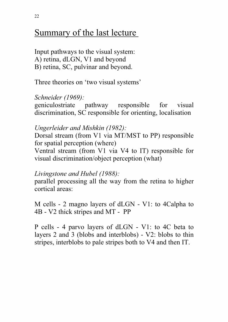
Summary of the last lecture
Input pathways to the visual system:A) retina, dLGN, V1 and beyondB) retina, SC, pulvinar and beyond.
Three theories on ‘two visual systems’
Schneider (1969):geniculostriate pathway responsible for visualdiscrimination, SC responsible for orienting, localisation
Ungerleider and Mishkin (1982):Dorsal stream (from V1 via MT/MST to PP) responsiblefor spatial perception (where)Ventral stream (from V1 via V4 to IT) responsible forvisual discrimination/object perception (what)
Livingstone and Hubel (1988):parallel processing all the way from the retina to highercortical areas:
M cells - 2 magno layers of dLGN - V1: to 4Calpha to4B - V2 thick stripes and MT - PP
P cells - 4 parvo layers of dLGN - V1: to 4C beta tolayers 2 and 3 (blobs and interblobs) - V2: blobs to thinstripes, interblobs to pale stripes both to V4 and then IT.

23
Problems with Livingstone and Hubel’smodel:
1. Is all of the information in higher extrastriate corticalregions coming from V1?No, Cells in area MT still respond when V1 is lesioned(must be another pathway)
2. Are the magno/parvo channels really that separate inV1 and V2?No, magno input into the blobs and interblobs
3. Does MT (dorsal stream) get only magnocellularinput?NO, magnocellular lesions of LGN do not eliminateall responsiveness in MT (although they do in a fairbit of it)
4. Does V4 (ventral stream) get only parvocellularinput??V4 neurons equally affected by inactivation of magnoor parvo layers in dLGN (magno should make nodifference), obvious that magno goes to blobs andinterblobs.
Conclusion: both dorsal and ventral stream appear toreceive magno and parvo input, although most of thedorsal stream appears to be magno.

24
Extrageniculate input to the cortex
Main source is the pulvinar:receives input directly from the retina and SCsends projections to the dorsal and ventral streams
(medial pulvinar to dorsal stream, lateral pulvinar toventral stream)
Functions of these two routes:SC - pulvinar - dorsal stream implicated in motion:
neurons in MT (dorsal stream) still active formotion when V1 lesioned
however, become inactive when SC lesioned as well
SC - pulvinar - ventral stream projections notunderstood
cells in V2, V3, V4 and IT no longer respond whenV1 is lesioned
seems to be no benefit from pulvinar route
Conclusions: ventral stream seems to rely on intactgeniculostriate pathway, dorsal stream seems to receiveother input as well.

25
Functional and anatomical properties ofthe two streams
Anatomical properties:Distinction between dorsal and ventral stream has beenrepeatedly confirmed and intermediate regions have beenidentified.Striking modularity of the dorsal stream (separatestreams and lots of interconnections), not the case for theventral stream (mainly one line of projections)
Functional properties:Ungerleider and Mishkin (1982):distinction in terms of input processing (what andwhere)
one system for spatial vision (dorsal stream,stimulus location)
the other for object vision (ventral stream, size,shape orientation and colour processing).
Livingstone and Hubel also focus entirely on theprocessing of incoming information.
New approach by Milner and Goodale (1992)Dorsal stream for visuomotor control (how)Ventral stream for visual perception (what)

26
Milner and Goodale (1992)
- Emphasis put on the output characteristics of the twosystems (how a stimulus is processed depends on what itis used for ): - The visual inputs and transformations required foraction are quite different from those leading to objectidentification:
Reaching and grasping:coding with respect to the viewer, i.e. motion and
location of the object have to be specified in viewer-centred coordinates.
Coordinate system used (retina, head or body)dependent on the effector system (eyes, hand or both).
object’s structure and shape has to be coded withrespect to the hand and fingers grasping it.
fast computations since the relative position of goaland observer changes quickly and has to be updatedquickly.
Object identification:constancies across different views, independent
from the observer.Objects structure and shape coded independently
from observerlong term storage of the identities of objects.

27
Milner and Goodale claim:
The different transformations required for action vsperception has driven the evolution of the two streams ofvisual processing.
Cells in the dorsal stream perform computationsnecessary for visually guided actions.Networks in the ventral stream form perceptual andcognitive representations of the enduring characteristicsof objects and their relations.
Division cuts right across the distinction between objectand spatial vision. (what needed for action, where neededfor identification)
28
Evidence for the theory (what vs how)
1) Taira et al. (1990) Processing of form in monkeydorsal stream
Response properties in area LIP in the monkeywhile performing different visuomotor tasks
1. four-fingered pull lever2. push button3. open pull knob (four fingers)4. pull knob in groove--finger & thumb
some units visually responsive, some respondduring hand movements
some respond to specific visual stimuli and themovements appropriate for those stimuli fairlyindependent of the target's location in space.Cells are sensitive to object parameters such as size andorientation but not location (very against U and M)
29
2) Multiple dorsal routes from V1 to PPC (support ideaof visuomotor processing in the dorsal stream):
a. The most prominent route passes through MTwhich has many motion directive cells. This route couldbe responsible for ocular pursuit and locomotion andposture.
b. route through V3A with many orientationselective cells. Route could be responsible for visualinformation to guide grasping.
c. route through PO least well known, seems tospecialised for dealing with stimuli located in periphery.(Could be responsible for providing visual information toguide saccades or movements towards peripheralstimuli.)
3) Specific projections from the PPC to the frontal cortexa. From LIP to the frontal eye fields, responsible
for eye movements)b. From VIP and 7a to the inferior premotor cortex
(implicated in control of arm movements).
4)Descending projections from dorsal stream to SC(eye-movements)
5) Projections from the dorsal stream to the pontinenuclei who in turn are closely linked to cerebellum(motor control).
No such projections exist from ventral stream.
Conclusion:Seems to be that the PPC provides visual information toa number of motor-related structures both in frontalcortex, basal ganglia and brainstem.

30
Information processing closely linked to visuomotorbehaviour (eye-movements, grasping, reaching)
31
Functional properties of the ventral stream(from V1 via V2, ventral part of V3, V4 and TEO to IT)
1) more detailed coding of visual features thefurther along one records from V1 though to V4 and IT.In IT cells can be very specific. Seem to respond best tocomplex visual stimuli such as hands, faces.
2)Cells sensitive to similar stimulus features areclustered together in columns.
3) Specific colour sensitive cells in IT.
4) IT primarily dependent on the geniculostriatesystem (from dLGN to V1) and V4 has both magno andparvo input.
5) many cells in IT have large receptive fields.(allows generalisation across views)
6) cells respond in anaesthetised monkeys, suggeststhat ventral stream cells are not involved in on-linecontrol of behaviour (here and now). (cells fireindependently of own limb movement; in sharp contrastto parietal cortex cells fire specifically for monkey’s ownarm movements (again action vs perception).
7) Ventral stream no projections to motor areas butinstead projections to amygdala (limbic system)
Conclusion:Properties in cells in IT consistent with objectrecognition and enduring characteristics (object centred).
32
Basis for recognition memory and long -termrepresentation of the visual world.

33
Milner and Goodale (1992):
Dorsal stream (HOW):
- viewer centred coding- transient (fast but not long lasting)- cell units related to movement- on line
Ventral stream (WHAT):
object-centred codingperceptual constancies (over time and space)colour and object perceptionoff line
34
Lecture 10 (Blindsight)
1) Evidence for Blindsight
2) Evidence against Blindsight
3) Explanation of Blindsight
4) Limits of the Explanations of Blindsight

35
Can Milner and Goodale’s model explainblindsight:
Prediction:damage to V1 should result in loss of the visualperception of the world (ventral stream function)but should still allow certain actions (dorsal stream andthis streams does not rely entirely on V1 input)
Patient DB studied by Weiskrantz (1974):damage to V1 (from removing a tumour) resulted in ahemianopia and thus complete blindness in that area
- D.B. insisted on not seeing anything in his blindfield but was able to make reasonably good eyemovements towards lights flashed into that field.
- Performed even better on a pointing task where hewas asked to point his finger to the light.
Weiskrantz named this phenomenon blindsight (accurateperformance despite lack of awareness)
Soon confirmed by Jeannerod and Perenin (1975 for sixmore patients) and others

36
Major critique of blindsight concept byCampion and Latto (1983)
Patients not completely blind. The intact parts of theirvisual fields can aide performance: Light from thestimulus could have scattered into the good region andthe patient might guess the appropriate location fromthis.
Inadequate fixation would allow the stimulus to fall intothe good field and thus be located.
There could be nonvisual cues such as sound or heat orexperimenter effects. The subjects is desperate for a cueand the examiner might unconsciously cue.
Areas of preserved vision in the hemianopic field couldbe responsible for the accurate response (cortex actuallyintact)

37
Replies to the critique:
Weiskrantz demonstrated that D.B. is unable to pointaccurately to stimuli that fall on the blind spot.
- Should be able to perform just as well at blind spotsince the light would scatter from there as well,inaccurate fixation would enable pointing, the other cuesshould help.
Jeannerod and Perenin (1978) tested patients withsevered optic chiasm as controls for cortically blindpatients:They should have no residual vision (Blindsight) in thehemianopic field (bitemporal) since the actual opticnerve fibres are interrupted.
Showed no evidence for blindsight although againthey should have been able to use scattered lightinformation.

38
What mediates blindsight
Milner and Goodale (1995):Damage to V1 and consequently impaired
input to the ventral stream is responsible for the lack ofreport. The patients do not see anything (perception).
Visual input still available throughtectopulvinar route: the dorsal stream networks continueto participate in the control of spatially organised eye andarm movements (this processing does not lead toconscious perception, action).
Blindsight collection of visuomotor responseselicited by the SC’s and the dorsal stream.
Certain input\output systems can be moreeffectively used than others. (Looking vs Pointing)
More evidence for action related processing inBlindsight:Perenin and Rossetti (1993):
Patients reach out and grasp objects reasonably accurately in the blind field. Can obviously use orientation and size information to guide a motor action.
39
Anatomical basis for blindsight (Milnerand Goodale, 1995):
Major input route via the SC route. ( SC aloneunlikely since there is no indication of orientation codingin the SC).
Tectopulvinar inputs reach the cortex as early as V2but cells in V2 and V3 loose their visual responsivenesswhen V1 is lesioned.
Area V3A responds when V1 is lesioned.
Cells in V3A code for orientation just as well ascells in V1. Likely that this area responsible for thevarious visuomotor behaviours shown.

40
Problems with Milner and Goodale’s explanation:
1) Blindsight can be demonstrated in patients with onlyone hemisphere:Not only is there no striate cortex but no extrastriatecortex and thus no dLGN and pulvinar (projectionsdegenerate when area of cortical projections are lesioned)
How does blindsight work in these patients?Either a puzzle how patients with hemispherectomy showblindsight or certain degree of cortical reorganisation(likely since operation in childhood).Maybe SC’s of lesioned hemisphere linked to cortex inthe intact hemisphere through intercollicularcommmissure and thalamus.
2) Cowey and Stoerig (1987, 1992) show colourdiscrimination in the blind field.Obviously colour processing after V1 lesions.
Problem for Goodale and Milner interpretation ofblindsight as visuomotor responses (i.e. SC and dorsalstream processing): No evidence for colour processing in SC and subsequentroute but
a very small number of cells (only a few thousand)project directly from the parvo cellular layers of thedLGN to V2 and V4. They survive lesions of V1. Very likely that these cells provide the necessaryinformation to subserve colour discrimination inblindsight.Not clear what function this pathway has in the normalbrain.
41
Conclusion:
Blindsight paradoxical if vision regarded as a unitaryprocess. If one accepts separate mechanisms for visualexperience and others for action the paradox disappears.It would mean that in such patients retinal stimulationdoes not lead to the experience of sight but insteadenables access to visuomotor control mechanisms.

42
Lecture 11
1) Balint’s patient (1909)
- hemispatial neglect- psychic paralysis of gaze- optic ataxia
2) Balint vs Holmes
- what vs how- what vs where
3) Specific impairments of patients with optic ataxia

43
Balint’s Patient (studied by Balint over 3 years):
- bilateral brain infarcts- normal visual fields- hemispatial neglect obvious when tested for acuity
and reading- psychic paralysis of gaze (only sees one object at a
time): also impairs reading
- Optic Ataxia:
not primary motor disturbance nor disturbance ofproprioception
only movements that required visual control werefaulty
only right hand affected
post mortem showed that patient had bilateral lesions inthe posterior parts of both parietal lobes
44
Holmes (1918, 1919):
- published two papers on patients whose symptomsvery similar to those of Balint’s patient.
- stressed the spatial rather than the visuomotorproblem
- both studies frequently cited unlike Balint’s- fit with Ungerleider and Mishkin (1982)

45
Problems with Ungerleider and Mishkin’smodel in relation to optic ataxia:
Assumption one
a deficit in space perception should be either incontralateral space or in case of a bilateral lesion inboth hemispaces (but independent of hand).
Balint’s patient impaired in either hemispace but only forhis right hand (Balint’s patient had a bilateral lesion).
but
optic ataxia also after unilateral lesion: onlycontralesional field affected or sometimes only quadrantif lesion is small (agrees with U and M)
but
not only space is important but effector system: even ifboth hands show an impairment, eye movements towardsthe target can be accurate (Ratcliff and Davies-Jones,1972)
Conclusion: misreaching cannot be a loss of the sensoryrepresentation of space but must be a deficit linked tospecific effector systems.Both hands or both hemifields can be impaired in anumber of combinations although the lesion tends to bein the same region (generally the superior part of theparietal lobe (Balint slide). Must be that the visuomotornetwork underlying reaching can be impaired at differentstages from in the transformation from visual to motor
46
coordinates.

47
Assumption two: Optic ataxics should fail to localise a stimulus but shouldbe able to use size and orientation cues to guide theirprocessing of the stimulus
Jeannerod (1988):in prehension transport and grasp components are
temporally coupled: the hand opens in anticipation overtime, opens wide and then homes in.
Perenin and Vighetto (1988):10 optic ataxia patients impaired in both the transport andgrasp component of a movement: made errors in handrotation as they reached toward an oriented slot.
but
could locoalise dots (align them with a number on thescreen). Some degree of spatial localisation still there.
Jakobson et al (1991) showed that their optic ataxiapatient only showed a very weak relationship betweenobject size and grip scaling.
Conclusion:Obvious that lesions to the parietal cortex produce morethan just spatial location deficits. They cause deficits inthe sensitivity of a grasp regarding an objects size, shapeand orientation. The lesions also damage specificeffector systems (hand, space, difference between handand eyes etc) rather than spatial location per se.

48

49
Lecture 12 (Agnosia I)
1) Definition of agnosia
2) Famous cases
3) Patient DF
4) Residual processing capacities of DF
5) Pathways mediating residual processing
50
Definition of Agnosia
-A profound deficit to visually recognise objects- patients fail to recognise objects which they
consciously detect.- problems in the visual domain only- very rare; requires intact V1- usually bilateral damage in the occipito-temporal
area, i.e. the ventral stream which is responsible forobject processing is cut off from the input of V1.
Historical account:
Lissauer (1890) [two types of agnosia}
- Apperceptive agnosia makes patients unable toperceive a coherent percept of the structure of an object.
a disruption at very early stages of perceptual processing
- A patient suffering from associative agnosia canperceive an object okay but is still unable to recognise it.
a disruption at a higher cognitive level where perceptswould normally be associated with stored semanticinformation.
51
Visual form agnosia
- severest form of apperceptive agnosia- only a few patients ever reportedall have a particular pathological history
suffered from an anoxic episode (usually suffered fromcarbon monoxide poisoning)
CASES:Adler (1944) : Patient H.C.)Benson and Greenberg (1969): Mr S.Campion and Latto (1985): Patient RCMilner and Goodale (1992): Patient DF
Tissue most vulnerable to anoxia are the watershed areas(Areas lying in the border regions between the territoriesof the different arterial systems)
MRI’s available of H.C. and D.F. indicate the existenceof dense lesions in the occipital cortex bilaterally
52
Mr S (extensively tested by Efron)
- severe object and pattern recognition deficits,- unable to recognise faces, identify line drawings or
even simple shapesbetter with real objects (more cues such as colour,
reflectance, texture)normal performance when haptic information
available
Efron argued that Mr S’s disorder was one of visualcontour, i.e. shape discrimination.
Devised the Efron test:
the subject has to judge whether pairs of rectanglesare the same or different (All shapes are matched forsurface area so that differences in brightness cannot beused as a cue)Mr S was unable to distinguish between any of therectangles apart from the 9:1 ratio (even then only 90%correct).
Now a standard test for visual form agnosia. Does notrequire shape perception (square vs oblong) but can bedone by comparing orientation or size information.Implies that visual form agnosics are impaired on thesesimple transformations as well.

53
Patient DF (Milner et al (1991)
- suffered from carbon monoxide poisoning whilehaving a shower.
- severe recognition disorder cannot be explained bylow-level visual deficits.
severely impaired on the Efron Task, cannotrecognise line drawings and cannot copy them
- cannot recognise letters of digits but can printthem from dictation, can also draw from memory.
- better at recognising real objects but again usescolour, texture and brightness cues
- unable to recognise faces but recognises people bytheir voice, brightly coloured hair etc.
- again normal recognition in the tactile andauditory domain.

54
Milner et al. (1991) Slot experiment
DF confronted with a disc in which a slot was cut whichcould be oriented at different angles
Asked to do two separate tasks:Demonstrate the orientation of the slot (unable to do soat all)
Asked to insert her hand into the slot (she was perfect)_
In the matching task the subjects were reporting theirperception of the slot.
To post the hand is a natural goal-directed movementwhich requires the rotation of the hand in order to besuccessful. It can be performed independently of aconscious perception of the orientation of the hand or theslot.
Special visuomotor system responsible that isindependent of object recognition.
Same dissociation observed with regard to size andshape: can grasp Efron shapes but cannot indicate theirwidth.
Findings strongly suggest that DF has a fully functioningvisuomotor system that uses orientation and sizeinformation although her perceptual system does not.
55
What mediates these residual functions?
Very likely that size and orientation calibration comesfrom the dorsal stream since her ventral stream (or atleast the access to her ventral stream) is damaged.The input to dorsal stream could come either from V1 orthe tectopulvinar route
Know that V1 is spared in DF, simple sensoryperception intact. Likely that human equivalents of V2,V3 and V4 are disrupted.
Likely that neither the magno (4B) nor the ParvoInterblob channel (Layers 2 and 3) can send informationto the ventral stream via pale stripes.If this region is no longer able to convey information tothe ventral stream, the ventral stream would be deprivedof both magno and parvo derived information on form.
Seems that DF’s vision can no longer rely on theInterblob channel. The magnochannel through V2 andV3 seems also impaired since she has trouble with lowspatial frequencies.
However, even a complete lesion in V2 and V3 and V4could still leave visuomotor abilities intact since thedorsal stream could still receive input from V1 direct (toMT, V3A and PO and there is also the tectothalamicpathway (Sc, pulvinar). These are routes on how thedorsal stream could receive input to allow the visuomotorbehaviour.

56
How come she can still use colour andtexture?If access to ventral stream is blocked she should beunable to do this?
Unlikely that this is mediated by the dorsal stream, noevidence for colour coding there just yet. Seems to be mediated by the ventral stream. Seemslikely that the blob channel has intact connectionsthrough V2 to V4 and IT. Seems to be colour only, notsize or shape, but possibly surface characteristics ofobjects (texture).

57
Lecture 13 (Agnosia II)
1) Limitations of visuomotor processing in DF
2) Can the visuomotor system aid object recognition?
3) Can Ungerleider and Mishkin’s Model explain DF?

58
Limitations of visuomotor processing inDF (1)
accurate processing of orientation and size
Processing of shape: impaired when asked to post a T-shape into a slot on half the trials she was correct on the other she
was incorrect. Suggests that she can take account of only one of
the two segments at once.
POSSIBLE PROBLEMS WITH SHAPEPROCESSING?
Experiment 1:presented with a series of everyday objects and
asked her to pick them up and demonstrate their use.
Is the object grasped appropriately as well asaccurately?
approach was fine, anticipatory opening of the handbut
when the object was rotated inappropriate grasp pointsand far more tactile exploration than the controls (needsnon-visual information)
Conclusion: her grasp is not guided by functionalknowledge of the object. She seems to use visualinformation of axis orientation and size to guide her
59
grasp but then uses tactile feedback to demonstrate theobjects function. Obvious that functional object knowledge is notaccessible via the dorsal stream but suggests that itcomes from the ventral stream and from there projectedto the dorsal stream.
Second experiment:
Can she combine size and orientation information toguide her actions?
Efron shapes presented at different orientations andasked to pick them up accurately (requires orientation ofthe hand adjustment of the grip size at the same time).
DF adjusts both her grip aperture and the orientationof her hand well in advance of target contact.
No problem in combining orientation and sizeinformation
Experiment 3:
Can she process shape to guide her actions?
Asked to reach out for a cross that was presented atdifferent orientations. (no principal axis she can use toguide her orientation)She does not rotate her hand in anticipation to therotation of the crossDoes not spread her fingers to engage with the shape ofthe cross.

60
Conclusion: Her visuomotor abilities seem to be limitedin so far as she is not able to integrate more than oneorientation in exercising her motor control. It seems thather general good grasping performance is due to heridentifying the principal axis and combine this with sizeprocessing.
Seems unlikely that DF’s visuomotor system can dealaccurately with different visual contour orientations tocombine them into a shape. DF as such does not giveevidence that the dorsal stream can handle form.
61
Can the abilities of her visuomotor systemaid her object recognition?
Yes, to some extent:
Asked to stamp shapes (orientation accurate)When asked not to stamp directly onto the stimulus butinstead on a separate sheet (perceptual task), surprisinglyshe was good. But:
moved towards the stimulus with her stamp and thenchanged in mid-flight to stamp the blank paper. (onlyapparent through careful video analysis)
Obviously used the shaping of her movement andtransferred that to the blank paper giving her the rightorientation.
She monitors her visuomotor performance to enhanceher perceptual judgements.
62
How does Ungerleider and Mishkin’smodel explain DF’s residual processing
abilities?She should be able to localise objects in space but
not much else.Should not be able manipulate objects without
knowledge of the objects identity.
BUT
She not only knows where an object is but also howto approach it.
Obvious that this is not done by the ventral stream sinceeven though she can reach for objects there is no objectrecognition and no functional knowledge of the object
Milner and Goodale argue that the distinction is betterexplained in terms of What for ventral and How fordorsal.
How requires a Where since otherwise one could not getthere in the first place but it also involves an approachthat is guided by the intrinsic properties of the object andan on-line handling of that object.
Comparison of DF to optic ataxia patients? Veryobvious dissociation.
Optic ataxics cannot orient their hand appropriate,DF no problem
They do not show anticipatory opening of the handto grasp an object, DF perfect
They can recognise object, DF cannotThey can orient their hand to match, DF cannot

63
Milner and Goodale’s assumptions for thetwo streams: What vs How
Ventral stream characteristics (perception):-object-centred coding (independent of
observer)-perceptual constancies (how does this look
like over different views, spatial positions)-colour, object perception-"off-line functions" (access to memory of
objects and object relations)
Dorsal stream characteristics (action)--viewer-centred coding (where are things in
relation to the person)--transient (quickly picks up even subtle
changes)--many units related to movement (action)--"on-line functions" (where is now, and how
does it change)
64
Lecture 16
Spatial Attention and Visual Neglect
1. Description of the neglect syndrome
2. Historical Overview: a) Issue of laterality b) Locus of lesions in neglect c) Task differences
3. Standard neglect tests today
4. Recovery from neglect
65
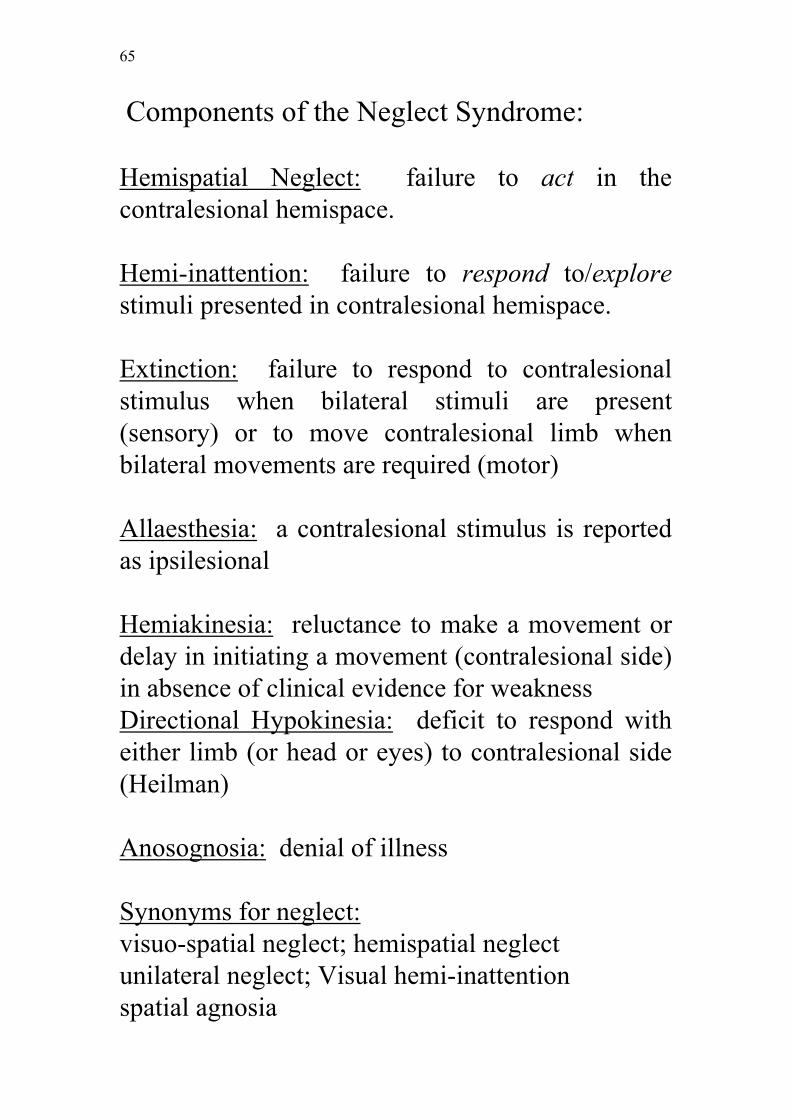
Components of the Neglect Syndrome:
Hemispatial Neglect: failure to act in thecontralesional hemispace.
Hemi-inattention: failure to respond to/explorestimuli presented in contralesional hemispace.
Extinction: failure to respond to contralesionalstimulus when bilateral stimuli are present(sensory) or to move contralesional limb whenbilateral movements are required (motor)
Allaesthesia: a contralesional stimulus is reportedas ipsilesional
Hemiakinesia: reluctance to make a movement ordelay in initiating a movement (contralesional side)in absence of clinical evidence for weaknessDirectional Hypokinesia: deficit to respond witheither limb (or head or eyes) to contralesional side(Heilman)
Anosognosia: denial of illness
Synonyms for neglect:visuo-spatial neglect; hemispatial neglectunilateral neglect; Visual hemi-inattentionspatial agnosia

66
Issue of Laterality:
Brain (1941) first to give a detailed description ofneglect:- three patients with large right parietal lesions inwhich he noticed a disorder of route finding:'agnosia for the left half of space'- restricted to patients with minor (i.e.) righthemisphere lesions- different from hemianopia, hemiplegia- possible that absence in left hemisphere lesionedpatients is due to masking (i.e. aphasia)
Since then debate in the literature: Does neglectoccur more frequently with right-than lefthemisphere lesions, and if so, what significancedoes that have?
Oliver Zangwill's group: left sided neglect presentin 14 out of 21 right hemisphere lesioned patientsbut only in one out of eight left hemisphere lesions
but
67
Battersby and colleagues (1956): tested 85 patientsand found no difference as to the side of lesion:-found the syndrome in 29 per cent of patients withnon-dominant (right) lesions and 9 per cent ofpatients with dominant (left) lesionsbut- 29 per cent of left hemisphere patients had to beexcluded because of aphasiaso- they judged 62 per cent of the left hemispherecases and 59 per cent of the right hemisphere casesto be without neglect
Gainotti (1968) designed a battery of tests simpleenough to include aphasics: - found that neglect is not only more frequent butalso more severe after right hemisphere damage
68
Localisation of Lesions in Neglect:
Zangwill's group: right and left hemispherelesioned group differed in incidence of visual fielddeficit and papilloedema suggesting that the righthemisphere lesioned patients had larger lesions
general problem:no CT, MRI scans, right hemisphere lesions tend tobe silent, only noticed when fairly large
Locus of lesion: -not only parietalVallar and Perani (1986): CT study on 110 righthemisphere lesioned patientsCortical neglect more frequent with retrorolandicthan frontal lesions- inferior parietal lobule most frequently involvedSubcortical neglect more frequent when grey nuclei(thalamus and basal ganglia) rather than whitematter damaged

69
Task Differences:
Problem: different investigators use different tasksto assess neglect (i.e. drawing, copying, reading)
Gainotti (1972): Right hemisphere lesionedpatients more impaired than left h. patients whentask required attention to be focused on smallportions of spacebutno difference between groups when task requiredexploration of large displays
Consensus of laterality debate: some people stillargue but majority thinks that neglect is morefrequent and more severe after right h. damageespecially after more than three days post onset

70
Standard Neglect Tests Today:
In Britain: Behavioural Inattention Test (BIT);Wilson, Cockburn and Halligan (1987)
-80 patients with right or left hemisphere damagecompared to 50 controls on:
Line Bisection (Schenkenberg, 1980)Line Crossing (Albert, 1973)Letter cancellation Star cancellationFigure and shape copyingRepresentational drawing
gives a cut-off score for each test and overall
71
Recovery from Neglect:
Clinical picture:
in severe cases very strong ispilesional eye andhead deviation
changes to neglect symptoms (i.e. failure to dress,shave etc)
changes to inattention (failure to respond)
Inattention can fade into extinction
all signs may disappear unless specifically testedfor, for example:
Goodale et al (1990):
tested 10 right h. damaged (recovered fromneglect) on a visuomotor bisection task: patientsshowed rightward errors
72
Lecture 17
Spatial Attention and Visual Neglect
Theories of Neglect ( from 1940 to recentthinking):
1. Sensory hypothesis (Battersby et al.)2. 3. Representational approach (Bisiach et al.)4. 3. Directional Hypokinesia (Heilman et al.)
5. Attentional approach (Posner et al.)6. 5. Spatial misperception (Milner et al.)

73
Brain (1941) and Zangwill group:neglect independent of sensory and generalintellectual disturbances
but
1. Sensory hypothesis: Bay (1950) andBattersby (1956):a combination of sensory defects and intellectualdeterioration can account for the disorder
but
2. Representational Approach: Bisiach (1978):demonstrated neglect in tasks without sensoryinflow (i.e. imagination)- RHD patients asked to face Milan cathedral anddescribe scene, asked to face away from cathedraland describe scenepatients omitted items on the left depending on theperspective taken-failure to exploit otherwise intact representation
74
3. Directional Hypokinesia (Heilman, 1979):a unilateral brain lesion hypoarouses thehemisphere and causes a selective loss of orientingresponses in the hemispace contralateral to thelesion- the left hemisphere controls orienting in rightspace only- the right hemisphere controls orienting in left andright spacetherefore: left hemisphere lesion can becompensated, right hemisphere lesions cannot
Experimental confirmation: RH patients initiateresponses into left space more slowly than LHpatients responses into right space (same formagnitude of errors)

75
4. Covert orienting (Posner, 1980; 1984): Posner's paradigm: + * +cues indicate to be attended locations:80 per cent valid20 per cent invalidNormal Subjects:benefit from valid trials because already engaged intargetbut cost of invalid trials as attention needs to bedisengaged and moved and reengaged on newtargetPatients with parietal lesions:benefit from valid cues like controls (in both leftand right space)also same cost when invalid cue in left space andthey have to reengage to right spacebuthigh cost when invalid cue in right space and theyhave to reengage to left (sometimes failed to detectstimulus completely)-problem to reallocate attention once it has beendirected to the right

76
5. Spatial misperception (Milner et al., 1992,1993)
Is it possible that neglect patients who fail to attendto left hemispace misperceive it as relativelyshrunk compared to right hemispace?- Impossible to tell with bisection task- but Landmark task can distinguish betweenperceptual neglect and directional hypokinesia
Landmark Task: centrally prebisected line, patientis asked to indicate to which end the central mark iscloser to- 7/8 neglect patients point to the left (perceptualneglect)- 1/8 points to the right (directional hypokinesia)
77
Other evidence for these two types of neglect:
Bisiach et al., 1990: pulley device on linebisectionCongruent condition: a rightward movement movespointer to the right (and VV)Incongruent condition: a rightward movementmoves pointer to the left (and VV)Results (incongruent condition): some patientsmove pointer leftwards to set transection to theright (perceptual neglect), others move pointerrightwards and transect to the left (directionalhypokinesia)
Tegner and Levander (1991): line cancellationby looking in mirrorall patients neglect left space in normal viewMirror view:4/18 cancel lines in right hemispace only(directional hypokinesia)10/18 patients cancel lines in left hemispace only(perceptual neglect)4/18 cancel only central lines (combination of thetwo deficits?)

78
Dissociation between two types with regard toline length (Harvey et al., 1994)
Patients with perceptual neglect make smaller and
smaller errors the shorter the line becomes (line
bisection), patients with DH make large errors throughout

79
Lecture 18
Neglect and the two visual systems
1. Revision of Theories2. Object-centered neglect (Driver & Halligan)3. Blindsight? (Marshall & Halligan but see alsoBisiach)4. Does neglect result from damage to the dorsalstream ?(Evidence for and against)

80
2. Object-centred neglect (Driver andHalligan, 1991)
- single case study- patient neglects the left side of objects even whenpresented in right hemispace- even neglects left side of stimuli when objectsrotated and left part of stimulus is on right side ofthe display(interpreted as impaired spatial representation ofobjects)but maybe viewer centred rather than objectcentred representational deficit (Rizzolatti)

81
3. Blindsight (Halligan and Marshall, 1988)
-single case study- patient presented with drawing of a burning andnon-burning house, she claimed houses are thesame- asked where she would prefer to live: pointed tothe non-burning house
replicated by Bisiach and Rusconi (1990)
- four patients, various stimuliresults inconsistent- some patients prefer broken glass- some unflowered pot- some the burning house

82
4. Is hemispatial neglect a result of dorsalstream damage:
Ungerleider and Mishkin (1982):
Claimed that the appreciation of an objects’ qualities(object identification) and of its spatial location (where isit) depends on the processing of information in theinferior temporal (IT) and posterior parietal cortex (PPC)respectively.Same distinction as Schneider between what and wherebut this time the dichotomy is entirely cortical: dorsal vsventral stream processing.
Main evidence:Found that monkeys with IT lesions are impaired in adiscrimination task but not in a landmark task (choosethe food well that is closer to the object), while monkeyswith PP lesions were impaired on using a spatiallandmark but not on discriminating between two objects.
Problem: response modality not taken into account.Model focuses on the animals decision about thestimulus array and neglects the animals visuomotorbehaviour.
83
evidence for it-monkey evidence showed that the PPC is
implicated in spatial attention (Mohler and Wurtz)
thusreasonable assumption that neglect in humans followingdamage to the posterior brain areas must be result ofdamage to the end-points of the dorsal stream
anda spatial imbalance of attention is present in most casesof neglect.in most patients the lesions include parts of the parietallobe.
Nonetheless there is serious doubt that neglect resultsform dorsal stream lesions.
84
Evidence against it:
1. Proved very difficult to mimic neglect properly inmonkeys. Monkeys do not show the attentional failure,the perceptual failure, object centered neglect (Milner,1987).
2. What is the human equivalent of PPC?
- monkey evidence associates visual guidance ofreaching, grasping and eye-movements with the dorsalstream. They are impaired by PPC lesions.
- in humans equivalent deficits are associated withlesions of the superior parietal areas: optic ataxia,psychic paralysis of gaze

85
hint that in humans the superior parietal areas are theequivalent of monkeys dorsal stream mechanisms
Human neglect most commonly associated with theinferior parietal lobe and parietotemporal lobe.
3. Object centered neglect:Argues against neglect as a deficit within the coordinatesof egocentric space (space with relation to the personacting) since object based neglect cannot be independentof object identity processing.
Possible that inferior parietal and temporo-parietalregions have evolved in the human to deal with this formof abstract spatial processing and that they rely heavilyon the input from the ventral stream
Possibility that impairment of abstract spatial processingon one side leads to an incorrect computation of stimulussize
86
Lecture 19: Are all neglect symptomsindependent of dorsal stream processing:
Extinction
Directional Hypokinesia
Attention and Awareness

87
Extinction within neglect syndrome problematic:
1. Double dissociation between neglect and extinction (itcan occur without neglect (De Renzi, 1984, Barbieri andDe Renzi, 89) and neglect can occur without extinction(cannot be a mild form).
2. Extinction found to an equal extend after right and lefthemisphere lesions while neglect is generally morefrequent after right h. lesions.
3. Evidence for an anatomical distinction: superior parietal lobe lesions produce optic ataxia
and extinction (Perenin and Vighetto, 1988) but notneglect.
Conclude that visual extinction may follow damage to thedorsal stream in both humans and monkeys but thatneglect does not.
Extinction is primarily a failure to orient. This functionis easily carried out by the dorsal stream or by the SC’s.It should be closely linked to visuomotor controlmechanisms. Should be more obvious when patients areasked to point than through verbal report (Bisiach (1989).
88
Directional hypokinesia
Most investigators now distinguish between premotorand perceptual forms of neglect.
Most studies have found that premotor neglect is relatedto frontal and subcortical damage while perceptualdamage results from inferior parietal damage.
Directional hypokinesia can be demonstrated in monkeysand even rats. Seems to be related to damage to motor orpremotor areas in the brain.
Quite different from the neglect which is a selectivefailure of perceptual awareness of incoming stimuli.
89
Attention (Milner and Goodale’shypotheses)
In the dorsal stream attention is closely linked to theparticular effector systems.
There are specific attentional mechanisms for eye-movements, arm movements etc.
In the ventral stream attention is linked to a particularstimulus (filtering of irrelevant stimuli) rather than to aselective response.
Only the attentional mechanisms associated with theventral stream allow conscious awareness of objects andevents in the world.
The dorsal stream although involved in selectiveattention does not give rise to visual awareness.

90
Evidence for these assumptions
1. Attentional modularity in the dorsal stream:
Goldberg and Wurtz studies:a typical cell in the superficial layers of SC would
respond to a visual stimulus flashed into its receptivefield but they also showed that the cell response wasgreatly enhanced when the stimulus was relevant to theanimal.Similar attentional effects were also found in the frontaleye-fields, PPC and pulvinar but not in the primaryvisual cortex.
In a covert attention ask the monkey was asked to fixateand respond to the stimulus by pressing a key. It wasfound that no enhancement was found in the SC orfrontal eye field neurons but in PPC and the pulvinar.
This was interpreted at the time the SC and frontal eyefields may be specifically associated with gaze shift(saccades) while the PPC mediates attention independentof gaze. as been derived from this that PPC mediated attentiongenerally (some sort of superordinate attentional process)
but
the monkey was pressing a key in response.
Same study can be taken as evidence for selective visualattention in relation to reaching and saccadic behaviour.
91
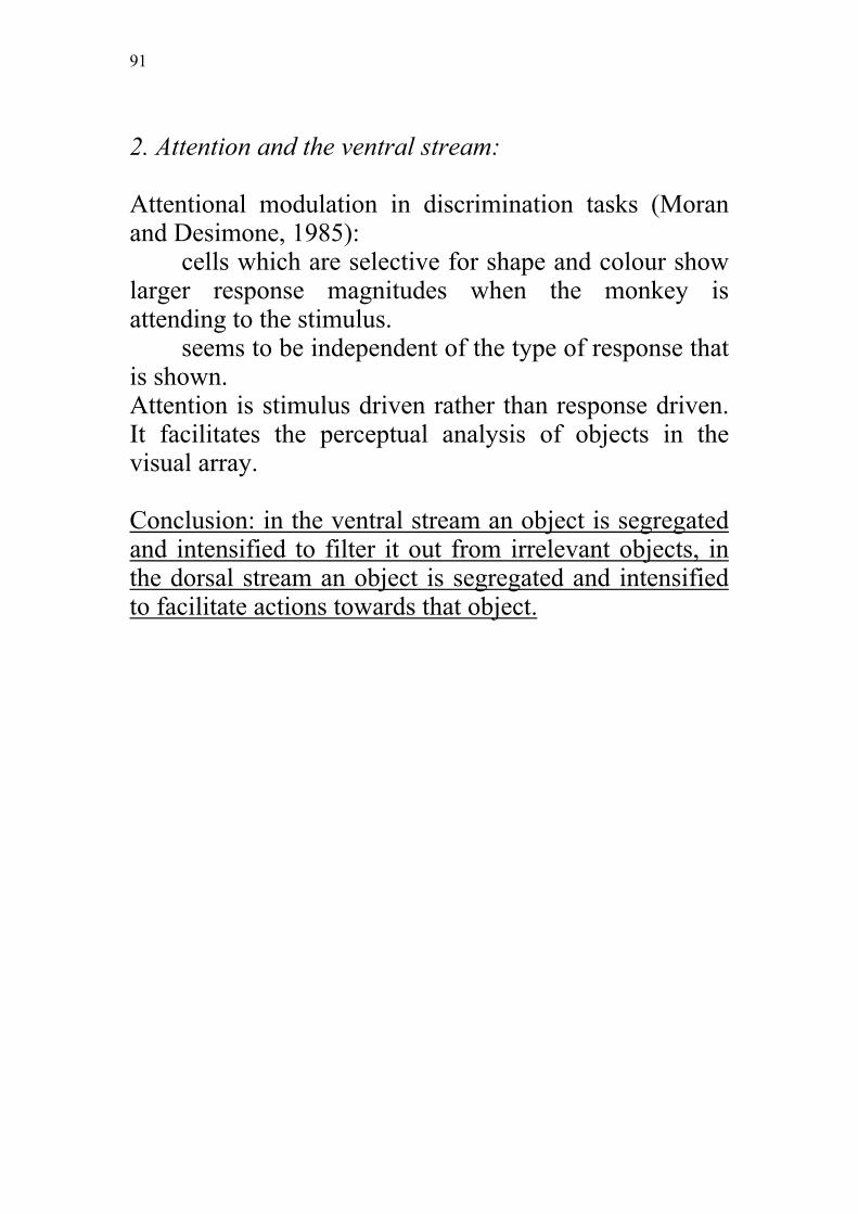
2. Attention and the ventral stream:
Attentional modulation in discrimination tasks (Moranand Desimone, 1985):
cells which are selective for shape and colour showlarger response magnitudes when the monkey isattending to the stimulus.
seems to be independent of the type of response thatis shown.Attention is stimulus driven rather than response driven.It facilitates the perceptual analysis of objects in thevisual array.
Conclusion: in the ventral stream an object is segregatedand intensified to filter it out from irrelevant objects, inthe dorsal stream an object is segregated and intensifiedto facilitate actions towards that object.

92
3. Evidence for linking ventral but not dorsal stream toawareness
Evidence from blindsight patients:Blindsight patients do not have conscious percept of
the stimulus but can nevertheless point.
Argued that this residual ability is performed by thedorsal stream through input from the pulvinar.