generic protocol to examine the incidence of lower
TRANSCRIPT

s
\~S 280 2000~JR
-=r.: --=-
Generic protocol to examine the incidence of lower respiratory infection due to respiratory syncytial virus in children less than five years of age
Field test version
DEPARTMENT OF VACCINES AND BIOLOGICALS ,
(@~) World Health Organization -Geneva __.~

WHON &B/00.08 ENGLISH ONLY
DISTR.: GENERAL
Generic protocol to examine the incidence of lower respiratory infection due to respiratory syncytial virus in children less than five years of age
Field test version
Peter F. Wright, MD H ead, Division of Infectious Diseases, Department of Pediatrics Vanderbilt University, Nashville, Tennessee, United States of Ameria
Felicity T. Cutts, MD, MSc WHO Collaborating Centre on Clinical Evaluation of Vaccines in Developing Countries, London School of Hygiene and Tropical Medicine, London, United Kingdom
DEPARTMENT OF VACCINES AND BIOLOGICALS . ) World Health Organization ~ ~ Geneva ~J? 2000

The Department of Vaccines and Biologicals thanks the donors whose unspecified financial support
has made the production of this document possible.
This document was produced by the Vaccine Assessment and Monitoring Team
of the Department of Vaccines and Biologicals
Ordering code: WHOIV&B/00.08 Printed : March 2000
This document is available on the Internet at! www. vaccines. who.int/vaccines-documents/
Copies may be requested from: World Health Organization
Department of Vaccines and Biologicals CH-1211 Geneva 27, Switzerland
• Fax: +22 791 4193/4192 • E-mail: [email protected] •
© World Health Organization 2000
This document is not a formal pubJjcatjon of the World Health Organjzation (WHO), and all rights are reserved by the Organjzation. The document may, however, be freely reviewed, abstracted, reproduced and translated, in part or in whole, but not for sale nor for use in conjunction with commercial purposes.
The views expressed in documents by named authors are solely the responsibility of those authors.
ii

Contents
Preface ............................................................................................................................... v
1. Introduction .............................................................................................................. 1
1.1 Types of severe RSV disease .................................................................... ....... 2 1.2 Specificity of RSV diagnosis ........................................................................... 2 1 . 3 RSV serotypes ................................................................................................... 3 1.4 Epidemiology of RSV disease in industrialized countries ........................... 3 1. 5 Epidemiology of RSV in developing countries ...................... , ...................... 4
2. Study objectives ........................................................................................................ 8
3. Methodology .............................................................................................................. 9
3 .1 Reference population .... ................................... .... ...... ...................................... 9 3.2 Size of population under surveillance .......................................................... 1 0 3.3 Case ascertainment ......................................................... ...................... .......... 11 3. 4 Case definition ................................................................................................ 12 3.5 Case report form ............................................................................................ 13 3. 7 Laboratory assays ......... .................................................................................. 1 5 3. 8 Laboratory log book ...................................................................................... 17 3. 9 Field work ................................................................................................... ... .. 17 3. 1 0 Data handling .................................................................................................. 1 7
References ...................................................................................................................... 18
Annex 1: Management of the child with cough or difficult breathing .......... 22
Annex 2: List of manufacturers of rapid diagnostic tests for RSV ................ 29
Annex 3: Respiratory rate timer ............................................................................ 3 0
iii

iv

Preface
The WHO Department of Vaccines and Biologicals has an increasing interest in vaccines against respiratory syncytial virus (RSV). Such vaccines are still in the early phases of development, but research is being pursued within a number of pharmaceutical companies and academic centres. The promise of having a vaccine available in the relatively near future emphasizes the need to understand the clinical impact of RSV in developing countries. It can be anticipated that the mortality from acute lower respiratory tract disease will be higher in developing countries than in industrialized countries, if only because of the general lack of respiratory support such as mechanical ventilation and supplemental oxygen. Viral pneumonias may predispose to the increased frequency and severity of bacterial pneumonias which are seen in developing countries (WHO 1991). Although the epidemiology of RSV disease is clearly defined in temperate climates, it is incomplete in developing countries.
The Steering Committee on Epidemiology and Field Research and the Steering Committee on Measles, Acute Respiratory Viruses and Poliomyelitis jointly identified the need to develop a practical method for immunization programme managers and clinical epidemiologists to assess the local burden of severe disease due to RSV. At the request of these Steering Committees, this generic protocol was prepared by staff at Vanderbilt University and the London School of Tropical Medicine and Hygiene.
This protocol provides a general outline for a study and describes the main procedures involved. It will need to be adapted to the local setting, and details of field work and operational procedures should be added by local investigators with experience of conducting field studies in the specific area. During 1998-2001, WH O is sponsoring studies based on this protocol in Indonesia, Mozambique, Nigeria and South Africa.
WHO provides this generic protocol free-of-charge. In return, the programme would appreciate being informed about studies conducted using this protocol. The generic protocol may be freely adapted, but this WHO document should be referenced in any publication resulting from its use. Comments or suggestions for improving this protocol are welcome and should be sent to: DrS. Robertson, Department of Vaccines and Biologicals, World Health Organization, 1211 Geneva 27, Switzerland.
v


1. Introduction
Severe respiratory syncytial virus (RSV) pulmonary disease, manifested as bronchiolitis and pneumonia, continues to play a major role in childhood morbidity and mortality that is best documented in industrialized countries. Estimates of impact in the USA are that 18 000 to 75 000 hospitalizations and 90 to 1900 deaths occur annually (Anderson et al. 1990). Vigorous initiatives are now under way for the prevention of RSV disease in industrialized countries. These include the development of live, attenuated RSV vaccines; subunit RSV vaccines for maternal or infant immunization; and passive antibody in the form of RSV immune globulin or monoclonal antibodies. The RSV immune globulin given prophylactically has been shown to be helpful in prevention of lower respiratory tract disease in those with underlying cardiopulmonary disease, particularly small, premature infants (Groothuis et al. 1995). It is anticipated that vaccines will be available for immunizatio n against RSV within a decade (Murphy et al. 1994). Nonetheless, because the pathogenesis of RSV infection appears to be at least in part immunologically mediated and because of past experience of disease enhancement after killed vaccines, the introduction of RSV vaccines will have to be undertaken with particular care.
The purpose of this generic protocol is to describe a surveillance system which will allow estimation of the local burden of RSV disease. This will assist government health officials and immunization programme managers in the future, when they consider the introduction of an RSV vaccine to their immunization programme. This protocol may also be adapted later to demons~rate the impact of immunization.
Other bacteria and viruses cause severe lower respiratory tract illness (LRI) in young children, including Streptococcus pneumoniae, Haemophilus influenzae, adenovirus, influenza virus, and parainfluenza virus. However, RSV represents a substantial amount of the burden of acute respiratory tract illness, particularly in the first year of life. It is the closest of the viral pathogens to having a vaccine available, although research is also being conducted to develop live attenuated influenza and parainfluenza type 3 vaccines. There have been previous attempts to define the totality of microbial pathogens causative in acute respiratory illness, notably the BOSTID1 studies in the late 1980s. In retrospect, the BOSTID studies seem to have suffered from being overly ambitious in attempting to define all respiratory illness. Investigators using this WHO generic protocol are encouraged to focus on RSV, which can now be identified by rapid viral techniques on adequately collected samples of nasopharyngeal aspirations (see section 3.6).
A series of studies sponsored by the United States National Academy of Sciences Board on Science and Technology for International Development (BOSTIO). These studies were reported in a 1991 supplement to Reviews of Infectious Diseases, entitled "Etiology and Epidemiology of Acute Respiratory Tract Infection in Children in Developing Countries" (Bale 1990).
WHON&B/00.08

Although RSV causes considerable upper respiratory tract disease, some of which is associated with o~itis media, RSV causes proportionately more LRI than other viruses. RSV vaccines are more likely to protect against severe disease than to give sterilizing immunity that protects against recurrent upper respiratory infection. Furthermore, as the main costs to the health service of RSV disease relate to LRI, the burden of RSV-associated LRI is of most interest to policy makers. For all of these reasons, this WHO generic protocol focuses on identification of RSV-associated LRI.
1.1 Types of severe RSV disease
Severe RSV disease has been traditionally divided into bronchiolitis and pneumonia, with RSV thought to account for approximately 85% of cases of bronchiolitis and approximately 20% of cases of childhood pneumonia. The clinical distinction between bronchiolitis and pneumonia is often not clear. In addition to hyperinflation, radiographs in bronchiolitis frequently show what is interpreted either as atelectasis or infiltrates, although the rarer lobar consolidation is likely to indicate pneumonia (Forgie et al. 1991). Wheezing, considered to be the hallmark of bronchiolitis, can be elicited in almost every infant when sequential observations are made. Thus, although there are suggestions that the pathogenesis of these two RSVassociated diseases is different, that is often not clear from clinical or radiographic examinations. However, one would still like to try to differentiate between these two illnesses, by documentation of wheezing and the findings of clinical chest auscultation, as well as radiographs where available.
1.2 Specificity of RSV diagnosis
Unlike some other respiratory viruses (e.g. adenovirus), it is uncommon to isolate or identify RSV from healthy children without evidence of respiratory illness. Some studies of RSV-related illness have included a control series of well children and found that less than 5% of controls have been positive for RSV (Chanock et al. 1961, Forgie et al. 1991, Phillips et al. 1990). RSV infection may be detected in appropriately collected specimens (nasal aspirates, nasal washes) by virus isolation or by rapid diagnostic methods (see section 3.6). Serology is not recommended for diagnosis of RSV infection, as up to 50% of young infants hospitalized with culture-proven RSV infection show no serological rise (unpublished data, Vanderbilt University, 1996). The detection of RSV in nasal secretions of a patient with respiratory infection is therefore taken as evidence that the illness is caused by RSV. This is likely to be an over-simplification, as there may be superimposed bacterial infections that contribute to the severity of disease. Although secondary bacterial infection is documented with RSV, it is not known whether this occurs more frequently in developing than in developed countries, though one study in the Gambia suggested that this is the case (Forgie et al. 1991). More recent data from the Gambia show that approximately 5% of children with documented RSV disease also had a positive blood culture for a bacterial agent (Weber and Greenwood, personal communication, 1996). The rate of mixed infections is difficult to estimate accurately, because of the difficulty in identifying a bacterial agent in lower respiratory tract infections (Shann 1986). In the Gambia, mixed viral-bacterial infections were identified in 13 (15%) pneumonia cases in infants less than one year of age, four of which involved RSV infection (Forgie et al. 1991).
2 Generic protocol to examine incidence of RSV·LRI

Children with mixed infections had higher mean temperatures and higher white blood cell counts than those with RSV infection alone, but other clinical parameters were similar in the two groups. In Uruguay, mixed infections were documented in 10 (5%) of 204 hospitalized children with pneumonia; seven of these involved RSV (H or tal et al. 1990). Although the potential for RSV co lead to increased susceptibility to bacterial infection is important in estimating the potencial impact of a future RSV vaccine, the study of this possible interaction is likely to be impractical in many centres. It might be studied more readily by evaluating the reduction in overall LRI in an intervention trial, when RSV vaccines reach the stage of phase III clinical trials.
1.3 RSV serotypes
Two serotypes of RSV exist: A and B. There is some evidence of heterotypic immunity between the two. The analogy has been made that the two RSV serotypes are as closely related as sequential influenza strains of the same serotype. All current vaccine efforts are directed towards the development of a vaccine that will incorporate both strains or w ill be directed against the F protein, which is quite homologous between the two strains. The clinical impact of the strains appears to be relatively similar, although RSV A may cause slightly more severe disease than RSV B (Hall et al. 1990). It may not be possible to differentiate between types A and B easily in field studies, as this requires isolation of RSV virus followed by typing. Polymerase chain reaction (PCR) technology at a central laboratory may, however, provide the opportunity to serotype strains on specimens that do not contain viable VI rUS.
1.4 Epidemiology of RSV disease in industrialized countries
RSV is now well documented as a cause of yearly winter epidemics of acute lower respiratory tract disease, including bronchiolitis and pneumonia. Studies have provided accurate estimates of invasive RSV disease burden in several industrialized countries. Data from Vanderbilt University, Nashville, Tennessee, USA, as an example, shows the rate of culture-proven RSV-associated LRI in otherwise healthy children to be 37 per 1000 child-years in the first _two years of life and the risk of hospitalization 6 per 1000 child-years (Fisher et al. 1997). These rates are higher in the first six months of life: 45 per 1000 child-years for LRI and 11 per 1000 childyears for hospitalization. Incidence will be higher fo r children with cardio-pulmonary disease and those with prematurity. Patients with these risk factors constitute almost half of RSV-related hospital admissions in the USA (Table 1).
WHON&B/00.08 3

Table 1: Risk factors for hospitalization with RSV-related illness amqng 95 hospitalized children, Vanderbilt University,
Nashville, Tennessee, USA
Risk factor Number(%) of children
Cardiac disease 15 (16)
Prematurity 27 (28)
Pulmonary disease 29 (31)
One or more of above risk factors 44 (46)
In eight European countries, 19% of LRI occurring in hospitalized patients under five years of age was attributed to RSV by virus isolation at the time of illness (Orstavik et aL 1984). This represented approximately 80% of the virally-proven LRI. Apnea is another presenting sign of RSV disease in 10-20% of hospitalized patients (Church et al. 1984). These studies serve as a basis for anticipating widespread use of RSV vaccines and other interventions in industrialized countries, where the costs of caring for patients with severe lower respiratory tract disease and their sequelae are substantial.
There is increasing recognition of the importance of RSV as a cause of substantial morbidity from influenza-like illness in the elderly in industrialized countries (Fleming & Cross 1993). We found no published data on the burden of RSV in the elderly in developing countries. While potentially an important question, a study of this age group is beyond the scope of this generic protocol.
1.5 Epidemiology of RSV in developing countries
1.5.1/ncidence of RSV-LRI
In developing countries there are few population-based estimates of the incidence of RSV disease. Table 2 shows the incidence of RSV-associated LRI in selected studies that were denominator based. Early population-based studies in India, that relied on throat swabs for virus isolation and appeared to have a low rate of RSV recovery (Ota et al. 1972) have not been included.
Other estimates have been derived from the incidence of LRI of all causes, and the proportion of LRI that is RSV-related. From community-based studies, the median incidence of pneumonia in under-five-year-olds in developing countries has been estimated as 0.4 episodes per child per year. This rate is as high as 0.7 per child-year in infants less than one year of age (Schwartz et al. in press). Pneumonia represents a median of 74% (range, 50-86%) of lower respiratory tract infections among hospitalized children, and 40% (range, 28-59%) of outpatient cases of LRI. Thus, the overall incidence of LRI in under-five-year-old children will lie between 0.54 (0.4 x 100/74) episode and one (0.4 x 100/40) episode per child per year, i.e. 540 to 1000 LRI episodes per 1000 child-years.
4 Generic Jl(Oiocol to examine incidence of RSV·LRI

Viruses have been found to cause a median of 37% of LRI (range, 15-76%). This proportion is higher among infants, and is higher fo r bronchiolitis than pneumonia (Cherian et al. 1990). Among viral aetiologies, RSV is by far the most frequent, causing a median of 18% of LRI (range, 7-33%) (Schwartz et al. in press), and representing 62% of viral LRis. This is similar to the proportion found in European countries. Applying this proportion to the incidence of all-cause LRI, the estimated incidence of RSV-associated LRl is 97 to 180 episodes per 1000 child-years. Although these are crude estimates, this developing country rate is 2.6 to 4.8 times the rate of RSV-associated LRI seen in the USA.
Table 2: Incidence of RSV-assodated LRI in selected studies from developing countries
Reference Site Case Age, Specimens Incidence Incidence ascertain- sample for of all LRI ofRSV-LRI
ment, dates size virology
Berman Cali, 5 PHC <5yr Throat 70/1000 36/1000 et al. Colombia clinics (n=4958), swab child-years child-yearsd 1983 (urban, 2!17- 10/78 5-15 yr (1977) (age 0·15 yr) (age 0-15 yr)
low SES•) (n=3790) NPAb,IFA<, 400/1000 & culture child-years (1978) (age 0-2 yr)
Borrero et al. Cali, Weekly Birth cohort NPA, IFA, 1700/1000 198/1000 1990 Colombia home visits, (n=340) & culture child-years child-years•
(urban, 10/86-4/88 (age 0-1 .5 yr) 0·5 mo: low SES) 192/1000
6-11 mo: 255/1000 12-17 mo: 125/1000
Sutmoller Rio de Weekly <5 yr NPA, IFA, 64/1000 14/1000 et al. 1995 Janeiro, community (n=262) & culture child-years child-years'
Brazil surveillance, (age 0-4 yr) (age 0-4 yr) (urban, 1/87- 12/89 low SES)
• SES, socioeconomic status . b NPA, nasopharyngeal aspirate.
• IFA, immunofluorescent antibody . d Total, 106 RSV isolates (9% of all LRI).
• Total, 67 RSV isolates . I Total, 11 RSV isolates.
Among factors which may influence the incidence and severity of RSV-associated disease in developing countries are exposure to passive smoke (Holberg et al. 1991), malnutrition (Tupasi et al. 1990a), HIV infection (King et al. 1993), and crowding (Holberg et al. 1991). Breast feeding and vitamin A supplementation (Dowell et al. 1996) have not been shown to alter the frequency or severity of RSV-associated LRI. An attempt should be made to identify such potential risk factors in the children studied as part of this WHO generic protocol.
WHON&B/00.08 5

1.5.2 Age distribution of RSV-LRI
In industria lized countries, RSV-associated hospitalization rates fall dramatically after six months of age, and are negligible after 24 months of age (only 5 of 95 hospitalizations for RSV were in children over 24 months of age in a typical year at Vanderbilt University Medical Center, Nashville, Tennessee, USA).
In developing countries, the highest incidence of RSV-LRI is also in infants less than six months of age, and around two-thirds of RSV-LRI (80% of inpatient cases and 60-70% of outpatient cases) occurs in children under two years of age (Vathanophas et al. 1990). Nonetheless, several studies suggest that the burden of RSV-LRI in developing countries is not negligible in o lder children. A WHO collaborative study in 10 developing countries in the early 1960s identified RSV in 19% of children < 6 years of age hosp italized with severe respiratory illness. This proportion was highest in under-6-month- olds (37%) but remained at 12-16% of cases in one, two, three, and four year old children, fall ing to 8% in over-fiveyear-olds (Chanock et al. 1967). Similarly, studies in Colombia (Berman et al. 1983), Brazil (Sutmoller et al. 1995), and Bangkok (Suwanjutha et al. 1990) show that RSV caused 20-30% of LRI cases in children from one to four years of age. In the Gambia, the peak age has varied in different areas and different years, depending on the density of the population and the frequency of outbreaks.
1.5.3 Limitations ofpreviotts studies
Although several hospital-based studies in developing countries have documented the relative importance of RSV as a cause of LRI, there have been few studies with a defined population denominator that allowed the calculation of incidence rates2•
There has also been little informacion on the severity of the disease in different age groups.
Few studies have included adequate numbers of young infants to obtain detailed information on the median age at first infection. This information is important for vaccination programme planners , when considering the optimal schedule for vaccination. Maternal immunization against RSV may protect the infant in the first months of life. Documentation of disease in this age group is particularly relevant to vacc10e strategy.
AJI of the community-based studies were conducted in urban populations, among lower socio-economic classes. It is not clear how generalizable these data are to rural areas, where exposure to environmental smoke, for example, may increase the
6
Several papers published after the development of this generic protocol provide additional information that may be helpful to groups planning a study of RSV infection. These include:
Weber MW, Mulholland EK, Greenwood BM. Respiratory syncytial vi rus infection in tropical and developing countries. Tropical Medicine and lnternatjonal Health 1998;3(4):268-280.
Weber MW, Dackour R, Usen S, et al. The clinical spectrum of respiratory syncytial virus disease in The Gambia. Pediatric Infectious Disease Journal 1997; 17(3):224-230.
Weber MW, Milligan P, Giadom B, et al. RespiratOry illness after severe respiratory syncytial virus disease in infancy in The Gambia. Journal of Pediatrics 1999 (December): 683-688.
Generic protocol to examine incidence of RSV-LAI

risk of wheezing-associated illness. In rural areas, outbreaks of RSV may not occur every year, whereas they are annual in urban areas, and the average age at infection may tend to be later in rural than urban areas (Weber and Greenwood, personal communication 1996).
Studies in countries with temperate climates, such as Argentina and Pakistan, have shown marked seasonal variation similar to that in industrialized countries (Ghafoor et al. 1990, Weissenbacher et al. 1990). Some studies in tropical countries have reported an increase in RSV in the rainy season (John et al. 1991, Nwanko et al. 1988, Phillips et al. 1990, Sung et al. 1987), but this has not been a constant finding (Berman et al. 1983, Forgie et al. 1991, Sutmoller et al. 1995, Nascimento et al. 1991, Tupasi et al. 1990b ). Some studies were only conducted for one year or less, and thus information on seasonality of infection over a number of years was not obtained. Although RSV has consistently appeared in epidemic form during some months of each year in urban areas, the timing of the epidemic has often varied from year to year (Forgie et al. 1991, Vathanophas et al. 1990, Suwanjutha et al. 1990). Thus, studies for more than one year are recommended.
The methods used for viral detection were standardized in the studies conducted under BOSTID funding, but laboratory assays in other studies were not standardized.
WHON&B/00.08 7

2. Study objectives
The objectives of this WHO generic protocol are to enable a developing-country site to:
1) Document the incidence of RSV-LRI in children less than five years of age.
2) Determine the degree and consistency of seasonal variation of RSV.
3) Determine the age-specific incidence of RSV-LRI in the following age categories: 0-2, 3-5, 6-8, 9- 11, 12-23, and 24-59 months.
4) Describe the clinical presentation, course, and severity of RSV-LRL
5) Determine if sufficient disease occurs in the first three months of life to warrant further investigation of maternal immunjzation in the prevention of RSV.
8 Generic protocol to examine incidence of RSV-LRI

3. Methodology
3.1 Reference population
A well-defined area should be selected for the study, in which an adequate number of children under five years of age are identified (see section 3.2). The proposed population for surveillance should be defined geographically and age-specific population data should be available, broken down in the fo llowing age categories: 0-2, 3-5, 6-8, 9-11, 12-23, and 24-59 months. This may necessitate a baseline census at the start of the study.
A cohort study would be the ideal study, with regular follow-up of children for active surveillance of LRI and identification of births, deaths, and migrations, with modification of the denominator accordingly, as was conducted in some previous studies of LRI (Borrero et al. 1990}. Such studies are expensive, and relatively complex to conduct and analyze. However, in order to estimate incidence rates of RSV-LRI, a cohort approach is ideal.
The alternative is to select a stable population in which the average demography of the population will not change greatly over a two-year period of study, and the agespecific profile of RSV-LRI cases can be compared to the age distribution of the population at the start of the study to obtain an approximation of the incidence rates.
If a cohort study is not feasible, it is important that the population is stable, and that episodes of RSV-LRI will be detected by the surveil.lance system. Thus, a population in which people commonly leave the area for several days or weeks, e.g. for seasonal work outside the area, would not be suitable. A community with one well-utilized primary source of medical care, for example a hospital or a well-equipped clinic, and with few other sources of medical treatment, would be ideal. Evidence of good access to and utilization of the clinic(s) or hospital selected for the study should be documented. For example, in planning the study, a retrospective record review of paediatric cases of LRI in the health facility (including outpatient cases, if registers are available) would be useful to compare the number of cases with that expected for that size of population (see section 1.5). This would also provide some insight into the seasonality of LRI, which will determine the seasonal variation in workload for the study. In communities with a variety of different sources of medical care, especially those with extensive use of private practitioners, case ascertainment would be much more difficult, and such sites are not recommended.
The existing population of children aged 0 up to five years should be enroled at the beginning of the study by informing parents about the study and requesting them to bring the child to a designated clinic or hospital during episodes of respiratory illness.
WHON&B/00.08 9

Because RSV is most severe in early life, provisions must be made to continue to include children. born after the start of the study, so that young infants are well represented at the time of an outbreak of RSV. This will most accurately be accomplished by establishing a birth registration system in the area, and explaining, or reinforcing, the objectives and importance of the study to mothers who deliver during the study period. At the same time, once children reach their fifth birthday they should no longer be included in the study, as this would increase the effort and cost of the study.
Multiple episodes of RSV are common in children. This is because of incomplete cross-protection between strains, and also because there is not long- lasting immunity against the same strain. Therefore, a system is needed whereby each child has a uniq ue identification number which is used for each consultation. T his system w ill ideally be established at the beginning of the study, e.g. by giving each child a family record in a baseline census. An alternative would be to issue a card at the time of the first presentation of the child with suspect LRI. If home-based child or family health records are already in wide use in the area, then the child's study number and information about each episode can be recorded in the existing record. Copies of the family records should be held at the clinics in the area in case of loss of the home-based record. Each episode of LRI should be documented o n the family record as well as on the case report forms used for the study.
Focusing on a community-based, defined population, should allow estimation of the incidence of lower respiratory tract illness with a reaso nably s ized population (see section 3.2). This would not be possible for hospitalization as the incidence is lower e.g. 2 to 10 per 1000 infants in the first year of life in the USA and Europe (Glezen et a/. 1981, Orstavik et al. 1980), and it would certainly not be possible with mortality.
3.2 Size of population under surveillance
The estimated pop ulation size needed for surveillance is based on the number of episodes of RSV-associated LRI required to obtain an estimate of incidence with a given precision in the 0-2 month age group. This group is selected because it is the narrowest age band of interest, and a population large enough to give rise to the desired number of episodes in this age group will also give adequate numbers in the other, larger age bands.
The expected rate of RSV-associated LRI in infants, detected by clinic-based surveillance, is in the order of 36 per 1000 child-years (Table 2, and data from Vanderbilt University, Nashville, Tennessee, USA). More intensive surveillance such as that in the study by Borrero et al. (1990) would produce higher incidence rates, but for the purposes of sample size estimation we have used the conservative figure of 36 per l 000 child-years. This is close to the experience of clinic- based surveillance in Vanderbilt University, USA (see section 1.4).
To measure an incidence rate of 36 per 1000 child-years, with a precision of +1- 20 per 1000 child-years, 346 child-years of follow-up would be required (Kirkwood 1988). Since the age band of 0-2 months represents only a one quarter- year period, 1384 child-quarters (346 x 4) are needed. Conceptually, this would be obtained in a cohort study by recruiting children at birth and following
10 Genetic protocol to examine incidence of RSV·LRI

each child until they reach the age of three months, so that a total of 1384 childmonths of observation of children less than three months old is obtained. Since the study will run for two years, we need a population large enough to generate 58 births each month (1384/24).
In areas with high neonatal mortality, this figure should be increased proportionally. For example, if the neonatal mortality rate is 60 per 1000 live births (e.g. 6 per 100 live births), the number of births required each month would be (58 x 106/ 100) = 61 births. Although some other deaths will occur between ages 1-three months, the contribution of this mortality is relatively small and has been omitted from the sample size calculations.
The total population required will depend on the crude birth rate (CBR) in the area of study. For example, if the CBR is 40 per 1000 population per year, then a total population of 18 500 people would have 740 births per year, or 61 births per month.
3.3 Case ascertainment
Cases of suspect LRI will be identified throughout the study at health facilities (and using outreach workers such as village health workers, if they are active in the area), and will be examined using standard methods (see below). Residents of the surveillance population should have good access to, and a high level of utilization of, health facilities for severe disease such as lower respiratory tract illness. The rapidity with which RSV occurs and can compromise a young infant suggests that access to the health facility be within an hour of the onset of serious illness to avoid deaths that may occur before children reach the facility.
Community participation ill the study should be sought to increase the Likelihood of case ascertainment and the ability to obtain the appropriate samples from children with LRI.
Since the incidence of RSV varies greatly at different times of the year, the number of positive RSV isolates from children with LRI each week should be monitored closely. When the RSV-detection rate begins to increase, more active surveillance for, and investigation of, LRI is encouraged. The methods used to institute active surveillance will vary depending on the local circumstances.
All health workers at health facilities and outreach workers who will be involved in the study should be trained in recognition of signs of LRI, preferably using the WHO methodologies (WHO 1991). The WHO methods have been validated and are readily taught to health care workers (Simoes 1994). Nevertheless, an experienced paediatrician should supervise the health workers regularly and monitor case ascertainment and diagnosis.
All children recognized with LRI who fit the WHO criteria for empiric antibiotic treatment because of age or severity of disease should be treated (regardless of the result of their screening test for RSV) because of the possibility of mixed bacterialviral infections. Such treatment will not influence the result of the RSV screening test.
WHON&.B/00.08 11

3.4 Case definition
3.4.1 LRI
For the purposes of this study, the definitions developed by WHO will be adapted (WHO 1991). The general signs of LRI are age-dependent. The management of children should follow the WHO guidelines (Annex 1). The WHO algorithms were developed to faci litate choice of treatment strategy, and include signs of severe disease that may not be related to pneumonia (Mulholland et al. 1992).
12
Figure 1: RSV-LRI surveillance flow chart
Child <5 years present with cough and/or difficult breathing
<2 months:
2-59 months:
Absent
Examine for signs of LRI
+ Fast breathing, ~ 60 per minute in a child < 2 months), OR Severe chest indrawing, OR Stridor in a calm child, OR Wheezing, OR Apnea
Fast breathing, ~ 50 per minute in a child age 2 to 11 months and; ~ 40 per minute in a child age 12 to 59 months) OR Chest indrawing, OR Stridor in a calm child, OR Wheezing, OR Apnea
Already seen and nasopharyngeal aspirate obtained in the previous two weeks.
Present
/ First consultation for this episode of LRI:
• Complete case report form;
• Obtain nasopharyngeal aspirate sample;
Manage child according to WHO guidelines (including urgent referral of children with .... ~ ... ----signs of very severe disease).
• Note date and presentation of this episode on child's clinic card and homebased record.
Generic protcx:ol to examine in<:idence of RSV·LRI

For the purposes of deciding from which children to take a sample for RSV detection, the signs of severe disease that are unrelated to LRI will not be included: see Figure 1. In addition, we have included apnea (history of or observation of cessation of respiratory effort for 20 seconds or greater) as an indication for taking a specimen for RSV detection.
3.4.2 Definition of an episode of LRI
Because children may present to a health facility more than once during the same episode of LRI, in this protocol we propose that a repeat visit within two weeks of onset of illness be considered part of the same episode. A visit more than two weeks after onset may be considered a new episode, and a further nasopharyngeal aspirate and new case report form would be completed in that instance.
3.5 Case report form
A standard case report form should be developed and suitably pretested for the study. A list of the type of information to include is provided here, but suitable questions will need to be formulated in order to obtain that information. Previous studies in the area might help to identify suitable formats and phrases for use in questions, especially with regard to the kind of information to use to define socio-economic status. Careful attention will need to be given to standardize the recording of clinical signs. The case report form should be completed on all cases meeting the criteria for RSV-detection sample collection (Figure 1). The case report form should have the child's unique identification number clearly marked. The episode should be indicated, i.e. whether this is the first, second, third, or subsequent episode of LRI in the same child. The case report form should document the above signs (especially by measuring the respiratory rate, and noting presence of lower chest indrawing), which are needed to classify the disease.
The following additional information would be helpful to investigate the feasibility of developing a clinical case definition for RSV-LRI, to assess the costs to the health services of RSV-LRI, and to describe the socio-demographic features of children with RSV-LRI.
• Clinical signs:
Presence of rhinitis
Presence of wheeze
Occurrence of apnea
Auscultatory findings
Hypoxaernia (if oxymeter available)
Central cyanosis
Temperature
Nutritional status (weight for age, or weight for height)
• Hospitalization:
Duration of hospitalization
Special care required
WHON&B/00.08 13

• Therapy given:
Bronchodilators
Oxygen
Mechanical ventilation
Other?
• Outcome:
If died, apparent cause of death
{Document the circumstances of any death as carefully as possible.)
Radiographic and clinical laboratory analyses are not required for this study, but if they are conducted, the results should be recorded.
• Socio-demographical data:
Child's age
Child's sex
Mother's age
Child's birth order
Breast feeding
Exposure to passive cigarette smoking
Exposure to environmental smoke
Attendance at day care
In addition to documenting the above information on the locally-developed case report form, background data should be sought on meteorological conditions:
• Recordings on maximum and minumum temperature, rainfall, barometric pressure, and relative humidity on a regular basis in the study area during the entire study period. Altitude of the population served should be recorded, since RSV induced hypoxia may be worse at high altitudes.
3.6 Specimen collection and p rocessing
RSV is an extremely unstable and heat labile virus. This makes virus isolation problematic except in well established laboratories immediately adjacent to the site of clinical care. For this reason, virus isolation is not required for this WHO generic protocol, even though it remains the gold standard for documentation of illness due to RSV. Study groups which already have good experience in RSV isolation should be encouraged to include this in their study.
14 Generic ptotocol to examine incidence of RSV·LRI

The technique used for specimen collection influences the likelihood of recovery of virus either by culture or by rapid diagnostic techniques. Nasal and throat swabs should not be used. The optimal techniques are nasopharyngeal aspiration or nasal wash. Nasal wash yields three logs more virus than a nasal swab (Hallet al. 1975}:
• Nasopharyngeal aspiration was used in all sites in the BOSTID trial. This involves deep nasopharyngeal suctioning of each nares into a sterile trap with flushing of the feedjng tube with one cc of Hank's solution.
• For the nasal wash, ten cc of phosphate buffered saline are placed in a 30 cc rubber bulb. With the patient held in a sitting position, the material is squirted into the nose and aspirated using the same bulb. A 30 cc plastic cup is used to retrieve flu id which comes out of the other nostril.
• Material collected from nasopharyngeal aspiration or nasal wash must be placed immediately on ice and transported to the laboratory as rapidly as possible.
3.7 Laboratory assays
A number of rapid diagnostic techniques are presently avai lable for RSV. Table 3 provides a comparison of four of these tests and Annex 2 includes addresses of the companies that produce RSV rapid diagnostic products. When ordering diagnostic tests, their shelf life and storage requirements should be considered. The number of tests required should be estimated based on the number of LRI cases expected, not the number of cases of RSV-LRI. The laboratory workload will vary by season, and it may be possible ro anticipate seasonality by a hospital record review at the start of the study.
These rapid diagnostic tests listed in Table 3 appear to be comparable in sensitivi ty and specificity and have the advantage that virus viability is not critical to their performance. According ro the manufacturers, the sensitivity and specificity of the ELISA kits are 94% and 95% respectively for the Abbott product, and 97% and 97% respectively for the Becton-Dickinson product. The sensitivity and specificity may vary, however. Data from Vanderbilt University, N ashville, Tennessee, USA, on the performance of each ELISA assay in comparison to culture is shown in Table 4.
Table 3: Comparison of four rapid diagnostic tests for RSV
Test Manufacturer Equipment Technical Reference needed expertise
IFA Chemicon IFA Moderate WHO 1992 monoclone microscope
IFA Dako IFA Moderate Domingues et al. 1993 monoclone microscope
ELISA Abbott None Minimal Sweirkosz et al. 1989
ELISA Becton-Dickinson None Minimal Waner et al. 1989
WHON&B/00.08 15

Table 4: Data from Vanderbilt University, NashviJle, Tennessee, USA, on the perfot:mance of each ELISA assay in comparison to culture
4a) ELISA {Abbott) versus culture: sensitivity 89%, specificity 94%
Culture+ Culture-
ELISA, Abbott+ 8 3
ELISA, Abbott- 1 49
4b) ELISA (8ecton-Dickinson, 8-D) versus culture: sensitivity 67%, specificity 97%
Culture+ Culture -
ELISA, 8-0+ 12 1
ELISA, 8-0- 6 36
Use of the IFA monoclone (WHO 1992) is encouraged as this is a sustainable test in the developing country setting, but it requires a fully functional fluorescent microscope, greater technical skill, and mo re time than an ELISA-based assay. One or more of these tests, or comparable assays, are suggested for this study. Assays should be performed according to the manufacturer 's instructions. WHO may consider making a discount purchase for this study of one or two tests that cou ld be used by all study groups.
The remaining nasopharyngeal aspirate or nasal wash should be frozen at -20°C. It is hoped that material can be submitted for PCR analysis to a central laboratory that will confirm the antigen positive samples and differentiate between A and B strains of RSV ( Cubie et al. 1992). PCR does n ot require live virus. (Further instructions on preservation of material for PCR will be supplied when the central laboratory is identified).
16 Generic protocol to examine ilcidence of RSV·lRI
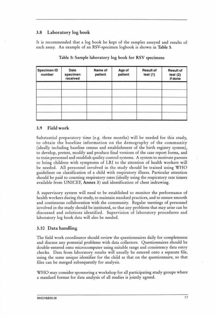
3.8 Laboratory log book
It is recommended that a log book be kept of the samples assayed and results of each assay. An example of an RSV-specimen logbook is shown in Table 5.
Table 5: Sample laboratory log book for RSV specimens
Specimen 10 Date Name of Age of Result of Result of number specimen patient patient test {1) test (2)
received if done
3. 9 F ield work
Substantial preparatory time (e.g. three months) will be needed for this study, to obtain the baseline information on the demography of the community (ideally including baseline census and establishment of the birth registry system), to develop, pretest, modify and produce final versions of the case report forms, and to train personnel and establish quality control systems. A system to motivate parents to bring child ren with symptoms of LRI to the attention of health workers will be needed. All personnel involved in the study should be trained using WHO guidelines on classification of a child with respiratory illness. Particular attention should be paid to counting respiratory rates (ideally using the respiratory rate timers available from UNICEF, Annex 3) and identification of chest indrawing.
A supervisory system will need to be established to monitor the performance of health workers during the study, to maintain standard practices, and to ensure smooth and continuous collaboration with the community. Regular meetings of personnel involved in the study should be instituted, so that any problems that may arise can be discussed and solutions identified. Supervision of laboratory procedures and laboratory log book data will also be needed.
3.10 Data handling
The field work coordinator should review the questionnaires daily for completeness and discuss any potential problems with data collectors. Questionnaires should be double-entered onto microcomputer using suitable range and consistency data entry checks. Data from laboratory results will usually be entered onto a separate file, using the same unique identifier for the child as that on the questionnaire, so that files can be merged subsequently for analysis.
WHO may consider sponsoring a workshop for all participating study groups where a standard format for data analysis of all studies is jointly agreed.
WHON&B/00.08 17

References
Anderson LJ, Parker RA, Strikas RL. Association between respiratory syncytial virus outbreaks and lower respiratory tract deaths of infants and young children. J Infect Dis 1990;161:640-646.
Bale JR. Creation of a research program to determine the etiology and epidemiology of acute respiratory disease among children in developing countries. Rev Infect Dis 1990;12 (suppl 8):S861-S866.
Berman S, Duenas A, Bedoya A, et al. Acute lower respiratory tract illness in Cali, Colombia:a two-year ambulatory study. Pediatrics 1983;71:210-218.
Borrero IH, Fajardo LP, Bedoya M, Zea A, Carmona F, de Borrero MF. Acute respiratory tract infection among a birth cohort of children from Cali, Colombia, who were studied through 17 months of age. Rev Infect Dis 1990;12 (suppl 8): S950-S9566.
Chanock RM, Kim HW, Vargosko AJ, et al. Respiratory syncytial virus. I. Virus recovery and other observations during 1960 outbreak of bronchiolitis, pneumonia, and minor respiratory diseases in children. JAMA 1961;176:647-653.
Chanock R, Chambon L, Change W, et al. WHO respiratory disease survey in children: a serological study. Bull World Health Organ 1967;37:363-369.
Cherian T, Simoes EAF, Steinhoff MC, et al. Bronchiolitis in tropical south India. A1DC 1990;144:1026-1030.
Church NR, Anas HG, Hall CB. Respiratory syncytial virus-related apnea in infants. Demographics and outcome. Am 1 Dis Child 1984;138:247-250.
Connochie KM, Hall CB, Walsh EE, Roghmann K1. Variation in severity of respiratory syncytial virus infections with subtype. J Pediatr 1990;117:52-62.
Cubie HA, Inglis 1M, Leslie EE, Edmunds AT, Totapally B. Detection of respiratory syncytial virus in acute bronchiolitis in infants. 1 Med Virol 1992;38:283-287.
Dominquez EA, Taber LH, Couch RB Comparison of rapid diagnostic techniques for respiratory syncytial and influenza A virus respiratory infections in young children. 1 Clin Microbiol 1993;31 :2286-2290.
18 Generic protocol lo examine incidence of RSV-LRJ

Dowell SF, Papic Z, Bresee 1S, Larranaga C, Mendez M, Sowell AL, Gary HE, Anderson L1, Avendano LF. Treatment of respiratory syncytial virus infection with vitamin A: a randomized placebo-controlled trial in Santiago, Chile. Pediatr Inf Dis J 1996;15:782-786.
Fisher RG, Gruber WC, Edwards KM, Reed GW, Tollefson SJ, Thompson 1M, Wright PF. Twenty years of outpatient respiratory syncytial virus infection: a framework for vaccine efficacy trials. Pediatrics 1997; 99(2):£7.
Fleming OM, Cross KW. Respiratory synctial virus or influenza? Lancet 1993;342:1507-1510.
Forgie IM, O'Neill KP, Lloyd-Evans N, et al. Etiology of acute lower respiratory tract infections in Gambian children. I. Acute lower respiratory tract infections in infants presenting at the hospital. Ped Infect Dis J 1991 ;1 0:33-41.
Gafoor A, Nomani NK, Ishaq Z, Zaidi SZ, Anwar F, Burney MI, Qureshi W, Ahmad SZ. Diagnoses of acute lower respiratory tract infections in children in Rawalpindi and Islamabad, Pakistan. Rev Infect Dis 1990;12 (suppl 8):S907-S914.
Glezen WP, Paredes A, Allison JE, Taber LH, Frank AL. Risk of respiratory syncytial virus infection for infants from low-income families in relationship to age, sex, ethnic group, and maternal antibody levels. 1 Pediatr 1981 ;98:708-715.
Groothuis JR, Simoe~ EA, Hemming VG. Respiratory syncytial virus (RSV) infection in preterm infants and the protective effects of RSV immune globulin (RSVIG). Respiratory Syncytial Virus Immune Globulin Study Group. Pediatrics 1995;95:463-467.
Hall CB, Walsh EE, Schnabel KC et al. Occurrence of groups A and B of respiratory syncytial virus over 15 years: associated epidemiologic and clinical characteristics in hospitalized and ambulatory children. J Infect Dis 1990;162: 1283-1290.
Hall CB, Douglas RG Jr. Clinically useful method for the isolation of respiratory syncytial virus. 1 Infect Dis 1975;131:1-5.
Holberg CJ, Wright AL, Martinez FD, Ray CG, Taussig LM, Lebowitz, MD. Risk factors for respiratory syncytial virus-associated lower respiratory illnesses in the first year of life. Am J Epidemic} 1991;133:1135-1151.
Horta! M, Mogdasy C, Russi 1C, Deleon C, Suarez A. Microbial agents associated with pneumonia in children from Uruguay. Rev Infect Dis 1990;12(suppl 8): S915-S922.
John TJ, Cherian T, Steinhoff MC, Simoes EAF, John M. Etiology of respiratory infections in children in tropical southern India. Rev Infect Dis 1991;13 (suppl 6):$463-$469.
King JC Jr, Burke AR, Clemens ML, Nair P, Farley JJ, Vink PE, Batlas SR Rao M, Johnson JP. Respiratory syncytial virus illnesses in human immunodeficiency virus and noninfected children. Pediatr Infect Dis J 1993;12:733-739.
WHON&B/00.08 19

Kirkwood BR. Essentials of medical statistics. Blackwell Scientific Publications, London 1988.
Mulholland EK, Simoes EAF, Costales MOD, McGrath EJ, Manalac EM, Gove S. Standardized diagnosis of pneumonia in developing countries. Pediatr Infect Dis J 1992;11:77- 81.
Murphy BR, Hall SL, Kulkarni AB, et al. An update on approaches to the development of respiratory syncytial virus (RSV) and parainfluenza virus type 3 (PIV3) vaccines. Virus Research 1994;32:13-36.
Nascimento JP, Siqueira M, Sutmoller F, et al. Longitudinal study of acute respiratory diseases in Rio de Janeiro: occurrence of respiratory viruses during four consecutive years. Rev Inst Med Trop Sao Paulo 1991 ;33:287 -296.
Nwankwo MU, Dym AM, Schuit KE, Offor E, Omene ]A. Seasonal variation in respiratory syncytial virus infections in Benin City, Nigeria. Trop Geogr Med 1988;40:309-313.
Orstavik I, Carlsen KH, Halvorsen K. Respiratory syncytial virus infections in Oslo 1972-1978. 1. Virological and epidemiological studies. Acta Paediatr Scand 1980;69:717 -722.
Orstavik I, Grandien M, Halonen P, et al. Viral diagnoses using the rapid immunofluorescence technique and epidemiological implications of acute respiratory infections among children in different European countries. Bull World Health Organ 1984;62:307-313.
Ota WK, Bang FB. A continuous study of viruses in the respiratory tract in families of a Calcutta bustee. Am J Epidemiol 1972;95:371-383.
Phillips PA, Lehmann D, Spooner V, et al. Viruses associated with acute lower respiratory tract infections in children from the Eastern highlands of Papua New Guinea (1983-1985). Southeast Asian J Trop Med Public Health 1990;21:373-382.
Schwartz B, Gove S, Lipman H, Lob-Levyt G. The etiology of acute lower respiratory tract infections among young children in developing countries. Pediatr Infect Dis], in press.
Simoes EAF. Recognizing and diagnosing pneumonia in developing countries. Curr Opin Inf Dis 1994;7:358-363.
Shann F. Etiology of severe pneumonia in children in developing countries. Pediatr Inf Dis J 1986;5:247-252.
Sung RYT, Murray HGS, Chan RCK, Davies DP, French GL. Seasonal patterns of respiratory syncytial virus infection in Hong Kong: a preliminary report. J Infect Dis 1987;156:527-528.
20 Generic protocol to examine incidence of RSV·LRI

Sutmoller F, Andrade Ferro P, Asensi MD, Ferreira V, Mazzei IS, Cunha BL. Etiology of acute respiratory tract infections among children in a combined community and hospital study in Rio de Janeiro. Clin Infect Dis 1995;20:854-860.
Suwanjutha S, C hantarojanasiri T, Watthana-Kasetr S, et al. A study of nonbacterial agents of acute lower respiratory tract infection in Thai children. Rev Infect Dis 1990; 12 (suppl 8):$923-928.
Swierkosz EM, Flanders R, Melvin L, Miller JD, Kline MW. Evaluation of the Abbott TESTPACK RSV enzyme immunoassay for detection of respiratory syncytial virus in nasopharyngeal swab specimens. J C lin Microbiol 1989;27:1151-1154.
Tupasi TE, Mangubat NV, Sunico MES, Magdangal DM, Navarro EE, Leonor ZA, Lupisan S, Medulla F, Lucero MG. Malnutrition and acute respiratory illness in Filipino children. Rev In£ Dis 1990a;12(suppl 8):S1047-S l054.
Tupasi TE, Lucero MG, Magdangal DM, et al. Etiology of acute lower respiratory tract infection in children from Alabang, metro Manila. Rev Infect Dis 1990b; 12(suppl 8):S929-S939.
Vathanophas K, Sangchai R, Raktham S, Pariyanonda A, et al. A community-based study of acute respiratory tract infection in Thai children. Rev Inf Dis 1990; 12:S957 -S965.
Waner JL, Whitehur~t NJ, Todd SJ, Shalaby H, Wall LV. Comparison of Directogen RSV with virus isolation and direct immunofluorescence for the identification of respiratory syncytial virus. J Clin Microbiol 1990;28:480-483.
Weissenbacher M, Carballal G, Avila M, et al. Etiologic and clinical evaluation of acute lower respiratory tract infections in young Argentinian children: an overview. Rev Infect Dis 1990;12(suppl 8):S889-S898.
WHO. Use of monoclonal antibodies for rapid diagnosis of respiratory viruses: Memorandum from a WHO meeting. Bull World Health Organ 1992;70:699-703.
WHO Programme for the Control of Acute Respiratory Infections. Technical bases for the WHO recommendations on the management of pneumonia in children at first-level health services. Document WHO/ ARI/91.20. Geneva: World Health Organization, 1991.
WHON&B/00.08 21

Annex 1: Management of the child with
cough or difficult breathing (adapted from WHO document WHO/ARI/94.31 )
Ask:
• How old is the child?
• Is the child coughing? For how long?
• Age 2 months up to 5 years: Is the child able to drink?
Age less than 2 months: Has the young infant stopped feeding well?
• Has the child had fever? For how long?
• Has the child had convulsions?
Look, listen: (Child must be calm)
• Count the breaths in one minute . • See if child is abnormally sleepy, or
• Look for chest indrawing. difficult to wake .
• Look and listen for stridor. • Feel for fever or low body temperature
• Look and listen for wheeze . (or measure temperature).
Is it recurrent? • Look for severe malnutrition.
Fast breathing
• Young infant aged less than 2 months: 60 breaths per minute or more .
• Child aged 2 months up to 12 months: 50 breaths per minute or more .
• Child aged 1 up to 5 years: 40 breaths per minute or more .
A child has chest lndrawing if:
• The lower chest wall goes in when the child breathes in .
• Chest indrawing is a sign of severe pneumonia in children aged 2 months up to 5 years .
• Only severe chest indrawing is a sign of severe pneumonia in young infants .
22 Generic protocol to examine ilcidence of RSV-LRI

• • • • •
-+ -+ -+ -+ -+
• • • • • •
-+ -+ -+
CLASSIFY THE ILLNESS Does the child have danger signs?
The child (age 2 months up to 5 years)
Signs:
Not able to drink,
Convulsions,
Abnormally sleepy or difficult to wake,
Stridor in calm child, or
Severe malnutrition .
Classify as very severe disease
Treatment:
Refer urgently to hospital.
Give first dose of an antibiotic.
Treat fever if present.
Treat wheezing, if present.
If cerebral malaria is possible, give an antimalarial.
The young infant (age less than 2 months)
Signs:
Stopped feeding well,
Convulsions,
Abnormally sleepy or difficult to wake,
Stridor in calm child,
Wheezing, or
Fever or low body temperature .
Classify as very severe disease
Treatment :
Refer urgently to hospital.
Keep young infant warm.
Give first dose of an antibiotic.
WHON&B/00.08 23

DOES THE CHILD HAVE PNEUMONIA?
The child (age 2 months up to 5 years)
Signs:
• Chest indrawing. • No chest indrawing, and • No chest indrawing, and
(If also recurrent • Fast breathing • No fast breathing wheezing, go directly to (50 per minute or more lf (Less than 50 per minute
• Treat wheezing, child 2 months up to 12 if child 2 months up to 12 page 28. months; 40 per minute or months; Less than 40 per
more if child 12 months up minute if child 12 months to 5 years.) up to 5 years.)
Classify as:
Severe pneumonia Pneumonia No pneumonia: cough or cold
-+ Refer urgentlyto -+ Advise mother to give -+ If coughing more than 30 hospital. home care. days, refer for
assessment.
-+ Give first dose of an -+ Give an antibiotic. -+ Assess and treat ear antibiotic. problem or sore throat, if
-+ Treat fever if present. -+ Treat fever if present. present.
-+ Treat wheezing, if -+ Treat wheezing, if present. -+ Assess and treat other present -+ Advise mother to return with problems.
(If referral is not feasible, child in 2 days for -+ Advise mother to give treat with an antibiotic reassessment, or eartier if home care. and follow closely.) the child is getting worse. -+ Treat fever, if present
-+ Treat wheezing, if present.
Reassess In 2 days a child who Is taking an antibiotic for pneumonia
Signs:
Worse The same Improving
• Not able to drink. • No change. • Breathing slower.
• Has chest indrawing. • Less fever .
• Has other danger signs . • Eating better.
Treatment:
-+ Refer urgentlyto -+ Change antibiotic or refer. -+ Finish 5 days of antibiotic. hospital.
24 Generic protocol to examine incidence of RSV·LRI

DOES THE CHILD HAVE PNEUMONIA? (continued)
The young infant (age less than 2 months)
Signs:
• Severe chest indrawing, or • No severe chest indrawing,
• Fast breathing and
(60 per minute or more). • No fast breathing (Less than 60 per minute).
Classify as:
Severe pneumonia No pneumonia: cough or cold
~ Refer urgently to ~ Advise mother to give hospital. following home care:
~ Keep young infant warm. - keep young infant warm; ~ Give first dose of an - breastfeed frequently;
antibiotic. - clear nose if it interferes with feeding.
(If referral is not feasible, ~ Retum quickly if: treat with an antibiotic and - breathing becomes difficult; follow closely.) . breathing becomes fast;
. feeding becomes a problem;
. the young infant becomes sicker . .
WHON&B/00.08 25

1\)
"' TREATMENT INSTRUCTIONS
• Give an antibiotic I Antibiotics should not be used for coughs or colds I -+ Give first dose of antibiotic in clinic.
-+ Instruct mother on how to give the antibiotic for five days at home (or return to clinic for daily procaine penicillin injection).
COTAIMOXAZOLE AMOXYCILLIN AMPICILLIN PROCAINE trimethoprim + sulphamethoxazole PENICIWN
-+ Two times daily for 5 days -+ Three times daily -+ Four times daily -+ Once daily for 5 days. for 5 days. for 5 days.
Adult tablet Pediatric tablet Syrup Tablet Syrup Tablet Syrup Age or weight single strength
(80mg trimethoprim (20mg trimethoprim (40mg trimethoprim 250mg 125mg 250mg 250mg Intramuscular +400mg + 100mg +200mg in5ml in5ml injection
sulphamethoxazole) sulphamethoxazole) sulphamethoxazole per 5ml)
Less than 2 months ( <5kg)" 1/4 .. 1 .. 2.5ml .. 1/4 2.5ml 1/2 2.5ml 200 000 units
2 months up to 12 months (6 -9kg) 1/2 2 S.Oml 1/2 S.Oml 1 5.0ml 400 000 units
12 months up to 5 years (10- 19kg) 1 3 7.5ml 1 10.0ml 1 S.Oml 800 000 units
. Give oral antibiotic for 5 days at home only if referral is not feasible . .. If the child is less than 1 month old, give 112 pediatric tablet or 1.25ml syrup twice daily. Avoid cotrimoxazole in infants less than one month of age who are premature or jaundiced.

TREATMENT INSTRUCTIONS (continued)
Advise mother to give home care (For the child age 2 months up to 5 years)'
-+ Feed the child - Feed the child during illness. - Increase feeding after illness. - Clear the nose if it interfered with feeding.
-+ Increase fluids
- Offer the child extra to drink. - Increase breast-feeding.
-+ Soothe the throat and relieve the cough with a safe remedy.
-+ Most important: In the child classified as having No Pneumonia: Cough or Cold, watch for the following signs and return quickly if they occur:
Breathing becomes difficult. }Breathing becomes fast. Child is not able to drink. Child becomes sicker.
This child may have pneumonia.
• See section on young infant in Classify the illness (page 23) for home care instructions for that age group
•
-+
TREAT FEVER
392 Centigrade or more means a HIGH FEVER
382 to 392 Centigrade means FEVER, NOT HIGH
35.52 Centigrade or less means LOW BODY TEMPERATURE
Fever is high • Fever is In a falciparum malarious area:
(~392 C). not high • Any fever, (382 - 392 C) or
• History of fever .
Give -+ Advise -+ Give an antimalarial
• Fever for
more than five days.
-+ Refer for paracetamol. mother to (or treat according to assessment.
~ give more your malaria programme fluids. recommendations).
~
Paracetamol doses: every six hours
Age or weight 1 OOmg tablet 500mg tablet
2 months up to 3 years 6 - 14kgs 1 Y-1
3 years up to 5 years 15 - 19kgs 1112 1f2
Fever alone is not a reason to give an antibiotic except in a young infant aged less than 2 months.
Treat a young infant with fever or low body temperature as very severe disease.
WHON&B/00.08 27

TREATMENT INSTRUCTIONS (continued)
TREATWHEEZJNG
Children with first episode of wheezing
If in respiratory distress -+ Give a rapid acting bronchodilator and refer If not in respiratory distress -+ Give oral salbutamol
Children with recurrent wheezing (asthma)
• Give a rapid acting bronchodilator
• Assess the child's condition 30 minutes later;
If: Then: Respiratory distress or any -+ Treat for severe pneumonia or very danger sign severe disease (refer).
No respiratory distress and:
Fast breathing -+ Treat for pneumonia Give oral salbutamol.
No fast breathing -+ Treat for no pneumonia: cough or cold. Give oral salbutamol.
Rapid acting Oral salbutamol bronchodilator three time dally for five days
Nebulized O.Sml Age or weight 2mg tablet 4mg tablet Salbutamol Salbutamol (5mg/ml) plus 2.0ml
sterile water 2 months up to % !4
Subcutaneous 12 months (<10kgs) Epinephrine 0 .01ml (adrenaline) per kg (1 :1000 = 0.1%) body weight 12 months up to 1 %
5 years (10- 19kg)
28 Generic protocol to examine Incidence of RSV-LRI

Annex 2: List of tnanufacturers of
rapid diagnostic tests for RSV
Abbott Diagnostics: Fax:
Test name:
Postfach 1303, 85003 Wiesbaden, Germany (06122) 5 81244
T ESTPACK RSV (list no. 2027), enzyme immunoassay for rapid detection of respiratory syncytial virus
Baxter-Bartels Diagnostics: PO Box 3093, Bellevue, Washington 98009, USA Tel: (1) 206-392-2992; Fax: (1) 206-392-0409
Test name:
Becton Dickinson:
Tel: Test name:
C hemicon Laboratories: Tel:
Test name:
Dako: Tel: Fax:
Test name:
Sanofi Diagnostics Pasteur: Tel: Fax:
Test name:
WHON&B/00.08
RSV ElA kit, PRIMA product line
PO Box 243, Cockeysville, Maryland 21030. USA (1) 800-638-8663
Directigen RSV
Temecula, California 92509, USA (1) 909-67 6-8080
Respiratory Syncytial Virus (Antigen Test) -DFA Kit, labeled monoclonal antibodies and control slide.
USA (1) 800-23505763 (1) 800-566-DAKO
IMAGEN Respiratory Syncytial Virus (RSV) (no. K6102 IVD)
Marnes Ia Coquette, France (33) 01 4795-6000 (33) 01 474-9133
PathFinder RSV ELISA (no. 79674)
29

Annex 3: Respiratory rate titner
SPECIFICATIONS Operating temperature Storage temperature Timing duration Timing accuracy Timing source Packaged Neck cord
COMMENTS
CODE PIS A2/01
Respiratory rate timer (ARI Timer)
COMPANY NAME AND ADDRESS UNICEF (Denmark) UNICEF Plads Freeport, D K 2100 Copenhagen Denmark Telephone: 45 35 27 35 27 Fax: 45 35 26 94 2/ Telex: 19813 unicef dk
Oto+50°C -10to+50°C 60 seconds Within 0. 1 second Crystal oscillatorof32768 Hz 40perbox Approximately I metre long
The Respiratory Rate Timer has been carefully made to provide a life span of 8 000 counting cycles of 60 seconds under normal usage. How to use the timer: - Push the Start/Stop button once. A short "beep" sound will be heard to show that the timer
is starting. Actual counting of the respiratory rate must begin as soon this initial beep is heard. Audible clicks will sound every second so that you can be sure that timing is in progress while you continue to observe the patient.
- The timer will give another short beep at the 30 second point of the timing cycle, and two short beeps at the end of 60 seconds when it will automatically shut off. The timer can be stopped at any time during the counting cycle, if needed, by pushing the Start/Stop button once.
Notes on handling: - Always store the timer in a cool, dry place after use. lf the timer is exposed to excessive
temperature and moisture, its life span will be shortened. - If it is necessary to clean the timer, use a moist, clean cloth. Do not use chemicals or
detergents as they may damage the unit.
2000 PRICES (FCA) Shipping weight I volume:
Manufacturer's minimum order:
boxof40 canon of 5 boxes ltimer
2 kg /0.008 ml JOkg/0.04 ml
Item Quantity UNICEF stock number Price
Respiratory rate timer
30
I timer box of 40 timers carton of200 timers
lruJIO~IDDDm rn11 *00066464*
0845010 0845010 0845010
US$ US$ US$
3.66 144.00 720.00
tric protocol to examine llcidence of RSV·LRI

lllllllllllllllll lllllllllllllllllllllllllllllllll *00066464*
The Department of Vaccines and Biologicals was established by the World Health Organization in 1998 to operate within the Cluster of Health Technologies and Pharmaceuticals. The Department's major goal is the achievement of a world in which all people at risk are protected against vaccine-preventable diseases.
The Department replaces the former Global Programme for Vaccines and Immunization. Five teams implement its "bench-to-bush" strategy, which starts with the establishment of norms and standards, focusing on major vaccine and technology issues, and ends with implementation and guidance for vaccination programmes. The work of the teams is outlined below.
The Quality Assurance and Safety of Biologicals Team ensures the quality and safety of vaccines and other biological medicines through the development and establishment of global norms and standards.
The Vaccine Development Team coordinates and facilitates the development of new vaccines and immunization-related technologies.
The Vaccine Assessment and Monitoring Team assesses strategies and activities for reducing morbidity and mortality caused by vaccine-preventable diseases.
The Access to Technologies Team endeavours to reduce financial and technical barriers to the introduction of new and established vaccines and immunizationrelated technologies.
The Expanding Immunization Team (EPI) develops policies and strategies for maximizing the use of vaccines of public health importance and their delivery. It supports the WHO regions and countries in acquiring the skills , competence and infrastructure needed for implementing these policies and strategies and for achieving disease control and/or elimination and eradication objectives.
For further information please contact:
Department of Vaccines and Biologicals World Health Organization • CH-1211 Geneva 27 • Switzerland
Fax: +41 22 791 4192/93 • E-mail: [email protected] or visit our web site at:
http://www.vaccines.who.int/