genetic analysis of fusion recombinants in bacillus
TRANSCRIPT

Copyright 0 1990 by the Genetics Society of America
Genetic Analysis of Fusion Recombinants in Bacillus subtilis: Function of the recE Gene
Nouzha Ftouhi’ and Nancy Guillin2 Institut de Microbiologie, Universiti Paris-Sud, 91405 Orsay Cedex, France
Manuscript received March 6, 1990 Accepted for publication July 15, 1990
ABSTRACT Bacillus subtilis protoplast fusion allows the study of the genetic recombination of an entire
procaryotic genome. Protoplasts from bacterial strains marked genetically by chromosomal mutations were fused using polyethylene glycol and the regenerated cells analyzed. Recombinants represent 19.3% of heterozygotic cells; they are haploids. Individual characterization of clones show a unique particular phenotype in each colony suggesting that recombination takes place immediately after fusion, probably before the first cellular division. Recombination occurs in the whole chromosome; in one-third of the cases both reciprocal recombinants could be shown in the colony. The genetic interval that includes the chromosome replication origin shows the highest recombination level. Our results suggest that the RecE protein accounts for most of the fused protoplast recombination; however, some “replication origin-specific” recombination events were independent of the recE gene product.
T HREE different approaches can be used to achieve genetic recombination in Bacillus sub-
tilis. (1) The natural transformation system requires the recE product gene activity and depends upon the development of a state of competence which involves the elaboration of gene products necessary for the binding and uptake of transforming DNA (DUBNAU et al. 1973; HAHN, ALBANO and DUBNAU 1987; DUBNAU 1989). It appears that the recombinant donor DNA molecule involved in the genetic exchange is con- verted to a single strand DNA intermediate before the establishment of the heterologous recombinant DNA complex (DUBNAU and DAVIDOFF-ABELSON 1971). (2) Transduction mediated by phages is also a recE dependent recombination pathway (DUBNAU et al. 1973). The recombinogenic DNA molecules are double stranded and homogeneity in the DNA modi- fication pattern is necessary to accomplish the trans- duction process (TRAUTNER et al. 1974). (3) The fu- sion of B. subtilis protoplasts allows the formation of cellular associations containing both complete paren- tal genomes (SCHAEFFER, CAMI and HOTCHKISS 1976); these segregate recombination products (reviewed by HOTCHKISS and GABOR 1985).
A complex genetic situation arises when protoplasts fuse and the resulting fusion bodies start to regenerate as a diploid bacteria. This diploid cell can survive to produce in some cases a genetic recombinant bacte-
I Present address: Dipartement de Biochimie, Universiti de Montrial, Montreal, Quebec H3C 357, Canada.
To whom correspondence should be sent at present address: Uniti de Pathoginie Microbienne Moliculaire, Institut Pasteur, 28 Rue du Docteur Roux, 75724 Paris Cedex 15, France.
Genetics 126 487-496 (November, 1990)
rium. The physiological factors implicated in the ge- netic exchange produced by fusion are completely unknown. In this paper, we present an analysis of this type of recombination. Using appropriate genetically marked strains, genetic exchanges were found in the whole chromosome. We show that recE gene product activity is involved in most of the genetic exchanges observed. However, genetic intervals that included the B. subtilis replication origin showed higher recom- bination frequencies than other similar sized regions. At least part of this excess in recombinogenic activity may be determined by recE-independent components.
MATERIALS AND METHODS
Culture media: Bacteria were generally grown in Schaef- fer’s medium (SCHAEFFER, MILLET and AUBERT 1965). Phage stocks and transduction experiments were grown in Pennassay Broth. Competence development during trans- formation and selection for auxotrophic markers were ac- complished in Spizizen minimal media supplemented as required (ANAGNOSTOPOULOS and SPIZIZEN 196 1). The pro- toplast wall regeneration media were as described by WY- RICK and ROGERS (1973) and by SANCHEZ-RIVAS (1982). Recombinants were selected by replica-plating on Spizizen minimal base medium supplemented with all the metabolites necessary for the growth of each particular recombinant; amino acids and bases were used at 40 rg/ml; rifampin and erythromycin were utilized at 5 rg/ml.
Bacterial strains and phages: Table 1 gives the charac- teristics of the strains used in this work. Figure 1 shows the map locations of relevant loci. In M0507 and M0508 the mutated recE allele is also present. These recE deficient strains were constructed using the Zahler method: PBS1- phage transduction of spectinomycin resistance marker into wild type recipient cells gave 45% coheritance with the recE mutation (unpublished results). The bacteriophage used was

488
TABLE 1
Bacterial strains and plasmids
N. Ftouhi and N. Gui1li.n
Strain or plasmid Genotype or description Origin or reference
Bacillus subtiliJ CU3497
BDlO8
BD224 QB944 S6
M0501
M0507
M0508
Plasmids pAK 1
pHV438
pHV33
trpC2 ilva2 recE4 spcB1 argA3 lys21 metB5 pheA12 purA16 rplVl thr-5 trpC2 recE4 cysA14 purA26 trpC2 thrA pyrA trpF7 rJin486
argA3 metB5 pheA.2 purA I6 trpC2 rplVl argA3 metB5 pheBl2 purA16 trpCZ recE4 rplVl spcBl thrA pyrA trpF7 recE4
rfm486 spcBl
pJH 10 1 vector with the SphI-EcoRI fragment of 8 kbp which con- tains the E . subtilis gyrA, gyrB and recF genes
pHV32 vector with a 4.4-kbp fragment contains the E. subtilis thyB gene and the X region
pBR322 and pC194 li- gated in the Hind111 site. Replicates in B. subtilis and in E. coli
DR. ZAHLER
D. DUBNAU
D. DUBNAU DEDONDER et a l . (1977) SCHAEFFER, CAMI and
HOTCHKISS (1976) This work
This work
This work
LAMPE and BOTT ( 1984)
NIAUDET, GOZE and EHRLICH (1 982)
PRIMROSE and EHR- LICH (1 98 1)
The strain M0501 was constructed by the transformation- congresion method, using QB944 DNA as donor and BD108 cells as recipient. Trypthophan deficient mutants were screened among the selected lysine proficients recombinants. The RecE- strains were constructed by PBSl transduction using the CU3497 strain as donor and the M0501 and S6 cells as recipients. The Rec deficient phenotype is coherited at 45% with the SpR marker.
the wild-type strain of PBS 1 (TAKAHACHI 196 1). Genetic transformation and transduction: Genetic trans-
formation and transduction were carried out as described by DUBNAU, DAVIDOFF-ABELSON and SMITH (1 969) and by DUBNAU and DAVIDOFF-ABELSON (1 97 1).
Fusion experiments and recombinant selection: Bacte- rial protoplast fusions were performed according to SCHAEF- FER, CAMI and HOTCHKIS (1976). Well isolated colonies growing in the wall regeneration medium were transferred with toothpicks to a rich medium (SCHAEFFER, MILLET and AUBERT 1965) and screened for recombinants by replica plating onto 16 different selective media. The same treat- ment was realized on protoplasts obtained from separate parental strains, no reversion of the genetic markers was observed. The phenotype of each recombinant was verified after purification of single cells which were screened on the same selective media. One pair of media was designed in order to visualize the reciprocal phenotypes formed by the genetic exchange into the intervals 1 and 10. These phe- noty es are: Rifamycin0-Adenine- and Erythromicin@-Ade- nine .
For each recombinant, we were able to score the number P
T e r .....................................................................................
INTERVAL GENETIC DISTANCE SIZE PERCENTAGE OF
MARKERS (Degrees) (kbp) CO-TRANSDUCITON
1 purA - rpoB 24 239
2 rpoB - W' 0.01 1
I .7
99
3 r p N - pyrA 127 1265 ND
4 pyrA - m t B 58 578 ND
5 merB - rrpF 6 60 31.5
6 trpF - rrpC 0.01 1 99
7 trpC - pheA 37 369 ND
8 pheA - argA 20 1 9 9 6.8
9 ar8A - thrA 24 239 1.7
10 thrA - purA 64 638 ND
.................................................................................................................
FIGURE 1.-Genetic intervals used in this work. Ten intervals are defined by genetic markers. Their size was calculated taking into account the recombinant frequencies obtained by transduction and utilized to propose the genetic map of B. subtilis (HENNER and HOCK 1980; PIGCOT 1989). The B. subtilis chromosome has an estimated size of 5500 kbp organized in 360". Using the conversion graph established by HENNER and HOCK (1 980); one degree of the map is equivalent to 9.96 kbp. Employing this factor; we converted the relative genetic distance in degrees to kbp. Taking into account the imprecision of linkage values derived from PBSl transduction these data represent an approximate physical distance. The two chromosomes (one of each parental strain fused) are represented by two circles (the sizes of which are drawn unequal for conven- ience). Inner circle corresponds to S6 strain and outside circle to M0501 strain. The origin of DNA replication is located in the interval 1 and the terminus in the interval 4. argA, arginine; metB, methionine; pheB, phenylalanine; p y A , uracil; purA, adenine; rplV, erythromycin resistance; rpoB, rifampin resistance; thrA, threonine; trpC and trpF, tryptophan; NL, markers nonlinked.
of genetic exchanges promoting a specific phenotype to each interval by detection of different growth on specific selective media. In this manner, the number of genetic exchanges in each recombinant and subsequently in the population were totalled. The frequency of recombination in a particular interval is calculated from the ratio of ex- changes in a given interval to the total exchanges in the recombinant population. The observed recombination fre- quencies from different experiments were pooled after ver- ifying that they satisfied a standard x* test with a 5% confidence limit and 12 degrees of freedom; theoretical = 2 1,06.
Chromosomal and plasmid DNA purification: Chro-

Fusion Recombination in B. subtilis 489
mosomal DNA purification from B. subtilis cells was realized according to SAUNDERS et al. (1984). Plasmid DNA was purified: (1) from Escherichia coli according to MANIATIS, FRITSCH and SAMBROOK (1982) and (2) from B. subtilis according to GUERRY, LEBLANC and FALKOW (1973).
Southern blots and hybridization: After endonuclease DNA digestions, the different DNA fragments were sepa- rated in a 0.8% agarose gel then transferred onto Hybond N-Nylon (Amersham catalog No. RPN2020N) according to SOUTHERN (1975). A probe was made from DNA of pAKl plasmid 32P-radiolabeled by the Multiprimer kit (Amersham catalog No. RPN 160 1 Z). Hybridization and washing con- ditions were done as described previously (GUILL~N, AMAR and HIRSCHBEIN 1985).
RESULTS
Fusion product analysis: Diploids created by poly- ethylene glycol (PEG) treatment of B. subtilis proto- plasts produce recombinants which are found among the primary regenerant cells (SCHAEFFER, CAMI and HOTCHKISS 1976) and among the segregation progeny produced during the growth of the unstable noncom- plementing diploids (Ncd) (HOTCHKISS and GABOR 1980; GABOR and HOTCHKISS 1983). However, no extensive analysis had yet been made of primary re- combinants resulting from PEG protoplast fusion. Nevertheless, in previous work the phenotypic analysis of the recombinant progeny from Ncd or proto- trophic diploid clones suggest a stimulation of genetic exchange into the intervals that include the origin or the terminus of DNA replication (SANCHEZ-RIVAS et al. 1982; GABOR and HOTCHKISS 1983; L~vI-MEY- RUEIS and SANCHEZ-RIVAS 1984).
We constructed an appropriate strain (M0501) to evaluate in the same protoplast fusion experiment with the S6 strain, the genetic recombination frequen- cies in different regions of the bacterial chromosome in primary recombinants. The position and size of the ten genetic intervals that we tested is shown in Figure 1 . The smallest of these corresponds to 1 kb (interval 6) and the largest to 1265 kb (interval 3). Protoplasts of strains M0501 and S6 were fused and the recom- binant phenotypes of the regenerated cells deter- mined. The values in Table 2 summarize the results of 1244 exfusant (3 independent crosses). These show that colonies with a recombinant phenotype appear with a percentage of 19.3%. In these fusion crosses, the bacteria with a biparental phenotype represent 5.7, 4.9, and 2.7 percent of the total exfusant popu- lation. They generate noncomplementing diploids (Ncd) in which only one chromosome is still expressed after several generations of growth (HOTCHKISS and GABOR 1980; GUILL~N, AMAR and HIRSCHBEIN 1985). In one cross (11), we found that after two passages on selective media (as described in MATERIALS AND METH- ODS), 8% of exfusant colonies conserved the diploid state, producing progeny with a prototrophic pheno- type. Such clones interpreted as complementing dip-
TABLE 2
Fusion crosses of strains MO 501 and s6
Fusion No' Of colonies Observed phenotypes
cross analyzed Biparental Parentals ::\, I 520 114 30 376 0
11 364 71 18 243 32
111 360 60 10 290 0
1244 245 909
21.9% 5.7% 72.3%
19.5% 4.9% 66.7% 8.7%
16.6% 2.7% 80.5%
The fusion crosses between S6 and M0501 strains were per- formed according to SCHAEFFER, CAMI and HOTCHKISS (1976). Isolated wall regenerated colonies were grown in rich medium and subsequently replica plated on the different selective media. In a similar manner 100 of wall-regenerated colonies obtained from treatment of parental strains were analyzed; nonreversion of genetic markers was observed.
loids were not further studied. The remaining exfu- sant population corresponded to the parental pheno- types in similar proportions. The addition of different diploid and recombinant phenotypes revealed that at least 30% of regenerated cells corresponded to het- erozygotic clones.
Genetic recombinant in the whole B. subtilis chro- mosome: We scored each type of genetic exchange occurring in a particular recombinant class according to their nutritional requirements. Among 245 stable recombinants recovered in the three independent ex- periments a total of 694 genetic exchanges were scored, which produced 48 different phenotypes.
Even if the minimal number of exchanges necessary to produce a stable recombinant phenotype is as- sumed; we are forced to conclude that in order to obtain certain complex phenotypes multiple ex- changes were necessary. Therefore, our data below suggest that multiple exchanges are frequent in the diploid cells. These are shown in Table 3. We found 156 recombinant colonies with a minimum of 2 cross- overs, 81 with a minimum of 4, 3 with 6, and 5 with 8 genetic exchanges. The stability of the recombinant phenotype was verified by growth in nonselective conditions followed by a supplementary phenotype analysis of these colonies on the different selective media.
The frequency of recombination in each genetic interval was calculated. The results show that recom- bination takes place along the whole B. subtilis chro- mosome (Table 3).
The statistical x' treatment of the individual recom- binant frequency values observed in each interval permit them to be considered as belonging to the same normal distribution. Thus, the B. subtilis chro- mosome genetic recombination values measured by fusion of protoplasts are quite reproducible (Table 3 and Figure 2). Any interpretation of the above data

490 N. Ftouhi and N. Guillin
TABLE 3
Genetic exchanges distribution on the B. subtilis chromosome
Fusion I + I 1 + 111
No. of genetic
Percent
Interval crossover
exchanges Percentage by kbp
1 246 35.4 0.14 2 108 15.5 1.55 3 34 4.8 0.003 4 96 13.8 0.02 5 5 0.7 0.01 1 6 4 0.57 0.57 7 6 0.86 0.002 8 13 1.8 0.009 9 86 12.39 0.05
10 96 13.8 0.02
Crossovers 694
Recombinants 245
Colonies with 2 crossovers 156 4 crossovers 81 6 crossovers 3 8 crossovers 5
Total phenotypes 48
Analyzed colonies 1244
The results represent the values of three independent experi- ments. From each experiment the number of recombinants was scored and the different crossovers were determinated in each recombinant. The total number of crossovers among the recombi- uant population corresponds to 100% recombination. According to MATERIALS AND METHODS, a x' statistical analysis of the data permits grouping of recombination frequencies values in each interval. The practical x' found was 13.5.
should take into account the physical size of each genetic interval since a deformation of the results should be created by the fact that for long intervals all even and all odd numbers of genetic exchanges will give the same phenotype, thus resulting in an underestimate of the recombinant frequency in such an interval. Moreover, comparison of the rates of recombination in DNA fragments that have similar sizes, confirms that the highest recombination fre- quency appears in intervals 1, 2, 9 and 10 (1.7 X 10' kbp of DNA with 8 genetic markers tested) which show 77% of the total recombination events.
Equivalent sized region within intervals 4, 5, 6, and 7 (1.71 X I O 3 kbp, 8 genetic markers tested) show a recombination rate of 16%. By this analysis, we can conclude that the genetic recombination obtained in diploids cells of B. subtilis in the absence of any selec- tive growth pressure is enhanced in the regions that include the replication origin of the chromosome.
Comparison of recombination frequencies ob- tained by protoplast fusion with those obtained by transduction: A comparison of recombination fre- quencies obtained by the two genetic systems that involve exchange between two double stranded DNA molecules was made. No extensive analysis is available
m Ori * 4 0 1 T
X 9 1 0 1 2 3 4 5 6 7
GENETIC INTERVALS
Inl
x 9 1 0 1 2 3 4 5 6 7
GENETIC INTERVALS
FIGURE 2.-Mode of chromosomal recombination during pro- toplasts fusion crosses. (A) The percentage of recombination in each interval was calculated from the number of exchanges in the interval divided by the total exchanges among the recombinant population. B. subtilis chromosome is represented in a linear form arbitrarily interrupted in interval 7. (B) The recombination frequencies ob- tained by protoplast fusion (Table 3, dark bars) and by transduction (Figure 1, light bars) are compared.
to elaborate a genetic map by fusion of protoplasts, so the conversion of cell-fusion recombination frequency between independent markers to genetic distance is not possible. However, genetic distances obtained by transduction can be transformed into recombination frequencies since the relationship used to constructed the B. subtilis map was previously determined (HEN- NER and HOCH 1980). In Figure 1, the position and size of each genetic interval in the B. subtilis chromo- some is shown. In addition, following the rules of construction of the B. subtilis genetic map, the trans- duction recombination frequency in intervals 1, 2, 5 , 6, 8 and 9 was calculated. The other genetic intervals used in this work are not suited to this kind of treat- ment; the sizes of these DNA regions are higher than the maximal size transduced by PBSl phage (275 kbp) utilized to construct the map and, by definition, no genetic linkage is possible between markers separated more than 275 kbp.
The recombination pattern visualized by protoplast

Fusion Recombination in B. subtilis
fusion is quite different from that observed by trans- duction, as shown in Figure 2B. The two sets of values do not match; in transduction, the recombination between close markers is significantly higher (intervals 2, 5 and 6), while in fused protoplasts the frequency of these phenomena seems to be influenced by other factors than the genetic distance between two partic- ular markers. Hence the hypothesis of hotspots of cell- fusion recombination is proposed.
Reciprocity of recombination by protoplast fu- sion: The phenotypic analysis of fusion products by subculturing of diploid cells had shown the presence of recombinants with reciprocal phenotype (GABOR and HOTCHKISS 1983).
During the performance of the experiments re- ported above, two media were used to evaluate the eventual reciprocity of genetic recombination con- cerning intervals 1 and 10. In this analysis, it was observed that only 15 colonies out of 1244 generate a mixture of two recombinant phenotypes and grow on the two media that allow visualizing the reciprocal phenotype. These are Ade-, Thr-, Uri-, Trp-, R i p and Met-, Trp-, Phe-, Arg- Ery@. They correspond to only one-third of the recombinants produced by the genetic exchange within these intervals. These results suggest that the reciprocality of recombination take place in one part of genetic exchanges observed in the origin of replication area. In addition, they support the first observation reported by GABOR and HOTCHKISS (1983) in which reciprocal recombination was observed in several regions of the chromosome.
Genetic exchanges in fused protoplasts after a selective pressure: In order to confirm our earlier results and to exclude the possibility of preferential growth of recombinants caused by exchanges in the origin area, an analysis of genetic recombination in exfusant clones growing in a selective medium was done. The desired type of genetic exchange giving the selected phenotype is imposed by the selection. According to the earlier results, the simplest hypoth- esis is that the second genetic exchange necessary for the formation of a stable recombinant occurs, for most recombinants, in the region of the replication origin. Tryptophan+ recombinants were consequently se- lected after fusion of M0501 and S6 strains in a minimal regeneration medium (this phenotype in- volves an exchange into interval 6).
among the regenerated protoplasts. From the pheno- typic analysis of the 75 Trp+ recombinants obtained, 4 17 genetic exchanges were scored. The comparison of the recombination rates in different intervals show that 70% of the recombinational events appear in intervals 1, 2, 9 and 10 (Figure 3). Therefore, 54.9% of genetic exchanges in the Trp+ recombinants oc- curred in the origin region (intervals 1, 2 and 10). A
The frequency of Trp+ recombinants is 2.5 X
20
10
0 8 9 1 0 1 2 3 4 5 6 7
GENETIC INTERVALS
rplV argA metE
pheEI M0501 I I t r p c
7 6
49 1
S6 I 1
thrA p W I rpoE
trpF
FIGURE 3.-Mode of chromosome recombination after selection of Trp+ recombinants. PEG-treated protoplasts (SCHAEFFER, CAMI and HOTCHKISS 1976) were plated on selective regeneration me- dium (SANCHEZ-RIVAS 1982) without tryptophane. The Trp+ phe- notype was verified in 75 recombinants; these were replicated on selective media to determine their complete phenotype. In these experiments recombinants with 1 1 different phenotypes were re- covered (a total of 342 events of recombination). N o reversion of Trp- phenotype was observed when 2.5 X IO' protoplasts of each parental strain were plated separate on the regeneration medium without tryptophan. (A) The percentage of recombination in each interval was calculated as described in MATERIALS AND METHODS. (B) Scheme of the cross. Only the mutant alleles are represented. The cross line points out the desired genetic exchange.
stimulation was also observed in interval 4, close to the selected exchange: 21.6% of recombination was localized in this DNA region which contains the ter- minus of chromosome replication. Thus, the conclu- sion reached is that the second exchange necessary for the production of these Trp+ recombinants oc- curred in the most part of them in the origin area. In this manner, we can exclude the possibility of trivial artifacts entering the phenotypic analysis in fusion crosses.
Protoplast fusions of recE mutant strains: The recE mutants of B. subtilis are inhibited in all known homologous recombination pathways. In order to de- termine the influence of the recE gene product in the recombination obtained by protoplast fusion, the recE4 mutation was introduced into the same strains used for the recombination experiments. The result- ing strains (M0507 and M0508) were, as expected, recombination deficient, (both transduction and trans- formation were decreased by at least lo', data not shown). The PEG treatment and regeneration of pro-
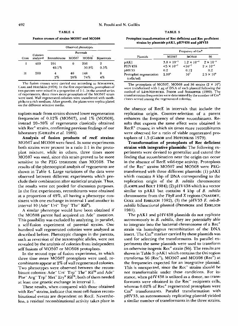
492 N. Ftouhi and N. Gui1li.n
TABLE 4
Fusion crosses of strains M0507 and M0508
Observed phenotypes
Parentals Colonies
Cross analyzed Recombinants M0507 M0508 Biparentals
1 409 20 1 0 206 2 49.1% 50.9% 0.5%
11 200 4 40 148 8 2% 20% 74% 4%
The fusion crosses were carried out according to SCHAEFFER, CAMI and Hotchkiss (1 976). In the first experiments, protoplasts of two parents were mixed in a proportion of 1 : 1. In the second series of experiments, three times more protoplasts of the M0507 strain were used. Wall regenerated colonies were transferred with tooth- picks to a rich medium. After growth, the plates were replica plated on the different selective media.
toplasts made from strains showed lower regeneration frequencies of 0.12% (M0507), and 1% (MOSOS), instead 20-30% of regenerants classically obtained with Rec+ strains, confirming previous findings of our laboratory (GUILLEN et al. 1986).
Analysis of fusion products of recE strains: M0507 and M0508 were fused. In some experiments both strains were present in a ratio 1:l in the proto- plast mixture, while in others, three times more M0507 was used, since this strain proved to be more sensitive to the PEG treatment than M0508. The results of the phenotypical analysis of regenerants are shown in Table 4. Large variations of the data were observed between different experiments which pre- clude their combination after statistical analysis. Thus, the results were not pooled for discussion purposes. In the first experiments, recombinants were obtained at a proportion of 49%, all having a phenotype con- sistent with one exchange in interval 1 and another in interval 10 (Ade- Uri- Trp- Thr- Rip).
A similar phenotype would have been obtained if the M0508 parent had acquired an Ade- mutation. This possibility was excluded by analyzing, in parallel, a self-fusion experiment of parental strains. One hundred wall regenerated colonies were analyzed as described before. Phenotypic changes in the parents, such as reversion of the auxotrophic alleles, were not revealed by the analysis of colonies from independent self fusions of M0507 or M0508 protoplasts.
In the second type of fusion experiment, in which three time more M0507 protoplasts were used, re- combinants appear at 2% of wall regenerated colonies. Two phenotypes were observed between the recom- binant colonies: Ade- Uri- Trp- Thr- Rif@ and Ade- Phe- Arg- Trp- Met- Ery@ Rif@; both of them needed at least one genetic exchange in interval 1.
These results, when compared with those obtained with Rec+ strains, indicate that most cell-fusion recom- binational events are dependent on RecE. Neverthe- less, a residual recombinational activity takes place in
TABLE 5
Protoplast transformation of Rec deficient and Rec proficient strains by plasmids pAK1, pHV438 and pHV33
Frequency of Cm'
Plasmids M0507 M0508 S6
pAK 1 3.8 x 1.2 X 10-4 2 X 1 0 - ~ PHV438 c5 X 10-7 ~ 1 0 - 7 2 X 1 0 - ~ pHV33 0.2 0.12 0.2 Protoplast regeneration 2.1 O6 1 o7 2.5 x loH
(cells/ml)
The protoplasts of M0507, M0508 and S6 strains (2 X 10") were transformed with 1 pg of DNA of each plasmid following the method of Lh-MEYRUEIS, FODOR and SCHAEFFER (1980). The transformation frequencies were determined by the number of CmR clones scored among the regenerated colonies.
the absence of RecE in intervals that include the replication origin. Counter-selection of a parent enhances the frequency of these recombinants. Re- sults that express the same effect were obtained in RecE+ crosses; in which six times more recombinants were observed for a ratio of viable regenerated pro- toplasts of 1.5 (GABOR and HOTCHKISS 1979).
Transformation of protoplasts of Rec deficient strains with integrative plasmids: The following ex- periments were devised to generalize and extend the finding that recombination near the origin can occur in the absence of RecE wild-type activity. Protoplasts of the Rec- strains M0507 and M0508 were PEG- transformed with three different plasmids: (1) pAKl which contains 8 kbp of DNA corresponding to the replication origin of the B. subtilis chromosome (LAMPE and BOLT 1984); (2) pHV438 which is a vector similar to pAKl but contains 4 kbp of B. subtilis chromosome from the ThyB and X regions (NIAUDET, GOZE and EHRLICH 1982), (3) the pHV33 E. coli-B- subtilis bifunctional plasmid (PRIMROSE and EHRLICH 198 1).
The pAKl and pHV438 plasmids do not replicate autonomously in B. subtilis, they are potentially able to integrate into the bacterial chromosome of a Rec+ strain via homologous recombination of the DNA insert. The Cm@ marker carried by these plasmids was used for selecting the transformants. In parallel ex- periments the same plasmids were used to transform an otherwise isogenic Rec+ strain (S6). The results are shown in Table 5: pAKl which contains the Orz region transforms S6 (Ret+), M0507 and M0508 (Rec-) at the frequencies expected for an integrative plasmid. This is unexpected, since the Rec- strains should be not transformable under these conditions. For in- stance, when pHV438 is utilized as a donor, no trans- formants were obtained in the Rec- recipients cells, whereas 0.02% of Rec+ regenerated protoplasts were Cm@ transformants. Parallel transformation with pHV33, an autonomously replicating plasmid yielded a similar number of transformants in the three strains.

Fusion Recombination in B. subtilis 493
A R C D E F G
kbp
f 1.3
f 2.9
e 2.5
SP \ I " P P , R
E P P
I I
111
4 L A r- b
43114 254X ZYX5 7367 25431 3825 (bp)
FIGURE 4.-Southern blot analysis of DNA from RecE-independ- em E. subtilis origin recombinant strains. Two micrograms of each DNA were digested by the Pstl endonuclease, electrophoresed in an agarose gel, blotted and probed with pAKl DNA plasmid. (I) A, M0.508 (Rec-); B, SGpAKl (Ret+); C, recombinant 1; D, recombi- nant 2; E, recombinant 3; F, recombinant 4; G , pAKl plasmid. (11) Restriction map of the Ori region on the B. subtilis chromosome. ( 1 1 1 ) Physical map of the Ori region after integration of the pAKl plasmid. B, BamHl; E, EcoRl, P, Pstl; S, Sall; Sp, SphI.
Genomic blot analysis of the Rex' Cm" recombi- nants: Studying the replication origin region of the B. subtilis chromosome an 8-kbp Ori-specific DNA fragment was cloned producing the pAKl plasmid (LAMPE and Born 1984). Since no autonomous rep licative capacities had been shown for pAKl plasmid, the results described above indicate the chromosomal integration of pAKl by a recE independent mecha- nism. In order to test this hypothesis, DNA purified from the Rec- recombinants strains and from a Rec+ recombinant was analyzed by Southern blot. The hy- bridization pattern of the PstI restricted DNA from the Cm@ recombinant cells with the pAKl labeled plasmid is shown in Figure 4.
The PstI restriction pattern of DNA obtained from Rec- Cm@ recombinants 1 and 2 is equivalent to those of the DNA from a Rec+ Cm@ recombinant; some differences could be relevant to the hybridization intensity of 4.4- and 3.8-kbp fragments, as a less
intense signal is observed with the Rec- DNA prepa- rations. Nevertheless, the Cm@ phenotype seems to occur by integration into the chromosome of pAK1, which is conducive to a duplication of the specific cloned DNA origin. A similar mode of recombination in these three strains seems to have occurred. For instance, two DNA deletions have appeared, in all of them the expected 7.3-kbp fragment appears as 6.5- kbp and the expected 2.9-kbp fragment as a 2.3-kbp DNA fragment. These DNA fragments contain the recF or the gyrA gene previously identified in the BS5 and BS6 DNA fragments that are the first to be replicated in B. subtilis (LEVINE et al. 1987). Con- versely, DNA patterns from recombinant 4 (Rec- Cm@) and from Rec- parental strain are the same. Finally, the DNA profile from recombinant 3 (Rec- Cm? is heterogeneous; probably a mixture of cells containing integrated plasmid with cells without plas- mid has been recovered.
The general view of the performed genomic analy- sis suggests that in some cases pAKl is able to be incorporated into the chromosome by a recE inde- pendent mechanism. However, the persistent Cm@ phenotype even in cells that do not show DNA inte- gration supports the existence of some free plasmid. Moreover, a phenotypic instability of the Cm@ marker carried by pAKl was observed when the recombinants were grown over 4 cellular generations without chlor- amphenicol. The loss of the Cm@ marker seems to coincide with the excision of DNA sequences carrying the cut gene, since a replicative plasmid can be re- covered in competent E. coli cells by use of DNA minipreparations from cultures grown in presence of chloramphenicol of the four Rec- recombinants stud- ied above (results not shown).
DISCUSSION
Homologous recombinant by protoplasts fusion: When B. subtilis protoplasts were fused by PEG treat- ment, heterozygous cells formed. This raised the pos- sibility of using fusion as a bidirectional way to ex- change genetic material in bacteria. Among the ex- fusants, haploid recombinants appeared at a frequency of 19.3%. Most of the recombinants were produced by two genetic exchanges between the pa- rental chromosomes. The proportion of recombinants produced by four crossovers was high (3.3%); those with six or more crossovers were rare. Nevertheless, the majority of colonies consisted of a single recom- binant genotype, although some contained both recip rocal phenotypes. Thus a single recombination event, presumably before any genome replication or/and cellular division, is the most common occurrence in fused protoplasts.
In fusion crosses the existence of diploid clones in which an entire chromosome is unexpressed (Ncd)

494 N. Ftouhi and N. Gui1li.n
was previously demonstrated. Unstable Ncd clones segregate during growth in nonselective medium in a phenotypically mixed population (HOTCHKISS and GA- BOR 1980). In addition, stabilization of these clones led to a homogeneous phenotype of bacterial popula- tion (GUILL~N, AMAR and HIRSCHBEIN 1985). The influence of these phenomena on the analysis in this study appears to be minor. In fact, all the recombi- nants subject to our study are phenotypically stable as judged by the absence of segregation of new pheno- types during their growth in rich medium. Neverthe- less, it is possible that among recombinants some stable Ncds were present. In this case, the chromosome inactivation had occured after genetic recombination since the silent chromosome in Ncds is devoid of recombination capacities (FTOUHI 1989).
Reciprocal recombination: Pairs of reciprocal re- combinants were found in a third of the population of recombinants having crossovers in intervals 1 and 10. They were the only reciprocal phenotypes searched for in these experiments. The production of reciprocal recombinants was first observed by GABOR and HOTCHKISS (1983). Nevertheless, in other inde- pendent crosses, reciprocal recombinants were not recovered (SANCHEZ-RIVAS et al. 1982). The finding implying that bacteria can display reciprocal cross- overs, was confirmed in our experiments. The recip- rocal recombinants are the products of genetic ex- change between two replicons that are entirely con- served after recombination and independently segregated into daughter cells. However, this pathway is not the usual fate of the exfusant bacteria. In fact, in most of the recombinant population, reciprocal recombination was not observed, indicating that other recombination pathways function in fused protoplasts.
High frequency of recombination in the chromo- somal replication origin: The recombinants analyzed in this work showed multiple genetic exchanges in different areas of the bacterial chromosome (Fig 2). Comparison of recombination frequencies in chro- mosomal regions equivalent in size show that the most important recombinogenic activity appears in the in- tervals 1 ,2 ,9 and 10. More frequent exchanges were found near the origin of chromosome replication (in- terval 1). This observation permits a clarification of a controversial point raised by previous work in which recombinants obtained after selection or during the growth of noncomplementing diploids were analyzed. In independent crosses, preferential occurrence of crossovers at sites close to the replication terminus was sometimes noted, while at other times preference was for sites near the origin of replication (SANCHEZ- RIVAS et al. 1982; GABOR and HOTCHKISS 1983). The scarceness of markers, the heterogeneity of the crosses or the small number of recombinants recovered could explain this variability. More homogeneous results
were obtained in this work in which we studied a large number of recombinants from the same cross. Our results reveal that the genetic interval bearing the replication origin of the chromosome has a high re- combinogenic activity and that a second area with an elevated recombination rate corresponds to a genetic interval containing the terminus of replication.
Diverse biological activities are classically rational- ized by the physical association of the DNA with cell envelope components. For example, a transformation assay demonstrated that genetic markers near the replication origin and terminus are stably associated with membrane fragments and wall material (YAMA- GUCHI and YOSHIKAWA 1973; HOROWITZ et al. 1979). It may be supposed that during protoplast fusion, these attachments enhance genetic recombination. In addition, it is plausible to imagine that the DNA replication activity by itself stimulates recombination in these chromosome areas. Generation of single stranded DNA at the replication origin and resolution of replicated molecules at the terminus involves reac- tions like DNA cutting, helix destabilization and DNA ligation; these enzymatic activities could enhance chromosome recombination. The possibility that DNA replication has a particular role in promoting recombination has been raised in the case of B. subtilis strains carrying an integrated copy of plasmid pE 194 between directly repeated DNA sequences (NOIROT, PETIT and EHRLICH 1987). When plasmid replication is induced, recombination between the repeated se- quences is stimulated by up to 450 times.
In the protoplast fusion system, the influence of the DNA replication activity was also invoked to explain the nonreciprocality observed in the majority of ge- netic exchanges. This hypothesis proposes an asyn- chronous DNA replication of chromosomes during the diploid state followed by a non-asymmetric chro- mosomes segregation. Thus, the reciprocal recombi- nant chromosome replicated with delay should by lost during segregation (HOTCHKISS and GABOR 1985).
RecE protein activity in recombination by proto- plast fusion: The RecA protein accounts for most of the homologous recombination in E. coli . One known activity of this protein is that it makes heteroduplexes between DNA molecules carrying long patches of sequence homology. This ATP dependent enzymatic reaction can form Holliday junctions which are the putative structural intermediate complexes leading to homologous recombination (for a review; SMITH 1988). Although the mechanism of recombination in bacteria other than E . coli are poorly understood, counterparts of the RecA protein have been found in several species. Much evidence indicates that the recE gene in B. subtilis encodes a protein equivalent to E . coli RecA. In vitro, the RecE protein enhances DNA strand

Fusion Recombination in B. subtilis 495
exchange in the presence of ATP (LOVETT and ROB- ERTS 1985). In vivo, RecE protein is required for the formation of the DNA donor-recipient complex dur- ing genetic recombination (DUBNAU et al. 1973). The cloned B. subtilis recE gene restores the SOS response and homologous recombination in the recE mutant cells; its product is a 45-kD protein (MARRERO and YASBIN 1988).
One of the main aims of this work was to determine the role of the RecE protein in recombination using the protoplast fusion system. These findings show that RecE is essential to accomplish recombination through most of the B. subtilis chromosome. By crossing mu- tants affected in this protein activity, a total inhibition of genetic exchange was observed in most genetic intervals. One exception was found; recE-independent recombinational activity was observed in chromo- somal areas encompassing the origin of replication. This observation was reinforced by results obtained from experiments using an origin-region-specific plas- mid to transform Rec deficient strains by the proto- plast fusion method. The integration of the Ori se- quences into the chromosome by a similar pathway to that used in Rec proficient strains was observed. A unique recombinant DNA structure similar to those found with the RecE proficient strains was obtained.
The origin region is particularly sensitive to recom- bination during protoplast fusion of B. subtilis. Work conducted by other groups had show that DNA am- plification in the chromosome origin area occurs when Rec+ cells were protoplasted; these chromosome re- arrangements generate a tetracycline resistant phe- notype (WILSON and MORGAN 1985; SHISHIDO et al. 1988). Recently, it has been shown that the amplifi- cation of a DNA fragment close to the replication origin is required for the expression of tetracycline resistance (IVES and BOTT 1989).
The general overview of these results is that by protoplastization of B. subtilis, diverse genetic ex- changes can be produced. When the fusion is stimu- lated by treatment with PEG, pairs of replicons car- rying the DNA at or near the replication origin can recombine even in the absence of a functional RecE protein. The role of DNA replication (during or after wall regeneration) in such recombination remains to be discovered. Nevertheless, it is reasonable to sup- pose that it could be mediated by one or more enzymes normally involved in the initiation of DNA replica- tion.
The ability of the Ori sequences to recombine in- dependently of the RecE gene product seems to be a more general fact in bacteria. This is also the case of plasmids carrying E. coli OriC, which in the absence of the RecA activity are able to integrate unstably into the chromosome (MASTERS, ANDRESDOTTIR and WOLF-WATZ 1978). It seems that the RecE activity is
necessary for the stable maintenance of the pAKl integrated sequences. This observation raises the questions of the role of the RecE activity as a regulator of the Ori specific recombination or rather as a part of the control of the initiation of chromosomal repli- cation. There are some observations indicating that in recA mutants of E. coli chromosomal replication is perturbed. It was suggested that the absence of RecA activity in E. coli causes initiation of newly formed replication forks to stall. DNA is accumulated in such a way that RecA deficient cells contain more DNA than wild-type strains growing at the same rate (SKAR- STAD and BOYE 1988). Genetic recombination could result in this case from the overproduction of repli- cating DNA. This hypothesis needs to be tested.
We are grateful to A N D R ~ BERKALOFF and LUISA HIRSCHBEIN who permitted us the use of their respective laboratories at various stages of this work. We wish also to thank DAVID THALER for suggesting the integrating plasmid experiment and for encourage- ment during the course of this work; KEN BOTT for the gift of tht pAKl plasmid and OSCAR REYES for his useful criticism of the manuscript. N. F. was financially supported by a fellowship of the Maroc Government and by a grant of the U.N.E.S.C.O. to L. HIRSCHBEIN. This study was supported by grants from the French Centre National de la Recherche Scientifique (URA 1354).
LITERATURE CITED ANAGNOSTOPOULOS C., and J. SPIZIZEN, 1961 Requirements for
transformation in Bacillus subtilis. J. Bacteriol. 81: 741-746. DEWNDER R. A., J. A. LEPESANT, A. LEPESANT-KEJZLAROVA, A.
BILLAUT, M. STEINMETZ and F. KUNST, 1977 Construction of a kit of reference strains for rapid genetic mapping in Bacillus subtilis 168. Appl. Env. Microbiol. 33: 989-993.
DUBNAU, D., 1989 The competence regulon of Bacillus subtilis, pp. 147-166 in Regulation of Procaryotic Development: A Struc- tural and Functional Analysis of Bacterial Sporulation and Ger- mination, edited by I . SMITH, R. A. SLEPECKY and P. SETLOW. America1 Society for Microbiology, Washington, D.C.
DUBNAU, D., and R. DAVIDOFF-ABELSON, 1971 Fate of transform- ing DNA following uptake by competent Bacillus subtilis. I. Formation and properties of donor-recipient complex. J. Bac- teriol. 117: 488-493.
DUBNAU, D., R. DAVIWFF-ABELSON and I . SMITH, 1969 Transformation and transduction in Bacillus subtilis: evidence for separate modes of recombinant formation. J. Mol. Biol. 45: 155-179.
DUBNAU, D., R. DAVIDOFF-ABELSON, B. SCHER and C. CIRIGLIANO, 1973 Fate of transforming deoxyribonucleic acid after uptake by competent Bacillus subtilis: phenotypic characterization of radiation sensitive recombination deficient mutants. J. Bacte- rial. 114 273-286.
FTOUHI, N., 1989 Etude de la recombinaison ginitique par fusion de protoplastes de Bacillus subtilis. Thke de Doctorat es- Sciences, Universiti Paris Sud, France.
GABOR, M., and R. D. HOTCHKISS, 1979 Parameters governing bacterial regeneration and genetic recombination after fusion of Bacillus subtilis protoplasts. J. Bacteriol. 137: 1346-1 353.
GABOR, M., and R. D. HOTCHKISS, 1983 Reciprocal and non reciprocal recombination in diploid clones from Bacillus subtilis protoplasts fusion: association with the replication origin and terminus. Proc. Natl. Acad. Sci. USA 8 0 1426-1430.
GUERRY, P., D. J. LE BLANC and S. FALKOW, 1973 General method for the isolation of plasmid deoxyribonucleic acid. J. Bacteriol. 116: 1064-1066.

496 N. Ftouhi and N. Guillin
GUILL~N, N., M. AMAR and L. HIRSCHBEIN, 1985 Stabilized non- complementing diploids (Ncd) from fused protoplast products of Bacillus subtilis. EMBO J. 4 1333-1338.
GUILL~N, N., A. ZAHRAOUI, R. D’ARI and L. HIRSCHBEIN, 1986 Rec E dependent lysogenic induction in the absence of repressor in Bacillus subtilis non-complementing diploids. J. Gen. Microbiol. 132 1703-1707.
HAHN, J., M. ALBANO and D. DUBNAU, 1987 Isolation and char- acterization of Tn917-lac generated competence mutants of Bacillus subtilis. J. Bacteriol. 169 3 104-31 09
HENNER, D., and J. HOCH, 1980 TheBacillussubtzlischromosome. Microbiol. Rev. 4 4 57-82.
HOROWITZ, S., R. J. DOYLE, F. E. YOUNG and U. N. STREIPS, 1979 Selective association of the chromosome with mem- brane in a stable L-form of Bacillus subtilis. J. Bacteriol. 138
HOTCHKISS, R. D., and M. H. GABOR, 1980 Biparental products of bacterial protoplast fusion showing unequal parental chro- mosome expression. Proc. Natl. Acad. Sci USA 77: 3553- 3557.
HOTCHKISS, R. D., and M. H. GABOR, 1985 Protoplast fusion in Bacillus subtilis and its consequences, pp 110-146 in Molecular Biology ofBacilli, Vol 11, edited by D. DUBNAU. Academic Press, New York.
IVES, C., and K. F. BOTT, 1989 Cloned Bacillus subtilis chromo- somal DNA mediates tetracycline resistance when present in multiple copies. J. Bacteriol. 171: 1801-1810.
LAMPE, M. F., and K. F. BOTT, 1984 Cloning of the Bacillus subtilis recF gene. Gene 38: 139- 144.
L~vI-MEYRUEIS, C., K. FODOR and P. SCHAEFFER, 1980 Polyethyleneglycol-induced transformation of Bacillus subtilis protoplasts by bacterial chromosomal DNA. Mol. Gen. Genet. 179 589-594.
L~vI-MEYRUEIS, C., and C. SANCHEZ-RIVAS, 1984 Complementation and genetic inactivation: T w o alternative mechanisms leading to prototrophy in diploid bacterial clones. Mol. Gen. Genet. 196: 488-493.
LEVINE, A., G. HENCKES, F. VANNIER and S. J. SEROR, 1987 Chromosomal initiation in Bacillus subtilis may involve two closely linked origins. Mol. Gen. Genet. 208: 37-44.
LOVEIT, C. M., and J. W. ROBERTS, 1985 Purification of a RecA protein analogue from Bacillus subtilis. J. Biol. Chem. 260:
MANIATIS, T., E. F. FRITSCH and J. SAMBROOK, 1982 Molecular cloning manual. Cold Spring Harbor Laboratory, Cold Spring Harbor, N.Y.
MARRERO, R., and R. E. YASBIN, 1988 Cloning the Bacillus subtilis recE gene product and functional expression of the recE in Bacillus subtilis. J. Bacteriol. 170: 335-344.
MASTERS, M., V. ANDRESDOTTIR and H. WOLF-WATZ, 1978 Plasmids carrying oriC can integrate at or near the chromosome origin of Escherichia coli in the absence of a functional recA product. Cold Spring Harbor Symp. Quant. Biol. 43: 1069- 1072.
NIAUDET, B. A,, A. GOZE and S. D. EHRLICH, 1982 Insertional
799-802.
3305-33 13.
mutagenesis in Bacillus subtilis mechanism and use in gene cloning. Gene 1 9 277-284.
NOIROT, P., M. A. PETIT and S. D. EHRLICH, 1987 Plasmid replication stimulated DNA recombination in Bacillus subtilis. J. Mol. Biol. 196 39-48.
PIGGOT, P., 1989 Revised genetic map of Bacillus subtilis 168, pp 1-43 in Regulation of Procaryotic Deuelopment, edited by I. SMITH, R. SLEPECKY and P. SETLOW. American Society for Microbiology, Washington, D.C.
PRIMROSE, S. B., and S. D. EHRLICH, 1981 Isolation of plasmid deletion mutants and study of their instability. Plasmid 6 193- 201.
SANCHEZ-RIVAS, C., 1982 Direct selection of complementing d i p loids from PEG induced fusion of Bacillus subtilis protoplasts. Mol. Gen. Genet. 188 272-278.
SANCHEZ-RIVAS, C., C. L~vI-MEYRUEIS, F. LAZARD-MONIER and P. SCHAEFFER, 1982 Diploid state of phenotypically recombi- nant progeny arising after protoplast fusion in Bacillus subtilis. Mol. Gen. Genet. 188 272-278.
SAUNDERS, C. W., B. J. SCHMIDT, M. S. MIROT, L. D. THOMPSON and M. S. GUYER, 1984 Use of chromosomal integration in the establishment and expression of bla-Z, Staphylococcus aureus /j”lactamase gene, in Bacillus subtilis. J. Bacteriol. 157: 718- 726.
SCHAEFFER, P., B. CAMI and R. D. HOTCHKISS, 1976 Fusion of bacterial protoplasts. Proc. Natl. Acad. Sci USA 73: 21 5 1 - 2155.
SCHAEFFER, P., J. MILLET and J. P. AUBERT, 1965 Catabolic repression of bacterial sporulation. Proc. Natl. Acad. Sci. USA
SHISHIDO, K., N. NOCUCHI, N. KIM and T. ANDO, 1983 Isolation of a tetracycline resistant plasmid excised from a chromosomal DNA sequence in Bacillus subtilis. Plasmid 10: 224-234.
SKARSTAD, K., and E, BOYE, 1988 Perturbed chromosomal repli- cation in recA mutants of Escherichia coli. J. Bacteriol. 170
SMITH, G . R., 1988 Homologous recombination in procaryotes. Microbiol. Rev. 52: 1-28.
SOUTHERN, E., 1975 Detection of specific sequences among DNA fragments separated by gel electrophoresis. J. Mol. Biol. 98: 503-5 17.
TAKAHASHI, I., 1961 Genetic transduction in Bacillus subtilis. Biochem. Biophys. Res. Commun. 8 8 1 1 19-1 124.
TRAUTNER, T. A., B. PAWLEK, S. BRON and C. ANAGNOSTOPOULOS, 1974 Restriction and modification in Bacillus subtilis: biolog- ical aspects. Mol. Gen. Genet. 131: 181-191.
WILSON, C. R., and A. MORGAN, 1985 Chromosomal DNA am- plification in Bacillus subtilis. J. Bacteriol. 163: 445-453.
WYRICK, P. B., and H. J. ROGERS, 1973 Isolation and character- ization of cell wall defective variants of Bacillus subtilis and Bacillus lichenqormis. J. Bacteriol. 116 456-465.
YAMAGUCHI, K., and H. YOSHIKAWA, 1973 Topography of chro- mosome membrane junction in Bacillus subtilis. Nature New Biol. 244: 204-206.
54: 504-5 1 1 .
2549-2554.
Communicating editor: J. R. ROTH