genetic and biochemical definition of the hedgehog receptor beachy genes&dev 10.pdf · genetic...
TRANSCRIPT

10.1101/gad.1870310Access the most recent version at doi: 2010 24: 57-71Genes Dev.
Xiaoyan Zheng, Randall K. Mann, Navdar Sever, et al. Genetic and biochemical definition of the Hedgehog receptor
MaterialSupplemental http://genesdev.cshlp.org/content/suppl/2009/12/14/24.1.57.DC1.html
References http://genesdev.cshlp.org/content/24/1/57.full.html#ref-list-1
This article cites 70 articles, 20 of which can be accessed free at:
Open Access Freely available online through the Genes & Development Open Access option.
serviceEmail alerting
click heretop right corner of the article orReceive free email alerts when new articles cite this article - sign up in the box at the
http://genesdev.cshlp.org/subscriptions go to: Genes & DevelopmentTo subscribe to
Copyright © 2010 by Cold Spring Harbor Laboratory Press
Cold Spring Harbor Laboratory Press on January 4, 2010 - Published by genesdev.cshlp.orgDownloaded from

Genetic and biochemical definitionof the Hedgehog receptor
Xiaoyan Zheng, Randall K. Mann, Navdar Sever, and Philip A. Beachy1
Department of Developmental Biology, Institute for Stem Cell Biology and Regenerative Medicine, Howard Hughes MedicalInstitute, Stanford University School of Medicine, Stanford, California 94305, USA
Although the transporter-like protein Patched (Ptc) is genetically implicated in reception of the extracellularHedgehog (Hh) protein signal, a clear definition of the Hh receptor is complicated by the existence of additionalHh-binding proteins and, in Drosophila, by the lack of physical evidence for direct binding of Hh to Ptc. Here weshow that activity of Ihog (Interference hedgehog), or of its close relative Boi (Brother of Ihog), is absolutelyrequired for Hh biological response and for sequestration of the Hh protein to limit long-range signaling. Wedemonstrate that Ihog interacts directly with Ptc, is required for presentation of Ptc on the cell surface, and thatIhog and Ptc are both required for high-affinity Hh binding. On the basis of their joint roles in ligand binding,signal transduction, and receptor trafficking, we conclude that Ihog and Ptc together constitute the Drosophila Hhreceptor.
[Keywords: Brother of Ihog; Hedgehog receptor; Hedgehog signaling; Ihog; Patched]
Supplemental material is available at http://www.genesdev.org.
Received October 1, 2009; revised version accepted November 9, 2009.
The Hedgehog (Hh) signaling pathway organizes patternformation in a variety of embryonic tissues ranging frominsect segments to the vertebrate limb and neural tube(Nusslein-Volhard and Wieschaus 1980; Chiang et al.1996; for review, see Jessell 2000; McMahon et al. 2003).The Hh pathway also functions post-embryonically inhomeostatic processes such as tissue maintenance andregeneration by acting on tissue stem or progenitor cells(Nystul and Spradling 2006; Mandal et al. 2007; Takashimaet al. 2008; Zhao et al. 2009). Hh pathway dysfunction thuscan lead to embryonic pattern disruptions, such as seg-mentation defects in Drosophila (for review, see Inghamand McMahon 2001) or holoprosencephaly (for review, seeMuenke and Beachy 2000) and other birth defects inhumans; post-embryonic dysfunction can result in pro-liferative disorders such as the growth of malignant tumors(for review, see Varjosalo and Taipale 2008) or tissuedegeneration (Lavine et al. 2008).
The secreted Hh protein heading this signaling pathwayis produced as a precursor that undergoes cleavage andcholesterol modification in an autoprocessing reaction,followed by further covalent addition of palmitate (forreview, see Mann and Beachy 2004). The mature, duallylipid-modified protein signal (HhNp) is then released from
cells by an active process that requires Dispatched (Burkeet al. 1999; Ma et al. 2002) and involves other protein andlipoprotein components (Han et al. 2004; Glise et al. 2005;Gorfinkiel et al. 2005; Hollway et al. 2006; Kawakamiet al. 2005; Panakova et al. 2005; Woods and Talbot 2005).Upon release, typically from a localized source, the Hhprotein then elicits concentration-dependent cellular dif-ferentiation or proliferation responses from cells in sur-rounding tissues and structures.
The Hh receptor has several unusual features, the moststriking of which may be a separation of its Hh-sensingfunction from signal transmission to the cell’s interior.The latter function (signal transmission) is mediated bySmoothened (Smo), a seven-transmembrane protein thatacts via an intracellular signal cascade to activate thelatent cytoplasmic transcription factor Ci (Cubitus inter-ruptus) in Drosophila and the homologous Gli proteins invertebrates. Smo is not involved, however, in directbinding and sensing of the extracellular Hh signal, whichinstead appears to involve the transporter-like protein,Patched (Ptc), which contains 12 transmembrane seg-ments. In the absence of Hh, Ptc indirectly inhibits Smo,possibly via transport of a small molecule intermediate(Taipale et al. 2002). In the presence of Hh, Ptc inhibitionof Smo is blocked, and pathway activation by Hh isfunctionally equivalent to loss of Ptc (for review, seeLum and Beachy 2004).
A role for Ptc in sensing the Hh protein is consistentwith genetic analysis (Ingham et al. 1991; Sampedro andGuerrero 1991), and studies in mammals suggest that
1Corresponding author.E-MAIL [email protected]; FAX (650) 725-7739.Article is online at http://www.genesdev.org/cgi/doi/10.1101/gad.1870310.Freely available online through the Genes & Development Open Accessoption.
GENES & DEVELOPMENT 24:57–71 � 2010 by Cold Spring Harbor Laboratory Press ISSN 0890-9369/10; www.genesdev.org 57
Cold Spring Harbor Laboratory Press on January 4, 2010 - Published by genesdev.cshlp.orgDownloaded from

Ptc interacts directly with the Hh protein (Marigo et al.1996; Stone et al. 1996; Fuse et al. 1999). However, severalother mammalian Hh-binding proteins that contribute tobiological activity of the pathway have been identified(Chuang and McMahon 1999; Okada et al. 2006; Tenzenet al. 2006; Yao et al. 2006; Zhang et al. 2006; Allen et al.2007; Martinelli and Fan 2007), thus complicating thesimple conclusion that Ptc is the binding componentof the Hh receptor. Genetic studies in Drosophila impli-cate Ptc in a second function beyond Smo regulation;namely, the sequestration of Hh protein within theimaginal disc epithelium to limit its long-range signalingability (Chen and Struhl 1996). This function might mostsimply be accounted for by Hh binding, but no directinteraction of Hh protein with Drosophila Ptc has beendemonstrated.
More recent studies in Drosophila cultured cells sug-gest that high-affinity Hh binding and transcriptionalresponse require expression of not only Ptc, but alsoIhog (Interference hedgehog) (Yao et al. 2006). Ihog isa type I single-span transmembrane protein with fourextracellular Ig domains, two extracellular fibronectintype III (FNIII) domains, and a cytoplasmic domain un-related to sequences of known structure or function.Biochemical and structural studies have shown thatFn1, the first FNIII domain, directly contacts HhN(McLellan et al. 2006; Yao et al. 2006). Fn1 alone,however, is insufficient for high-affinity binding of Hh,either alone or in synergy with Ptc, and the physical basisfor interaction between Ihog and Ptc is unknown. Inaddition, ihog mutant phenotypes in embryos and imag-inal discs are mild (Yao et al. 2006), possibly due tofunctionally overlapping expression of a related Drosoph-ila protein, Boi (Brother of Ihog), that in cultured cells canfunctionally substitute for Ihog. Curiously, although themammalian members of the Ihog family, Cdo and Boc,both contribute to aspects of Hh signaling (Okada et al.2006; Tenzen et al. 2006; Yao et al. 2006; Zhang et al.2006), they bind to mammalian Hh proteins via a non-orthologous FNIII repeat (Tenzen et al. 2006; Yao et al.2006; McLellan et al. 2008).
To further define the nature of the Hh receptor andelucidate the mechanistic roles of Ihog proteins in Hhreceptor function, we focus here on the Drosophila ihogand boi genes and their protein products. We demonstrateby genetic analysis that maternal and zygotic loss of ihogand boi function produces severe defects in Hh targetgene expression and segmental patterning in embryos.We further demonstrate that Ihog or Boi protein activityis required for all Hh-dependent target gene expressionand patterning functions in the wing imaginal disc, andfor sequestration of Hh protein to limit long-range sig-naling. We demonstrate biochemically that the Fn2domains of Ihog/Boi interact physically with Ptc, andthat this domain is required for presentation of Ptc on thecell surface. Both Fn1 and Fn2 domains are required forformation of a high-affinity multimolecular complex ofIhog with Ptc and HhN. On the basis of these studies, weconclude that Ihog and Ptc together constitute the Hhreceptor in Drosophila.
Results
Mutation of the ihog locus
Although ptc function is known to be required geneti-cally for sequestration of the Hh signal (Chen and Struhl1996) and Ptc protein has been suggested to bind to Hh(Lu et al. 2006), these studies did not incorporate analysisof the more recently identified Hh-binding proteins, Ihogand Boi (Yao et al. 2006). In addition, previous geneticanalysis revealed only mild or intermediate phenotypesof ihog mutations in embryos and imaginal discs, and didnot account for the overlapping expression and functionof boi, for which no mutant allele existed (Yao et al. 2006).
To definitively examine the role of Ihog family proteinsin Hh pathway function, we designed and introducedprecise DNA lesions into the Drosophila germline (Gongand Golic 2003) that inactivate ihog and boi function.The ihog mutation was designed to replace a 2781-base-pair (bp) contiguous genomic segment containing the fullihog coding sequence by the mini white (w+) marker (Fig.1A), thus resulting in a clean ihog mutation that leavesadjacent genes intact (Figs. 1A; Supplemental Fig. S1).Introduction of the ihog DNA lesion into the Drosophilagermline was confirmed by RT–PCR analysis (Supple-mental Fig. S2A), and the ihog mutant allele was found tobe homozygous viable and fertile, unlike previously de-scribed deletion alleles that also disrupt function of a vitalgene ;200 bp to the 39 side of ihog (Yao et al. 2006).
Mutation of the boi locus
The boi gene comprises three predicted transcripts de-rived from a 26-kb genomic region (Figs. 1B; Supplemen-tal Fig. S3). Two of these predicted transcripts encodeproteins (Boi-RB and Boi-RD) that, like Ihog, contain fourextracellular Ig domains and two FNIII domains, witha single transmembrane domain and a cytoplasmic do-main lacking homology with Ihog or with any otherdomain of known function. The larger Boi-RD protein ispredicted to contain 112 internal amino acid residueswithin its cytoplasmic domain that are absent from Boi-RB, due to exclusion of an internal exon in the Boi-RBtranscript; our cDNA clone contains coding sequenceslike those in Boi-RB. The third predicted transcript isinitiated from an internal location, and its encoded pro-tein (Boi-RA) therefore lacks the signal sequence andthe four Ig domains, and thus would be unlikely to besecreted. A coding sequence like Boi-RA appears to beexpressed in cl-8 Drosophila cultured cells, but is notfunctional in Hh signaling (Yao et al. 2006).
As coding sequences for Boi-RD and Boi-RB are distrib-uted among multiple exons that span another gene, zeste,that is transcribed from the opposite strand, we selected forreplacement by the (w+) marker a 1586-bp region of contig-uous genomic sequence that includes the entire fourthexon and a portion of the fifth (Fig. 1B). This replacementdoes not affect the zeste gene but results in deletion of theAUG initiation codon, the signal sequence, and the four Igdomains from the coding sequence of Boi-RD and Boi-RB(Supplemental Fig. S3). If any protein could be translated
Zheng et al.
58 GENES & DEVELOPMENT
Cold Spring Harbor Laboratory Press on January 4, 2010 - Published by genesdev.cshlp.orgDownloaded from

from a residual transcript from this modified locus, it wouldlack a signal sequence and therefore should not be secreted.
Introduction of the boi DNA lesion into the Drosophilagermline was confirmed by RT–PCR analysis (Supple-mental Fig. S2B), and the boi mutant allele was found tobe homozygous viable and fertile. Animals bred to ho-mozygosity for both boi and ihog mutations, however, diein early larval stages (data not shown). Of note, the levelof Ihog protein expression increased significantly in boi
homozygotes as compared with wild type (SupplementalFig. S1), suggesting that loss of function for one gene maybe compensated by increase in expression of the other, atleast in stocks maintained as homozygotes.
Roles of Ihog proteins in embryonic patterning
As maternal expression is known to contribute to ihogfunction (Yao et al. 2006), we generated ihog maternal
Figure 1. ihog/boi mutations disrupt embryonic pat-terning and Hh signal transduction in the wing imagi-nal disc. (A) Generation of ihog mutation by genetargeting. A genomic region within the ihog locus wasreplaced by the white marker gene, as shown. Primersused in RT–PCR (Supplemental Fig. S3) are representedas blue arrows. (B) Generation of boi mutation by genetargeting. Splicing patterns of three predicted boi tran-scripts (boi-RA, red; boi-RB, blue; boi-RD, black) areindicated. The boi-RA transcript initiates from an in-ternal initiation site, thus defining a sixth exon uniqueto boi-RA. The exon present in Boi-RD, but excludedfrom Boi-RB, is a darker green. A genomic region wasreplaced by the white marker gene as shown. Primersused in RT–PCR (Supplemental Fig. S3) are locatedwithin exons and are shown as blue arrows. Note thatthe gene zeste (light green) is transcribed from theopposite strand. (C–E) Cuticle preparations of embryoslacking maternal and zygotic ihog and boi either alone(C,D) or in combination (E). (F–H) Wingless proteinexpression in stage 10 embryos lacking maternal andzygotic ihog and boi either alone (F,G) or in combina-tion (H). (I) Reduction of Ptc expression (red) in ihog
homozygous boi heterozygous mutant clones (boi/+;
ihog), marked by loss of GFP expression (yellow arrows).(J) Loss of Ptc expression (red) in boi; ihog homozygousmutant clones, marked by loss of GFP expression(yellow arrow).
Ihog/Boi and Ptc, Drosophila Hh receptor
GENES & DEVELOPMENT 59
Cold Spring Harbor Laboratory Press on January 4, 2010 - Published by genesdev.cshlp.orgDownloaded from

germline clones in a boi homozygous mutant back-ground, and thus were able to examine embryos lackingmaternal as well as zygotic ihog and boi function. Theseindividuals derive from crosses between boi; ihogGLC
females and boi; ihog/+ males, and hereafter are referredto as boi; ihogGLC. Cuticle preparations from theseembryos revealed a fully penetrant phenotype in whichnaked cuticle normally present in the posterior of eachsegment fails to develop, resulting in a continuous lawnof poorly patterned denticles (Fig. 1E). This cuticlephenotype greatly resembles that of embryos homozy-gous for hh null alleles (Lee et al. 1992), and is far moresevere than that of embryos lacking maternal and zygoticihog or boi alone (Fig. 1C,D). We also assessed the effectsof ihog and boi loss on segmentally repeated expression ofthe wingless (wg) gene, which depends on Hh signaling forits maintenance (for review, see DiNardo et al. 1994). Wesimilarly noted a severe effect on Wg protein expressionin boi; ihogGLC individuals (Fig. 1H), and only a mildeffect, if any, on Wg expression in embryos lackingmaternal and zygotic ihog or boi alone (Fig. 1F,G). Al-though maternal loss was required for full expression ofthe boi; ihog loss-of-function phenotype, such loss did notproduce any phenotype in the presence of a paternallyprovided wild-type allele of ihog. Maternal boi and ihogfunction is thus neither essential nor sufficient fornormal development.
Ihog proteins essential for Hh transduction and targetgene expression
As loss of Wg expression and naked cuticle in eachsegment of the developing embryo can be caused byeffects other than simple loss of Hh signaling, we furtherassessed the function of ihog/boi by examining expres-sion of Hh target genes in the wing imaginal disc, theprecursor of the adult wing. Hh expression in the disc isrestricted to cells of the posterior compartment, whichlack expression of Ci and are not responsive to the Hhsignal; target gene expression consequently is limited toa stripe of adjacent cells within the anterior compart-ment. As the ptc gene not only encodes a Hh receptorcomponent but also is a transcriptional target for Hhpathway activity, its expression serves as an indicator ofHh response and occurs at its highest level in a stripe ofanterior cells immediately adjacent to the compartmentboundary (Fig. 1I). We generated clones of cells homozy-gous for ihog in a boi mutant background by FLP-mediatedrecombination (see the Materials and Methods), and foundthat loss of both ihog and boi caused a complete loss of Ptcexpression (Fig. 1J). Loss of ihog function in a heterozygousboi mutant background, in contrast, only caused partiallydecreased expression of ptc (Fig. 1I), consistent with pre-vious findings of a partial loss of Hh response in ihoghomozygous mutant clones (Yao et al. 2006).
Smo protein levels normally increase in A cells near theanterior/posterior (A/P) compartment boundary in re-sponse to Hh signaling (Denef et al. 2000). We noted,however, that Smo levels were not higher in clones ofcells lacking ihog/boi function (Supplemental Fig. S4),
indicating that Ihog/Boi activity is required for the Hh-dependent stabilization of Smo. This confirms the role ofIhog/Boi in Hh transduction upstream of Smo.
Ihog proteins sequester Hh and limit its range of action
A previously reported aspect of Ptc protein function is thesequestration of Hh protein, thus limiting distal action ofthe Hh signal (Chen and Struhl 1996). As Ihog/Boi wereunknown and therefore were not tested at the time of thisanalysis, we assessed their contribution to this functionby examining the effects of large clones positionedbetween the P-cell source of Hh protein and A cells thatare capable of responding. The normal expression of ptc inwild-type discs is restricted to a stripe of five to 10 cellsimmediately adjacent to the Hh-expressing P cells. Asimilar pattern, albeit with some reduction in level ofexpression, was noted in boi/+; ihog clones with reducedbut not absent expression of the Ihog/Boi proteins (Fig.2A). In contrast, and as noted above, all expression of ptcwas lost within A-cell clones lacking both Ihog and Boi(boi; ihog); expression of ptc, however, occurred in cellsjust outside and to the anterior of such clones, even atdistances from the Hh-expressing P cells that greatlyexceed the normal five- to 10-cell range of ptc expression(Fig. 2B, see arrow).
Expression of the dpp target of Hh signaling occurs ina stripe of cells near the A/P boundary, although thisexpression is first seen a few cells distant from theboundary and extends beyond the normal range of Ptcexpression. A similar pattern of dpp expression, indicatedby a dpp-lacZ reporter, also was noted in large clones ofboi/+; ihog cells with a reduced dosage of Ihog/Boi pro-teins (Fig. 2C). In clones lacking all Ihog/Boi proteins, dppexpression was completely abolished, but dpp wasexpressed in cells to the anterior of such clones, againat distances from the Hh-expressing P cells that greatlyexceeded the normal range of dpp expression (Fig. 2D, seearrow). These results suggest that Ihog/Boi proteins playa role in sequestration of the Hh signal and limitation ofits action on distal cells.
In clones lacking Ihog/Boi, the Ptc protein is notexpressed, and the loss of sequestration noted could bedue to loss of Ptc (Figs. 1J, 2B). To determine whetherIhog/Boi proteins are required for sequestration in thepresence of Ptc, we generated large clones of cells lackingnot only Ihog/Boi, but also the catalytic subunit of cAMP-dependent protein kinase (PKA-C1). The loss of PKA-C1expression causes transcriptional activation of Hh path-way targets through stabilizing effects on Ci, thus result-ing in expression of endogenous Ptc (Jiang and Struhl 1995;Lepage et al. 1995; Li et al. 1995; Pan and Rubin 1995;Strutt et al. 1995); as demonstrated previously, loss of PKA-C1 restores sequestration of Hh in clones lacking Smofunction (Chen and Struhl 1996). In striking contrast,however, we noted that Ptc expression occurred on theanterior side of clones lacking Ihog/Boi, at an abnormallylarge distance from the Hh-expressing posterior cells (Fig.2E, see arrow). Similarly, anterior dpp expression alsooccurred at distances from the Hh-expressing P cells that
Zheng et al.
60 GENES & DEVELOPMENT
Cold Spring Harbor Laboratory Press on January 4, 2010 - Published by genesdev.cshlp.orgDownloaded from

greatly exceed the normal range of dpp expression (Fig. 2F,see arrow). These results confirm that Ihog/Boi expressionis absolutely required for sequestration of Hh to limit itsrange of action, and that expression of the Ptc proteinalone is not sufficient for this sequestration activity.
Ihog/Boi is required for Hh response, not Hh release
Our results thus far demonstrate a requirement for theactivity of Ihog/Boi in various aspects of Hh response,including segmental patterning, activation of specific Hhpathway targets, and sequestration of Hh protein to limit
distal signaling. We did not, however, examine a potentialrole for Ihog/Boi in Hh-producing cells. In order to addressthis possibility, we removed Ihog/Boi function in cellsthat are activated simultaneously by MARCM (Lee andLuo 1999) for ectopic Hh expression. When such Hh-expressing anterior clones were examined, we noted thatendogenous Ptc protein expression was induced withinboi/+; ihog clones (Fig. 3A,B), but not within boi; ihogdouble-mutant clones (Fig. 3C,D). This result confirmsthe cell-autonomous requirement for Ihog/Boi in Hhresponse noted at the compartment boundary (see above),and further demonstrates that this requirement applies
Figure 2. ihog/boi mutations preventsequestration of the Hh signal. Each setof panels shows a wing imaginal discimmunostained for GFP (green), Ptc ordppZ (red), and Ci (blue, marking cells ofA compartment origin in A–D). Homo-zygous ihog mutant clones (yellow as-terisk) are marked by the loss of GFPexpression, and their ihog+/ihog+ sisterclones (red asterisk) are marked by ele-vated levels of GFP expression. (A,C)Homozygous ihog and heterozygous boi
mutant clones (boi/+; ihog) in the Acompartment behave as wild-type inthat only cells a short distance fromthe Hh-secreting posterior cells (lackingCi expression) express Ptc (A) and dppZ(C). No Ptc or dppZ expression wasdetected anterior to the mutant clones.(B,D) Homozygous boi; ihog mutantclones (boi; ihog) originating in the Acompartment (as judged by expression ofCi and by position of the ihog+/ihog+
sister clone) fail to express Ptc (B) ordppZ (D). However, Ptc and dppZ ex-pression is noted in thin strips of cellsimmediately anterior to the borders ofthese boi; ihog mutant clones, abnor-mally far from the posterior cell sourceof the Hh signal, and indicating thatsequestration of the Hh signal in theboi; ihog cells failed. White arrows in-dicate abnormally far-ranging Hh actionacross mutant clones lacking both Ihogand Boi to induce Ptc or dppZ. AlthoughCi levels in boi; ihog mutant clones atthe compartment boundary are lowerthan in surrounding cells, these Ci levelsare slightly higher than in cells far to theanterior. (E,F) Homozygous boi; ihog;pka-C1 mutant clones originating inthe A compartment autonomously ex-press Ptc (E) and dppZ (F), but still fail tosequester the Hh signal. Note expressionof Ptc or dppZ in thin strips of cells (bluearrows) immediately anterior to the bor-der (yellow lines) of these boi; ihog; pka-C1 mutant clones, indicating that ele-vated Ptc expression alone is not suffi-
cient to sequester the Hh signal, and that Ihog family proteins are absolutely required to limit the range of Hh activity. Whitelines indicate the border of Hh-secreting posterior cells.
Ihog/Boi and Ptc, Drosophila Hh receptor
GENES & DEVELOPMENT 61
Cold Spring Harbor Laboratory Press on January 4, 2010 - Published by genesdev.cshlp.orgDownloaded from

even when cells are exposed to high concentrations of Hhprotein such as those produced by GAL4-driven expres-sion within the MARCM clone.
We also examined Hh response in cells outside theclones lacking Ihog/Boi function and noted that Ptcexpression extends beyond the boundaries of Hh-express-ing clones at a range and intensity at least as great as thatobserved at the A/P compartment boundary (Fig. 3C,D).We also noted that large clones of cells lacking Ihog/Boiwithin the posterior compartment did not decrease thelevel or range of endogenous Ptc expression in adjacentanterior cells (Supplemental Fig. S5). From these ob-servations, we conclude that loss of Ihog/Boi function inHh-expressing cells does not decrease the range or po-tency of Hh action on adjacent cells, and that Ihog/Boifunction is not required for the export of Hh protein fromcells producing it.
Ihog interacts physically with Ptc via Fn2
The genetic requirement for Ihog/Boi and Ptc in allaspects of Hh response, particularly in activities such asthe sequestration of the Hh ligand (see above), raises thequestion of whether the genetic collaboration of Ihog/Boi and Ptc may depend on a physical interaction.We addressed this question by immunoprecipitation of
wild-type or variant forms of these proteins from tran-siently transfected Drosophila S2R+ cultured cells. Wefound that Ptc coprecipitated with Ihog (Fig. 4B), and thatthis coprecipitation was not affected by deletion of theFn1 domain, previously shown to interact directly withHhN (McLellan et al. 2006; Yao et al. 2006). Deletion ofFn2 or mutations of particular Fn2 surface residues(Supplemental Fig. S6), in contrast, disrupted or reducedPtc coprecipitation (Fig. 4B). HhN added to the externalmedium was also coprecipitated with Ihog from tran-siently transfected cells, but, unlike the Ptc interaction,this association was dependent on Fn1 and not Fn2 (Fig.4C). Neither the interaction with Ptc nor Hh was affectedby deletion of the Ihog cytoplasmic domain.
Ihog mediates surface presentation of Ptc
Given that Ptc and Ihog interact physically, we wonderedwhether they influence each other’s localization. Toexamine these issues, we used S2R+ cultured cells andfocused initially on detection of surface proteins by usingantibodies that recognize extracellular epitopes and a pro-tocol that omits detergent permeabilization (Fig. 4D–I),or, in other experiments, through use of surface biotiny-lation to specifically mark and monitor behavior ofsurface proteins (Fig. 4J).
Figure 3. Ihog/Boi is required for Hh response, notexport. Wing discs are immunostained for GFP (green)and Ptc (red). ihog homozygous mutant clones alsoexpress active GAL4 (from ActP-Gal4, activated by theMARCM method and indicated by expression ofmCD8GFP) and have been induced in boi heterozygous(boi/+; ihog in A,B) or boi hemizygous (boi; ihog in C,D)larvae; cells in these clones simultaneously expressa UAS-Hh transgene. Note that in the A compartmentHh is expressed only in GFP-positive cells. (A,B) Ptcexpression was induced both inside and outside of theHh-expressing ihog mutant clone carried by boi hetero-zygous animals (boi/+; ihog; ActP-Gal4 > UAS-Hh).(C,D) Ptc expression was induced only in cells surround-ing the Hh-expressing ihog mutant clones carried by boi
hemizygous animals (boi; ihog; ActP–Gal4 > UAS–Hh).boi; ihog mutant cells are thus not capable of respondingto the Hh signal, but do produce and release an activeHh signal that stimulates pathway activation in sur-rounding cells.
Zheng et al.
62 GENES & DEVELOPMENT
Cold Spring Harbor Laboratory Press on January 4, 2010 - Published by genesdev.cshlp.orgDownloaded from

Immunostaining of cells transfected for expression ofIhog protein confirmed its predominant localization onthe cell surface (Fig. 4D). Ptc protein expressed in S2R+
cells, in contrast, is localized primarily in intracellularvesicles, albeit with a trace of surface expression. Thepredominantly intracellular staining in S2R+ cells reflectsthe intracellular localization of the great bulk of Ptcprotein and indicates some degree of cell permeabiliza-tion despite the absence of detergent, possibly due to thefixation step in the protocol. Coexpression with Ihogdramatically shifted the apparent localization of Ptc tothe cell surface (Fig. 4F). This Ihog-mediated shift in Ptclocalization was not affected by deletion of the C-termi-nal domain of Ihog (IhogDCTD) (Fig. 4G), but was dra-matically reduced by deletion of Fn2 (Fig. 4I), the Ihogdomain that mediates interaction with Ptc. Ihog proteinlacking the Hh-interacting Fn1 domain retained the
ability to shift Ptc localization to the surface (Fig. 4H).Given this dramatic effect of Ihog on Ptc surface locali-zation, we speculate that the trace levels of Ptc on thesurface of cells not transfected for Ihog expression (Fig.4E) may be due to endogenous expression of Ihog/Boi inthis cell line (Supplemental Figs. S1, S7; Yao et al. 2006).
We further examined the trafficking of Ptc using a cell-impermeable biotinylation agent to mark and monitorthe surface proteins of transfected S2R+ cells. We notedthat, although only trace amounts of Ptc protein ex-pressed alone could be surface-biotinylated, the level ofsurface-labeled Ptc proteins was increased dramaticallyby coexpression with Ihog, IhogDCTD, or IhogDFn1, butnot by IhogDFn2 (Fig. 4J). These results confirm ourconclusions based on immunostaining that Ihog mediatessurface expression of Ptc in a manner dependent on theinteraction between Ihog and Ptc, mediated by Ihog Fn2.
Figure 4. Cell surface presentation of Ptc is dependent on Ihog. (A) A schematic diagram of the Ihog protein. (B) Drosophila S2R+ cellscoexpressing Ptc with wild-type or variant Ihog proteins were lysed, Ihog proteins were immunoprecipitated with anti-Ihog antibodyand Protein G beads, and proteins were detected by immunoblotting for Ihog (top panel) or Ptc (bottom panel). Ihog Fn2 but not Fn1 iscritical for Ptc binding. Note that two surface residues on the Fn2 domain are mutated in IhogFn2* (see Supplemental Fig. S6). (C)Drosophila S2R+ cells expressing wild-type or variant Ihog proteins were incubated with HhN-conditioned medium, cells were lysed,Ihog proteins were immunoprecipitated with anti-Ihog antibody and Protein G beads, and proteins were detected by immunoblottingfor Ihog (top panel) or Hh (bottom panel). Ihog Fn1 but not Fn2 is critical for HhN binding. (D–I) Confocal microscope images showinglocalization of Ihog (red) and Ptc (green) proteins. Drosophila S2R+ cells were transfected for expression of Ptc and/or Ihog, as indicated.Ihog protein was detected mainly on the cell surface (D), whereas Ptc was localized mainly in intracellular vesicles with a trace of cellsurface expression (E). Surface localization of Ptc increased with coexpression of Ihog (F), IhogDCTD (G), or IhogDFn1 (H), but not withIhogDFn2 (I). (J) S2R+ cells transfected for expression of Ptc and Ihog variants were labeled by surface biotinylation. Immunoblots in thetop panels show 5% of the biotinylated cell lysate, and the bottom panel shows the biotin-labeled proteins recovered by Streptavidin-agarose beads. Coexpression with Ihog and IhogDCTD increased levels of Ptc expressed on the surface.
Ihog/Boi and Ptc, Drosophila Hh receptor
GENES & DEVELOPMENT 63
Cold Spring Harbor Laboratory Press on January 4, 2010 - Published by genesdev.cshlp.orgDownloaded from

To confirm the importance of these interactions invivo, we tested the ability of Ihog variants to function inHh signaling in the wing imaginal disc. We noted thateither wild-type Ihog or Boi are capable of rescuing Hhresponse in clones of cells lacking boi and ihog functions(Fig. 5A–C). The C-terminal domain of Ihog was dispens-able for this rescuing activity (Fig. 5D). In strikingcontrast, Ihog expression constructs lacking Fn1 or Fn2(Fig. 5E,F, respectively) completely failed to rescue Hh
response, indicating that both of these domains arecritical for Ihog function in vivo.
Surface presentation of Ptc is insufficientfor high-affinity Hh binding
We noted previously in cultured cell-based experimentsthat Ihog and Ptc act synergistically to mediate binding ofHhN (Yao et al. 2006). Given our observation that Ihog
Figure 5. Sequence requirements for function of Ihogproteins in Hh transduction. Each set of panels showsa wing disc immunostained for GFP (green) and Ptc(red). The yellow outlines indicate marked clones inthe A compartment near the A/P boundary, where Ptcnormally is highly expressed. (A) Loss of Ptc expressionin boi hemizygous larvae containing MARCM clones(indicated by expression of mCD8GFP) that lack ihog
function (boi; ihog). (B–F) Hemizygous boi larvae withMARCM clones (indicated by expression of mCD8GFP)lacking ihog function (boi; ihog) and specifically ex-pressing wild-type Ihog (B), wild-type Boi (C), or variousaltered Ihog proteins as indicated (D–F). Note that Ptcexpression is rescued by IhogDCTD, but not by variantswith altered or missing Fn1 or Fn2 domains. Note thattwo surface residues on Fn2 domain are mutated inIhogFn2* (see Supplemental Fig. S6).
Zheng et al.
64 GENES & DEVELOPMENT
Cold Spring Harbor Laboratory Press on January 4, 2010 - Published by genesdev.cshlp.orgDownloaded from

expression increases surface localization of Ptc, we con-sidered the possibility that synergistic binding could bedue simply to Ihog enhancement of Ptc surface presenta-tion. This possibility is especially relevant in light of thepreviously reported findings that Ptc proteins lackingC-terminal cytotail residues 1181–1286 (Ptc1180) or1131–1286 (Ptc1130) are more prominently localized onthe cell surface (Johnson et al. 2000; Zhu et al. 2003; Luet al. 2006), and that S2 cells expressing these truncatedforms of Ptc are able to retain HhN (Lu et al. 2006).
Indeed, we also observed immunostaining of HhN onthe surface of S2 cells transfected for expression ofPtc1130 and incubated at 4°C with HhN (SupplementalFig. S8B). Immunostaining for HhN also was noted on thesurface of cells expressing Ptc, albeit at lower levels(Supplemental Fig. S8A). For both Ptc and Ptc1130,however, the amount of HhN immunostaining dramati-cally increased upon cotransfection with Ihog (Supple-mental Fig. S8C,D). To measure this effect, we made useof HhN-Ren, a quantifiable form of HhN fused to theRenilla luciferase enzyme. Consistent with the immu-nostaining results, we noted that coexpression of Ihogwith full-length Ptc, Ptc1180, or Ptc1130 dramaticallyincreased the binding of HhN-Ren by 30-fold or more ascompared with cells cotransfected with ihog dsRNA (Fig.6A). Given the greater surface localization of Ptc1180 andPtc1130 (Supplemental Fig. S8B; Lu et al. 2006), theseresults together suggest that the Ihog-mediated increasein HhN binding is not solely dependent on increasedsurface localization of Ptc.
To remove surface localization as a variable in Ptcaccess to and binding of extracellular HhN, we developedan assay in which membranes from heterologous cellswere isolated and tested for their ability to bind andcoprecipitate HhN-Ren. We found that binding of HhN-Ren depended on coexpression of Ihog and Ptc, as dra-matically higher binding was observed for combinedexpression even with lower levels of Ihog and Ptc proteins(Fig. 6B). As membrane vesicles in these cell-free extractsare not shielded from exposure to HhN-Ren, we concludethat Ihog contributes directly to binding of HhN by Ptc, inaddition to its effect on Ptc surface localization.
Ptc expression alone is insufficient for sequestration ofthe Hh ligand in vivo, and this activity requires expres-sion of Ihog/Boi (see above). As wild-type Ptc is localizedmainly inside cells, we tested the ability of surface-localized Ptc1130 to sequester Hh ligand in the absenceof Ihog/Boi function. For this purpose, we examined wingdisc clones with MARCM-activated expression of Ptc1130for their ability to sequester Hh ligand when occurringadjacent to Hh-expressing posterior cells. We found thatHhNp accumulated dramatically in boi/+; ihog clonesexpressing Ptc1130 (Fig. 6C,D, blue arrows); this accu-mulation occurred in the cells that immediately abutHh-expressing posterior cells, but not in other boundarycells outside the clones that lack expression of Ptc1130(Fig. 6D, yellow arrows). The accumulation of HhNp isthus dependent on expression of Ptc1130; this accumu-lation is also dependent on expression of Ihog/Boi, as noaccumulation is seen in clones expressing Ptc1130 but
lacking Ihog/Boi function (boi; ihog) (Fig. 6E,F). Binding ofHh by Ptc in imaginal discs or cultured cells thus requiresIhog/Boi, even if Ptc surface localization is increased bydeletion of its C-terminal cytoplasmic tail.
Ihog, Ptc, and HhN form a complex
We noted enhanced binding of HhN in imaginal discs,cultured cells, and membrane preparations in a mannerdependent on expression of both Ihog/Boi and Ptc; thiseffect could be due to formation of a complex incorporat-ing favorable energetic interactions between all three ofthe components: HhN, Ihog, and Ptc. Indeed, we alreadyestablished pairwise interactions between Ihog and Ptc(Fig. 4B) and between Ihog and HhN (Fig. 4C; McLellanet al. 2006; Yao et al. 2006). Alternatively, the high-affinitycomplex might only involve HhN and Ptc, with the role ofIhog being to help recruit HhN to the membrane.
To investigate the possibility of an interaction involv-ing all three components, we used the GluGlu mono-clonal antibody attached to Sepharose beads to purifya form of GluGlu epitope-tagged HhN (SupplementalFig. S9), and incubated this immobilized HhN withdetergent-solubilized extracts from S2R+ cells expressingPtc. Despite testing many buffer and salt conditions anda variety of detergent types and concentrations, we didnot observe a HhN/Ptc interaction (Fig. 7, lane 1; datanot shown). Knowing that Ihog and Ptc in vesicles bindHhN at much higher affinity than either alone (Fig. 6B),we produced detergent extracts from cells coexpressingPtc and Ihog, and in this setting indeed observed pre-cipitation of Ptc by HhN (Fig. 7, lane 4). Of course, giventhe already established interactions between HhN andIhog, and between Ihog and Ptc, it is possible that Ptc isprecipitated indirectly by interacting with Ihog that isbound to immobilized HhN. We therefore compared theamount of Ptc detected from these same extracts whenIhog is precipitated via its HA epitope tag in the absenceof HhN protein (Fig. 7, lane 7). We noted that, despitea higher quantity of Ihog precipitated, significantly lessPtc was detected (Fig. 7, cf. lanes 7 and 4), indicating thatthe presence of HhN enriches Ptc in the complex.Similar results were noted for Boi, HhN, and Ptc (Sup-plemental Fig. S10). Together with our previous obser-vations, these results indicate that Ptc and HhN contrib-ute favorable energetic interactions to a complex thatalso benefits from the previously identified interactionsbetween Ptc/Ihog and Ihog/HhN (see above). We notethat Ptc variants lacking either of its two large extracel-lular loops are able to interact with Ihog (Fig. 7, lanes 8,9)as well as or better than wild-type Ptc (Fig. 7, lane 7), butthat these deleted variants are impaired in their ability tobe enriched by HhN in the presence of Ihog (Fig. 7, cf.lanes 5 and 8, lanes 6 and 9). The Ptc extracellular loopsthus appear to be involved in formation of a complexcontaining Ptc, Ihog, and HhN. Fn1 and Fn2 from Ihogor Boi also appear to be involved in formation of thiscomplex, as deletion of either one similarly impairs theability of Ihog or Boi to participate in this complex(Supplemental Fig. S10).
Ihog/Boi and Ptc, Drosophila Hh receptor
GENES & DEVELOPMENT 65
Cold Spring Harbor Laboratory Press on January 4, 2010 - Published by genesdev.cshlp.orgDownloaded from
Discussion
Ihog proteins function in all aspects of Hh response
Using the targeted alleles of ihog and boi developed inthis study, we provided evidence that Ihog proteins are anessential component required for all biological responsesto the Hh signal, including target gene induction andpatterning in the embryonic segment and in the wingimaginal disc. The central role of Ihog proteins in Hhresponse was not noted previously because of the func-tionally overlapping expression of Ihog and Boi in em-
bryos and imaginal discs, which complicates geneticscreens and analysis and accounts for our observationthat neither the ihog nor boi targeted alleles are lethal inhomozygous form. The cl-8 cells used in our genome-scale RNAi screen (Lum et al. 2003a), in contrast, do notexpress Boi, and this exposed a critical role for Ihog andfacilitated initial discovery of this essential component.In addition to functional overlap, analysis of these func-tions has been complicated by the required removal of allmaternal function for fully penetrant expression of em-bryonic phenotypes, although maternal expression isneither necessary nor sufficient for Hh response.
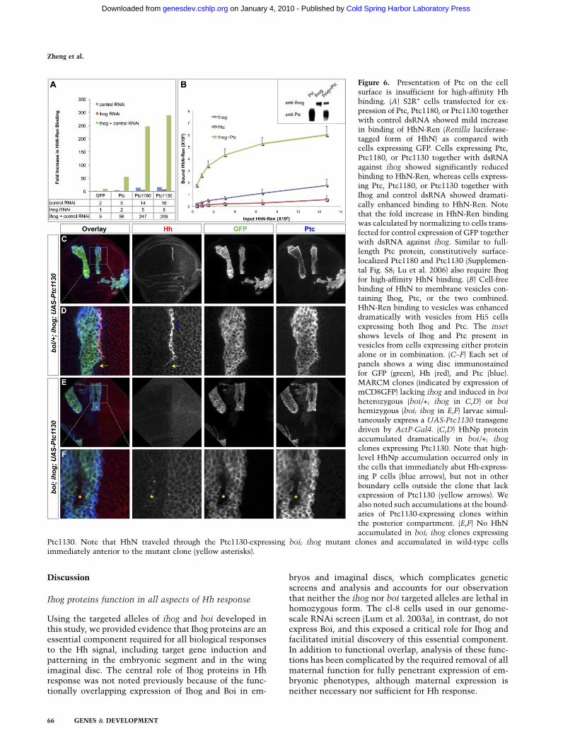
Figure 6. Presentation of Ptc on the cellsurface is insufficient for high-affinity Hhbinding. (A) S2R+ cells transfected for ex-pression of Ptc, Ptc1180, or Ptc1130 togetherwith control dsRNA showed mild increasein binding of HhN-Ren (Renilla luciferase-tagged form of HhN) as compared withcells expressing GFP. Cells expressing Ptc,Ptc1180, or Ptc1130 together with dsRNAagainst ihog showed significantly reducedbinding to HhN-Ren, whereas cells express-ing Ptc, Ptc1180, or Ptc1130 together withIhog and control dsRNA showed dramati-cally enhanced binding to HhN-Ren. Notethat the fold increase in HhN-Ren bindingwas calculated by normalizing to cells trans-fected for control expression of GFP togetherwith dsRNA against ihog. Similar to full-length Ptc protein, constitutively surface-localized Ptc1180 and Ptc1130 (Supplemen-tal Fig. S8; Lu et al. 2006) also require Ihogfor high-affinity HhN binding. (B) Cell-freebinding of HhN to membrane vesicles con-taining Ihog, Ptc, or the two combined.HhN-Ren binding to vesicles was enhanceddramatically with vesicles from Hi5 cellsexpressing both Ihog and Ptc. The inset
shows levels of Ihog and Ptc present invesicles from cells expressing either proteinalone or in combination. (C–F) Each set ofpanels shows a wing disc immunostainedfor GFP (green), Hh (red), and Ptc (blue).MARCM clones (indicated by expression ofmCD8GFP) lacking ihog and induced in boi
heterozygous (boi/+; ihog in C,D) or boi
hemizygous (boi; ihog in E,F) larvae simul-taneously express a UAS-Ptc1130 transgenedriven by ActP-Gal4. (C,D) HhNp proteinaccumulated dramatically in boi/+; ihog
clones expressing Ptc1130. Note that high-level HhNp accumulation occurred only inthe cells that immediately abut Hh-express-ing P cells (blue arrows), but not in otherboundary cells outside the clone that lackexpression of Ptc1130 (yellow arrows). Wealso noted such accumulations at the bound-aries of Ptc1130-expressing clones withinthe posterior compartment. (E,F) No HhNaccumulated in boi; ihog clones expressing
Ptc1130. Note that HhN traveled through the Ptc1130-expressing boi; ihog mutant clones and accumulated in wild-type cellsimmediately anterior to the mutant clone (yellow asterisks).
Zheng et al.
66 GENES & DEVELOPMENT
Cold Spring Harbor Laboratory Press on January 4, 2010 - Published by genesdev.cshlp.orgDownloaded from

Ihog signaling function integrates modular activitiesof Fn1 and Fn2
The interaction of Ihog Fn2 with Ptc is essential forpresentation of wild-type Ptc on the cell surface. Wecannot, at present, distinguish between the possibilitiesthat Ihog-mediated surface presentation of Ptc is due toan increased rate of transport to the surface or to anincreased duration of residence on the surface. Whateverthe mechanism, Fn2 can interact with Ptc in vitro andpromote surface presentation of Ptc in cells, even in theabsence of Fn1. Similarly, Fn1 alone can interact withHhN in vitro (McLellan et al. 2006), and Fn1 and Fn2 thushave demonstrably independent functions. Neither do-main alone, however, suffices for formation of a high-affinity complex, and the presence of both domains isrequired for Hh signal reception and transduction andparticipation in signaling in vivo (see Fig. 5).
The role of Ihog proteins in Hh binding
In addition to surface presentation of Ptc (discussedabove), our evidence indicates that Ihog proteins alsoplay a direct role in binding the Hh ligand in a multimo-lecular receptor complex that is critical for transduction.We thus found, as reported previously (Lu et al. 2006),that Hh ligand is bound to the surface of cultured cellsexpressing a variant of Ptc (Ptc1130) with increasedlocalization on the surface (Supplemental Fig. S8). Wealso found, with the use of quantitative assays, thatendogenous Ihog expressed in these cultured cells con-tributes critically to binding, and that additional Ihogexpression can dramatically enhance binding (Fig. 6A). Inaddition, expression of Ptc1130 in the wing imaginal discclearly produces visible accumulation of the Hh proteinon what appears to be the surface of anterior cells atthe compartment boundary (Fig. 6C,D); this accumula-tion depends critically on the expression of Ihog/Boi(Fig. 6E,F).
Consistent with the role of Ihog in binding, we alsonoted a striking contribution of Ihog to binding inmembrane vesicle preparations when present in combi-nation with Ptc (Fig. 6B). In addition, we were able to usepurified, immobilized HhN and detergent-solubilizedextracts containing Ptc and Ihog to demonstrate Ihog-dependent, enhanced precipitation of Ptc (Fig. 7). In thesebiochemical experiments, we observed that immobilizedHhN fails to precipitate detergent-solubilized Ptc alone,but does so in the presence of detergent-solubilized Ihog,and that Ihog alone precipitates Ptc much less efficientlythan when HhN is present. This enhancement of Ptcprecipitation was dependent on the presence of both theHhN-binding Fn1 domain and the Ptc-binding Fn2 do-main of Ihog, consistent with the formation of a multi-molecular complex involving HhN, Ptc, and Ihog (Sup-plemental Fig. S10). Similar results were noted for Boi(Supplemental Fig. S10).
It is interesting to note that we were unable to observemuch if any interaction between HhN and Ptc in theabsence of Ihog. Formally, it is possible that the interac-tion of Ptc with HhN is indirect and mediated throughenhanced Ihog interaction due to Ptc-induced multime-rization or allosteric effects on Ihog. We believe this isunlikely, however, because Ihog is capable of dimeriza-tion in the absence of Ptc (McLellan et al. 2006), andbecause the HhN-interacting surface of Ihog is located onthe Fn1 domain, which folds independently and is quitedistinct from the Ptc-interacting Fn2 domain, thus mak-ing allostery unlikely (Supplemental Fig. S6; McLellanet al. 2006). We thus favor the interpretation that favor-able energetic contributions in the multimolecular re-ceptor/ligand complex derive from Ptc–HhN contacts aswell as contacts between Ihog–Ptc and Ihog–HhN.
It is important to note that, despite a direct physicalinteraction of Ihog and Ptc and their mutual contribu-tions to formation and surface presentation of receptor,and to ligand binding, these two pathway componentshave opposing roles in pathway regulation. Ihog proteinsare thus absolutely required for pathway activation in
Figure 7. A complex of Ihog, Ptc, and HhN. Immobilized HhNpreferentially binds detergent-solubilized Ptc in the presence ofIhog. GluGlu-tagged HhN protein was purified using Sepharosebeads containing the anti-GluGlu monoclonal antibody, andthese beads were incubated with detergent-solubilized extractsfrom S2R+ cells expressing Ptc variants (lanes 1–3), or Ptcvariants coexpressed with HA-tagged Ihog (lanes 4–6). Precipi-tation of Ptc by Sepharose HhN-GluGlu beads depended criti-cally on the presence of Ihog, and was much reduced by deletionof the large extracellular Ptc loops (Dloop1,2 or Dloop7,8). (Lane7) In comparison, precipitation of Ihog with anti-HA beadsprecipitated significantly less Ptc.
Ihog/Boi and Ptc, Drosophila Hh receptor
GENES & DEVELOPMENT 67
Cold Spring Harbor Laboratory Press on January 4, 2010 - Published by genesdev.cshlp.orgDownloaded from

response to Hh ligand, whereas Ptc alone suffices forsuppression of Smo activity in the absence of ligands.
Mammalian Ihog proteins
Functional genetic analyses of the mammalian Ihog pro-teins Cdo and Boc have revealed roles in Hh signaling.Cdo mutant mice thus display mild to intermediateforms of holoprosencephaly, a classic manifestation ofHh signaling deficiency, with the severity of the effectdepending on strain background and subject to modifyingeffects of mutations in other Hh pathway components(Cole and Krauss 2003; Tenzen et al. 2006; Zhang et al.2006; Allen et al. 2007). Boc mutant mice also showdefects in Hh signal-dependent axonal pathfinding bydorsal neurons with ventral commissural projections inthe developing neural tube (Okada et al. 2006; Yam et al.2009). Neither of these mutants displays phenotypes assevere as those seen in the Shh mutant mouse, or in theSmo mutant, which affects all aspects of Hh signaling. Itis possible, however, that a systematic analysis of thedouble mutant Cdo; Boc animals might reveal moresevere phenotypes, as we noted here for ihog; boi inDrosophila. In addition, our phenotypic characterizationof ihog and boi mutants was not designed to revealdefects in axonal pathfinding functions like that ofmurine Boc, and the possibility of such a function inDrosophila remains to be explored.
Materials and methods
Constructs
Constructs used in generating transgenic flies were sublonedinto pW25 (ihog and boi target constructs) or pUAST. Expressionconstructs of Ihog variants and Ptc variants used in Drosophilacell culture were fused in-frame with a C-terminal HA tag or3xMyc tag, respectively, and were cloned into pAcSV plasmid.Constructs used in the baculovirus expression system weresubcloned into pVL1392 or pVL1393 (BD Biosciences). GluGlu-tagged HhN was generated by inserting the GluGlu tag betweenHh codons for amino acids 254 and 255, followed by codons foramino acids 256 and 257 and a stop codon TAA.
Antibodies
Antibodies and dilutions used were mouse anti-Ptc, 1:50 (Capdevilaet al. 1994); mouse anti-bgal, 1:100 (Promega, Z378); mouse anti-Smo, 1:50 (Lum et al. 2003b); mouse anti-En 4D9, 1:50 (Patel et al.1989); mouse anti-Wg 4D4, 1:50 (Brook and Cohen 1996); mouseanti-HA, 1:1000 (HA.11, Covance); mouse anti-b-tubulin E7,1:5000 (Developmental Studies Hybridoma Bank, developed byM. Klymkowsky); rabbit anti-HhN, 1:1000 (Tabata and Kornberg1994) for immunostaining; rabbit anti-HhN, 1:1000 for Westernblotting (Lee et al. 1992); rabbit anti-Myc, 1:1000 (A-14, Santa CruzBiotechnologies); rabbit anti GFP, 1:1000 (Invitrogen, A-11122); ratmonoclonal anti-Ci 2A1, 1:50 (Motzny and Holmgren 1995); ratanti-Ihog, 1:200 for immunostaining and 1:1000 for immunoblot-ting (Yao et al. 2006); HRP-conjugated goat anti-mouse (Promega,W4021); HRP-conjugated goat anti-rabbit (Jackson Immuno-Re-search Laboratory, 111-035-144); and HRP-conjugated goat anti-rat (Jackson Immuno-Research Laboratory, 112-035-167). Fluoro-phore-conjugated secondary antibodies were from Invitrogen.
Purification and immobilization of HhN
Culture of Sf9 and Hi5 cells and production of recombinantbaculovirus were as described (Dukkipati et al. 2006). Hi5 cellsused for protein production were infected with baculovirus forexpression of HhN-GluGlu. Forty-eight hours after infection,cells were cleared by centrifugation at 1000g and the supernatantwas concentrated and incubated with GluGlu monoclonal anti-body-affinity matrix (Covance, AFC-115P) overnight at 4°C.Beads were washed and stored at 4°C until use.
Protein immunoprecipitation
Drosophila S2R+, S2, and S2-HhN stable cells were cultured asdescribed (Lum et al. 2003b). Transfection was carried out usingFuGENE 6 transfection reagent (Roche). Forty-eight hours aftertransfection, S2R+ cells were lysed in 0.5% Digitonin (50 mMTris-HCl at pH 7.5, 150 mM NaCl, 40 mM low-molecular-weightHeparin, 1 mM CaCl2, protease inhibitors) for 30 min at roomtemperature. The lysate was clarified by centrifugation, andproteins were immobilized by binding to anti-HA-affinity matrix(Roche, 11815016001) or GluGlu monoclonal antibody-affinitymatrix with prebound HhN for 2 h at room temperature. Beadswere washed and proteins were recovered directly in SDS-PAGEsample buffer (for the anti-HA matrix) or by elution witha peptide containing the GluGlu epitope (Anaspec, 62189).
Cell immunostaining
Forty-eight hours after transfection, S2R+ cells were then washedtwice with PBS, fixed in 4% formaldehyde (Ted Pella) in PBS,blocked by 1.5% normal goat serum (NGS) in PBS, incubatedwith primary antibody in PBS containing 1.5% NGS for 1 h atroom temperature, washed three times with 0.1% Tween-20/PBS, incubated with secondary antibody, and washed.
Cell surface biotinylation
S2R+ cells were washed with PBS and incubated for 30 min inPBS containing 1 mg/mL Sulfo-NHS-LC-Biotin (Pierce, 21335).The reaction was quenched by washing the cells with 100 mMglycine in PBS three times. Cells were then lysed in 0.5%Digitonin (50 mM Tris-HCl at pH 7.5, 150 mM NaCl, proteaseinhibitors) for 30 min. The lysate was clarified by centrifugation,and biotinylated protein was recovered by binding to Streptavi-din-Sepharose beads (GE Healthcare, 17-5113-01) for 2 h at roomtemperature. Beads were washed and protein was recovered inSDS-PAGE sample buffer.
Cell-based binding assay
S2R+ cells were transfected for 48 h followed by incubation withconditioned medium containing HhN-Ren (Yao et al. 2006)protein for 1 h at 4°C. Cells then were washed three times withcold PBS and lysed in 250 mL (per well of a six-well plate) ofpassive lysis buffer (Promega), and 50 mL of lysate were used tomeasure luciferase activities.
Cell-free binding assay
Hi5 cells were infected with baculovirus for expression of Ihog, Ptc,or Ihog plus Ptc. Cells were harvested 48 h after infection bycentrifugation at 800g for 15 min. The cell pellet was resuspendedin 3 vol of buffer A (Brown et al.2002), homogenized with50strokesin a tight Dounce homogenizer, and centrifuged at 800g for 10 minat 4°C. The supernatant from this spin was collected to prepare
Zheng et al.
68 GENES & DEVELOPMENT
Cold Spring Harbor Laboratory Press on January 4, 2010 - Published by genesdev.cshlp.orgDownloaded from

the membrane fraction by centrifugation at 18,000g for 15 min at4°C. The resulting membrane pellets were resuspended in condi-tioned medium containing HhN-Ren. After 1 h of incubation at4°C, the membrane pellets were washed three times with PBS andsolubilized in 200 mL of passive lysis buffer (Promega), and 50 mL oflysate were used to measure luciferase activity.
Drosophila strains
Mutant and transgenic strains are described in the followingreferences: pka-C1B3 (Lane and Kalderon 1993), Actin-Gal4 (Itoet al. 1997), hs-FLP (Golic and Lindquist 1989), tubP-Gal80 (Leeand Luo 1999), dpp-lacZ (BS3.0, from Bloomington Stock Cen-ter), UAS-mCD8GFP (Lee and Luo 1999), UAS-Hh (Azpiazu et al.1996),UAS-Ptc and UAS-Ptc1130 (Johnson et al. 2000), and2Xubi-nGFP FRT40A (Bloomington Stock Center). ORFs encod-ing full-length or altered Ihog and Boi were cloned into pUASTto generate UAS-Ihog, UAS-Boi, UAS-IhogCTD (1-742), UAS-
IhogDFn1 (delete aa467-577), UAS-IhogFn2* (K653E,Q655E;mutations in Fn2) (see Supplemental Fig. S6). ihog pka-C1B3
FRT40A recombinant chromosome was selected from maleoffspring of a mating of ihog/pka-C1B3 FRT40A heterozygousfemales and Sco/Cyo balancer males.
Generating ihog and boi mutations by gene targeting
The ends-out gene targeting strategy (Gong and Golic 2003) wasused to generate ihog and boi mutations. DNA fragmentsflanking the target locus were prepared by PCR of 4- to 5-kb frag-ments from genomic DNA using two pairs of primers for ihog:59-TTAGCGGCCGCAGAGCGAGATAAGCTGGCACAGG-39/59-GACGGTACCTGTGCATCCCTACGCCCGATG-39 and 59-AATGGGCCGCCGACTGATTCTAGGTGGGGAAACG-39/59-GCGCGTACGTCACCTTGTATGAGGTTTCGCCA-39; and two pairs ofprimers for boi: 59-TACGCGGCCGCTCCACTGCCTTATTGGCTGGCAC-39/59-GACCGCATGCTCCAAAAACAAGAGCGGCAGAC-39 and 59-ATAGGCGCGCCCTCTAACCATTTGACAGGCGAGG-39/59-GTCCGTACGTGTGTGTGAGTGTGTGGCTTGG-39.These fragments were cloned into the vector pW25. Transgenicflies carrying the ihog target construct on the X chromosome orboi target construct on the second chromosome were crossed toflies carrying the hs-FLP recombinase and I-SceI endonuclease,and the progeny were screened for precise gene targeting asdescribed (Gong and Golic 2003) .
We note here that cuticle phenotypes for our new ihog and boi
alleles in embryos lacking maternal and zygotic function maydiffer from those reported previously for ihog due to compensa-tory gains in expression of one gene that might be adaptive andcould occur when stocks are maintained as homozygous mu-tants for the other gene. Thus, for example, Ihog protein levelsare significantly higher in boi mutant homozygotes as comparedwith wild-type (Supplemental Fig. S1). The homozygous em-bryos in our current studies derive from homozygous stockswhere such compensatory gain might occur, whereas our pre-vious studies used an allele maintained as a heterozygote be-cause of an associated lethal mutation in an adjacent gene (Yaoet al. 2006). Such compensatory gain in homozygous stockscould also have influenced the effects of reduced boi/ihog genedosage in our current wing imaginal disc studies (i.e., loss of allihog and half of boi function [75% of gene dosage lost] producesa mild reduction of Ptc, similar to loss of ihog only [50% of genedosage lost] in our previous study).
Genotype of larvae for generating mosaic clones
Germline clones and somatic clones were generated by FRT-FLPrecombination (Golic 1991; Chou et al. 1993; Xu and Rubin 1993;
Lee and Luo 1999). The genotypes used in our analysis were asfollows.
Wing disc clones marked by absence of GFP. ihog mutant clones ina boi heterozygous background: y w boi hs-FLP/X; ihog FRT40A/ubi-nGFP FRT40A; dppZ/+. ihog mutant clones in a boi homo-zygous or hemizygous background: y w boi hs-FLP/y w boi or Y;
ihog FRT40A/ubi-nGFP FRT40A; dppZ/+. ihog and pka-C1B3
double mutant clones in a boi homozygous or hemizygousbackground: y w boi hs-FLP/y w boi or Y; ihog pka-C1B3
FRT40A/ubi-nGFP FRT40A; dppZ/+.
Wing disc MARCM clones. ihog mutant clones in a boi heterozy-gous background: y w boi hs-FLP UAS-mCD8GFP/X; ihogFRT40A/tubP-GAL80 FRT40A; ActP-Gal4/Tm6B. Positivelymarked ihog mutant clones in a boi homozygous or hemizygousbackground: y w boi hs-FLP UAS-mCD8GFP/y w boi or Y; ihog
FRT40A/tubP-GAL80 FRT40A; ActP-Gal4/Tm6B. Positivelymarked ihog mutant clones in a boi homozygous or hemizygousbackground with specific expression of a gene of interest to betested for rescue: y w boi hs-FLP UAS-mCD8GFP/y w boi or Y;
ihog FRT40A/tubP-GAL80 FRT40A; ActP-Gal4/UAS-gene-of-interest (for specific expression of rescue constructs).
Germline clones. ihog germline clones in a boi homozygous orhemizygous background: y w boi hs-FLP; ihog FRT40A/P[ovoD1]2La P[ovoD1]2Lb FRT40A X y w boi; ihog FRT40A/
Cyo[act-GFP].
RT–PCR analysis
Total RNA was extracted from cultured cells or tissues usingRNeasy Mini-Kit (Qiagen). cDNA was synthesized using iScriptcDNA Synthesis Kit (Bio-Rad), and PCR reactions (iTaq DNAPolymerase from Bio-Rad) were performed using 5 mL of cDNAas a template.
Image collection and quantification of flurescence intensity
Confocal images were collected via Zeiss LSM 510 and wereprocessed with LSM software and ImageJ.
Acknowledgments
We thank M. Frasch, K.C. Garcia, J. Jiang, T.B. Kornberg, R.Krauss, T. Lee, L. Luo, D. Pan, M. Scott, G. Struhl, T. Tabata, A.Zhu, Developmental Studies Hybridoma Bank, and BloomingtonStock Center for fly strains and reagents; L. Luo for criticalreview of the manuscript; Ya-Hui Chou, M. Kato, and S. Park forexperimental advice; and J.S. McLellan and D.J. Leahy for de-signing the IhogK653E, Q655E mutant. P.A.B. is an Investigatorof the Howard Hughes Medical Institute. N.S. and X.Z. areDamon Runyon Fellows supported by the Damon RunyonCancer Research Foundation (DRG-1891-05 and DRG-1915-06,respectively).
References
Allen BL, Tenzen T, McMahon AP. 2007. The Hedgehog-bindingproteins Gas1 and Cdo cooperate to positively regulate Shhsignaling during mouse development. Genes & Dev 21:1244–1257.
Azpiazu N, Lawrence PA, Vincent JP, Frasch M. 1996. Segmen-tation and specification of the Drosophila mesoderm. Genes &
Dev 10: 3183–3194.
Ihog/Boi and Ptc, Drosophila Hh receptor
GENES & DEVELOPMENT 69
Cold Spring Harbor Laboratory Press on January 4, 2010 - Published by genesdev.cshlp.orgDownloaded from

Brook WJ, Cohen SM. 1996. Antagonistic interactions betweenwingless and decapentaplegic responsible for dorsal–ventralpattern in the Drosophila Leg. Science 273: 1373–1377.
Brown AJ, Sun L, Feramisco JD, Brown MS, Goldstein JL. 2002.Cholesterol addition to ER membranes alters conformationof SCAP, the SREBP escort protein that regulates cholesterolmetabolism. Mol Cell 10: 237–245.
Burke R, Nellen D, Bellotto M, Hafen E, Senti KA, Dickson BJ,Basler K. 1999. Dispatched, a novel sterol-sensing domainprotein dedicated to the release of cholesterol-modifiedhedgehog from signaling cells. Cell 99: 803–815.
Capdevila J, Pariente F, Sampedro J, Alonso JL, Guerrero I. 1994.Subcellular localization of the segment polarity proteinpatched suggests an interaction with the wingless receptioncomplex in Drosophila embryos. Development 120: 987–998.
Chen Y, Struhl G. 1996. Dual roles for patched in sequesteringand transducing Hedgehog. Cell 87: 553–563.
Chiang C, Litingtung Y, Lee E, Young KE, Corden JL, WestphalH, Beachy PA. 1996. Cyclopia and defective axial patterningin mice lacking Sonic hedgehog gene function. Nature 383:407–413.
Chou TB, Noll E, Perrimon N. 1993. Autosomal P[ovoD1]dominant female-sterile insertions in Drosophila and theiruse in generating germ-line chimeras. Development 119:1359–1369.
Chuang PT, McMahon AP. 1999. Vertebrate Hedgehog signal-ling modulated by induction of a Hedgehog-binding protein.Nature 397: 617–621.
Cole F, Krauss RS. 2003. Microform holoprosencephaly in micethat lack the Ig superfamily member Cdon. Curr Biol 13:411–415.
Denef N, Neubuser D, Perez L, Cohen SM. 2000. Hedgehoginduces opposite changes in turnover and subcellular local-ization of patched and smoothened. Cell 102: 521–531.
DiNardo S, Heemskerk J, Dougan S, O’Farrell PH. 1994. Themaking of a maggot: Patterning the Drosophila embryonicepidermis. Curr Opin Genet Dev 4: 529–534.
Dukkipati A, Vaclavikova J, Waghray D, Garcia KC. 2006. Invitro reconstitution and preparative purification of com-plexes between the chemokine receptor CXCR4 and itsligands SDF-1a, gp120-CD4 and AMD3100. Protein Expr
Purif 50: 203–214.Fuse N, Maiti T, Wang B, Porter JA, Hall TM, Leahy DJ, Beachy
PA. 1999. Sonic hedgehog protein signals not as a hydrolyticenzyme but as an apparent ligand for patched. Proc Natl
Acad Sci 96: 10992–10999.Glise B, Miller CA, Crozatier M, Halbisen MA, Wise S, Olson
DJ, Vincent A, Blair SS. 2005. Shifted, the Drosophila
ortholog of Wnt inhibitory factor-1, controls the distributionand movement of Hedgehog. Dev Cell 8: 255–266.
Golic KG. 1991. Site-specific recombination between homolo-gous chromosomes in Drosophila. Science 252: 958–961.
Golic KG, Lindquist S. 1989. The FLP recombinase of yeastcatalyzes site-specific recombination in the Drosophila ge-nome. Cell 59: 499–509.
Gong WJ, Golic KG. 2003. Ends-out, or replacement, genetargeting in Drosophila. Proc Natl Acad Sci 100: 2556–2561.
Gorfinkiel N, Sierra J, Callejo A, Ibanez C, Guerrero I. 2005. TheDrosophila ortholog of the human Wnt inhibitor factorShifted controls the diffusion of lipid-modified Hedgehog.Dev Cell 8: 241–253.
Han C, Belenkaya TY, Wang B, Lin X. 2004. Drosophila
glypicans control the cell-to-cell movement of Hedgehog bya dynamin-independent process. Development 131: 601–611.
Hollway GE, Maule J, Gautier P, Evans TM, Keenan DG, Lohs C,Fischer D, Wicking C, Currie PD. 2006. Scube2 mediates
Hedgehog signalling in the zebrafish embryo. Dev Biol 294:104–118.
Ingham PW, McMahon AP. 2001. Hedgehog signaling in animaldevelopment: Paradigms and principles. Genes & Dev 15:3059–3087.
Ingham PW, Taylor AM, Nakano Y. 1991. Role of the Dro-
sophila patched gene in positional signalling. Nature 353:184–187.
Ito K, Awano W, Suzuki K, Hiromi Y, Yamamoto D. 1997. TheDrosophila mushroom body is a quadruple structure ofclonal units each of which contains a virtually identical setof neurones and glial cells. Development 124: 761–771.
Jessell TM. 2000. Neuronal specification in the spinal cord:Inductive signals and transcriptional codes. Nat Rev Genet 1:20–29.
Jiang J, Struhl G. 1995. Protein kinase A and hedgehog signalingin Drosophila limb development. Cell 80: 563–572.
Johnson RL, Milenkovic L, Scott MP. 2000. In vivo functions ofthe patched protein requirement of the C terminus for targetgene inactivation but not hedgehog sequestration. Mol Cell6: 467–478.
Kawakami A, Nojima Y, Toyoda A, Takahoko M, Satoh M,Tanaka H, Wada H, Masai I, Terasaki H, Sakaki Y, et al. 2005.The zebrafish-secreted matrix protein you/scube2 is impli-cated in long-range regulation of hedgehog signaling. Curr
Biol 15: 480–488.Lane ME, Kalderon D. 1993. Genetic investigation of cAMP-
dependent protein kinase function in Drosophila develop-ment. Genes & Dev 7: 1229–1243.
Lavine KJ, Kovacs A, Ornitz DM. 2008. Hedgehog signaling iscritical for maintenance of the adult coronary vasculature inmice. J Clin Invest 118: 2404–2414.
Lee T, Luo L. 1999. Mosaic analysis with a repressible cellmarker for studies of gene function in neuronal morphogen-esis. Neuron 22: 451–461.
Lee JJ, von Kessler DP, Parks S, Beachy PA. 1992. Secretion andlocalized transcription suggest a role in positional signaling forproducts of the segmentation gene hedgehog. Cell 71: 33–50.
Lepage T, Cohen SM, Diaz-Benjumea FJ, Parkhurst SM. 1995.Signal transduction by cAMP-dependent protein kinase A inDrosophila limb patterning. Nature 373: 711–715.
Li W, Ohlmeyer JT, Lane ME, Kalderon D. 1995. Function ofprotein kinase A in hedgehog signal transduction and Dro-
sophila imaginal disc development. Cell 80: 553–562.Lu X, Liu S, Kornberg TB. 2006. The C-terminal tail of the
Hedgehog receptor Patched regulates both localization andturnover. Genes & Dev 20: 2539–2551.
Lum L, Beachy PA. 2004. The Hedgehog response network:Sensors, switches, and routers. Science 304: 1755–1759.
Lum L, Yao S, Mozer B, Rovescalli A, Von Kessler D, NirenbergM, Beachy PA. 2003a. Identification of Hedgehog pathwaycomponents by RNAi in Drosophila cultured cells. Science
299: 2039–2045.Lum L, Zhang C, Oh S, Mann RK, von Kessler DP, Taipale J,
Weis-Garcia F, Gong R, Wang B, Beachy PA. 2003b. Hedge-hog signal transduction via Smoothened association witha cytoplasmic complex scaffolded by the atypical kinesin,Costal-2. Mol Cell 12: 1261–1274.
Ma Y, Erkner A, Gong R, Yao S, Taipale J, Basler K, Beachy PA.2002. Hedgehog-mediated patterning of the mammalianembryo requires transporter-like function of dispatched. Cell111: 63–75.
Mandal L, Martinez-Agosto J, Evans C, Hartenstein V, BanerjeeU. 2007. A Hedgehog- and Antennapedia-dependent nichemaintains Drosophila haematopoietic precursors. Nature446: 320–324.
Zheng et al.
70 GENES & DEVELOPMENT
Cold Spring Harbor Laboratory Press on January 4, 2010 - Published by genesdev.cshlp.orgDownloaded from

Mann RK, Beachy PA. 2004. Novel lipid modifications ofsecreted protein signals. Annu Rev Biochem 73: 891–923.
Marigo V, Davey RA, Zuo Y, Cunningham JM, Tabin CJ. 1996.Biochemical evidence that patched is the Hedgehog receptor.Nature 384: 176–179.
Martinelli DC, Fan CM. 2007. Gas1 extends the range ofHedgehog action by facilitating its signaling. Genes & Dev
21: 1231–1243.McLellan JS, Yao S, Zheng X, Geisbrecht BV, Ghirlando R,
Beachy PA, Leahy DJ. 2006. Structure of a heparin-dependentcomplex of Hedgehog and Ihog. Proc Natl Acad Sci 103:17208–17213.
McLellan JS, Zheng X, Hauk G, Ghirlando R, Beachy PA, LeahyDJ. 2008. The mode of Hedgehog binding to Ihog homologuesis not conserved across different phyla. Nature 455: 979–983.
McMahon AP, Ingham PW, Tabin CJ. 2003. Developmental rolesand clinical significance of hedgehog signaling. Curr Top Dev
Biol 53: 1–114.Motzny CK, Holmgren R. 1995. The Drosophila cubitus inter-
ruptus protein and its role in the wingless and hedgehogsignal transduction pathways. Mech Dev 52: 137–150.
Muenke M, Beachy PA. 2000. Genetics of ventral forebraindevelopment and holoprosencephaly. Curr Opin Genet Dev
10: 262–269.Nusslein-Volhard C, Wieschaus E. 1980. Mutations affecting
segment number and polarity in Drosophila. Nature 287:795–801.
Nystul T, Spradling A. 2006. Breaking out of the mold: Diversitywithin adult stem cells and their niches. Curr Opin Genet
Dev 16: 463–468.Okada A, Charron F, Morin S, Shin DS, Wong K, Fabre PJ,
Tessier-Lavigne M, McConnell SK. 2006. Boc is a receptor forsonic hedgehog in the guidance of commissural axons.Nature 444: 369–373.
Pan D, Rubin GM. 1995. cAMP-dependent protein kinase andhedgehog act antagonistically in regulating decapentaple-gic transcription in Drosophila imaginal discs. Cell 80:543–552.
Panakova D, Sprong H, Marois E, Thiele C, Eaton S. 2005.Lipoprotein particles are required for Hedgehog and Winglesssignalling. Nature 435: 58–65.
Patel NH, Martin-Blanco E, Coleman KG, Poole SJ, Ellis MC,Kornberg TB, Goodman CS. 1989. Expression of engrailed pro-teins in arthropods, annelids, and chordates. Cell 58: 955–968.
Sampedro J, Guerrero I. 1991. Unrestricted expression of theDrosophila gene patched allows a normal segment polarity.Nature 353: 187–190.
Stone DM, Hynes M, Armanini M, Swanson TA, Gu Q, JohnsonRL, Scott MP, Pennica D, Goddard A, Phillips H, et al. 1996.The tumour-suppressor gene patched encodes a candidatereceptor for Sonic hedgehog. Nature 384: 129–134.
Strutt DI, Wiersdorff V, Mlodzik M. 1995. Regulation of furrowprogression in the Drosophila eye by cAMP-dependent pro-tein kinase A. Nature 373: 705–709.
Tabata T, Kornberg TB. 1994. Hedgehog is a signaling proteinwith a key role in patterning Drosophila imaginal discs. Cell
76: 89–102.Taipale J, Cooper MK, Maiti T, Beachy PA. 2002. Patched acts
catalytically to suppress the activity of Smoothened. Nature
418: 892–897.Takashima S, Mkrtchyan M, Younossi-Hartenstein A, Merriam
JR, Hartenstein V. 2008. The behaviour of Drosophila adulthindgut stem cells is controlled by Wnt and Hh signalling.Nature 454: 651–655.
Tenzen T, Allen BL, Cole F, Kang JS, Krauss RS, McMahon AP.2006. The cell surface membrane proteins Cdo and Boc are
components and targets of the Hedgehog signaling pathwayand feedback network in mice. Dev Cell 10: 647–656.
Varjosalo M, Taipale J. 2008. Hedgehog: Functions and mecha-nisms. Genes & Dev 22: 2454–2472.
Woods IG, Talbot WS. 2005. The you gene encodes an EGF-CUBprotein essential for Hedgehog signaling in zebrafish. PLoS
Biol 3: 476–487.Xu T, Rubin GM. 1993. Analysis of genetic mosaics in de-
veloping and adult Drosophila tissues. Development 117:1223–1237.
Yam PT, Langlois SD, Morin S, Charron F. 2009. Sonic hedgehogguides axons through a noncanonical, Src-family-kinase-dependent signaling pathway. Neuron 62: 349–362.
Yao S, Lum L, Beachy P. 2006. The ihog cell-surface proteinsbind Hedgehog and mediate pathway activation. Cell 125:343–357.
Zhang W, Kang JS, Cole F, Yi MJ, Krauss RS. 2006. Cdo functionsat multiple points in the Sonic Hedgehog pathway, and Cdo-deficient mice accurately model human holoprosencephaly.Dev Cell 10: 657–665.
Zhao C, Chen A, Jamieson C, Fereshteh M, Abrahamsson A,Blum J, Kwon H, Kim J, Chute J, Rizzieri D, et al. 2009.Hedgehog signalling is essential for maintenance of cancerstem cells in myeloid leukaemia. Nature 458: 776–779.
Zhu AJ, Zheng L, Suyama K, Scott MP. 2003. Altered localiza-tion of Drosophila Smoothened protein activates Hedgehogsignal transduction. Genes & Dev 17: 1240–1252.
Ihog/Boi and Ptc, Drosophila Hh receptor
GENES & DEVELOPMENT 71
Cold Spring Harbor Laboratory Press on January 4, 2010 - Published by genesdev.cshlp.orgDownloaded from
Figure S1. Verification of ihog Mutation by Protein Immunoblotting Extracts from S2R+ cells, embryos, and adults were analyzed by immunoprecipitation
and immunoblotting with anti-Ihog antibody. The Ihog protein is absent in ihog
homozygotes. An anti-tubulin immunoblot is included as a loading control.
Figure S2. Verification of ihog and boi Alleles Generated by Targeted Recombination
(A, B) Primers used for RT-PCR are shown as blue arrows in Figure 5; pgk was used as
an internal control. (A) An ihog+ transcript (P1+P2, 124 bp) was detected in wild-type
and ihog mutant heterozygotes, but not in ihog mutant homozygotes. A hybrid ihog-
white transcript (P1+P3, 153bp) was detected in ihog heterozygous or homozygous
mutants, but not in wild-type. (B) A boi+ transcript (P5+P6, 160bp) was detected
exclusively in wild-type and a larger transcript (P5+P6, 3233 bp) exclusively in boi
homozygotes. The absence of the 160 bp wild-type product presumably is due to
deletion of the splice acceptor for exon 5 in the boi mutant, thus preventing
incorporation of P5 sequences into transcripts. The 3233 bp fragment in the mutant
corresponds to an unspliced transcript, absent in the wild-type. With primers P4 and P6,
the 1620 bp fragment (P4+P6, 1620 bp) represents a normally spliced boi-RB or boi-RD
transcript, whereas the 207 bp product (P4+P6, 207 bp) corresponds to a mutant
transcript corresponding to direct splicing from exon 3 to exon 7 (exon 6 within boi-RA
begins at an internal initiation site and lacks a splice acceptor).
Figure S3. Boi Proteins Encoded by Predicted Transcripts from the Wild-Type and
Mutant boi Locus
The predicted Boi-RA, Boi-RB and Boi-RD proteins are aligned by sequence.
Replacement of exon 4 and part of exon 5 of boi by white results in deletion of the
signal sequence (SS, orange) and the four Ig domains (blue) from the coding
sequences of the altered Boi proteins in the boi mutant (Boi-RB* and Boi-RD*), making
it unlikely that they would be normally produced and secreted; the predicted Boi-RA
protein in wild-type and mutant alleles is also unlikely to be secreted.
2
Figure S4. Smo Functions Downstream of Ihog/Boi Wing imaginal discs from boi hemizygous larvae carrying ihog mutant MARCM clones
(boi/Y; ihog) were immunostained for GFP (green), Ci (red) and Smo (blue). Smo
protein levels were reduced in the mutant clone located adjacent to the AP
compartment boundary (yellow asterisk), originating from the A compartment (as judged
by the expression of Ci).
Figure S5. Ihog/Boi Is Not Required for Hh Release Normal Ptc expression (red) adjacent to a large boi; ihog homozygous mutant clone
(marked by loss of GFP expression) originating from the P compartment (marked by
lack of Ci expression).
Figure S6. IhogFn1,2 Complexed with HhN The molecular surface of a crystallographic complex between HhN (green) and Ihog
Fn1, 2 (wheat) is shown, with the location of two missense mutations in surface
residues of Fn2 (K653E and Q655E) in red. As shown, HhN contacts IhogFn1 (McLellan
et al. 06); IhogFn2 is required for interaction with Ptc (see text).
Figure S7. Surface Localization of Ptc Is Ihog/Boi Dependent
Confocal microscope images showing localization of Ptc proteins in S2R+ cells co-
transfected with Ptc and either control dsRNA or dsRNA against both ihog and boi.
Cells were fixed with 4% paraformaldehyde and immunostaining with anti-Ptc antibodies
was done in the absence of detergent permeabilization to emphasize proteins on the
cell surface (same as in Figure 4); some intracellular proteins nevertheless were
detected.
(A) Ptc was mainly localized in intracellular vesicles with a trace of cell surface
expression (arrow; see also Figure 4E) in the presence of control RNAi.
(B) Cell-surface expression of Ptc protein is reduced in the presence of dsRNA against
both ihog and boi.
3
Figure S8. Co-expression of Ihog with either Ptc or Ptc1130 Increased the Amount of HhN Bound to Cells Confocal microscope images of S2 cells transfected by Ptc or Ptc1130 with or without
Ihog, as indicated. After incubation with HhN conditioned medium for 2 hours at 4°C (to
prevent endocytosis), cells were fixed, permeabilized (with 0.1% Triton X-100 in PBS)
and immunostained.
(A) Ptc was mainly localized in intracellular vesicles with a trace of surface expression,
and very little HhN was detected on the surface of Ptc expressing cells.
(B) Ptc1130 was mainly localized on the cell surface and more HhN was detected on
the surface of cells expressing Ptc1130 as compared to cells expressing full length Ptc
(Lu et al., 2006).
(C) Co-expression of Ihog dramatically increased the amount of surface localized Ptc
protein and significantly more HhN was detected on the cell surface as compared to A.
(D) Co-expression of Ihog dramatically increased the amount of HhN bound to cells
expressing Ptc1130 as compared to cells expressing Ptc1130 alone (B).
Figure S9. Purification and Immobilization of HhN-GluGlu Anti-GluGlu affinity matrix with bound HhN-GluGlu (2 µl) was incubated with sample
loading buffer and analyzed by SDS-PAGE (15% gel) and Coommassie Brilliant Blue
staining. The migration of molecular mass markers is indicated on the left, and bands
corresponding to the antibody chains and HhN-GluGlu on the right. Note the nearly
equivalent intensities of bands corresponding to HhN-GluGlu and antibody light chain,
which indicates the near-saturation of the anti-GluGlu matrix.
Figure S10. A Complex of Ihog/Boi, Ptc, and Hh Requires Both Fn1 and Fn2 GluGlu tagged HhN protein was purified using Sepharose beads containing the anti-
GluGlu monoclonal antibody and these beads were incubated with detergent-solubilized
(1% NP40, 50 mM Tris-HCl at pH 6.8, 150 mM NaCl, 40uM low molecular weight
Heparin, 1mM CaCl2, and protease inhibitors) extracts from S2R+ cells expressing Ptc
(lane 1), or Ptc together with HA-tagged Ihog variants (lanes 2-4) or Boi variants (lanes
5-7). Precipitation of Ptc by Sepharose HhN-GluGlu beads depended critically on the

4
presence of Ihog or Boi (lanes 2 and 5), and was abolished by deletion of either Fn1 or
Fn2 domain of Ihog/Boi (lanes 3,4, 6 and 7).
In comparison, precipitation of Ihog/Boi with anti-HA beads precipitated significantly less
Ptc (compare lanes 9 and 12 with lanes 2 and 5, respectively). Note that, similar to Ihog
(Figure 4B and 4C), the Fn1 of Boi is required to interact with HhN (lane 6, middle
panel) and Fn2 of Boi is required to interact with Ptc (lane 14, lower panel).
Table S1. Primers Used in Figure 1 and Figure S2

IP anti-IhogWB anti-Ihog
5% Input LysateWB anti-tubulin
wt boi
ihog/+
ihog
wt ihog
S2R+Cells
Fly embryos Adult flies
Zheng_FigS1
1 2 3 4 5 6 7
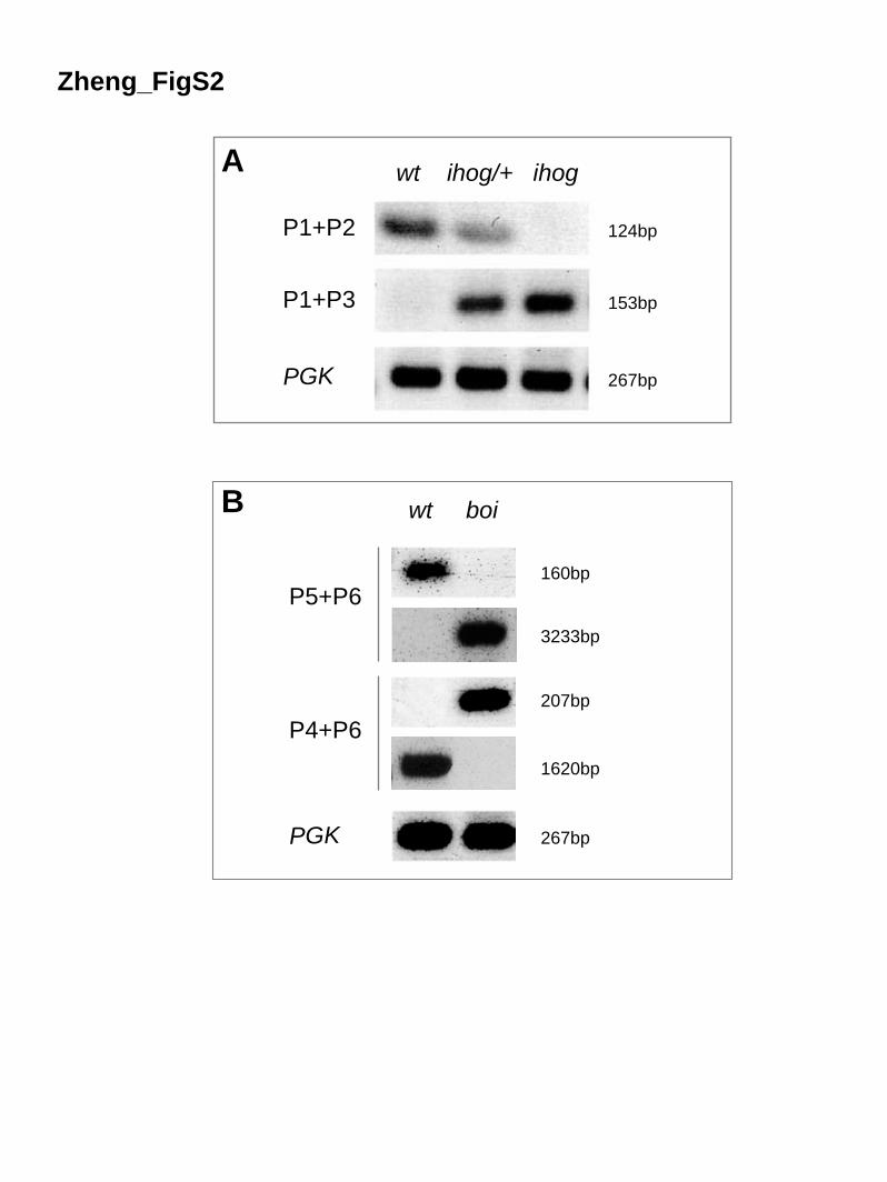
Zheng_FigS2
A wt ihog/+ ihog
P1+P2
P1+P3
PGK
B
P5+P6
P4+P6
PGK
wt boi
160bp
3233bp
207bp
1620bp
267bp
124bp
153bp
267bp

Boi-RA 1 0Boi-RB 1 MAEIRLVVVLLVLMTTCIYTLQASDSNSSVATTLGVLFERAPESAVAPKGDEVVFECE LNLKPDRLEWRFRRGDSSEPYLYLRPSAGYNVTSGSDGLAWRLRIYVSAQTAGDYQCVAWYGPGALASTPARLALVSIALDV 140Boi-RD 1 MAEIRLVVVLLVLMTTCIYTLQASDSNSSVATTLGVLFERAPESAVAPKGDEVVFECE LNLKPDRLEWRFRRGDSSEPYLYLRPSAGYNVTSGSDGLAWRLRIYVSAQTAGDYQCVAWYGPGALASTPARLALVSIALDV 140Boi-RB* 1 0Boi-RD* 1 0
Boi-RA 1 0Boi-RB 141 GQGSMGARSSIRWSVAPKNCLLIRCGSVISNPPAIWSFYRNGKKLPQSELLPGAAGALVLDTVTAKDAGNYSCVATNSITGDELRLPQTIELR VDYTDRTPPYFLQRPPIEYSARPGETVVLECPGVGSPRP RVIWSSPN 280Boi-RD 141 GQGSMGARSSIRWSVAPKNCLLIRCGSVISNPPAIWSFYRNGKKLPQSELLPGAAGALVLDTVTAKDAGNYSCVATNSITGDELRLPQTIELR VDYTDRTPPYFLQRPPIEYSARPGETVVLECPGVGSPRP RVIWSSPN 280Boi-RB* 1 0Boi-RD* 1 0
Boi-RA 1 0Boi-RB 281 VVEIYNNRSTILSYGLQITDVKPEDQGSYICMLDNGIAPPIDHTIKLSVLQRPTILR GPAATLTNESNPLLLDCSASGNPQPDIYWLINGEDATKDPEAVVDNRSLQLKRVQKRHAGVVQCFAKNILGETSEGNILRVNP 420Boi-RD 281 VVEIYNNRSTILSYGLQITDVKPEDQGSYICMLDNGIAPPIDHTIKLSVLQRPTILR GPAATLTNESNPLLLDCSASGNPQPDIYWLINGEDATKDPEAVVDNRSLQLKRVQKRHAGVVQCFAKNILGETSEGNILRVNP 420Boi-RB* 1 0Boi-RD* 1 0
Boi-RA 1 MIWSGNARYSNVQN------S SMVPPSRPNVTRLSDESVMLRWSVPQNSGLQITFFKVQYRMLGDGKNRRKNWQTTSDNIPYGNAHGSGPGQGNSHWRARNRDREYEMGNPPKNF 109Boi-RB 421 LEITGEDDEPLGEVPVWPVHESNTGMGTSSTSGGSKNKSGRRKF KASMVPPSRPNVTRLSDESVMLRWSVPQNSGLQITFFKVQYRMLGDGKNRRKNWQTTSDNIPYGNAHGSGPGQGNSHWRARNRDREYEMGNPPKNF 560Boi-RD 421 LEITGEDDEPLGEVPVWPVHESNTGMGTSSTSGGSKNKSGRRKF KASMVPPSRPNVTRLSDESVMLRWSVPQNSGLQITFFKVQYRMLGDGKNRRKNWQTTSDNIPYGNAHGSGPGQGNSHWRARNRDREYEMGNPPKNF 560Boi-RB* 1 MVPPSRPNVTRLSDESVMLRWSVPQNSGLQITFFKVQYRMLGDGKNRRKNWQTTSDNIPYGNAHGSGPGQGNSHWRARNRDREYEMGNPPKNF 93Boi-RD* 1 MVPPSRPNVTRLSDESVMLRWSVPQNSGLQITFFKVQYRMLGDGKNRRKNWQTTSDNIPYGNAHGSGPGQGNSHWRARNRDREYEMGNPPKNF 93 *********************************************************************************************
Boi-RA 110 TSSVTGLTAGKYYRFRI VAVYSNNDNKEGNTSLKFFLQNGTSKSNLPVPELREVETLSESAVVLHWSLVSSTQNEDIDGYYAYYRPADSAAEYLKATVDGGMS RSFKIDLLRPGTPYEFKLQSFNSDAASEFSAIRQART 249Boi-RB 561 TSSVTGLTAGKYYRFRI VAVYSNNDNKEGNTSLKFFLQNGTSKSNLPVPELREVETLSESAVVLHWSLVSSTQNEDIDGYYAYYRPADSAAEYLKATVDGGMS RSFKIDLLRPGTPYEFKLQSFNSDAASEFSAIRQART 700Boi-RD 561 TSSVTGLTAGKYYRFRI VAVYSNNDNKEGNTSLKFFLQNGTSKSNLPVPELREVETLSESAVVLHWSLVSSTQNEDIDGYYAYYRPADSAAEYLKATVDGGMS RSFKIDLLRPGTPYEFKLQSFNSDAASEFSAIRQART 700Boi-RB* 94 TSSVTGLTAGKYYRFRI VAVYSNNDNKEGNTSLKFFLQNGTSKSNLPVPELREVETLSESAVVLHWSLVSSTQNEDIDGYYAYYRPADSAAEYLKATVDGGMS RSFKIDLLRPGTPYEFKLQSFNSDAASEFSAIRQART 233Boi-RD* 94 TSSVTGLTAGKYYRFRI VAVYSNNDNKEGNTSLKFFLQNGTSKSNLPVPELREVETLSESAVVLHWSLVSSTQNEDIDGYYAYYRPADSAAEYLKATVDGGMS RSFKIDLLRPGTPYEFKLQSFNSDAASEFSAIRQART 233 ********************************************************************************************************************************************
Boi-RA 250 RKTHEQPASSSATPVPANKAVEQHQNSLYPLIAGAAGGGILLLIASLVVCLCLKRRENSQPEDENKPQLEHIQADFVTSAVLGVGGHHKSGDVRRLNGVIPRMNITPNPLAQETASDK---------------------- 367Boi-RB 701 RKTHEQPASSSATPVPANKAVEQHQNSLYPLIAGAAGGGILLLIASLVVCLCLKRRENSQPEDENKPQLEHIQADFVTSAVLGVGGHHKSGDVRRLNGVIPRMNITPNPLAQETASDK---------------------- 818Boi-RD 701 RKTHEQPASSSATPVPANKAVEQHQNSLYPLIAGAAGGGILLLIASLVVCLCLKRRENSQPEDENKPQLEHIQADFVTSAVLGVGGHHKSGDVRRLNGVIPRMNITPNPLAQETASDKSPMFQNRNVMELRFLPPLANGN 840Boi-RB* 234 RKTHEQPASSSATPVPANKAVEQHQNSLYPLIAGAAGGGILLLIASLVVCLCLKRRENSQPEDENKPQLEHIQADFVTSAVLGVGGHHKSGDVRRLNGVIPRMNITPNPLAQETASDK---------------------- 351Boi-RD* 234 RKTHEQPASSSATPVPANKAVEQHQNSLYPLIAGAAGGGILLLIASLVVCLCLKRRENSQPEDENKPQLEHIQADFVTSAVLGVGGHHKSGDVRRLNGVIPRMNITPNPLAQETASDKSPMFQNRNVMELRFLPPLANGN 373 **********************************************************************************************************************
Boi-RA 368 ------------------------------------------------------------------------------------------NNSGLHQNGLHHAQPYHPPGTPTLMHKRLDYHQQPPPVPPHSAYYQQAPT 417Boi-RB 819 ------------------------------------------------------------------------------------------NNSGLHQNGLHHAQPYHPPGTPTLMHKRLDYHQQPPPVPPHSAYYQQAPT 868Boi-RD 841 GNGNCMARDLALERSNAEEQQDEDGHEDEKGLPAVASSSCETLEACDAAIKLNLPQKHDQEQLDAPNAVLEAPSKPLPPLPKKPANGAGQNNSGLHQNGLHHAQPYHPPGTPTLMHKRLDYHQQPPPVPPHSAYYQQAPT 980Boi-RB* 352 ------------------------------------------------------------------------------------------NNSGLHQNGLHHAQPYHPPGTPTLMHKRLDYHQQPPPVPPHSAYYQQAPT 401Boi-RD* 374 GNGNCMARDLALERSNAEEQQDEDGHEDEKGLPAVASSSCETLEACDAAIKLNLPQKHDQEQLDAPNAVLEAPSKPLPPLPKKPANGAGQNNSGLHQNGLHHAQPYHPPGTPTLMHKRLDYHQQPPPVPPHSAYYQQAPT 513 **************************************************
Boi-RA 418 HQPSPMMERSVRGHQQPGGFHMEEGTPTPTRIPSLRRQRRTSGNQQVHNSHSNLNIGLSHHNNNNNNLPPHHQHLHHHPGHPGGLVGIPIVPGSPRVQRSPMPSRAMIKRTRLGSHTDNISSGSLNSIEV 547Boi-RB 869 HQPSPMMERSVRGHQQPGGFHMEEGTPTPTRIPSLRRQRRTSGNQQVHNSHSNLNIGLSHHNNNNNNLPPHHQHLHHHPGHPGGLVGIPIVPGSPRVQRSPMPSRAMIKRTRLGSHTDNISSGSLNSIEV 998Boi-RD 981 HQPSPMMERSVRGHQQPGGFHMEEGTPTPTRIPSLRRQRRTSGNQQVHNSHSNLNIGLSHHNNNNNNLPPHHQHLHHHPGHPGGLVGIPIVPGSPRVQRSPMPSRAMIKRTRLGSHTDNISSGSLNSIEV 1110Boi-RB* 402 HQPSPMMERSVRGHQQPGGFHMEEGTPTPTRIPSLRRQRRTSGNQQVHNSHSNLNIGLSHHNNNNNNLPPHHQHLHHHPGHPGGLVGIPIVPGSPRVQRSPMPSRAMIKRTRLGSHTDNISSGSLNSIEV 531Boi-RD* 514 HQPSPMMERSVRGHQQPGGFHMEEGTPTPTRIPSLRRQRRTSGNQQVHNSHSNLNIGLSHHNNNNNNLPPHHQHLHHHPGHPGGLVGIPIVPGSPRVQRSPMPSRAMIKRTRLGSHTDNISSGSLNSIEV 643 **********************************************************************************************************************************
Zheng_FigS3
SS Ig
Fn1
Fn2
TM
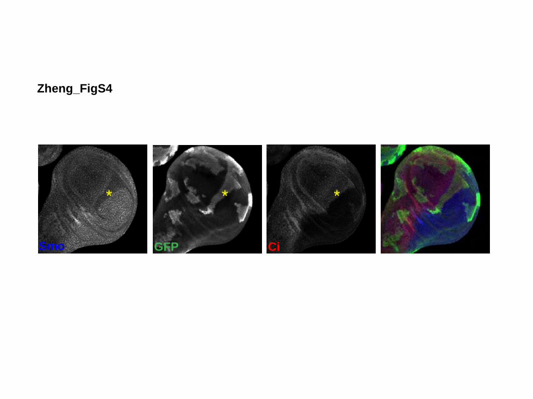
GFP Ci
Zheng_FigS4
* *
Smo
*
B

Overlay anti-GFP anti-Ptc anti-Ci
**
Zheng_FigS5

!Q655E
K653E
Ihog Fn2
Ihog Fn1
HhN
Zheng_FigS6

A
B
Ptc
+ yf
p R
NA
iPt
c +
boi
RN
Ai +
ihog
RN
Ai
Zheng_FigS7

Ptc
Ptc1
130
HhNPtc Overlay
A
B
C
D
Zheng_FigS8
Ptc1
130+
Ihog
Ptc+
Ihog
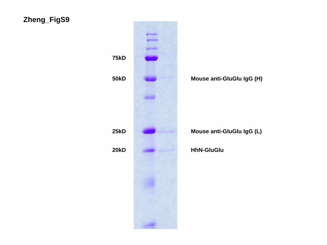
75kD
50kD
25kD
20kD
Mouse anti-GluGlu IgG (H)
Mouse anti-GluGlu IgG (L)
HhN-GluGlu
Zheng_FigS9

anti-GluGlu (HhN) anti-HA (Ihog or Boi)
1 2 3 4 5 76 8 9 10 11 12 1413
GFP
+ P
tc
Ihog
+ P
tc
Ihog
∆Fn1
+ P
tc
Ihog
∆Fn2
+ P
tc
Boi
∆Fn2
+ P
tc
Boi
∆Fn1
+ P
tc
Boi
+ P
tc
GFP
+ P
tc
Ihog
+ P
tc
Ihog
∆Fn1
+ P
tc
Ihog
∆Fn2
+ P
tc
Boi
∆Fn2
+ P
tc
Boi
∆Fn1
+ P
tc
Boi
+ P
tc
anti-HA
anti-Ptc
anti-HhN
Prec
ipita
tion
Imm
obili
zed
Prot
ein
Zheng_FigS10

Table S1. Primers Used in Figure 1 and Figure S2
Primers Location Sequence P1 exon 1 of ihog ATAAGTGATGCGCGATAGCC P2 exon 2 (deleted
region) of ihog AGGATGTGAGCAGCGTCATT
P3 white AGGTCGACTCTAGAGGATCA P4 exon 3 of boi AGCAGATACGCCTGGAAAGA P5 exon 5 (not deleted
region) of boi GCGGCAGACGAAAGTTTAAG
P6 exon 7 of boi ACATGCGATACTGCACCTTG