genetic and epigenetic mechanisms in the development of
TRANSCRIPT

1Wu Y, et al. World Jnl Ped Surgery 2021;4:e000196. doi:10.1136/wjps-2020-000196
Open access
Genetic and epigenetic mechanisms in the development of congenital heart diseases
Yue Wu,1 Xiaosi Jin,2 Yuhao Zhang,3 Jing Zheng,1 Rulai Yang1
To cite: Wu Y, Jin X, Zhang Y, et al. Genetic and epigenetic mechanisms in the development of congenital heart diseases. World Jnl Ped Surgery 2021;4:e000196. doi:10.1136/wjps-2020-000196
► Additional supplemental material is published online only. To view, please visit the journal online (http:// dx. doi. org/ 10. 1136/ wjps- 2020- 000196).
YW and XJ contributed equally.
Received 8 August 2020Revised 28 March 2021Accepted 30 March 2021
1Department of Genetic and Metabolic Diseases, The Children’s Hospital, Zhejiang University School of Medicine, National Clinical Research Center for Child Health, Hangzhou, China2Department of Obstetrics, Hangzhou Women’s Hospital, Hangzhou First People’s Hospital Qianjiang New City Campus, Hangzhou, China3Cardiovascular Key Lab of Zhejiang Province, Department of Cardiology, The Second Affiliated Hospital, Zhejiang University School of Medicine, Hangzhou, China
Correspondence toDr Rulai Yang; chsczx@ zju. edu. cn
Review
© Author(s) (or their employer(s)) 2021. Re- use permitted under CC BY- NC. No commercial re- use. See rights and permissions. Published by BMJ.
ABSTRACTCongenital heart disease (CHD) is the most common of congenital cardiovascular malformations associated with birth defects, and it results in significant morbidity and mortality worldwide. The classification of CHD is still elusive owing to the complex pathogenesis of CHD. Advances in molecular medicine have revealed the genetic basis of some heart anomalies. Genes associated with CHD might be modulated by various epigenetic factors. Thus, the genetic and epigenetic factors are gradually accepted as important triggers in the pathogenesis of CHD. However, few literatures have comprehensively elaborated the genetic and epigenetic mechanisms of CHD. This review focuses on the etiology of CHD from genetics and epigenetics to discuss the role of these factors in the development of CHD. The interactions between genetic and epigenetic in the pathogenesis of CHD are also elaborated. Chromosome abnormalities and gene mutations in genetics, and DNA methylations, histone modifications and on- coding RNAs in epigenetics are summarized in detail. We hope the summative knowledge of these etiologies may be useful for improved diagnosis and further elucidation of CHD so that morbidity and mortality of children with CHD can be reduced in the near future.
INTRODUCTIONCongenital heart disease (CHD) is a group of disorders attributed by abnormalities in fetal heart and large vessels that lead to actual or potential impairment of cardiac function in infants. CHD is the most common type of congenital defect worldwide and is the most common and the most life- threatening class of birth defects in infants, affecting approx-imately 1% live births annually worldwide.1 Epidemiological investigations indicate that the overall incidence of CHD varies across countries and continents, and the preva-lence of CHD in Asia is higher than that in North America.2 Despite benefits from the remarkable progress in therapeutic strategies of surgery and catheter intervention, CHD is still the principal source of mortality in infants. However, owing to medical, surgical and technological evolutions during the past decades, more than 90% of CHD infants now survive to adulthood.3 Improvement in
surgical intervention techniques and peri-operative care has dramatically changed the management of these populations with CHD. However, CHD is still a bothersome question owing to its undesirable outcomes and expen-sive healthcare costs, which bring substantial physiological, emotional and socioeconomic challenges to patients, families and society.
According to the final anatomical and pathophysiological complexities, CHD can be classified as mild, moderate or severe. Detailed classification is shown in box 1.4 The prognosis, morbidity and mortality vary with the severity of the anomalies. Despite the rapid advances in medical care and detection technology, the etiology of most CHD remains poorly understood. It is therefore imperative to improve our understanding of the disease mechanisms to reduce the frequent occur-rence of CHD. During the past decades a consensus has emerged that both genetic (eg, chromosomal abnormalities, smaller copy number variants and point mutations) and environmental (extrinsic factors, such as teratogen exposure and nutrient deficiencies; intrinsic factors, including maternal disease, illness and viral infection)5 factors are related to the occurrence of CHD. Progress in molec-ular genetic diagnosis has provided a valu-able opportunity to investigate the genetic factors of CHD. Furthermore, a multitude of animal models (eg, mouse, zebrafish, frog and fruit fly) have witnessed the significant effects of genetic etiology of CHD. These in vivo studies on animal models, in turn, have resulted in the identification of numerous structural genes, transcriptional regulators and signaling molecules that are critical for normal cardiac morphogenesis.6
To our knowledge, although numerous literatures have discussed the genetic mech-anisms of CHD, few have comprehensively elaborated the genetic and epigenetic mech-anisms of CHD. In this review, we focus on CHD origin from the etiology of genetics and epigenetics. Chromosomal abnormalities
on Decem
ber 17, 2021 by guest. Protected by copyright.
http://wjps.bm
j.com/
World Jnl P
ed Surgery: first published as 10.1136/w
jps-2020-000196 on 29 April 2021. D
ownloaded from

2 Wu Y, et al. World Jnl Ped Surgery 2021;4:e000196. doi:10.1136/wjps-2020-000196
Open access
and gene mutations in genetics, and DNA methylations, histone modifications and on- coding RNAs in epigenetics are summarized in detail. Moreover, we expect that
rapidly emerging data could provide a further under-standing of genetics and epigenetics in the development of CHD and also a basis for further exploring the early diagnosis and individualized therapy of CHD.
CHROMOSOME ABNORMALITIES UNDERLYING CHDChromosome abnormalities refer to abnormal chro-mosome numbers and structural aberrations including aneuploidies and copy number variations (CNVs), respec-tively. Conventional chromosome anomalies associated with CHD were identified half a century ago. A study by Pierpont et al7 suggested that ~30% of children with a chromosomal abnormality would suffer from CHD. In the following section, we summarize the involvement of chromosomal aneuploidies and CNVs in CHD in detail.
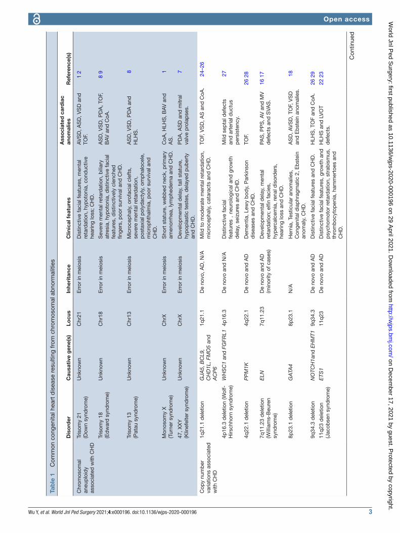
Aneuploidy in CHDChromosomal aneuploidy is the earliest recognized genetic cause of CHD and accounts for a great propor-tion of CHD (table 1). Approximately 50% of individ-uals born with trisomy 21 have the phenotypes of CHD, ranging from atrial septal defect (ASD)/ventricular septal defect (VSD) to atrioventricular canal lesions.1 2 The prevalence of CHD in newborns with trisomy 13 and trisomy 18 increases to 80%, and the major phenotypes of CHD are heterotaxy, laterality and septal defects.8 9 CHD is observed in approximately 33% of females with Turner syndrome or monosomy X, and the cardiac malformations are usually diagnosed as VSD, coarctation of aorta (CoA), bicuspid aortic valve and hypoplastic left heart.1 Although abnormal X chromosome numbers are rare, they also could result in CHD. For example, males with Klinefelter syndrome or 47, XXY have a 50% chance of CHD with the phenotypes of patent ductus arteriosus (PDA) and ASD.7 Moreover, sporadic 49, XXXXX cases with the phenotypes of ASD and vascular malformations have also been reported.10 At present, chromosomal G- banded karyotype analysis has been applied to detect the anomalies of chromosomes in spite of its limitation for the restrictive base resolution in exploring the tiny abnormalities of chromosomes.
CNVs in CHDConventional chromosomal microscopy in clinic could only detect the alterations of structure, numbers of chro-mosomes and abnormalities of large fragments, causing incomplete diagnosis of CHD. Numerous detection technologies include fluorescent in situ hybridization and multiplex ligation- dependent amplification. Chro-mosomal microarrays have been applied to explore the submicroscopic chromosomal anomalies and to eluci-date the pathogenic mechanisms of CHD, which may become a better approach for diagnosis.
CNVs refer to structural aberrations consisting of dele-tions or duplications, which are too small to be detected by routine karyotype analysis. A few CNVs can alter one or more contiguous genes and then inappropriately
Box 1 Classification of congenital heart disease (the copyright can be viewed in the online supplemental file)
Mild: ► Isolated congenital aortic valve disease and bicuspid aortic disease. ► Isolated congenital mitral valve disease (except parachute valve and cleft leaflet).
► Mild isolated PS (infundibular, valvular and supravalvular). ► Isolated small ASD, VSD or PDA. ► Repaired secundum ASD, sinus venosus defect, VSD or PDA without residuae or sequellae, such as chamber enlargement, ventricular dysfunction or elevated pulmonary artery pressure.
Moderate (repaired or unrepaired where not specified; alphabetical order):
► Anomalous pulmonary venous connection (partial or total). ► Anomalous coronary artery arising from the PA. ► Anomalous coronary artery arising from the opposite sinus. ► AS subvalvular or supravalvular. ► AVSD, partial or complete, including primum ASD (excluding pulmo-nary vascular disease).
► Coarctation of the aorta. ► Double- chambered right ventricle. ► Ebstein anomaly. ► Marfan syndrome and related HTAD and Turner syndrome. ► PDA, moderate or large unrepaired (excluding pulmonary vascular disease).
► PPS. ► PS (infundibular, valvular and supravalvular), moderate or severe. ► Sinus of Valsalva aneurysm/fistula. ► Sinus venosus defect. ► TOF repaired. ► Transposition of the great arteries after arterial switch operation. ► VSD with associated abnormalities (excluding pulmonary vascular disease) and/or moderate or greater shunt.
Severe (repaired or unrepaired where not specified; alphabetical order):
► Any CHD (repaired or unrepaired) associated with pulmonary vas-cular disease (including Eisenmenger syndrome).
► Any cyanotic CHD (unoperated or palliated). ► Double- outlet ventricle. ► Fontan circulation. ► IAA. ► Pulmonary atresia (all forms). ► Transposition of the great arteries (except for patients with arterial switch operation).
► Univentricular heart (including double inlet left/right ventricle, tri-cuspid/mitral atresia, hypoplastic left heart syndrome and any other anatomic abnormalitywith a functionally single ventricle).
► Truncus arteriosus. ► Other complex abnormalities of atrioventricular and ventriculoar-terial connection (ie, crisscross heart, heterotaxy syndromes and ventricular inversion).
AS, aortic stenosis; ASD, atrial septal defect; AVSD, atrioventricular septal defect; CHD, congenital heart disease; HTAD, heritable thoracic aortic disease; IAA, interrupted aortic arch; PA, pulmonary artery; PDA, patent ductus arteriosus; PPS, peripheral pulmonary stenosis; PS, pulmonary stenosis; TOF, tetralogy of Fallot ; VSD, ventricular septal defect.
on Decem
ber 17, 2021 by guest. Protected by copyright.
http://wjps.bm
j.com/
World Jnl P
ed Surgery: first published as 10.1136/w
jps-2020-000196 on 29 April 2021. D
ownloaded from
3Wu Y, et al. World Jnl Ped Surgery 2021;4:e000196. doi:10.1136/wjps-2020-000196
Open access
Tab
le 1
C
omm
on c
onge
nita
l hea
rt d
isea
se r
esul
ting
from
chr
omos
omal
ab
norm
aliti
es
Dis
ord
erC
ausa
tive
gen
e(s)
Locu
sIn
heri
tanc
eC
linic
al f
eatu
res
Ass
oci
ated
car
dia
c an
om
alie
sR
efer
ence
(s)
Chr
omos
omal
an
eup
loid
y as
soci
ated
with
CH
D
Tris
omy
21(D
own
synd
rom
e)U
nkno
wn
Chr
21E
rror
in m
eios
isD
istin
ctiv
e fa
cial
feat
ures
, men
tal
reta
rdat
ion,
hyp
oton
ia, c
ond
uctiv
e he
arin
g lo
ss; C
HD
.
AV
SD
, AS
D, V
SD
and
TO
F.1
2
Tris
omy
18(E
dw
ard
syn
dro
me)
Unk
now
nC
hr18
Err
or in
mei
osis
Sev
ere
men
tal r
etar
dat
ion,
bili
ary
atre
sia,
hyp
oton
ia, d
istin
ctiv
e fa
cial
fe
atur
es, d
istin
ctiv
ely
clen
ched
fin
gers
, poo
r su
rviv
al a
nd C
HD
.
AS
D, V
SD
, PD
A, T
OF,
B
AV
and
CoA
.8
9
Tris
omy
13(P
atau
syn
dro
me)
Unk
now
nC
hr13
Err
or in
mei
osis
Mic
roce
pha
ly, o
rofa
cial
cle
fts,
se
vere
men
tal r
etar
dat
ion,
p
osta
xial
pol
ydac
tyly
, om
pha
loce
le,
mic
rop
htha
lmia
, poo
r su
rviv
al a
nd
CH
D.
AS
D, V
SD
, PD
A a
nd
HLH
S.
8
Mon
osom
y X
(Tur
ner
synd
rom
e)U
nkno
wn
Chr
XE
rror
in m
eios
isS
hort
sta
ture
, web
bed
nec
k, p
rimar
y am
enor
rhea
, lym
phe
dem
a an
d C
HD
.C
oA, H
LHS
, BA
V a
nd
AS
.1
47, X
XY
(Klin
efel
ter
synd
rom
e)U
nkno
wn
Chr
XE
rror
in m
eios
isD
evel
opm
enta
l del
ay, t
all s
tatu
re,
hyp
opla
stic
tes
tes,
del
ayed
pub
erty
an
d C
HD
.
PD
A, A
SD
and
mitr
al
valv
e p
rola
pse
s.7
Cop
y nu
mb
er
varia
tions
ass
ocia
ted
w
ith C
HD
1q21
.1 d
elet
ion
GJA
5, B
CL9
, C
HD
1L, F
MO
5 an
d
AC
P6
1q21
.1D
e no
vo, A
D, N
/AM
ild t
o m
oder
ate
men
tal r
etar
dat
ion,
m
icro
cep
haly
, cat
arac
ts a
nd C
HD
.TO
F, V
SD
, AS
and
CoA
.24
–26
4p16
.3 d
elet
ion
(Wol
f-
Hirs
chho
rn s
ynd
rom
e)W
HS
C1
and
FG
FRL1
4p16
.3D
e no
vo a
nd N
/AD
istin
ctiv
e fa
cial
fe
atur
es,
neur
olog
ical
and
gro
wth
d
elay
, sei
zure
s an
d C
HD
.
Mild
sep
tal d
efec
ts
and
art
eria
l duc
tus
per
sist
ency
.
27
4q22
.1 d
elet
ion
PP
M1K
4q22
.1D
e no
vo a
nd A
DD
emen
tia, L
ewy
bod
y, P
arki
nson
d
isea
ses
and
CH
D.
TOF.
26 2
8
7q11
.23
del
etio
n (W
illia
ms-
Beu
ren
synd
rom
e)
ELN
7q11
.23
De
novo
and
AD
(m
inor
ity o
f cas
es)
Dev
elop
men
tal d
elay
, men
tal
reta
rdat
ion;
elfi
n fa
cies
, hy
per
calc
emia
, ren
al d
isor
der
s,
hear
ing
loss
and
CH
D.
PAS
, PP
S, A
V a
nd M
V
def
ects
and
SVA
S.
16 1
7
8p23
.1 d
elet
ion
GA
TA4
8p23
.1N
/AH
erni
a, T
estic
ular
ano
mal
ies,
C
onge
nita
l dia
phr
agm
atic
2, E
bst
ein
anom
aly,
CH
D.
AS
D, A
VS
D, T
OF,
VS
D
and
Eb
stei
n an
omal
ies.
18
9q34
.3 d
elet
ion
NO
TCH
1and
EH
MT1
9q34
.3D
e no
vo a
nd A
DD
istin
ctiv
e fa
cial
feat
ures
and
CH
D.
HLH
S, T
OF
and
CoA
.26
29
11q
23 d
elet
ion
(Jac
obse
n sy
ndro
me)
ETS
111
q23
De
novo
and
AD
Dis
tinct
ive
faci
al fe
atur
es, g
row
th a
nd
psy
chom
otor
ret
ard
atio
n, s
trab
ism
us,
thro
mb
ocyt
open
ia, h
amm
erto
es a
nd
CH
D.
HLH
S a
nd L
VO
T d
efec
ts.
22 2
3
Con
tinue
d
on Decem
ber 17, 2021 by guest. Protected by copyright.
http://wjps.bm
j.com/
World Jnl P
ed Surgery: first published as 10.1136/w
jps-2020-000196 on 29 April 2021. D
ownloaded from

4 Wu Y, et al. World Jnl Ped Surgery 2021;4:e000196. doi:10.1136/wjps-2020-000196
Open access
affect their expression, leading to the development of CHD.11 12 CNVs can occur de novo in sporadic cases, or they can be inherited familiarly causing complex congen-ital heart malformations. However, the underlying mech-anisms of CNVs in CHD still need to be elucidated. One of the most common CNVs syndromes in CHD, 22q11.2 deletion syndrome (DiGeorge syndrome or velocardio-facial syndrome), is caused by a microscopic deletion on chromosome 22q11.2. Variable phenotypes, such as craniofacial abnormalities, neurocognitive disabilities, palate abnormalities, hypocalcemia and immunodefi-ciencies, can be observed in this syndrome. The main cardiac malformations of this syndrome contain VSD, arch abnormalities and tetralogy of Fallot (TOF).13 To date, more than 30 gene deletions have been identified in the 22q11.2 locus by sequencing. Deletions in the T- box transcription factor TBX1 account for the major molec-ular basis of these cardiac malformations.14 Further work revealed that distal 22q11.2 deletion presented atypical clinical features of DiGeorge syndrome; therefore, this deletion was proposed as one of the etiologies of CHD.15 Williams- Beuren syndrome, which is characterized by supravalvar aortic stenosis (SVAS), peripheral pulmon-onic stenosis, coronary artery stenosis, pulmonary artery sling, developmental delays, typical elfin facies, infantile hypercalcemia and cognitive disability, has been report-edly caused by microdeletion of over 25 genes in the 7q11.23 region.16 It has been known that cardiovascular abnormalities, such as SVAS, can be induced by haploin-sufficiency of the elastin gene (ELN).17
Some CNVs encompass previously identified CHD genes, or genes known to be implicated in heart develop-ment. For example, 8 p deletion syndrome and mutations in GATA4, a cardiac transcription factor, are reported to be associated with CHD.18 The 8p23.1 delete region that overlaps with the locus of GATA4 could elucidate this causal outcome. However, Kumar et al reported an interesting case of 8p23.3p23.1 deletion and 8p23.1p11.1 interstitial duplication syndrome that a male toddler with global developmental delay, dysmorphic facies, seizures and large doubly committed VSD occurred without the GATA4 gene involvement.19 Duplications at the 8p23.1 locus have also been identified in CHD, including ASD, VSD and TOF.20 21 Other studies have confirmed that the CNVs at chromosome 11q23 were associated with Jacobsen syndrome.22 23 Moreover, several CNVs have been identified from larger cohorts of patients with CHD, including 1q21.1,24–26 4p16.3,27 4q22.1,26 28 9q34.326 29 and 15q11.2.28 All of these CNVs mentioned above are detailed in table 1 for their relationship to CHD in human.
GENE MUTATIONS UNDERLYING CHDNumerous mutations are implicated in the develop-ment of CHD (table 2). Some mutations are identified in pedigrees of CHD, while others are initially observed in sporadic cases of CHD.2 Currently, mutations in more
Dis
ord
erC
ausa
tive
gen
e(s)
Locu
sIn
heri
tanc
eC
linic
al f
eatu
res
Ass
oci
ated
car
dia
c an
om
alie
sR
efer
ence
(s)
15q
11.2
del
etio
nTU
BG
CP
5, C
YFI
P1,
N
IPA
2 an
d N
IPA
115
q11
.2N
/AD
elay
ed p
sych
omot
or d
evel
opm
ent,
sp
eech
del
ay, a
utis
m s
pec
trum
d
isor
der
, att
entio
n d
efici
t-
hyp
erac
tivity
dis
ord
er, o
bse
ssiv
e–co
mp
ulsi
ve d
isor
der
, pos
sib
ly
seiz
ures
and
CH
D.
AS
D, V
SD
, CoA
, TA
PV
D a
nd c
omp
lex
left
sid
ed.
28
22q
11.2
del
etio
n (D
iGeo
rge
Syn
dro
me)
TBX
122
q11
.2D
e no
vo a
nd A
D
(28%
of c
ases
).Th
ymus
and
par
athy
roid
ap
lasi
a or
hyp
opla
sia,
imm
unod
efici
ency
, hy
poc
alce
mia
, dis
tinct
ive
faci
al
feat
ures
, OFT
ab
norm
aliti
es a
nd C
HD
.
TOF,
IAA
typ
e B
, V
SD
, TA
, aor
tic a
rch
anom
alie
s an
d t
runc
us
arte
riosu
s.
13 1
4
Dis
tal 2
2q11
.2 d
elet
ion
CR
KL
and
ER
K2/
MA
PK
122
q11
.22
De
novo
and
AR
(m
inor
ity o
f cas
es)
Dis
tinct
ive
faci
al fe
atur
es, p
sych
iatr
ic
and
cog
nitiv
e d
efici
ts, s
epsi
s an
d
CH
D.
Inte
rrup
ted
aor
tic a
rch
and
tru
ncus
art
erio
sus.
15
AD
, aut
osom
al d
omin
ant;
AR
, aut
osom
e re
cess
ive;
AS
, aor
tic s
teno
sis;
AS
D, a
tria
l sep
tal d
efec
t; A
V, a
ortic
val
ve; A
VS
D, a
trio
vent
ricul
ar s
epta
l def
ect;
BA
V, b
icus
pid
aor
tic v
alve
; CH
D, c
onge
nita
l hea
rt
dis
ease
; Chr
, chr
omos
ome;
CoA
, coa
rcta
tion
of t
he a
orta
; HLH
S, h
ypop
last
ic le
ft h
eart
syn
dro
me;
IAA
, int
erru
pte
d a
ortic
arc
h; L
VO
T, le
ft v
entr
icul
ar o
utflo
w t
ract
; MV,
mitr
al v
alve
; N/A
, not
ava
ilab
le;
OFT
, out
flow
tra
ct; P
AS
, pul
mon
ary
arte
ry s
teno
sis;
PD
A, p
aten
t d
uctu
s ar
terio
sus;
PP
S, p
erip
hera
l pul
mon
ic s
teno
sis;
SVA
S, s
upra
valv
ar a
ortic
ste
nosi
s; T
A, t
ricus
pid
atr
esia
; TA
PV
D, t
otal
ano
mal
ous
pul
mon
ary
veno
us d
rain
age;
TO
F, t
etra
logy
of F
allo
t; V
SD
, ven
tric
ular
sep
tal d
efec
t.
Tab
le 1
C
ontin
ued
on Decem
ber 17, 2021 by guest. Protected by copyright.
http://wjps.bm
j.com/
World Jnl P
ed Surgery: first published as 10.1136/w
jps-2020-000196 on 29 April 2021. D
ownloaded from

5Wu Y, et al. World Jnl Ped Surgery 2021;4:e000196. doi:10.1136/wjps-2020-000196
Open access
Table 2 Common congenital heart disease resulting from single gene defects
Gene Locus Protein Cardiac phenotype OMIM
Gene associated with transcription factors of cardiac development
GATA4 8p23.1 GATA4 transcription factor ASD, VSD, AVSD, PS, PAPVR and TOF.
600 576
GATA5 20q13.33 GATA5 transcription factor Congenital bicuspid aortic valve and VSD.
617 912
GATA6 18q11.2 GATA6 transcription factor ASD, VSD, AVSD, OFT defects, PDA, PS and TOF.
601 656
NKX2.5 5q35.1 Homeobox containing transcription factor ASD, VSD, TOF, HLHS, CoA, TGA, DORV, IAA and OFT defects.
600 584
NKX2.6 8p21.2 Homeobox containing transcription factor PTA and conotruncal heart malformations.
217 095
TBX1 22q11.2 T- Box 1 transcription factor TOF. 602 054
TBX5 12q24.21 T- Box 5 transcription factor ASD, VSD and AVSD. 601 620
TBX20 7p14.2 T- Box 20 transcription factor ASD, VSD and MS. 611 363
TFAP2β 6p12.3 Transcription factor AP-2 beta PDA. 601 601
ZIC3 Xq26.3 Zinc finger transcription factor ASD, VSD, HLHS, DORV, PS, TGA, TAPVR, dextrocardia, L- R axis defects and heterotaxy.
300 265
Gene associated with signaling pathways of cardiac development
AXIN2 17q24.1 Axin- related protein 2 Congenital valve defect. /
BRAF 7q34 Serine/threonine- protein kinase B- raf ASD, and PAS. 164 757
CBL 11q23.3 E3 ubiquitin- protein ligase CBL AVSD, HCM and PS. 613 563
DLL4 15q15.1 Delta- like protein 4 Left- sided obstructive lesions, septal and conotruncal defects and tricuspid atresia.
616 589
FOXH1 8q24.3 Forkhead box protein H1 TOF and TGA. /
GALNT11 7q36.1 Polypeptide N- acetylgalactosaminyltransferase 11
Heterotaxy. /
GLI1 12q13.3 Zinc finger protein GLI1 Abnormity of atrioventricular separation and cardiac OFT.
165 220
HHEX 10q23.33 Hematopoietically expressed homeobox Ventricular aplasia, dense myocardial dysplasia and intracardiac membrane dysplasia.
/
HRAS 11p15.5 GTPase HRas PAS and tachycardia. 190 020
JAG1 20p12.2 Protein Jagged-1 PAS and TOF. 601 920
KRAS 12p12.1 GTPase KRas ASD and PAS. 190 070
MAML1 5q35.3 Mastermind- like protein 1 Aortic valve disease. /
MEK1 15q22.31 Dual specificity mitogen- activated protein kinase kinase 1
ASD and PAS. 176 872
MEK2 19p13.3 Dual specificity mitogen- activated protein kinase kinase 2
ASD and PAS. 601 263
NOTCH1 9q34.3 Notch receptor 1 Aortic valve disease. 190 198
NOTCH2 1p12 Notch receptor 2 AS, TOF and PAS. 610 205
NRAS 1p13.2 GTPase NRas HCM and PS. 164 790
PPP1CB 2p23.2 Serine/threonine- protein phosphatase PP1- beta catalytic subunit
ASD, VSD, HCM, PAS and TOF.
617 506
PTPN11 12q24.13 Protein tyrosine phosphatase non- receptor type 11
ASD, VSD and PAS. 176 876
Continued
on Decem
ber 17, 2021 by guest. Protected by copyright.
http://wjps.bm
j.com/
World Jnl P
ed Surgery: first published as 10.1136/w
jps-2020-000196 on 29 April 2021. D
ownloaded from

6 Wu Y, et al. World Jnl Ped Surgery 2021;4:e000196. doi:10.1136/wjps-2020-000196
Open access
than 50 genes have been found to be associated with CHD by the application of high- throughput sequencing of whole- genome and whole- exome, and many of these affected genes have been confirmed to be involved in transcriptional regulation, signal transduction and cardiac development.
Mutations of genes encoding transcription factors in CHDHeart development is regulated by several transcriptional circuits that are members of a core group of transcrip-tion factors, including NKX2.5, GATA4 and TBX5.30 31 Therefore, transcription factors have been considered as the prime inducer of CHD. NKX2.5 is the earliest known marker of myocardial progenitor cells in all species.32 The mechanisms of NKX2.5 regulation and its interac-tion with other transcription factors in early cardiac development have been studied extensively. It has been found that mutations in NKX2.5 could result in variable types of CHD, including ASD, VSD, TOF, hypoplastic left heart syndrome, CoA, transposition of the great arteries, double outlet right ventricle (DORV), interrupted aortic arch and cardiac outflow tract defects.33 Furthermore, NKX2.5 mutations are the most common cause of ASD in individuals with defects of conduction system.34 35
However, some individuals with CHD caused by the mutations of NKX2.5 can manifest an individual pheno-type of ASD and/or conduction defects.36 To date, approx-imately 80 different mutations have been identified in
NKX2.5, including missense mutations [eg, c.44A>T (p.K15I), c.232A>G (p.N19S), c.673C>A (p.N188K) and c.1089A>G (p.S305G)], synonymous mutations [eg, c.543G>A (p.Q181Q), c.677A>G (p.E167E), c.902C>G (p.G242G) and c.1142A>G (p.R322R)] and nonsense mutations [eg, c.1149T>C (stop→Gln)].33 The location of some of these mutations is depicted in figure 1A. Holt- Oram syndrome, which is characterized by CHD (eg, ASD, VSD and atrioventricular conduction system disease) and upper limb malformations, could be caused by the loss- of- function mutations in TBX5, which is notably expressed in the upper limbs and heart.37 38 Nonsense or frameshift mutations of TBX5 may be responsible for this syndrome.12 Functional deficiency of the conserved DNA- binding motif in the transcription factor encoded by TBX5 may be the etiology of Holt- Oram syndrome.39
GATA4, encoding one of the GATA zinc- finger tran-scription factors, is a deeply studied gene and is essential for cardiogenesis. Mutations in GATA4 are reported to be implicated in cardiac septal defects.31 Over 100 muta-tions in GATA4 coding region have been identified in patients with CHD, and more mutations will be identi-fied with intensive research.40 Among these mutations, 11 sites (two synonymous mutations, seven missense muta-tions and two frameshift mutations) have been studied in familial cases, which highlight the significance of these sites in the development of CHD (figure 1B). Multiple mutations identified from other transcription factors,
Gene Locus Protein Cardiac phenotype OMIM
RAF1 3p25.2 RAF proto- oncogene serine/threonine- protein kinase
ASD and TOF. 164 760
RIT1 1q22 GTP- binding protein Rit1 VSD, TOF and PAS. 615 355
SHOC2 10q25.2 Leucine- rich repeat protein SHOC-2 ASD, VSD, HCM, PAS and TOF.
607 721
SMAD6 15q22.31 Mothers against decapentaplegic- related protein 6
AV disease. 602 931
SOS1 2p22.1 Son of sevenless homolog 1 ASD, VSD and TOF. 182 530
SOS2 14q21.3 Son of sevenless homolog 2 ASD, VSD and TOF. 616 559
TGF-β1 19q13.2 Transforming growth factor beta-1 proprotein
CoA, HLHS, BAV and AS. /
Gene associated with structural proteins of cardiac development
ACTC 15q14 Alpha cardiac actin ASD. 102 540
ELN 7q11.23 Elastin AS, PAS, PS and SVAS. 130 160
MYH6 14q11.2 Alpha myosin heavy chain AS, ASD, PFO, TA and TGA. 160 710
MYH7 14q11.2 Beta myosin heavy chain ASD, NVM and Ebstein anomaly.
160 760
MYH11 16p13.11 Myosin heavy chain 11 Aortic aneurysm and PDA. 132 900
AS, aortic stenosis; ASD, atrial septal defect; AV, aortic valve; AVSD, atrioventricular septal defect; BAV, bicuspid aortic valve; CoA, coarctation of the aorta; DORV, double outlet right ventricle; HCM, hypertrophic cardiomyopathy; HLHS, hypoplastic left heart syndrome; IAA, interrupted aortic arch; L- R, Left- right; MS, mitral stenosis; NVM, non- compaction of ventricular myocardium; OFT, outflow tract; OMIM, online mendelian inheritance in man; PAPVR, partial anomalous pulmonary venous return; PAS, pulmonary artery stenosis; PDA, patent ductus arteriosus; PFO, patent foramen ovale; PS, pulmonary (valve) stenosis; PTA, persistent truncus arteriosus; SVAS, supravalvar aortic stenosis; TA, tricuspid atresia; TAPVR, total anomalous pulmonary venous return; TGA, transposition of the great arteries; TOF, tetralogy of Fallot; VSD, ventricular septal defect.
Table 2 Continued
on Decem
ber 17, 2021 by guest. Protected by copyright.
http://wjps.bm
j.com/
World Jnl P
ed Surgery: first published as 10.1136/w
jps-2020-000196 on 29 April 2021. D
ownloaded from

7Wu Y, et al. World Jnl Ped Surgery 2021;4:e000196. doi:10.1136/wjps-2020-000196
Open access
including NKX2.6,41 GATA5,42–44 GATA6,45 TFAP2β,46 TBX1,47TBX2048 49 and ZIC3,50 have also been reported to be associated with the incidence of CHD.
Mutations of genes encoding signal proteins in CHDSignaling pathways involved in the occurrence of CHD are widely studied. Genes involved in these different sign-aling pathways can converge into a large and sophisti-cated regulatory network that plays an important role in cardiac development and pathogenesis of CHD. Recent studies have also suggested the potential contributions of vascular endothelial growth factor- A (VEGF- A), Notch signaling, Wnt signaling, transforming growth factor-β (TGF-β), bone morphogenic protein (BMP) signaling and cpathway to the occurrence of CHD.51–57 In these pathways, some are essential for the formation of cardiac septum, valves and the construction of cardiac outflow tracts, while others are associated with the asymmetric development of the heart. Gene mutations in renin–angi-otensin system mitogen- activated protein kinase (RAS- MAPK) signal transduction pathway can lead to Noonan syndrome with the typical phenotype of pulmonary valve stenosis and hypertrophic cardiomyopathy.58–61
Six missense variants (COL6A1, COL6A2, CRELD1, FBLN2, FRZB and GATA5), acting in the VEGF- A pathway, were found to be damaged in individuals with complete atrioventricular septal defect (AVSD),51 suggesting that rare variants in the VEGF- A pathway might play a role in the development of AVSD. In addition, mutations in VEGF- A have been reported to be associated with congenital left ventricular outflow tract obstruction.62 Notch signaling is a highly conserved pathway involved in developmental process of heart. JAG1 encodes a ligand in
the Notch signaling pathway, which leads to localization of Notch to the nucleus and downstream activation of target genes. Mutations in JAG1 have been found in over 90% cases of Alagille syndrome.63 64 Some cases (~2%) that have mutations in NOTCH2, a NOTCH receptor gene, are also correlated with Alagille syndrome.65 However, variants of NOTCH1 that belongs to the Notch signaling pathway have been identified to be associated with Adams- Oliver syndrome.15 Mind bomb 1 (Mib1) is a vital protein that promotes ubiquitination, endocytosis and subsequent activation of Notch ligands to activate the Notch signaling pathway. Mutations in MIB1 have been identified to be associated with cardiac deformity such as ASD, AVSD and VSD, through a lower level of JAG1 ubiquitination and Notch signaling induction.66 In addition, mutations in other genes of Notch signaling pathway include MAML1,67 DLL468 and GALNT1169 are also involved in CHD.
AXIN2 is involved in the regulation network of cardiac valve formation and elongation, and its expres-sion product is a negative regulator of Wnt/β-catenin signaling pathway.70 71 It has been found that mutations in AXIN2 can result in CHD with the phenotype of congen-ital valve defect.72 HHEX, a member of the Homeobox gene family, is an important cardiac determinant and controls the early differentiation, migration and devel-opment of cardiomyocytes.73 Foley et al74 reported that mutations in HHEX could lead to a phenotype of abnormal developmental endogenous cardiac or ectopic heart, which was similar to the antagonistic effect of Dick-kopf-1 to Wnt signaling pathway. Aberrant Wnt signaling pathways implicated in CHD have been summarized in a previous excellent literature.75 We describe the aberrant expression of genes associated with CHD within the Wnt signaling pathways in figure 2. TGF-β signaling pathway has important role in the development and remodeling of cardiovascular system. Aberrant TGF-β signaling pathway is involved in the pathogenesis of several human cardiovascular diseases through the epithelial- to- mesenchymal transition (EMT) of resident fibroblasts, circulating progenitors, pericytes, epithelial cells and/or the endothelial- mesenchymal transdifferentiation of endothelial cells.76 77 For example, mutations in TGF-β1, one major subtype of TGF-β family, have been reported to be associated with CHD in pediatric patients.78 In general, BMP synergizes with TGF-β signaling to activate the downstream genes, such as Smad1, Smad5 or Smad8, with the changed transcription of target genes. Muta-tions in the genes that encode transducers of the TGF-β and BMP signaling pathway have been identified in the pathogenesis of cardiovascular diseases, such as Marfan syndrome and Loeys- Dietz syndrome.55 79 Nodal signaling pathway is involved in the left–right patterning and development of the heart and in abnormal gene prod-ucts throughout the pathway that are clearly associated with CHD. Roessler et al56 previously demonstrated that reduced nodal signaling strength via mutation of FOXH1 was linked to human heart defects. SHH, one morphogen
Figure 1 Spectrum of some significant mutant sites of (A) NKX2.5 and (B) GATA4 observed in the occurrence of congenital heart defects. CZF, C- terminal zinc finger domain; HD, homeodomain; NK2, NK-2- specific domain; NLS, nuclear localization signal; NZF, N- terminal zinc finger domain; TAD1, transcription activation domain 1; TAD2, transcription activation domain 2; TN, tin- man domain/transcriptional repression domain.
on Decem
ber 17, 2021 by guest. Protected by copyright.
http://wjps.bm
j.com/
World Jnl P
ed Surgery: first published as 10.1136/w
jps-2020-000196 on 29 April 2021. D
ownloaded from

8 Wu Y, et al. World Jnl Ped Surgery 2021;4:e000196. doi:10.1136/wjps-2020-000196
Open access
of hedgehog (HH) family proteins, is involved in a remarkably wide variety of process, including cardiovas-cular development. Previous evidence demonstrated the mutations in genes of HH signaling were implicated in the occurrence of CHD with a phenotype of ASD, VSD or AVSD, and the responsible mutated genes include SHH, Gli3 and MKS1.80–83 Noonan syndrome, one of the most common genetic syndromes of CHD, is caused by muta-tions in genes of the RAS- MAPK pathway.84 At present, several genes have been verified to be responsible for the development of Noonan syndrome and other disorders. Mutations in genes, encoding molecules implicated in the RAS- MAPK signaling pathway, account for approxi-mately 90% of affected CHD cases. These genes include: PTPN11, SOS1, KRAS, RAF1, BRAF, SHOC2, NRAS, HRAS, CBL, MEK1, MEK2, PPP1CB, RIT1 and SOS2.15 58 61 84
Currently, most studies have only stayed on the basic exploration of a single gene, which cannot fully explain the pathogenesis of CHD. The interactions of various signaling pathways involved in heart development are still incompletely understood. Some responsible mutated genes have been proposed to clarify the interactions of the signaling pathways implicated in CHD (figure 2).
More researches are needed to help elaborate the patho-genic mechanisms of CHD for the intricate network of signaling pathway.
Mutations of genes encoding cardiac structural proteins in CHDSeveral studies use targeted whole- genome sequencing to investigate the genes that encode cardiac structural proteins to elucidate the monogenic cause of CHD. Some rare missense mutations or premature termination muta-tions in myosin heavy chain 6 (MYH6), a marker gene of myocardial cell, could result in ASD.85 86 However, mutations in MYH7 have been confirmed in bilobal aortic valve.87 CHD with apical hypertrophic cardiomyo-pathy, left ventricular non- compaction and ASD could be caused by the mutations in ACTC, which is a cardiac actin gene and is essential for cardiac contraction.88 89 MYH11, encoding smooth muscle myosin heavy chain, is another gene that has been reported to be involved in CHD with the phenotype of dominant thoracic aortic aneurysm.90 91 Moreover, loss- of- function mutations in the elastin gene (ELN) could result in CHD with the presence of Williams- Beuren syndrome and non- syndromic SVAS.17
Figure 2 The relationships of different signaling pathways and some responsible mutated genes within these pathways in cardiac development and CHD. CHD, congenital heart disease; ECM, extracellular matrix; EMT, epithelial- mesenchymal transition; PAS, pulmonary artery stenosis; TA, tricuspid atresia; TOF, tetralogy of Fallot; VSD, ventricular septal defect.
on Decem
ber 17, 2021 by guest. Protected by copyright.
http://wjps.bm
j.com/
World Jnl P
ed Surgery: first published as 10.1136/w
jps-2020-000196 on 29 April 2021. D
ownloaded from

9Wu Y, et al. World Jnl Ped Surgery 2021;4:e000196. doi:10.1136/wjps-2020-000196
Open access
EPIGENETIC MODIFICATIONS UNDERLYING CHDEpigenetics, which refers to the mechanisms of changed gene expression that are independent of DNA sequence, provides a new way to understand the pathogenesis of CHD. To date, three canonical mechanisms of epige-netics include DNA methylation, histone modification and non- coding RNAs. The increasing evidence suggests that the aberrant regulation of gene expression by epige-netics is a key factor in the development of cardiovascular diseases, which have attracted attention to focus on the role of epigenetics in CHD.
DNA methylation modifications in CHDDNA methylation, the most widely studied epigenetic mechanism, refers to the formation of a methyl group (−CH3) in the 5′ carbon of cytosine (CpG islands), which induces an alteration of the structure of DNA. The meth-ylation process is catalyzed by DNA methyltransferases (DNMTs), comprising DNMT1, DNMT3A and DNMT3B. The dysregulation of DNA methylation during different stages of development could lead to the transcriptional repression and functional inactivation of tissue- specific genes, resulting in increased risk for several diseases, including cardiac malformation. Alterations of DNA methylation, especially in CpG islands, which are close to core transcription factors and genes in signaling pathway, have been reported in patients with cardiac malforma-tions.92
DNA methylation plays a critical role in the develop-ment of the heart. The expression of hyaluronan synthase 2 (Has2) is necessary for the formation of the heart valves, septa and epicardium. However, Has expression was found to be downregulated via DNA methylation in the heart at E14.5 embryos.93 Furthermore, gene knockout model indicated that expression of Has2 is downregu-lated via DNA methyltransferase 3B (DNMT3B), which was coexpressed with Has2 in the region of cardiac valve, suggesting that changes in DNA methylation might be involved in the regulatory function of Has2 enhancer. Aberrant methylation of CITED2 may play an important role in the development of VSD, ASD and TOF.94 Aber-rant methylations of CITED2 could decrease its mRNA expression and be regarded as the prime cause of CHD.95 The presence of aberrant hypomethylation in the CpG region of BRG1 was found to exist in patients with ASD and interventricular septal defect.96 It has been shown that the promoter region of CX43 plays an essential role in the development of the heart outflow tract, and the aberrant hypomethylation of this enhancer region of CX43 could be considered as one of the etiologies of CHD.97 98
In another study, two encoding transcription factors, zinc- finger in cerebellum 3 and nuclear receptor subfamily 2 group F member 2, were found to be hyper-methylated in monozygotic twins with DORV, suggesting that the differential methylation of these transcription factors could be regarded as a potential pathogenesis of the diseased twin.99 Hypermethylation of the promoter
region of SCO2, a cytochrome oxidase, has been found in patients with TOF and VSD. The methylation of CpG islands located in the promoter of SCO2 result in reduced expression of SCO2, which may be the mech-anism of the occurrence of these diseases.100 Further studies discovered that hypermethylation of CpG islands of NKX2.5, HAND1 and RXRα was also essential for CHD, including VSD and TOF.101 102 Multiple differential meth-ylated genes have been identified from cardiomyocytes of newborn and adults, and adult failing hearts have revealed a highly dynamic of DNA methylation under specific developmental or pathological conditions. DNA methylation is tightly regulated during cardiac differen-tiation and maturation.103 Taken together, these findings highlight the importance of DNA methylation in cardiac morphogenesis and CHD formation.
Histone modifications in CHDHistones, including H1, H2A, H2B, H3 and H4, and DNA constitute the nucleosome, which is the basic struc-tural unit of chromatin in eukaryotic cells.104 Altering the histone- DNA contacts effectively via histone, post- translational modifications could loosen or tighten the chromatin architecture to control the availability of gene transcription or expression. Histone post- translational modifications could be modified by the catalysis of histone- modifying enzymes, such as histone methylases, demethylases, acetylases and deacetylases, ubiquitin enzymes and phosphorylases. Aberrant expression and mutation of the histone modifiers during the develop-ment of heart can influence the response of heart to pathological stresses.105 Moreover, increasing researches show that the interplay between these different cardiac transcription factors and histone modifiers plays a signif-icant role in heart development. Numerous studies have shown that methylation and acetylation of histone is an emerging epigenetic mechanism for the regulation of gene transcription. Therefore, we focused only the mech-anism of histone methylation and acetylation in CHD.
Methylation mainly occurs at the core histones H3 and H4, which is performed by the catalysis of histone methyl-transferases. H3K4, H3K36 and H3K79 methylation leads to the transcriptional activation, whereas transcriptional repression can be induced by H3K9, H3K27 and H4K20 methylation.106 CHD associated with Wolf- Hirschhorn syndrome exists deletion of Wolf - Hirschhorn candidate protein 1 (Whsc1), which encodes the H3K36me3- specific methyltransferase. Nimura et al107 found that the patho-genic role of Whsc1 was associated with the transcrip-tional activation of Nkx2.5 in its target sites through the model of Whsc1- knockout murine. Another studies revealed that the interactions of JARID2 and SETDB1, which was an H3K9me3- specific methyltransferase, could elucidate the role of Jarid2 in the occurrence of VSD, DORV and impaired ventricular compaction induced by hypertrabeculation.108 109 TBX1, one member of T- box transcription factor family, was shown to interact with H3K4 and H3K27 via two domains of T- box to regulate
on Decem
ber 17, 2021 by guest. Protected by copyright.
http://wjps.bm
j.com/
World Jnl P
ed Surgery: first published as 10.1136/w
jps-2020-000196 on 29 April 2021. D
ownloaded from

10 Wu Y, et al. World Jnl Ped Surgery 2021;4:e000196. doi:10.1136/wjps-2020-000196
Open access
gene expression, and aberrant expression could result in CHD including TOF, VSD and aortic arch interrup-tion.110 111 A later study further identified the interactions of TBX1 and BAF chromatin remodeling complex regu-late Wnt5a expression, and the insufficient expression of TBX1 or Wnt5a could result in the phenotypes of hypo-plastic right heart.112 DPF3, an evolutionarily conserved protein, binds methylated and acetylated lysine residues of histone 3 and 4 to regulate gene expression. Dpf3 is expressed in the heart during development and has been found as significantly upregulated in the right ventricular myocardium of patients with TOF.113
Histone acetylation and deacetylation are always in a dynamic state, which is associated with transcriptional activation or transcriptional repression, gene silencing and cell cycle. Histone acetylation and deacetylation have been studied extensively in recent years.114 It has been shown that some histone acetylases, such as EP300, KAT2A and hNAT1, are associated with the develop-ment of the heart. Aberrant expression of these acetyl-ases could lead to CHD with ASD, VSD, AVSD and valve dysplasia.115 116 Previous studies have revealed that histone deacetylases (HDACs) are also implicated in CHD. For example, HDAC3 regulates the cellular acetylation level affecting cardiogenesis, and the absence of HDAC3 could cause PDA and TOF.117 In addition, down- regulation of HDAC5 and HDAC9 simultaneously could result in CHD with VSD.118 119 Abnormal regulation of SIRT1 has been reported to be involved in ventricular hypoplasia.120 Park et al121 revealed that histone methyltransferase SMYD1 was associated with ventricular hypoplasia. Some other histone modifiers have been shown to be important for the development of the heart through numerous studies, such as G9a, Ezh2/PRC2, Baf60c/Brg1, Jmjd3, UTX and MLL2,122–130 and the aberrant modification of them could yield critical CHD phenotypes. The activity of cardiac transcription factor could be changed by histone modifiers, which means these modifiers could be strong candidates for CHD etiology and therapeutic targeting.
Non-coding RNA in CHDNon- coding RNA is another type of epigenetic modi-fication involved in the control of gene expression by post- transcriptional regulation. Among the various non- coding RNAs, microRNAs (miRNAs) and long non- coding RNAs (LncRNAs) are the two best studied groups. miRNAs are capable of regulating gene expres-sion by interacting with mRNA transcript 3′ untranslated regions (UTRs) to repress translation, whereas LncRNAs are able to regulate transcription by directly interacting with chromatin remodeling complexes.131 Emerging evidence that indicates the impact of non- coding RNAs to cardiac development was previously unappreciated but is becoming valuable.
A number of miRNAs have recently been shown to func-tion in the heart.132 It has been shown miR-1 is important in cardiac development and is regarded as the most rele-vant miRNA leading to CHD.133 134 Li et al found that
expression of miRNA-1 decreased in patient with VSD.135 Molecularly, miRNA-1 binds to its target gene GJA1 and SOX9, regulating the formation of cardiac valves and septa, and therefore miRNA-1 dysregulation could result in CHD in human.135 Further studies have demonstrated that myocyte enhancer factor 2 (Mef2) could upregu-late the expression of miR-1, which suppress the cardiac transcription factor Hand2 and HDAC translation.133 136 Hand2 is known as a target of miRNA-1 and is involved in the growth of the embryonic heart; thus, Hand2 could be associated with CHD.133 Overexpression of miRNA- 27b could repress the expression of Mef2c and affect the development of myocardial, leading to cardiac hyper-trophy.137 A recent study has confirmed that differentially expressed frataxin (FXN) regulates the development of CHD and that differential expression of FXN is under the control of miRNA-145.138 Further investigation of this study demonstrated that overexpression of miRNA-145 could regulate apoptosis and mitochondrial function by repressing the expression of FXN, leading to the devel-opment of CHD.138 In recent years, increasing evidence has confirmed that alterations of miRNAs expression are associated with human cardiovascular diseases, including CHD (reviewed by Nagy).139 Furthermore, miRNAs are extensively studied as clinical biomarkers for their stability in blood, urine and other biological fluids and their ability to evade RNA degrading enzymes. The researchers demonstrated that miRNAs in maternal serum could be used as candidate biomarkers for prenatal detection of fetal CHD in early pregnancy.140 141 These authors identified miR- 19b and miR- 29c significantly upregulated in patients with VSD, while miR-19, miR-22, miR- 29c and miR-375 upregulated in patients with TOF. In recent years, increasing new miRNAs have been found to be associated with CHD, suggesting a potential value of miRNAs as diagnostic markers in human cardiovascular diseases.
LncRNA regulates cell growth, differentiation, cell proliferation and apoptosis by controlling gene expres-sion. To date, little information of the functional role of lncRNAs in CHD has been reported, and only scant evidence has demonstrated the involvement of lncRNAs in CHD, particularly on VSD and TOF. Jiang et al142 reported the increased expression of SNHG6 in fetal cardiac tissues of VSD patients and suggested SNHG6 might be involved in VSD by the mechanistic link between SNHG6 upregula-tion, miR-100 downregulation, Wnt/β-catenin activation and the formation of VSD. The author also identified that expression of HOTAIR was increased in right atrial biopsies of CHD patients with ASD and VSD and postu-lated HOTAIR as a biomarker for CHD. Another study identified a polymorphism of MALAT1 associated with ASD and VSD.143 LncRNA TUC40 has been reported to reduce the expression of PBX1 and to affect the differ-entiation of cardiomyocytes, which may be a potential pathological etiology of VSD.144 In addition to the impor-tance of LncRNAs in cardiac septal defects, the role of LncRNAs in the development of cyanotic heart disease
on Decem
ber 17, 2021 by guest. Protected by copyright.
http://wjps.bm
j.com/
World Jnl P
ed Surgery: first published as 10.1136/w
jps-2020-000196 on 29 April 2021. D
ownloaded from

11Wu Y, et al. World Jnl Ped Surgery 2021;4:e000196. doi:10.1136/wjps-2020-000196
Open access
such as TOF has been reported. Wang et al145 indentified that high expression of HA117 was associated with adverse outcomes in TOF patients, although the mechanism of HA117 in TOF remained unclear. In a recent study by Gu et al,146 the circulating plasma LncRNAs have been implicated in cardiovascular diseases for its potential as new biomarkers of diagnostic and prognostic in clinical treatments. The aberrant expression of the following LncRNAs: ENST00000436681, ENST00000422826, AA584040, AA709223 and BX478947 are associated with CHD. These specific LncRNAs identified from the plasma of pregnant women with typical fetal CHD may play an important role in the development and prenatal diagnosis of fetal CHD.
CONCLUSIONSNormal development of the heart is a complex process involving many regulatory factors, including genetics and epigenetics. Cardiac malformations are congenital devel-opmental disorders that can be induced by the dysreg-ulations of genetic and epigenetic factors. Abundant seminal studies have found that conventional genetic factors could not elucidate the pathogenesis of CHD alone. Epigenetic regulation is also an important aspect of normal cardiac development as well as of defective func-tion in disease situations such as CHD. The interactions at multiple levels provide insights of the combinatorial regulation into the morphogenesis of heart and suggest they can partially compensate each other’s function. Phenotypic heterogeneity and incomplete penetrance of CHD complicate our understanding of the interactions between genetics and epigenetics in CHD. Future studies to focus on elucidating the epigenetic signals of genes associated with cardiac development pathways could throw light on the genetic and epigenetic mechanisms in the development of CHD.
With the great progress in basic researches carried out in model systems (eg, in vivo: animal models; in vitro: cell and tissue engineered models) and with the improving limits of detection, the study of CHD has entered a new era for clearer understanding of its etiology. The wide application of these new detection technologies would provide an effective method for the prevention, diagnosis and treatment of CHD, opening up new avenues of indi-vidualized care for patients with CHD.
Contributors YW and XJ contributed to conceptualization and data curation. YW, XJ, YZ and JZ performed writing and revising the original draft. RY was responsible for conceptualization and conducted all procedures, including data curation, writing the original draft and revised manuscript. All authors read and approved the final manuscript.
Funding This work was supported by the National Key R&D Program of China (grant number 2018YFC1002700) and the National Key R&D Program of China (grant number 2018YFC1002703).
Disclaimer The funder of the work had no role in: study design, or collection, analysis, and interpretation of data, or writing the manuscript or decision to submit.
Competing interests The authors declare that they have no conflicts of interest to report regarding the present study.
Patient consent for publication Not required.
Ethics approval Not required for this review article.
Provenance and peer review Not commissioned; externally peer reviewed.
Data availability statement Data are available in a public, open access repository.
Open access This is an open access article distributed in accordance with the Creative Commons Attribution Non Commercial (CC BY- NC 4.0) license, which permits others to distribute, remix, adapt, build upon this work non- commercially, and license their derivative works on different terms, provided the original work is properly cited, appropriate credit is given, any changes made indicated, and the use is non- commercial. See: http:// creativecommons. org/ licenses/ by- nc/ 4. 0/.
REFERENCES 1 Zaidi S, Brueckner M. Genetics and genomics of congenital heart
disease. Circ Res 2017;120:923–40. 2 Fahed AC, Gelb BD, Seidman JG, et al. Genetics of congenital
heart disease: the glass half empty. Circ Res 2013;112:707–20. 3 Marelli AJ, Ionescu- Ittu R, Mackie AS, et al. Lifetime prevalence
of congenital heart disease in the general population from 2000 to 2010. Circulation 2014;130:749–56.
4 Baumgartner H, De Backer J, Babu- Narayan SV, et al. 2020 ESC guidelines for the management of adult congenital heart disease. Eur Heart J 2021;42:563–645.
5 Kalisch- Smith JI, Ved N, Sparrow DB. Environmental risk factors for congenital heart disease. Cold Spring Harb Perspect Biol 2020;12. doi:10.1101/cshperspect.a037234. [Epub ahead of print: 02 Apr 2020].
6 Li YJ, Yang YQ. An update on the molecular diagnosis of congenital heart disease: focus on loss- of- function mutations. Expert Rev Mol Diagn 2017;17:393–401.
7 Pierpont ME, Basson CT, Benson DW, et al. Genetic basis for congenital heart defects: current knowledge: a scientific statement from the American heart association congenital cardiac defects Committee, Council on cardiovascular disease in the young: endorsed by the American Academy of pediatrics. Circulation 2007;115:3015–38.
8 Pont SJ, Robbins JM, Bird TM, et al. Congenital malformations among liveborn infants with trisomies 18 and 13. Am J Med Genet A 2006;140:1749–56.
9 Petry P, Polli JB, Mattos VF, et al. Clinical features and prognosis of a sample of patients with trisomy 13 (Patau syndrome) from Brazil. Am J Med Genet A 2013;161A:1278–83.
10 Cho YG, Kim DS, Lee HS, et al. A case of 49,XXXXX in which the extra X chromosomes were maternal in origin. J Clin Pathol 2004;57:1004–6.
11 Costain G, Silversides CK, Bassett AS. The importance of copy number variation in congenital heart disease. NPJ Genom Med 2016;1:16031.
12 Geng J, Picker J, Zheng Z, et al. Chromosome microarray testing for patients with congenital heart defects reveals novel disease causing loci and high diagnostic yield. BMC Genomics 2014;15:1127.
13 Momma K. Cardiovascular anomalies associated with chromosome 22q11.2 deletion syndrome. Am J Cardiol 2010;105:1617–24.
14 Merscher S, Funke B, Epstein JA, et al. Tbx1 is responsible for cardiovascular defects in velo- cardio- facial/digeorge syndrome. Cell 2001;104:619–29.
15 Digilio MC, Marino B. What is new in genetics of congenital heart defects? Front Pediatr 2016;4:120.
16 Pober BR. Williams- Beuren syndrome. N Engl J Med 2010;362:239–52.
17 Metcalfe K, Rucka AK, Smoot L, et al. Elastin: mutational spectrum in supravalvular aortic stenosis. Eur J Hum Genet 2000;8:955–63.
18 Pehlivan T, Pober BR, Brueckner M, et al. Gata4 haploinsufficiency in patients with interstitial deletion of chromosome region 8p23.1 and congenital heart disease. Am J Med Genet 1999;83:201–6.
19 Kumar V, Roy S, Kumar G. An interesting and unique case of 8p23.3p23.1 deletion and 8p23.1p11.1 interstitial duplication syndrome. J Pediatr Genet 2018;7:125–9.
20 Carey AS, Liang L, Edwards J, et al. Effect of copy number variants on outcomes for infants with single ventricle heart defects. Circ Cardiovasc Genet 2013;6:444–51.
21 Osoegawa K, Iovannisci DM, Lin B, et al. Identification of novel candidate gene loci and increased sex chromosome aneuploidy among infants with conotruncal heart defects. Am J Med Genet A 2014;164A:397–406.
on Decem
ber 17, 2021 by guest. Protected by copyright.
http://wjps.bm
j.com/
World Jnl P
ed Surgery: first published as 10.1136/w
jps-2020-000196 on 29 April 2021. D
ownloaded from

12 Wu Y, et al. World Jnl Ped Surgery 2021;4:e000196. doi:10.1136/wjps-2020-000196
Open access
22 Grossfeld PD, Mattina T, Lai Z, et al. The 11q terminal deletion disorder: a prospective study of 110 cases. Am J Med Genet A 2004;129A:51–61.
23 Ye M, Coldren C, Liang X, et al. Deletion of Ets-1, a gene in the Jacobsen syndrome critical region, causes ventricular septal defects and abnormal ventricular morphology in mice. Hum Mol Genet 2010;19:648–56.
24 Christiansen J, Dyck JD, Elyas BG, et al. Chromosome 1q21.1 contiguous gene deletion is associated with congenital heart disease. Circ Res 2004;94:1429–35.
25 Silversides CK, Lionel AC, Costain G, et al. Rare copy number variations in adults with tetralogy of Fallot implicate novel risk gene pathways. PLoS Genet 2012;8:e1002843.
26 Greenway SC, Pereira AC, Lin JC, et al. De novo copy number variants identify new genes and loci in isolated sporadic tetralogy of Fallot. Nat Genet 2009;41:931–5.
27 Battaglia A, Filippi T, Carey JC. Update on the clinical features and natural history of Wolf- Hirschhorn (4p-) syndrome: experience with 87 patients and recommendations for routine health supervision. Am J Med Genet C Semin Med Genet 2008;148C:246–51.
28 Soemedi R, Wilson IJ, Bentham J, et al. Contribution of global rare copy- number variants to the risk of sporadic congenital heart disease. Am J Hum Genet 2012;91:489–501.
29 Kleefstra T, Brunner HG, Amiel J, et al. Loss- of- function mutations in euchromatin histone methyl transferase 1 (EHMT1) cause the 9q34 subtelomeric deletion syndrome. Am J Hum Genet 2006;79:370–7.
30 Bruneau BG. Signaling and transcriptional networks in heart development and regeneration. Cold Spring Harb Perspect Biol 2013;5:a008292.
31 Garg V, Kathiriya IS, Barnes R, et al. Gata4 mutations cause human congenital heart defects and reveal an interaction with Tbx5. Nature 2003;424:443–7.
32 Tong YF. Mutations of Nkx2.5 and GATA4 genes in the development of congenital heart disease. Gene 2016;588:86–94.
33 Chung IM, Rajakumar G. Genetics of congenital heart defects: the Nkx2-5 gene, a key player. Genes 2016;7. doi:10.3390/genes7020006. [Epub ahead of print: 23 Jan 2016].
34 McElhinney DB, Geiger E, Blinder J, et al. Nkx2.5 mutations in patients with congenital heart disease. J Am Coll Cardiol 2003;42:1650–5.
35 Schott JJ, Benson DW, Basson CT, et al. Congenital heart disease caused by mutations in the transcription factor Nkx2-5. Science 1998;281:108–11.
36 Benson DW, Silberbach GM, Kavanaugh- McHugh A, et al. Mutations in the cardiac transcription factor Nkx2.5 affect diverse cardiac developmental pathways. J Clin Invest 1999;104:1567–73.
37 Basson CT, Bachinsky DR, Lin RC, et al. Mutations in human TBX5 [corrected] cause limb and cardiac malformation in Holt- Oram syndrome. Nat Genet 1997;15:30–5.
38 Li QY, Newbury- Ecob RA, Terrett JA, et al. Holt- Oram syndrome is caused by mutations in Tbx5, a member of the Brachyury (T) gene family. Nat Genet 1997;15:21–9.
39 Basson CT, Huang T, Lin RC, et al. Different Tbx5 interactions in heart and limb defined by Holt- Oram syndrome mutations. Proc Natl Acad Sci U S A 1999;96:2919–24.
40 Yu Y, Lei W, Yang J, et al. Functional mutant GATA4 identification and potential application in preimplantation diagnosis of congenital heart diseases. Gene 2018;641:349–54.
41 Heathcote K, Braybrook C, Abushaban L, et al. Common arterial trunk associated with a homeodomain mutation of NKX2.6. Hum Mol Genet 2005;14:585–93.
42 Wei D, Bao H, Zhou N, et al. Gata5 loss- of- function mutation responsible for the congenital ventriculoseptal defect. Pediatr Cardiol 2013;34:504–11.
43 Padang R, Bagnall RD, Richmond DR, et al. Rare non- synonymous variations in the transcriptional activation domains of GATA5 in bicuspid aortic valve disease. J Mol Cell Cardiol 2012;53:277–81.
44 Lentjes MHFM, Niessen HEC, Akiyama Y, et al. The emerging role of GATA transcription factors in development and disease. Expert Rev Mol Med 2016;18:e3.
45 Zheng GF, Wei D, Zhao H, et al. A novel GATA6 mutation associated with congenital ventricular septal defect. Int J Mol Med 2012;29:1065–71.
46 Satoda M, Zhao F, Diaz GA, et al. Mutations in TFAP2B cause char syndrome, a familial form of patent ductus arteriosus. Nat Genet 2000;25:42–6.
47 Gong W, Gottlieb S, Collins J, et al. Mutation analysis of Tbx1 in non- deleted patients with features of DGS/VCFS or isolated cardiovascular defects. J Med Genet 2001;38:45e–45.
48 Kirk EP, Sunde M, Costa MW, et al. Mutations in cardiac T- box factor gene TBX20 are associated with diverse cardiac pathologies, including defects of septation and valvulogenesis and cardiomyopathy. Am J Hum Genet 2007;81:280–91.
49 Posch MG, Gramlich M, Sunde M, et al. A gain- of- function TBX20 mutation causes congenital atrial septal defects, patent foramen ovale and cardiac valve defects. J Med Genet 2010;47:230–5.
50 Ware SM, Peng J, Zhu L, et al. Identification and functional analysis of ZIC3 mutations in heterotaxy and related congenital heart defects. Am J Hum Genet 2004;74:93–105.
51 Ackerman C, Locke AE, Feingold E, et al. An excess of deleterious variants in VEGF- A pathway genes in Down- syndrome- associated atrioventricular septal defects. Am J Hum Genet 2012;91:646–59.
52 Luxán G, D'Amato G, MacGrogan D, et al. Endocardial Notch signaling in cardiac development and disease. Circ Res 2016;118:e1–18.
53 Gillers BS, Chiplunkar A, Aly H, et al. Canonical Wnt signaling regulates atrioventricular junction programming and electrophysiological properties. Circ Res 2015;116:398–406.
54 Kodo K, Ong SG, Jahanbani F, et al. iPSC- derived cardiomyocytes reveal abnormal TGF-β signalling in left ventricular non- compaction cardiomyopathy. Nat Cell Biol 2016;18:1031–42.
55 Quarto N, Li S, Renda A, et al. Exogenous activation of BMP-2 signaling overcomes TGFβ-mediated inhibition of osteogenesis in Marfan embryonic stem cells and Marfan patient- specific induced pluripotent stem cells. Stem Cells 2012;30:2709–19.
56 Roessler E, Ouspenskaia MV, Karkera JD, et al. Reduced nodal signaling strength via mutation of several pathway members including FoxH1 is linked to human heart defects and holoprosencephaly. Am J Hum Genet 2008;83:18–29.
57 Thomas NA, Koudijs M, van Eeden FJM, et al. Hedgehog signaling plays a cell- autonomous role in maximizing cardiac developmental potential. Development 2008;135:3789–99.
58 Tartaglia M, Mehler EL, Goldberg R, et al. Mutations in PTPN11, encoding the protein tyrosine phosphatase SHP-2, cause Noonan syndrome. Nat Genet 2001;29:465–8.
59 Tartaglia M, Pennacchio LA, Zhao C, et al. Gain- of- function Sos1 mutations cause a distinctive form of Noonan syndrome. Nat Genet 2007;39:75–9.
60 Pandit B, Sarkozy A, Pennacchio LA, et al. Gain- of- function Raf1 mutations cause Noonan and LEOPARD syndromes with hypertrophic cardiomyopathy. Nat Genet 2007;39:1007–12.
61 Romano AA, Allanson JE, Dahlgren J, et al. Noonan syndrome: clinical features, diagnosis, and management guidelines. Pediatrics 2010;126:746–59.
62 Zhao W, Wang J, Shen J, et al. Mutations in VEGFA are associated with congenital left ventricular outflow tract obstruction. Biochem Biophys Res Commun 2010;396:483–8.
63 Li L, Krantz ID, Deng Y, et al. Alagille syndrome is caused by mutations in human Jagged1, which encodes a ligand for Notch1. Nat Genet 1997;16:243–51.
64 Oda T, Elkahloun AG, Pike BL, et al. Mutations in the human Jagged1 gene are responsible for Alagille syndrome. Nat Genet 1997;16:235–42.
65 McDaniell R, Warthen DM, Sanchez- Lara PA, et al. Notch2 mutations cause Alagille syndrome, a heterogeneous disorder of the Notch signaling pathway. Am J Hum Genet 2006;79:169–73.
66 Li B, Yu L, Liu D, et al. Mib1 mutations reduce Notch signaling activation and contribute to congenital heart disease. Clin Sci 2018;132:2483–91.
67 Preuss C, Capredon M, Wünnemann F, et al. Family based whole exome sequencing reveals the multifaceted role of Notch signaling in congenital heart disease. PLoS Genet 2016;12:e1006335.
68 Meester JAN, Southgate L, Stittrich AB, et al. Heterozygous loss- of- function mutations in Dll4 cause Adams- Oliver syndrome. Am J Hum Genet 2015;97:475–82.
69 Boskovski MT, Yuan S, Pedersen NB, et al. The heterotaxy gene GALNT11 glycosylates Notch to orchestrate cilia type and laterality. Nature 2013;504:456–9.
70 Jho EH, Zhang T, Domon C, et al. Wnt/beta- catenin/Tcf signaling induces the transcription of Axin2, a negative regulator of the signaling pathway. Mol Cell Biol 2002;22:1172–83.
71 Bosada FM, Devasthali V, Jones KA, et al. Wnt/β-catenin signaling enables developmental transitions during valvulogenesis. Development 2016;143:1041–54.
72 Hulin A, Moore V, James JM, et al. Loss of AXIN2 results in impaired heart valve maturation and subsequent myxomatous valve disease. Cardiovasc Res 2017;113:40–51.
73 Hallaq H, Pinter E, Enciso J, et al. A null mutation of Hhex results in abnormal cardiac development, defective vasculogenesis and elevated VEGFA levels. Development 2004;131:5197–209.
on Decem
ber 17, 2021 by guest. Protected by copyright.
http://wjps.bm
j.com/
World Jnl P
ed Surgery: first published as 10.1136/w
jps-2020-000196 on 29 April 2021. D
ownloaded from

13Wu Y, et al. World Jnl Ped Surgery 2021;4:e000196. doi:10.1136/wjps-2020-000196
Open access
74 Foley AC, Mercola M. Heart induction by Wnt antagonists depends on the homeodomain transcription factor Hex. Genes Dev 2005;19:387–96.
75 Mohamed IA, El- Badri N, Zaher A. Wnt signaling: the double- edged sword diminishing the potential of stem cell therapy in congenital heart disease. Life Sci 2019;239:116937.
76 Pardali E, Goumans MJ, ten Dijke P. Signaling by members of the TGF- beta family in vascular morphogenesis and disease. Trends Cell Biol 2010;20:556–67.
77 ten Dijke P, Arthur HM. Extracellular control of TGFbeta signalling in vascular development and disease. Nat Rev Mol Cell Biol 2007;8:857–69.
78 Guerri- Guttenberg RA, Castilla R, Francos GC, et al. Transforming growth factor β1 and coronary intimal hyperplasia in pediatric patients with congenital heart disease. Can J Cardiol 2013;29:849–57.
79 Loeys BL, Mortier G, Dietz HC. Bone lessons from Marfan syndrome and related disorders: fibrillin, TGF- B and BMP at the balance of too long and too short. Pediatr Endocrinol Rev 2013;10:417–23.
80 Currier DG, Polk RC, Reeves RH. A sonic hedgehog (Shh) response deficit in trisomic cells may be a common denominator for multiple features of Down syndrome. Prog Brain Res 2012;197:223–36.
81 Petrova R, Garcia ADR, Joyner AL. Titration of Gli3 repressor activity by sonic hedgehog signaling is critical for maintaining multiple adult neural stem cell and astrocyte functions. J Neurosci 2013;33:17490–505.
82 Sund KL, Roelker S, Ramachandran V, et al. Analysis of Ellis van Creveld syndrome gene products: implications for cardiovascular development and disease. Hum Mol Genet 2009;18:1813–24.
83 Abdelhamed ZA, Wheway G, Szymanska K, et al. Variable expressivity of ciliopathy neurological phenotypes that encompass Meckel- Gruber syndrome and Joubert syndrome is caused by complex de- regulated ciliogenesis, Shh and Wnt signalling defects. Hum Mol Genet 2013;22:1358–72.
84 Tidyman WE, Rauen KA. The RASopathies: developmental syndromes of Ras/MAPK pathway dysregulation. Curr Opin Genet Dev 2009;19:230–6.
85 Ching YH, Ghosh TK, Cross SJ, et al. Mutation in myosin heavy chain 6 causes atrial septal defect. Nat Genet 2005;37:423–8.
86 Granados- Riveron JT, Ghosh TK, Pope M, et al. Alpha- cardiac myosin heavy chain (MYH6) mutations affecting myofibril formation are associated with congenital heart defects. Hum Mol Genet 2010;19:4007–16.
87 Postma AV, van Engelen K, van de Meerakker J, et al. Mutations in the sarcomere gene MYH7 in Ebstein anomaly. Circ Cardiovasc Genet 2011;4:43–50.
88 Monserrat L, Hermida- Prieto M, Fernandez X, et al. Mutation in the alpha- cardiac actin gene associated with apical hypertrophic cardiomyopathy, left ventricular non- compaction, and septal defects. Eur Heart J 2007;28:1953–61.
89 Matsson H, Eason J, Bookwalter CS, et al. Alpha- cardiac actin mutations produce atrial septal defects. Hum Mol Genet 2008;17:256–65.
90 Zhu L, Vranckx R, Khau Van Kien P, et al. Mutations in myosin heavy chain 11 cause a syndrome associating thoracic aortic aneurysm/aortic dissection and patent ductus arteriosus. Nat Genet 2006;38:343–9.
91 Pucci L, Pointet A, Good JM, et al. A new variant in the MYH11 gene in a familial case of thoracic aortic aneurysm. Ann Thorac Surg 2020;109:e279- e281.
92 Moore- Morris T, van Vliet PP, Andelfinger G, et al. Role of epigenetics in cardiac development and congenital diseases. Physiol Rev 2018;98:2453–75.
93 Chamberlain AA, Lin M, Lister RL, et al. Dna methylation is developmentally regulated for genes essential for cardiogenesis. J Am Heart Assoc 2014;3:e000976.
94 Sperling S, Grimm CH, Dunkel I, et al. Identification and functional analysis of Cited2 mutations in patients with congenital heart defects. Hum Mutat 2005;26:575–82.
95 Xu M, Wu X, Li Y, et al. Cited2 mutation and methylation in children with congenital heart disease. J Biomed Sci 2014;21:7.
96 Hang CT, Yang J, Han P, et al. Chromatin regulation by BRG1 underlies heart muscle development and disease. Nature 2010;466:62–7.
97 Thibodeau IL, Xu J, Li Q, et al. Paradigm of genetic mosaicism and lone atrial fibrillation: physiological characterization of a connexin 43- deletion mutant identified from atrial tissue. Circulation 2010;122:236–44.
98 Gu R, Xu J, Lin Y, et al. The role of histone modification and a regulatory single- nucleotide polymorphism (rs2071166) in the Cx43 promoter in patients with TOF. Sci Rep 2017;7:10435.
99 Lyu G, Zhang C, Ling T, et al. Genome and epigenome analysis of monozygotic twins discordant for congenital heart disease. BMC Genomics 2018;19:428.
100 Grunert M, Dorn C, Cui H, et al. Comparative DNA methylation and gene expression analysis identifies novel genes for structural congenital heart diseases. Cardiovasc Res 2016;112:464–77.
101 Sheng W, Qian Y, Wang H, et al. Dna methylation status of Nkx2-5, GATA4 and HAND1 in patients with tetralogy of Fallot. BMC Med Genomics 2013;6:46.
102 Zhang J, Ma X, Wang H, et al. Elevated methylation of the RXRA promoter region may be responsible for its downregulated expression in the myocardium of patients with TOF. Pediatr Res 2014;75:588–94.
103 Gilsbach R, Preissl S, Grüning BA, et al. Dynamic DNA methylation orchestrates cardiomyocyte development, maturation and disease. Nat Commun 2014;5:5288.
104 Richmond TJ, Davey CA. The structure of DNA in the nucleosome core. Nature 2003;423:145–50.
105 Zaidi S, Choi M, Wakimoto H, et al. De novo mutations in histone- modifying genes in congenital heart disease. Nature 2013;498:220–3.
106 Barski A, Cuddapah S, Cui K, et al. High- resolution profiling of histone methylations in the human genome. Cell 2007;129:823–37.
107 Nimura K, Ura K, Shiratori H, et al. A histone H3 lysine 36 trimethyltransferase links Nkx2-5 to Wolf- Hirschhorn syndrome. Nature 2009;460:287–91.
108 Mysliwiec MR, Bresnick EH, Lee Y. Endothelial Jarid2/Jumonji is required for normal cardiac development and proper Notch1 expression. J Biol Chem 2011;286:17193–204.
109 Mysliwiec MR, Carlson CD, Tietjen J, et al. Jarid2 (Jumonji, at rich interactive domain 2) regulates Notch1 expression via histone modification in the developing heart. J Biol Chem 2012;287:1235–41.
110 Miller SA, Huang AC, Miazgowicz MM, et al. Coordinated but physically separable interaction with H3K27- demethylase and H3K4- methyltransferase activities are required for T- box protein- mediated activation of developmental gene expression. Genes Dev 2008;22:2980–93.
111 Zhang M, Li FX, Liu XY, et al. Tbx1 loss- of- function mutation contributes to congenital conotruncal defects. Exp Ther Med 2018;15:447–53.
112 Chen L, Fulcoli FG, Ferrentino R, et al. Transcriptional control in cardiac progenitors: Tbx1 interacts with the BAF chromatin remodeling complex and regulates Wnt5a. PLoS Genet 2012;8:e1002571.
113 Lange M, Kaynak B, Forster UB, et al. Regulation of muscle development by DPF3, a novel histone acetylation and methylation reader of the BAF chromatin remodeling complex. Genes Dev 2008;22:2370–84.
114 Jiang YZ, Manduchi E, Jiménez JM, et al. Endothelial epigenetics in biomechanical stress: disturbed flow- mediated epigenomic plasticity in vivo and in vitro. Arterioscler Thromb Vasc Biol 2015;35:1317–26.
115 Han Y, Tanios F, Reeps C, et al. Histone acetylation and histone acetyltransferases show significant alterations in human abdominal aortic aneurysm. Clin Epigenetics 2016;8:3.
116 Matthis AL, Zhang B, Denson LA, et al. Importance of the evaluation of N- acetyltransferase enzyme activity prior to 5- aminosalicylic acid medication for ulcerative colitis. Inflamm Bowel Dis 2016;22:1793–802.
117 Lewandowski SL, Janardhan HP, Trivedi CM. Histone deacetylase 3 coordinates deacetylase- independent epigenetic silencing of transforming growth factor-β1 (TGF-β1) to orchestrate second heart field development. J Biol Chem 2015;290:27067–89.
118 Chang S, McKinsey TA, Zhang CL, et al. Histone deacetylases 5 and 9 govern responsiveness of the heart to a subset of stress signals and play redundant roles in heart development. Mol Cell Biol 2004;24:8467–76.
119 Guise AJ, Cristea IM. Approaches for studying the subcellular localization, interactions, and regulation of histone deacetylase 5 (HDAC5). Methods Mol Biol 2016;1436:47–84.
120 Cheng HL, Mostoslavsky R, Saito S, Saito S, et al. Developmental defects and p53 hyperacetylation in Sir2 homolog (SIRT1)- deficient mice. Proc Natl Acad Sci U S A 2003;100:10794–9.
121 Park CY, Pierce SA, von Drehle M, et al. skNAC, a Smyd1- interacting transcription factor, is involved in cardiac development and skeletal muscle growth and regeneration. Proc Natl Acad Sci U S A 2010;107:20750–5.
on Decem
ber 17, 2021 by guest. Protected by copyright.
http://wjps.bm
j.com/
World Jnl P
ed Surgery: first published as 10.1136/w
jps-2020-000196 on 29 April 2021. D
ownloaded from

14 Wu Y, et al. World Jnl Ped Surgery 2021;4:e000196. doi:10.1136/wjps-2020-000196
Open access
122 Ow JR, Palanichamy Kala M, Rao VK, et al. G9A inhibits MEF2C activity to control sarcomere assembly. Sci Rep 2016;6:34163.
123 Chen L, Ma Y, Kim EY, et al. Conditional ablation of EZH2 in murine hearts reveals its essential roles in endocardial cushion formation, cardiomyocyte proliferation and survival. PLoS One 2012;7:e31005.
124 He HH, Meyer CA, Shin H, et al. Nucleosome dynamics define transcriptional enhancers. Nat Genet 2010;42:343–7.
125 Lickert H, Takeuchi JK, Von Both I, Both V I, et al. Baf60C is essential for function of BAF chromatin remodelling complexes in heart development. Nature 2004;432:107–12.
126 Takeuchi JK, Lou X, Alexander JM, et al. Chromatin remodelling complex dosage modulates transcription factor function in heart development. Nat Commun 2011;2:187.
127 Ohtani K, Zhao C, Dobreva G, et al. Jmjd3 controls mesodermal and cardiovascular differentiation of embryonic stem cells. Circ Res 2013;113:856–62.
128 Lee S, Lee JW, Lee SK. Utx, a histone H3- lysine 27 demethylase, acts as a critical switch to activate the cardiac developmental program. Dev Cell 2012;22:25–37.
129 Wan X, Liu L, Ding X, et al. Mll2 controls cardiac lineage differentiation of mouse embryonic stem cells by promoting H3K4me3 deposition at cardiac- specific genes. Stem Cell Rev Rep 2014;10:643–52.
130 Ng SB, Bigham AW, Buckingham KJ, et al. Exome sequencing identifies MLL2 mutations as a cause of Kabuki syndrome. Nat Genet 2010;42:790–3.
131 Tsai MC, Manor O, Wan Y, et al. Long noncoding RNA as modular scaffold of histone modification complexes. Science 2010;329:689–93.
132 Dueñas A, Expósito A, Aranega A, et al. The role of non- coding RNA in congenital heart diseases. J Cardiovasc Dev Dis 2019;6. doi:10.3390/jcdd6020015. [Epub ahead of print: 01 Apr 2019].
133 Zhao Y, Samal E, Srivastava D. Serum response factor regulates a muscle- specific microRNA that targets HAND2 during cardiogenesis. Nature 2005;436:214–20.
134 Zhao Y, Ransom JF, Li A, et al. Dysregulation of cardiogenesis, cardiac conduction, and cell cycle in mice lacking miRNA-1-2. Cell 2007;129:303–17.
135 Li J, Cao Y, Ma XJ, et al. Roles of miR-1-1 and miR- 181c in ventricular septal defects. Int J Cardiol 2013;168:1441–6.
136 Chen JF, Mandel EM, Thomson JM, et al. The role of microRNA-1 and microRNA-133 in skeletal muscle proliferation and differentiation. Nat Genet 2006;38:228–33.
137 Chinchilla A, Lozano E, Daimi H, et al. Microrna profiling during mouse ventricular maturation: a role for miR-27 modulating MEF2C expression. Cardiovasc Res 2011;89:98–108.
138 Wang L, Tian D, Hu J, et al. Mirna-145 regulates the development of congenital heart disease through targeting FXN. Pediatr Cardiol 2016;37:629–36.
139 Nagy O, Baráth S, Ujfalusi A. The role of microRNAs in congenital heart disease. EJIFCC 2019;30:165–78.
140 Yu Z, Han S, Hu P, et al. Potential role of maternal serum microRNAs as a biomarker for fetal congenital heart defects. Med Hypotheses 2011;76:424–6.
141 Zhu S, Cao L, Zhu J, et al. Identification of maternal serum microRNAs as novel non- invasive biomarkers for prenatal detection of fetal congenital heart defects. Clin Chim Acta 2013;424:66–72.
142 Jiang Y, Zhuang J, Lin Y, et al. Long noncoding RNA SNHG6 contributes to ventricular septal defect formation via negative regulation of miR-101 and activation of Wnt/β-catenin pathway. Pharmazie 2019;74:23–8.
143 Li Q, Zhu W, Zhang B, et al. The MALAT1 gene polymorphism and its relationship with the onset of congenital heart disease in Chinese. Biosci Rep 2018;38. doi:10.1042/BSR20171381. [Epub ahead of print: 29 May 2018].
144 Li H, Jiang L, Yu Z, et al. The role of a novel long noncoding RNA TUC40- in cardiomyocyte induction and maturation in P19 cells. Am J Med Sci 2017;354:608–16.
145 Wang B, Shi G, Zhu Z, et al. Sexual difference of small RNA expression in tetralogy of Fallot. Sci Rep 2018;8:12847.
146 Gu M, Zheng A, Tu W, et al. Circulating lncRNAs as novel, non- invasive biomarkers for prenatal detection of fetal congenital heart defects. Cell Physiol Biochem 2016;38:1459–71.
on Decem
ber 17, 2021 by guest. Protected by copyright.
http://wjps.bm
j.com/
World Jnl P
ed Surgery: first published as 10.1136/w
jps-2020-000196 on 29 April 2021. D
ownloaded from