genetic and pharmacological inhibition of microrna-92a
TRANSCRIPT

HAL Id: inserm-02439029https://www.hal.inserm.fr/inserm-02439029
Submitted on 14 Jan 2020
HAL is a multi-disciplinary open accessarchive for the deposit and dissemination of sci-entific research documents, whether they are pub-lished or not. The documents may come fromteaching and research institutions in France orabroad, or from public or private research centers.
L’archive ouverte pluridisciplinaire HAL, estdestinée au dépôt et à la diffusion de documentsscientifiques de niveau recherche, publiés ou non,émanant des établissements d’enseignement et derecherche français ou étrangers, des laboratoirespublics ou privés.
Genetic and pharmacological inhibition ofmicroRNA-92a maintains podocyte cell cycle quiescence
and limits crescentic glomerulonephritisCarole Henique, Guillaume Bollée, Xavier Loyer, Florian Grahammer, Neeraj
Dhaun, Marine Camus, Julien Vernerey, Léa Guyonnet, François Gaillard,Hélène Lazareth, et al.
To cite this version:Carole Henique, Guillaume Bollée, Xavier Loyer, Florian Grahammer, Neeraj Dhaun, et al.. Geneticand pharmacological inhibition of microRNA-92a maintains podocyte cell cycle quiescence and lim-its crescentic glomerulonephritis. Nature Communications, Nature Publishing Group, 2017, 8 (1),pp.1829. �10.1038/s41467-017-01885-7�. �inserm-02439029�

ARTICLE
Genetic and pharmacological inhibition ofmicroRNA-92a maintains podocyte cell cyclequiescence and limits crescenticglomerulonephritisCarole Henique1,2,3,4, Guillaume Bollée1,2,5, Xavier Loyer1,2, Florian Grahammer6,7,8, Neeraj Dhaun1,9,
Marine Camus1, Julien Vernerey1, Léa Guyonnet1,2, François Gaillard1,2, Hélène Lazareth1,2, Charlotte Meyer8,
Imane Bensaada1,2, Luc Legrès10, Takashi Satoh11, Shizuo Akira11, Patrick Bruneval2,12,13, Stefanie Dimmeler14,
Alain Tedgui1,2, Alexandre Karras1,2,13,15, Eric Thervet1,2,13,15, Dominique Nochy2,12,13, Tobias B. Huber6,7,8,
Laurent Mesnard16,17, Olivia Lenoir1,2 & Pierre-Louis Tharaux 1,2,15
Crescentic rapidly progressive glomerulonephritis (RPGN) represents the most aggressive
form of acquired glomerular disease. While most therapeutic approaches involve potentially
toxic immunosuppressive strategies, the pathophysiology remains incompletely understood.
Podocytes are glomerular epithelial cells that are normally growth-arrested because of the
expression of cyclin-dependent kinase (CDK) inhibitors. An exception is in RPGN where
podocytes undergo a deregulation of their differentiated phenotype and proliferate. Here we
demonstrate that microRNA-92a (miR-92a) is enriched in podocytes of patients and mice
with RPGN. The CDK inhibitor p57Kip2 is a major target of miR-92a that constitutively
safeguards podocyte cell cycle quiescence. Podocyte-specific deletion of miR-92a in mice de-
repressed the expression of p57Kip2 and prevented glomerular injury in RPGN. Administration
of an anti-miR-92a after disease initiation prevented albuminuria and kidney failure, indicating
miR-92a inhibition as a potential therapeutic strategy for RPGN. We demonstrate that miRNA
induction in epithelial cells can break glomerular tolerance to immune injury.
DOI: 10.1038/s41467-017-01885-7 OPEN
1 Paris Cardiovascular Research Centre-PARCC, Institut National de la Santé et de la Recherche Médicale (INSERM), Paris 75015, France. 2 Paris DescartesUniversity, Sorbonne Paris Cité, Paris 75006, France. 3 Institut Mondor de Recherche Biomédicale, team 21, Unité Mixte de Recherche (UMR) 955, InstitutNational de la Santé et de la Recherche Médicale (INSERM), Créteil 94000, France. 4 Université Paris-Est Créteil, Créteil 94000, France. 5 Centre deRecherche, Centre Hospitalier de l’Université de Montréal, Montréal, H2X 0A9 QC, Canada. 6 III. Medizinische Klinik, Universitätsklinikum Hamburg-Eppendorf, Hamburg 20246, Germany. 7 Department of Medicine IV, Medical Center–University of Freiburg, Faculty of Medicine, University of Freiburg,Freiburg im Breisgau, P.O. Box 79085, Germany. 8 BIOSS Centre for Biological Signalling Studies and Center for Biological Systems Analysis (ZBSA), Albert-Ludwigs-University, Freiburg 79104, Germany. 9 British Heart Foundation Centre of Research Excellence (BHF CoRE), Edinburgh, EH16 4TJ, UK. 10 UnitéMixte de Recherche (UMR_S) 1165, Institut National de la Santé et de la Recherche Médicale (INSERM), Plateforme MicroLaser Biotech, Paris 75010, France.11 Laboratory of Host Defense, WPI Immunology Frontier Research Center (IFReC), Osaka University, Osaka 565-0871, Japan. 12 Department of Pathology,Hôpital Européen Georges Pompidou, Assistance Publique–Hôpitaux de Paris, Paris 75015, France. 13 Département Hospitalo-Universitaire, Paris DescartesUniversity–Hôpitaux Universitaires Paris Ouest, Paris 75015, France. 14 Institute of Cardiovascular Regeneration, Centre for Molecular Medicine, GoetheUniversity Frankfurt, Frankfurt 60590, Germany. 15 Renal Division, Hôpital Européen Georges Pompidou, Assistance Publique–Hôpitaux de Paris, Paris 75015,France. 16 Unité Mixte de Recherche (UMR) 702, Institut National de la Santé et de la Recherche Médicale (INSERM), Paris 75020, France. 17 Faculty ofMedicine, University Pierre and Marie Curie, Paris 75020, France. Correspondence and requests for materials should be addressed toC.H. (email: [email protected]) or to P.-L.T. (email: [email protected])
NATURE COMMUNICATIONS |8: 1829 |DOI: 10.1038/s41467-017-01885-7 |www.nature.com/naturecommunications 1
1234
5678
90

Necrotizing and crescentic rapidly progressive glomer-ulonephritis (RPGN) is one of the severest forms of glo-merular disease. RPGN can occur in the setting of a
number of immunological disorders including anti-glomerularbasement membrane (anti-GBM) disease, anti-neutrophil cyto-plasmic antibody (ANCA)-associated vasculitis, and systemiclupus erythematosus1, 2. Importantly, crescent formation within
the glomerulus appears to occur downstream of the originalinflammatory insult with common mechanisms that are inde-pendent of the initial injury. This might be because the patho-genesis of RPGN involves local factors as well as inflammatorycells and immune mediators.
The proliferation of podocytes3, 4 and of parietal epithelialcells5 plays a key role in extracapillary crescent formation. Mature
a
e
f
c
WT1 / miR-92a WT1 / miR-92a WT1 / miR-92a WT1/ miR-92a
GAPLNMN
5*
4
3
2
1
0
MPA
b
NTNControl
miR
-A
miR
-B
miR
-C
miR
-92a
miR
-D
miR
-E
miR
-F
miR
-G
miR
-H
miR
-I
WT
1 / m
iR-9
2a
***
*
*
*
Non-crescenticglomerular diseases Crescentic RPGN
MPA LNMN
MCD MPAGPA
dc
Non-crescentic glomerular diseasesCrescentic RPGN
Control
NTN**
** *
* *
*8
6
Rel
ativ
e m
iR e
xpre
ssio
n
Rel
ativ
e m
iR-9
2aex
pres
sion
Rel
ativ
e m
iR-9
2aex
pres
sion
4
2
00.0
2.0
1.5
1.0
0.5
Control
NTN
ARTICLE NATURE COMMUNICATIONS | DOI: 10.1038/s41467-017-01885-7
2 NATURE COMMUNICATIONS | 8: 1829 |DOI: 10.1038/s41467-017-01885-7 |www.nature.com/naturecommunications

podocytes are glomerular epithelial cells that are normallygrowth-arrested because of the expression of cyclin-dependentkinase (CDK) inhibitors6–10. Under pathological conditions,podocytes may undergo mitosis but fail to complete cell division.Crescentic RPGN is an exceptional condition where podocytesundergo a dysregulation of their differentiated phenotype andstart to proliferate and migrate resulting in the so-called “extra-capillary glomerulopathy”.
Studies in human kidney biopsies3 and in a relevant mousemodel11 demonstrate that podocytes are dysregulated in RPGN;they lose their typical cell markers and switch to a proliferativephenotype. Convincing evidence for podocyte involvement inRPGN has come from two studies, one in which podocyte-specificdeletion of the Vhl gene resulted in podocyte proliferation,crescent formation, and the rapid onset of renal failure12, and theother in which podocyte-specific deletion of the Egfr gene pre-vented such features13. Numerous signaling pathways and pro-teins can be activated downstream of EGFR activation, includingproteins of the signal transducer and activator of transcription(STAT) family, namely STAT514 and STAT315. The STAT3-SH2domain can directly bind phosphorylated EGFR at tyrosine 1068and tyrosine 108616. STAT3 transduces signals from growthfactors and cytokines and plays an important role in develop-ment, cell growth, prevention of apoptosis, proliferation, andinflammation17.
MicroRNAs (miRNAs) are endogenous, short-length, single-stranded non-coding RNAs that can disrupt gene expression byinducing translation inhibition and mRNA degradation. Recentevidence indicates that miRNAs may have a pivotal role in anumber of renal disorders18. MiRNA profiling in isolated glo-meruli from mice with nephrotoxic serum-induced crescenticnephritis (NTN) and control mice unraveled upregulation ofmiR92a. MiR92a belongs to the first discovered microRNAcluster as a potential human oncogene19. The miR-92a family isrelated to the formation of vascular endothelial cells20, 21. Aber-rant expression of miR-92a family was detected in multiplecancers, and the disturbance of miR-92a family was related withtumorigenesis and tumor development22.
Here we report that the abundance of miR-92a is high inkidney biopsies from patients diagnosed with crescentic RPGN,especially within podocytes, as well as in mice exposed to NTN.We next investigated the role of miR-92a in primary cultures ofpodocytes as well as in the NTN model of crescentic RPGN.Given the promoter region of the miR-17/92 gene contains ahighly conserved functional STAT3-binding site23, we examinedmiR-92a expression dependency on STAT3 in podocytes and inthe context of severe extracapillary glomerulonephritis. We havediscovered that the STAT3−miR-92a activation governs a ded-ifferentiation program in podocytes with acquisition of pro-liferative capability. We went on to examine the involvement
miR-92a activation in the glomerular injurious process. Condi-tional podocyte-specific miR-92a deletion reduces albuminuriaand glomerular injury and fully prevents renal failure after NTN.Inhibition of miR-92a in vitro upregulates the expression of itsdirect target the CDK inhibitor p57Kip2 that regulates the podo-cyte cell cycle, and results in impairment of cell proliferation. Totranslate our findings to a potentially novel therapy for RPGN, wefind that specific blockade of miR-92a in vivo markedly preventsalbuminuria, crescent formation, and renal failure even when thisstrategy is initiated in a therapeutic manner after the onset ofNTN.
ResultsGlomerular miR-92a induction in mouse nephrotoxic nephri-tis. We carried out miRNA profiling in isolated decapsulatedglomeruli from NTS-challenged and control mice. We detectedseveral significantly and differentially expressed miRNAs on day10, a time when the onset of crescent formation is detectable withproliferating (Ki67+) podocytes (Fig. 1a). We then refined ouranalysis to select miRNA species that were enriched in podocytesusing freshly sorted podocytes from nephritic and control micewith podocyte-specific GFP expression (NPHS2-Cre x mT/mG).MiR-92a was selectively induced in glomerular epithelial cells innephritic mice as demonstrated both by in situ hybridization inWT1-expressing cells in kidney sections (Fig. 1b) and by RT-qPCR in freshly sorted podocytes from NPHS2-Cre x mT/mGmice (Fig. 1c).
High expression of miR-92a in human kidneys with RPGN. Toevaluate the clinical relevance of our findings, we analyzed miR-92a expression in kidney biopsies from patients with RPGN andcontrol patients with non-proliferative glomerulopathies(Table 1). Whereas miR-92a labeling was weak and restricted tothe endothelium in control human kidneys (non-crescentic glo-merular diseases) (Fig. 1d), miR-92a was significantly upregulatedin kidney biopsies from patients with RPGN, regardless ofetiology. In situ hybridization revealed the expression of miR-92ain glomerular epithelial cells of patients with RPGN, particularlyin podocytes and crescents, and to a lesser extent in parietalepithelial cells (Fig. 1d). These results were independently con-firmed by RT-qPCR analysis in laser capture microdissectedglomeruli showing a 3.5- to 4-fold increase of miR-92a toU6snRNA ratio in RPGN cases compared to cases diagnosed withnon-proliferative glomerular diseases (Fig. 1e).
This prominent induction of miR-92a was further demon-strated in WT1-expressing podocytes in kidney biopsies fromindividuals diagnosed with crescentic RPGN but not in thosepatients with non-proliferative glomerulopathies such as mem-branous nephropathy (Fig. 1f). In situ hybridization of U6snRNA
Fig. 1 Increased miR-92a expression in crescents and podocytes during nephrotoxic nephritis and human crescentic glomerulonephritis. a RepresentativemicroRNA profiling on dynabeads-isolated glomeruli from control or NTS-challenged mice (NTN). Values are means± s.e.m., *p< 0.05 vs. controlcondition (n= 3 samples per condition). b Relative abundance of miR-92a assessed by RT-qPCR in sorted podocytes from normal healthy mice (control)and NTS-challenged mice (NTN). Values are means± s.e.m., *p< 0.05 vs. control condition (n= 5 mice per condition). c Double miR-92a in situhybridization (blue staining) and WT1 staining (brown staining) of kidney sections from normal mice (control) and NTS-challenged mice (NTN). Pictures inthe bottom show a higher magnification of the top panel. Scale bars, 10 μm. d miR-92a in situ hybridization of kidney sections from random biopsies fromindividuals diagnosed with non-crescentic glomerulopathies, including minimal change disease (MCD) and membranous nephropathy (MN), and frompatients with RPGN of various etiologies including stage III and IV lupus nephritis (LN), microscopic polyangiitis (MPA), and granulomatosis withpolyangiitis (GPA). Scale bars, 50 μm. The lower panel shows higher magnification of middle panels (black box). Black stars (*) show miR-92a-positivecells. e RT-qPCR analysis of the relative abundance of miR-92a in microdissected glomeruli from four patients with non-crescentic glomerulopathies (blackbars) and six patients with crescentic RPGN (white bars). Values are means± s.e.m., *p< 0.05 vs. non-crescentic glomerular diseases. f Fluorescent in situhybridization of miR-92a (red) and WT1 (green) on patients biopsies described in d. Bottom panel shows higher magnification of top panel (white box).White arrows show colocalization of WT1-positive cells and miR-92a expression. Scale bars, 50 μm. Statistical analysis: Mann–Whitney test to comparetwo groups
NATURE COMMUNICATIONS | DOI: 10.1038/s41467-017-01885-7 ARTICLE
NATURE COMMUNICATIONS |8: 1829 |DOI: 10.1038/s41467-017-01885-7 |www.nature.com/naturecommunications 3

was used as a control (Supplementary Fig. 1a). Moreover,utilizing RT-qPCR analysis of miRNA in whole kidney biopsysections (a more practical alternative to laser capture micro-dissection of glomeruli or in situ hybridization), we showed thatthe expression of miR-92a was 2- to 7.5-fold higher in samplesfrom patients with RPGN than in those from patients with otherchronic proteinuric glomerular diseases (minimal change disease(MCD) and membranous nephropathy (MN)) (SupplementaryFig. 1b) with no overlap of relative miR-92a levels between theRPGN and non-RPGN groups. Interestingly, a similar pattern ofmiR-92a expression was observed in all kidney samples frompatients with RPGN regardless of etiology.
As miR-92a is part of the miR-17–92 cluster that encodes apolycistronic transcript that produces six individual maturemiRNAs24, we expected increased glomerular expression of allmembers of the cluster. Surprisingly, miR-92a was the onlymember to be dysregulated in human (Supplementary Fig. 1c)and murine RPGN (Supplementary Fig. 1d).
These studies show that miR-92a is highly abundant inconditions associated with glomerular epithelial cell proliferationand crescent formation.
miR-92a expression is modulated downstream of the STAT3cascade in podocytes. We focused our analysis on STAT3-dependent miRNAs given our evidence for EGFR and STAT3activation in freshly isolated glomeruli of NTS-challenged andcontrol mice. Phosphorylation of EGFR at tyrosine 1068, amarker of EGFR activation, and phosphorylation of STAT3 attyrosine 705, a marker of STAT3 activation, were stimulated byNTS administration (Supplementary Fig. 2a, b). Furthermore,nephritic mice displayed a 10-fold increase in podocyte nuclearlocalization of phosphorylated STAT3 (pY705) compared tohealthy mice (Supplementary Fig. 2c, d). Notably, STAT3 acti-vation was recently shown to aggravate experimental crescenticglomerulonephritis25 and is also known to activate several
Table 1 Patients clinical details
Gender Age Diagnosis Group ofpatients
First episode/relapse
Treatment Urinary protein (g/mmolcreatinine)
eGFR (ml/min/1.73m2)
F 29 MCD Control Relapse Corticosteroids 1.07 79F 30 MCD Control Relapse No treatment 0.19 73M 66 MCD Control First episode No treatment 0.56 55F 31 MCD Control First episode No treatment NA 94F 30 MN Control First episode No treatment 0.08 89M 46 MN Control First episode No treatment 0.6 90M 50 MN Control First episode No treatment >3 72M 62 MPA RPGN First episode No treatment 0.1 63
MPO-ANCA-positive
M 78 MPA RPGN First episode Corticosteroids 0.23 39PR3-ANCA-positive
M 52 MPA RPGN First episode No treatment 0.41 47MPO-ANCA-positive
F 46 MPA RPGN First episode No treatment 0.26 18MPO-ANCA-positive
M 47 MPA RPGN First episode No treatment 0.27 6MPO-ANCA-positive
F 58 MPA RPGN First episode No treatment Traces 6MPO-ANCA-positive
M 44 MPA RPGN First episode No treatment 0.08 55MPO-ANCA-positive
M 68 GPA RPGN First episode Corticosteroids 0.15 58PR3-ANCA-positive
M 71 GPA RPGN First episode No treatment 0.56 75PR3-ANCA-positive
M 55 GPA RPGN Relapse Corticosteroids 5 52PR3-ANCA-positive
F 35 LN (class IV) RPGN Relapse Corticosteroids 0.34 35F 27 LN (class IV) RPGN Relapse Corticosteroids 0.26 41M 17 LN (class IV) RPGN First episode No treatment 0.13 37F 41 LN (class III) RPGN Relapse Corticosteroids 0.19 25F 25 LN (class III) RPGN First episode No treatment 0.25 50
Patients’ characteristics at the time of the kidney biopsy used in the study. The eGFR is calculated according to the Chronic Kidney Disease Epidemiology Collaboration (CKD-EPI) equationMCD minimal change disease; MN membranous nephropathy; MPA microscopic polyangiitis; GPA granulomatosis with polyangiitis (GPA); ANCA anti-neutrophil cytoplasmic antibodies; MPOmyeloperoxidase; PR3 proteinase 3; LN class III and class IV lupus nephritis, control for non-crescentic glomerular disease and RPGN for crescentic RPGN
ARTICLE NATURE COMMUNICATIONS | DOI: 10.1038/s41467-017-01885-7
4 NATURE COMMUNICATIONS | 8: 1829 |DOI: 10.1038/s41467-017-01885-7 |www.nature.com/naturecommunications

miRNAs in a number of proliferative disorders26–29. We focusedour bioinformatics analysis on those STAT3-dependent miRNAswhose predicted targets control cell proliferation (SupplementaryFig. 2e). MiR-92a was the most differentially upregulated candi-date fulfilling these criteria. A bioinformatics screen for STAT3-binding sites in the promoter of the chromosome 13 open readingframe 25 (C13orf25) containing the miR-17–92 cluster revealed ahighly conserved binding site. Furthermore, this binding site for
STAT3 was previously functionally confirmed by Brock et al. witha reporter gene assay study. The authors showed a down-regulation of luciferase activity when the predicted STAT3-binding site on C13orf25 promoter was mutated (mutated pro-moter construct)23.
We next investigated upstream activators of the STAT3-miR-92a cascade in primary cultures of podocytes. STAT3 signaling isactivated by ligands binding to the gp130 receptor and EGFR30–32
aControlAnti-miR-ctrlAnti-miR-92a
1.5
1.0R
elat
ive
miR
-92a
exp
ress
ion
Out
grow
th a
rea
(mm
2 )
Fire
fly/r
enill
alu
cife
rase
rat
io
Ki6
7/G
AP
DH
mR
NA
0.5
0.0
0.00
1.2
***
***
1.0
0.8
0.6
0.4
0.2
0.0
0.0
0.5
1.0
1.50.25
0.20
0.15
0.10
0.05
h
fControl
Anti-miR-ctrl
Tubulin
p57
Anti-miR-92a
ControlAnti-miR-ctrlAnti-miR-92a
Control
p57/DAPI p57/DAPI p57/DAPI
dControlAnti-miR-ctrlAnti-miR-92a
ControlAnti-miR-ctrlAnti-miR-92a
b Control
c
e
g
0.0
0.0
0.5
1.0
1.5p5
7 / T
ubul
in
2.00.2
0.4
0.6
0.8
1.0
1.2
Fire
fly/r
enill
alu
cife
rase
rat
io
Mutated p57 3'UTRp57 3'UTR
Pre-miR-ctrl
Pre-miR-92a
Pre-miR-126
Pre-miR-ctrl
Pre-miR-92a
Pre-miR-126
+
+
+
–
–
–
–
–
–+
+
+
–
–
–
–
–
–
3′ 5′ miR-92a
5′ 3′ 3′UTR p57
5′ 3′ mutated 3′UTR p57 57
52
kDa
**
****
Anti-miR-ctrl Anti-miR-92a
Anti-miR-ctrl Anti-miR-92a
Fig. 2 Inhibition of miR-92a in podocytes upregulates its target p57 and impairs proliferation. a RT-qPCR analysis of the relative abundance of miR-92a inpodocytes transfected with an anti-miR-control (anti-miR-ctrl) or an anti-miR-92a. Values are normalized to U6snRNA and are relative to control (non-transfected cells). b, c Representative pictures (b) and quantification (c) of podocyte proliferation assay involving decapsulated mouse glomeruli. Podocyteproliferation was assessed after 4 days. Scale bars, 50 μm. d RT-PCR analysis of Ki67 mRNA abundance in intact and anti-miR-ctrl primary podocytes or inanti-miR-92a primary podocytes. e Dual luciferase assay wild-type or mutated p57 3′UTR in HEK293 cells transfected with pre-miR-ctrl, pre-miR-92a, orpre-miR-126. n= 2 independent experiments (each experiment assayed each condition in triplicate). p57 3′UTR miR-92a-binding sequence binding site isindicated in bold and mutated sequence is labeled in red. ***p< 0.001 vs. 3′UTR +Pre-miR- ctrl. f, g Western blot analysis (f) and quantification of p57protein abundance (g) in untreated or anti-miR-ctrl primary podocytes vs. in anti-miR-92a primary podocytes. Tubulin is shown as a loading control.h Staining of p57 protein (green) in podocyte outgrowths. DAPI-stained nuclei (blue). Adherent glomeruli are indicated by arrows. Scale bars, 100 μm.Statistical analysis: Kruskal–Wallis one-way analysis of variance followed by Dunn’s multiple comparaison test. Values are means± s.e.m. (n= 4 pergroup). *p< 0.05, **p< 0.01, and ***p< 0.001 vs. control podocytes (control)
NATURE COMMUNICATIONS | DOI: 10.1038/s41467-017-01885-7 ARTICLE
NATURE COMMUNICATIONS |8: 1829 |DOI: 10.1038/s41467-017-01885-7 |www.nature.com/naturecommunications 5

b
c
f
p57/DAPI
Non-transfected siRNA-Control
p57/DAPI p57/DAPI
MN MCD
MPALN
Non
-cre
scen
ticgl
omer
ular
dise
ases
p57-
stai
ned
cells
per
glom
erul
ar s
ecio
nO
utgr
owth
are
a (m
m2 )
Cre
scen
tic R
PG
N
p57 staining
d
e
gh
Non-transfected
0.8
0.6
0.4
0.2
0.0
siRNA-ControlsiRNA-p57
Non-crescenticglomerular diseasesCrescentic RPGN
12
*
***
8
4
0
0.0
0.4
0.8
1.2
p57
/ Tub
ulin
*
0
4
8
12
p57-
stai
ned
cells
per g
lom
erul
ar s
ectio
n
*
Control NTN
p57
stai
ning
WT1 / p57 WT1 / p57
Control
NTN
Control
NTN
a Control NTN
Tubulin
p5757
52
kDa
siRNA-p57
Fig. 3 p57Kip2 expression is lost during RPGN and p57Kip2 silencing decreases podocyte proliferation. a, b Western blot analysis (a) and quantification (b)of p57 on magnetic beads isolated glomeruli from unchallenged (control) or NTS-challenged (NTN) mice. Values are means± s.e.m. (n= 5 per group). *p< 0.05 vs. control. c Immunostaining of p57 (strong brown staining) and immunofluorescent p57 (red)/WT1 (green) staining in kidney sections fromnormal mice (control) and NTS-challenged mice (NTN). Scale bars, 10 µm. d Quantification of p57-positive cells per glomerular section in mice describedin a. e Representative photomicrographs of p57 immunostaining (strong brown staining) in kidney sections from random biopsies from individualsdiagnosed with non-crescentic glomerulopathies, including minimal change disease (MCD) and membranous nephropathy (MN), and from patients withRPGN of various etiologies including stage III and IV lupus nephritis (LN) and microscopic polyangiitis (MPA). Scale bars, 50 µm. f Quantification of p57-positive cells per glomerular section in biopsies described in e. Values are means± s.e.m. *p< 0.05 vs. non-crescentic glomerular diseases. gRepresentative pictures of p57 staining (red) and podocyte proliferation assay in control cells (non-transfected) or transfected with a control siRNA(siRNA-control) or a siRNA for p57 (siRNA-p57). DAPI-stained nuclei (blue). Top panel (scale bars, 50 μm); bottom panel (scale bars, 100 µm). hQuantification of podocyte proliferation assay involving decapsulated mouse glomeruli. Podocyte proliferation was assessed after 4 days. Values aremeans± s.e.m. (n= 6 per group). ***p< 0.001 vs. non-transfected cells. Statistical analysis: Kruskal–Wallis one-way analysis of variance followed byDunn’s multiple comparaison test or Mann–Whitney test to compare groups. Values are means± s.e.m
ARTICLE NATURE COMMUNICATIONS | DOI: 10.1038/s41467-017-01885-7
6 NATURE COMMUNICATIONS | 8: 1829 |DOI: 10.1038/s41467-017-01885-7 |www.nature.com/naturecommunications

or IL-633–35. As previously found, we measured activation of theHbegf gene in cultured podocytes, as found in crescent13. We alsomeasured a fourfold increase in IL6 mRNA expression byprimary podocytes undergoing a dedifferentiation and prolifera-tive program (Supplementary Fig. 3a).
To examine whether IL-6 and EGFR activate STAT3 inpodocytes, we blocked IL-6 and EGFR in cultured primarypodocytes and performed western blotting to determine STAT3phosphorylation (Tyr705), which is a marker of STAT3activation. Cultured podocytes displayed autocrine constitutive
STAT3 (Supplementary Fig. 3b, c, d, f) and EGFR activation(Supplementary Fig. 3d, e). We found that both addition ofexogenous recombinant IL-6 or HB-EGF-stimulated STAT3phosphorylation and miR-92a expression (Supplementary Fig. 3g,h). Furhermore, an anti-mIL-6 monoclonal antibody and aspecific inhibitor of EGFR kinase, AG1478 impaired STAT3phosphorylation, miR-92a expression, and Ki67 levels (Supple-mentary Fig. 3f, h, i). These data indicate that the IL-6 receptor(IL-6R) and EGFR pathways are tonically activated by autocrineligands synthesized by activated podocytes. Taken together, these
b
a
iPod-miR92a WT
1.5 2000
1600
1200
800
4004
2
0
0
20
40
60
0
20
40
60 80
1.0
* *0.5
0.0Baseline Day 10
iPod-miR92a lox
iPod
-miR
92a
WT
Rel
ativ
e m
iR-9
2a e
xpre
ssio
n
Urin
e al
bum
in /c
reat
inin
e(g
mol
–1)
Blo
od u
rea
nitr
ogen
(mg
dl–1
)
% g
lom
erul
ar c
resc
ents
with
/with
out f
ibrin
oid
necr
osis
iPod
-miR
92a
lox WT1 / miR-92a
WT1 / miR-92ac iPod-miR92a WT
iPod-miR92a lox
diPod-miR92a WT
iPod-miR92a lox
eiPod-miR92a WTiPod-miR92a lox
f
g
h iPod-miR92a WTiPod-miR92a lox
iPod-miR92a lox
Silv
er s
tain
ing
iPod-miR92a WT
iPod-miR92a loxiPod-miR92a WT
**
**
*
*
6
4
p57-
stai
ned
cells
per
glom
erul
ar s
ectio
n
2
0
p57
stai
ning
i
WT1 / p57 WT1 / p57
p57
WT1
p57
WT1
iPod-miR92a loxiPod-miR92a WT
Mas
son
tric
hrom
e
***
NATURE COMMUNICATIONS | DOI: 10.1038/s41467-017-01885-7 ARTICLE
NATURE COMMUNICATIONS |8: 1829 |DOI: 10.1038/s41467-017-01885-7 |www.nature.com/naturecommunications 7
findings suggest that both EGFR and IL-6R stimulate the STAT3-miR-92a cascade in activated proliferating podocytes.
To further confirm the mechanisms whereby miR-92a isinduced in vivo, we went on to abolish STAT3 expressionspecifically in podocytes by generating mice with floxed Stat3 andpodocyte-specific expression of Cre (Pod). Double staining ofSTAT3 protein and podocalyxin (a cell surface marker ofpodocytes) on kidney sections revealed marked constitutiveexpression of STAT3 in podocytes of glomeruli from nephriticPod-Stat3 WT mice but STAT3 staining was almost absent inpodocytes from Pod-Stat3 lox mice (Supplementary Fig. 4a).Deletion of STAT3 was also confirmed in cultures of primarypodocytes isolated from Pod-Stat3 lox or Pod-Stat3 WT mice forwhich the prurity of the primary culture was confimed byexamining podocin, nephrin, and WT1 staining (SupplementaryFig. 4b); western blot analysis showed ~90% reduction in STAT3expression in cultured podocytes from Pod-Stat3 lox compared tocontrols (Supplementary Fig. 4c, d).
Genetic Stat3 allele deletion limited miR-92a expression inprimary culture of podocytes (Supplementary Fig. 4e). A similareffect was observed in Pod-Stat3 WT cells treated with Stattic, apharmacological STAT3 inhibitor (not shown)36.
Given we had found STAT3 to be a strong modulator of miR-92a in cultured podocytes, we studied the expression of miR-17/92 in experimental NTN on Pod-Stat3 lox mice. MiR-92aupregulation was confirmed in vivo by miR-92a RT-qPCR inisolated glomeruli (Supplementary Fig. 4f) and by in situhybridization (Supplementary Fig. 4f, g) that showed a wide-spread high abundance of miR-92a throughout glomerularsections from NTS-challenged nephritic Pod-Stat3 WT mice,particularly in glomerular epithelial cells. MiR-92a glomerularexpression was not induced in Pod-Stat3 lox mice despite NTSchallenge. Overall, miR-92a expression was significantly lower inglomeruli isolated from Pod-Stat3 lox mice compared to thatmeasured in Pod-Stat3 WT podocytes.
miR-92a inhibition preserves p57Kip2 expression and lim-its podocyte proliferation. To decipher the role of miR-92a inpodocyte function, we inhibited miR-92a expression in primarycultures of podocytes (Fig. 2a). We established an in vitro assayfor podocyte crescent formation and measured outgrowth ofpodocytes from isolated, decapsulated mouse glomeruli13. Bothpodocyte outgrowth area and the abundance of Ki67 mRNA werelower in podocytes transfected with anti-miR-92a than in controlcells transfected with an anti-miR control (Fig. 2b, c, d). There-fore, miR-92a is involved in the regulation of podocyte pro-liferation. We next searched for potential targets of miR-92ausing the target prediction algorithms miRWalk and TargetS-can37, 38. We focused on genes relevant to the maintenance ofpodocyte quiescence. Among potential candidates, the p57/Kip2/Cyclin-dependent kinase inhibitor 1C is a member of the Cip/Kipfamily and is a strong inhibitor of several G1 cyclin/Cdk
complexes and a negative regulator of cell proliferation39, 40.Overexpression of p57Kip2 leads to G1 phase cell cycle arrest. Thisprotein has been shown to be constitutively expressed in maturepodocytes10, 41, an observation that we confirmed. Interestingly,impairment in p57Kip2 expression during glomerular disease isassociated with a high rate of podocyte proliferation6. p57Kip2
displays a 3′UTR miR-92a-binding sequence. Utilizing a 3′UTRluciferase assay, we confirmed that miR-92a directly targetsp57Kip2 (Fig. 2e and Supplementary Fig. 6a, b, c). Furthermore,we showed by western blot and immunofluorescence that theabundance of p57Kip2 was higher in anti-miR-92a-transfectedpodocytes and correlated with a lower rate of podocyte pro-liferation (Fig. 2f, g, h).
We next studied whether the p57Kip2 expression pattern inpodocytes during RPGN corresponded to the loss of expressionobserved in proliferating cultured podocytes with high miR-92aexpression. We found significantly reduced p57Kip2 abundance inglomerular lysates from NTS-challenged mice (Fig. 3a, b) andimmunostaining for p57Kip2 in kidney sections of mice confirmedthis loss was exclusively from the podocytes (Fig. 3c, d).Importantly, in keeping with our pre-clinical findings, we foundalmost complete disappearance of p57Kip2 expression in glomer-uli from individuals diagnosed with crescentic RPGN due toANCA vasculitis and lupus nephritis (Fig. 3e, f).
In order to investigate whether downregulation of p57Kip2 isassociated with defective cell cycle exit of podocytes, we knockeddown p57Kip2 in primary mouse podocytes cultures (Fig. 3g andSupplementary Fig. 6d, e) that significantly boosted podocyteproliferation (Fig. 3g, h). These data indicate that p57Kip2 is a“master safeguard” of podocyte quiescence.
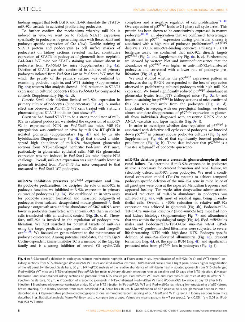
miR-92a deletion prevents crescentic glomerulonephritis andrenal failure. To determine if miR-92a expression in podocytesin vivo is necessary for crescent formation and renal failure, weselectively deleted miR-92a from podocytes. We used a condi-tional expression model (Tet-On system) to achieve temporalpodocyte-specific deletion of the miR-92a gene in mice. Mice ofall genotypes were born at the expected Mendelian frequency andappeared healthy. Ten weeks after doxycycline administration,marked reduction of miR-92a abundance in podocytes wasachieved (Fig. 4a), with most of residual signal being in endo-thelial cells. Overall, a ~50% reduction in relative miR-92aexpression was achieved in glomeruli (Fig. 4b). Podocin-rtTA-Tet-O-Cre miR-92a loxP/loxP (iPod-miR92a lox) mice had nor-mal kidney histology (Supplementary Fig. 7) and albuminuriathat was within the physiological range (Fig. 4c). iPod-miR92a loxmales and Podocin-rtTA-Tet-O-Cre miR-92a wt/wt (iPod-miR92a wt) gender-matched littermates were subjected to severe,life-threatening NTN with high-dose NTS. Podocyte-specificdeletion of miR-92a-alleviated albuminuria (Fig. 4c), crescentformation (Fig. 4d, e), the rise in BUN (Fig. 4f), and significantlyprotected mice from p57Kip2 loss in podocytes (Fig. 4g–i).
Fig. 4 miR-92a-specific deletion in podocytes reduces nephrotoxic nephritis. a Fluorescent in situ hybridization of miR-92a (red) and WT1 (green) onkidney sections from NTS-challenged iPod-miR92aWT mice and iPod-miR92a lox mice. DAPI-stained nuclei (blue). Right panel shows higher magnificationof the left panel (white box). Scale bars, 50 μm. b RT-qPCR analysis of the relative abundance of miR-92a in freshly isolated glomeruli from NTS-challengediPod-miR92a WT mice and NTS-challenged iPod-miR92a lox mice. c Urinary albumin excretion rates at baseline and 10 days after NTS injection. d Massontrichrome- and silver-stained kidney sections of glomeruli from NTS-challenged iPod-miR92a WT mice and iPod-miR92a lox mice at day 10 after NTSinjection. Scale bars, 10 μm. e Proportion of crescentic glomeruli in NTS-challenged iPod-miR92a WT and iPod-miR92a lox mice at day 10 after NTSinjection. f Blood urea nitrogen concentration at day 10 after NTS injection in iPod-miR92aWT and iPod-miR92a lox mice. g Immunostaining of p57 (strongbrown staining, *) in kidney sections from mice described in a. Scale bars 10 µm. h Quantification of p57-positive cells per glomerular section in micedescribed in a. i Representative photomicrophotographs of dual immunofluorescent satining of p57 (red) and WT1 (green) in kidney sections from micedescribed in a. Statistical analysis: Mann–Whitney test to compare two groups. Values are means± s.e.m. (n= 7 per group). *p< 0.05, **p< 0.01 vs. iPod-miR-92a WT mice
ARTICLE NATURE COMMUNICATIONS | DOI: 10.1038/s41467-017-01885-7
8 NATURE COMMUNICATIONS | 8: 1829 |DOI: 10.1038/s41467-017-01885-7 |www.nature.com/naturecommunications
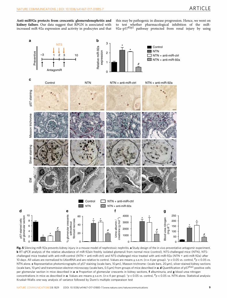
Anti-miR92a protects from crescentic glomerulonephritis andkidney failure. Our data suggest that RPGN is associated withincreased miR-92a expression and activity in podocytes and that
this may be pathogenic in disease progression. Hence, we went onto test whether pharmacological inhibition of the miR-92a–p57Kip2 pathway protected from renal injury by using
cControl
Silv
er s
tain
ing
EM
NTN + anti-miR-ctrl NTN + anti-miR-92a
p57
stai
ning
b
d e f g
a
Pre
vent
ive
Rel
ativ
e m
iR-9
2aex
pres
sion
p57-
stai
ned
cells
per
glom
erul
ar s
ectio
n
Blo
od u
rea
nitr
ogen
(mg
dl–1
)
Urin
e al
bum
in /
crea
tinin
e (g
mol
–1)
% g
lom
erul
ar c
resc
ents
with
/with
out
fibrin
oid
necr
osis
–3 10
3
#
**
2
1
0
NTS
AntagomiR
1 2 3
Control
10
* *
* * * *
**
## #
#
8
6
4
2
0
NTN
NTN + anti-miR-ctrl
NTN + anti-miR-92a
2504000
40
20
0
60
3000
2000
1000
0
200
150
100
50
0
ControlNTNNTN + anti-miR-ctrlNTN + anti-miR-92a
NTN
Mas
son
tric
hrom
e
Fig. 5 Silencing miR-92a prevents kidney injury in a mouse model of nephrotoxic nephritis. a Study design of the in vivo preventative antagomir experiment.b RT-qPCR analysis of the relative abundance of miR-92ain freshly isolated glomeruli from normal mice (control), NTS-challenged mice (NTN), NTS-challenged mice treated with anti-miR-control (NTN + anti-miR-ctrl) and NTS-challenged mice treated with anti-miR-92a (NTN + anti-miR-92a) after10 days. All values are normalized to U6snRNA and are relative to control. Values are means± s.e.m. (n= 4 per group). *p< 0.05 vs. control, #p< 0.05 vs.NTN alone. c Representative photomicrographs of p57 staining (scale bars, 10 µm), Masson trichrome- (scale bars, 20 µm), silver-stained kidney sections(scale bars, 10 µm) and transmission electron microscopy (scale bars, 0.5 µm) from groups of mice described in a. d Quantification of p57Kip2-positive cellsper glomerular section in mice described in a. e Proportion of glomerular crescents in kidney sections, f albuminuria, and g blood urea nitrogenconcentrations in mice as described in a. Values are means± s.e.m. (n= 4 per group). *p< 0.05 vs. control, #p< 0.05 vs. NTN alone. Statistical analysis:Kruskal–Wallis one-way analysis of variance followed by Dunn’s multiple comparaison test
NATURE COMMUNICATIONS | DOI: 10.1038/s41467-017-01885-7 ARTICLE
NATURE COMMUNICATIONS |8: 1829 |DOI: 10.1038/s41467-017-01885-7 |www.nature.com/naturecommunications 9

chemically engineered oligonucleotides, termed “antagomirs”(anti-miRs), efficient and specific silencers of endogenous miR-NAs in mice. Anti-miRs are taken up into the kidney cortex afterintravenous injection into mice42.
We first administered an anti-miR-92a strategy in a pre-ventative fashion. Anti-miR-92a injections to mice inhibited miR-92a expression in glomeruli during NTN (Fig. 5a, b) withoutmodifying the expression of other miRNAs from the 17–92
cluster (not shown). Induction of NTN was associated with a lowabundance of p57Kip2 in podocytes that was partially butsignificantly rescued by delivery of anti-miR-92a (Fig. 5c, d andSupplementary Fig. 8a). Importantly, NTS-induced glomerularinjury was less severe in mice that received anti-miR92a (NTN+anti-miR92a) than in mice that received a control anti-miR(NTN+anti-miR-ctrl) or in mice that received vehicle alone(NTN). Indeed, the proportion of glomerular crescents was ~70%
baC
urat
ive
Rel
ativ
e m
iR-9
2a e
xpre
ssio
n
Blo
od u
rea
nitr
ogen
(mg
dl–1
)
% g
lom
erul
ar c
resc
ents
with
/with
out f
ibrin
oid
necr
osis
Urin
e al
bum
in /
crea
tinin
e (g
mol
–1)
1 10 2.5 8000
6000
4000
2000
0
#
# #
#
#
#*
** *
*
2.0
1.5
1.0
0.5
0.0
150
100
50
0
NTS AntagomiR
2 3 4 5 8
c
dS
ilver
sta
inin
g Control NTN
NTN + anti-miR-ctrl NTN + anti-miR-92a
e f
g
h
p57
stai
ned
cells
per
glom
erul
ar s
ectio
n
p57
stai
ning Control NTN
NTN + anti-miR-ctrl NTN + anti-miR-92a
**
**
**
*
**
*
*
**
**
*
Control
NTN
NTN + anti-miR-ctrl
NTN + anti-miR-92a
Control
NTN
NTN + anti-miR-ctrl
NTN + anti-miR-92a80
60
40
8
6
4
2
0
20
0
Control
NTN
NTN + anti-miR-ctrl
NTN + anti-miR-92a
**
*
*
*
*
Control
NTN
NTN + anti-miR-ctrl
NTN + anti-miR-92a
Control
NTN
NTN + anti-miR-ctrl
NTN + anti-miR-92a
Fig. 6 miR-92a in vivo silencing abolishes nephrotoxic nephritis development. a Study design of the in vivo antagomir experiment. b Relative miR-92aexpression in dynabeads-isolated glomeruli from normal mice (control), NTS-challenged mice (NTN), NTS-challenged mice treated with anti-miR-control(NTN + anti-miR-ctrl), and NTS-challenged mice treated with anti-miR-92a (NTN + anti-miR-92a) after 10 days. All values are normalized to U6 and arerelative to control. Values are means± s.e.m. (n= 5 per group). *p< 0.05 vs. control, #p< 0.05 vs. NTS alone. c Urinary albumin excretion rates at day 4(before antagomir injection) and at day 10 after NTS injection. d Blood urea nitrogen concentration at day 10 after NTS injection in control or NTS-challenged mice. e Silver-stained kidney sections of mice described in b. Scale bars, 10 µm. f Proportion of crescentic glomeruli in kidney from micedescribed in b. Values are means± s.e.m. (n= 10 mice per group). g Representative immunostaining of p57 (strong brown staining) in kidney sections frommice described in a. Scale bars, 10 µm. h Quantification of p57-positive cells per glomerular section in mice described in a. Values are means± s.e.m. (n=10 per group). *p< 0.05, #p< 0.05 vs. NTS alone (NTN). Statistical analysis: Kruskal–Wallis one-way analysis of variance followed by Dunn’s multiplecomparaison test
ARTICLE NATURE COMMUNICATIONS | DOI: 10.1038/s41467-017-01885-7
10 NATURE COMMUNICATIONS | 8: 1829 |DOI: 10.1038/s41467-017-01885-7 |www.nature.com/naturecommunications

lower in NTN+anti-miR92a mice than in either control condition(Fig. 5c, e). This improvement in kidney structure was alsoreflected by less severe damage to the podocyte ultrastructure inanti-miR-92a-injected mice relative to untreated and anti-miR-ctrl injected littermate controls (Fig. 5c). These improvedstructural parameters corresponded to improved functionalparameters: urinary albumin excretion was lower (Fig. 5f), andkidney dysfunction minimal (Fig. 5g) in anti-miR-92a-treatedmice.
We next administered anti-miR-92a, on day 4 after infusion ofNTS (Fig. 6a). This time-point was chosen as it is clinicallyrelevant, associated with nephrotic range albuminuria and highserum creatinine and BUN. As previous, this regimen wascompared with the effects of vehicle alone and with theadministration of an anti-miR-ctrl. Anti-miR-92a given afterNTS still effectively inhibited glomerular miR-92a levels at day 10(Fig. 6b) and these mice displayed a marked reduction inalbuminuria (Fig. 6c). Furthermore, whereas vehicle-only-treatedmice (NTN) or anti-miR-ctrl-treated mice (NTN+anti-miR-ctrl)developed rapid and life-threatening kidney failure, mice treatedtherapeutically with anti-miR-92a had BUN levels within thenormal range (Fig. 6d). This functional protection conferred byanti-miR-92a administration was associated with marked allevia-tion of histologic damage as measured using silver staining ofrenal cortex (Fig. 6e, f) and significantly preserved p57Kip2
expression in podocytes (Fig. 6g, h and Supplementary Fig. 8b).
DiscussionRPGN with extracapillary proliferation of epithelial glomerularcells is an area of unmet clinical need. Despite current immu-nosuppressive therapies, a significant number of patients withRPGN fail to respond to treatment and their disease progresses toend-stage kidney disease with its associated significant morbidityand mortality. RPGN involves the response of podocytes toimmune injury. Therefore, better understanding of the mechan-isms that regulate podocyte function are critical. Here weexplored the role of miRNAs. Furthermore, the mechanismswhereby terminally differentiated, post-mitotic podocytes reentercell cycle upon immune-mediated stress are unknown.
Our data demonstrate the involvement of miR-92a in thedeleterious response to immune injury that leads to glomerulardestruction and functional demise. Our investigation furtheridentified a key miR-92a target, the p57/Kip2/Cyclin-dependentkinase inhibitor 1C, which is involved in cell cycle regulation andcontrol of the quiescent state of podocytes.
MiR-92a is part of the miR-17–92 cluster, which contains sixmiRNAs24. The human MIR17(MIR17HG) cluster has beenlinked to developmental, apoptotic, and oncogenic pathways inother organs, and the locus is conserved between mouse and man.Mice deficient for miR-17/92 die shortly after birth and havecardiac and lung abnormalities. Thus, this cluster plays animportant role during development. Among the members in thecluster, miR-92a is the least characterized. Whereas endothelialmiR-92a has been characterized with pro-inflammatory and anti-angiogenic effects43–45, ours is the first description of epithelialinduction in post-mitotic cells. As miR-92a is part of the miR-17–92 cluster that encodes a polycistronic transcript that pro-duces six individual mature miRNAs24, we expected increasedglomerular expression of all members of the cluster. Surprisingly,miR-92a was the only member to be dysregulated in human andmurine RPGN. In fact, this phenomenon was also previouslyfound in experimental atherosclerosis45. Moreover, these in vivodata are consistent with the recent demonstration that expressionof individual miRNAs from pri-miR-17/92 is dynamicallyregulated46.
MiR-92a was significantly overexpressed in human glomerulifrom patients with RPGN compared to those with other non-proliferative glomerular diseases. This difference highlights thedifference in pathogenesis of these diseases, the former beingcritically dependent on loss of podocyte post-mitotic quiescence.However, miR-92a expression level did not correlate with histo-logical variant of crescent (cellular vs. fibrocellular), proteinuria,or kidney function. This may be explained by clinical hetero-geneity in terms of stage of disease at the time of diagnosis orrelationship between miRNA expression and cell density. Forexample, we observed intense miR92a expression in fully con-stitued crescents and less in pseudo-crescents (when a paucity ofcells are proliferating) and fibrocellular cellular crescents (whenmany epithelial cells have already disappeared).
We identified a miR-92a target relevant to podocyte prolifera-tion. In contrast to immature podocytes, which proliferate duringglomerular development, differentiated podocytes have a quies-cent phenotype47. This is required for podocytes to perform theirspecialized functions47. Two independent target prediction algo-rithms and luciferase assay identified p57Kip2 as a relevant miR-92a target. The CDK inhibitor p57Kip2 regulates cell proliferationand differentiation48. This protein is typically present in differ-entiated and post-mitotic non-renal cells. Several groups observedthat the de novo expression of p57Kip2 in podocytes duringglomerulogenesis coincides with the acquisition of a terminallydifferentiated quiescent phenotype6, 49. Loss of p57Kip2 expressionin podocytes is recognized as an early feature of proliferativeglomerular diseases and dedifferentiation in vitro3, 6, 8, 49–52,although the mechanisms involved remained unclear.
Identifying p57Kip2 as a key effector of mi-92a in podocytedrove us to test whether p57Kip2 participates in the extrinsicstimuli-dependent abrogation of podocyte quiescence. Our resultssuggest that a reduction in p57Kip2 protein, a brake on podocytecell cycle, contributes to trigger activation of crescent formationin response to immune-mediated glomerular injury. We do notexclude that there are other potential miR-92a targets involved inthe proliferative response of podocyte. For instance, the integrinsubunit alpha5 is a target of miR-92a in ischemic tissues20.Alpha5 integrin subunit showed a gradual loss in early FSGS andbecame undetectable in advanced FSGS53, meanwhile, its reg-ulation in crescentic RPGN remain to be characterized. Change inintegrin pattern may be important for podocyte functions in thissetting, as shown in other conditions54, 55.
The combination of our results from in vitro experiments,mouse models, and human tissues indicate that the high abun-dance of miR-92a can initiate a cascade of podocyte-destabilizingmolecular events, starting with the downregulation of p57Kip2 andproliferation. Mechanistically, binding of miR-92a to the targetregion of p57Kip2 acts as negative regulator and causes a lack inthe p57Kip2 that is available for podocyte cell cycle resistance toambient mitogenic stimuli. Moreover, specific blockade of miR-92a in vivo by an antagomir markedly prevented proteinuria,crescent formation, and renal failure. Together, these findingsindicate that miR-92a control of podocyte phenotype may be ageneral paradigm for proliferative extracapillary diseases.
Several animal studies have used antagomirs to block targetmicroRNA at a concentration ranging from 0.33 to 100 mg/kg ofbody weight. A potential concern of using high doses of theseagents is that they may non-specifically block genes other thanthe target. Since previous studies have shown that a dose of 8 mg/kg produced an effective tissue response20, we chose a similar,albeit slightly higher, dose for our own study. We felt that 12 mg/kg of body weight would account for the potential loss of drug inthe urine of heavily nephrotic animals. Encouragingly, our resultsrevealed a threefold decrease in miR-92 levels in glomeruli withthis dose compared to the control antagomir. In contrast,
NATURE COMMUNICATIONS | DOI: 10.1038/s41467-017-01885-7 ARTICLE
NATURE COMMUNICATIONS |8: 1829 |DOI: 10.1038/s41467-017-01885-7 |www.nature.com/naturecommunications 11
expression levels of 5 other microRNAs, miR-17, -18a, -19a, -19b,and -20a were not different between the two groups of mice, thussuggesting both efficacy and specificity of our antagomir. Inter-estingly, prolonged anti-92a blockade at higher dose for morethan 10 weeks did not display detectable side effects in mice45.Another notable finding of our study is that STAT3 activation isrequired to trigger miR-92a pathogenic expression. This may leadone to consider anti-STAT3 strategies as anti-miR-92a options.Meanwhile, from a therapeutic perspective, the identification of amolecular target that is as cell-specific as possible may beimportant to limit the undesired side effects associated with moreupstream ubiquitous targets such as STAT3.
Finally, we also provide proof of principle that delayed anti-miR-92a strategy could display therapeutic actions on glomerularfunction and structure in a severe model of RPGN. Although thistreatment showed a preventive effect in our mouse model, itremains to be seen whether this holds true for human RPGN.Furthermore, although most of the pathophysiological actions ofmiR-92a were found in podocytes, it is important to note thatanti-miR-92a strategies would be expected to alleviate endothelialinflammation, cardiac ischemia, and atherosclerosis45, potentiallyimportant given the high risk of cardiovascular disease in indi-viduals with RPGN56–59.
MethodsAnimals. Mice with podocyte-specific GFP expression (NPHS2-Cre x mT/mG)were obtained by crossing podocin-Cre-positive mice60 with mT/mG mice (Gt(2)26Sortm4(ACTB-tdTomato,-EGFP)Luo/J)61 on a C57BL6/J background that werepurchased from The Jackson Laboratories (Bar Harbor, ME). Mice with podocyte-specific disruption of Stat3 were generated by crossing podocin-Cre-positive micewith Stat3 floxed mice62 on a C57BL6/J background. Age-matched littermates thathad no deletion of Stat3 in any cells were considered as controls. To generate atime-specific and podocyte-specific knockout of miR-92a, we crossed mice carryingreverse tetracycline transactivator protein under control of the podocin promoter(iPod) with mice carrying the Tet-O-Cre transgene as previously described13, 63
and with mice carrying a loxP-flanked miR-92a allele64. Doxycycline was admi-nistered for 3 weeks before administration of nephrotoxic serum after 1 week ofwashout. Age-matched littermates that had no deletion of miR-92a in any cellswere considered as controls. Experiments were conducted according to the Frenchveterinary guidelines and those formulated by the European Community forexperimental animal use (L358-86/609EEC), and were approved by the InstitutNational de la Santé et de la Recherche Médicale and local University ResearchEthics Committee (file 12–62, Comité d’Ethique en matière d’ExpérimentationAnimale, Paris Descartes).
Induction of nephrotoxic nephritis. Nephrotoxic nephritis was induced in malemice (10–12 weeks of age) by intravenous injection of 15 µl of sheep anti-glomerular basement membrane (GBM) nephrotoxic serum (NTS), which wasdiluted with 85 µl of sterile phosphate buffer solution as previously described65, 66.Serum injections were repeated twice: once on day 2 at 6 µl/g of body weight and asecond time on day 3 at 7 µl/g of body weight.
Biochemical measurements in blood and urine. Urinary creatinine and bloodurea nitrogen (BUN) concentrations were analyzed by a standard colorimetricmethod (Olympus AU400) at the Biochemistry Laboratory of Institut ClaudeBernard (IFR2, Faculté de Médecine Paris Diderot). Urinary albumin excretion wasmeasured using a specific ELISA assay for the quantitative determination ofalbumin in mouse urine (CellTrend GmbH).
Human tissues. Formalin-fixed, paraffin-embedded renal tissue specimens wereobtained from the Hôpital Européen Georges Pompidou, Assistance Publique-Hôpitaux de Paris, Paris, France. Human tissue was used after informed consent bythe patients and approval from, and following the guidelines of, the local EthicsCommittee (IRB00003888, FWA00005831). Renal biopsy specimens with sufficienttissue for immunohistochemical evaluation after the completion of diagnosticworkup were included.
Histology. Kidneys were harvested and fixed in 4% formol or in Alcohol-Formol-Acetic acid. Paraffin-embedded sections (5 μm thick) were stained by Masson’strichrome to evaluate kidney morphology and determine the proportion of cres-centic glomeruli by a blind examination of at least 50 glomeruli per section. Silverstaining of paraffin sections was also used for quantification of crescents.
Immunohistochemistry and immunofluorescence. Deparaffinized kidney sec-tions were incubated for 30 min at 95 °C in target retrieval solution (S1699, Dako),then in peroxidase blocking reagent (S2001, Dako), were blocked in PBS containing5% BSA, and were immunostained for phospho-STAT3 (Tyr705) (clone EP2147Y,Millipore, 1:50), nephrin (GP2, Progen, 1:400), WT1 (ab15249, Abcam, 1:400)STAT3 (D1A5, Cell Signaling Technology, 1:200), podocalyxin (AF1556, R&Dsystems, 1:100), and p57Kip2 (Clone M-20, Santa Cruz Technology, 1:200). Forphospho-STAT3, and p57Kip2, staining was detected by Histofine reagents(Nichirei Biosciences), which contained anti-rabbit (414341F) or anti-goat(414161F) immune-peroxidase polymer for mouse tissue sections. WT1 and p-STAT3 primary antibodies were followed by a secondary rabbit anti-goat IgGAF488-conjugated antibody (Invitrogen, 1:400), p57Kip2 primary antibody wasfollowed by a secondary anti-goat IgG AF594 (Invitrogen, 1:400), nephrin primaryantibody was followed by a secondary anti-guinea pig IgG AF594-conjugatedantibody (Invitrogen, 1:400). Podocyte culture cells were immunostained forpodocin (ab50339, Abcam, 1:100), nephrin (ab58968, Abcam, 1:400), WT1(ab15249, Abcam, 1:400), or p57Kip2 (Clone M-20, Santa Cruz Biotechnology,1:200). The nuclei were stained using DAPI. Images were obtained with anAxioimager Z1 apotome (Zeiss) with Axiovision microscopy software. For quan-tification of p57-positive nuclei or p-STAT3-positive cells, 10 glomeruli per miceand five randomly chosen mice from each group were examined to calculate thenumber of stained nuclei.
miR-92a in situ hybridization. In situ hybridization was performed on 5 μm-thickkidney paraffin-embedded sections cut and fixed in PFA 4% for 10 min. Thensections were washed with 1× PBS and were acetylated for 10 min. After washes,sections were incubated with protein kinase K (Sigma-Aldrich) for 10 min at 37 °C.After subsequent washes, sections were incubated with hybridization buffer for 5 hat room temperature. miRNA probes (miR-92a probe double-DIG labeled LNAprobes, Exiqon, final concentration 20 nM) were mixed with denaturation bufferand added to the sections and were incubated over night at 56 °C. U6snRNA probe(3′-DIG labeled LAN, probe, Exiqon) was used at 10 nM final concentration and asa positive control. The following morning, sections were washed in successivelydecreasing SSC buffers for 5 min at 56 °C (5× 1 time, 1× 2 times, 0.2× 3 times) andwere then washed. Sections were incubated for 1 h in blocking solution (B1 solution+ 3% fetal calf serum + 0.1% Tween-20), and were then incubated with anti-DIGAP antibody (Roche; 1:2000) over night at 4 °C. After washes, sections wereincubated with NBT/BCIP (Promega) in NTMT + levamisole (0,2 mM/L) for 48 hin the dark at RT. NBT/BCIP was changed every 12 h. Slides were fixed in PFA 4%for 30 min and mounted with Fluoprep mounting medium (Biomerieux).
For fluorescent in situ hybridization (FISH), sections were incubated twice infreshly prepared 3% H2O2 for 3 min to inactive endogenous peroxidases afterfixation, acetylation, and incubation with proteinase K. Slides were then rinsedthree times in PBS. Then, sections were incubated with hybridization buffer for 1 hat 37 °C. MicroRNA probes (miR-92a, 5′-3′–Digoxigenin-labeled Locked NucleicAcid probe, Exiqon, 100 nmol/L; U6snRNA, 3′-Digoxigenin-labeled LockedNucleic Acid probe, 2 nmol/L) were mixed with denaturation buffer and thenincubated with the sections over night at 56 °C. Washes with decreasingconcentrations of SSC were the same as for ISH. Then, slides were again twiceincubated in freshly prepared 3% H2O2 for 5 min and washed three times in PBS.Blocking solution (Tris + 3% fetal calf serum + 1% BSA) was applied to slide for 1 hat room temperature, then incubated in anti-DIG-FAB peroxidase (POD) (Roche)diluted 1:400 in blocking solution for 1 h at room temperature. After washes withPBS, TSA Plus Cy3 system working solution was applied onto the sections for 10min at room temperature in the dark according to the manufacturer’s protocol(PerkinElmer Life sciences). The slides were washed three times in PBS.
To assess whether miR-92a was localized specifically in glomerulus, sectionswere processed for double fluorescence staining to visualize the simultaneouslocalization of miR-92a (red; Cy3) and a primary antibody for WT1 (green;AF488), a podocyte-specific marker. Sections were incubated in demasking citratebuffer during 20 min at 95 °C and solution with a primary anti-WT1 antibody(Abcam) was applied on slide over night at 4 °C. The next day, slides were twiceimmerged in PBS and incubated with a secondary AlexaFluor488-conjugatedantibody (Invitrogen) during 1 h at RT or with a Histofine-AEC system (DAKOand Nichirei Biosciences). Nuclei were stained with DAPI for FISH and sectionswere mounted using a drop of Fluorescent mounting medium (DAKO).
Laser capture microdissection of glomeruli. Laser microdissection was per-formed with a PALM® RoboSoftware 4.6 MicroBeam system (PALM MicrolaserTechnologies, Zeiss Micro Imaging, Munich, Germany) coupled to an invertedmicroscope Axio Observer.Z1. Serial 20 μm-thick cryosections, stored either at −80°C for a maximum of 48 h or processed immediately, were spread onto poly-ethylene naphthalate (PEN) membrane-coated slides (Carl Zeiss Micro Imaging,Munich, Germany) previously treated for 20 min under UV exposure. After sec-tions, the slide is stained with toluidine blue solution under RNase-free conditions.Glomerular tufts were delineated using a graphical computer wizard and isolatedfrom surrounding tissue by laser catapulting into the cap of a single micro-centrifuge Eppendorf Tube® filled with 20 μl of RLT plus buffer/1% NucleoGuardreagent (Amsbio) for subsequent RNA isolation with the AllPrep DNA/RNA
ARTICLE NATURE COMMUNICATIONS | DOI: 10.1038/s41467-017-01885-7
12 NATURE COMMUNICATIONS | 8: 1829 |DOI: 10.1038/s41467-017-01885-7 |www.nature.com/naturecommunications

Micro Kit (Qiagen). One cap per “section collection” was used and caps werereplaced on their tube and stored on dry ice prior to RNA extraction.
Transmission electron microscopy procedure. Small pieces of renal cortex werefixed in 4% glutaraldehyde, postfixed in 1% osmium tetroxide, and embedded inepoxy resin. Ultrathin sections were counterstained with uranyl acetate andexamined in a JEOL 1011 transmission electron microscope with Digital Micro-graph software for acquisition.
Glomeruli preparation and isolation of podocytes. We used the magnetic beadmethod described by Takemoto et al. with appropriate modifications67. In brief,dynabeads perfusion was performed through the abdominal aorta and harvestedkidneys were transferred in fresh Hank’s buffered salt solution (HBSS). Then,kidneys were minced into 1-mm3 pieces using a scalpel in digest solution (col-lagenase 210 U/ml (Gibco), DNase I 40 U/ml (Euromedex)) and incubated at 37 °Cfor 15 min on a rotator (100 rpm) The solution was pipetted up and down with acut 1000 μl pipette tip every 5 min. After incubation, all steps were carried out at 4 °C or on ice. The digested kidneys were gently pressed twice through a 100-μm cellstrainer and the flow through was washed extensively with HBSS. After spinningdown, the supernatant was discarded and the pellet resuspended in 2 ml HBSS.These tubes were inserted into a magnetic particle concentrator and the separatedglomeruli were washed five times. Glomeruli were lysed in RIPA buffer for proteinanalysis or in Trizol for RNA study. For podocyte isolation, in a second digestionstep, glomeruli were enzymatically and mechanically disrupted to yield a single cellsuspension. Subsequently, green fluorescent protein (GFP)-positive podocytes fromNPHS2-Cre x mT/mG animals were separated from the GFP-negative non-podocyte glomerular fraction by fluorescence-activated cell sorting (FACS) aspublished68.
Glomeruli isolation for culture of primary podocytes and in vitro assays.Magnetic beads infused-mouse kidneys were extracted, minced, and digested in 2mg/ml collagenase I solution (Gibco) in RPMI 1640 (Invitrogen) at 37 °C for 3 min,then filtered through a 70-µm cell strainer and once more through a 40-µm cellstrainer. The homogenate was centrifuged at 720×g for 6 min and the cells wereplated. Podocyte primary cultures consisted of freshly isolated glomeruli plated in6-well dishes in RPMI 1640 (Invitrogen) supplemented with 10% fetal calf serum(Biowest) and 1% penicillin–streptomycin (Invitrogen). Purity of culture of dif-ferentiated primary podocytes was verified as previously described13, 69 and shownin Supplementary Fig. 3b. Podocyte primary cultures used in this study was alwaysP0. The outgrowth of podocytes started between days 2 and 3. Podocyte outgrowtharea was quantified at day 4 using ImageJ software. Differentiated podocytes wereexposed to HB-EGF (10 ng/ml, Preprotech), AG1478 (1 µM, Calbiochem), anti-mIL-6 monoclonal antibody MP5-20F3, monoclonal rat IgG1, κ isotype controlimmunoglobulin (both functional grade purified, 10 µg/ml, eBiosciences), Stattic (2µM, Calbiochem), or recombinant IL6 (10 ng/ml, Preprotech) for 16 h. After sti-mulation, podocytes were scrapped in Phosphosafe buffer (Novagen) for proteinextraction or in Trizol (Invitrogen) for total RNA extraction.
miR-92a in vitro modulation. MicroRNA-92a inhibition was achieved in vitro bytransfecting primary culture podocytes with anti-miR-92a inhibitor using Hiperfecttransfection reagent (Qiagen). Anti-miR-Control was used as a control (All fromAmbion, 50 nM).
p57Kip2 3′UTR luciferase assay. For validation of p57 as a target of miR-92a,3′UTR or mutated 3′UTR of mouse p57Kip2 were cloned into a mammalianexpression vector with dual luciferase reporter system (GeneCopoeia). HEK293cells were transfected using Hiperfect (Qiagen). Transfections were performedusing 1 μg dual luciferase reporter plasmids and a final concentration of 100 nMsynthetic miR-92a mimic, or miR-126 as an irrelevant miRNA mimic (AppliedBiosystems). Twenty-four hours after transfection, dual luciferase assays wereperformed using Luc-Pair miR luciferase assay kit (GeneCopoeia) according tothe manufacturer's instructions. Firefly luciferase activity was normalized toRenilla luciferase expression control.
p57Kip2 silencing in vitro. p57Kip2 was silenced using ON-TARGETplus mousep57 siRNA SMARTpool (Dharmacon). Primary podocytes were transfected with50 nM ON-TARGETplus mouse p57 siRNA SMARTpool using Hiperfect follow-ing the manufacturer’s instructions over night. Thereafter, medium was changedand cells were harvested after 72 h.
Western blotting. Proteins were extracted from glomeruli or podocytes with lysisbuffer and were quantified by BCA protein assay (iNtRON Biotechnology). Sam-ples were resolved on 4–12% Bis-Tris Criterion XT gels (Bio-Rad) then transferredto a polyvinylidene difloride membrane. Membranes were incubated with theappropriate primary antibodies: rabbit anti-phospho-EGFR (Tyr1068) (D7A5, CellSignaling Technology, 1:1000), rabbit anti-EGFR (D38B1, Cell Signaling Tech-nology, 1:1000), rabbit anti-STAT3 (D1A5, Cell Signaling Technology, 1:1000),rabbit anti-phospho-STAT3 (Tyr705) (D3A7, Cell Signaling Technology, 1:1000),
goat anti-p57Kip2 (Clone M-20, Santa Cruz Biotechnology, 1:500). Protein loadingwas monitored by the rat anti-tubulin antibody (ab6160, Abcam, 1:5000). Sec-ondary antibodies were donkey anti-rabbit HRP and donkey anti-goat HRP (GEHealthcare Life Sciences) with no cross reaction to sheep serum. Antigens weredetected by enhanced chemiluminescence (Supersignal West Pico, Pierce) using aLAS-4000 imaging system (Fuji). Densitometric analysis with ImageJ software wasused for quantification. The uncropped versions of western blotting are shown inSupplementary Fig. 10.
Real-time PCR. Total RNA was extracted from mice glomeruli, cultured podo-cytes, or human biopsies with Trizol reagent according to the manufacturer’sinstructions (Invitrogen). Total RNA was reverse transcribed into cDNA using theQuantitect Reverse Transcription kit (Qiagen). cDNA and standards were ampli-fied with the Maxima SYBR Green/Rox qPCR mix (Fermentas) using an ABIPRISM thermo cycler. The comparative method of relative quantification (2–ΔΔCT)was used to calculate the relative expression level of each target gene. Mouse orhuman GAPDH was used as an internal control. The data were presented as thefold change in gene expression. The following oligonucleotides served as primers:Mouse Ki67 forward 5′- CCTCAAAAGCAGACGAGCAAGA-3′, Mouse Ki67reverse 5′- GAGAGTTTGCATGGCCTGTAGT-3′.
Following extraction by Trizol extraction, miRNA expression was determinedusing Taqman miRNA assay (Life Technologies) according to the manufacturer’sprotocols. U6snRNA was used as an endogenous control. Total miR contentanalysis of isolated glomeruli from control mice or NTS-challenged mice wasperformed with the miRCURY LNATM Universal RT microRNA PCR Mouse&Ratpanel I+II (Exiqon).
In vivo miR-92a inhibition in mice. For preventive strategy, antagomiR treatment(12 mg/kg) was started 3 days before NTS injection. AntagomiRs (VBC Biotech,Vienna) were delivered by retro-orbital IV injections under brief anesthesia. Sec-ond and third injections were performed on days 1 and 3 accompanying the NTSinjection. For curative strategy, antagomir were injected on days 4, 5, and 8 afterNTS. A scramble antagomiR (Antagomir-Control) was used as control. Thesequences (AntagomiR-Control (anti-miR-ctrl): 5′-AAGGCAAGCUGACCCU-GAAGUU-3′ and antagomiR-92a (anti-miR-92a): 5′-CAGGCCGGGACAA-GUGCAAUA-3′) were obtained from a previously published study20. In the miR-92 antagomir and control antagomir, the 2′O RNA base are methylated and thefirst two bases and the last three bases are phosphorothiated to increase the stabilityof antagomir and hence protect it from degradation. In addition, a cholesterol-TEGwas added at the 3′ for easy entry of the antagomir to the cells. AntagomiR-Controland antagomiR-92a were previously successfully used in vivo following adminis-tration in kidneys42, 70. Saline-treated mice were used as a control of the scrambleantagomiR.
Statistical analyses. All values were expressed as means + s.e.m. When samplesize was less than five per group, exact, two-sided comparisons were performedwith exact test using StatXact 8.0 software (Cytel Software Corporation, CambridgeMA, USA). In other cases, the two-tailed Mann–Whitney test, was used asappropriate. For experiments with more than two subgroups, the nonparametricKruskal–Wallis ANOVA followed by Dunn's multiple comparison test were used.Values of p < 0.05 were considered significant. Statistical analyses were calculatedusing Prism v5.04 software (GraphPad Inc, La Jolla, CA, USA).
Data availability. The authors declare that data supporting the findings of thisstudy are available within the paper and its supplementary information files orfrom the corresponding author on reasonable request.
Received: 1 May 2017 Accepted: 23 October 2017
References1. Couser, W. G. Rapidly progressive glomerulonephritis: classification,
pathogenetic mechanisms, and therapy. Am. J. Kidney Dis. 11, 449–464 (1988).2. Jennette, J. C. & Thomas, D. B. Crescentic glomerulonephritis. Nephrol. Dial.
Transplant. 16, 80–82 (2001).3. Bariety, J. et al. Podocyte involvement in human immune crescentic
glomerulonephritis. Kidney Int. 68, 1109–1119 (2005).4. Thorner, P. S., Ho, M., Eremina, V., Sado, Y. & Quaggin, S. Podocytes
contribute to the formation of glomerular crescents. J. Am. Soc. Nephrol. 19,495–502 (2008).
5. Smeets, B. et al. Tracing the origin of glomerular extracapillary lesions fromparietal epithelial cells. J. Am. Soc. Nephrol. 20, 2604–2615 (2009).
6. Hiromura, K. et al. Podocyte expression of the CDK-inhibitor p57 duringdevelopment and disease. Kidney Int. 60, 2235–2246 (2001).
NATURE COMMUNICATIONS | DOI: 10.1038/s41467-017-01885-7 ARTICLE
NATURE COMMUNICATIONS |8: 1829 |DOI: 10.1038/s41467-017-01885-7 |www.nature.com/naturecommunications 13
7. Griffin, S. V. et al. The role of cell cycle proteins in glomerular disease. Semin.Nephrol. 23, 569–582 (2003).
8. Barisoni, L. et al. Podocyte cell cycle regulation and proliferation in collapsingglomerulopathies. Kidney Int. 58, 137–143 (2000).
9. Nitta, K. et al. Glomerular expression of cell-cycle-regulatory proteins in humancrescentic glomerulonephritis. Virchows Arch. 435, 422–427 (1999).
10. Nagata, M., Nakayama, K., Terada, Y., Hoshi, S. & Watanabe, T. Cell cycleregulation and differentiation in the human podocyte lineage. Am. J. Pathol.153, 1511–1520 (1998).
11. Moeller, M. J. et al. Podocytes populate cellular crescents in a murine model ofinflammatory glomerulonephritis. J. Am. Soc. Nephrol. 15, 61–67 (2004).
12. Ding, M. et al. Loss of the tumor suppressor Vhlh leads to upregulation ofCxcr4 and rapidly progressive glomerulonephritis in mice. Nat. Med. 12,1081–1087 (2006).
13. Bollee, G. et al. Epidermal growth factor receptor promotes glomerular injuryand renal failure in rapidly progressive crescentic glomerulonephritis. Nat.Med. 17, 1242–1250 (2011).
14. Mirmohammadsadegh, A. et al. STAT5 phosphorylation in malignantmelanoma is important for survival and is mediated through SRC and JAK1kinases. J. Invest. Dermatol. 126, 2272–2280 (2006).
15. Park, O. K., Schaefer, T. S. & Nathans, D. In vitro activation of Stat3 byepidermal growth factor receptor kinase. Proc. Natl Acad. Sci. USA 93,13704–13708 (1996).
16. Shao, H., Cheng, H. Y., Cook, R. G. & Tweardy, D. J. Identification andcharacterization of signal transducer and activator of transcription 3recruitment sites within the epidermal growth factor receptor. Cancer Res. 63,3923–3930 (2003).
17. Aaronson, D. S. & Horvath, C. M. A road map for those who don't know JAK-STAT. Science 296, 1653–1655 (2002).
18. Gebeshuber, C. A. et al. Focal segmental glomerulosclerosis is induced bymicroRNA-193a and its downregulation of WT1. Nat. Med. 19, 481–487(2013).
19. He, L. et al. A microRNA polycistron as a potential human oncogene. Nature435, 828–833 (2005).
20. Bonauer, A. et al. MicroRNA-92a controls angiogenesis and functional recoveryof ischemic tissues in mice. Science 324, 1710–1713 (2009).
21. Kaluza, D. et al. Histone deacetylase 9 promotes angiogenesis by targeting theantiangiogenic microRNA-17-92 cluster in endothelial cells. Arterioscler.Thromb. Vasc. Biol. 33, 533–543 (2013).
22. Li, M. et al. miR-92a family and their target genes in tumorigenesis andmetastasis. Exp. Cell Res. 323, 1–6 (2014).
23. Brock, M. et al. Interleukin-6 modulates the expression of the bonemorphogenic protein receptor type II through a novel STAT3-microRNAcluster 17/92 pathway. Circ. Res. 104, 1184–1191 (2009).
24. Mendell, J. T. miRiad roles for the miR-17-92 cluster in development anddisease. Cell 133, 217–222 (2008).
25. Dai, Y. et al. Podocyte-specific deletion of signal transducer and activator oftranscription 3 attenuates nephrotoxic serum-induced glomerulonephritis.Kidney Int. 84, 950–961 (2013).
26. Iliopoulos, D., Jaeger, S. A., Hirsch, H. A., Bulyk, M. L. & Struhl, K. STAT3activation of miR-21 and miR-181b-1 via PTEN and CYLD are part of theepigenetic switch linking inflammation to cancer. Mol. Cell 39, 493–506 (2010).
27. Loffler, D. et al. Interleukin-6 dependent survival of multiple myeloma cellsinvolves the Stat3-mediated induction of microRNA-21 through a highlyconserved enhancer. Blood 110, 1330–1333 (2007).
28. Lin, H. Y., Chiang, C. H. & Hung, W. C. STAT3 upregulates miR-92a to inhibitRECK expression and to promote invasiveness of lung cancer cells. Br. J. Cancer109, 731–738 (2013).
29. Bourguignon, L. Y., Earle, C., Wong, G., Spevak, C. C. & Krueger, K. Stem cellmarker (Nanog) and Stat-3 signaling promote MicroRNA-21 expression andchemoresistance in hyaluronan/CD44-activated head and neck squamous cellcarcinoma cells. Oncogene 31, 149–160 (2012).
30. Zhong, Z., Wen, Z. & Darnell, J. E. Jr. Stat3: a STAT family member activatedby tyrosine phosphorylation in response to epidermal growth factor andinterleukin-6. Science 264, 95–98 (1994).
31. Grandis, J. R. et al. Requirement of Stat3 but not Stat1 activation for epidermalgrowth factor receptor- mediated cell growth In vitro. J. Clin. Invest. 102,1385–1392 (1998).
32. Gao, S. P. et al. Mutations in the EGFR kinase domain mediate STAT3activation via IL-6 production in human lung adenocarcinomas. J. Clin. Invest.117, 3846–3856 (2007).
33. Lutticken, C. et al. Association of transcription factor APRF and protein kinaseJak1 with the interleukin-6 signal transducer gp130. Science 263, 89–92 (1994).
34. Kishimoto, T. Signal transduction through homo- or heterodimers of gp130.Stem Cells 12, 37–44 (1994).
35. Wegenka, U. M., Buschmann, J., Lutticken, C., Heinrich, P. C. & Horn, F.Acute-phase response factor, a nuclear factor binding to acute-phase response
elements, is rapidly activated by interleukin-6 at the posttranslational level.Mol.Cell Biol. 13, 276–288 (1993).
36. Schust, J., Sperl, B., Hollis, A., Mayer, T. U. & Berg, T. Stattic: a small-moleculeinhibitor of STAT3 activation and dimerization. Chem. Biol. 13, 1235–1242(2006).
37. Dweep, H., Gretz, N. & Sticht, C. miRWalk database for miRNA-targetinteractions. Methods Mol. Biol. 1182, 289–305 (2014).
38. Lewis, B. P., Burge, C. B. & Bartel, D. P. Conserved seed pairing, often flankedby adenosines, indicates that thousands of human genes are microRNA targets.Cell 120, 15–20 (2005).
39. Matsuoka, S. et al. p57KIP2, a structurally distinct member of the p21CIP1 Cdkinhibitor family, is a candidate tumor suppressor gene. Genes Dev. 9, 650–662(1995).
40. Lee, M. H., Reynisdottir, I. & Massague, J. Cloning of p57KIP2, a cyclin-dependent kinase inhibitor with unique domain structure and tissuedistribution. Genes Dev. 9, 639–649 (1995).
41. Shankland, S. J. & Wolf, G. Cell cycle regulatory proteins in renal disease: rolein hypertrophy, proliferation, and apoptosis. Am. J. Physiol. Renal Physiol. 278,F515–F529 (2000).
42. Krutzfeldt, J. et al. Silencing of microRNAs in vivo with 'antagomirs'. Nature438, 685–689 (2005).
43. Hinkel, R. et al. Inhibition of microRNA-92a protects against ischemia/reperfusion injury in a large-animal model. Circulation 128, 1066–1075 (2013).
44. Doebele, C. et al. Members of the microRNA-17-92 cluster exhibit a cell-intrinsic antiangiogenic function in endothelial cells. Blood 115, 4944–4950(2010).
45. Loyer, X. et al. Inhibition of microRNA-92a prevents endothelial dysfunctionand atherosclerosis in mice. Circ. Res. 114, 434–443 (2014).
46. Du, P., Wang, L., Sliz, P. & Gregory, R. I. A biogenesis step upstream ofmicroprocessor controls miR-17 approximately 92 expression. Cell 162,885–899 (2015).
47. Pavenstadt, H., Kriz, W. & Kretzler, M. Cell biology of the glomerular podocyte.Physiol. Rev. 83, 253–307 (2003).
48. Zhang, P., Wong, C., DePinho, R. A., Harper, J. W. & Elledge, S. J. Cooperationbetween the Cdk inhibitors p27(KIP1) and p57(KIP2) in the control of tissuegrowth and development. Genes Dev. 12, 3162–3167 (1998).
49. Shankland, S. J. et al. Differential expression of cyclin-dependent kinaseinhibitors in human glomerular disease: role in podocyte proliferation andmaturation. Kidney Int. 58, 674–683 (2000).
50. Srivastava, T., Garola, R. E. & Singh, H. K. Cell-cycle regulatory proteins in thepodocyte in collapsing glomerulopathy in children. Kidney Int. 70, 529–535(2006).
51. Wang, S., Kim, J. H., Moon, K. C., Hong, H. K. & Lee, H. S. Cell-cyclemechanisms involved in podocyte proliferation in cellular lesion of focalsegmental glomerulosclerosis. Am. J. Kidney Dis. 43, 19–27 (2004).
52. Bariety, J. et al. Glomerular epithelial-mesenchymal transdifferentiation inpauci-immune crescentic glomerulonephritis. Nephrol. Dial. Transplant. 18,1777–1784 (2003).
53. Kemeny, E., Mihatsch, M. J., Durmuller, U. & Gudat, F. Podocytes loose theiradhesive phenotype in focal segmental glomerulosclerosis. Clin. Nephrol. 43,71–83 (1995).
54. Pozzi, A. & Zent, R. Integrins in kidney disease. J. Am. Soc. Nephrol. 24,1034–1039 (2013).
55. Hayek, S. S. et al. A tripartite complex of suPAR, APOL1 risk variants andalphavbeta3 integrin on podocytes mediates chronic kidney disease. Nat. Med.23, 945–953 (2017).
56. Roman, M. J. et al. Prevalence and correlates of accelerated atherosclerosis insystemic lupus erythematosus. N. Engl. J. Med. 349, 2399–2406 (2003).
57. Suppiah, R. et al. A model to predict cardiovascular events in patients withnewly diagnosed Wegener's granulomatosis and microscopic polyangiitis.Arthritis Care Res 63, 588–596 (2011).
58. Wall, N. & Harper, L. Complications of long-term therapy for ANCA-associated systemic vasculitis. Nat. Rev. Nephrol. 8, 523–532 (2012).
59. Morgan, M. D. et al. Increased incidence of cardiovascular events in patientswith antineutrophil cytoplasmic antibody-associated vasculitides: a matched-pair cohort study. Arthritis Rheum. 60, 3493–3500 (2009).
60. Moeller, M. J., Sanden, S. K., Soofi, A., Wiggins, R. C. & Holzman, L. B.Podocyte-specific expression of cre recombinase in transgenic mice. Genesis 35,39–42 (2003).
61. Muzumdar, M. D., Tasic, B., Miyamichi, K., Li, L. & Luo, L. A global double-fluorescent Cre reporter mouse. Genesis 45, 593–605 (2007).
62. Moh, A. et al Role of STAT3 in liver regeneration: survival, DNA synthesis,inflammatory reaction and liver mass recovery. Lab. Invest. 87, 1018–1028(2007).
63. Shigehara, T. et al. Inducible podocyte-specific gene expression in transgenicmice. J. Am. Soc. Nephrol. 14, 1998–2003 (2003).
ARTICLE NATURE COMMUNICATIONS | DOI: 10.1038/s41467-017-01885-7
14 NATURE COMMUNICATIONS | 8: 1829 |DOI: 10.1038/s41467-017-01885-7 |www.nature.com/naturecommunications

64. Daniel, J. M. et al. Inhibition of miR-92a improves re-endothelialization andprevents neointima formation following vascular injury. Cardiovasc. Res. 103,564–572 (2014).
65. Huang, J. et al. Lutheran/basal cell adhesion molecule accelerates progression ofcrescentic glomerulonephritis in mice. Kidney Int. 85, 1123–1136 (2014).
66. Henique, C. et al. Nuclear factor erythroid 2-related factor 2 drives podocyte-specific expression of peroxisome proliferator-activated receptor gamma essentialfor resistance to crescentic GN. J. Am. Soc. Nephrol. 27, 172–188 (2016).
67. Takemoto, M. et al. A new method for large scale isolation of kidney glomerulifrom mice. Am. J. Pathol. 161, 799–805 (2002).
68. Boerries, M. et al. Molecular fingerprinting of the podocyte reveals novel geneand protein regulatory networks. Kidney Int. 83, 1052–1064 (2013).
69. Lenoir, O. et al. Direct action of endothelin-1 on podocytes promotes diabeticglomerulosclerosis. J. Am. Soc. Nephrol 25, 1050–1062 (2014).
70. Sengul, A., Santisuk, R., Xing, W. & Kesavan, C. Systemic administration of anantagomir designed to inhibit miR-92, a regulator of angiogenesis, failed tomodulate skeletal anabolic response to mechanical loading. Physiol. Res. 62,221–226 (2013).
AcknowledgmentsThis work was supported by INSERM, the European Research Council-ERC Grant107037 to P.-L.T., and the Association des Malades d’un Syndrome Néphrotique(AMSN). We are grateful to La Fondation du Rein for supporting Dr G. Bollée and theFrench National Agency for Research (ANR Grant “SWITCHES” to P.-L.T.) for sup-porting Dr C. Hénique. T.B.H. was supported by the DFG (CRC1140, CRC 992, HU1016/8-1), by the BMBF (01GM1518C), by the European Research Council-ERC Grant616891, and by the H2020-IMI2 consortium BEAt-DKD. We also thank Elizabeth Hucand the ERI970 team for assistance in animal care and handling, Chantal Mandet forexcellent technical assistance, Nicolas Sorhaindo for biochemical measurements (ICB-IFR2, laboratoire de Biochimie, Hôpital Bichat, Paris, France), and Alain Schmitt andJean-Marc Masse for transmission electron microscopy (Institut Cochin, Paris, France).We thank Soraya Taleb for providing Stat3 lox/lox mice. We acknowledge administrativesupport from Véronique Oberweis, Annette De Rueda, Martine Autran, Bruno Pillard,and Philippe Coudol.
Author contributionsC.H., G.B., and P.-L.T. designed research; C.H., G.B., X.L., N.D., F.G., M.C., L.G., H.L.,C.M., I.B., and L.L. performed research; D.N., P.B., O.L, A.K., and E.T. participated inexperimental design and provided tissues; C.H., G.B., X.L., and P.-L.T. analyzed data; C.H., N.D., G.B., O.L, A.T., A.K., E.T., P.B., T.B.H., L.M., and P.-L.T. wrote and/or dis-cussed the paper; P.-L.T. participated in experimental design, supervised, and funded theproject and wrote the manuscript. All authors discussed the results and commented onthe manuscript.
Additional informationSupplementary Information accompanies this paper at doi:10.1038/s41467-017-01885-7.
Competing interests: The authors declare no competing financial interests.
Reprints and permission information is available online at http://npg.nature.com/reprintsandpermissions/
Publisher's note: Springer Nature remains neutral with regard to jurisdictional claims inpublished maps and institutional affiliations.
Open Access This article is licensed under a Creative CommonsAttribution 4.0 International License, which permits use, sharing,
adaptation, distribution and reproduction in any medium or format, as long as you giveappropriate credit to the original author(s) and the source, provide a link to the CreativeCommons license, and indicate if changes were made. The images or other third partymaterial in this article are included in the article’s Creative Commons license, unlessindicated otherwise in a credit line to the material. If material is not included in thearticle’s Creative Commons license and your intended use is not permitted by statutoryregulation or exceeds the permitted use, you will need to obtain permission directly fromthe copyright holder. To view a copy of this license, visit http://creativecommons.org/licenses/by/4.0/.
© The Author(s) 2017
NATURE COMMUNICATIONS | DOI: 10.1038/s41467-017-01885-7 ARTICLE
NATURE COMMUNICATIONS |8: 1829 |DOI: 10.1038/s41467-017-01885-7 |www.nature.com/naturecommunications 15