genetic manipulation of human embryonic stem cells …€¦ · 16 genetic manipulation of human...
TRANSCRIPT
16
Genetic Manipulation of Human Embryonic Stem Cellsby Transfection
Rachel Eiges
SummaryOne of the great advantages of embryonic stem (ES) cells over other cell types is their
accessibility to genetic manipulation. They can easily undergo genetic modifications whileremaining pluripotent, and can be selectively propagated, allowing the clonal expansion ofgenetically altered cells in culture. Since the first isolation of ES cells in mice, many effec-tive techniques have been developed for gene delivery and manipulation of ES cells. Theseinclude transfection, electroporation, and infection protocols, as well as differentapproaches for inserting, deleting, or changing the expression of genes. These methodsproved to be extremely useful in mouse ES cells, for monitoring and directingdifferentiation, discovering unknown genes and studying their function, and are now beinginitiated in human ES (hES) cells. This chapter describes the different approaches andmethodologies that have been applied for the genetic manipulation of hES cells and theirapplications. Specifically, two detailed protocols that can be used to generate clones ofgenetically modified hES cells by transfection will be described, with special emphasis onthe important technical details that are required for this purpose.
Key Words: Human ES cells; genetic manipulation; transfection; overexpression;targeted mutagenesis; homologous recombination; knock-down by RNAi.
1. Introduction
1.1. Genetic Modification Approaches and Their Potential Applications
There are basically four types of strategies that can be applied for geneticengineering of hES cells: overexpression, knockout, knock-in, and knock-downexperiments.
From: Methods in Molecular Biology, vol. 331: Human Embryonic Stem Cell ProtocolsEdited by: K. Turksen © Humana Press Inc., Totowa, NJ
221
16_chap_Eigesed.qxd 2/27/06 8:48 AM Page 221

222 Eiges
1.1.1. Overexpression
Overexpression of genes is usually based on random integration of anexogenous DNA sequence into the genome. It can be applied for constitutive orfacultative expression of either cellular or foreign genes. It may also be used forthe introduction of reporter or selection genes, under the regulation of tissuespecific promoters. These procedures allow us to label and track specific celllineages following induced differentiation of human embryonic stem (hES) cellsin culture. Moreover, it can be employed for the isolation of pure populations ofspecific cell types, by the use of selectable markers. The marker gene may eitherbe a selectable reporter, such as green fluorescent protein (GFP), which can beselected for by fluorescent activated cell sorter (FACS), or a drug resistance gene(1, 2). Likewise, the introduction of selectable reporters under the regulation ofan inner cell mass-specific promoter, may allow the selection for or againstundifferentiated cells in culture. This has been previously demonstrated byintroduction of the Rex1-EGFP expression construct into hES cells, which isexpressed in undifferentiated cells only (3). The ability to isolate pure populationsof specific cell types and eliminate undifferentiated cells prior to transplantationhas great importance in cell-based therapy; this is because transplantation ofundifferentiated cells may lead to teratoma formation.
Overexpression experiments may also be employed for directing the cell fate ofdifferentiating ES cells in culture. This can be achieved by introducing mastergenes that play a dominant role in cell commitment, forcing the cells to differenti-ate into specific lineages that otherwise are rarely obtained among many other celltypes in culture (4– 6). In addition, overexpression may be employed for the gen-eration of cell-based delivery systems by producing therapeutic agents at the siteof damaged tissue. The use of ES-derived cells as therapeutic vectors has beenpreviously shown to be possible in mice, where grafting of ES-derived insulin-secreting cells normalized glycemia in streptozotocin-induced diabetic mice (7).
Apart from tagging, selecting, and directing the differentiation of specificcell types, it is possible to inactivate endogenous genes to study their function.This can be achieved either by disrupting both copies of the gene or by down-regulating its activity in trans.
1.1.2. Knockout
The most widely used technique for inactivating genes in ES cells is site-directed mutagenesis. This procedure involves the replacement of a specificsequence in the genome by a mutated copy through homologous recombinationwith a targeting vector. The targeting vector that contains the desired mutationand a selectable marker, flanked by sequences that are interchangeable with thegenomic target, pairs with the wild-type chromosomal sequence and replaces itthrough homologous recombination. By targeting both alleles, using distinctselection markers, it is possible to create “loss-of-function” or so-called
16_chap_Eigesed.qxd 2/27/06 8:48 AM Page 222

Transfection and Maintenance of hES Cell Clones In Vitro 223
knockout phenotypes in ES cells that can be used for functional studies ofspecific genes. This technology has been well practiced in mice for gene func-tion studies, in which genetically altered cells are introduced into wild-typeembryos, resulting in the creation of germ-line transmitting chimeras (8). Thegenetically manipulated animals can be further mutated to generate animals thatare homozygous for the desired mutation. The creation of hES cells with a nullgenotype for specific genes may have great importance for modeling humandiseases, and for the study of crucial developmental genes that in their absenceare embryonic lethal. Recently, two independent reports demonstrated thesuccessful targeting of the X-linked gene hypoxanthine phosphoribosyltransferase 1 (HPRT-1) in hES cells. Mutations in the HPRT-1 are the cause forLesch-Nyhan syndrome (9, 10). In both cases, this was performed by introduc-ing a large deletion at the HPRT-1 locus in hES cells of an XY karyotype.The resulting cell lines recapitulate the major biochemical defect that charac-terizes Lesch-Nyhan affected individuals, which involves the accumulation ofuric acid (10). Thus, these cells should be valuable for basic research, but moreimportantly for exploration of new gene therapy-based treatments and drugdiscovery.
1.1.3. Knock-In
Similar to the knockout strategy, it is possible to generate clones of hES cellsin which the gene of interest is deleted by inserting a promoterless reporter genethrough homologous recombination. The method, termed knock-in, allows thepositioning of a reporter gene under the regulation of a native gene. Therefore,it can be applied to monitor the expression of a target gene in situ during EScell differentiation. Accordingly, Zwaka and Thomson have created humanknock-in ES cell lines that express either GFP or a neomycin resistance geneunder the regulation of the endogenous OCT4 promoter (9). The OCT4 geneencodes for a transcription factor that is specifically expressed by pluripotentstem cells. Thus, by replacing OCT4 with such reporters, the authors were ableto monitor and select for undifferentiated hES cells in culture.
The relative ease by which ES cells can be genetically manipulated has madethem particularly useful for the search of unknown genes whose pattern of expres-sion suggests that they might have developmental importance. The identificationof such genes is performed by the gene trap method, which is based on the randomdisruption of endogenous genes (reviewed by Stanford et al., 2001) (11). Asopposed to targeted mutagenesis, it involves the random insertion of a reportergene that lacks essential regulatory elements into the genome. Because the expres-sion of the reporter gene is conditioned by the presence of an active endogenousregulatory element, it may serve to identify only transcribed sequences. Using thismethod, a large-scale gene disruption assay is possible, allowing the discovery ofnew genes and the creation of wide variety of mutations.
16_chap_Eigesed.qxd 2/27/06 8:48 AM Page 223

224 Eiges
1.1.4. Knock-Down
Downregulation of particular genes can also be achieved by overexpressingspecific RNA molecules that inhibit the activity of a given gene through thegeneration of small interfering RNA molecules (siRNAs). Because siRNAsoperate in trans and are not involved in the modification of the targeted gene, itis relatively simple to achieve transient or conditional gene silencing using thismethod. The use of RNA interference (RNAi) was demonstrated to be feasiblein mouse ES cells to inactivate genes and was shown to be equally effective asthe knockout models in the generation of null mutant embryos (12).Downregulation by RNAi in hES cells was recently demonstrated for the HPRTand OCT4 genes (13, 14). Applications of this loss-of-function approach willhave widespread use, not only to study developmental roles of specific genes inhuman, but also for their utility in modulating hES cell differentiation in vitro.
1.2. Methods for Genetic Manipulation
There are many factors that may influence transfection efficiency: phase of cellgrowth, number of passages, size and source of the transgene, vector type and size,and the selection system. However, the most important factor is the transfectionmethod. Several gene transfer techniques are now available for manipulating geneexpression in hES cells. The latter include chemical-based (transfection), physical(electroporation), and viral-mediated (infection) techniques.
1.2.1. Transfection
Transfection is probably the most commonly used method for introducingtransgenes into hES cells. It is straightforward, relatively easy to calibrate,provides a sufficient number of cells for clonal expansion, can be performed onadherent cell cultures, and allows the insertions of constructs of virtually unlim-ited size. This system is based on the use of carrier molecules that bind to for-eign nucleic acids and introduce them into the cells through the plasma mem-brane. In general, the uptake of exogenous nucleic acids by the cell is thought tooccur through endocytosis, or in the case of lipid-based reagents, through fusionof lipid vesicles to the plasma membrane. The first study to describe stable trans-fection in hES cells (3) was based on the use of a commercially availablereagent, ExGen 500, which is a linear polyethylenimine (PEI) molecule that hasa high cationic charge density. The unique property of this molecule is due to itsability to act as a “proton sponge,” which buffers the endosomal pH, leading toendosome rupture and DNA release. This method routinely produces transienttransfection rates of approx 10–20% and stable transfection efficiencies of1:10–5–10–6 (3). Since then, other chemical-based transfection methods havebeen found to be equally effective. The calcium phosphate precipitation method
16_chap_Eigesed.qxd 2/27/06 8:48 AM Page 224
Transfection and Maintenance of hES Cell Clones In Vitro 225
is a widely used method for transfecting many different cell types. It is alsobased on negatively charged molecules that interact with DNA to form precipi-tates that are incorporated by the cells. The calcium phosphate transfectionsystem seems to be slightly more efficient in gene delivery in comparison toExGen 500. Lipofectamine 2000 reagent is a positively charged cationic lipidcompound that forms small unilamellar liposomes and was recently shown to beuseful in obtaining transient and stable transfections in hES cells as well (13, 14).
1.2.2. Electroporation
Electroporation is a method that employs the administration of short electri-cal impulses that create transient pores in the cell membrane, allowing foreignDNA to enter into the cells. Although efficient and most popular in mouse EScells, this procedure gave poor results in hES cells, both in transient and stabletransfection experiments. This was most probably because of the low survivalrates of hES cells after the voltage shock. Recently, Zwaka and Thomson man-aged to increase the yield of electroporation 100-fold, thereby achieving anintegration rate of approx 1:10–5 (9). This was performed by carrying out theprocedure on cell clumps rather than on single cell suspension, and altering theparameters of the protocol used in mouse ES cells. Using this method, 2–40%homologous recombination events were reported, subject to vector properties.A substantial number of hES clones obtained by homologous recombinationhas been created thus far using different constructs, demonstrating the feasibil-ity of this technique for site directed mutagenesis in hES cells.
1.2.3. Infection
Unlike in all nonviral-mediated methods (transfection and electroporation),gene manipulation by viral infection can produce a very high percentage ofmodified cells. To date, genetic manipulation of hES cells by viral infection hasbeen reported by several groups using adeno- as well as lenti-viral vectors (15–18). Infection studies with RNA and DNA viruses have demonstrated that theseviral vectors have two distinct advantages over other systems: high efficiencyof DNA transfer (almost 100% efficiency) and single-copy integrations.However, integration occurs randomly and cannot be targeted to a specific sitein the genome. In addition, the vector size is limited. Yet, because of its highefficiency, this method could prove useful for bypassing the need for selectionand time consuming clonal expansion, as well as for experiments that aim forrandom insertion mutagenesis or gene trap.
1.2.4. Short- vs Long-Term Expression
Gene transfer experiments can be subdivided into short-term (transient) andlong-term (stable) expression systems. In transient expression, the foreign
16_chap_Eigesed.qxd 2/27/06 8:48 AM Page 225

226 Eiges
DNA is introduced into the cells and its expression is examined within 1–2 d.The advantage of this assay is its simplicity and rapidity. Furthermore, becausethe foreign DNA remains episomal, there are no problems associated with siteof integration and the copy number of the transgene. Yet, it does not allowconducting experiments over long periods. Moreover, transfection efficiencyusually does not exceed 20%. For short-term induction, efficient transientexpression can be achieved through the insertion of supercoiled plasmid DNArather than the linear form. Transient expression in hES cells usually peaksroughly 48 h after transfection, and frequently results in high expression levelsattributed to the high copy number of plasmid DNA molecules that occupy the cell.
During long-term assays, one isolates a clone of hES cells that has stablyintegrated the foreign DNA into its chromosomal genome. The major advantageof this method is the ability to isolate stable ES cell lines that have beengenetically modified and can be grown indefinitely in culture. In this type ofexperiment, it is important to linearize the vector, leading to greater integrationand targeting efficiencies. When the target gene is nonselectable, one mustintroduce also a positive selection marker under the regulation of a strongconstitutive promoter. This can be performed either by cotransfecting theselectable marker on a separate vector, or as is frequently done, by fusing theselectable marker to the targeting vector. Selection should not be carried outimmediately after transfection but at least 24 h later, giving the cells time torecover, integrate the foreign DNA and express the resistance conferring gene.
2. Materials2.1. Tissue Culture (see Notes 1 and 2)
1. Knockout DMEM-optimized Dulbecco’s modified Eagle’s medium for ES cells(Gibco BRL, Carlsbad, CA; cat. no. 10829-018).
2. DMEM 4.5 g/L glucose (Sigma, Dorset, UK; cat. no. D5796). 3. 1 M β-mercaptoethanol (Sigma; cat. no. M7522).4. Nonessential amino acids 100X stock (Biological Industries, Kibutz Beit-Haemek,
Israel; cat. no. 01-340-1B). 5. Insulin-transferrin-selenium 100X (Gibco BRL; cat. no. 41400-045). 6. Bovine serum albumin (Sigma; cat. no. A-4919). 7. Mitomycin C (Sigma; cat. no. M-0503). 8. 0.1% gelatin (Sigma; cat. no. G-1890).9. Hygromycin B (Sigma; cat. no. H-3274).
10. 6-thioguanine (Sigma; cat. no. A-4660).11. KnockOut SR—serum-free formulation (Gibco BRL; cat. no. 10828-028).12. Fetal calf serum (Biological Industries).13. L-glutamine 100X stock (200 mM/L, Biological Industries; cat. no. 03-020-1).14. Penicillin (10,000 U/mL) and streptomycin (10 mg/mL) 100X stock (Biological
Industries; cat. no. 03-031-1B).
16_chap_Eigesed.qxd 2/27/06 8:48 AM Page 226

Transfection and Maintenance of hES Cell Clones In Vitro 227
15. Human basic fibroblast growth factor (bFGF) stock solution (2 ng/µL) (humanrecombinant; Gibco BRL; cat. no. 13256029).
16. Trypsin-EDTA: 0.25% trypsin and 0.05% EDTA (Biological Industries; cat. no.03-052-1).
17. G418 (Geneticin; Sigma; cat. no. G-9516). 18. Puromycin (Sigma; cat. no. P8833).19. Dimethylsulfoxide (DMSO; Sigma; cat. no. D-2650). 20. 1X phosphate-buffered saline (PBS) without Ca2+/Mg2+. For 1 L: mix 3.58 g sodium
phosphate (Na2HPO4⋅12H2O), 0.24 g potassium phosphate monobasic (KH2PO4),8 g sodium chloride (NaCl), 0.2 g potassium chloride (KCl), in a final volume of 1 Lof double-distilled water (ddH2O). Aliquot 200 mL solution per bottle and autoclave,store at room temperature.
21. 10 mM β-mercaptoethanol: dilute 1:100 in PBS, filter, sterilize, and store at 4°C.22. 50X Mitomycin-C: dissolve 2 mg in 4 mL MEF medium, store in 4°C. 23. bFGF solution: add 10 µg of bFGF solution to 5 mL of filter-sterilized 0.1%
bovine serum albumin dissolved in 1X PBS (with Ca2+/Mg2+), to give a final con-centration of 2 µg/mL, store 1-mL aliquots in –20°C.
24. 0.1% gelatin solution: add 0.1 g of gelatin into a bottle containing 100-mLdistilled water and autoclave immediately. The gelatin is dissolved while boilingin the autoclave, store at 4°C.
25. MEF media: add to a 500-mL bottle of DMEM (high glucose and L-glutamine)50-mL fetal calf serum and 2.5 mL penicillin/streptomycin.
26. hES medium: add to a 500-mL bottle of Knockout DMEM: 75 mL KnockOut SR,6 mL nonessential amino acids, 6 mL glutamine (2 mM), 3 mL insulin-transfer-rin-selenium, 60 µL β-mercaptoethanol (0.1 mM), 3 mL penicillin/streptomycin,and 1 mL bFGF. ES media should be protected from light (see Note 3), and storedin 4°C up to 1 mo.
27 Freezing medium: add 1 mL of DMSO to 9 mL of appropriate media (either hESor MEF media). Media should be prepared fresh.
28. Leishman’s stain (BDH, Poole, England) in 100% methanol.
2.1.1. Equipment and Supplies for Tissue Culture
1. Laminar flow hood.2. Humidified incubator set at 37°C and 5% CO2.3. Phase contrast microscope (objective range from ×10 to ×40).4. Liquid nitrogen storage tank.5. Refrigerator (4°C) and freezers (–20°C, –70°C).6. 37°C water bath.7. Swing-out centrifuge for conical tubes (15- and 50-mL). 8. Cell counter.9. Pipetmen (2, 10, 20, 200, and 1000 µL) designated for tissue culture use only.
10. Sterile forceps and scissors for dissecting mouse embryos.11. Falcon tissue culture plates (100 × 20 mm) and 6-, 12-, and 24-multiwell trays
(Falcon, Bedford, MA; cat. no. 353047, 353047, 353043, 353046).
16_chap_Eigesed.qxd 2/27/06 8:48 AM Page 227

12. Falcon 15-mL and 50-mL (Falcon; cat. no. 352097, 352098) polypropyleneconical tubes.
13. Cryo vials (1.8-mL CryTube; Nunc, Roskilde, Denmark; cat. no. 363401).14. Plastic pipets (1-, 2-, 5-, and 10-mL).15. Tips for 2-, 10-, 20-, 200- and 1000-µL pipetmen.16. Eppendorf tubes (1.5-mL).
2.2. Transfection
1. 2X HBS: 50 mM HEPES and 280 mM NaCl; dissolve 1.57 g NaCl, and 1.19 gHEPES in approx 80 mL sterile ddH2O. Adjust pH to 6.8 and bring to a final volumeof 100 mL with ddH2O. Filter-sterilize and store in 15-mL aliquots at −20°C.
2. 70 mM Na2HPO4: dissolve 2.5 g of Na2HPO4⋅12H2O in 100 mL of ddH2O. Filter-sterilize and store in 15-mL aliquots at –20°C.
3. Transfection buffer: mix 485 µL of 2X HBS with 15 µL of 70 mM Na2HPO4.4. 2 M CaCl2: dissolve 27.75 g CaCl2 in ddH2O to a final volume of 100 mL. Filter-
sterilize and store 15-mL aliquots at −20°C. 5. Humidified incubator set at 34°C, 3% CO2.6. Tips for 2-, 10-, 20-, 200- and 1000-µL pipetmen.7. 10-mL tubes.8. Eppendorf tubes (1.5-mL).9. ExGen 500 (Fermentas, Hanover, MD; cat. no. R0511).
10. Vortex.11. Swing out centrifuge for microplates.
2.3. Colony Picking
1. hES medium (see Subheading 2.1., item 26).2. G418 (200 µg/mL).3. Puromycin (0.5–1 µg/mL). 4. Hygromycin (100 µg/mL).5. 6-Thioguanine (1 µg/mL).6. 6-, 12-, and 24-well Falcon tissue culture plates (see Subheading 2.1.1., item 11).7. Mouth apparatus consisting of an aspirator mouthpiece, tubing and Pasteur pipet
pulled on flame for collecting single colonies (see Note 4).
3. Methods3.1. Tissue Culture (see Notes 5 and 6)
3.1.1. MEFs
The special growth conditions that are required for supporting undifferenti-ated growth of hES cells in culture rely mostly on the presence of inactivatedfibroblasts, serving as a feeder layer. The feeder layer sustains undifferentiatedgrowth by secreting unknown growth factors, and by serving as a growth matrixthat allows the cells to adhere and grow as monolayer culture.
228 Eiges
16_chap_Eigesed.qxd 2/27/06 8:48 AM Page 228

Transfection and Maintenance of hES Cell Clones In Vitro 229
So far, primary mouse embryonic fibroblasts (MEFs) were the mostcommonly used in the propagation and derivation of hES cells. However, STOcells (19), fetal muscle (20), foreskin fibroblasts (21, 22), and marrow cells (23)were also reported to be equally effective in supporting undifferentiated growth.The feeders are prepared only from early passage MEFs (up to passage 5).Their mitotic inactivation is carried out by the treatment with mitomycin-C(24), but can also be achieved through irradiation (25).
Normally we prepare MEFs from 13.5-d-old ion cyclotron resonance -derivedembryos. However, inactivated primary fibroblasts are required not only forroutine maintenance of ES cells in culture, but also for stable transfectionexperiments, where drug selection is applied. Therefore, it is a prerequisite thatfeeder cells be resistant to the drug employed. For this purpose, one mustseparately prepare MEFs from different strains of mice that bear resistance to thedesired drug or alternatively, use feeders that carry multidrug-resistant genes byintercrossing between different strains. For instance, the transgenic strain ofmice DR-4, expresses four different drug-selected genes and can be used for theproduction of MEFs, which confer resistance to G418, puromycin, Hygromycin,and 6-thioguanine drugs (26). The DR-4 strain, therefore, represents a suitableand an economical donor for the production of drug-resistant MEFs, and isespecially advantagous for gene targeting experiments, which normally involvesequential selection for multidrug-resistant markers.
There may be a significant variability between various batches of MEFs, withrespect to their capacity for supporting undifferentiated proliferation of hES cells.To overcome this problem, the competence of different batches of MEFs to sup-port undifferentiated growth can be assessed by testing their ability to maintainundifferentiated proliferation of mouse or primate ES cell lines before their use.
3.1.1.1. ISOLATION OF MEFS
1. Coat plates with 0.1% gelatin by incubation for 1 h at room temperature.2. Collect 13.5-d-old fetuses from pregnant mice using sterile equipment: sacrifice
pregnant mice and dissect the embryos by removing the uterus and transferring itinto a sterile PBS-containing Petri dish.
3. Rinse twice in PBS and relocate all work to laminar flow hood.4. Using sterile tweezers and scissors, remove the fetuses from the uterus, separate
them from extraembryonic tissues (amniotic and yolk sacs) and transfer them to aclean Petri dish with PBS.
5. Count the number of collected fetuses and prepare, for later use, 1X 10-cmgelatin-coated tissue culture dish for every three fetuses.
6. Remove head and internal parts (liver, heart, kidney, lung, and intestine) with ster-ile tweezers under a stereomicroscope.
7. Cut the remaining tissues into small pieces in a minimal volume of PBS (1–2 mL)and transfer into a sterile 50-mL Falcon tube.
16_chap_Eigesed.qxd 2/27/06 8:48 AM Page 229

230 Eiges
8. Disaggregate the cell clumps obtained by passing them through a 5-mL syringewith an 18-gauge needle, no more than 10 times.
9. Add MEF media to reach 10 mL per three embryos, distribute cell suspensionevenly into 10-cm tissue culture dishes and incubate.
10. Change media the following day. When plates are confluent (2–3 d after dissection)split 1:3 by trypsinization.
11. Change media (10 mL) every 2 d. When cell density reaches confluence, trypsinizethe cells and freeze each 10-cm plate in one cryovial, store in liquid nitrogen.
3.1.1.2. MITOMYCIN-C INACTIVATION OF MEFS
1. Thaw contents of one cryotube into 3X 10-cm culture dishes. 2. Grow the cells to confluence by changing the media every other day. 3. Further propagate the cells by splitting them twice at a 1:3 dilution (sums to
27 plates).4. To inactivate the cells, add 40 µL of mitomycin-C stock solution (1 mg/mL) to
5 mL culture media (final concentration of 8 µg/mL) and incubate at 37°C, 5%CO2, for 3 h.
5. Aspirate the mitomycin-containing medium and wash the plates twice with 6 mLPBS.
6. Tripsinize cells by adding 1 mL of trypsin-EDTA and incubate at 37°C, 5% CO2,for 5 min.
7. Add 5 mL medium and suspend the cells by vigorous pipetting. 8. Collect cell suspension into a 50-mL Falcon tube. 9. Centrifuge mitomycin-treated cell pool at 1000g for 5 min.
10. Aspirate supernatant and add fresh medium to reach a final cell concentration of4 ° 106 cells/10-cm dish. Feeder plates can be stored in the incubator for 3–4 d, butshould be examined under the microscope before use.
11. It is possible to freeze mitomycin-C treated MEFs and keep them for later use. Forthis purpose freeze 1.5–7 × 10⁶ cells in each cryotube and later thaw and plate togive 1–5X 10-cm dishes, respectively.
3.1.2. Maintenance of hES Cells and Genetically Modified Clones
The maintenance of hES cells in culture relies on the continuous andselective propagation of undifferentiated cells. Controlling culture conditionsand minimizing the effect of spontaneous differentiation, which constantlyoccurs, can achieve this. When passing the cells, care must be taken so thatthe cell number will not drop below a certain density, because this increasestheir tendency to differentiate, possibly from a lack of autocrine signaling.The differentiation status of the cultures should be followed daily byobservation through a phase-contrast microscope. Undifferentiated coloniesare easily recognized by their typical appearance, which includes small andequal-sized cells that are defined by a discrete border, pronounced nucleusand clear cellular boundaries. As differentiation begins, the cells at the
16_chap_Eigesed.qxd 2/27/06 8:48 AM Page 230
Transfection and Maintenance of hES Cell Clones In Vitro 231
periphery of the colonies lose their typical morphology. At that stage, splittingmust be performed (see Note 7).
3.1.2.1. SUBCULTURE OF HES CELLS
1. Remove culture media and rinse with 6 mL PBS.2. Add 1 mL of trypsin-EDTA and incubate for 5 min.3. Add 5 mL growth medium and suspend the cells by vigorous pipetting.4. Collect suspension into a conical tube and pellet by centrifugation 1000g
for 5 min.5. Resuspend with fresh media and plate on mitotically inactivated feeders prepared
the previous day.
3.1.2.2. FREEZING HES CELLS
1. Trypsinize hES cells and pellet them, as described in Subheading 3.1.2.1.,steps 1–4.
2. Resuspend cells in an appropriate amount of growth media supplemented with10% DMSO.
3. Mix the cells are gently by pipetting up and down and place in a properly markedcryotube.
4. Store at –70°C in a low temperature vial container filled with isopropanol for atleast 1 d.
5. For long-term storage, vials must be kept in liquid nitrogen.
3.1.2.3. THAWING HES CELLS (SEE NOTE 8)
1. Incubate the frozen cryovial in a 37°C water bath until it is completely thawed.2. Transfer and resuspend the cells with 5 mL growth media in a conical tube.3. Pellet the cells by centrifugation at 1000g for 5 min.4. Resuspend again in an appropriate amount of fresh media. 5. Plate cells and incubate overnight.
3.1.2.4. MOUSE ES CELLS CLONAL ASSAY TO TEST COMPETENCE AND QUALITY OF
KO-SERUM BATCH
Batch-to-batch variability in the competence of the KO-serum replacer tosupport undifferentiated proliferation may be remarkable. Clonal assays withmouse ES cells may be used to test the quality of the serum substitute batchbefore its use. An established culture of mouse ES cells is used as previouslydescribed (27) and all medium components should be those that will be used toculture the hES cells (see Note 9).
1. Trypsinize mouse ES cells (27) and plate individual cells in pre-gelatinized 6-cmPetri culture dishes at a low density (1000 cells per plate).
2. Culture either with the medium that was in current use or the new tested mediumat 37°C in a 5% CO2 atmosphere.
16_chap_Eigesed.qxd 2/27/06 8:48 AM Page 231

3. Change medium once on the fifth day after plating.4. On the seventh day, rinse the cultures with PBS and stain for 5 min with 0.15%
Leishman’s fix and stain. 5. Wash the stained cultures thoroughly with water and let them air-dry. 6. Compare the number of colonies per plate as well as the size and degree of
differentiation and select the batch of serum with the best performance comparedwith the batch in use.
3.2 Transfection
3.2.1. DNA Preparation for Transfection
1. Prepare DNA vector by any commonly used technique to obtain OD280/OD260absorption ratio value of 1.8 or greater (see Note 10).
2. To linearize the vector by digesting it with the appropriate restriction enzyme. 3. Assess the completion of the restriction digest by electrophoresis of a small
aliquot on a 1% gel agarose. 4. Ethanol precipitate the DNA and resuspend in a small volume (20–50 µL) of TE
or sterile water. Adjust concentration to 1 µg/µL.
3.2.2. Growing hES Cells for Transfection
1. Split (1:2 or 1:3) a morphologically undifferentiated and confluent hES cellculture 2 d before transfection (see Note 11).
3.2.2.1. TRANSFECTION BY CALCIUM PHOSPHATE (SEE NOTE 12)
1. Harvest hES cells and split 1:4 into 10-cm culture dishes containing MEFs thatwere plated the previous day.
2. Prepare for each 10-cm plate transfection buffer and DNA in separate tubes.Dilute 10–20 µg of DNA in 240 mM CaCl2 by bringing the DNA to a final volumeof 0.5 mL with DDW and then slowly adding 60 µL of 2 M CaCl2 (and not thereverse order).
3. Add very slowly the DNA solution (one to two drops/s) to the transfection buffer,while gently mixing by generating small air bubbles with a sterile disposable tip.
4. Incubate 10 min at room temperature (see Note 13). 5. Add the 1-mL solution dropwise on to the cells without swirling or rotating the
dish.6. Incubate at 34°C, 3% CO2, for 4 h and then change the growth media by
aspirating it and washing twice with PBS. Add fresh media and return to theincubator.
7. Apply selection the following day by adding the appropriate drug to the growthmedia.
8. Refeed the cells with selection media when the medium starts to turn yellow,usually every day during the first 5 d and then every other day. By d 10–12 ofselection, colonies should visible and large enough to be picked for furtherexpansion and analysis.
232 Eiges
16_chap_Eigesed.qxd 2/27/06 8:48 AM Page 232

Transfection and Maintenance of hES Cell Clones In Vitro 233
3.2.2.2. TRANSFECTION BY EXGEN 500 (SEE NOTE 14)
1. Two days before transfection by Exgene 500, harvest and split hES cells into six-well trays containing inactivated and drug resistant MEFs.
2. About 1 h before transfection, change the growth media by rinsing the cells withPBS and adding 1 mL of fresh media to each well.
3. For each well of a six-well tissue culture tray prepare a tube containing 2 µg ofDNA to a final volume of 50 µL of 150 mM NaCl and vortex.
4. In a separate tube mix 10 µL ExGen 500 to 40 µL of 150 mM NaCl and vortex.5. Mix DNA and transfecting agent by rapidly adding diluted ExGen 500 to DNA
(not the reverse order). Vortex-mix the solution immediately for 10 s and thenincubate for 10 min at room temperature.
6. Add 100 µL of ExGen/DNA mixture to each well.7. Gently rock the plate back and forth to equally distribute the complexes on
the cells.8. Centrifuge culture trays immediately for 5 min at 280g.9. Incubate at 37°C, 5% CO2, for 30 min.
10. Wash twice with PBS and return to incubator (see Note 15).
3.3. Colony Picking and Expansion
After 10–12 d in selection media, individual hES cell-resistant clonesbecome visible and are big enough to be isolated for expansion.
1. Screen transfected culture plates using an inverted microscope for the presence ofresistant clones and mark their location at the bottom of the dish.
2. Manually pick selected hES cell colonies (see Note 16). 3. Disconnect the cell colony from the feeders by dissociating it into small cell pieces
using the sharp edge of the glass micropipet while collecting them by aspirationinto the tip of the pipet.
4. Plate the small cell clumps on fresh drug-resistant feeder layer, in a single well ofa 24-well culture tray and return to incubator for further growth. The replated cellclumps, which have originated from a single cell clone, give rise to round flatcolonies with well-defined borders in 3–5 d, while changing the selection mediaas necessary (see Note 17).
5. Scale up the clone population by splitting 1:2 with trypsin, twice.6. When the wells (2 × 12-well) are approaching confluence, freeze each well in
individual cryovial. The remaining cells can either be further expanded (Fig. 1C), by splitting 1:4, or directly used for DNA, RNA, or protein extraction (see Note 18) (Table 1).
4. Notes1. Subheading 2.1., items 1–10 are stored at 4°C, items 11–18 at –20°C, and item
19 at room temperature. As a rule, all tissue culture protocols must be performedunder sterile conditions, in a laminar flow hood, using sterile disposable plasticsand clean, detergent-free, glassware.
16_chap_Eigesed.qxd 2/27/06 8:48 AM Page 233

234 Eiges
Fig. 1. (A) Human embryonic stem (hES) cell culture on day of transfection. Theculture should be composed of many small (8–32 cells) colonies. (B) Transientexpression of CMV-EGFP in hES cells after 48 h to transfection. (C) Established cellline of hES cells after transfection, selection, and clonal expansion of geneticallymodified cells.
16_chap_Eigesed.qxd 2/27/06 8:48 AM Page 234
Transfection and Maintenance of hES Cell Clones In Vitro 235
2. Media should be stored in 4°C and can be used for up to 1 mo. 3. Serum replacement is sensitive to light. Protect supplemented hES media by cov-
ering it with aluminum foil.4. The mouth-controlled device is the same as the one that is commonly used for
handling oocytes and preimplantation embryos in mice. The mouthpiece isavailable as a part of an aspiration tube assembly from Drummond (model no.2-000-0001). Sterile glass Pasteur pipets are pulled on a flame to create longtubing with a narrow opening. Soften the glass tubing by rotating it in a fineflame until the glass becomes soft. Then, withdraw the glass quickly from theheat and pull both ends smoothly to produce a tube with an internal diameter ofabout 200 µm. Neatly break the tube and fire polish its tip by quickly touchingthe flame.
5. All tissue culture procedures are performed under sterile conditions, using pre-warmed media and gelatin-precoated plates.
6. Protocols for cell freezing, thawing and splitting are basically the same for all celltypes (feeders and hES cells).
7. As in other cell lines growing in vitro, chromosomal aberrations may occur.Working with cells of low passage number can minimize this. Thus, it is advisableto monitor the karyotype of the cells following prolonged growth in culture andsubsequent to stable transfection.
8. Cell thawing must be performed as quickly as possible.9. The culture medium is supplemented with 10% of the tested batch of knockout-
serum substitute (instead of 15%) and mouse recombinant LIF at 1000 U/mL.10. The purity of the DNA is very critical for successful transfection.
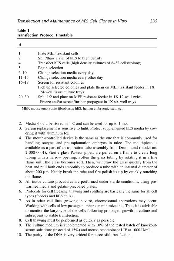
Table 1Transfection Protocol Timetable
d
1 Plate MEF resistant cells 2 Split/thaw a vial of hES to high density4 Transfect hES cells (high density cultures of 8–32 cells/colony)5 Begin selection6–10 Change selection media every day11–15 Change selection media every other day16–18 Screen for resistant colonies
Pick up selected colonies and plate them on MEF resistant feeder in 1X24-well tissue culture trays
20–30 Split 1:2 and plate on MEF resistant feeder in 1X 12-well twice Freeze and/or screen/further propagate in 1X six-well trays
MEF, mouse embryonic fibroblasts; hES, human embryonic stem cell.
16_chap_Eigesed.qxd 2/27/06 8:48 AM Page 235

236 Eiges
Fig. 2. Schematic illustration describing the methods for generating geneticallymodified hES cells by transfection.
16_chap_Eigesed.qxd 2/27/06 8:48 AM Page 236

Transfection and Maintenance of hES Cell Clones In Vitro 237
11. The cells should be transfected during the lag phase of cell division. The transfectionrate is most efficient when the cell density reaches 50–70% and the colonies aresmall (8–32 cells per colony) (Fig. 1A). The colonies should have discrete bordersand be composed of similar sized cells, with a pronounced nucleus.
12. The calcium phosphate (CaPO4) transfection protocol is basically similar to theprotocols used for other cell types.
13. At this time, fine DNA-calcium phosphate precipitates should be formed withoutagitation.
14. ExGen 500 (polyethylenimine, PEI) is a cationic polymer, which is capable oftransfecting a wide range of cell types at relatively high efficiency. It interacts withthe negatively charged DNA molecules by forming small, stable, and highlydiffusible particles, which settle on the cell surface by gravity and absorb into thecell by endocytosis.
15. In parallel to the experiment, one may consider to carrying out transient transfec-tion on a small number of cells with a construct carrying a constitutive expressedreporter gene, such as CMV-EGFP, to asses transfection efficiency beforeapplying selection (Fig. 1B).
16. The colonies are picked up by the aid of a mouth apparatus connected to a sterilepulled and fire polished paster pipet, as is commonly used for handling oocytesand preimplantation embryos (see Note 4).
17. We find this pickup method more suitable and efficient for isolating single hEScolonies than the method applied in mouse, where individual ES colonies arecollected with a disposable tip, trypsinzed, and then plated.
18. In some cases, it is crucial that no feeders will be present during the screen. Forthis purpose, cells must be propagated in feeder-free gelatinized plates, for at leastone passage. Under such conditions the cells must be grown in conditioned media(CM, hES cell media conditioned by MEFs for 24 h), preventing from differenti-ation and consequently culture loss.
References1. Li, M., Pevny, L., Lovell-Badge, R. and Smith, A. (1998) Generation of purified neural
precursors from embryonic stem cells by lineage selection. Curr. Biol. 8, 971–974.2. Klug, M. G., Soonpaa, M. H., Koh, G. Y. and Field, L. J. (1996) Genetically
selected cardiomyocytes from differentiating embronic stem cells form stableintracardiac grafts. J. Clin. Invest. 98, 216–224.
3. Eiges, R., Schuldiner, M., Drukker, M., Yanuka, O., Itskovitz-Eldor, J. andBenvenisty, N. (2001) Establishment of human embryonic stem cell-transfectedclones carrying a marker for undifferentiated cells. Curr. Biol. 11, 514–518.
4. Levinson-Dushnik, M. and Benvenisty, N. (1997) Involvement of hepatocytenuclear factor 3 in endoderm differentiation of embryonic stem cells. Mol. Cell.Biol. 17, 3817–3822.
5. Dekel, I., Magal, Y., Pearson-White, S., Emerson, C. P. and Shani, M. (1992)Conditional conversion of ES cells to skeletal muscle by an exogenous MyoD1gene. New Biol. 4, 217–224.
16_chap_Eigesed.qxd 2/27/06 8:48 AM Page 237

238 Eiges
6. Kim, J. H., Auerbach, J. M., Rodriguez-Gomez, J. A., et al. (2002) Dopamine neuronsderived from embryonic stem cells function in an animal model of Parkinson’sdisease. Nature 418, 50–56.
7. Soria, B., Roche, E., Berna, G., Leon-Quinto, T., Reig, J. A. and Martin, F. (2000)Insulin-secreting cells derived from embryonic stem cells normalize glycemia instreptozotocin-induced diabetic mice. Diabetes 49, 157–162.
8. Capecchi, M. R. (1989) Altering the genome by homologous recombination.Science 244, 1288–1292.
9. Zwaka, T. P. and Thomson, J. A. (2003) Homologous recombination in humanembryonic stem cells. Nat. Biotechnol. 21, 319–321.
10. Urbach, A., Schuldiner, M. and Benvenisty, N. (2004) Modeling for Lesch-Nyhandisease by gene targeting in human embryonic stem cells. Stem Cells 22, 635–641.
11. Stanford, W. L., Cohn, J. B. and Cordes, S. P. (2001) Gene-trap mutagenesis: past,present and beyond. Nat. Rev. Genet. 2, 756–768.
12. Kunath, T., Gish, G., Lickert, H., Jones, N., Pawson, T. and Rossant, J. (2003)Transgenic RNA interference in ES cell-derived embryos recapitulates a geneticnull phenotype. Nat. Biotechnol. 21, 559–561.
13. Vallier, L., Rugg-Gunn, P. J., Bouhon, I. A. andersson, F. K., Sadler, A. J. andPedersen, R. A. (2004) Enhancing and diminishing gene function in human embry-onic stem cells. Stem Cells 22, 2–11.
14. Hay, D. C., Sutherland, L., Clark, J. and Burdon, T. (2004) Oct-4 knockdowninduces similar patterns of endoderm and trophoblast differentiation markers inhuman and mouse embryonic stem cells. Stem Cells 22, 225–235.
15. Ma, Y., Ramezani, A., Lewis, R., Hawley, R. G. and Thomson, J. A. (2003) High-level sustained transgene expression in human embryonic stem cells usinglentiviral vectors. Stem Cells 21, 111–117.
16. Pfeifer, A., Ikawa, M., Dayn, Y. and Verma, I. M. (2002) Transgenesis by lentiviralvectors: lack of gene silencing in mammalian embryonic stem cells and preim-plantation embryos. Proc. Natl. Acad. Sci. USA 99, 2140–2145.
17. Smith-Arica, J. R., Thomson, A. J., Ansell, R., Chiorini, J., Davidson, B. andMcWhir, J. (2003) Infection efficiency of human and mouse embryonic stem cellsusing adenoviral and adeno-associated viral vectors. Cloning Stem Cells 5, 51–62.
18. Gropp, M., Itsykson, P., Singer, O., et al. (2003) Stable genetic modification ofhuman embryonic stem cells by lentiviral vectors. Mol. Ther. 7, 281–287.
19. Park, J. H., Kim, S. J., Oh, E. J., et al. (2003) Establishment and maintenance ofhuman embryonic stem cells on STO, a permanently growing cell line. Biol.Reprod. 69, 2007–2014.
20. Richards, M., Fong, C. Y., Chan, W. K., Wong, P. C. and Bongso, A. (2002) Humanfeeders support prolonged undifferentiated growth of human inner cell masses andembryonic stem cells. Nat. Biotechnol. 20, 933–936.
21. Amit, M., Margulets, V., Segev, H., et al. (2003) Human feeder layers for humanembryonic stem cells. Biol. Reprod. 68, 2150–2156.
22. Hovatta, O., Mikkola, M., Gertow, K., et al. (2003) A culture system using humanforeskin fibroblasts as feeder cells allows production of human embryonic stemcells. Hum. Reprod. 18, 1404–1409.
16_chap_Eigesed.qxd 2/27/06 8:48 AM Page 238

Transfection and Maintenance of hES Cell Clones In Vitro 239
23. Cheng, L., Hammond, H., Ye, Z., Zhan, X. and Dravid, G. (2003) Human adultmarrow cells support prolonged expansion of human embryonic stem cells inculture. Stem Cells 21, 131–142.
24. Reubinoff, B. E., Pera, M. F., Fong, C. Y., Trounson, A. and Bongso, A. (2000)Embryonic stem cell lines from human blastocysts: somatic differentiation in vitro.Nat. Biotechnol. 18, 399–404.
25. Thomson, J. A., Itskovitz-Eldor, J., Shapiro, S. S., et al. (1998) Embryonic stemcell lines derived from human blastocysts. Science 282, 1145–1147.
26. Tucker, K. L., Wang, Y., Dausman, J. and Jaenisch, R. (1997) A transgenic mousestrain expressing four drug-selectable marker genes. Nucleic Acids Res. 25,3745–3746.
27. Robertson, E. J. (1987) Teratocarcinomas and Embryonic Stem Cells: A PracticalApproach. IRL Press, Oxford.
16_chap_Eigesed.qxd 2/27/06 8:48 AM Page 239

16_chap_Eigesed.qxd 2/27/06 8:48 AM Page 240