genetic markers involved in inflammatory digestive pathology
TRANSCRIPT
1
UNIVERSITY OF MEDICINE AND PHARMACY OF CRAIOVA
PhD Thesis
Abstract
Genetic markers involved in
inflammatory digestive pathology
PhD Tutor Prof. univ dr. Florica Popescu PhD Joint Tutor Prof. univ. dr. Francisc Mixich
PhD Student Elena-Raluca Nicoli
Craiova, 2013
2
Investeşte în oameni !
FONDUL SOCIAL EUROPEAN
Programul Operaţional Sectorial Dezvoltarea Resurselor Umane 2007 – 2013
Axa prioritară 1
„Educaţia şi formarea profesională în sprijinul creşterii
economice şi dezvoltării societăţii bazate pe cunoaştere”
Domeniul major de intervenţie 1.5
„Programe doctorale şi postdoctorale în sprijinul cercetării”
Titlul proiectului
"Creşterea calităţii şi vizibilităţii rezultatelor cercetării ştiinţifice a doctoranzilor cu frecvenţă prin acordarea de burse doctorale"
Contract nr: POSDRU/107/1.5/S/82705
Beneficiar
Universitatea de Medicină şi Farmacie din Craiova

3
INTRODUCTION
Inflammatory bowel disease (IBD) represents a heavy burden for the patients
and national health systems, due to their onset in adolescence or early adulthood
with potentially debilitating symptoms and chronic and unpredictable course
characterized by an increased risk of developing colorectal cancer (Høivik M. et al,
2012). With all the therapeutic improvements that have been made in the last years,
there are still a surprisingly large number of patients with IBD who have to undergo
surgery due to disease complications (Podolsky D.K., 2002). Thus, any molecular
marker that could be used for early diagnosis and to avoid complications like
colorectal cancer development is beneficial.
Key Words: TPC2, Inflammatory Bowel Diseases, Colorectal Cancer, Autophagy
PART I - STATE OF KNOWLEDGE
Chapter 1. General introduction
Main risk factors influence on IBD epidemiology
IBD represents an important public health problem, as it affects mostly young
adults and is marked by episodes of debilitating relapse and remission with negative
effects on socioeconomic status and quality of life. The epidemiology of IBD is
constantly changing, suggesting that environmental factors including infections, diet,
lifestyle factors, and medication play a major role in modifying disease progression
(Ng Siew C et al., 2013). Generally, incidence rates of ulcerative colitis (UC) and
Crohn’s Disease (CD) vary between 2.2 to 14.3 cases per 100,000 person/years for
UC and from 3.1 to 14.6 cases per 100,000 person/years for CD (Longo D.L., 2012).
4
Autophagy – an important pathogenic pathway
Genome-wide association studies identified in their intent to discover the
genetic basis of IBD, beyond genes that modulate the innate immunity, a group of
genes related to autophagy that seem to be associated with increased disease
susceptibility, mainly CD (Nys K. et al., 2013; Van Limbergen J. et al., 2009). The
autophagy genes connected with an increased susceptibility to IBD (CD or UC or
both) identified through several genome-wide association studies are ATG16L1,
IRGM1 and LRRK2. It was demonstrated on several knockout animal models that
autophagy is modulated by LRRK2 activity: an increased activity of the gene tempers
the autophagic flux while reduced activity would amplify it (Gómez-Suaga P. et al.,
2012). Pathology involving LRRK2 defects depends on the cellular enviroment in
which the gene works (Lewis P. A. & Manzoni C., 2012). LRRK2 expression level
and defects influence the number of autophagosome in a calcium-dependent
manner: the calcium chelator BAPTA inhibits their number increase and, genetically
depletion of ER calcium stores blocks LRRK2 effects on autophagosome numbers,
increases the pH of a population of lysosomes and the number of lipid droplets
(Gómez-Suaga P. et al., 2012).
A similar mechanism of action was described in the case of nicotinic acid
adenine dinucleotide phosphate (NAADP), an agonist-generated second
messenger, able to trigger complex calcium signals, initiated from acidic stores and
subsequently amplified by ER calcium release channels (Churchill G. C. et al., 2002;
Patel S. & Docampo R., 2010; Morgan A. J. et al., 2011). It was showed that NAADP
main action is on the endolysosomal two-pore channels TPC1 and TPC2 (Calcraft
P.J. et al., 2009; Brailoiu E. et al., 2009; Patel S. et al., 2011), but it seems that the
5
molecule is not directly attached to TPCs, an unidentified associated low-molecular
weight binding protein being responsible for this connection (Lin-Moshier Y. et al.,
2012). TPCs role is to release calcium under NAADP action, a molecule for which
TPC2 presents high and low affinity binding sites, suggesting that TPC2 may be one
of the components of an NAADP-dependent, lysosome-targeted Ca2+ release
channel that is neither activated, nor inhibited by Ca2+ (Zhu M.X. et al., 2009).
Thus, demonstration and definition of TPC2 potential role in the
pathophysiological mechanisms involved in autophagy defective diseases such as
Parkinson’s disease or IBD require more elaborate studies on knockout animal
models associated with the assessment of TPC2 gene expression levels in the
damaged tissues versus healthy ones .
PART II - PERSONAL CONTRIBUTIONS
Chapter 2. TPC2 Mouse Model – preliminary result
The Aim of this chapter was to obtain more information about TPC2 KO and
WT animals that could be used in further studies to show the role played by TPC2 in
pathophysiological mechanisms involved in autophagy defective diseases such as
neurodegenerative diseases, IBD or tumour processes.
Materials and Methods
Animals. All studied animals were TPC2 KO and strain-matched WT
littermates provided by the Parrington/Galione labs (Department of Pharmacology,
University of Oxford). Animals were housed under standard conditions, in solid-
bottomed polycarbonate cages that were individually ventilated, at constant
temperature (22°C), 45-65% relative humidity and a 12:12-h light–dark cycle and

6
were provided with irradiated water and food ad libitum. All procedures were carried
out in accordance with the UK Animals (Scientific Procedures) Act, 1986 and
experiments were carried out with UK Home Office approval. This experiment was
conducted on a number of twelve animals from which five were TPC2 KO (two
females and three males) and six TPC2 WT (three males and three females)
animals.
Cell preparation. Single cell suspensions of spleen, lymph nodes (pooled
inguinal) and thymus were accomplished according to standard procedures, were
prepared using DPBS between two glass microscopic slides. FACS staining. The
multicolour antibody cocktails used for labelling the cells of interest were: FITC-
CD69, PE-CD25 (IL2Rα, p55), Alexa Flour-CD335 (NKp46), PerCP-Cy5.5-CD45,
APC-H7-CD19 and Pacific Blue-CD3e.
Flow cytometry. All samples were analysed on a FACSCanto II (BD
Biosciences) that was standardised using BD Cytometer Setup and Tracking beads
and software. Data acquisition was controlled with BD FACS Diva v6 software.
Results and Discussions
Comparison of the two animal model TPC2 (KO and WT) organs between
them was revealed:
in spleen, lymphocytes expression is significant different in cell population of:
T cells especially for CD69 positive, NK cells mostly CD25 positive and B
cells both for CD69 and CD25 positive;
in thymus was revealed significant differences for: T cells expression both for
CD69 and CD25 positive, and B cells expression especially for CD69 positive.
NK cells population presented no differences;
7
in lymph nodes was remarked a significant difference in: T cells CD25
positive expression, NK and B cells expression both for CD69 positive and
CD25 positive;
In this study, we have used the previously characterised TPC2 KO and WT
mouse models to assess lymphocytes expression in the spleen, lymph nodes
(pooled inguinal) and thymus of TPC2 KO compared with TPC2 WT animals based
on that isolation of lymphocytes and other hematopoietic-derived cells from tissues
to highlight their phenotype that has become increasingly relevant to immunology
over the last decade (Sheridan, B. S et al, 2012).
Immune assessment of animal models revealed that both lymphocytes and
hematopoietic-derived cells are characterized by significant heterogeneity in
morphology, surface antigen expression, and function (Croitoru K., et al, 1991).
Based on the above preliminary results and comparing with literature data, we
can say that the immune cells expression in TPC2 KO model tissues might be
different compared with TPC2 WT mouse, suggesting that TPC2 could be an
important player in immune response. More studies are needed in order to fully
characterize and understand the immune system of TPC2 KO animals.
8
Chapter 3. The role of acidic-store calcium in susceptibility to colitis:
Insights gained from the TPC2 knockout mouse – preliminary results
The aim of this chapter was to assess the response of the TPC2 KO mouse
model to dextran sodium sulphate (DSS)-induced acute colitis compared to wild-type
(TPC2 WT) animals.
Material and methods
Animals. The studied animals were TPC2 KO and strain-matched WT
colonies provided by the Parrington/Galione labs (Department of Pharmacology,
University of Oxford). All animal studies were conducted using protocols approved by
the UK Home Office for the conduct of regulated procedures under licence (Animal
scientific Procedures Act, 1986) and experiments were carried out with UK Home
Office approval under PPL 30/2524. Twenty four animals were included in this study.
For induction of colitis, twelve animals (six TPC2 KO and six TPC2 WT, mixed
groups with equal numbers of males and females), at 6 weeks of age were
administered with 2% DSS in drinking water over the course of one week then
switched to normal drinking water for three days. Twelve animals (six TPC2 KO and
six TPC2 WT, mixed groups with equal numbers of males and females) were also
included in the study as control. Ten days following the first day of DSS
administration, mice were sacrificed by CO2 asphyxiation and tissues were collected
for histopathology and molecular biology.
Drug Administration. Dextran sodium sulphate (DSS, MP Biomedicals,
Catalog no.: 160110, 500g, M.W.=36,000-50,000) was dissolved in water in a
concentration of 2% and administered to animals in drinking water for 7 days starting
on day 0 (D0) ad libitum. All animals were provided the normal or 2% DSS drinking
water ad libitum.
9
Assessment of the disease. Throughout the course of the ten days of colitis
induction, mice weight, physical condition, stool consistency, and the presence of
gross and occult blood in the faeces and/or from the anus were examined and
documented daily. Weigh loss of animals was assessed by daily monitoring of body
weight loss compared to D0.
Histopathology Analysis. Evaluation of histological changes and inflammatory
cell infiltration in colon was performed on colon sections fixed in 10% neutral-
buffered formalin. Samples were processed, paraffin-embedded, and thereafter
sectioned using a Leica RM2126 microtome. Tissue sections (4 µm) were stained
with hematoxylin and eosin (H&E). Five sections, 50 mm apart per colon sample
were evaluated and repeated in all mice per group in a blinded manner, and scored.
Sample preparation
Paraffin embedding protocol. All samples were kept in formalin for exactly 7
days before processing and were processed identically following the same protocol.
H&E staining protocol. The staining with hematoxylin (Harrison Hematoxylin,
Sigma UK) was done by incubating the sections directly in hematoxylin a couple of
dips and then rinsed in tap water followed by one or two dips to differentiate in acid
alcohol (1% HCL in 70% ethanol) and 30 seconds in Scott’s tap water. The next step
was the dipping in eosin (water eosin) and a brief rinse under tap water. The
sections were dehydrated though graded alcohol, cleared in Histoclear Solution and
mounted with a permanent mounting medium (DePeX).
Tissue collection. Animals were sacrificed by CO2 asphyxiation followed by
decapitation. Blood was removed by cardiac puncture. Peripheral tissues were
perfused with 0.9% saline at 20ml/minute for 1-2 minutes. Whole liver lobes were
dissected and part was kept in 10% formalin and part snap frozen on isopentane on
10
dry ice (approximately -78°C). Intestines were removed for histopathology and
molecular biology and dissected into regions for qPCR.
Tissue collection for histopathology. Weight loss and histology are the most
important ways to assess disease. The colon (starting from 0.5 cm above the anus
up to the cecum), small intestine (starting from above the cecum up to the stomach),
spleen, thymus, and part of the liver were carefully dissected, removed, and
weighted. For histology, sections of mid, distal, proximal colon, ileum, jejunum,
duodenum (~1 cm) as well as cecum and stomach were collected into 10% formalin
(in PBS) and then embedded in paraffin, sectioned (4 μm) and stained with H&E.
Tissue collection for RT-PCR. For RT-PCR; slices (~1 mm) of mid, distal,
proximal colon, cecum, ileum, jejunum, duodenum, stomach and liver, were snap-
frozen in liquid nitrogen and stored at -80°C until extraction of mRNA.
mRNA extraction from intestinal tissue. Intestine specimens (~10 mg of tissue
per sample) were homogenized using a pestle homogenizer in lysis buffer (Buffer
RLT, Qiagen) with 1% β-MCE (mercaptoethanol, Sigma, UK), and total RNA
extraction was performed according to the manufacturer instructions
(QiagenRNEasy Mini Kit).
cDNA synthesis from intestinal tissue. To determine the mRNA expression in
the mouse colonic tissue, quantitative real-time reverse-transcription polymerase
chain reaction (qRT-PCR) was performed using High Capacity cDNA Reverse
Transcription Kit from Applied Biosystems, UK.
qRT-PCR from intestinal tissue. Quantification of the different cytokines and
genes for mRNA expression in colon tissues was assessed by qRT-PCR with
specific primers for each gene (IL1β, IL6, TNFα, TLR4, TGFβ, Caveolin, ICAM,
VCAM, β-Actin). The expression level of each transcript within the different samples
11
was quantified as the relative value against β-Actin. The primers used in qRT-PCR
reactions were purchased Roche, UK.
Results and Discussions
Daily measurements during the experiment. Daily body weight measurements
revealed that DSS-induced colitis was associated with a higher weight loss in the
group TPC2 KO animals compared with TPC2 WT group (***p<0.0001).
Measurements of organs parameters. Analysing the colon weight/length ratio,
we observed an extremely significant differences between the two groups: TPC2 KO
and WT, whilst in small intestine, this ratio was not significantly different between the
two groups. The weight/length ratio recorded in spleen showed also no significant
differences between the two genotypes.
Histopathological assessment of colonic sections:
At the end of the experiment, no histological differences were found between
WT and KO control groups (on water). However, when comparing the DSS groups
with their control groups, histological assessment revealed important differences in
the mid and distal colon of WT and KO groups as follows:
for TPC2 WT group receiving DSS, a moderate hyperplasia associated with
mild goblet cell depletion and low epithelial cells loss was noticed. Also, the
area of affected tissue was up to 25% coverage, showing a gentle infiltration
with leukocytes in lamina propria;
the TPC2 KO group receiving DSS has shown unrelenting depletion of the
goblet cell accompanied by a substantial hyperplasia and epithelial cells loss.
Also, several histological features of severe inflammation were observed
including loss of crypt structure, oedema, and crypt abscesses associated

12
with stern submucosal inflammation. More than 50% of the colonic tissue was
affected, having almost no structural architecture.
Histolopathology scoring. Histopathology scores of proximal colon were not
significantly different between the two genotypes. Due to measurements of length
and weight before processing the sections for histology (embedding in paraffin
blocks and staining with H&E), a lot of the H&E sections have shown processing
artefacts and hence not all the sections from each animal have been scored.
Expression analysis of inflammatory gene markers mRNAs in TPC2 animals
colonic sections after induction of colitis:
the studied model of colitis induced by DSS in TPC2 animals is suggesting a
possible confirmation of the histopathology results, showing a relative
increased percentage fold changes of IL-6 and IL-1β mRNA expression.
Assessment of IL-6 and IL-1β expression levels in both TPC2 models, WT
and KO, revealed a significant difference between TPC2 KO receiving DSS
compared with TPC2 KO on water and Wild-type on water or DSS;
the TPC2 group receiving DSS showed a significant increase in relative
percentage fold change of Caveolin and VCAM mRNA expression when
comparing between KO and WT animals;
relative percentage fold change of TLR4 mRNA expression in TPC2 KO group
treated with DSS was significantly higher compared to TPC2 KO and also
Wild-type groups receiving water (*p = 0.0189);
relative percentage fold change of TNF-α mRNA expression in the Wild-type
group treated with DSS was very significantly higher than both in the TPC2
KO receiving water and the Wild-type group on water.
13
assessment of TGF-β and ICAM expression levels in both TPC2 groups (WT
and KO) revealed no significant difference.
We have assessed the response of the previously characterized TPC2 KO
and WT mouse models (Chapter 2) to DSS-induced acute colitis. We have also tried
to evaluate if their response to DSS is associated with altered gene expression
levels of some inflammatory gene markers such as IL-6, IL-1β, Caveolin, VCAM,
ICAM, TNF-α, TLR-4 or TGF-β that have previously shown to be implicated in IBD
pathogenesis (Carta, S. et al, 2013).
DSS is the most commonly polysaccharide used to induce colitis in animal
models in the study of chronic gastrointestinal inflammation pathogenesis and
preclinical studies (Neurath, M. et al, 2000; Wirtz, S., & Neurath, M. F. 2007; Wirtz,
S. et al, 2007). Mice are given cycles of DSS added to their drinking water resulting
in the development of an acute or chronic intestinal inflammation. While chronic DSS
colitis is especially driven by T lymphocytes, acute DSS colitis can occur in the
absence of T cells (Wirtz S. et al 2007). Several studies showed that genetic factors
modulate susceptibility and responsiveness to DSS-induced colitis in animal models,
like the interleukin-10 deficient mouse model or C3H mouse model (Wirtz S. et al
2007; Berg DJ, et al, 2002; Perše, M. et al 2012; Mähler, M. et al 1998). Several
studies carried out between1999–2005 tried to show the involvement of genetic
factors in regulations of inflammatory response in mouse models, which represents
an important direction for future research (Stevceva, L. et al, 1999; Kitajima, S. et al,
2001; Melgar, S. et al, 2005)
Based on the above preliminary results and comparing them with literature
data, we can conclude that the TPC2 KO mouse model is more susceptible to DSS
induced colitis compared with TPC2 WT.

14
Chapter 4. Determination of TPCN Gene Expression in Human Colorectal
Cancer Samples
Main objectives of this chapter was to quantify the expression of total TPCN1
and TPCN2 mRNA expression level in a Romanian cohort of patients diagnosed with
colorectal cancer (CRC), for further analysis to see whether there might be a
correlation between the gene expression and clinicopathological features of the
tumours in Romanian population.
Material and Methods
Patients and samples. 25 patients with colorectal tumour who had undergone
surgery at the County Clinical Emergency Hospital of Craiova, Romania between
2008 and 2012 were included. The Ethics Committee of University of Medicine and
Pharmacy of Craiova, Romania approved this study and written informed consent
was obtained from all subjects. The age of the patients included in the study was
64.24 ± 10.81 years (mean+/- sem), with a sex ratio of 14:11 (m:f).Both tumour (T)
and peritumour (PT) corresponding to large intestine mucosa were harvested during
surgery for all the patients. The samples were collected in RNAlater solution
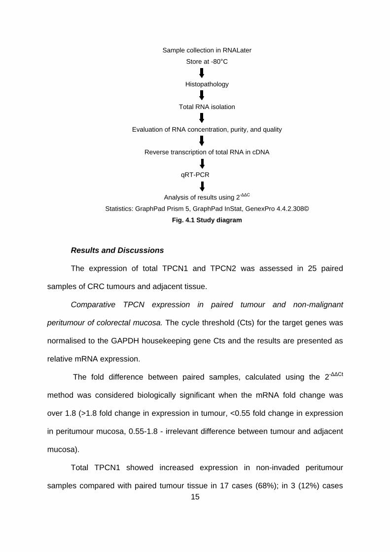
(Ambion) and stored at -80°C until RNA isolation. The representative steps are
presented in next figure (Fig. 4.1).
15
Sample collection in RNALater
Store at -80°C
Histopathology
Total RNA isolation
Evaluation of RNA concentration, purity, and quality
Reverse transcription of total RNA in cDNA
qRT-PCR
Analysis of results using 2-ΔΔC
Statistics: GraphPad Prism 5, GraphPad InStat, GenexPro 4.4.2.308©
Fig. 4.1 Study diagram
Results and Discussions
The expression of total TPCN1 and TPCN2 was assessed in 25 paired
samples of CRC tumours and adjacent tissue.
Comparative TPCN expression in paired tumour and non-malignant
peritumour of colorectal mucosa. The cycle threshold (Cts) for the target genes was
normalised to the GAPDH housekeeping gene Cts and the results are presented as
relative mRNA expression.
The fold difference between paired samples, calculated using the 2-ΔΔCt
method was considered biologically significant when the mRNA fold change was
over 1.8 (>1.8 fold change in expression in tumour, <0.55 fold change in expression
in peritumour mucosa, 0.55-1.8 - irrelevant difference between tumour and adjacent
mucosa).
Total TPCN1 showed increased expression in non-invaded peritumour
samples compared with paired tumour tissue in 17 cases (68%); in 3 (12%) cases
16
the expression was higher in tumour tissue and in the remaining 5 cases (20%) the
difference between paired samples was biologically irrelevant.
The total TPCN2 expression was increased in non-invaded peritumour
samples compared with paired tumour tissue in 15 cases (60%); in 3 (12%) cases
the expression was higher in tumour tissue and in the remaining 7 cases (28%) the
difference between paired samples was biologically irrelevant.
The expression of total TPCN1 and total TPCN2 were significantly
(statistically) higher in non-invaded peritumour tissue compared with tumour tissue
(p=0.005 and p=0.001 respectively, Wilcoxon matched-pairs signed rank test).
The expression for the TPCN genes (TPCN1 and TPCN2) was extremely
significant in each individual sample (tumour or peritumour tissue) (p<0.0001,
Wilcoxon Signed Rank Test).
Gene expression analysis in relation to tumour pathological features.
The analysis of TPCN1 and TPCN2 expression according to tumour location
revealed that mRNA levels tend to be lower in rectosigmoid colon compared with
ascending and descending colon reaching a statistically significant different level
(p=0.0210, p=0.0323 respectively, Kruskal-Wallis test).
When assessed according to tumour Dukes stage (A, B, C, D), total TPCN1
and TPCN2 showed a similar expression pattern. Even though there’s a tendency
towards a higher mRNA levels in the first two stages (stage A+B), however, this
difference was not statistically significant (NS), (p=0.26, p=0.47 respectively,
Kruskal-Wallis test). Regarding the assessment according to tumour stage (I, II, III,
IV), total TPCN1 and TPCN2 showed a similar expression pattern. The mRNA levels
were similar in all four tumour stages, the difference was not statistically significant
(NS, Kruskal-Wallis test).
17
In the majority of the cases, both TPCN1 and TPCN2 levels were similar
between well (G1), moderate (G2) and poor (G3) tumours (p=0.74, p=0.55
respectively, Kruskal-Wallis test). In two G3, two G2 and one G1 tumours TPCN1
and TPCN2 levels were biologically higher compared with the rest of the tumours but
this was not representative for the entire group.
Correlations between genes in tumour and peritumour mucosa. A very strong
correlation between TPCN1 and TPCN2 expression in both tumour (r=0.94,
p<0.0001) and peritumour tissue (r=0.97, p<0.0001) was identified. In both tumour
and peritumour tissue, Spearman coefficient also highlights an aggregation of
TPCN1 and TPCN2 mRNA levels (r=0.60, p<0.004 for tumor and r=0.64, p<0.002 for
peritumour tissue).
The role of TPCs to discrete acidic calcium stores as in the case of TPC2 for
lysosomes and of TPC1 and TPC3 for endosomes has been confirmed (Zhu, M.X. et
al 2010).
Calcium is important for maintaining normal trafficking, vesicular fusion events
and recycling within the endolysosomal system (Lloyd-Evans and Platt, 2011). Via
two-pore channel family (TPC) lysosomal calcium can be released by intracellular
calcium-releasing second messenger nicotinic acid adenine dinucleotide phosphate
(NAADP) (Churchill et al., 2002).
Our results on human colorectal cancer subjects show a higher expression of
total TPCN1 and TPCN2 in non-invaded peritumour tissue compared with tumour
tissue. Multivariate analyses showed that the studied TPCs gene expression level in
mucosa compared with tumour, could be a prognostic parameter independent of the
clinicopathological factors with regard to a possible risk to colorectal cancer. These
findings would require a further research to answer some other key questions that
18
need to be answered about the involvement of TPCs in carcinogenesis. More of
these systematic research will probably reveal new and exciting targets for cancer
therapy.
Chapter 5. Determination of an autophagy polymorphism in the Human
Colorectal Cancer Samples
The purpose of this chapter was to collect and genotype a Romanian cohort
of patients diagnosed with CRC for ATG16L1 polymorphism. Once this was
accomplished, the final goal was assessing whether ATG16L1 T300A (+898A>G)
influences the risk of CRC in Romanian population. This polymorphism was selected
on the basis of functional data relating to its potential role in autophagy and
carcinogenesis, particularly within the gastrointestinal tract (Deretic, V. et al, 2013;
Gillen, C. D., et al, 1994; Bolan, C. R. et al, 2010)
Material and Methods
Patients and samples. A total of 351 Romanian subjects (109 patients
diagnosed with sporadic CRC and 242 healthy controls) were included in this study.
Matched control (healthy) subjects of the same ethnic and geographical origins were
recruited among unrelated volunteers admitted at the same time to the same
hospital, with no previous history of tumour, inflammatory or infectious disease.
Blood samples were obtained from both groups. The Ethics Committee of University
of Medicine and Pharmacy of Craiova, Romania approved this study and written
informed consent was obtained from all subjects.
DNA extraction and genotyping. Genomic DNA was extracted from the
peripheral blood leukocytes using Wizard® Genomic DNA Purification Kit (Promega,
Madison, WI, USA), following the manufacturer’s protocol. Polymorphism genotyping
19
was performed with a predesigned TaqMan assay from Applied Biosystems (Foster
City, CA, USA): ATG16L1 T300A (+898A>G, rs2241880, assay C_9095577_20).
Results and Discussions
Subjects characteristics. Genotyping was performed in 109 CRC patients and
242 controls. There were no differences in median age and gender between cases
and controls.
Genotype frequency and the risk of CRC in Romanian population. A
significant association was observed for male subjects carrying GG genotype that
were at a higher risk for CRC (OR 2.76, 95% CI: 1.12-6.79, p = 0.019) when
compared with the more frequent AA genotype. Furthermore, there was no
correlation between CRC cases and controls for female subjects carrying GG
genotype (OR 1.19, 95% CI: 0.445-3.2, p =0.722) or AG genotype (OR 1.26, 95% CI:
0.51-3.15, p =0.607).
Association of ATG16L1 T300A (+898A>G) polymorphism with Dukes' stage
and histological subtype was examined separately (A, B, C, D). In a stratified
analysis, the only association between CRC and autophagy polymorphism was
found for carriers of GG allele and was restricted to moderately and poorly
differentiated cases (OR 5.15, 95% CI: 1.47-18.06, p=0.005). No significant
differences were observed between tumour stage (A + B), localisation or histological
grading and controls in stratified analysis.
Autophagy is a catabolic intracellular process in which cytoplasmic
components are sequestered within vesicles and delivered to the lysosomes. It is the
pathway used by cells during nutrient starvation to break down non-vital components
and use them as nutrients (Naser, S. A et al, 2012). Autophagy is involved in
infectious processes by clearing the cells of foreign antigens released from the

20
breakdown of the pathogen (Naser, S. A et al, 2012). This pathway can also be
implemented as a repair mechanism to degrade damaged organelles and proteins.
Autophagy is a complex pathway, modulated by several molecular mechanisms that
remain elusive. There have been approximately 30 autophagy-related (Atg) genes
identified, with 2 proteins having ubiquitin-like characteristics, Atg12 and Atg8 (Read,
S. et al, 2000; Mehrpour, M. et al, 2012). The ATG16L1 protein is expressed in the
colon, small intestine, intestinal epithelial cells, leukocytes, and spleen (Fujita, N. et
al 2008). In 2010, a study has shown that an ATG16L mutation: ATG16L1 -
Thr300Ala (rs2241880), located on chromosome 2, is associated with the onset of
ileal CD, and is therefore a key molecule in elucidating the genetic aspects of this
disease (Sventoraityte, J. et al, 2010).
A second report from the North American CD genome-wide study also shows
an association with ATG16L using a case-control analysis in 988 CD patients and
1007 controls (Rioux, J. D. et al, 2007). Taking into account that patients with IBD,
especially CD, have a six-fold higher risk of developing colorectal cancer, several
studies investigating the link between autophagy genes, and susceptibility to this
type of cancer, were conducted (Brest P. et. al., 2010). Thus, a study conducted by
Kang M.R. and colleagues in 2009, indicates that frameshift mutations in ATG genes
with mononucleotide repeats appear in gastric and colorectal carcinomas suggesting
that these mutations may contribute to cancer development by deregulating the
autophagy process (Kang M.R. et al., 2009). In our hospital-based case-control
study, we assessed whether an autophagy polymorphism influenced sporadic CRC
development and progression in Romanian population.
It is necessary to study also if there is a link between ATG16L1 gene
expression, CD course and colorectal cancer predisposition. It is clear that variants
21
of this gene have been proven to be directly associated with CD because autophagy
plays a critical role in disease pathogenesis. Further research needs to focus on
understanding how ATG16L1 variants contribute to disease susceptibility in IBD
patients, and their possible therapeutic implications, and, allows the link between the
ATG16L1 variants in IBD and colorectal cancer.
In this study, it was found that ATG16L1 T300A (+898A>G) polymorphism
influences the risk of CRC in Romanian population. The male carriers of GG
genotype are at a higher risk for CRC. While this study indicates a strong association
in the moderately and poorly differentiated CRC cases, the small subgroups size
ousts drawing conclusions. Further research is required in different ethnic
populations to improve the level of knowledge of autophagy in colon cancer
tumorigenesis.
Final conclusions
Our results showed that between TPC2 KO and WT model there are
significant differences regarding expression of immune surface antigens in
lymphocyte population from spleen, thymus and lymph nodes.
Also, we identified that TPC2 KO mouse model might be more susceptible to
DSS-induced colitis and develops more severe lesions after DSS
administration than the WT model.
Several genes involved in inflammation seem to have a higher expression
level in TPC2 KO mice compared with TPC2 WT model after DSS ingestion.
These results are important for sustaining future experimental studies,
especially on TPC2 KO model, in order to improve management and therapy
22
of chronic inflammatory diseases, such as Crohn’s disease, ulcerative colitis
or colorectal cancer.
TPCN1 and TPCN2 gene expression level in non-invaded peritumour tissue is
higher compared with tumour tissue indicating that TPCN1 and TPCN2 may
serve as a useful new marker for the study of colorectal cancer.
ATG16L1 T300A (+898A>G) polymorphism influences the risk of CRC in
Romanian population; the male carriers of GG genotype being at a higher risk
for CRC.
Further research is required in different ethnic populations to improve the level
of knowledge of autophagy in colon cancer tumorigenesis and to show if
exists a strong link between TPCN, autophagy and development of chronic
inflammatory lesion like IBD or CRC.
Selective References Berg, D. J., Zhang, J., Weinstock, J. V., Ismail, H. F., Earle, K. A., Alila, H., ... & Lynch, R. G. (2002). Rapid development of colitis in NSAID-treated IL-10–deficient mice. Gastroenterology, 123(5), 1527-1542. Boland, C. R. (2010). Chronic inflammation, colorectal cancer and gene polymorphisms. Digestive Diseases, 28(4-5), 590-595. Brailoiu, E., Churamani, D., Cai, X., Schrlau, M. G., Brailoiu, G. C., Gao, X., & Patel, S. (2009). Essential requirement for two-pore channel 1 in NAADP-mediated calcium signaling. The Journal of cell biology, 186(2), 201-209. Brest, P., Corcelle, E. A., Cesaro, A., Chargui, A., Belaid, A., Klionsky, D. J. ... & Mograbi, B. (2010). Autophagy and Crohn's disease: at the crossroads of infection, inflammation, immunity, and cancer. Current molecular medicine, 10(5), 486.
23
Calcraft, P. J., Ruas, M., Pan, Z., Cheng, X., Arredouani, A., Hao, X., & Zhu, M. X. (2009). NAADP mobilizes calcium from acidic organelles through two-pore channels. Nature, 459(7246), 596-600. Carta, S., Lavieri, R., & Rubartelli, A. (2013). Different members of the IL-1 family come out in different ways: DAMPs vs. cytokines? Frontiers in immunology, 4. Churchill, G. C., Okada, Y., Thomas, J. M., Genazzani, A. A., Patel, S., & Galione, A. (2002). NAADP Mobilizes Ca< sup> 2+</sup> from Reserve Granules, Lysosome-Related Organelles, in Sea Urchin Eggs. Cell, 111(5), 703-708. Croitoru, K., & Ernst, P. B. (1991). Leukocytes in the intestinal epithelium: an unusual immunological compartment revisited. Regional immunology, 4(2), 63-69. Deretic, V., Saitoh, T., & Akira, S. (2013). Autophagy in infection, inflammation and immunity. Nature Reviews Immunology, 13(10), 722-737. Fujita, N., Itoh, T., Omori, H., Fukuda, M., Noda, T., & Yoshimori, T. (2008). The Atg16L complex specifies the site of LC3 lipidation for membrane biogenesis in autophagy. Molecular biology of the cell, 19(5), 2092-2100. Gillen, C. D., Andrews, H. A., Prior, P., & Allan, R. N. (1994). Crohn's disease and colorectal cancer. Gut, 35(5), 651-655. Gómez-Suaga, P., Fdez, E., Blanca Ramírez, M., &Hilfiker, S. (2012). A Link between Autophagy and the Pathophysiology of LRRK2 in Parkinson's Disease.Parkinson's disease, 2012. Høivik, M. L., Moum, B., Solberg, I. C., Henriksen, M., Cvancarova, M., & Bernklev, T. (2013). Work disability in inflammatory bowel disease patients 10 years after disease onset: results from the IBSEN Study. Gut, 62(3), 368-375. Kang, M. R., Kim, M. S., Oh, J. E., Kim, Y. R., Song, S. Y., Kim, S. S., ... & Lee, S.
H. (2009). Frameshift mutations of autophagy‐related genes ATG2B, ATG5, ATG9B and ATG12 in gastric and colorectal cancers with microsatellite instability. The Journal of pathology, 217(5), 702-706. Kitajima, S., Morimito, M., Sagara, E., Shimizu, C., & Ikeda, Y. (2001). Dextran sodium sulfate-induced colitis in germ-free IQI/Jic mice.Experimental animals, 50(5), 387-395. Lewis, P. A., & Manzoni, C. (2012). LRRK2 and human disease: a complicated question or a question of complexes?. Science Signaling, 5(207), pe2. Lin-Moshier, Y., Walseth, T. F., Churamani, D., Davidson, S. M., Slama, J. T., Hooper, R., & Marchant, J. S. (2012). Photoaffinity labeling of nicotinic acid adenine dinucleotide phosphate (NAADP) targets in mammalian cells. Journal of Biological Chemistry, 287(4), 2296-2307.
24
Lloyd-Evans, E., & Platt, F. M. (2011). Lysosomal Ca2+homeostasis: Role in pathogenesis of lysosomal storage diseases. Cell calcium, 50(2), 200-205. Longo DL, Fauci AS, Kasper DL, Hauser SL, Jameson JL, Loscalzo J, editors. Harrison's Principles of internal medicine. 18th edition. Vol. 1. New York: Mc Graw Hill; 2012. pp.1870-1879. Mähler, M., Bristol, I. J., Leiter, E. H., Workman, A. E., Birkenmeier, E. H., Elson, C. O., & Sundberg, J. P. (1998). Differential susceptibility of inbred mouse strains to dextran sulfate sodium-induced colitis. American Journal of Physiology-Gastrointestinal and Liver Physiology, 274(3), G544-G551. Mehrpour, M., Botti, J., & Codogno, P. (2012). Deep Insight Section.http://AtlasGeneticsOncology. org, 165. Melgar, S., Karlsson, A., & Michaëlsson, E. (2005). Acute colitis induced by dextran sulfate sodium progresses to chronicity in C57BL/6 but not in BALB/c mice: correlation between symptoms and inflammation. American Journal of Physiology-Gastrointestinal and Liver Physiology, 288(6), G1328-G1338. Morgan A. J., Platt F. M, Lloyd-Evans E. & Galione A. (2011). Molecular mechanisms of endolysosomal Ca2+ signalling in health and disease.Biochemical Journal, 439(3), 349-374. Naser, S. A., Arce, M., Khaja, A., Fernandez, M., Naser, N., Elwasila, S., & Thanigachalam, S. (2012). Role of ATG16L, NOD2 and IL23R in Crohn’s disease pathogenesis. World journal of gastroenterology: WJG, 18(5), 412. Neurath, M., Fuss, I., & Strober, W. (2000). TNBS-colitis. International reviews of immunology, 19(1), 51-62. Ng, Siew. C., Bernstein, C. N., Vatn, M. H., Lakatos, P. L., Loftus, E. V., Tysk, C., ... & Colombel, J. F. (2013). Geographical variability and environmental risk factors in inflammatory bowel disease. Gut, 62(4), 630-649. Nys, K., Agostinis, P., & Vermeire, S. (2013). Autophagy: a new target or an old strategy for the treatment of Crohn's disease?. Nature Reviews Gastroenterology and Hepatology. Patel, S., Ramakrishnan, L., Rahman, T., Hamdoun, A., Marchant, J. S., Taylor, C. W., & Brailoiu, E. (2011). The endo-lysosomal system as an NAADP-sensitive acidic Ca< sup> 2+</sup> store: Role for the two-pore channels. Cell calcium, 50(2), 157-167. Patel, S., & Docampo, R. (2010). Acidic calcium stores open for business: expanding the potential for intracellular Ca< sup> 2+</sup> signaling. Trends in cell biology, 20(5), 277-286.
25
Perše, M., & Cerar, A. (2012). Dextran sodium sulphate colitis mouse model: traps and tricks. BioMed Research International, 2012. Podolsky, D. K. (2002). The current future understanding of inflammatory bowel disease. Best practice & research Clinical gastroenterology, 16(6), 933-943. Read, S., Malmström, V., & Powrie, F. (2000). Cytotoxic T lymphocyte–associated antigen 4 plays an essential role in the function of CD25+ CD4+ regulatory cells that control intestinal inflammation. The Journal of experimental medicine, 192(2), 295-302. Rioux, J. D., Xavier, R. J., Taylor, K. D., Silverberg, M. S., Goyette, P., Huett, A., ... & Brant, S. R. (2007). Genome-wide association study identifies new susceptibility loci for Crohn disease and implicates autophagy in disease pathogenesis. Nature genetics, 39(5), 596-604. Sheridan, B. S., & Lefrançois, L. (2012). Isolation of Mouse Lymphocytes from Small Intestine Tissues. Current Protocols in Immunology, 3-19. Stevceva, L., Pavli, P., Buffinton, G., Wozniak, A., & Doe, W. (1999). Dextran sodium
sulphate‐induced colitis activity varies with mouse strain but develops in lipopolysaccharide‐unresponsive mice. Journal of gastroenterology and hepatology, 14(1), 54-60. Sventoraityte, J., Zvirbliene, A., Franke, A., Kwiatkowski, R., Kiudelis, G., Kupcinskas, L., & Schreiber, S. (2010). NOD2, IL23R and ATG16L1 polymorphisms in Lithuanian patients with inflammatory bowel disease. World journal of gastroenterology: WJG, 16(3), 359. Van Limbergen, J., Stevens, C., Nimmo, E. R., Wilson, D. C., & Satsangi, J. (2009). Autophagy: from basic science to clinical application. Mucosal immunology, 2(4), 315-330. Zhu, M. X., Ma, J., Parrington, J., Calcraft, P. J., Galione, A., & Evans, A. M. (2010). Calcium signaling via two-pore channels: local or global, that is the question. American Journal of Physiology-Cell Physiology, 298(3), C430-C441. Wirtz, S., & Neurath, M. F. (2007). Mouse models of inflammatory bowel disease. Advanced drug delivery reviews, 59(11), 1073-1083. Wirtz, S., Neufert, C., Weigmann, B., & Neurath, M. F. (2007). Chemically induced mouse models of intestinal inflammation. Nature protocols, 2(3), 541-546.