geneticfidelityassessmentof invitro-regeneratedplantsof ... · julibrissin using scotand irap...
TRANSCRIPT

MICROPROPAGATION
Genetic fidelity assessment of in vitro-regenerated plants ofAlbiziajulibrissin using SCoT and IRAP fingerprinting
Mohammad-Shafie Rahmani1 & Paula M. Pijut2 &
Naghi Shabanian1& Mona Nasri1
Received: 6 October 2014 /Accepted: 31 March 2015 /Published online: 8 May 2015 / Editor: John Finer# The Society for In Vitro Biology 2015
Abstract A protocol was established for callus induction andplant regeneration of Albizia julibrissin Durazz., a multipur-pose tree. Calli were induced on hypocotyl explants excisedfrom 10- to 14-d-old in vitro seedlings cultured on Murashigeand Skoog (MS) medium supplemented with α -naphthaleneacetic acid (NAA) alone or in combination with6-benzylaminopurine (BA) or 6-furfurylaminopurine (kine-tin). The highest frequency of organogenic callus (82.2±3.6%) was obtained on MS medium with 10.8 μM NAAand 4.4 μMBA. Calli were then cultured onMSmediumwithBA or zeatin, singly or in combination, for shoot regeneration.Calli cultured on MS medium with 13.2 μM BA and 4.6 μMzeatin produced the highest frequency of adventitious shootregeneration (75.3±6.3%). Maximum rooting of shoots(73.3±5%) was achieved using half-strength MS mediumwith 4.9 μM indole-3-butyric acid. The genetic fidelity of 12plants acclimatized to the greenhouse was assessed based onanalyses of start codon targeted (SCoT) polymorphism andinter-retrotransposon amplified polymorphism (IRAP). The14 SCoT and 7 IRAP adapted primers produced 71 and 34scoreable fragments, of which 33 (46%) and 12 (35%) werepolymorphic, respectively. The in vitro-raised plants exhibited0.129–0.438 genetic distance from the mother plant and0.000–0.788 distance from one another according to the
SCoT and IRAP analyses. Although the culture method de-scribed here may not be suitable for clonal propagation of elitegenotypes, it can be used for conservation of this plant.
Keywords Adventitious shoot regeneration . Albizia .
Genetic fidelity . IRAP . SCoT . Silk tree
Introduction
Silk tree or mimosa (Albizia julibrissin Durazz.: Fabaceae:Mimosoideae) is an important ecological and medicinal de-ciduous tree legume native to Azerbaijan, Iran, China, Nepal,Taiwan, Korea, and Japan (Cheatham et al. 1995; Orwa et al.2009). Atmospheric nitrogen fixation, high frost tolerance,fast-growth, and response to coppicing make A. julibrissin abeneficial tree with many potential uses including soil fertilityimprovement and erosion control (Rhoades et al. 1998; Jordan2004; Pitman 2008). In addition to the use of seeds as a sourceof oil and the foliage as fodder for livestock, goats, and otherruminants and wildlife (Wang et al. 2006; Nehdi 2011;Bouazza et al. 2012), butterflies and honeybees consume thenectar from the silk tree flowers. This tree has been extensive-ly planted in gardens for ornamental purposes. The ornamen-tal importance of this species has been limited by sensitivity toa soil-borne fungus, Fusarium oxysporum f. sp. perniciosum,which infects the root system, causing vascular wilting andeventual death (Phipps and Stipes 1976). High-quality timberfrom A. julibrissin can be used for building materials andfurniture manufacturing (Zheng et al. 2004), and it could bea valuable resource for producing gum.
Flower heads of A. julibrissin are used in traditional med-icine for the treatment of insomnia, anxiety, and depression(Kang et al. 2007), while an infusion made from the stem barkis used in folk medicine to treat sprains, fractures, abscesses,
* Paula M. [email protected]; [email protected]
1 Laboratory of Forest Biology and Biotechnology, Department ofForestry, Faculty of Natural Resources, University of Kurdistan,Khanagah Campus, Sanandaj 66177-1-5175, Iran
2 USDA Forest Service, Northern Research Station, Hardwood TreeImprovement and Regeneration Center, 715 West State Street,West Lafayette, IN 47907, USA
In Vitro Cell.Dev.Biol.—Plant (2015) 51:407–419DOI 10.1007/s11627-015-9692-y

hemorrhoids, boils, and ulcers (Orwa et al. 2009; Kokila et al.2013). Active natural products of great pharmaceutical interestinclude triterpenoid saponins (Kinjo et al. 1992; Liang et al.2005; Han et al. 2011), phenolic glycosides (Jung et al. 2004),and flavonoids (Lau et al. 2007). It has been reported that twosecondary metabolites from the tree pods have the potential toact as biocides and antioxidants (Lv et al. 2011). Thetriterpenoid julibroside J28 from the bark of the plant signifi-cantly inhibited the growth of three tumor cell lines in vitro(Liang et al. 2005). The anti-tumor properties of three minorsaponin extracts (Zheng et al. 2006) and the cytotoxic effect ofbark extracts on human acute leukemia Jurkat T cells havealso been reported (Won et al. 2006). In addition, dried tissuesof this plant contain 0.83% hyperoside and 0.9% quercitrin,which may play a role in anti-inflammation and intestinalrepair, respectively (Ekenseair et al. 2006).
Silk tree is conventionally propagated by seed. However,seeds can be heavily infested by the beetle Bruchidiusterrenus (Sharp) (Coleoptera: Chrysomelidae: Bruchinae)(Hoebeke et al. 2009; Hizal and Nihan Parlak 2013). Seed-based propagation is currently not providing sufficient mate-rial to conserve or improve the germplasm resource of thisspecies in the declining Hyrcanian forests of northern Iran.Therefore, plant tissue culture may provide a viable approachfor mass propagation, genetic conservation, and developmentof genetic engineering technology for the improvement ofA. julibrissin, as well as for the commercial production ofpharmaceutically important compounds. There is, however,limited information available on in vitro plant regenerationfor this species. A. julibrissin can be regenerated via adventi-tious shoot morphogenesis from cotyledons, hypocotyls, androots (Sankhla et al. 1993, 1994, 1995, 1996; Zhou et al.2001) or through somatic embryogenesis (Burns andWetzstein 1998).
The genetic fidelity or variability of in vitro-regeneratedplants can be successfully verified by different DNA-basedmolecular markers, such as inter-simple sequence repeats(ISSR; Rawat et al. 2013), inter-retrotransposon amplifiedpolymorphism (IRAP; Campbell et al. 2011), or start codontargeted (SCoT) polymorphism (Rathore et al. 2014). In thisinvestigation, SCoT and IRAP were used to assess geneticconstancy of in vitro-raised plants of A. julibrissin.
The objectives of our study were to develop an efficientadventitious shoot regeneration and rooting method fromin vitro cultured hypocotyls excised from seeds ofA. julibrissin and to assess the genetic stability or variabilityof regenerated plants.
Materials and Methods
Plant material and culture conditions. Mature pods ofA. julibrissin were collected from a 40- to 50-yr-old tree
growing in the Sisangan area of the Hyrcanian forest,Nowshahr, Iran. Seeds were excised from the pods and storedat 6°C in the dark until use. Seeds were rinsed under runningtap water for 20 min, immersed in 90–95°C water (Fordham1965) that was then allowed to gradually cool to room tem-perature, and incubated in the water overnight at 28°C in anincubator to stimulate germination. Treated seeds were surfacedisinfested by soaking in 70% (v/v) ethanol for 2 min, follow-ed by immersion in 20% (v/v) bleach solution (5.25% sodiumhypochlorite) containing three drops of Tween 20 per 100 mlfor 10 min, followed by three rinses (3 min each) in steriledistilled water. Seeds were then germinated onMurashige andSkoog (MS) medium (Murashige and Skoog 1962) supple-mented with 3% (w/v) sucrose and 0.8% (w/v) agar(Duchefa, Haarlem, Netherlands, Plant Agar P1001).Hypocotyl segmentswere excised from10- to 14-d-old in vitroseedlings for experiments. Unless noted otherwise, all in vitromaterial were cultured in 300-ml jam jars (120×80 mm) con-taining 50 ml medium, and maintained at 25±2°C under a 16-h photoperiod provided by cool-white fluorescent lamps(Philips, Eindhoven, Netherlands): 40 μmol m−2 s−1 for callusinduction, 60 μmol m−2 s−1 for adventitious shoot regenera-tion and rooting. All media contained 3% (w/v) sucrose and0.8% agar with the pH of the medium adjusted to 5.8 beforeautoclaving at 121°C for 20 min.
C a l l u s i n d u c t i o n a n d a d v e n t i t i o u s s h o o tregeneration. Hypocotyl segments (5–6 mm) were culturedhorizontally onMSmedium containing 0.2 g L−1myo-inositoland supplemented with 5.4, 8.1, 10.8, 13.4, or 16.2 μM α-naphthaleneacetic acid (NAA) alone or in combination with2.2 or 4.4 μM 6-benzylaminopurine (BA) or 2.3 or 4.7 μM 6-furfurylaminopurine (kinetin) for callus induction (Table 1).Medium without plant growth regulators (PGR) servedas the control. Calli induced on hypocotyl explants fromthe different PGR treatments were subcultured twice tothe same treatment medium every 4 wk. Replicate treat-ments were conducted using eight jars, each containingsix explants, and the experiment was performed twice(n=96). The percentage of explants with callus and thenumber of total organogenic, white-friable calli pertreatment were recorded 4- and 6-wk after culture initi-ation, respectively.
For shoot induction, an aliquot (150–200 mg) of callusobtained from hypocotyl sections was cultured on MS medi-um supplemented with 4.4, 8.8, 11.1, or 13.2 μM BA or 2.3,4.6, or 6.8 μM zeatin alone or in combination (Table 2).Medium without PGRs was used as a control. After selectionof the best PGRs for shoot regeneration (13.2 μM BA plus4.6 μM zeatin; see Results), basal MS medium, woody plantmedium (WPM; Lloyd and McCown 1981), and Gamborg(B5) medium (Gamborg et al. 1968) were supplemented with0.2 or 0.3 g L−1 glycine and 0.2 or 0.3 g L−1 myo-inositol to
408 RAHMANI ETAL.
determine the optimum basal medium and organic additivesrequired for improvement of shoot regeneration and growth(Table 3). All treatments were performed twice with six repli-cations and four callus aliquots per replication per treatment(n=48). The number of green shoot buds per callus was ob-served 2 wk after treatment. Data on the frequency of callusthat formed shoots, number of shoots per callus, and numberof internodes per shoot were recorded after 6 wk.
Root induction and plantlet acclimatization. Well-developedshoots (2–3 cm in length) were cultured on root induction
medium consisting of half-strength MS medium or WPMcontaining 2.5, 4.9, or 7.4 μM indole-3-butyric acid (IBA)or 2.7, 5.4, or 8.1 μMNAA. Medium without PGRs was usedas the control (Table 4). Percent root formation, number ofroots per shoot, and root length (cm) were recorded 5 wk afterculture of shoots on root induction medium. Shoots were cul-tured in 12 jars, each containing two shoots, and the entireexperiment was performed twice (n=48). Rooted shoots wereremoved from the culture jars, rinsed with distilled wa-ter to remove remnants of agar from the roots, andtransferred to plastic pots containing hardening medium(autoclaved perlite: garden soil [1:1v/v]). To ensure highhumidity, plantlets in pots were covered with transparentplastic bags for 4 wk. The potted plantlets were wateredwith half-strength MS medium without sucrose at week-ly intervals. After 1 mo, bags were gradually removedto acclimatize the plants to ex vitro conditions.Acclimatized plantlets were transplanted after 1 mo topots containing garden soil and maintained in the green-house under normal daylight conditions in Iran.
Data analysis. The experimental PGR treatments were con-ducted in a completely randomized design. All data weresubjected to statistical analysis of variance (ANOVA)using SAS 9.1 (SAS Institute 2004), and when theANOVA of treatment means was statistically significant,Duncan’s multiple range test at 5% level of probabilitywas used to distinguish differences between treatments.
Genomic DNA isolation and marker analyses. The develop-ing and newly formed leaves from 12 regenerated plants alongwith leaves from the original tree were sampled (100 mg) andground for genomic DNA extraction using the CTAB-basedprotocol of Doyle and Doyle (1990). Quality and quantity ofisolated DNAwere checked using a spectrophotometer at 260and 280 nm (BioPhotometer, Eppendorf, Hamburg,Germany) and comparative loading in 1% (w/v) agarosegel with a known concentration of 100-bp DNA ladder(SinaClon, Karaj, Iran). Working samples of DNA ali-quots were adjusted to 20–25 ng μL−1 final concentra-tion in TE buffer and stored at −20°C until used.Eighteen SCoT and 10 IRAP primer sequences werescreened, of which 14 and 7 adapted primers(Table 5), respectively, amplified scoreable and repro-ducible bands for evaluation of genetic variability ofthe plants.
SCoT amplification was performed in a 25-μL reactionmixture containing 50 ng template DNA, 12.5 μL 2xMaster-Mix buffer (0.08 U μL−1 Taq polymerase, 3 mMMgCl2, 0.4 mM of each dNTP (SinaClon), 0.8 μM of eachSCoT primer, and sterile nuclease-free distilled water. ThePCR program (Bio-Rad C-1000 Thermal Cycler) consistedof an initial denaturation for 5 min at 94°C; followed by
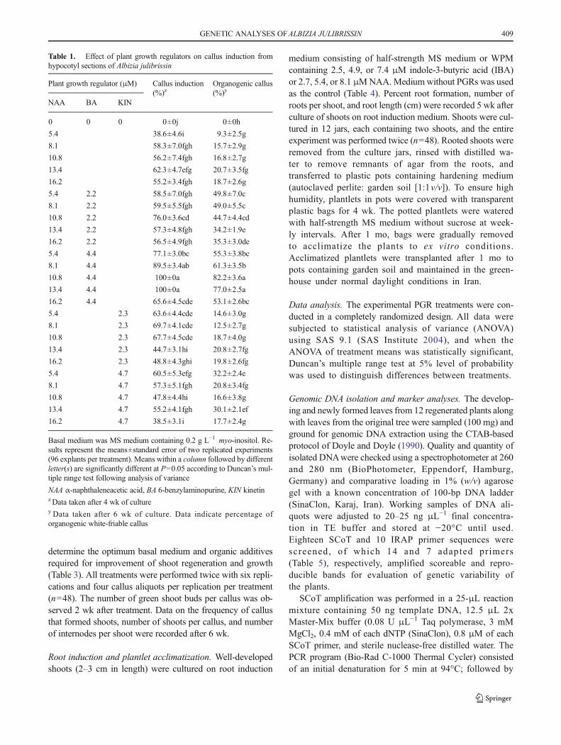
Table 1. Effect of plant growth regulators on callus induction fromhypocotyl sections of Albizia julibrissin
Plant growth regulator (μM) Callus induction(%)z
Organogenic callus(%)y
NAA BA KIN
0 0 0 0±0j 0±0h
5.4 38.6±4.6i 9.3±2.5g
8.1 58.3±7.0fgh 15.7±2.9g
10.8 56.2±7.4fgh 16.8±2.7g
13.4 62.3±4.7efg 20.7±3.5fg
16.2 55.2±3.4fgh 18.7±2.6g
5.4 2.2 58.5±7.0fgh 49.8±7.0c
8.1 2.2 59.5±5.5fgh 49.0±5.5c
10.8 2.2 76.0±3.6cd 44.7±4.4cd
13.4 2.2 57.3±4.8fgh 34.2±1.9e
16.2 2.2 56.5±4.9fgh 35.3±3.0de
5.4 4.4 77.1±3.0bc 55.3±3.8bc
8.1 4.4 89.5±3.4ab 61.3±3.5b
10.8 4.4 100±0a 82.2±3.6a
13.4 4.4 100±0a 77.0±2.5a
16.2 4.4 65.6±4.5cde 53.1±2.6bc
5.4 2.3 63.6±4.4cde 14.6±3.0g
8.1 2.3 69.7±4.1cde 12.5±2.7g
10.8 2.3 67.7±4.5cde 18.7±4.0g
13.4 2.3 44.7±3.1hi 20.8±2.7fg
16.2 2.3 48.8±4.3ghi 19.8±2.6fg
5.4 4.7 60.5±5.3efg 32.2±2.4e
8.1 4.7 57.3±5.1fgh 20.8±3.4fg
10.8 4.7 47.8±4.4hi 16.6±3.8g
13.4 4.7 55.2±4.1fgh 30.1±2.1ef
16.2 4.7 38.5±3.1i 17.7±2.4g
Basal medium was MS medium containing 0.2 g L−1 myo-inositol. Re-sults represent the means±standard error of two replicated experiments(96 explants per treatment). Means within a column followed by differentletter(s) are significantly different at P=0.05 according to Duncan’s mul-tiple range test following analysis of variance
NAA α-naphthaleneacetic acid, BA 6-benzylaminopurine, KIN kinetinz Data taken after 4 wk of culturey Data taken after 6 wk of culture. Data indicate percentage oforganogenic white-friable callus
GENETIC ANALYSES OFALBIZIA JULIBRISSIN 409

36 cycles of 60, 60, and 120 s at 94, 50, and 72°C, respective-ly; with a final extension at 72°C for 10 min. PCR amplifica-tion reactions for IRAP analysis consisted of 25-ng templateDNA, 10 μL 2× Master-Mix buffer (0.08 U μL−1 Taq poly-merase, 3 mM MgCl2, 0.4 mM of each dNTP (SinaClon),0.4 μM of each IRAP primer, and sterile nuclease-free dis-tilled water to a final volume of 20 μL. The PCR programconsisted of an initial denaturation for 3 min at 94°C; followedby 36 cycles of 30 s at 94°C, 1.5 min annealing at 55°C and2min extension at 72°C; followed by a final extension at 72°Cfor 10 min. Mineral oil (7 μL) was added to eachmicrocentrifuge tube for PCR reactions. A negative-controlPCR reaction with sterile distilled water in place of DNAtemplate was conducted to visualize possible self-polymerization or DNA contamination. Additionally, apositive PCR reaction with a known DNA templateand the specific forward and reverse primers was alsoconducted to check the stability of PCR conditions. AllSCoT and IRAP amplifications were performed withtwo independent PCR reactions. Amplified PCR
products were visualized under ultraviolet light using agel documentation system (Bio-Rad Molecular ImagerXR+ system) after electrophoresis (1.2% agarose [w/v]at a constant voltage of 80 V for 70 min with a 1×TAE buffer followed by staining with 1 μg ml−1
ethidium bromide).SCoTand IRAP bands were visually scored as 1 (presence)
or 0 (absence) for each reaction. Because these marker sys-tems are dominant, each bandwas representative of a bi-alleliclocus (Williams et al. 1990). In scoring, bands with equalmolecular weight from the same primer were considered asamplicons of the same locus. The size of the DNA amplifica-tion products were estimated comparatively using a 100-bpmolecular weight marker and the Gel Doc system software(Bio-Rad; Image Lab Software v. 4.0). Ambiguousbands with low visual intensity were not scored. Thelevel of polymorphism was calculated as the ratio ofthe number of polymorphic bands to the total numberof bands and expressed as a percentage. Binary matricesof SCoT and IRAP scoring data were constructed. Using
Table 2. Effect of plant growthregulators on adventitious shootregeneration from white-friablecallus induced on hypocotylsections of Albizia julibrissin
Plant growth regulator (μM) Mean no. green shootbuds per callusz
Shoot organogenesis(%)y
Mean no. microshootsper callusy
BA Zeatin
0 0 0±0h 0±0h 0±0h
4.4 1.8±0.3g 33.6±5.2g 1.1±0.3g
8.8 3.1±0.3ef 46.1±4.1def 2.0±0.2fg
11.1 3.3±0.2def 46.1±4.1def 1.6±0.2fg
13.2 3.0±0.3ef 39.8±3.8g 1.5±0.2fg
2.3 3.6±0.4fg 52.3±7.5def 2.5±0.2ef
4.6 2.8±0.5fgh 44.1±7.7efg 2.0±0.4fg
6.8 3.1±0.3ef 44.0±5.3efg 2.5±0.2ef
4.4 2.3 3.3±0.5def 48.1±5.8def 2.1±0.3fg
8.8 2.3 4.0±0.6def 52.1±8.1def 3.0±0.4de
11.1 2.3 4.0±0.4def 56.5±5.3cde 4.5±0.4ab
13.2 2.3 4.1±0.5def 58.6±8.3cde 3.3±0.5cd
4.4 4.6 4.8±0.6abc 71.0±8.3ab 4.6±0.3a
8.8 4.6 4.8±0.3abc 64.8±3.8bcd 4.1±0.3bc
11.1 4.6 5.0±0.4ab 69.0±6.9ab 3.5±0.6cd
13.2 4.6 5.5±0.4a 75.3±6.3a 4.8±0.4a
4.4 6.8 5.1±0.5ab 66.8±7.6bc 4.1±0.3bc
8.8 6.8 4.5±0.2bcd 62.6±4.5cde 4.3±0.3bc
11.1 6.8 5.0±0.3ab 69.0±7.1ab 4.5±0.4ab
13.2 6.8 4.3±0.3cde 58.5±6.1cde 4.1±0.4bc
Basal medium was MS medium. Results represent the means±standard error of two replicated experiments (n=48; about 250 mg callus was cultured as one explant). Means within a column followed by different letter(s) aresignificantly different at P=0.05 according to Duncan’s multiple range test following analysis of variance
BA 6-benzylaminopurinez Data taken after 2 wk of culturey Data taken after 6 wk of culture
410 RAHMANI ETAL.
the program GenAlEx 6.5 (Peakall and Smouse 2012), Nei’spairwise genetic distance (Dij; Nei 1987) among all possiblepairs of the 12 in vitro plants and themother plant was calculated
asDij=1−Sij [Sij=2Nij/(Ni+Nj), whereNij is the number of com-mon amplicons in genotypes i and j, and Ni and Nj are the totalnumber of amplicons in genotypes i and j, respectively.
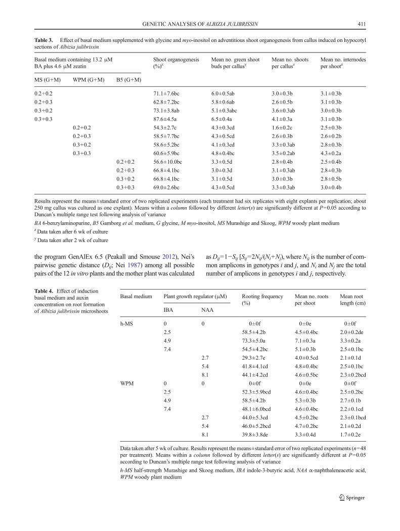
Table 3. Effect of basal medium supplemented with glycine and myo-inositol on adventitious shoot organogenesis from callus induced on hypocotylsections of Albizia julibrissin
Basal medium containing 13.2 μMBA plus 4.6 μM zeatin
Shoot organogenesis(%)z
Mean no. green shootbuds per callusy
Mean no. shootsper callusz
Mean no. internodesper shootz
MS (G+M) WPM (G+M) B5 (G+M)
0.2+0.2 71.1±7.6bc 6.0±0.5ab 3.0±0.3b 3.1±0.3b
0.2+0.3 62.8±7.2bc 5.8±0.6ab 2.6±0.5b 3.1±0.3b
0.3+0.2 73.1±3.8ab 5.1±0.3abc 3.6±0.3ab 3.0±0.3b
0.3+0.3 87.6±4.5a 6.5±0.4a 4.1±0.3a 3.1±0.3b
0.2+0.2 54.3±2.7c 4.3±0.3cd 1.6±0.2c 2.5±0.3b
0.2+0.3 58.5±7.7bc 4.3±0.5cd 2.6±0.3b 2.6±0.2b
0.3+0.2 58.6±5.2bc 4.1±0.3cd 3.3±0.3ab 2.8±0.3b
0.3+0.3 60.6±5.9bc 4.8±0.4bc 3.5±0.2ab 4.3±0.2a
0.2+0.2 56.6±10.0bc 3.3±0.5d 2.8±0.4b 2.5±0.4b
0.2+0.3 66.8±4.1bc 3.0±0.3d 3.1±0.3ab 2.8±0.3b
0.3+0.2 66.8±4.1bc 3.1±0.5d 3.0±0.3b 2.8±0.5b
0.3+0.3 69.0±2.6bc 4.3±0.5cd 3.3±0.3ab 3.0±0.4b
Results represent the means±standard error of two replicated experiments (each treatment had six replicates with eight explants per replication; about250 mg callus was cultured as one explant). Means within a column followed by different letter(s) are significantly different at P=0.05 according toDuncan’s multiple range test following analysis of variance
BA 6-benzylaminopurine, B5 Gamborg et al. medium, G glycine, M myo-inositol, MS Murashige and Skoog, WPM woody plant mediumzData taken after 6 wk of culturey Data taken after 2 wk of culture
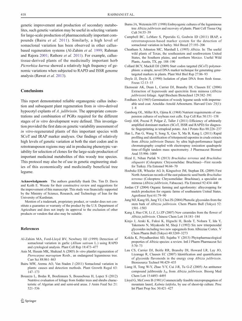
Table 4. Effect of inductionbasal medium and auxinconcentration on root formationof Albizia julibrissin microshoots
Basal medium Plant growth regulator (μM) Rooting frequency(%)
Mean no. rootsper shoot
Mean rootlength (cm)
IBA NAA
h-MS 0 0 0±0f 0±0e 0±0f
2.5 58.5±4.2b 4.5±0.4bc 2.0±0.2de
4.9 73.3±5.0a 7.1±0.3a 3.3±0.2a
7.4 54.5±4.2bc 5.1±0.3b 2.5±0.1bc
2.7 29.3±2.7e 4.0±0.5cd 2.1±0.1d
5.4 41.8±4.1cd 4.8±0.4bc 2.5±0.1bc
8.1 44.1±4.2cd 4.6±0.5bc 2.3±0.2bcd
WPM 0 0 0±0f 0±0e 0±0f
2.5 52.3±5.9bcd 4.6±0.4bc 2.5±0.2bc
4.9 58.5±4.2b 5.3±0.3b 2.7±0.1b
7.4 48.1±6.0bcd 4.6±0.4bc 2.2±0.1cd
2.7 44.0±5.3cd 4.5±0.2bc 2.3±0.1bcd
5.4 46.0±5.2bcd 4.7±0.2bc 2.1±0.2d
8.1 39.8±3.8de 3.3±0.4d 1.7±0.2e
Data taken after 5 wk of culture. Results represent the means±standard error of two replicated experiments (n=48per treatment). Means within a column followed by different letter(s) are significantly different at P=0.05according to Duncan’s multiple range test following analysis of variance
h-MS half-strength Murashige and Skoog medium, IBA indole-3-butyric acid, NAA α-naphthaleneacetic acid,WPM woody plant medium
GENETIC ANALYSES OFALBIZIA JULIBRISSIN 411

Results and Discussion
Callus induction. Hot-water-scarified seeds of A. julibrissingerminated at 80–90% on MS medium lacking PGRs andproduced in vitro seedlings within 7–10 d. Callus was inducedfrom the cut ends of the hypocotyl explants, began to form 8–10 d after initial culture (Fig. 1a), and covered the entire sur-face of the explants within 3–4 wk (Fig. 1b, c). From 38.5–100% hypocotyl explants formed callus and organogenic cal-lus formation varied from 9.3–82.2% (P=0.05) depending onthe type and concentration of PGRs (Table 1). Medium devoidof PGRs (control) failed to induce callus even after 6 wk ofculture. Media without cytokinin (BA or kinetin), but with5.4–16.2 μM NAA, showed the highest percentage (62.3%)of callus induction on MS medium with 13.4 μM NAA.Callus induction frequencies were significantly increasedwhen NAA was combined with either BA or kinetin(Table 1). Maximum callus formation (100%) was achieved
on MS medium with 10.8 or 13.4 μM NAA plus 4.4 μM BA(Table 1). MS medium containing only 5.4 μM NAAyieldedmostly yellowish-white, soft callus that was not capable ofshoot regeneration (Fig. 1b). Calli induced and maintainedfor 6–8 wk on MS medium with 10.8 μM NAA plus4.4 μM BAwere white and friable (Fig. 1c), and 82.2% wereorganogenic on the basis of callus and shoot bud appearance(Fig. 1d). Callus induced on MS medium with NAA in com-bination with kinetin (38.5–69.7% callus induction) produceda low percentage of organogenic callus (12.5–32.2%).
Many leguminous trees are recalcitrant to in vitro culture(Anis et al. 2005), and limited information is available oncallus induction and subsequent adventitious shoot regenera-tion in A. julibrissin. Hypocotyl explants excised from 10- to12-d-old in vitro seedlings of A. julibrissin and cultured on B5medium with 0–4 μM paclobutrazol, uniconazole, orprohexadione calcium also produced callus at high frequency(90–100%) (Sankhla et al. 1993). The roots from 8- to 10-d-old
Table 5. SCoT and IRAP primers and sequences, number and size range of generated fragments, and number of polymorphic bands in Albiziajulibrissin in vitro-regenerated plants
Primer 5′→3′ sequence Size (kb) Total no. SB No. PB PB (%)
SCoT
SCoT2 CAACAATGGCTACCACCA 0.6–1.3 4 1 25
SCoT3 CAACAATGGCTACCACCG 0.3–1.0 5 3 60
SCoT5 CAACAATGGCTACCACGA 0.5–1.1 5 0 0
SCoT6 CAACAATGGCTACCACGC 0.1–0.9 5 4 80
SCoT9 CAACAATGGCTACCAGCA 0.6–1.3 6 3 50
SCoT12 ACGACATGGCGACCAACG 0.3–0.9 5 3 60
SCoT14 ACGACATGGCGACCACGC 0.2–1.1 6 3 50
SCoT16 ACCATGGCTACCACCGAC 0.3–1.1 6 3 50
SCoT22 AACCATGGCTACCACCAC 0.3–1.0 5 4 80
SCoT25 ACCATGGCTACCACCGGG 0.3–1.2 4 0 0
SCoT27 ACCATGGCTACCACCGTG 0.3–1.1 5 3 60
SCoT28 CCATGGCTACCACCGCCA 0.2–1.1 4 0 0
SCoT30 CCATGGCTACCACCGGCG 0.2–0.8 5 3 60
SCoT35 CATGGCTACCACCGGCCC 0.2–1.1 6 3 50
IRAP
Hana CACGATTCACCTTAATATCTGACA 0.5–1.4 5 3 60
Gaga GGGAACCAACCGTCACA 0.4–1.5 5 0 0
5′LTR1 TTGCCTCTAGGGCATATTTCCAACA 0.3–1.3 6 2 33
LTR6149 CTCGCTCGCCCACTACATCAACCGCGTTTATT 0.5–1.0 4 3 75
LTR6150 CTGGTTCGGCCCATGTCTATGTATCCACACATGTA 0.4–1.3 5 1 20
3′LTR TGTTTCCCATGCGACGTTCCCCAACA 0.5–1.2 4 2 50
Nikita CGCATTTGTTCAAGCCTAAACC 0.6–1.4 5 1 20
Summary of data
Marker system No. primer Total no. SB Mean no. SBper primer
Total no. PB Mean no. PBper primer
% P
SCoT 14 71 5.1 33 2.4 46.48
IRAP 7 34 4.9 12 1.7 35.29
IRAP inter-retrotransposon amplified polymorphism, SB scoreable bands, SCoT start codon targeted, PB polymorphic bands, P polymorphism
412 RAHMANI ETAL.

intact in vitro-grown silk tree seedlings split and formed prolificcallus on MSmedium containing 0.1–1 μM thidiazuron (TDZ)(Sankhla et al. 1994). Roots excised from 15- to 20-d-oldin vitro A. julibrissin seedlings and cultured onB5mediumwiththe ethylene precursors/generators 1-amino-cyclopropane-1-carboxylic acid and 2-chloroethylphosphonic acid formed cal-lus, but subsequent shoot formation was inhibited (Sankhlaet al. 1995). Excised root segments from 15- to 20-d-old in vitrosilk tree seedlings cultured on B5 medium containing NAA,indole-3-acetic acid (IAA), IBA, kinetin, BA, 6-(γ,γ-dimethylallylamino)purine (2iP), zeatin, or TDZ formed somecallus after 1 wk in culture (Sankhla et al. 1996). As in ourstudy, a combination of an auxin with a cytokinin has beenwidely used for successful induction of organogenic callus inmany commercially or medicinally important regeneration sys-tems. For example, Sha Valli Khan et al. (2002) reported 64%
induction of white, friable callus on Bixa orellana seeds cul-tured on MS medium with 5 μM NAA and 2.5 μM BA.
Adventitious shoot regeneration. Both yellowish-white andwhite-friable callus types were cultured separately onMSme-dium either without PGRs as a control, or with different con-centrations and combinations of BA and zeatin, for shoot or-ganogenesis (Table 2). Ten days after culture on shoot regen-eration medium containing PGRs individually or in combina-tion, white-friable calli proliferated rapidly and green shootbuds appeared (Fig. 1d). The yellowish-white callus type,however, failed to exhibit shoot differentiation and did notshow further growth. Shoot regeneration medium lackingPGRs did not induce shoot organogenesis from theorganogenic white-friable callus. The number of green shootbuds, shoot organogenesis frequency, and number of
Figure 1. Callus induction and microshoot organogenesis fromhypocotyl sections of Albizia julibrissin. a, b Non-organogenicyellowish-white, soft callus from hypocotyl cultured on Murashige andSkoog (MS) medium with 16.2 and 5.4 μM α-naphthaleneacetic acid(NAA) after 2 wk and 6 wk, respectively. c Organogenic white-friablecallus from hypocotyl on MS medium with 10.8 μM NAA plus 4.4 μM
6-benzylaminopurine (BA) after 6 wk. d Callus proliferation and greenshoot bud formation from organogenic white-friable callus on MSmedium with 13.2 μM BA plus 4.6 μM zeatin after 2 wk. Arrowsindicate green shoot buds. e, f Adventitious microshoot formation fromorganogenic white-friable callus after 3- and 4-wk, respectively, on MSmedium with 13.2 μM BA plus 4.6 μM zeatin. Bars=5 mm.
GENETIC ANALYSES OFALBIZIA JULIBRISSIN 413

microshoots per callus varied by PGR type, concentration, andcombination (Table 2). Compared to MS medium containingBA or zeatin alone, the combination of these PGRs promotedshoot regeneration. MS medium with 13.2 μM BA plus4.6 μM zeatin produced the highest number of green shootbuds (5.5±0.4) per callus. The best shoot regeneration wasachieved on this medium with 75.3% of the culturesresponding and the highest mean number of shoots (4.8±0.4) per callus (Table 2).
BA, either alone or in combination with other PGRs, hasbeen very effective for the induction of callus or shoot organ-ogenesis in several Albizia species. Sankhla et al. (1994) re-ported good adventitious shoot regeneration (16±2 shoots) onroots of intact seedlings of A. julibrissin cultured on MS me-dium with B5 vitamins and 10 μM BA, as compared to 5.2±0.3 shoots on excised root explants cultured on B5 mediumwith 10 μM BA (Sankhla et al. 1996). The addition of3 mg L−1 hymexazol to MS medium containing 2.22 μMBA and 0.05 μM NAAwas reported to increase adventitiousshoot development on cotyledons and hypocotyls ofA. julibrissin (Zhou et al. 2001). Mamun et al. (2004) reported100% callus induction on Albizia lebbeck nodal sections cul-tured on MS medium with 8.88 μM BA plus 1.07 μM NAA,with the best shoot regeneration (7.3 per callus) occurring onMSmediumwith 11.1 μMBA plus 1.07μMNAA. However,cotyledonary explants (83.3%) regenerated the greatest num-ber of shoots on MS medium with only 8.88 μM BA. Rootexplants from 15-d-old in vitro A. lebbeck seedlings culturedonMSmedium with 7.5 μMBA plus 0.5 μMNAA produced16±1.9 shoot buds per root explant (Perveen et al. 2011).Organogenic callus induced on nodal explants of A. lucidaregenerated shoots when cultured on MS medium containing8.88 μM BA plus 0.54 μM NAA (Saha et al. 2013).Hypocotyls of Albizia odoratissima cultured on MS mediumwith 7.5 μM BA and 0.5 μM NAA in the light differentiatedadventitious shoots from callus, whereas hypocotyls culturedin the dark produced non-morphogenic calli (Rajeswari andPaliwal 2008). In our study, we found that increasing zeatin to6.8 μM in combination with 13.2 μM BA decreased the per-centage of shoot organogenesis to 58.5% (Table 2).Vengadesan et al. (2003), also showed that the number ofadventitious shoots regenerated from cotyledon-derived callusin the leguminous tree Acacia sinuata was significantly de-creased (to 8.5 per callus) with 5 μM zeatin in combinationwith 13.3 μM BA, as compared to 25.3 shoots regeneratedwhen 2.5 μM zeatin plus 13.3 μM BAwas used. Originationof adventitious shoots from callus (Fig. 1e, f ) and the 6 wkduration required for complete formation of shoots (Fig. 2a, b)indicated indirect organogenesis in our study.
Shoot growth on the regeneration medium was asynchro-nous and poor. Therefore, experiments were conducted tocompare the efficiency of different basal media (MS medium,WPM, and B5 medium) containing 13.2 μMBA plus 4.6 μM
zeatin, supplemented with glycine and myo-inositol for im-provement of adventitious shoot regeneration and growth(Table 3). The frequency of shoot regeneration, number ofgreen shoot buds, number of shoots per callus, and numberof internodes per shoot varied depending on the basal mediumas well as on the concentrations of glycine and myo-inositol(Table 3). The highest frequency of shoot-producing callus(87.6±4.5%; Fig. 2c), highest average number of green shootbuds (6.5±0.4), and maximum mean number of shoots percallus (4.1±0.3) were observed on MS medium containing0.3 g L−l glycine plus 0.3 g L−lmyo-inositol, while the highestaverage number of internodes per shoot (4.3±0.2) was obtain-ed on WPM with 0.3 g L−l glycine and 0.3 g L−l myo-inositol(Table 3; Fig. 2d). The relatively improved elongation ofshoots on WPM may be a result of the lower concentrationof ionic nutrients than in the other full-strength media. Thisfinding is in accordance with results by Vengadesan et al.(2003) where half-strength MS medium favored adventitiousshoot formation from callus of Acacia sinuate. In contrast,Perveen et al. (2011) reported that full-strength MS mediumwasmore effective than other media for increasing shoot num-ber and shoot length ofA. lebbeck using root explants. Glycineand myo-inositol at 0.3 g L−1 each were found to be a signif-icant factors in enhancing the percentage of shoot organogen-esis and mean number of shoots per callus as compared toMSmedium with only 0.2 g−1 L glycine but did not increase thenumber of shoots per callus in our study. The addition ofglycine and myo-inositol at 0.3 g L−l yielded an optimumresponse in over 60% of cultures, regardless of the basal me-dium (Table 3). Although elongation of shoots was improvedon medium augmented with these organics, only one or twoshoots per callus elongated and exhibited continued growth,while other microshoots failed to elongate or died.
Roo t i n g o f a d v e n t i t i o u s s h o o t s a n d p l a n t l e tacclimatization. Shoots failed to form roots on rooting medi-um lacking PGRs. Adventitious roots became visible at thebase of shoots within 3 wk after culture onmedium containingIBA or NAA. The best rooting percentage (73.3%), meannumber of roots per shoot (7.1±0.3), and mean root length(3.3±0.2 cm) were obtained when shoots were cultured onhalf-strength MS medium with 4.9 μM IBA (Table 4;Fig. 2e). In comparison, a lower number of roots and shorterroots were obtained when IBA was replaced with NAA(Table 4). In vitro rooting experiments carried out with otherleguminous species including A. julibrissin (Sankhla et al.1994; Tudor Radu and Radomir 2010), A. chinensis (Sinhaet al. 2000), A. procera (Swamy et al. 2004), A. odoratissima(Rajeswari and Paliwal 2006), A. lebbeck (Perveen et al.2011), Acacia sinuata (Vengadesan et al. 2000), and Cassiasiamea (Parveen et al. 2010) all showed successful root for-mation on half- or full-strength MSmediumwith various con-centrations of IBA. Sankhla et al. (1994) reported that
414 RAHMANI ETAL.

A. julibrissin shoots induced on roots of in vitro intact seed-lings by kinetin or BA rooted faster than TDZ-induced shoots,when cultured on MS medium with or without 4.9 μM IBA.Tudor Radu and Radomir (2010) obtained 85% rooting ofA. julibrissin Bmicro cuts^ on half-strength MS medium with0.98μM IBAversus 40% rooting with 3.94 μM IBA. Perveenet al. (2011) reported the best rooting (66%) of A. lebbeckmicroshoots on half-strength MS medium with 2 μM IBA.Normal growth and development of rooted plantlets in ourstudy was observed 5 wk after transfer to the greenhouse(Fig. 2f ). Over 60% of in vitro rooted plants were successfullyacclimatized to the greenhouse.
Genetic variability of regenerated plants. SCoT is a simple,gene-targeted, and cost-effective DNA-based marker systemwhereby primers target the short conserved region flankingthe ATG translation start codon of plant genes (Collard andMackill 2009). SCoT polymorphism has proven to be infor-mative, effective, and reproducible for the evaluation of ge-netic stability of plants (Gorji et al. 2011; Rathore et al. 2014).The retrotransposon-based IRAP marker system reveals
insertional polymorphisms of retrotransposon regions ofDNA by a simple PCR of single primers followed by electro-phoresis for analysis of the PCR amplicons. An investigationwith barley in vitro-regenerated plants showed that the IRAPmethod was an informative tool for the detailed elucidation ofmutation profiles that occur under in vitro conditions(Campbell et al. 2011).
The genetic variability of A. julibrissin regenerated plantswas assessed initially with 18 SCoT and 10 IRAP screenedprimers, of which 14 and 7 primers, respectively, producedintense and re-amplifiable bands. A total of 71 amplicons weregenerated from PCR amplification with SCoT primers usingamplicons ranging in size from 100 to 1300 bp. Depending onthe primer sequence, the scoreable bands from SCoT primersranged from four (SCoT2, SCoT25, and SCoT28) to six(SCoT9, SCoT14, SCoT16, and SCoT35) with an averageof five bands per primer (Table 5). The number of polymor-phic bands from each SCoT primer also varied from zero(SCoT5, SCoT25, and SCoT28) to four (SCoT6, 80% andSCoT22, 80%). SCoT6 and SCoT22 each amplified fivebands in total, so the polymorphism for these two markers
Figure 2. In vitro organogenesis from white-friable callus of Albiziajulibrissin. a, b Adventitious shoot formation from callus cultured onMurashige and Skoog (MS) medium containing 13.2 μM 6-benzylaminopurine (BA) plus 4.6 μM zeatin (a) and 11.1 μM BA plus4.6 μM zeatin (b), after 6 wk. c Induction of multiple shoots on MSmedium with 13.2 μM BA, 4.6 μM zeatin, 0.3 g L−1 glycine, and
0.3 g L−1 myo-inositol after 6 wk. d In vitro elongated shoot on woodyplant medium (WPM) with 13.2 μM BA, 4.6 μM zeatin, 0.3 g L−1
glycine, and 0.3 g L−1 myo-inositol after 6 wk. e Root formationinduced on half-strength MS medium with 4.9 μM indole-3-butyricacid. f In vitro-regenerated plants approximately 2 mo afteracclimatization. Bars=5 mm.
GENETIC ANALYSES OFALBIZIA JULIBRISSIN 415

was 4/5 or 80%. The banding pattern of the regeneratedA. julibrissin genotypes and the mother plant obtained withthe SCoT22 primer are shown in Fig. 3a. Of the 34 totalscoreable bands obtained with IRAP primers, six(maximum) and four (minimum) scoreable bands were gener-ated from primers 5′LTR1, and LTR6149 and 3′LTR, respec-tively, with an average of 4.9 scoreable bands per primer.Among these primers, 75% polymorphism was observed forprimer LTR6149 (shown in Fig. 3b), while no polymorphismwas recorded for primer Gaga (Table 5).
Investigation of genetic variability generated by in vitroconditions using more than one marker technique has beensuggested to be advantageous because each marker may elu-cidate changes in different regions of the species’ genomicDNA (Palombi and Damiano 2002; Martins et al. 2004). Acomparative summary of our results with the SCoT and IRAPmarker systems is presented in Table 5. Genetic similarityrevealed between in vitro-regenerated plants and the motherplant was higher with the SCoT system than with the IRAP
system (Fig. 4). This observation confirms that in vitro cultureconditions can affect different genome regions of regeneratedplantlets, which indicated that the use of at least two markertechniques was more effective than any one technique forscreening the genome variability in tissue culture plants(Martins et al. 2004; Rathore et al. 2011).
The SCoT and IRAP profiles were used individually toestimate pairwise genetic distances among the 12 in vitroplants and between those plants and the mother plant(Table 6). According to both SCoT and IRAP analyses, over-all, the in vitro plants and the mother plant showed a relativelyhigh degree of genetic dissimilarity. SCoT-based genetic dis-tance values between in vitro plants varied from 0.095 to0.788, with an average of 0.352 (Table 6). IRAP-based geneticdistance values among the in vitro plants ranged from 0.000 to0.693, with an average of 0.292 (Table 6). The calculatedgenetic distance between in vitro plants and the mother plantwere 0.129–0.405 for SCoT analysis and 0.182–0.438 forIRAP analysis (Table 6, Fig. 4). It was obvious from these
Figure 3. Genetic variability of in vitro-regenerated plants (IP-1–IP12)of Albizia julibrissin compared with each other and with the mother plant(MP). a Banding pattern of SCoT amplification products with primerSCoT22. b Banding pattern of IRAP amplification products with primer
LTR6149. (M 100-bp molecular marker). Asterisk indicates some of thescored SCoT or IRAP amplified bands (red: polymorphic bands; green:monomorphic bands).
416 RAHMANI ETAL.

observations that there was a relatively high level of geneticdivergence of in vitro-regenerated plants from the originalmother plant. This high frequency of genetic variation maybe a result of an accumulation of somatic mutations during thetissue culture period, induced by several determining factorsincluding the various concentrations and types of PGRs thatwere applied (Martin et al. 2006; Bairu et al. 2011), and theoutcrossing nature of seed production in this species. Theinduction of somaclonal variation in plants regenerated from
a single mother plant under in vitro conditions has been rou-tinely observed (Rathore et al. 2011; Rawat et al. 2013).DNA-based methods such as simple sequence repeat (SSR),ISSR, random amplified polymorphic DNA (RAPD), SCoT,IRAP, and amplified fragment length polymorphism (AFLP)analysis have been used to evaluate the genetic stability ofregenerants (Rahman and Rajora 2001; Palombi andDamiano 2002; Campbell et al. 2011; Rawat et al. 2013).Considering the importance of tissue culture techniques in
Table 6. Pairwise genetic distance matrix showing data from SCoT and IRAP analyses for in vitro-raised plants and the mother plant of Albiziajulibrissin
MP IP1 IP2 IP3 IP4 IP5 IP6 IP7 IP8 IP9 IP10 IP11 IP12 Ave.
MP 0.00 0.405 0.288 0.405 0.288 0.288 0.288 0.182 0.405 0.438 0.431 0.182 0.182
IP1 0.385 0.00 0.288 0.405 0.539 0.288 0.087 0.182 0.182 0.288 0.288 0.182 0.405
IP2 0.361 0.405 0.00 0.288 0.182 0.182 0.182 0.087 0.288 0.405 0.405 0.087 0.288
IP3 0.278 0.405 0.278 0.00 0.539 0.087 0.288 0.182 0.405 0.539 0.539 0.182 0.405
IP4 0.318 0.552 0.606 0.318 0.00 0.405 0.405 0.288 0.539 0.693 0.405 0.288 0.288
IP5 0.164 0.552 0.501 0.238 0.278 0.00 0.182 0.087 0.288 0.405 0.405 0.087 0.288
IP6 0.238 0.552 0.405 0.318 0.201 0.201 0.00 0.087 0.087 0.182 0.182 0.087 0.288
IP7 0.164 0.361 0.318 0.238 0.361 0.129 0.278 0.00 0.182 0.288 0.288 0.000 0.182
IP8 0.201 0.318 0.361 0.361 0.405 0.238 0.318 0.095 0.00 0.087 0.288 0.182 0.405
IP9 0.129 0.405 0.278 0.278 0.318 0.164 0.164 0.095 0.129 0.00 0.405 0.288 0.539
IP10 0.318 0.452 0.606 0.606 0.452 0.361 0.361 0.361 0.318 0.238 0.00 0.288 0.539
IP11 0.405 0.788 0.724 0.501 0.452 0.361 0.361 0.361 0.318 0.318 0.552 0.00 0.182
IP12 0.361 0.606 0.663 0.452 0.405 0.318 0.405 0.318 0.161 0.278 0.501 0.164 0.00 0.292z
Ave. 0.352y
SCoT-based genetic distances are given below the bold diagonal; IRAP-based genetic distances are given above the bold diagonal
MP mother plant, IP in vitro-regenerated plant (IP1–IP12), Ave. averagez Average genetic distance between pairs of in vitro-raised plants according to IRAP analysisy Average genetic distance between pairs of in vitro-raised plants according to SCoT analysis
Figure 4. Histogram of the pairwise Nei’s genetic distance values between the in vitro-raised plants (IP-1–IP12) and the mother plant (MP) based onSCoT and IRAP fingerprinting data.
GENETIC ANALYSES OFALBIZIA JULIBRISSIN 417
genetic improvement and production of secondary metabo-lites, such genetic variation may be useful in selecting variantsfor large-scale production of pharmaceutically important com-pounds (Bairu et al. 2011). Similarly, a high level ofsomaclonal variation has been observed in other callus-based regeneration systems (Al-Zahim et al. 1999; Rahmanand Rajora 2001; Rathore et al. 2011). For example, callus-tissue-derived plants of the medicinally important herbPicrorhiza kurroa showed a relatively high frequency of ge-nomic variations when subjected to RAPD and ISSR genomeanalysis (Rawat et al. 2013).
Conclusions
This report demonstrated reliable organogenic callus induc-tion and subsequent plant regeneration from in vitro-derivedhypocotyl explants of A. julibrissin. The appropriate concen-trations and combination of PGRs required for the differentstages of in vitro development were defined. This investiga-tion provided the first data on genetic variability assessment ofin vitro-regenerated plants of this important species withSCoT and IRAP marker analyses. Our findings of relativelyhigh levels of genetic variation at both the start codon and inretrotransposon regions may aid in producing phenotypic var-iability for selection of clones for the large-scale production ofimportant medicinal metabolites of this woody tree species.This protocol may also be of use in genetic engineering stud-ies of this economically and medicinally beneficial treelegume.
Acknowledgments The authors gratefully thank Drs. Tim D. Davisand Keith E. Woeste for their constructive review and suggestions forthe improvement of this manuscript. This study was financially supportedby the Ministry of Science, Research, and Technology of Iran for theUniversity of Kurdistan.
Mention of a trademark, proprietary product, or vendor does not con-stitute a guarantee or warranty of the product by the U.S. Department ofAgriculture and does not imply its approval to the exclusion of otherproducts or vendors that also may be suitable.
References
Al-Zahim MA, Ford-Lloyd BV, Newbury HJ (1999) Detection ofsomaclonal variation in garlic (Allium sativum L.) using RAPDand cytological analysis. Plant Cell Rep 18:473–477
Anis M, Husain MK, Shahzad A (2005) In vitro plantlet regeneration ofPterocarpus marsupium Roxb., an endangered leguminous tree.Curr Sci 88:861–863
Bairu MW, Aremu AO, Van Staden J (2011) Somaclonal variation inplants: causes and detection methods. Plant Growth Regul 63:147–173
Bouazza L, Bodas R, Boufennara S, Bousseboua H, Lopez S (2012)Nutritive evaluation of foliage from fodder trees and shrubs charac-teristic of Algerian arid and semi-arid areas. J Anim Feed Sci 21:521–536
Burns JA, Wetzstein HY (1998) Embryogenic cultures of the leguminoustree Albizia julibrissin and recovery of plants. Plant Cell Tissue OrgCult 54:55–59
Campbell BC, LeMare S, Piperidis G, Godwin ID (2011) IRAP, aretrotransposon-based marker system for the detection ofsomaclonal variation in barley. Mol Breed 27:193–206
Cheatham S, Johnston MC, Marshall L (1995) Albizia. In: The usefulwild plants of Texas, the southeastern and southwestern UnitedStates, the Southern plains, and northern Mexico. Useful WildPlants, Austin, TX, pp. 188–190
Collard BCY, Mackill DJ (2009) Start codon targeted (SCoT) polymor-phism: a simple, novel DNA marker technique for generating gene-targeted markers in plants. Plant Mol Biol Rep 27:86–93
Doyle JJ, Doyle JL (1990) Isolation of plant DNA from fresh tissue.Focus 12:13–15
Ekenseair AK, Duan L, Carrier DJ, Bransby DI, Clausen EC (2006)Extraction of hyperoside and quercitrin from mimosa (Albiziajulibrissin) foliage. Appl Biochem Biotechnol 129:382–391
Fordham AJ (1965) Germination of woody legume seeds with imperme-able seed coat. Arnoldia–Arnold Arboreturm. Harvard Univ 25(1):1–8
Gamborg OL, Miller RA, Ojima K (1968) Nutrient requirements of sus-pension cultures of soybean root cells. Exp Cell Res 50:151–158
Gorji AM, Poczai P, Polgar Z, Taller J (2011) Efficiency of arbitrarilyamplified dominant markers (SCoT, ISSR and RAPD) for diagnos-tic fingerprinting in tetraploid potato. Am J Potato Res 88:226–237
Han L, Pan G, Wang Y, Song X, Gao X, Ma B, Kang L (2011) Rapidprofiling and identification of triterpenoid saponins in crude extractsfrom Albizia julibrissin Durazz. by ultra high-performance liquidchromatography coupled with electrospray ionization quadrupoletime-of-flight tandem mass spectrometry. J Pharmaceut BiomedAnal 55:996–1009
Hizal E, Nihan Parlak N (2013) Bruchidius terrenus and Bruchidiussiliquastri (Coleoptera: Chrysomelidae: Bruchinae)—First recordsfor Turkey. Fla Entomol 96:66–70
Hoebeke ER, Wheeler AG Jr, Kingsolver JM, Stephan DL (2009) FirstNorth American records of the east palearctic seed beetle Bruchidiusterrenus (Coleoptera: Chrysomelidae: Bruchinae), a specialist onmimosa (Albizia julibrissin, Fabaceae). Fla Entomol 92:434–440
Jordan CF (2004) Organic farming and agroforestry: alleycropping formulch production for organic farms of southeastern United States.Agroforest Syst 61:79–90
JungMJ, Kang SS, Jung YJ, Choi JS (2004) Phenolic glycosides from thestem bark of Albizzia julibrissin. Chem Pharm Bull (Tokyo) 52:1501–1503
Kang J, Huo CH, Li Z, Li ZP (2007) New ceramides from the flower ofAlbizia julibrissin. Chinese Chem Lett 18:181–184
Kinjo J, Araki K, Fukui K, Higuchi H, Ikeda T, Nohara T, Ida Y,Takemoto N, Miyakoshi M, Shoji J (1992) Six new triterpenoidalglycosides including two new sapogenols from Albizziae Cortex. VChem Pharm Bull (Tokyo) 40:3269–3273
Kokila K, Priyadharshini SD, Sujatha V (2013) Phytopharmacologicalproperties of Albizia species: a review. Intl J Pharm Pharmaceut Sci5:70–73
Lau CS, Carrier DJ, Beitle RR, Bransby DI, Howard LR, Lay JO,Liyanage R, Clausen EC (2007) Identification and quantificationof glycoside flavonoids in the energy crop Albizia julibrissin.Bioresource Technol 98:429–435
Liang H, Tong W-Y, Zhao Y-Y, Cui J-R, Tu G-Z (2005) An antitumorcompound julibroside J28 from Albizia julibrissin. Bioorg MedChem Lett 15:4493–4495
Lloyd G, McCown B (1981) Commercially feasible micropropagation ofmountain laurel, Kalmia latifolia, by use of shoot-tip culture. ProcInt Plant Prop Soc 30:421–427
418 RAHMANI ETAL.

Lv JS, Zhang LN, SongYZ,WangXF, ChuXZ (2011) Biological activityexhibited by secondarymetabolites of the Albizia julibrissinDurazz.pod. Intl Biodeterior Biodegradation 65:258–264
Mamun ANK, Matin MN, Bari MA, Siddique NA, Sultana RS, RahmanMH, Musa ASM (2004) Micropropagation of woody legume(Albizia lebbeck) through tissue culture. Pak J Biol Sci 7:1099–1103
Martin KP, Pachathundikandi SK, Zhang C-L, Slater A, Madassery J(2006) RAPD analysis of a variant of banana (Musa sp.) cv.Grande Naine and its propagation via shoot tip culture. In VitroCell Dev Biol-Plant 42:188–192
Martins M, Sarmento D, Oliveira MM (2004) Genetic stability ofmicropropagated almond plantlets, as assessed by RAPD andISSR markers. Plant Cell Rep 23:492–496
Murashige T, Skoog F (1962) A revised medium for rapid growth and bioassays with tobacco tissue cultures. Physiol Plant 15:473–497
Nehdi I (2011) Characteristics, chemical composition and utilisation ofAlbizia julibrissin seed oil. Ind Crop Prod 33:30–34
Nei M (1987) Molecular evolutionary genetics. Columbia UniversityPress, New York
Orwa C, Mutua A, Kindt R, Jamnadass R, Simons A (2009) Albiziajulibrissin. Agroforestry Database: a tree reference and selectionguide. Version 4.0. Accessed 27 June 2014 (http://www.worldagroforestry.org/treedb2/AFTPDFS/Albizia_julibrissin.pdf)
Palombi MA, Damiano C (2002) Comparison between RAPD and SSRmolecular markers in detecting genetic variation in kiwifruit(Actinidia deliciosa A. Chev). Plant Cell Rep 20:1061–1066
Parveen S, Shahzad A, Saema S (2010) In vitro plant regeneration systemforCassia siamea Lam., a leguminous tree of economic importance.Agroforest Syst 80:109–116
Peakall R, Smouse PE (2012) GenAlEx 6.5: genetic analysis in Excel.Population genetic software for teaching and research–an update.Bioinformatics 28:2537–2539
Perveen S, Varshney A, Anis M, Aref IM (2011) Influence of cytokinins,basal media and pH on adventitious shoot regeneration from excisedroot cultures of Albizia lebbeck. J For Res 22:47–52
Phipps PM, Stipes RJ (1976) Histopathology of mimosa (Albiziajulibrissin) infected with Fusarium oxysporum f. sp. perniciosum.Phytopathology 66:839–843
Pitman WD (2008) Establishment and regrowth responses of Albiziajulibrissin on Louisiana USA coastal plain soils. Agroforest Syst74:259–266
Rahman MH, Rajora OP (2001) Microsatellite DNA somaclonal varia-tion in micropropagated trembling aspen (Populus tremuloides).Plant Cell Rep 20:531–536
Rajeswari V, Paliwal K (2006) In vitro propagation of Albiziaodoratissima L F. (Benth.) from cotyledonary node and leaf nodalexplants. In Vitro Cell Dev Biol–Plant 42:399–404
Rajeswari V, Paliwal K (2008) Peroxidase and catalase changes duringin vitro adventitious shoot organogenesis from hypocotyls of Albiziaodoratissima L. f. (Benth). Acta Physiol Plant 30:825–832
Rathore MS, Chikara J, Mastan SG, Rahman H, Anand KGV, ShekhawatNS (2011) Assessment of genetic stability and instability of tissueculture-propagated plantlets of Aloe vera L. by RAPD and ISSRmarkers. Appl Biochem Biotechnol 165:1356–1365
Rathore NS, Rai MK, Phulwaria M, Rathore N, Shekhawat NS (2014)Genetic stability in micropropagated Cleome gynandra revealed bySCoT analysis. Acta Physiol Plant 36:555–559
Rawat JM, Rawat B,Mehrotra S, Chandra A, Nautiyal S (2013) ISSR andRAPD based evaluation of genetic fidelity and active ingredientanalysis of regenerated plants of Picrorhiza kurroa. Acta PhysiolPlant 35:1797–1805
Rhoades CC, Nissen TM, Kettler JS (1998) Soil nitrogen dynamics inalley cropping and no-till systems on ultisols of the GeorgiaPiedmont, USA. Agroforest Syst 39:31–44
Saha D, Kumar R, Ramani R (2013) Regeneration of plantlets from nodalexplant-derived callus cultures of Albizia lucida Benth. Indian JBiotechnol 12:265–268
Sankhla D, Davis TD, Sankhla N (1993) Effect of gibberellin biosynthe-sis inhibitors on shoot regeneration from hypocotyl explants ofAlbizzia julibrissin. Plant Cell Rep 13:115–118
Sankhla D, Davis TD, Sankhla N (1994) Thidiazuron-induced in vitroshoot formation from roots of intact seedlings of Albizzia julibrissin.Plant Growth Regul 14:267–272
Sankhla D, Sankhla N, Davis TD (1995) Promotion of in vitro shootformation from excised roots of silktree (Albizzia julibrissin) by anoxime ether derivative and other ethylene inhibitors. Plant Cell Rep15:143–146
Sankhla D, Davis TD, Sankhla N (1996) In vitro regeneration of silktree(Albizzia julibrissin) from excised roots. Plant Cell Tissue OrganCult 44:83–86
SAS Institute Inc. (2004) SAS® 9.0, 9.1, 9.1.2, and 9.1.3. SAS InstituteInc, Cary, NC
Sha Valli Khan PS, Prakash E, Rao KR (2002) Callus induction andplantlet regeneration in Bixa orellana L., an annatto-yielding tree.In Vitro Cell Dev Biol-Plant 38:186–190
Sinha RK, Majumdar K, Sinha S (2000) In vitro differentiation and plantregeneration of Albizia chinensis (Osb.) Merr. In Vitro Cell DevBiol–Plant 36:370–373
Swamy SL, Ganguli JL, Puri S (2004) Regeneration andmultiplication ofAlbizia procera Benth. through organogenesis. Agroforest Syst 60:113–121
Tudor Radu CM, Radomir A-M (2010) Researches concerning Albizziajulibrissin species behaviour in the in vitro rooting faze [siz.]. JHortic For Biotechnol 14:286–289
Vengadesan G, Ganapathi A, Prem Anand R, Ramesh Anbazhagan V(2000) In vitro organogenesis and plant formation in Acacia sinuate.Plant Cell Tissue Organ Cult 61:23–28
Vengadesan G, Ganapathi A, Amutha S, Selvaraj N (2003) High-frequency plant regeneration from cotyledon callus of Acaciasinuata (Lour.) Merr. In Vitro Cell Dev Biol-Plant 39:28–33
Wang FQ, Wang ET, Zhang YF, Chen WX (2006) Characterization ofrhizobia isolated from Albizia spp. in comparison withmicrosymbionts of Acacia spp. and Leucaena leucocephala grownin China. Syst Appl Microbiol 29:502–517
Williams JGK, Kubelik AR, Livak KJ, Rafalski JA, Tingey SV (1990)DNA polymorphisms amplified by arbitrary primers are useful asgenetic markers. Nucleic Acids Res 18:6531–6535
Won HJ, Han CH, Kim YH, Kwon HJ, Kim BW, Choi JS, Kim KH(2006) Induction of apoptosis in human acute leukemia Jurkat Tcells by Albizia julibrissin extract is mediated via mitochondria-dependent caspase-3 activation. J Ethanopharmacol 19:383–389
Zheng H, Wu Y, Ding J, Binion D, Fu W, Reardon R (2004) Invasiveplants of Asian origin established in the United States and theirnatural enemies, vol. 1. FHTET 2004–05. USDA Forest Service,Morgantown, WV, pp 15–18
Zheng L, Zheng J, Zhao Y, Wang B, Wu L, Liang H (2006) Three anti-tumor saponins from Albizia julibrissin. Bioorg Med Chem Lett 16:2765–2768
Zhou Y, Zhang ZQ, Zhang JJ (2001) Use of hymexazol (HMI) in rapidpropagation of Albizia julibrissin Durazz. Acta Agr Shanghai 17:31–34
GENETIC ANALYSES OFALBIZIA JULIBRISSIN 419