genome-wide characterization of endogenous retroviruses in the bat myotis lucifugus reveals
TRANSCRIPT
Genome-Wide Characterization of Endogenous Retroviruses in the BatMyotis lucifugus Reveals Recent and Diverse Infections
Xiaoyu Zhuo,a Mina Rho,b Cédric Feschottea
Department of Human Genetics, University of Utah School of Medicine, Salt Lake City, Utah, USAa; Department of Biostatistics and Bioinformatics, Roswell Park CancerInstitute, Buffalo, New York, USAb
Bats are increasingly recognized as reservoir species for a variety of zoonotic viruses that pose severe threats to human health.While many RNA viruses have been identified in bats, little is known about bat retroviruses. Endogenous retroviruses (ERVs)represent genomic fossils of past retroviral infections and, thus, can inform us on the diversity and history of retroviruses thathave infected a species lineage. Here, we took advantage of the availability of a high-quality genome assembly for the little brownbat, Myotis lucifugus, to systematically identify and analyze ERVs in this species. We mined an initial set of 362 potentially com-plete proviruses from the three main classes of ERVs, which were further resolved into 13 major families and 86 subfamilies byphylogenetic analysis. Consensus or representative sequences for each of the 86 subfamilies were then merged to the Repbasecollection of known ERV/long terminal repeat (LTR) elements to annotate the retroviral complement of the bat genome. Theresults show that nearly 5% of the genome assembly is occupied by ERV-derived sequences, a quantity comparable to findingsfor other eutherian mammals. About one-fourth of these sequences belong to subfamilies newly identified in this study. Usingtwo independent methods, intraelement LTR divergence and analysis of orthologous loci in two other bat species, we found thatthe vast majority of the potentially complete proviruses identified in M. lucifugus were integrated in the last �25 million years.All three major ERV classes include recently integrated proviruses, suggesting that a wide diversity of retroviruses is still circu-lating in Myotis bats.
With 1,116 known extant species in 202 genera, bats (orderChiroptera) constitute more than 20% of living mammal
species (1). The family Vespertilionidae, which contains aboutone-third of all bat species and more than 100 species in the genusMyotis, ranks among the most species rich of all mammal families.Bats display many exceptional developmental and physiologicalcharacteristics, including the extreme elongation of digits to formwebbed wings enabling powered flight, the capacity of several spe-cies to undergo extended hibernation, and extraordinary lifespans for their size and metabolic rate (up to 34 years in the wildfor Myotis), making them emerging models for research in limbdevelopment (2, 3) and aging (4). Bats have also gained attentionin biomedical research because a number of bat species have beenidentified as zoonotic reservoirs for some of the most sinister vi-ruses infecting humans, such as rabies, Ebola, Marburg, Hendra,Nipah, and SARS-like viruses (5–10). A recent study suggests thatbats host almost twice as many zoonotic viruses per species asrodents, another important reservoir of zoonotic viruses (11).
The growing notoriety of bats as reservoirs for zoonotic viruseshas generated considerable interest in the scientific communityand prompted a broad effort to characterize the viruses naturallyinfecting bats, including recent metagenomic surveys of the “vi-rome” of several bat species (12–15). Together, these studies haveled to the detection of a large number of viruses affiliated withdiverse mammalian families of (mostly) RNA viruses, as well asinsect and plant viruses (12–15).
Retroviruses are unique among vertebrate viruses in that theypossess an obligatory chromosomal integration stage in their rep-lication cycle. Integration may occasionally occur in the germ line,which can result in vertical inheritance and fixation in the hostpopulation (16–18). Such endogenous retroviruses (ERVs) havebeen identified in nearly all vertebrate genomes examined (16–18), and they often occupy a substantial fraction of mammalian
genomes, accounting for about 8% of human (19) and 10% ofmouse nuclear genome sequences (20). The infiltration and am-plification of ERVs in vertebrate genomes are pervasive and rep-resent a source of genetic variation thought to have had a strongimpact on the biology and evolution of host species (18, 21, 22).Furthermore, because ERV integration events can often be dated,they provide a precious fossil record of past retroviral infectionsthat have afflicted the host species or its ancestors (22–25).
Despite the prevalence of ERVs in mammalian genomes andtheir biological relevance, relatively few bat retroviral sequenceshave been reported in the literature (26–29). Initially, these wereonly short ERV fragments isolated by PCR with degenerate prim-ers designed to amplify conserved pol domains of retroviruses (26,27). More recently, traces of foamy viruses (spumaviruses) wereidentified in bat viromes (15), and Cui et al. reported an appar-ently complete sequence for an exogenous gammaretrovirus (Rhi-nolophus ferrumequinum retrovirus [RfRV]) in the greater horse-shoe bat, as well as defective gammaretroviral sequences in otherbat species (30). Lastly, the same group identified �50 copies ofendogenous gammaretroviruses in the draft genome sequences ofM. lucifugus and of the megabat Pteropus vampyrus and were ableto recover a total of 16 proviruses with both of the long terminalrepeats (LTRs) but apparently defective coding capacity (28).
Received 5 April 2013 Accepted 17 May 2013
Published ahead of print 29 May 2013
Address correspondence to Cédric Feschotte, [email protected].
Supplemental material for this article may be found at http://dx.doi.org/10.1128/JVI.00892-13.
Copyright © 2013, American Society for Microbiology. All Rights Reserved.
doi:10.1128/JVI.00892-13
August 2013 Volume 87 Number 15 Journal of Virology p. 8493–8501 jvi.asm.org 8493
Dow
nloa
ded
from
http
s://j
ourn
als.
asm
.org
/jour
nal/j
vi o
n 20
Dec
embe
r 20
21 b
y 17
5.21
4.23
5.75
.

These results suggested that bats are host to a large diversity ofgammaretroviruses, including endogenous elements (28, 30).Early this year, endogenous betaretroviruses were also identifiedand reported in megabats and microbats (29). However, the over-all diversity and evolution of ERVs in bat genomes remain largelyunexplored.
In this study, we take advantage of the recent public release of ahigh-quality, 7� genome assembly (http://www.genome.gov/25521745) of the little brown bat Myotis lucifugus, one of the mostcommon species in North America, to perform a comprehensivemining and analysis of ERVs in a bat species. We found that theamount and diversity of ERVs in M. lucifugus rival those observedin other mammalian genomes and include both ancient and re-cent integration events. Our study suggests that the vespertilionidbats have been subject to considerable levels of retroviral infec-tions over the last �25 million years (My) and that diverse retro-viruses are likely still circulating among natural populations of M.lucifugus.
MATERIALS AND METHODSERV mining. The M. lucifugus 7� genome assembly Myoluc 2.0 wasdownloaded from NCBI (NCBI accession number AAPE02000000) andused as the input for two ERV identification pipelines (Fig. 1). First, weidentified pairs of putative LTRs separated by 1 to 15 kb and flanked bytarget site duplications (TSD) by using LTRharvest (31). The LTR nucle-otide similarity threshold used in LTRharvest was �80%, with other pa-rameters set to their defaults. Internal retroviral sequence features of ERVcandidates, including protein domains, primer-binding sites (PBS), andpolypurine tracts (PPT), were predicted using LTRdigest (32). The M.
lucifugus tRNA library used for PBS annotation was generated for theMyoluc 2.0 genome assembly using tRNAscan-SE (33), and 32 retroviral-related protein domain profiles (see File S1 in the supplemental material)used for putative domain annotation were downloaded from the Pfamdatabase (34). To remove false positives and arrive at a list of high-confi-dence full-length ERVs, we applied two additional filters. First, we per-formed a tblastn search against all repeat libraries in Repbase (version17.11) (45) to remove candidates whose reverse transcriptase (RT) do-mains were most closely related to those of non-LTR retrotransposons.Second, we required each candidate to contain at least 3 of the 5 canonicalretroviral protein domains (Gag, PR, RT, IN, and RH) identified byLTRharvest. We also observed that some of the predicted LTR boundarieswere truncated, so we manually refined the LTR termini for each of thefiltered full-length ERVs using genomic alignments with blastn. The sec-ond ERV identification pipeline employed MGEScan-LTR (36) with thedefault parameters. The outputs from LTRdigest and MGEScan-LTRwere also submitted to CENSOR (37) to systematically identify any otherknown repetitive elements inserted within the candidate ERVs. This ap-proach was also used to eliminate several false positives where a pair ofshort interspersed elements (SINEs) flanking putative retroviral domainswas misidentified as LTRs.
ERV classification and phylogenetic analysis. We used MUSCLE(38), complemented by manual refinements, to build an amino acid mul-tiple alignment of the RT domain from 177 full-length bat ERVs and 20known exogenous and endogenous retroviruses (see File S2 in the supple-mental material). A neighbor-joining phylogeny was built from the RTdomain alignment using MEGA5 (39) with 1,000 bootstrap replicates,applying the pairwise deletion option and using JTT as the amino acidsubstitution model (40). A Bayesian phylogenetic reconstruction wasbuilt using MrBayes 3.1.2 (41) with two runs of 5 million generations,employing a mixed-rate model. The tree was sampled every 100 genera-tions. Posterior probabilities supporting family clustering are summa-rized in Table 1. For subfamily clustering of LTR sequences, we usedVmatch with parameters set according to the LTRdigest protocol (32).
Dating ERV insertions by using LTR divergence. LTR pairs from 362full-length ERVs were aligned using the Smith-Waterman algorithm (42).CpG sites in all LTR sequences were removed, and the pairwise evolution-ary distance K of LTR pairs was corrected using the Jukes-Cantor model(43). A previously estimated substitution rate (r) of 2.692 � 109 for the M.lucifugus lineage (44) was used for dating each insertion. The date of ERVintegration was calculated as K/2r.
RepeatMasker analysis. To generate a systematic annotation of ERVsin the M. lucifugus genome assembly, we first collected the LTR sequencesand internal regions of potentially complete proviruses identified abinitio. We first separate the LTR sequences and internal regions fromcomplete elements and extracted representative or consensus sequencesfrom each of the 86 subfamilies. These 172 sequences formed our M.lucifugus ERV (MLERV) library. To remove any repetitive elementsnested within the MLERV library, we screened this library using Repeat-Masker (version 3.3.0) (3.0.1996-2010 [http://www.repeatmasker.org])with a library of non-ERV repetitive elements from Repbase (version17.11) (45). We then combined our 172 MLERV entries with a RepbaseERV library (version 17.11) (45) to build a custom ERV library. Thislibrary was used to subsequently run RepeatMasker on the M. lucifugusgenome assembly, using the sensitive Crossmatch alignment programwith the default parameters.
Estimation of full-length ERVs and solitary LTRs. A Perl script wasused to parse the RepeatMasker output to systematically identify LTRpairs flanking internal ERV regions masked on the same DNA strand. Apotential full-length ERV was considered when a pair of similar LTR frag-ments were separated by less than 20 kb and the alignment of the pair ofLTR fragments spanned at least 100 bp. We also required that at least 500bp of internal region were masked as internal ERV sequences.
To estimate solitary LTR numbers, we parsed the RepeatMasker out-put to map solo LTRs. In many cases, we found LTRs to be fragmented. To
FIG 1 Potential complete ERV identification pipeline. We identified 25,239LTR candidates with pairs of putative LTRs and TSD using LTRharvest, and allof them were annotated using LTRdigest. LTR candidates with canonical ret-roviral features were extracted as potential complete ERVs. We also used theindependent pipeline MGEScan-LTR to identify potential complete ERVs. Bycombining the two independent pipelines, we identified 362 potential com-plete ERVs. They were further classified into 13 families based on RT domainphylogeny and into 86 subfamilies based on LTR sequence similarity.
Zhuo et al.
8494 jvi.asm.org Journal of Virology
Dow
nloa
ded
from
http
s://j
ourn
als.
asm
.org
/jour
nal/j
vi o
n 20
Dec
embe
r 20
21 b
y 17
5.21
4.23
5.75
.
better estimate solitary LTR copy numbers from fragmented pieces in thegenome, we aligned fragmented LTR sequence to their consensus se-quence and calculated the occurrence of each base in the alignment. The-oretically, the abundance of each base should be the same. In reality, itfluctuates because of genome rearrangements. Therefore, we used themedian occurrence of an LTR as a proxy for its genomic copy number.Paired LTRs from full-length elements are included in the genomic copynumber as well, so we subtracted twice the full-length ERV copy numberfrom the genomic copy number and used it as the solitary LTR number.
Identification of orthologous MLERV insertion sites in other batgenomes. A Perl script was designed to find orthologous loci of MLERVsin other bat genome assemblies. The first and last 100 bp of each MLERVplus 300 bp of their flanking sequences were extracted from the M. lucifu-gus genome assembly and used as queries to search other genome assem-blies using blastn. Genome sequences matching only the flanking regionin the queries were labeled “empty sites,” while sequences matching boththe flanking and repeat regions were labeled “occupied.” A given MLERVwas considered present or absent at an orthologous locus when at least oneend could be unambiguously labeled an occupied or empty site. All of theorthologous loci were validated by manual inspection.
RESULTSDe novo detection of ERVs in the M. lucifugus genome. We usedtwo different ERV mining pipelines (Fig. 1). The first strategyrelies on the combination of LTRharvest (31) and LTRdigest (32).We used LTRharvest to define ERV candidates by scanning thegenome sequence for putative LTR pairs (100 to 1,000 bp) sepa-rated by 1,000 to 15,000 bp and flanked by target site duplications(TSD). LTRdigest then screens and annotates each internal se-quence of the ERV candidates for putative protein-coding do-mains (e.g., reverse transcriptase, integrase, etc.), primer-bindingsites (PBS), and polypurine tracts (PPT), characteristic of com-plete proviruses. A filter is then applied to retain complete ornearly complete ERVs based on the presence of a subset of thesefeatures (see Materials and Methods). To complement this ap-proach, we applied a second computational tool, MGEScan-LTR,designed to identify full-length LTR retrotransposons, includingERVs (36). LTRdigest and MGEScan-LTR both use HMMER (46)to identify protein domains; however, LTRdigest outputs all ret-roviral protein domains with an E value of �1�6 for further iden-tification, while MGEScan-LTR retains candidates with a set ofprotein domains with a combined E value of �1�10 or a longestopen reading frame (ORF) length of �700 bp.
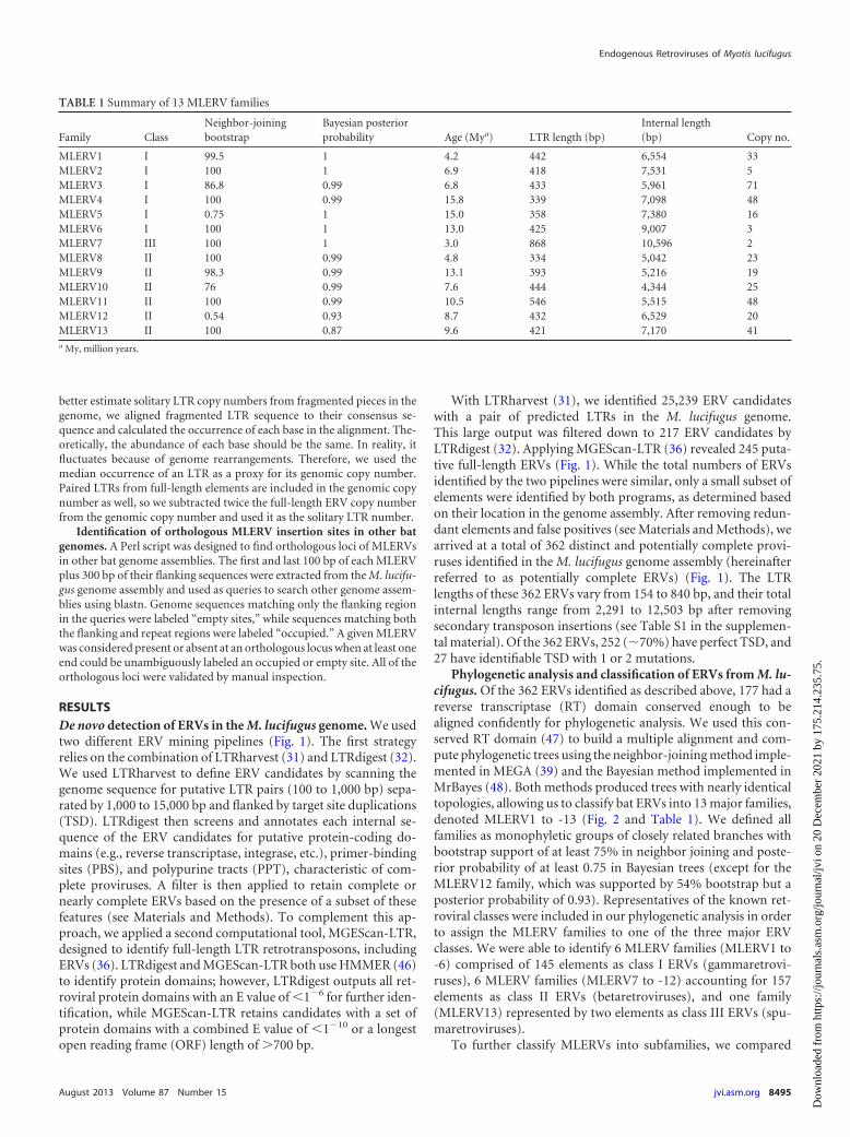
With LTRharvest (31), we identified 25,239 ERV candidateswith a pair of predicted LTRs in the M. lucifugus genome.This large output was filtered down to 217 ERV candidates byLTRdigest (32). Applying MGEScan-LTR (36) revealed 245 puta-tive full-length ERVs (Fig. 1). While the total numbers of ERVsidentified by the two pipelines were similar, only a small subset ofelements were identified by both programs, as determined basedon their location in the genome assembly. After removing redun-dant elements and false positives (see Materials and Methods), wearrived at a total of 362 distinct and potentially complete provi-ruses identified in the M. lucifugus genome assembly (hereinafterreferred to as potentially complete ERVs) (Fig. 1). The LTRlengths of these 362 ERVs vary from 154 to 840 bp, and their totalinternal lengths range from 2,291 to 12,503 bp after removingsecondary transposon insertions (see Table S1 in the supplemen-tal material). Of the 362 ERVs, 252 (�70%) have perfect TSD, and27 have identifiable TSD with 1 or 2 mutations.
Phylogenetic analysis and classification of ERVs from M. lu-cifugus. Of the 362 ERVs identified as described above, 177 had areverse transcriptase (RT) domain conserved enough to bealigned confidently for phylogenetic analysis. We used this con-served RT domain (47) to build a multiple alignment and com-pute phylogenetic trees using the neighbor-joining method imple-mented in MEGA (39) and the Bayesian method implemented inMrBayes (48). Both methods produced trees with nearly identicaltopologies, allowing us to classify bat ERVs into 13 major families,denoted MLERV1 to -13 (Fig. 2 and Table 1). We defined allfamilies as monophyletic groups of closely related branches withbootstrap support of at least 75% in neighbor joining and poste-rior probability of at least 0.75 in Bayesian trees (except for theMLERV12 family, which was supported by 54% bootstrap but aposterior probability of 0.93). Representatives of the known ret-roviral classes were included in our phylogenetic analysis in orderto assign the MLERV families to one of the three major ERVclasses. We were able to identify 6 MLERV families (MLERV1 to-6) comprised of 145 elements as class I ERVs (gammaretrovi-ruses), 6 MLERV families (MLERV7 to -12) accounting for 157elements as class II ERVs (betaretroviruses), and one family(MLERV13) represented by two elements as class III ERVs (spu-maretroviruses).
To further classify MLERVs into subfamilies, we compared
TABLE 1 Summary of 13 MLERV families
Family ClassNeighbor-joiningbootstrap
Bayesian posteriorprobability Age (Mya) LTR length (bp)
Internal length(bp) Copy no.
MLERV1 I 99.5 1 4.2 442 6,554 33MLERV2 I 100 1 6.9 418 7,531 5MLERV3 I 86.8 0.99 6.8 433 5,961 71MLERV4 I 100 0.99 15.8 339 7,098 48MLERV5 I 0.75 1 15.0 358 7,380 16MLERV6 I 100 1 13.0 425 9,007 3MLERV7 III 100 1 3.0 868 10,596 2MLERV8 II 100 0.99 4.8 334 5,042 23MLERV9 II 98.3 0.99 13.1 393 5,216 19MLERV10 II 76 0.99 7.6 444 4,344 25MLERV11 II 100 0.99 10.5 546 5,515 48MLERV12 II 0.54 0.93 8.7 432 6,529 20MLERV13 II 100 0.87 9.6 421 7,170 41a My, million years.
Endogenous Retroviruses of Myotis lucifugus
August 2013 Volume 87 Number 15 jvi.asm.org 8495
Dow
nloa
ded
from
http
s://j
ourn
als.
asm
.org
/jour
nal/j
vi o
n 20
Dec
embe
r 20
21 b
y 17
5.21
4.23
5.75
.
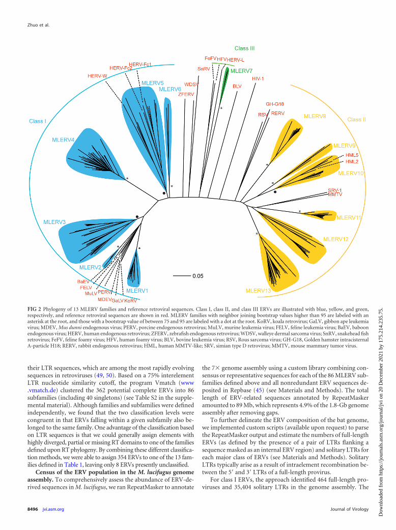
their LTR sequences, which are among the most rapidly evolvingsequences in retroviruses (49, 50). Based on a 75% interelementLTR nucleotide similarity cutoff, the program Vmatch (www.vmatch.de) clustered the 362 potential complete ERVs into 86subfamilies (including 40 singletons) (see Table S2 in the supple-mental material). Although families and subfamilies were definedindependently, we found that the two classification levels werecongruent in that ERVs falling within a given subfamily also be-longed to the same family. One advantage of the classification basedon LTR sequences is that we could generally assign elements withhighly diverged, partial or missing RT domains to one of the familiesdefined upon RT phylogeny. By combining these different classifica-tion methods, we were able to assign 354 ERVs to one of the 13 fam-ilies defined in Table 1, leaving only 8 ERVs presently unclassified.
Census of the ERV population in the M. lucifugus genomeassembly. To comprehensively assess the abundance of ERV-de-rived sequences in M. lucifugus, we ran RepeatMasker to annotate
the 7� genome assembly using a custom library combining con-sensus or representative sequences for each of the 86 MLERV sub-families defined above and all nonredundant ERV sequences de-posited in Repbase (45) (see Materials and Methods). The totallength of ERV-related sequences annotated by RepeatMaskeramounted to 89 Mb, which represents 4.9% of the 1.8-Gb genomeassembly after removing gaps.
To further delineate the ERV composition of the bat genome,we implemented custom scripts (available upon request) to parsethe RepeatMasker output and estimate the numbers of full-lengthERVs (as defined by the presence of a pair of LTRs flanking asequence masked as an internal ERV region) and solitary LTRs foreach major class of ERVs (see Materials and Methods). SolitaryLTRs typically arise as a result of intraelement recombination be-tween the 5= and 3= LTRs of a full-length provirus.
For class I ERVs, the approach identified 464 full-length pro-viruses and 35,404 solitary LTRs in the genome assembly. The
FIG 2 Phylogeny of 13 MLERV families and reference retroviral sequences. Class I, class II, and class III ERVs are illustrated with blue, yellow, and green,respectively, and reference retroviral sequences are shown in red. MLERV families with neighbor joining bootstrap values higher than 95 are labeled with anasterisk at the root, and those with a bootstrap value of between 75 and 95 are labeled with a dot at the root. KoRV, koala retrovirus; GaLV, gibbon ape leukemiavirus; MDEV, Mus dunni endogenous virus; PERV, porcine endogenous retrovirus; MuLV, murine leukemia virus; FELV, feline leukemia virus; BaEV, baboonendogenous virus; HERV, human endogenous retrovirus; ZFERV, zebrafish endogenous retrovirus; WDSV, walleye dermal sarcoma virus; SnRV, snakehead fishretrovirus; FeFV, feline foamy virus; HFV, human foamy virus; BLV, bovine leukemia virus; RSV, Rous sarcoma virus; GH-G18, Golden hamster intracisternalA-particle H18; RERV, rabbit endogenous retrovirus; HML, human MMTV-like; SRV, simian type D retrovirus; MMTV, mouse mammary tumor virus.
Zhuo et al.
8496 jvi.asm.org Journal of Virology
Dow
nloa
ded
from
http
s://j
ourn
als.
asm
.org
/jour
nal/j
vi o
n 20
Dec
embe
r 20
21 b
y 17
5.21
4.23
5.75
.

sizes of full-length class I ERVs in M. lucifugus typically range from6 to 9 kb (Table 1). MLERV3 is the most diverse family in thisclass, including 15 distinct subfamilies (Fig. 2 and Table 1). To-gether, the total genomic length occupied by class I elements isestimated at 31.5 Mb (1.66% of the genome assembly).
Class II ERVs were represented by 638 full-length provirusesand 10,858 solitary LTRs. The lengths of full-length class II ERVsrange from 4.5 to 9.5 kb. The most abundant class II family isMLERV11 (Fig. 2 and Table 1). Notably, subfamily MLERV11_2includes 123 potentially full-length copies, more than any otherMLERV subfamily. In total, class II ERVs occupy 9.1 Mb (0.48%)of the genome assembly.
Covering 49.2 Mb (or 2.6%) of DNA, class III ERVs accountfor the largest amount of ERV-derived sequences in the genomeassembly. This result was somewhat surprising in light of our ini-tial ab initio mining of ERVs, which had retrieved a single class IIIfamily (MLERV7) represented by only 2 complete canonical cop-ies (Fig. 2 and Table 1). Nonetheless, our parsing of the Repeat-Masker output identified 571 full-length and 81,967 solitary LTRsaffiliated with class III ERVs. Manual inspection of a subset ofthese sequences revealed that they represent relatively ancient andoften nonautonomous class III elements previously identified inother mammalian genomes, such as mammalian apparent LTRretrotransposons (MaLRs) (51). Thus, the discrepancy betweenthe results of the ab initio search and the RepeatMasker annotationcan be explained by the fact that most class III ERVs are repre-sented by highly decayed copies and nonautonomous elements, aswell as abundant solitary LTRs derived from ancient families (see
below). By design, such incomplete or highly diverged copies can-not be identified by the two pipelines used for our ab initio mining(31, 32, 36). The difficulty in identifying class III ERVs using abinitio approaches has been reported for other mammals (52–58).
Overall, the ERV coverage of the bat genome (89 Mb, 4.9%) isless than that in the human (261 Mb, 9.0%) and mouse (285 Mb,10.9%) genomes but similar to the ERV coverage of the dog ge-nome (115 Mb, 4.8%) (RepeatMasker) (Fig. 3a). However, the batgenome assembly is less complete and of poorer quality than themouse and human genome assemblies. Because ERVs and otherrepeats tend to be overrepresented in nonassembled regions ofsequenced genomes (gaps), our estimate of ERV abundance in M.lucifugus should be viewed as a conservative estimate.
Comparative demography of ERVs in bat, human, andmouse. The RepeatMasker output provides a measure of sequencedivergence for each DNA segment annotated to its closest consen-sus sequence in our ERV library, enabling us to examine thetempo and evolutionary dynamics of ERV invasions in M. lucifu-gus in comparison to those in human and mouse (Fig. 3b). Over-all, the demographic profile of M. lucifugus ERVs is more similarto that of human ERVs: class III ERVs are the most abundant andthe most diverged (ancient), class II ERVs are the least abundantbut the most recent, while class I ERVs occupy an intermediateposition both in abundance and divergence. The similar historiesof ERV accumulation in the bat and human (and to some extentmouse) lineages are to be contrasted with the dramatic differencesin DNA transposon activity, which is strikingly elevated in the M.
FIG 3 Comparison of ERV abundance and dynamics in different genomes. Different ERV classes and DNA transposons are labeled with different colors. (a)Comparison of percentages of genomes derived from different classes of ERVs in little brown bat and other mammals. (b) ERV and DNA transposon dynamicsin little brown bat, human, and mouse genomes. Distance to consensus was corrected using the Jukes-Cantor model. Older elements are more distant from theconsensus. The abundance is illustrated also, using the percentage of the genome.
Endogenous Retroviruses of Myotis lucifugus
August 2013 Volume 87 Number 15 jvi.asm.org 8497
Dow
nloa
ded
from
http
s://j
ourn
als.
asm
.org
/jour
nal/j
vi o
n 20
Dec
embe
r 20
21 b
y 17
5.21
4.23
5.75
.
lucifugus lineage (Fig. 3b), consistent with previous reports (59,60).
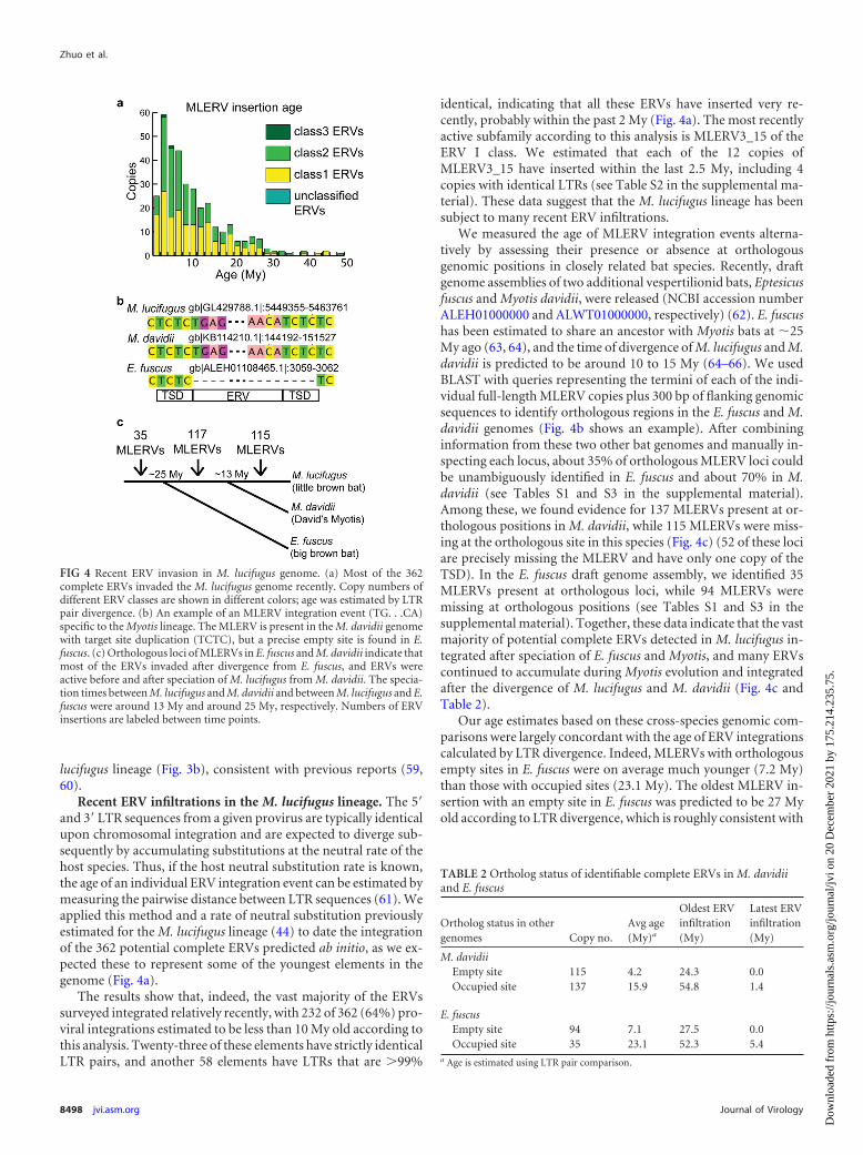
Recent ERV infiltrations in the M. lucifugus lineage. The 5=and 3= LTR sequences from a given provirus are typically identicalupon chromosomal integration and are expected to diverge sub-sequently by accumulating substitutions at the neutral rate of thehost species. Thus, if the host neutral substitution rate is known,the age of an individual ERV integration event can be estimated bymeasuring the pairwise distance between LTR sequences (61). Weapplied this method and a rate of neutral substitution previouslyestimated for the M. lucifugus lineage (44) to date the integrationof the 362 potential complete ERVs predicted ab initio, as we ex-pected these to represent some of the youngest elements in thegenome (Fig. 4a).
The results show that, indeed, the vast majority of the ERVssurveyed integrated relatively recently, with 232 of 362 (64%) pro-viral integrations estimated to be less than 10 My old according tothis analysis. Twenty-three of these elements have strictly identicalLTR pairs, and another 58 elements have LTRs that are �99%
identical, indicating that all these ERVs have inserted very re-cently, probably within the past 2 My (Fig. 4a). The most recentlyactive subfamily according to this analysis is MLERV3_15 of theERV I class. We estimated that each of the 12 copies ofMLERV3_15 have inserted within the last 2.5 My, including 4copies with identical LTRs (see Table S2 in the supplemental ma-terial). These data suggest that the M. lucifugus lineage has beensubject to many recent ERV infiltrations.
We measured the age of MLERV integration events alterna-tively by assessing their presence or absence at orthologousgenomic positions in closely related bat species. Recently, draftgenome assemblies of two additional vespertilionid bats, Eptesicusfuscus and Myotis davidii, were released (NCBI accession numberALEH01000000 and ALWT01000000, respectively) (62). E. fuscushas been estimated to share an ancestor with Myotis bats at �25My ago (63, 64), and the time of divergence of M. lucifugus and M.davidii is predicted to be around 10 to 15 My (64–66). We usedBLAST with queries representing the termini of each of the indi-vidual full-length MLERV copies plus 300 bp of flanking genomicsequences to identify orthologous regions in the E. fuscus and M.davidii genomes (Fig. 4b shows an example). After combininginformation from these two other bat genomes and manually in-specting each locus, about 35% of orthologous MLERV loci couldbe unambiguously identified in E. fuscus and about 70% in M.davidii (see Tables S1 and S3 in the supplemental material).Among these, we found evidence for 137 MLERVs present at or-thologous positions in M. davidii, while 115 MLERVs were miss-ing at the orthologous site in this species (Fig. 4c) (52 of these lociare precisely missing the MLERV and have only one copy of theTSD). In the E. fuscus draft genome assembly, we identified 35MLERVs present at orthologous loci, while 94 MLERVs weremissing at orthologous positions (see Tables S1 and S3 in thesupplemental material). Together, these data indicate that the vastmajority of potential complete ERVs detected in M. lucifugus in-tegrated after speciation of E. fuscus and Myotis, and many ERVscontinued to accumulate during Myotis evolution and integratedafter the divergence of M. lucifugus and M. davidii (Fig. 4c andTable 2).
Our age estimates based on these cross-species genomic com-parisons were largely concordant with the age of ERV integrationscalculated by LTR divergence. Indeed, MLERVs with orthologousempty sites in E. fuscus were on average much younger (7.2 My)than those with occupied sites (23.1 My). The oldest MLERV in-sertion with an empty site in E. fuscus was predicted to be 27 Myold according to LTR divergence, which is roughly consistent with
FIG 4 Recent ERV invasion in M. lucifugus genome. (a) Most of the 362complete ERVs invaded the M. lucifugus genome recently. Copy numbers ofdifferent ERV classes are shown in different colors; age was estimated by LTRpair divergence. (b) An example of an MLERV integration event (TG. . .CA)specific to the Myotis lineage. The MLERV is present in the M. davidii genomewith target site duplication (TCTC), but a precise empty site is found in E.fuscus. (c) Orthologous loci of MLERVs in E. fuscus and M. davidii indicate thatmost of the ERVs invaded after divergence from E. fuscus, and ERVs wereactive before and after speciation of M. lucifugus from M. davidii. The specia-tion times between M. lucifugus and M. davidii and between M. lucifugus and E.fuscus were around 13 My and around 25 My, respectively. Numbers of ERVinsertions are labeled between time points.
TABLE 2 Ortholog status of identifiable complete ERVs in M. davidiiand E. fuscus
Ortholog status in othergenomes Copy no.
Avg age(My)a
Oldest ERVinfiltration(My)
Latest ERVinfiltration(My)
M. davidiiEmpty site 115 4.2 24.3 0.0Occupied site 137 15.9 54.8 1.4
E. fuscusEmpty site 94 7.1 27.5 0.0Occupied site 35 23.1 52.3 5.4
a Age is estimated using LTR pair comparison.
Zhuo et al.
8498 jvi.asm.org Journal of Virology
Dow
nloa
ded
from
http
s://j
ourn
als.
asm
.org
/jour
nal/j
vi o
n 20
Dec
embe
r 20
21 b
y 17
5.21
4.23
5.75
.

the divergence time of �25 My estimated between these two batspecies (63). However, we note that the age of the youngestMLERV insertions with occupied orthologous sites in E. fuscuswas significantly underestimated by LTR divergence (5.4 My).Similar trends were found in M. davidii (summarized in Table 2).This discrepancy between the results of the two dating methodscould be caused by gene conversion homogenizing LTR se-quences, leading to underestimation of the timing of integration,as previously reported in other genomes (67). These data empha-size the need to apply multiple methods to confidently date ERVintegration events.
DISCUSSIONCensus of ERVs in the M. lucifugus genome. By combining twodifferent ab initio mining strategies, we identified 362 potentiallycomplete proviruses in the M. lucifugus genome. Nearly all of theseelements fall within 86 subfamilies that enabled us to identify amultitude of related sequence fragments using RepeatMasker, in-cluding nearly 1,700 full-length ERVs and 130,000 solitary LTRsin the M. lucifugus genome assembly. When used in conjunctionwith mammalian ERV sequences catalogued in Repbase, our col-lection allowed us to estimate that ERVs occupy 4.9% of the batgenome, a substantial fraction comparable to that observed inother eutherian genomes (Fig. 3a) (19, 20, 69).
Our data complement previous findings by Cui et al. (28), whoidentified 3 major groups (A, B, and C) of gammaretroviruses inthe M. lucifugus genome by BLAST searches. Our approach iden-tified these three groups as the MLERV2, MLERV1, and MLERV3families, respectively. We discovered three additional gammaret-rovirus families (MLERV4 to -6) (Fig. 2 and Table 1). The totallength of sequences derived from the MLERV4 family alone is 9.2Mb, or �0.5% of the genome assembly. At the time of this study,there were 5 entries of internal (coding) ERV regions and 132entries of LTR sequences for M. lucifugus in Repbase, a compre-hensive database for transposable element sequences, includingERVs (45). We identified both LTR and internal sequences for 13families and 86 subfamilies of ERVs, most of which were not re-ported in Repbase (see Table S2 in the supplemental material).Furthermore, through manual examination, we found that severalof the M. lucifugus LTR sequences deposited in Repbase were ac-tually truncated at their 5= end (data not shown). Thus, our man-ually curated collection of 86 reference ERV sequences will beuseful to replace or complement existing Repbase entries. Overall,the coverage of MLERV families newly identified in this studyamounts to 23 Mb of the genome assembly, thereby substantiallyimproving the census of ERVs in this bat species.
Comparison of ERV diversity in M. lucifugus with that ofother mammals. With regard to ERV diversity within M. lucifu-gus, we found that class I (gammaretroviruses) and class II (be-taretroviruses) ERVs are similarly diverse (each composed of 6major families), but the total amount of genomic DNA derivedfrom class II ERVs (9.1 Mb) is considerably smaller than thatderived from class I ERVs (31.5 Mb). Class III (spumaviruses)ERVs are the most abundant (49.2 Mb) in the genome, but theyare generally older and more degraded than class I and II elements,which hampered the identification of full-length class III ERVsusing ab initio methods, as reported for other mammalian ge-nomes (52–58, 70). Using RepeatMasker, we identified 571 appar-ently full-length class III ERVs, but we observed that a large frac-tion of these elements are nonautonomous MaLR-like elements
that are comparable to those abundantly populating the humanand mouse genomes (19, 20). Nonetheless, we note that the onlyclass III family we detected ab initio in M. lucifugus (MLERV7) is arelatively young family, with an age estimated at �4 My (Table 1).Thus, all three major ERV classes are represented by relativelyrecent insertions in the M. lucifugus genome.
Overall, the demographic profile of the three ERV classes in M.lucifugus was more similar to that seen in the human genome (Fig.3b). While the bulk of class III ERVs likely predate the radiation ofeutherian mammals and, thus, have essentially been inheritedthrough vertical decent, the amplification of class I and II ERVs ismuch more recent and largely lineage specific (Fig. 3). We con-clude that there was a parallel invasion and expansion of these twoclasses of ERV in the human and bat lineages.
An important motivation for our analysis of ERVs in M. lucifu-gus relates to recent findings of massive lineage-specific DNAtransposon activity in M. lucifugus (Fig. 3b) (59, 60). There isstrong evidence that several of these DNA transposons have beenacquired horizontally (71, 72), possibly reflecting a peculiar sen-sitivity of the germ line of this group of bats to lateral infiltration ofmobile elements. Because retroviral endogenization also repre-sents a form of horizontal transfer to the germ line, it was ofinterest to see whether these bats also display a greater vulnerabil-ity to ERV invasions. While we found clear evidence of recent ERVcolonization in the genome of M. lucifugus, neither the diversitynor the sheer amount of ERV sequences depart dramatically fromthe diversity or amount observed in other mammalian genomes(Fig. 3). Thus, while the mobile element landscape of M. lucifugusis exceptional in terms of recent DNA transposon invasions, M.lucifugus does not appear to be an outlier among eutherian mam-mals in terms of its ERV population. We conclude that the appar-ent vulnerability of vespertilionid bats to horizontal transfer ofDNA transposons is not generalizable to all types of mobile ele-ments.
Superspreader hypothesis. Recently, Magiorkinis et al. (73)proposed the “superspreader” hypothesis, which postulates thatERVs lacking coding capacity for an envelope (env-less ERVs)amplify more efficiently within the genome than those encodingan intact envelope. The hypothesis was supported by a detailedphylogenetic analysis of intracisternal A-type particles (IAPs)from several mammalian genomes (73) and for several primateERV families (74). In M. lucifugus, we classified MLERVs to 13families and 86 subfamilies. At the family level, we found no clearrelationship between the presence of an envelope domain andfamily copy number; however, at the subfamily level, we observedthat the most successful subfamilies are predominantly composedof env-less elements (see Table S2 in the supplemental material).For example, the two largest subfamilies in our data set(MLERV4_6 and MLERV11_2) are entirely composed of copieslacking an identifiable envelope domain. Thus, the pattern ofMLERV subfamily expansion brings further support to the super-spreader hypothesis.
Bats as possible zoonotic reservoirs of retroviruses. Wefound several clear examples of very recent ERV families in M.lucifugus. A good illustration is MLERV3_15, a subfamily of classI elements. Four of the 12 copies identified in the genome haveidentical LTR pairs, while the other eight have LTR pairs that are�99% identical, indicative of nearly contemporary integrationevents (see Table S2 in the supplemental material). All 12 copiesare also absent at orthologous positions in M. davidii (see Table
Endogenous Retroviruses of Myotis lucifugus
August 2013 Volume 87 Number 15 jvi.asm.org 8499
Dow
nloa
ded
from
http
s://j
ourn
als.
asm
.org
/jour
nal/j
vi o
n 20
Dec
embe
r 20
21 b
y 17
5.21
4.23
5.75
.

S3). Nonetheless, none of the MLERV3_15 copies identified ap-pear to retain intact coding capacity, suggesting that they are cur-rently incapable of replicating autonomously.
However, in a recently active class I ERV subfamily,MLERV2_2 (0 to 3 My old), we identified one copy (entry 74) withapparently intact gag, pro, pol, and env coding regions, suggestingthat this copy might be replication competent. In addition, an-other apparently intact and functional class II ERV was recentlyidentified in M. lucifugus (29). Together, these results suggest thatboth class I and II ERVs in M. lucifugus are potentially capable ofautonomous replication and of producing infectious viral parti-cles.
Among the most recently integrated (�10 My ago) potentiallycomplete proviruses supported by both LTR-LTR divergence andcross-species analysis, we were able to detect members of all threemain retroviral classes (see Table S2 in the supplemental mate-rial). Our finding of recently integrated spumaretroviruses andgammaretroviruses is consistent with the identification of exoge-nous members of these retroviral taxa in several bat species, in-cluding microbats (15, 30). We also identified proviral copies ofbetaretroviruses (e.g., MLERV12_4) that have retained identicalLTRs flanked by perfect TSD and are absent in M. davidii (seeTables S2 and S3), which suggests that M. lucifugus was also in-fected by exogenous betaretroviruses in the recent past. Together,these data indicate that a wide diversity of retroviruses have re-cently infected these bats and are likely still circulating in naturalpopulations of M. lucifugus. Given the apparent propensity of batsto act as reservoir species for zoonotic viruses that are highlypathogenic to humans, these observations raise concerns thatthese animals may also be capable of transmitting zoonotic retro-viruses to humans.
ACKNOWLEDGMENTS
We thank Aurelie Kapusta, Ellen Pritham, and Claudia Marquez for help-ful discussions and Ray Malfavon-Borja for critical reading of the manu-script.
X.Z. and C.F. were supported by grant R01-GM077582 from the Na-tional Institutes of Health.
REFERENCES1. Simmons NB. 2005. Order Chiroptera, p 312–529. In Wilson DE, Reeder
DM (ed), Mammal species of the world: a taxonomic and geographicreference, 3rd ed, vol 1. Johns Hopkins University Press, Baltimore, MD.
2. Hockman D, Mason MK, Jacobs DS, Illing N. 2009. The role of earlydevelopment in mammalian limb diversification: a descriptive comparison ofearly limb development between the Natal long-fingered bat (Miniopterusnatalensis) and the mouse (Mus musculus). Dev. Dyn. 238:965–979.
3. Behringer RR, Rasweiler JJ, Chen CH, Cretekos CJ. 2009. Geneticregulation of mammalian diversity. Cold Spring Harbor Symp. Quant.Biol. 74:297–302.
4. Munshi-South J, Wilkinson GS. 2010. Bats and birds: exceptional lon-gevity despite high metabolic rates. Ageing Res. Rev. 9:12–19.
5. Calisher CH, Childs JE, Field HE, Holmes KV, Schountz T. 2006. Bats:important reservoir hosts of emerging viruses. Clin. Microbiol. Rev. 19:531–545.
6. Dobson AP. 2005. What links bats to emerging infectious diseases? Sci-ence 310:628 – 629.
7. Lau SKP, Woo PCY, Li KSM, Huang Y, Tsoi H-W, Wong BHL, WongSSY, Leung S-Y, Chan K-H, Yuen K-Y. 2005. Severe acute respiratorysyndrome coronavirus-like virus in Chinese horseshoe bats. Proc. Natl.Acad. Sci. U. S. A. 102:14040 –14045.
8. Li W, Shi Z, Yu M, Ren W, Smith C, Epstein JH, Wang H, Crameri G,Hu Z, Zhang H, Zhang J, McEachern J, Field H, Daszak P, Eaton BT,Zhang S, Wang L-F. 2005. Bats are natural reservoirs of SARS-like coro-naviruses. Science 310:676 – 679.
9. Wong S, Lau S, Woo P, Yuen K-Y. 2007. Bats as a continuing source ofemerging infections in humans. Rev. Med. Virol. 17:67–91.
10. Drexler JF, Corman VM, Wegner T, Tateno AF, Zerbinati RM, Gloza-Rausch F, Seebens A, Müller MA, Drosten C. 2011. Amplification ofemerging viruses in a bat colony. Emerg. Infect. Dis. 17:449 – 456.
11. Luis AD, Hayman DTS, O’Shea TJ, Cryan PM, Gilbert AT, PulliamJRC, Mills JN, Timonin ME, Willis CKR, Cunningham AA, Fooks AR,Rupprecht CE, Wood JLN, Webb CT. 2013. A comparison of bats androdents as reservoirs of zoonotic viruses: are bats special? Proc. Biol. Sci.280:20122753. doi:10.1098/rspb.2012.2753.
12. Li L, Victoria JG, Wang C, Jones M, Fellers GM, Kunz TH, Delwart E.2010. Bat guano virome: predominance of dietary viruses from insects andplants plus novel mammalian viruses. J. Virol. 84:6955– 6965.
13. Donaldson EF, Haskew AN, Gates JE, Huynh J, Moore CJ, FriemanMB. 2010. Metagenomic analysis of the viromes of three North Americanbat species: viral diversity among different bat species that share a com-mon habitat. J. Virol. 84:13004 –13018.
14. Ge X, Li Y, Yang X, Zhang H, Zhou P, Zhang Y, Shi Z. 2012. Meta-genomic analysis of viruses from bat fecal samples reveals many novelviruses in insectivorous bats in China. J. Virol. 86:4620 – 4630.
15. Wu Z, Ren X, Yang L, Hu Y, Yang J, He G, Zhang J, Dong J, Sun L, DuJ, Liu L, Xue Y, Wang J, Yang F, Zhang S, Jin Q. 2012. Virome analysisfor identification of novel mammalian viruses in bat species from Chineseprovinces. J. Virol. 86:10999 –11012.
16. Gifford R, Tristem M. 2003. The evolution, distribution and diversity ofendogenous retroviruses. Virus Genes 26:291–315.
17. Blikstad V, Benachenhou F, Sperber GO, Blomberg J. 2008. Evolution ofhuman endogenous retroviral sequences: a conceptual account. Cell. Mol.Life Sci. 65:3348 –3365.
18. Jern P, Coffin JM. 2008. Effects of retroviruses on host genome function.Annu. Rev. Genet. 42:709 –732.
19. Lander ES, Linton LM, Birren B, Nusbaum C, Zody MC, Baldwin J,Devon K, Dewar K, Doyle M, FitzHugh W, Funke R, Gage D, Harris K,Heaford A, Howland J, Kann L, Lehoczky J, LeVine R, McEwan P,McKernan K, Meldrim J, Mesirov JP, Miranda C, Morris W, Naylor J,Raymond C, Rosetti M, Santos R, Sheridan A, Sougnez C, Stange-Thomann N, Stojanovic N, Subramanian A, Wyman D, Rogers J,Sulston J, Ainscough R, Beck S, Bentley D, Burton J, Clee C, Carter N,Coulson A, Deadman R, Deloukas P, Dunham A, Dunham I, Durbin R,French L, Grafham D, et al. 2001. Initial sequencing and analysis of thehuman genome. International Human Genome Sequencing Consortium.Nature 409:860 –921.
20. Waterston RH, Lindblad-Toh K, Birney E, Rogers J, Abril JF, Agarwal P,Agarwala R, Ainscough R, Alexandersson M, An P, Antonarakis SE, At-twood J, Baertsch R, Bailey J, Barlow K, Beck S, Berry E, Birren B, BloomT, Bork P, Botcherby M, Bray N, Brent MR, Brown DG, Brown SD, BultC, Burton J, Butler J, Campbell RD, Carninci P, Cawley S, Chiaromonte F,Chinwalla AT, Church DM, Clamp M, Clee C, Collins FS, Cook LL, CopleyRR, Coulson A, Couronne O, Cuff J, Curwen V, Cutts T, Daly M, DavidR, Davies J, Delehaunty KD, Deri J, Dermitzakis ET, et al. 2002. Initialsequencing and comparative analysis of the mouse genome. Mouse GenomeSequencing Consortium. Nature 420:520–562.
21. Kurth R, Bannert N. 2010. Beneficial and detrimental effects of humanendogenous retroviruses. Int. J. Cancer 126:306 –314.
22. Feschotte C, Gilbert C. 2012. Endogenous viruses: insights into viralevolution and impact on host biology. Nat. Rev. Genet. 13:283–296.
23. Katzourakis A, Gifford RJ, Tristem M, Gilbert MTP, Pybus OG. 2009.Macroevolution of complex retroviruses. Science 325:1512.
24. Emerman M, Malik HS. 2010. Paleovirology—modern consequences ofancient viruses. Plos Biol. 8:e1000301. doi:10.1371/journal.pbio.1000301.
25. Holmes EC. 2011. The evolution of endogenous viral elements. Cell HostMicrobe 10:368 –377.
26. Tristem M, Kabat P, Lieberman L, Linde S, Karpas A, Hill F. 1996.Characterization of a novel murine leukemia virus-related subgroupwithin mammals. J. Virol. 70:8241– 8246.
27. Baillie GJ, van de Lagemaat LN, Baust C, Mager DL. 2004. Multiplegroups of endogenous betaretroviruses in mice, rats, and other mammals.J. Virol. 78:5784 –5798.
28. Cui J, Tachedjian G, Tachedjian M, Holmes EC, Zhang S, Wang L-F.2012. Identification of diverse groups of endogenous gammaretrovirusesin mega- and microbats. J. Gen. Virol. 93:2037–2045.
29. Hayward JA, Tachedjian M, Cui J, Field H, Holmes EC, Wang L-F,Tachedjian G. 2013. Identification of diverse full-length endogenous be-
Zhuo et al.
8500 jvi.asm.org Journal of Virology
Dow
nloa
ded
from
http
s://j
ourn
als.
asm
.org
/jour
nal/j
vi o
n 20
Dec
embe
r 20
21 b
y 17
5.21
4.23
5.75
.

taretroviruses in megabats and microbats. Retrovirology 10:35. doi:10.1186/1742-4690-10-35.
30. Cui J, Tachedjian M, Wang L, Tachedjian G, Wang L-F, Zhang S. 2012.Discovery of retroviral homologs in bats: implications for the origin ofmammalian gammaretroviruses. J. Virol. 86:4288 – 4293.
31. Ellinghaus D, Kurtz S, Willhoeft U. 2008. LTRharvest, an efficient andflexible software for de novo detection of LTR retrotransposons. BMCBioinform. 9:18. doi:10.1186/1471-2105-9-18.
32. Steinbiss S, Willhoeft U, Gremme G, Kurtz S. 2009. Fine-grained an-notation and classification of de novo predicted LTR retrotransposons.Nucleic Acids Res. 37:7002–7013.
33. Lowe TM, Eddy SR. 1997. tRNAscan-SE: a program for improved detectionof transfer RNA genes in genomic sequence. Nucleic Acids Res. 25:955–964.
34. Punta M, Coggill PC, Eberhardt RY, Mistry J, Tate J, Boursnell C, PangN, Forslund K, Ceric G, Clements J, Heger A, Holm L, SonnhammerELL, Eddy SR, Bateman A, Finn RD. 2012. The Pfam protein familiesdatabase. Nucleic Acids Res. 40:D290 –D301.
35. Reference deleted.36. Rho M, Choi J-H, Kim S, Lynch M, Tang H. 2007. De novo identifica-
tion of LTR retrotransposons in eukaryotic genomes. BMC Genomics8:90. doi:10.1186/1471-2164-8-90.
37. Kohany O, Gentles AJ, Hankus L, Jurka J. 2006. Annotation, submissionand screening of repetitive elements in Repbase: RepbaseSubmitter andCensor. BMC Bioinform. 7:474. doi:10.1186/1471-2105-7-474.
38. Edgar RC. 2004. MUSCLE: multiple sequence alignment with high accu-racy and high throughput. Nucleic Acids Res. 32:1792–1797.
39. Tamura K, Peterson D, Peterson N, Stecher G, Nei M, Kumar S. 2011.MEGA5: Molecular Evolutionary Genetics Analysis using maximum like-lihood, evolutionary distance, and maximum parsimony methods. Mol.Biol. Evol. 28:2731–2739.
40. Jones DT, Taylor WR, Thornton JM. 1992. The rapid generation ofmutation data matrices from protein sequences. Comput. Appl. Biosci.8:275–282.
41. Ronquist F, Huelsenbeck JP. 2003. MrBayes 3: Bayesian phylogeneticinference under mixed models. Bioinformatics 19:1572–1574.
42. Smith TF, Waterman MS. 1981. Identification of common molecularsubsequences. J. Mol. Biol. 147:195–197.
43. Jukes TH, Cantor CR. 1969. Evolution of protein molecules, p 21–132. InMunro HN, Mammalian protein metabolism, vol III. Academic Press, SanDiego, CA.
44. Pace JK, Gilbert C, Clark MS, Feschotte C. 2008. Repeated horizontaltransfer of a DNA transposon in mammals and other tetrapods. Proc.Natl. Acad. Sci. U. S. A. 105:17023–17028.
45. Jurka J, Kapitonov VV, Pavlicek A, Klonowski P, Kohany O, Walichie-wicz J. 2005. Repbase Update, a database of eukaryotic repetitive ele-ments. Cytogenet. Genome Res. 110:462– 467.
46. Eddy SR. 2011. Accelerated profile HMM searches. PLoS Comput. Biol.7:e1002195. doi:10.1371/journal.pcbi.1002195.
47. Xiong Y, Eickbush TH. 1990. Origin and evolution of retroelementsbased upon their reverse transcriptase sequences. EMBO J. 9:3353–3362.
48. Ronquist F, Teslenko M, van der Mark P, Ayres DL, Darling A, HöhnaS, Larget B, Liu L, Suchard MA, Huelsenbeck JP. 2012. MrBayes 3.2:efficient Bayesian phylogenetic inference and model choice across a largemodel space. Syst. Biol. 61:539 –542.
49. Slattery JP, Franchini G, Gessain A. 1999. Genomic evolution, patternsof global dissemination, and interspecies transmission of human and sim-ian T-cell leukemia/lymphotropic viruses. Genome Res. 9:525–540.
50. Fernández-Medina RD, Ribeiro JMC, Carareto CMA, Velasque L,Struchiner CJ. 2012. Losing identity: structural diversity of transposableelements belonging to different classes in the genome of Anophelesgambiae. BMC Genomics 13:272. doi:10.1186/1471-2164-13-272.
51. Smit AF. 1993. Identification of a new, abundant superfamily of mamma-lian LTR-transposons. Nucleic Acids Res. 21:1863–1872.
52. McCarthy EM, McDonald JF. 2004. Long terminal repeat retrotransposonsof Mus musculus. Genome Biol. 5:R14. doi:10.1186/gb-2004-5-3-r14.
53. Polavarapu N, Bowen NJ, McDonald JF. 2006. Identification, character-ization and comparative genomics of chimpanzee endogenous retrovi-ruses. Genome Biol. 7:R51.
54. Polavarapu N, Bowen NJ, McDonald JF. 2006. Newly identified familiesof human endogenous retroviruses. J. Virol. 80:4640 – 4642.
55. Garcia-Etxebarria K, Jugo BM. 2010. Genome-wide detection and charac-terization of endogenous retroviruses in Bos taurus. J. Virol. 84:10852–10862.
56. Martínez Barrio Á, Ekerljung M, Jern P, Benachenhou F, Sperber GO,Bongcam-Rudloff E, Blomberg J, Andersson G. 2011. The first se-quenced carnivore genome shows complex host-endogenous retrovirusrelationships. PLoS One 6:e19832. doi:10.1371/journal.pone.0019832.
57. Garcia-Etxebarria K, Jugo BM. 2012. Detection and characterization ofendogenous retroviruses in the horse genome by in silico analysis. Virol-ogy 434:59 – 67.
58. Brown K, Moreton J, Malla S, Aboobaker AA, Emes RD, Tarlinton RE.2012. Characterisation of retroviruses in the horse genome and their tran-scriptional activity via transcriptome sequencing. Virology 433:55– 63.
59. Ray DA, Feschotte C, Pagan HJT, Smith JD, Pritham EJ, ArensburgerP, Atkinson PW, Craig NL. 2008. Multiple waves of recent DNA trans-poson activity in the bat, Myotis lucifugus. Genome Res. 18:717–728.
60. Pritham EJ, Feschotte C. 2007. Massive amplification of rolling-circletransposons in the lineage of the bat Myotis lucifugus. Proc. Natl. Acad.Sci. U. S. A. 104:1895–1900.
61. Dangel AW, Baker BJ, Mendoza AR, Yu CY. 1995. Complement com-ponent C4 gene intron 9 as a phylogenetic marker for primates: longterminal repeats of the endogenous retrovirus ERV-K(C4) are a molecularclock of evolution. Immunogenetics 42:41–52.
62. Zhang G, Cowled C, Shi Z, Huang Z, Bishop-Lilly KA, Fang X, WynneJW, Xiong Z, Baker ML, Zhao W, Tachedjian M, Zhu Y, Zhou P, JiangX, Ng J, Yang L, Wu L, Xiao J, Feng Y, Chen Y, Sun X, Zhang Y, MarshGA, Crameri G, Broder CC, Frey KG, Wang L-F, Wang J. 2013.Comparative analysis of bat genomes provides insight into the evolutionof flight and immunity. Science 339:456 – 460.
63. Miller-Butterworth CM, Murphy WJ, O’Brien SJ, Jacobs DS, SpringerMS, Teeling EC. 2007. A family matter: conclusive resolution of thetaxonomic position of the long-fingered bats, miniopterus. Mol. Biol.Evol. 24:1553–1561.
64. Lack JB, Roehrs ZP, Stanley CE, Jr, Ruedi M, Van Den Bussche RA.2010. Molecular phylogenetics of Myotis indicate familial-level divergencefor the genus Cistugo (Chiroptera). J. Mammal. 91:976 –992.
65. Agnarsson I, Zambrana-Torrelio CM, Flores-Saldana NP, May-ColladoLJ. 2011. A time-calibrated species-level phylogeny of bats (Chiroptera,Mammalia). PLoS Curr. 3:RRN1212. doi:10.1371/currents.RRN1212.
66. Stadelmann B, Lin LK, Kunz TH, Ruedi M. 2007. Molecular phylogenyof New World Myotis (Chiroptera, Vespertilionidae) inferred from mito-chondrial and nuclear DNA genes. Mol. Phylogenet. Evol. 43:32– 48.
67. Kijima TE, Innan H. 2010. On the estimation of the insertion time of LTRretrotransposable elements. Mol. Biol. Evol. 27:896 –904.
68. Reference deleted.69. Lindblad-Toh K, Wade CM, Mikkelsen TS, Karlsson EK, Jaffe DB,
Kamal M, Clamp M, Chang JL, Kulbokas EJ, Zody MC, Mauceli E, XieX, Breen M, Wayne RK, Ostrander EA, Ponting CP, Galibert F, SmithDR, deJong PJ, Kirkness E, Alvarez P, Biagi T, Brockman W, Butler J,Chin C-W, Cook A, Cuff J, Daly MJ, DeCaprio D, Gnerre S, GrabherrM, Kellis M, Kleber M, Bardeleben C, Goodstadt L, Heger A, Hitte C,Kim L, Koepfli K-P, Parker HG, Pollinger JP, Searle SMJ, Sutter NB,Thomas R, Webber C, Baldwin J, Abebe A, Abouelleil A, Aftuck L,Ait-zahra M, et al. 2005. Genome sequence, comparative analysis andhaplotype structure of the domestic dog. Nature 438:803– 819.
70. Han K, Konkel MK, Xing J, Wang H, Lee J, Meyer TJ, Huang CT,Sandifer E, Hebert K, Barnes EW, Hubley R, Miller W, Smit AFA,Ullmer B, Batzer MA. 2007. Mobile DNA in Old World monkeys: aglimpse through the rhesus macaque genome. Science 316:238 –240.
71. Gilbert C, Schaack S, Pace JK, Brindley PJ, Feschotte C. 2010. A role forhost-parasite interactions in the horizontal transfer of transposons acrossphyla. Nature 464:1347–1350.
72. Thomas J, Schaack S, Pritham EJ. 2010. Pervasive horizontal transfer ofrolling-circle transposons among animals. Genome Biol. Evol. 2:656 –664.
73. Magiorkinis G, Gifford RJ, Katzourakis A, De Ranter J, Belshaw R.2012. Env-less endogenous retroviruses are genomic superspreaders.Proc. Natl. Acad. Sci. U. S. A. 109:7385–7390.
74. Belshaw R, Katzourakis A, Paces J, Burt A, Tristem M. 2005. High copynumber in human endogenous retrovirus families is associated with copy-ing mechanisms in addition to reinfection. Mol. Biol. Evol. 22:814 – 817.
Endogenous Retroviruses of Myotis lucifugus
August 2013 Volume 87 Number 15 jvi.asm.org 8501
Dow
nloa
ded
from
http
s://j
ourn
als.
asm
.org
/jour
nal/j
vi o
n 20
Dec
embe
r 20
21 b
y 17
5.21
4.23
5.75
.